Embed Size (px)
Citation preview
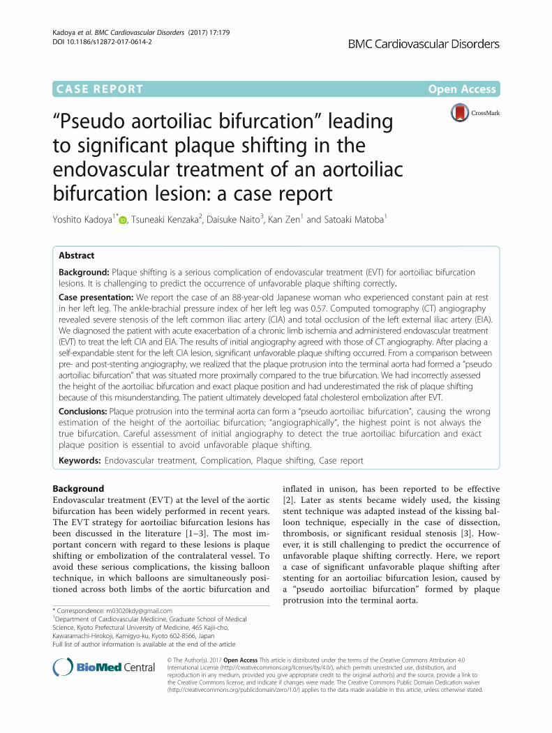
Kadoya et al. BMC Cardiovascular Disorders (2017) 17:179 DOI 10.1186/s12872-017-0614-2
CASE REPORT Open Access
“Pseudo aortoiliac bifurcation” leadingto significant plaque shifting in theendovascular treatment of an aortoiliacbifurcation lesion: a case report
Yoshito Kadoya1* , Tsuneaki Kenzaka2, Daisuke Naito3, Kan Zen1 and Satoaki Matoba1Abstract
Background: Plaque shifting is a serious complication of endovascular treatment (EVT) for aortoiliac bifurcationlesions. It is challenging to predict the occurrence of unfavorable plaque shifting correctly.
Case presentation: We report the case of an 88-year-old Japanese woman who experienced constant pain at restin her left leg. The ankle-brachial pressure index of her left leg was 0.57. Computed tomography (CT) angiographyrevealed severe stenosis of the left common iliac artery (CIA) and total occlusion of the left external iliac artery (EIA).We diagnosed the patient with acute exacerbation of a chronic limb ischemia and administered endovascular treatment(EVT) to treat the left CIA and EIA. The results of initial angiography agreed with those of CT angiography. After placing aself-expandable stent for the left CIA lesion, significant unfavorable plaque shifting occurred. From a comparison betweenpre- and post-stenting angiography, we realized that the plaque protrusion into the terminal aorta had formed a “pseudoaortoiliac bifurcation” that was situated more proximally compared to the true bifurcation. We had incorrectly assessedthe height of the aortoiliac bifurcation and exact plaque position and had underestimated the risk of plaque shiftingbecause of this misunderstanding. The patient ultimately developed fatal cholesterol embolization after EVT.
Conclusions: Plaque protrusion into the terminal aorta can form a “pseudo aortoiliac bifurcation”, causing the wrongestimation of the height of the aortoiliac bifurcation; “angiographically”, the highest point is not always thetrue bifurcation. Careful assessment of initial angiography to detect the true aortoiliac bifurcation and exactplaque position is essential to avoid unfavorable plaque shifting.
Keywords: Endovascular treatment, Complication, Plaque shifting, Case report
BackgroundEndovascular treatment (EVT) at the level of the aorticbifurcation has been widely performed in recent years.The EVT strategy for aortoiliac bifurcation lesions hasbeen discussed in the literature [1–3]. The most im-portant concern with regard to these lesions is plaqueshifting or embolization of the contralateral vessel. Toavoid these serious complications, the kissing balloontechnique, in which balloons are simultaneously posi-tioned across both limbs of the aortic bifurcation and
* Correspondence: [email protected] of Cardiovascular Medicine, Graduate School of MedicalScience, Kyoto Prefectural University of Medicine, 465 Kajii-cho,Kawaramachi-Hirokoji, Kamigyo-ku, Kyoto 602-8566, JapanFull list of author information is available at the end of the article
© The Author(s). 2017 Open Access This articInternational License (http://creativecommonsreproduction in any medium, provided you gthe Creative Commons license, and indicate if(http://creativecommons.org/publicdomain/ze
inflated in unison, has been reported to be effective[2]. Later as stents became widely used, the kissingstent technique was adapted instead of the kissing bal-loon technique, especially in the case of dissection,thrombosis, or significant residual stenosis [3]. How-ever, it is still challenging to predict the occurrence ofunfavorable plaque shifting correctly. Here, we reporta case of significant unfavorable plaque shifting afterstenting for an aortoiliac bifurcation lesion, caused bya “pseudo aortoiliac bifurcation” formed by plaqueprotrusion into the terminal aorta.
le is distributed under the terms of the Creative Commons Attribution 4.0.org/licenses/by/4.0/), which permits unrestricted use, distribution, andive appropriate credit to the original author(s) and the source, provide a link tochanges were made. The Creative Commons Public Domain Dedication waiverro/1.0/) applies to the data made available in this article, unless otherwise stated.
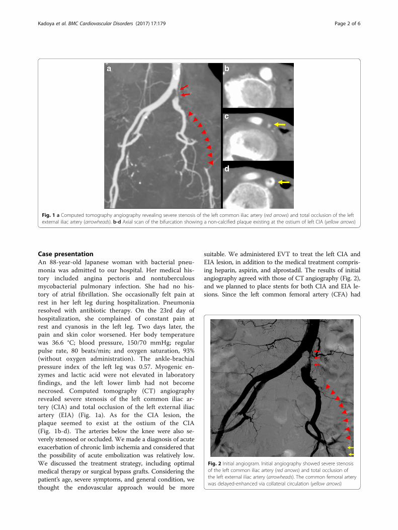
Fig. 1 a Computed tomography angiography revealing severe stenosis of the left common iliac artery (red arrows) and total occlusion of the leftexternal iliac artery (arrowheads). b-d Axial scan of the bifurcation showing a non-calcified plaque existing at the ostium of left CIA (yellow arrows)
Fig. 2 Initial angiogram. Initial angiography showed severe stenosisof the left common iliac artery (red arrows) and total occlusion ofthe left external iliac artery (arrowheads). The common femoral arterywas delayed-enhanced via collateral circulation (yellow arrows)
Kadoya et al. BMC Cardiovascular Disorders (2017) 17:179 Page 2 of 6
Case presentationAn 88-year-old Japanese woman with bacterial pneu-monia was admitted to our hospital. Her medical his-tory included angina pectoris and nontuberculousmycobacterial pulmonary infection. She had no his-tory of atrial fibrillation. She occasionally felt pain atrest in her left leg during hospitalization. Pneumoniaresolved with antibiotic therapy. On the 23rd day ofhospitalization, she complained of constant pain atrest and cyanosis in the left leg. Two days later, thepain and skin color worsened. Her body temperaturewas 36.6 °C; blood pressure, 150/70 mmHg; regularpulse rate, 80 beats/min; and oxygen saturation, 93%(without oxygen administration). The ankle-brachialpressure index of the left leg was 0.57. Myogenic en-zymes and lactic acid were not elevated in laboratoryfindings, and the left lower limb had not becomenecrosed. Computed tomography (CT) angiographyrevealed severe stenosis of the left common iliac ar-tery (CIA) and total occlusion of the left external iliacartery (EIA) (Fig. 1a). As for the CIA lesion, theplaque seemed to exist at the ostium of the CIA(Fig. 1b-d). The arteries below the knee were also se-verely stenosed or occluded. We made a diagnosis of acuteexacerbation of chronic limb ischemia and considered thatthe possibility of acute embolization was relatively low.We discussed the treatment strategy, including optimalmedical therapy or surgical bypass grafts. Considering thepatient’s age, severe symptoms, and general condition, wethought the endovascular approach would be more
suitable. We administered EVT to treat the left CIA andEIA lesion, in addition to the medical treatment compris-ing heparin, aspirin, and alprostadil. The results of initialangiography agreed with those of CT angiography (Fig. 2),and we planned to place stents for both CIA and EIA le-sions. Since the left common femoral artery (CFA) had
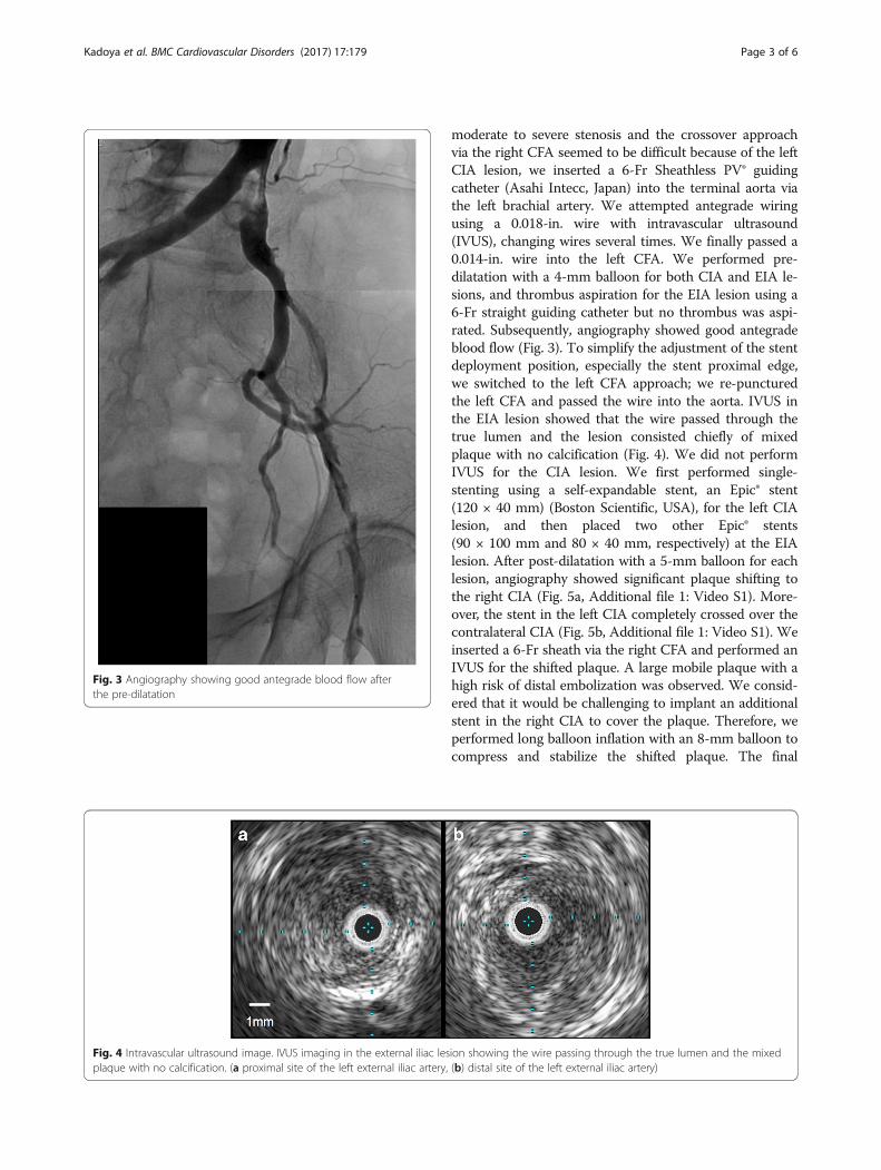
Fig. 3 Angiography showing good antegrade blood flow afterthe pre-dilatation
Fig. 4 Intravascular ultrasound image. IVUS imaging in the external iliac lesplaque with no calcification. (a proximal site of the left external iliac artery,
Kadoya et al. BMC Cardiovascular Disorders (2017) 17:179 Page 3 of 6
moderate to severe stenosis and the crossover approachvia the right CFA seemed to be difficult because of the leftCIA lesion, we inserted a 6-Fr Sheathless PV® guidingcatheter (Asahi Intecc, Japan) into the terminal aorta viathe left brachial artery. We attempted antegrade wiringusing a 0.018-in. wire with intravascular ultrasound(IVUS), changing wires several times. We finally passed a0.014-in. wire into the left CFA. We performed pre-dilatation with a 4-mm balloon for both CIA and EIA le-sions, and thrombus aspiration for the EIA lesion using a6-Fr straight guiding catheter but no thrombus was aspi-rated. Subsequently, angiography showed good antegradeblood flow (Fig. 3). To simplify the adjustment of the stentdeployment position, especially the stent proximal edge,we switched to the left CFA approach; we re-puncturedthe left CFA and passed the wire into the aorta. IVUS inthe EIA lesion showed that the wire passed through thetrue lumen and the lesion consisted chiefly of mixedplaque with no calcification (Fig. 4). We did not performIVUS for the CIA lesion. We first performed single-stenting using a self-expandable stent, an Epic® stent(120 × 40 mm) (Boston Scientific, USA), for the left CIAlesion, and then placed two other Epic® stents(90 × 100 mm and 80 × 40 mm, respectively) at the EIAlesion. After post-dilatation with a 5-mm balloon for eachlesion, angiography showed significant plaque shifting tothe right CIA (Fig. 5a, Additional file 1: Video S1). More-over, the stent in the left CIA completely crossed over thecontralateral CIA (Fig. 5b, Additional file 1: Video S1). Weinserted a 6-Fr sheath via the right CFA and performed anIVUS for the shifted plaque. A large mobile plaque with ahigh risk of distal embolization was observed. We consid-ered that it would be challenging to implant an additionalstent in the right CIA to cover the plaque. Therefore, weperformed long balloon inflation with an 8-mm balloon tocompress and stabilize the shifted plaque. The final
ion showing the wire passing through the true lumen and the mixed(b) distal site of the left external iliac artery)
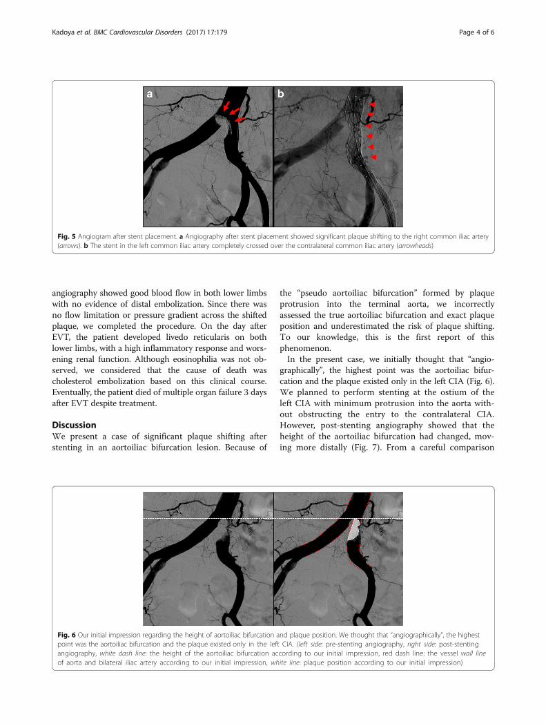
Fig. 5 Angiogram after stent placement. a Angiography after stent placement showed significant plaque shifting to the right common iliac artery(arrows). b The stent in the left common iliac artery completely crossed over the contralateral common iliac artery (arrowheads)
Kadoya et al. BMC Cardiovascular Disorders (2017) 17:179 Page 4 of 6
angiography showed good blood flow in both lower limbswith no evidence of distal embolization. Since there wasno flow limitation or pressure gradient across the shiftedplaque, we completed the procedure. On the day afterEVT, the patient developed livedo reticularis on bothlower limbs, with a high inflammatory response and wors-ening renal function. Although eosinophilia was not ob-served, we considered that the cause of death wascholesterol embolization based on this clinical course.Eventually, the patient died of multiple organ failure 3 daysafter EVT despite treatment.
DiscussionWe present a case of significant plaque shifting afterstenting in an aortoiliac bifurcation lesion. Because of
Fig. 6 Our initial impression regarding the height of aortoiliac bifurcation apoint was the aortoiliac bifurcation and the plaque existed only in the leftangiography, white dash line: the height of the aortoiliac bifurcation acof aorta and bilateral iliac artery according to our initial impression, wh
the “pseudo aortoiliac bifurcation” formed by plaqueprotrusion into the terminal aorta, we incorrectlyassessed the true aortoiliac bifurcation and exact plaqueposition and underestimated the risk of plaque shifting.To our knowledge, this is the first report of thisphenomenon.In the present case, we initially thought that “angio-
graphically”, the highest point was the aortoiliac bifur-cation and the plaque existed only in the left CIA (Fig. 6).We planned to perform stenting at the ostium of theleft CIA with minimum protrusion into the aorta with-out obstructing the entry to the contralateral CIA.However, post-stenting angiography showed that theheight of the aortoiliac bifurcation had changed, mov-ing more distally (Fig. 7). From a careful comparison
nd plaque position. We thought that “angiographically”, the highestCIA. (left side: pre-stenting angiography, right side: post-stenting
cording to our initial impression, red dash line: the vessel wall lineite line: plaque position according to our initial impression)
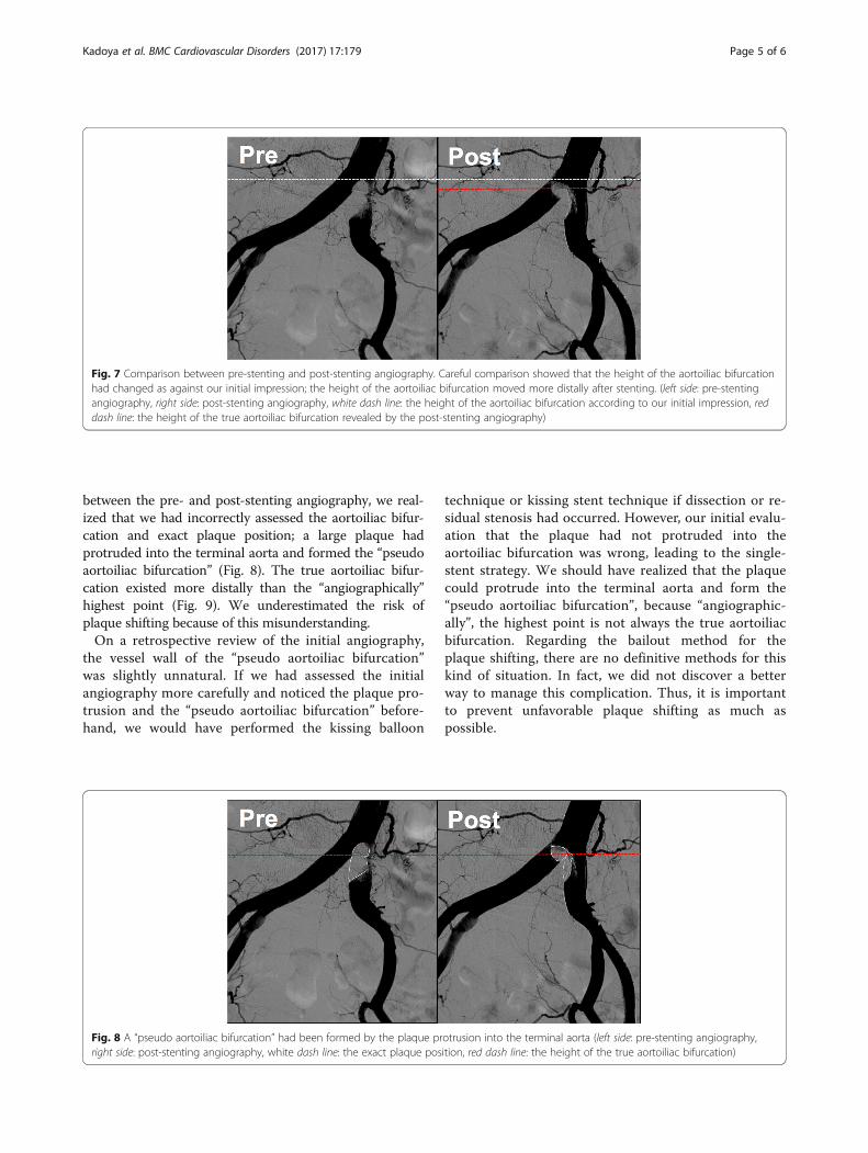
Fig. 7 Comparison between pre-stenting and post-stenting angiography. Careful comparison showed that the height of the aortoiliac bifurcationhad changed as against our initial impression; the height of the aortoiliac bifurcation moved more distally after stenting. (left side: pre-stentingangiography, right side: post-stenting angiography, white dash line: the height of the aortoiliac bifurcation according to our initial impression, reddash line: the height of the true aortoiliac bifurcation revealed by the post-stenting angiography)
Kadoya et al. BMC Cardiovascular Disorders (2017) 17:179 Page 5 of 6
between the pre- and post-stenting angiography, we real-ized that we had incorrectly assessed the aortoiliac bifur-cation and exact plaque position; a large plaque hadprotruded into the terminal aorta and formed the “pseudoaortoiliac bifurcation” (Fig. 8). The true aortoiliac bifur-cation existed more distally than the “angiographically”highest point (Fig. 9). We underestimated the risk ofplaque shifting because of this misunderstanding.On a retrospective review of the initial angiography,
the vessel wall of the “pseudo aortoiliac bifurcation”was slightly unnatural. If we had assessed the initialangiography more carefully and noticed the plaque pro-trusion and the “pseudo aortoiliac bifurcation” before-hand, we would have performed the kissing balloon
Fig. 8 A “pseudo aortoiliac bifurcation” had been formed by the plaque prright side: post-stenting angiography, white dash line: the exact plaque pos
technique or kissing stent technique if dissection or re-sidual stenosis had occurred. However, our initial evalu-ation that the plaque had not protruded into theaortoiliac bifurcation was wrong, leading to the single-stent strategy. We should have realized that the plaquecould protrude into the terminal aorta and form the“pseudo aortoiliac bifurcation”, because “angiographic-ally”, the highest point is not always the true aortoiliacbifurcation. Regarding the bailout method for theplaque shifting, there are no definitive methods for thiskind of situation. In fact, we did not discover a betterway to manage this complication. Thus, it is importantto prevent unfavorable plaque shifting as much aspossible.
otrusion into the terminal aorta (left side: pre-stenting angiography,ition, red dash line: the height of the true aortoiliac bifurcation)
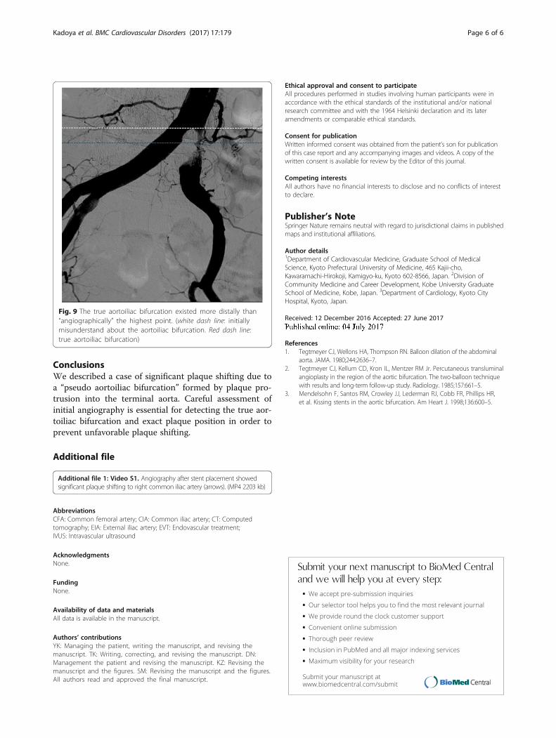
Fig. 9 The true aortoiliac bifurcation existed more distally than“angiographically” the highest point. (white dash line: initiallymisunderstand about the aortoiliac bifurcation. Red dash line:true aortoiliac bifurcation)
Kadoya et al. BMC Cardiovascular Disorders (2017) 17:179 Page 6 of 6
ConclusionsWe described a case of significant plaque shifting due toa “pseudo aortoiliac bifurcation” formed by plaque pro-trusion into the terminal aorta. Careful assessment ofinitial angiography is essential for detecting the true aor-toiliac bifurcation and exact plaque position in order toprevent unfavorable plaque shifting.
Additional file
Additional file 1: Video S1. Angiography after stent placement showedsignificant plaque shifting to right common iliac artery (arrows). (MP4 2203 kb)
AbbreviationsCFA: Common femoral artery; CIA: Common iliac artery; CT: Computedtomography; EIA: External iliac artery; EVT: Endovascular treatment;IVUS: Intravascular ultrasound
AcknowledgmentsNone.
FundingNone.
Availability of data and materialsAll data is available in the manuscript.
Authors’ contributionsYK: Managing the patient, writing the manuscript, and revising themanuscript. TK: Writing, correcting, and revising the manuscript. DN:Management the patient and revising the manuscript. KZ: Revising themanuscript and the figures. SM: Revising the manuscript and the figures.All authors read and approved the final manuscript.
Ethical approval and consent to participateAll procedures performed in studies involving human participants were inaccordance with the ethical standards of the institutional and/or nationalresearch committee and with the 1964 Helsinki declaration and its lateramendments or comparable ethical standards.
Consent for publicationWritten informed consent was obtained from the patient’s son for publicationof this case report and any accompanying images and videos. A copy of thewritten consent is available for review by the Editor of this journal.
Competing interestsAll authors have no financial interests to disclose and no conflicts of interestto declare.
Publisher’s NoteSpringer Nature remains neutral with regard to jurisdictional claims in publishedmaps and institutional affiliations.
Author details1Department of Cardiovascular Medicine, Graduate School of MedicalScience, Kyoto Prefectural University of Medicine, 465 Kajii-cho,Kawaramachi-Hirokoji, Kamigyo-ku, Kyoto 602-8566, Japan. 2Division ofCommunity Medicine and Career Development, Kobe University GraduateSchool of Medicine, Kobe, Japan. 3Department of Cardiology, Kyoto CityHospital, Kyoto, Japan.
Received: 12 December 2016 Accepted: 27 June 2017
References1. Tegtmeyer CJ, Wellons HA, Thompson RN. Balloon dilation of the abdominal
aorta. JAMA. 1980;244:2636–7.2. Tegtmeyer CJ, Kellum CD, Kron IL, Mentzer RM Jr. Percutaneous transluminal
angioplasty in the region of the aortic bifurcation. The two-balloon techniquewith results and long-term follow-up study. Radiology. 1985;157:661–5.
3. Mendelsohn F, Santos RM, Crowley JJ, Lederman RJ, Cobb FR, Phillips HR,et al. Kissing stents in the aortic bifurcation. Am Heart J. 1998;136:600–5.
• We accept pre-submission inquiries
• Our selector tool helps you to find the most relevant journal
• We provide round the clock customer support
• Convenient online submission
• Thorough peer review
• Inclusion in PubMed and all major indexing services
• Maximum visibility for your research
Submit your manuscript atwww.biomedcentral.com/submit
Submit your next manuscript to BioMed Central and we will help you at every step: