Embed Size (px)
Citation preview
Animal and Plant Health Agency
Access to Information Team
Weybourne Building
Ground Floor
Woodham Lane
New Haw
Addlestone
Surrey
KT15 3NB
T 01932 341111
F 01932 357608
www.gov.uk/apha
The Animal and Plant Health Agency is an Executive Agency of the Department for Environment, Food and Rural Affairs working to
safeguard animal and plant health for the benefit of people, the environment and the economy.
Our Ref: ATIC0815
{By Email}
17 March 2016
Dear
PROVISION OF REQUESTED INFORMATION
Thank you for your request for information about Mycoplasma disease, which we
received on 29 February 2016. Your request has been handled under the Freedom of
Information Act 2000.
The information you requested and our response is detailed below:
“I am writing to request information under the Freedom of Information Act about the
extent of mycoplasma diseases affecting UK cattle herds.
To this end, I have listed six questions which I would be grateful if you could provide
responses to by email.
The questions are:-
1. How many confirmed cases of Mycoplasma have been recorded in the UK since
2006? Of these, how many were: a) a single strain of mycoplasma b) multiple
infections of mycoplasma (if possible, please provide a detailed breakdown of the
different confirmed types of mycoplasma infection).
Please see our response to your request at Appendix 1, with additional
information and comments.
2. How many cattle have been slaughtered due to Mycoplasma diseases since
2006?
APHA do not hold this information. Government has not directed the culling of
any cattle due to Mycoplasmosis. Farmers may have culled cattle privately and
may or may not have known at the time that a mycoplasma was part of the
problem.
3. How much screening of UK cattle herds is being done to determine Mycoplasma
infections in UK herds?
See Table 1 in Appendix 1 for serological testing. See Table 3 in Appendix 1for
antigen detection. Mycoplasma diseases will be considered on any cases
submitted to APHA for diagnosis (numbers not available) where Mycoplasma is
potential differential diagnosis. Details of a testing carried out on suspect
Mycoplasma cases is detailed in Table 3.
4. What measures does Defra take to test imported cattle for Mycoplasma
diseases?
Imported cattle are certified as coming from countries or regions free from
contagious bovine pleuropneumonia (CBPP), so no official testing is carried out.
At present the risk is negligible because the whole of the EU is free. If that
changes then import testing could be instated. APHA does not test imported
cattle for non-notifiable mycoplasmas that are considered to be largely endemic
diseases. This is a decision for the farmer on the advice of their private vet.
5. What training are government vets being given to help them identify mycoplasma
in UK cattle herds?
APHA Veterinary Investigation Officers are trained to work to protocols which
direct them to investigate mycoplasmal disease when it is a potential diagnosis.
In addition there is training via teaching at the British Cattle Veterinary
Association (BCVA) Congress; and raised awareness through attendance at
BCVA. The Cattle Health and Welfare Group (CHAWG) information note for vets
and farmers is also published in 2014.
Please also see attached at Appendix 2 a guide for veterinary practitioners on
Mycoplasma bovis in BRD.
6. What efforts have Defra made to create awareness of Mycoplasma diseases in
the UK livestock industry (Please provide any supporting evidence).”
Defra and APHA vets have spoken at a number of meetings attended by private veterinary surgeons. In addition to these activities, the following publications have been produced:
CHAWG information note for vets and farmers published 2014 http://beefandlamb.ahdb.org.uk/wp/wp-content/uploads/2015/01/Mycoplasma-Bovis-Briefing-Dec-2014.pdf
Technical consultancy for Zoetis: Business solutions: A guide for Veterinary Practitioners on Mycoplasma bovis in BRD.
Book: Robin Nicholas, Roger Ayling, Laura McAuliffe (2008). Mycoplasma Diseases of Ruminants. CAB International ISBN-13:9780851990125
Discontools review Mycoplasma bovis: http://www.discontools.eu/Diseases/Detail/82
Publications (that include APHA staff as authors)
Ayling, R. D., Gosney, F., Hlusek, M. (2015). Mycoplasma diagnostics, some results, and what we still don’t know about Mycoplasma bovis disease. Cattle Practice. 23: 2, 248-251
Nicholas, R.A.J., Ayling, R.D. (2003) Mycoplasma bovis: disease, diagnosis, and control. Research in Veterinary Science, 74: 105-112.
Dudek, K., Bednarek, D., Ayling, R. D., E. Szacawa (2015). Flow cytometry analysis of peripheral blood leukocyte subpopulations in calves experimentally infected with field isolates of Mycoplasma bovis. Acta Veterinaria Hungarica, 63:2,
Szacawa, E., Niemczujk, K., Dudek, K., Bednarek, D., Rosales, R., Ayling, R. (2015). Mycoplasma bovis infections and co-infections with other Mycoplasma spp. with different clinical manifestations in affected cattle herds in eastern region of Poland. Bull Vet Inst Pulawy. 59: 331-337.
Otter, A., Wright, T., Leonard, D., Richardson, M., Ayling, R. (2015). Mycoplasma bovis mastitis in dry dairy cows. Veterinary Record, 12 December 2015. doi:10.1136/vr.h6663
Dudek, K., Bednarek, D., Szacawa, E., Ayling, R. D., Krzysiak, M. K., Marczuk, J. (2015). A serological and molecular study of the occurrence of mycoplasmas in European bison (Bison bonasus) from two areas of Eastern Poland. Polish Journal of Veterinary Sciences. 18: 881-883.
Lysnyansky, I., Freed, M., Rosales, R. S., Mikula, I., Khateb, N., Gerchman, I., van Straten, M., Levisohn, S. (2015). An overview of Mycoplasma bovis mastitis in Israel (2004-2014). The Veterinary Journal. Doui.org/10.1016/j.tvjl.2015.10.057
Ayling, R.D., Barden, G., Rosales, R. S., Gosney, F. L. (2014). Changes in antimicrobial susceptibility of Mycoplasma bovis isolates from Great Britain Veterinary Record. Sept 3rd doi: 10.1136/vr.102303
Ayling, R. D., Hlusek, M., Gosney, F., Rosales, R. S. (2014). Po rozpoznaniu, kolejne kroki na drodze zwalczania zakażenia Mycoplasma bovis u bydła. (After diagnosis, the next steps towards control of Mycoplasma bovis in cattle). Lecznica dużych zwierząt monografia. 2: 4-6.
Ayling, R. D., Bisgaard-Frantzen, S., Adler, A., Blowey, R. W., Barlow, A. M., Millar, M. F., van der Burgt, G. M. (2012) Detection of Candidatus “Mycoplasma haemobos” Mycoplasma wenyonii and Anaplasma phagocytophilum from cattle in England. Veterinary Record. Doi:10.1136/vt100636.
Ayling, R. D., Nicholas, R. A. J. (2008). Zastosowanie antybiotyków w leczeneniu infkcji wywołanych przez mykoplazmy u bydła. (Antimicrobial treatment of bovine mycoplasma infections). Najważniejsze czynniki etiologiczne, patogeneza I najnowsze trendy w profilakyce I terapii syndrome oddechowego bydła (BRD). pp.42-47. Proceedings of BRD Conference at the National Veterinary Research institute, Pulawy, Poland. (In Polish).
Ayling, R. D., Godinho, K., Nicholas, R. A. J. (2007). Comparative studies on the in vitro antimicrobial sensitivities of Mycoplasma mycoides subsp. mycoides small colony type and Mycoplasma bovis. Proceedings of the FAO-AU/IBAR-IAEA Consultative Group Meeting on CBPP in Africa, FAO, Rome pp51-61.
Strugnell, B. W., Glover, M., Wessels, M., Ayling, R. D. (2013). Ear droop and stertor in dairy calves associated with Mycoplasma bovis. Veterinary Record. 173: 299-300.
Lerner, U., Amram, E., Ayling, R. D., Mikula, I., Gerchman, I., Harrus, S., Teff, D., Yogev, D., Lysnyansky, I. (2014). Acquired resistance to the 16-membered macrolides tylosin and tilmicosin by Mycoplasma bovis. Veterinary Microbiology. 168: 365-371.
Dudek, K., Bednarek, D., Ayling, R. D. E. Szacawa. (2013). Immunomodulatory effect of Mycoplasma bovis in experimentally infected calves. Bull Vet Inst Pulawy, 57, 499-506.
Bednarek, D., Ayling, R. D., Nicholas, R. A. J., Dudek, K., Szymanska-Cerwinska, M. (2012). Serological survey to determine the occurrence of respiratory Mycoplasma infections in the Polish cattle population. Veterinary Record. 171: 45. doi:10.1136/vr.100545
Watson, P., Mason, C., Stevenson, H., Scholes, S., Schock, A., Mearns, R., Ayling, R., Nicholas, R. (2012). Laboratory diagnosis of Mycoplasma/Ureaplasma abortion in cattle. Veterinary Record. 170: 82-84.
Foster, A. P., Naylor, R. D., Howie, N. M., Nicholas, R. A. J., Ayling, R. D. (2009). Mycoplasma bovis and otitis in dairy calves in the United Kingdom. The Veterinary Journal. 179: 455-457.
Van der Burgt, G., Main, W., Ayling, R. (2008). Bovine mastitis caused by Mycoplasma bovis. Veterinary Record 163: 666.
Wrathall, A. E., Ayling, R. D., Simmons, H. (2007). Risks of transmitting mycoplasmas by semen and embryo transfer techniques in cattle, sheep, goats and pigs. CAB Reviews: Perspectives in Agriculture, Veterinary Science, Nutrition and Natural Resources. 2: 36, 1-31. on line http://www.cabastractsplus.org/cabreviews
Houlihan, M., Veenstra, B., Christian, M. K., Nicholas, R., Ayling, R. (2007). Mastitis and arthritis in two dairy herds caused by Mycoplasma bovis. Veterinary Record. 160: 126-127.
Nicholas, R., Ayling, R., McAuliffe, L. (2007). Mycoplasma mastitis. Veterinary Record. 160: 382-383.
Nicholas, R. A. J., Ayling, R. D., Woodger, N., Wessells, M. E., Houlihan, M. G. (2006). Mycoplasmas in adult cattle: Bugs worth bothering about? Irish Veterinary Journal. 59: 568-572.
McAuliffe, L., Lawes, J., Bell, S., Barlow, A., Ayling, R. D., Nicholas, R. A. J. (2006). The detection of Mycoplasma (formerly Eperythrozoon) wenyonii by 16S rDNA PCR and denaturing gradient gel electrophoresis. Veterinary Microbiology. 117: 292-296.
Nicholas, R.A.J., Ayling, R.D., Stipkovits, L. (2002) An experimental vaccine for calf pneumonia caused by Mycoplasma bovis: clinical, cultural, serological and pathological findings. Vaccine, 20: 3569-3575.
McAuliffe, L., Kokotovic, B., Ayling, R.D., Nicholas, R.A.J. (2004) Molecular epidemiological analysis of Mycoplasma bovis isolates from the United Kingdom shows two genetically distinct clusters. Journal of Clinical Microbiology, 42: 4556-4565.
Bashiruddin, J.B, Frey, J., Heldtander Königsson, M., Johansson, K.E., Hotzel, H., Diller, R., de Santis, P., Botelho, A., Ayling, R.D., Nicholas, R.A.J., Thiaucourt, F., Sachse, K. (2005) Evaluation of PCR systems for the identification and differentiation of Mycoplasma agalactiae and Mycoplasma bovis: A collaborative trial. The Veterinary Journal, 169: 268-275.
Miles, K., McAuliffe, L., Persson, A., Ayling, R.D., Nicholas, R.A.J. (2005) Insertion sequence profiling of UK Mycoplasma bovis field isolates. Veterinary Microbiology, 107: 301-306.
Strugnell, B., McAuliffe, L. (2012) Mycoplasma wenyonii infection in cattle. In Practice 2012;34:3 146-154 doi:10.1136/inp.e1550
Recent meeting abstracts
Ayling, R. (2015). Insights into cattle mycoplasmoses in England and Wales. Cattle Association Veterinary Ireland Abstracts –October 2015.
I attach an Annex which explains the copyright that applies to the information being
released to you and contact details should you be unhappy with the service you have
received.
If you have any queries about this letter, please contact the Access to Information Team
at the email address below or postal address at the top of this letter.
Yours sincerely
ACCESS TO INFORMATION TEAM
Email: [email protected]

Annex
Copyright
The information supplied to you is Crown copyright, unless otherwise stated, and is protected by the Copyright, Designs and Patents Act 1988. You are free to use it for your own purposes, and for the purposes of news reporting. You can find details on the arrangements for re-using Crown copyright information at:
http://www.nationalarchives.gov.uk/doc/open-government-licence/open-government-licence.htm
Information you receive which is not subject to Crown Copyright continues to be protected by the copyright of the person, or organisation, from which the information originated. You must ensure that you gain their permission before reproducing any third party (non Crown Copyright) information.
In keeping with the spirit and effect of the Freedom of Information Act 2000/Environmental Information Regulations 2004, all information is assumed to be releasable to the public unless exempt. The information released to you may now be published on our website together with any related information that will provide a key to its wider context.
Complaints If you are unhappy with the result of your request for information you may request an internal review within 40 working days of the date of this letter. If you wish to request an internal review, please contact: The Access to Information Team at [email protected] or at the postal address at the top of this letter, who will arrange for an internal review of your case. If you are not content with the outcome of the internal review, you have the right to apply directly to the Information Commissioner for a decision. Please note that generally the Information Commissioner cannot make a decision unless you have first exhausted APHA’s own complaints procedure. The Information Commissioner can be contacted at: Information Commissioner’s Office Wycliffe House Water Lane Wilmslow Cheshire SK9 5AF
Appendix 1
The Veterinary Investigation Diagnosis Analysis (VIDA) data is available at: https://www.gov.uk/government/statistics/veterinary-investigation-diagnosis-analysis-vida-report-2014 Please note Mycoplasma bovis is not a notifiable disease. Comment: many of the clinical signs seen in cattle that may be associated with Mycoplasma infections are often multi-factorial. Therefore the detection of a mycoplasma species may not be the concluding diagnosis of the cause of a disease. Bovine respiratory disease (BRD) develops as a result of complex interactions between environmental factors, host factors, and pathogens. Environmental factors (eg, weaning, transport, commingling, crowding, inclement weather, dust, and inadequate ventilation) serve as stressors that adversely affect the immune and nonimmune defense mechanisms of the host. In addition, certain environmental factors (eg, crowding and inadequate ventilation) can enhance the transmission of infectious agents among animals. Many infectious agents have been associated with BRD, these include viruses: Bovine Respiratory Syncytial Virus (BRSV), ParaInfluenza 3 (PI3), Adenovirus, Bovine Viral Diarrhea Virus (BVDV), and Infectious Bovine Rhinotracheitis (IBR) and bacteria: Pasteurella multocida, Mannheimia haemolytica, Histophilus somni, Mycoplasma bovis and some other Mycoplasma species. Mastitis may also be a clinical sign of infection with Mycoplasma bovis but more commonly caused by other bacterial pathogens including: Pseudomonas species; Staphylococcus species; Streptococcus species; Brucella species; Corynebacterium species; Escherichia coli; Klebsiella species; Enterobacter species; Pasteurella species, Trueperella pyogenes; Proteus species; and other Mycoplasma species. Diagnosis of Mycoplasma at APHA. Note: samples may have been tested by non-APHA labs but we have no data on this. Serology for Mycoplasma bovis: This shows previous exposure to Mycoplasma bovis. Samples are submitted from suspect cases usually showing clinical signs of BRD, so this data is biased to clinical cases and cannot be extrapolated to the whole cattle population. Ref: Ayling, R. D., Gosney, F., Hlusek, M. (2015) Mycoplasma diagnostics, some results, and what we still don’t know about Mycoplasma bovis disease. Cattle Practice. 23: 2, 248-251.
Appendix 1
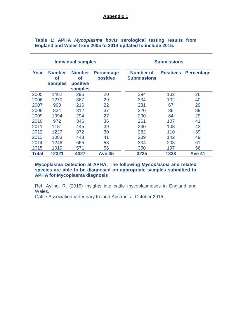
Table 1: APHA Mycoplasma bovis serological testing results from England and Wales from 2005 to 2014 updated to include 2015.
Individual samples
Submissions
Year Number of
Samples
Number of
positive samples
Percentage positive
Number of Submissions
Positives Percentage
2005 1462 296 20 394 102 26 2006 1270 367 29 334 132 40 2007 963 216 22 231 67 29 2008 834 312 37 220 86 39 2009 1094 294 27 290 84 29 2010 972 346 36 261 107 41 2011 1151 445 39 240 103 43 2012 1227 372 30 282 110 39 2013 1083 443 41 289 142 49 2014 2015
1246 1019
665 571
53 56
334 350
203 197
61 56
Total 12321 4327 Ave 35 3225 1333 Ave 41
Mycoplasma Detection at APHA; The following Mycoplasma and related species are able to be diagnosed on appropriate samples submitted to APHA for Mycoplasma diagnosis Ref: Ayling, R. (2015) Insights into cattle mycoplasmoses in England and Wales. Cattle Association Veterinary Ireland Abstracts –October 2015.
Appendix 1
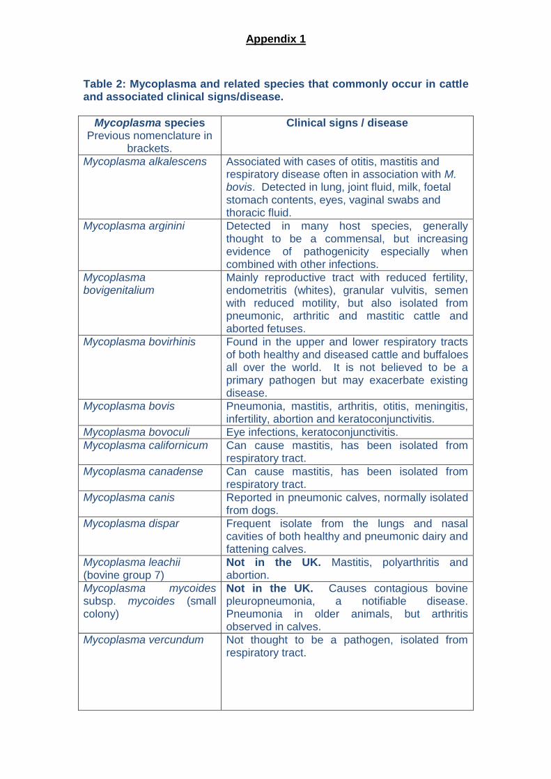
Table 2: Mycoplasma and related species that commonly occur in cattle and associated clinical signs/disease.
Mycoplasma species Previous nomenclature in
brackets.
Clinical signs / disease
Mycoplasma alkalescens Associated with cases of otitis, mastitis and respiratory disease often in association with M. bovis. Detected in lung, joint fluid, milk, foetal stomach contents, eyes, vaginal swabs and thoracic fluid.
Mycoplasma arginini Detected in many host species, generally thought to be a commensal, but increasing evidence of pathogenicity especially when combined with other infections.
Mycoplasma bovigenitalium
Mainly reproductive tract with reduced fertility, endometritis (whites), granular vulvitis, semen with reduced motility, but also isolated from pneumonic, arthritic and mastitic cattle and aborted fetuses.
Mycoplasma bovirhinis Found in the upper and lower respiratory tracts of both healthy and diseased cattle and buffaloes all over the world. It is not believed to be a primary pathogen but may exacerbate existing disease.
Mycoplasma bovis Pneumonia, mastitis, arthritis, otitis, meningitis, infertility, abortion and keratoconjunctivitis.
Mycoplasma bovoculi Eye infections, keratoconjunctivitis.
Mycoplasma californicum Can cause mastitis, has been isolated from respiratory tract.
Mycoplasma canadense Can cause mastitis, has been isolated from respiratory tract.
Mycoplasma canis Reported in pneumonic calves, normally isolated from dogs.
Mycoplasma dispar Frequent isolate from the lungs and nasal cavities of both healthy and pneumonic dairy and fattening calves.
Mycoplasma leachii (bovine group 7)
Not in the UK. Mastitis, polyarthritis and abortion.
Mycoplasma mycoides subsp. mycoides (small colony)
Not in the UK. Causes contagious bovine pleuropneumonia, a notifiable disease. Pneumonia in older animals, but arthritis observed in calves.
Mycoplasma vercundum Not thought to be a pathogen, isolated from respiratory tract.
Appendix 1
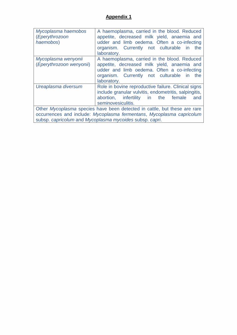
Mycoplasma haemobos (Eperythrozoon haemobos)
A haemoplasma, carried in the blood. Reduced appetite, decreased milk yield, anaemia and udder and limb oedema. Often a co-infecting organism. Currently not culturable in the laboratory.
Mycoplasma wenyonii (Eperythrozoon wenyonii)
A haemoplasma, carried in the blood. Reduced appetite, decreased milk yield, anaemia and udder and limb oedema. Often a co-infecting organism. Currently not culturable in the laboratory.
Ureaplasma diversum Role in bovine reproductive failure. Clinical signs include granular vulvitis, endometritis, salpingitis, abortion, infertility in the female and seminovesiculitis.
Other Mycoplasma species have been detected in cattle, but these are rare occurrences and include: Mycoplasma fermentans, Mycoplasma capricolum subsp. capricolum and Mycoplasma mycoides subsp. capri.
Appendix 1
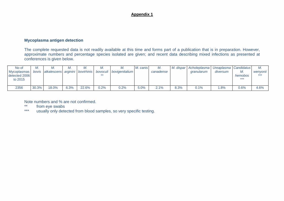
Mycoplasma antigen detection The complete requested data is not readily available at this time and forms part of a publication that is in preparation. However, approximate numbers and percentage species isolated are given; and recent data describing mixed infections as presented at conferences is given below.
No of Mycoplasmas detected 2006
to 2015
M. bovis
M. alkalescens
M. arginini
M. bovirhinis
M. bovoculi
**
M. bovigenitalium
M. canis M. canadense
M. dispar Acholeplasma granularum
Ureaplasma diversum
Candidatus M.
hemobos ***
M. wenyonii
***
2356 30.3% 18.0% 6.3% 22.6% 0.2% 0.2% 5.0% 2.1% 8.3% 0.1% 1.8% 0.6% 4.6%
Note numbers and % are not confirmed. ** from eye swabs *** usually only detected from blood samples, so very specific testing.
Appendix 1
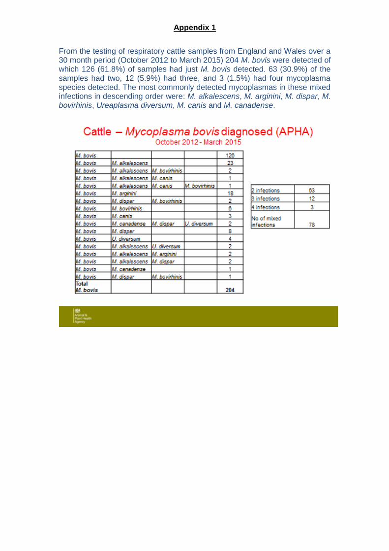
From the testing of respiratory cattle samples from England and Wales over a 30 month period (October 2012 to March 2015) 204 M. bovis were detected of which 126 (61.8%) of samples had just M. bovis detected. 63 (30.9%) of the samples had two, 12 (5.9%) had three, and 3 (1.5%) had four mycoplasma species detected. The most commonly detected mycoplasmas in these mixed infections in descending order were: M. alkalescens, M. arginini, M. dispar, M. bovirhinis, Ureaplasma diversum, M. canis and M. canadense.
Business solutionsA GUIDE FOR VETERINARY PRACTITIONERS on Mycoplasma bovis in BRD
Technical consultancy provided by: the Mycoplasma Group AHVLA
For further information please contact Zoetis UK Ltd, Walton Oaks, Dorking Road, Walton on the Hill, Tadworth, Surrey, KT20 7NS. Technical Services and Customer Support 0845 3008034 Use medicines responsibly (www.noah.co.uk/responsible) Date of preparation: September 2013. AH572/13
ServicesAPPENDIX 2
2 3
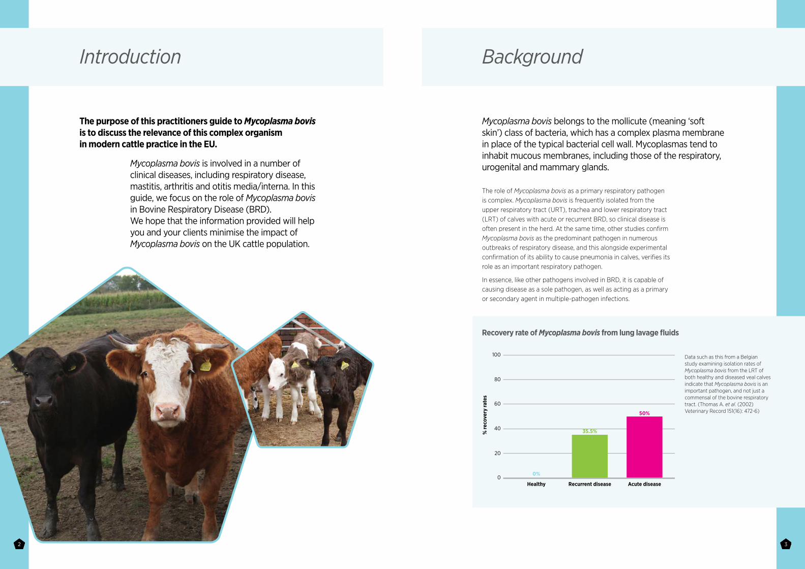
Mycoplasma bovis is involved in a number of clinical diseases, including respiratory disease, mastitis, arthritis and otitis media/interna. In this guide, we focus on the role of Mycoplasma bovis in Bovine Respiratory Disease (BRD). We hope that the information provided will help you and your clients minimise the impact of Mycoplasma bovis on the UK cattle population.
Introduction
The purpose of this practitioners guide to Mycoplasma bovis is to discuss the relevance of this complex organism in modern cattle practice in the EU.
The role of Mycoplasma bovis as a primary respiratory pathogen is complex. Mycoplasma bovis is frequently isolated from the upper respiratory tract (URT), trachea and lower respiratory tract (LRT) of calves with acute or recurrent BRD, so clinical disease is often present in the herd. At the same time, other studies confirm Mycoplasma bovis as the predominant pathogen in numerous outbreaks of respiratory disease, and this alongside experimental confirmation of its ability to cause pneumonia in calves, verifies its role as an important respiratory pathogen.
In essence, like other pathogens involved in BRD, it is capable of causing disease as a sole pathogen, as well as acting as a primary or secondary agent in multiple-pathogen infections.
Background
Mycoplasma bovis belongs to the mollicute (meaning ‘soft skin’) class of bacteria, which has a complex plasma membrane in place of the typical bacterial cell wall. Mycoplasmas tend to inhabit mucous membranes, including those of the respiratory, urogenital and mammary glands.
Recovery rate of Mycoplasma bovis from lung lavage fluids
Data such as this from a Belgian study examining isolation rates of Mycoplasma bovis from the LRT of both healthy and diseased veal calves indicate that Mycoplasma bovis is an important pathogen, and not just a commensal of the bovine respiratory tract. (Thomas A. et al. (2002) Veterinary Record 151(16): 472-6)
100
80
60
40
20
Recurrent disease
35.5%
0%
50%
Healthy Acute disease0
% re
cove
ry ra
tes
4 5
Initial infection of an animal or herd comes from exposure to Mycoplasma bovis through a variety of routes, the main sources being respiratory secretions and infected milk. Chronically infected cows are capable of shedding large numbers of bacteria in their milk.
In infected herds, calves become infected when they are very young, either through contact with contaminated vaginal mucus during parturition or in the maternity pen, being fed milk from chronically infected cows or through close contact with individuals shedding Mycoplasma bovis in respiratory secretions. Calves then go on to shed Mycoplasma bovis in large numbers during the first 2 months of life.
In many infected herds, the role of contaminated milk is crucial. Small numbers of infected cows can potentially contaminate large volumes of milk. Calves fed contaminated milk are much more likely to be colonised by Mycoplasma bovis.
Large numbers of Mycoplasma bovis can be isolated from the air of sheds housing infected animals. In calf barns, poor air circulation will significantly increase the bacterial load of the environment, and therefore the rate of transmission of the mycoplasma. Studies have demonstrated very rapid spread of infection within calf populations via this route.
In terms of cow-to-cow spread in milking herds, the role of fomite transmission is important, so it is plausible that a similar role may be important in terms of BRD. The bacterium is able to survive for prolonged periods of time, with survival rates being improved by lower ambient temperatures. Transmission of bacteria from calf to calf via infected feeding equipment, pen divisions and bedding could all play a role in spreading the bacteria and subsequent clinical disease, although the role of direct transfer remains more important.
In diseased herds, the prevalence of colonisation of the URT can be very high, with reports of 100% of animals being infected. Given the routes of transmission of the bacteria, it is not surprising that herds which experience high rates of Mycoplasma bovis-associated disease tend to have a higher prevalence of infection, and that once a herd is infected, eradication of Mycoplasma bovis is extremely hard due to the continual cycle of infection, shedding and transmission.
Epidemiology
Mycoplasma bovis is well adapted to causing chronic, asymptomatic infections, and therefore the role of ‘carrier’ animals is an important part of the Mycoplasma bovis story. Animals may remain infected for many years, shedding bacteria intermittently.
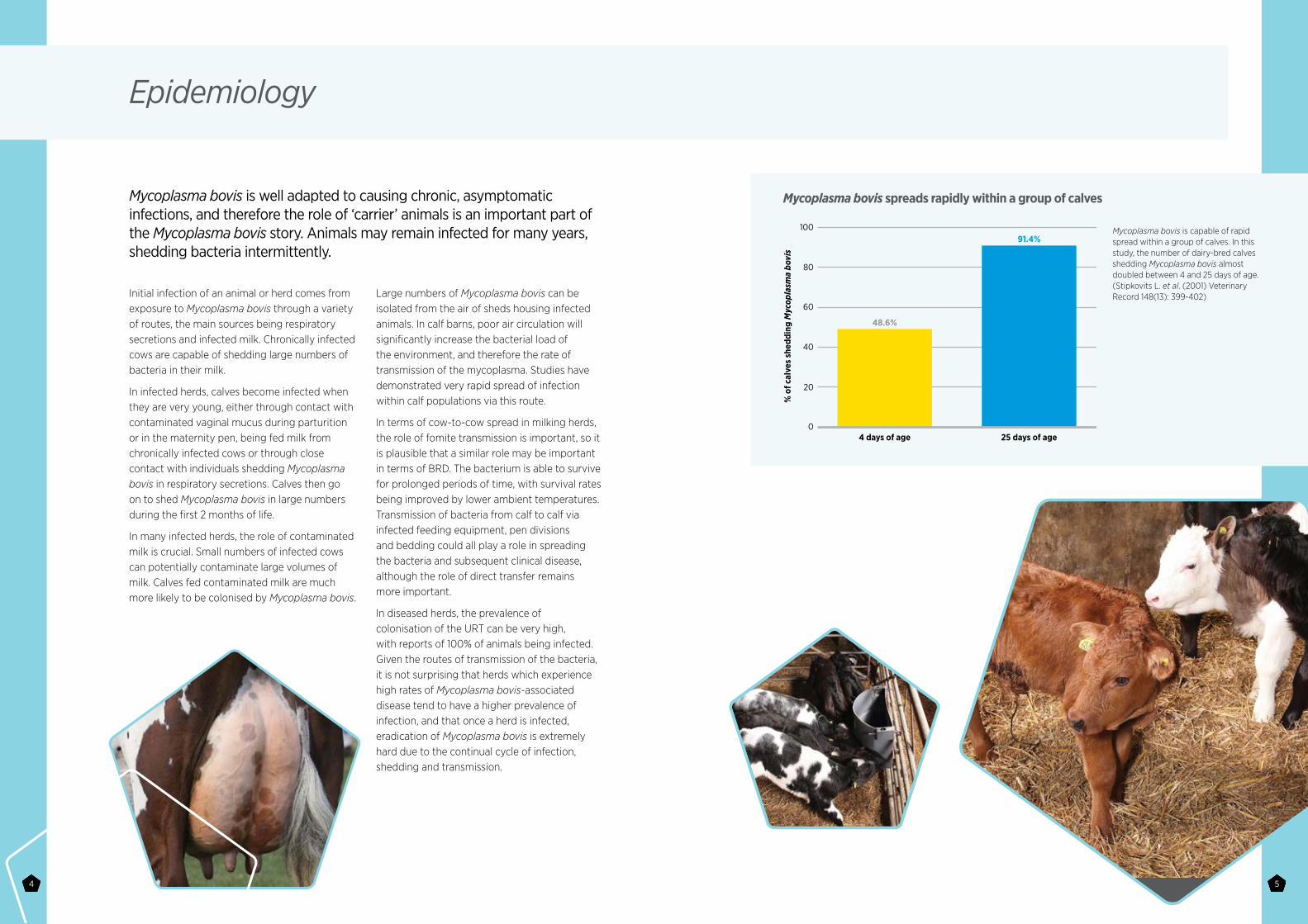
Mycoplasma bovis spreads rapidly within a group of calves
Mycoplasma bovis is capable of rapid spread within a group of calves. In this study, the number of dairy-bred calves shedding Mycoplasma bovis almost doubled between 4 and 25 days of age. (Stipkovits L. et al. (2001) Veterinary Record 148(13): 399-402)
100
80
60
40
20
4 days of age
% o
f cal
ves
shed
ding
Myc
opla
sma
bovi
s
48.6%
91.4%
25 days of age0
6 7
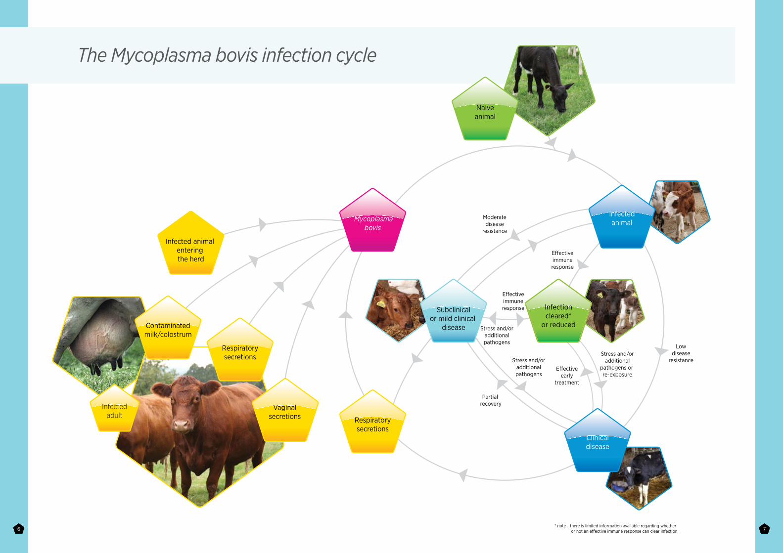
The Mycoplasma bovis infection cycle
E�ectiveimmuneresponse
Moderatedisease
resistance
Lowdisease
resistance
E�ectiveimmuneresponse
Stress and/oradditionalpathogens
Stress and/oradditionalpathogens
E�ectiveearly
treatment
Stress and/oradditional
pathogens or re-exposure
Partial recovery
* note - there is limited information available regarding whether or not an e�ective immune response can clear infection
Contaminatedmilk/colostrum
Respiratorysecretions
Infectedadult
Vaginalsecretions
Mycoplasmabovis
Naiveanimal
Infectioncleared*
or reduced
Infectedanimal
Subclinicalor mild clinical
disease
Clinicaldisease
Respiratorysecretions
Infected animal entering the herd
8 9
When we talk about prevalence, it is important to make the distinction between exposure to Mycoplasma bovis and its role as a key causal agent of BRD.
Based on exposure studies (seroprevalence), there is no doubt that Mycoplasma bovis is a highly prevalent bacteria. Recent studies identify high rates of infection and exposure in many EU countries, although prevalence does vary from region to region and between production systems. Very high rates of exposure are seen in those systems which rely on the mixing of animals from multiple sources, coupled with husbandry systems that encourage transmission from carrier animals (high-stress levels, overcrowding and poor air quality).
In terms of the prevalence of Mycoplasma bovis as a key pathogen in BRD outbreaks, the evidence is growing. Newer, more sensitive diagnostic techniques have added much to the understanding of the significance of Mycoplasma bovis as a BRD pathogen. The wider adoption of PCR techniques for pathogen identification in BRD samples will continue to increase the awareness of Mycoplasma bovis as a key pathogen.
Prevalence
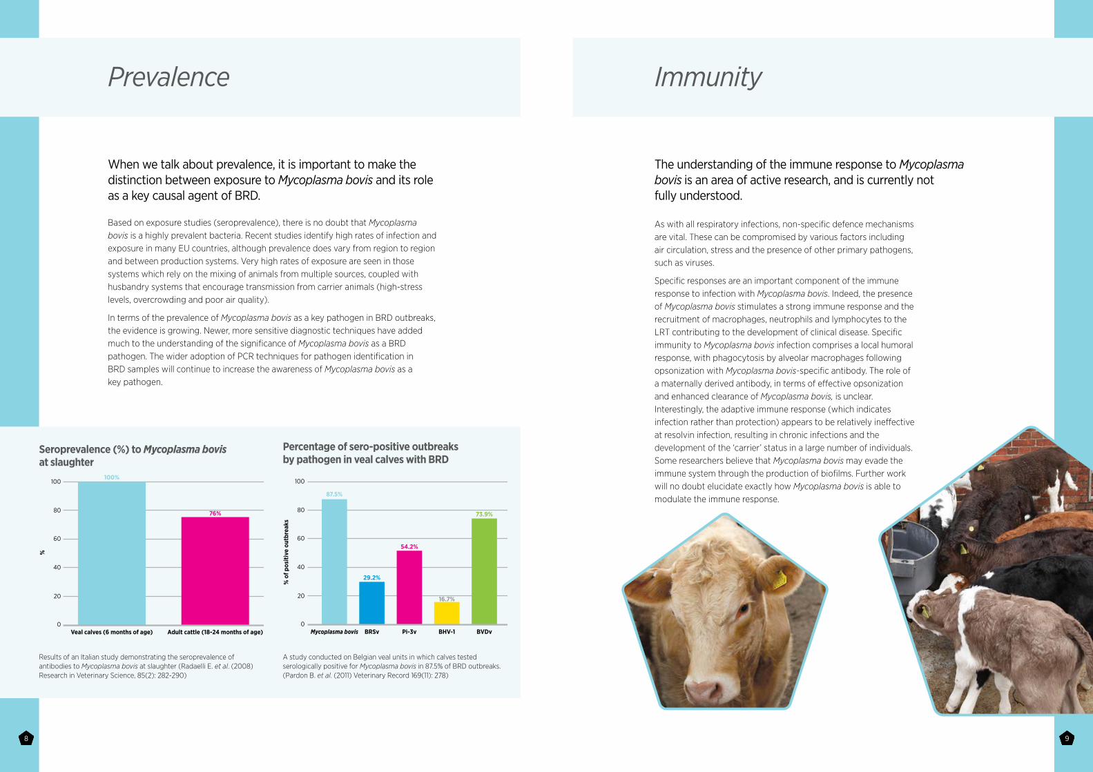
Seroprevalence (%) to Mycoplasma bovis at slaughter
Percentage of sero-positive outbreaks by pathogen in veal calves with BRD
Results of an Italian study demonstrating the seroprevalence of antibodies to Mycoplasma bovis at slaughter (Radaelli E. et al. (2008) Research in Veterinary Science, 85(2): 282-290)
A study conducted on Belgian veal units in which calves tested serologically positive for Mycoplasma bovis in 87.5% of BRD outbreaks. (Pardon B. et al. (2011) Veterinary Record 169(11): 278)
100
80
60
40
20
Veal calves (6 months of age)
%
100%
76%
Adult cattle (18-24 months of age)0
100
80
60
40
20
Mycoplasma bovis
% o
f pos
itive
out
brea
ks
87.5%
54.2%
29.2%
16.7%
73.9%
BHV-1 BVDvPi-3vBRSv0
As with all respiratory infections, non-specific defence mechanisms are vital. These can be compromised by various factors including air circulation, stress and the presence of other primary pathogens, such as viruses.
Specific responses are an important component of the immune response to infection with Mycoplasma bovis. Indeed, the presence of Mycoplasma bovis stimulates a strong immune response and the recruitment of macrophages, neutrophils and lymphocytes to the LRT contributing to the development of clinical disease. Specific immunity to Mycoplasma bovis infection comprises a local humoral response, with phagocytosis by alveolar macrophages following opsonization with Mycoplasma bovis-specific antibody. The role of a maternally derived antibody, in terms of effective opsonization and enhanced clearance of Mycoplasma bovis, is unclear. Interestingly, the adaptive immune response (which indicates infection rather than protection) appears to be relatively ineffective at resolvin infection, resulting in chronic infections and the development of the ‘carrier’ status in a large number of individuals. Some researchers believe that Mycoplasma bovis may evade the immune system through the production of biofilms. Further work will no doubt elucidate exactly how Mycoplasma bovis is able to modulate the immune response.
Immunity
The understanding of the immune response to Mycoplasma bovis is an area of active research, and is currently not fully understood.
10 11
Pathology
The impact of Mycoplasma bovis in the LRT is significant. Naturally occurring Mycoplasma bovis infection leads to similar, though typically more severe, lesions to experimentally induced disease.
Gross appearance
The affected lung lobes are a deep red colour, with degrees of consolidation. The distribution of the lesions is mainly focused on, but not restricted to, the cranioventral portions of the lung. In many chronic cases (not uncommon in animals affected by Mycoplasma bovis), caseo-necrotic lesions can vary from a few millimetres to several centimetres in diameter, and are distinct from typical lung abscesses as they are not surrounded by a well-defined fibrous capsule. These changes are considered by many to be pathognomonic for Mycoplasma bovis infection. Additional signs include a diffuse fibrinous or chronic fibrosing pleuritis and the observation of linear yellow necrotic lesions with oedema fluid in the interlobular septae. Occasionally, lung sequestration, fibrinosuppurative tracheitis and caseous necrosis of regional lymph nodes have also been observed.
In terms of histological appearance, the mixed nature of infections can often complicate interpretation, however, typical observations would include the bronchioles being filled with dry caseous exudate accompanied by peribronchiolar lympho-histiocytic cuffing, thickening of the alveolar septa as a result of cellular infiltration and atelectasis. Various lab techniques, such as immunohistochemical (IHC) staining of bronchiolar contents and both ELISA and qPCR on pneumonic lung, are able to confirm the presence of Mycoplasma bovis and its importance in the development of these significant lesions.
As previously discussed, Mycoplasma bovis is well adapted to colonisation of the URT without causing clinical disease. Clinical disease occurs where host and/or pathogen factors result in the replication and spread of bacteria to the LRT (as well as other sites, such as the middle ear).
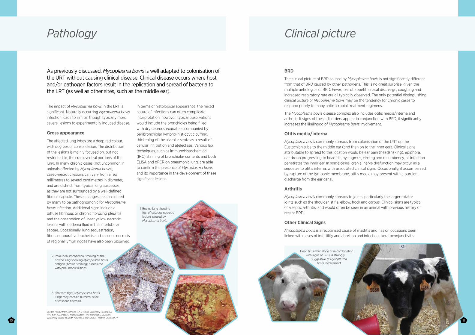
1. Bovine lung showing foci of caseous necrotic lesions caused by Mycoplasma bovis.
2. Immunohistochemical staining of the bovine lung showing Mycoplasma bovis antigen (brown staining) associated with pneumonic lesions.
3. (Bottom right) Mycoplasma bovis lungs may contain numerous foci of caseous necrosis.
Images 1 and 2 from Nicholas R.A.J. (2011). Veterinary Record 168 (17): 459-462. Image 3 from Maunsell FP & Donovan GA (2009). Veterinary Clinics of North America, Food Animal Practice; 25(1):139-77
Clinical picture
BRD
The clinical picture of BRD caused by Mycoplasma bovis is not significantly different from that of BRD caused by other pathogens. This is no great surprise, given the multiple aetiologies of BRD. Fever, loss of appetite, nasal discharge, coughing and increased respiratory rate are all typically observed. The only potential distinguishing clinical picture of Mycoplasma bovis may be the tendency for chronic cases to respond poorly to many antimicrobial treatment regimens.
The Mycoplasma bovis disease complex also includes otitis media/interna and arthritis. If signs of these disorders appear in conjunction with BRD, it significantly increases the likelihood of Mycoplasma bovis involvement.
Otitis media/interna
Mycoplasma bovis commonly spreads from colonisation of the URT up the Eustachian tube to the middle ear (and then on to the inner ear). Clinical signs attributable to spread to this location would be ear pain (headshaking), epiphora, ear droop progressing to head tilt, nystagmus, circling and recumbency, as infection penetrates the inner ear. In some cases, cranial nerve dysfunction may occur as a sequelae to otitis interna, with associated clinical signs. Occasionally, if accompanied by rupture of the tympanic membrane, otitis media may present with a purulent discharge from the ear canal.
Arthritis
Mycoplasma bovis commonly spreads to joints, particularly the larger rotator joints such as the shoulder, stifle, elbow, hock and carpus. Clinical signs are typical of a septic arthritis, and would often be seen in an animal with previous history of recent BRD.
Other Clinical Signs
Mycoplasma bovis is a recognised cause of mastitis and has on occasions been linked with cases of infertility and abortion and infectious keratoconjunctivitis.
Head tilt, either alone or in combination with signs of BRD, is strongly
suggestive of Mycoplasma bovis involvement
12 13
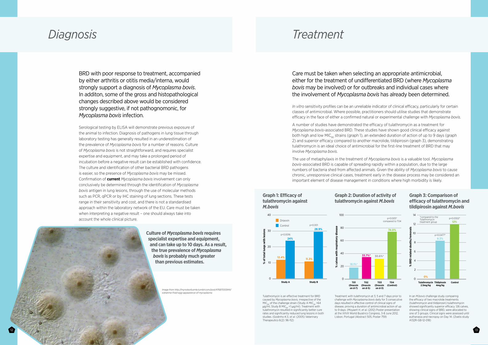
BRD with poor response to treatment, accompanied by either arthritis or otitis media/interna, would strongly support a diagnosis of Mycoplasma bovis. In additon, some of the gross and histopathological changes described above would be considered strongly suggestive, if not pathognomonic, for Mycoplasma bovis infection.
Serological testing by ELISA will demonstrate previous exposure of the animal to infection. Diagnosis of pathogens in lung tissue through laboratory testing has generally resulted in an underestimation of the prevalence of Mycoplasma bovis for a number of reasons. Culture of Mycoplasma bovis is not straightforward, and requires specialist expertise and equipment, and may take a prolonged period of incubation before a negative result can be established with confidence. The culture and identification of other bacterial BRD pathogens is easier, so the presence of Mycoplasma bovis may be missed. Confirmation of current Mycoplasma bovis involvement can only conclusively be determined through the identification of Mycoplasma bovis antigen in lung lesions, through the use of molecular methods such as PCR, qPCR or by IHC staining of lung sections. These tests range in their sensitivity and cost, and there is not a standardised approach within the laboratory network of the EU. Care must be taken when interpreting a negative result – one should always take into account the whole clinical picture.
Diagnosis
Culture of Mycoplasma bovis requires specialist expertise and equipment, and can take up to 10 days. As a result, the true prevalence of Mycoplasma bovis is probably much greater than previous estimates.
In vitro sensitivity profiles can be an unreliable indicator of clinical efficacy, particularly for certain classes of antimicrobial. Where possible, practitioners should utilise studies that demonstrate efficacy in the face of either a confirmed natural or experimental challenge with Mycoplasma bovis.
A number of studies have demonstrated the efficacy of tulathromycin as a treatment for Mycoplasma bovis-associated BRD. These studies have shown good clinical efficacy against both high and low MIC90 strains (graph 1), an extended duration of action of up to 9 days (graph 2) and superior efficacy compared to another macrolide, tildipirosin (graph 3), demonstrating tulathromycin is an ideal choice of antimicrobial for the first-line treatment of BRD that may involve Mycoplasma bovis.
The use of metaphylaxis in the treatment of Mycoplasma bovis is a valuable tool. Mycoplasma bovis-associated BRD is capable of spreading rapidly within a population, due to the large numbers of bacteria shed from affected animals. Given the ability of Mycoplasma bovis to cause chronic, unresponsive clinical cases, treatment early in the disease process may be considered an important element of disease management in conditions where high morbidity is likely.
Treatment
Care must be taken when selecting an appropriate antimicrobial, either for the treatment of undifferentiated BRD (where Mycoplasma bovis may be involved) or for outbreaks and individual cases where the involvement of Mycoplasma bovis has already been determined.
Graph 1: Efficacy of tulathromycin against M.bovis
Graph 3: Comparison of efficacy of tulathromycin and tildipirosin against M.bovis
Graph 2: Duration of activity of tulathromycin against M.bovis
Tulathromycin is an effective treatment for BRD caused by Mycoplasma bovis, irrespective of the MIC90 of the challenge strain (Study A MIC90 >64 µg/ml, Study B MIC90 <1 µg/ml). Treatment with tulathromycin resulted in significantly better cure rates and significantly reduced lung lesions in both studies. (Godinho K.S. et al. (2005) Veterinary Therapeutics 6(2): 96-112)
Treatment with tulathromycin at 3, 5 and 7 days prior to challenge with Mycoplasma bovis daily for 3 consecutive days resulted in effective control of clinical signs of disease, proving a duration of antimicrobial action of up to 9 days. (Moyaert H. et al. (2012) Poster presentation at the XXVII World Buiatrics Congress, 3-8 June 2012, Lisbon, Portugal (Abstract 505, Poster 759)
In an M.bovis challenge study comparing the efficacy of two macrolide treatments (tulathromycin and tildipirosin) tulathromycin showed significantly superior efficacy. 126 calves, showing clinical signs of BRD, were allocated to one of 3 groups. Clinical signs were assessed until euthanasia and necropsy on Day 14. (Zoetis study A132R-GB-12-018)
40
Draxxin
Control
30
20
10
Study A
% o
f tot
al lu
ngs
with
lesi
ons
Study B0
100
80
60
40
20
T01(Draxxin on d-7)
% c
alve
s w
ith re
spira
tory
dis
ease
T03(Draxxin on d-3)
T04 (Control)
T02(Draxxin on d-5)
p=0.0477*
p=0.0302**compared to the Tulathromycin treatment group
0
14
12
10
8
6
4
2
Tulathromycin2.5mg/kg
0%
8.3%
12%
12.4%
24%
11.3%
28.9%
% B
RD re
late
d de
aths
/rem
oval
s
ControlTildipirosin4mg/kg
0
p<0.005*compared to T04
p=0.001
p=0.0016
18.1%*
74.0%
30.6%*33.7%*
13
Image from http://mynotes4usmle.tumblr.com/post/47687055944/sidratime-fried-egg-appearence-of-mycoplasma
14 15
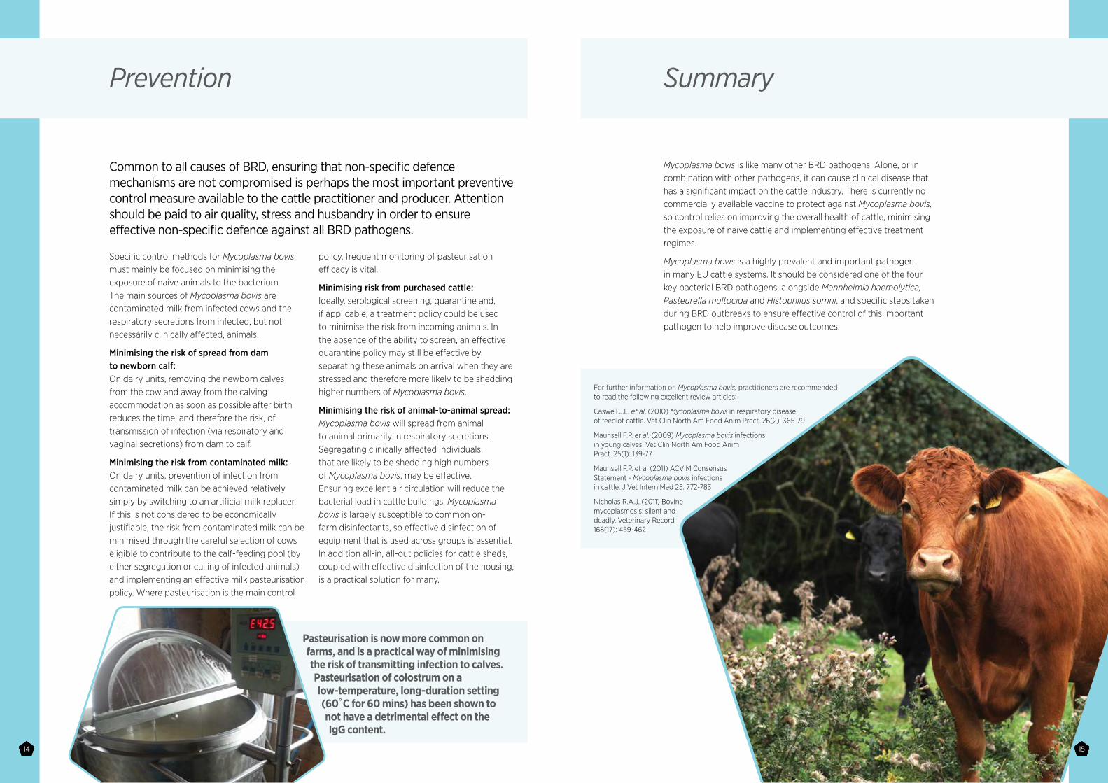
Pasteurisation is now more common on farms, and is a practical way of minimising the risk of transmitting infection to calves. Pasteurisation of colostrum on a low-temperature, long-duration setting (60̊ C for 60 mins) has been shown to not have a detrimental effect on the IgG content.
Specific control methods for Mycoplasma bovis must mainly be focused on minimising the exposure of naive animals to the bacterium. The main sources of Mycoplasma bovis are contaminated milk from infected cows and the respiratory secretions from infected, but not necessarily clinically affected, animals.
Minimising the risk of spread from dam to newborn calf: On dairy units, removing the newborn calves from the cow and away from the calving accommodation as soon as possible after birth reduces the time, and therefore the risk, of transmission of infection (via respiratory and vaginal secretions) from dam to calf.
Minimising the risk from contaminated milk: On dairy units, prevention of infection from contaminated milk can be achieved relatively simply by switching to an artificial milk replacer. If this is not considered to be economically justifiable, the risk from contaminated milk can be minimised through the careful selection of cows eligible to contribute to the calf-feeding pool (by either segregation or culling of infected animals) and implementing an effective milk pasteurisation policy. Where pasteurisation is the main control
policy, frequent monitoring of pasteurisation efficacy is vital.
Minimising risk from purchased cattle: Ideally, serological screening, quarantine and, if applicable, a treatment policy could be used to minimise the risk from incoming animals. In the absence of the ability to screen, an effective quarantine policy may still be effective by separating these animals on arrival when they are stressed and therefore more likely to be shedding higher numbers of Mycoplasma bovis.
Minimising the risk of animal-to-animal spread: Mycoplasma bovis will spread from animal to animal primarily in respiratory secretions. Segregating clinically affected individuals, that are likely to be shedding high numbers of Mycoplasma bovis, may be effective. Ensuring excellent air circulation will reduce the bacterial load in cattle buildings. Mycoplasma bovis is largely susceptible to common on-farm disinfectants, so effective disinfection of equipment that is used across groups is essential. In addition all-in, all-out policies for cattle sheds, coupled with effective disinfection of the housing, is a practical solution for many.
Prevention
Common to all causes of BRD, ensuring that non-specific defence mechanisms are not compromised is perhaps the most important preventive control measure available to the cattle practitioner and producer. Attention should be paid to air quality, stress and husbandry in order to ensure effective non-specific defence against all BRD pathogens.
Mycoplasma bovis is like many other BRD pathogens. Alone, or in combination with other pathogens, it can cause clinical disease that has a significant impact on the cattle industry. There is currently no commercially available vaccine to protect against Mycoplasma bovis, so control relies on improving the overall health of cattle, minimising the exposure of naive cattle and implementing effective treatment regimes.
Mycoplasma bovis is a highly prevalent and important pathogen in many EU cattle systems. It should be considered one of the four key bacterial BRD pathogens, alongside Mannheimia haemolytica, Pasteurella multocida and Histophilus somni, and specific steps taken during BRD outbreaks to ensure effective control of this important pathogen to help improve disease outcomes.
Summary
For further information on Mycoplasma bovis, practitioners are recommended to read the following excellent review articles:
Caswell J.L. et al. (2010) Mycoplasma bovis in respiratory disease of feedlot cattle. Vet Clin North Am Food Anim Pract. 26(2): 365-79
Maunsell F.P. et al. (2009) Mycoplasma bovis infections in young calves. Vet Clin North Am Food Anim Pract. 25(1): 139-77
Maunsell F.P. et al (2011) ACVIM Consensus Statement - Mycoplasma bovis infections in cattle. J Vet Intern Med 25: 772-783
Nicholas R.A.J. (2011) Bovine mycoplasmosis: silent and deadly. Veterinary Record 168(17): 459-462