Embed Size (px)
Citation preview

Provided to you by:
The Nervous The Nervous SystemSystem
The Wonderful World of THE NERVOUS SYSTEMTHE NERVOUS SYSTEM
Jaclynn Chen
Katie Tang
Winnie Tema
That lab group in the back.
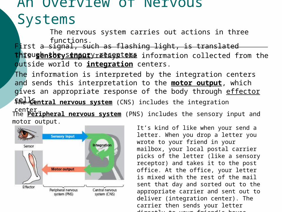
An Overview of Nervous SystemsThe nervous system carries out actions in three functions.
First a signal, such as flashing light, is translated through the sensory receptors . This sensory input relays the information collected from the outside world to integration centers.
The information is interpreted by the integration centers and sends this interpretation to the motor output, which gives an appropriate response of the body through effector cells.
It’s kind of like when your send a letter. When you drop a letter you wrote to your friend in your mailbox, your local postal carrier picks of the letter (like a sensory receptor) and takes it to the post office. At the office, your letter is mixed with the rest of the mail sent that day and sorted out to the appropriate carrier and sent out to deliver (integration center). The carrier then sends your letter directly to your friend’s house where he or she can read the lovely letter your wrote just for him or her (motor output).
The Central nervous system (CNS) includes the integration center.
The Peripheral nervous system (PNS) includes the sensory input and motor output.
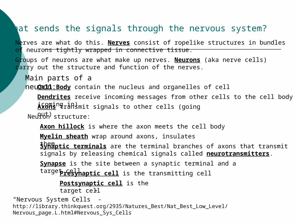
What sends the signals through the nervous system?
Nerves are what do this. Nerves consist of ropelike structures in bundles of neurons tightly wrapped in connective tissue.
Groups of neurons are what make up nerves. Neurons (aka nerve cells) carry out the structure and function of the nerves.
Main parts of a neuron:Cell Body contain the nucleus and organelles of cell
Dendrites receive incoming messages from other cells to the cell body (coming in)
Axons transmit signals to other cells (going out)
Myelin sheath wrap around axons, insulates them
Neuron structure:
Axon hillock is where the axon meets the cell body
Synaptic terminals are the terminal branches of axons that transmit signals by releasing chemical signals called neurotransmitters.
Synapse is the site between a synaptic terminal and a target cell
Presynaptic cell is the transmitting cell
Postsynaptic cell is the target cell
“Nervous System Cells” - http://library.thinkquest.org/2935/Natures_Best/Nat_Best_Low_Level/Nervous_page.L.html#Nervous_Sys_Cells

Can you label the structures of the neuron and the direction of the neurotransmitter?
A.
B.
C.
D.
E.
F. G.
H.
Answers:
A.Dendrites
B.Cell Body
C.Nucleus
D.Axon Hillock
E. AxonF. Myelin SheathsG. Synaptic TerminalH. Synapse
Presynaptic Cell
Postsynaptic Cell

Not all nervous systems are the same you know…
The simplest animals with nervous systems have an expansive nervous system, which are arranged in diffused nerve nets
More complex animals have nervous systems with systems of nerves.

Ever wonder what makes those knee jerking reactions?Go ahead…Try it.
Tap the tendon connected to the quadriceps muscle
What causes this to happen?
This is a perfect example that will allow us to observe the different parts of the nervous system.
http://www.dushkin.com/connectext/psy/ch02/reflex.gif
http://www.bbc.co.uk/science/humanbody/body/factfiles/reflexes/reflexes.shtml
See the movement on this site!

Reflexes are caused by the automatic responses of the reflex arc between the spinal cord and brain.
There are two kinds of nerve cells involved:
Sensory neuron transmits information from a sensory receptor to a motor neuron, which signals an effector cell to carry out the response.
The knee jerking reaction goes through the sensory neurons which relays the information to the stretch receptor in the thigh muscle, to interneurons in the spinal cord, which finally inhibit motor neurons to the flexor muscles.
Motor neurons and interneurons are located in the gray matter of the CNS. Motor & sensory axons are in the white matter.
Outside the spinal cord structure is a ganglion (a cluster of nerve cell bodies) in the PNS. Nuclei are similar to ganglion but are in the brain.

Glia – supporting cells of the nervous systemAstrocytes support the structure of the neuron and regulate the concentration of ions and neurotransmitters.
These induces the formation of the blood brain barrier which restricts the passage of substances into the brain.
Radial glia create and track newly formed neurons from stem cells
Oligogendrocytes and Schwann cells are glia that support the axons in the mylein sheath.
These membranes are mostly lipids which are poor conductors of electrical current. Multiple Sclerosis is a disease the degrades the myelin sheaths.
http://www.youtube.com/watch?v=qgySDmRRzxY
See more of a description of multiple sclerosis also known as MS.

Explore more of the world of the nervous system & reflexes
http://www.bbc.co.uk/science/humanbody/body/factfiles/reflexes/reflexes.shtml

Neurotransmitters travel by electrical impulsesThe membrane potential is the difference of charges across the plasma membrane
When the membrane is at resting potential, there is no transmitting of signals. The voltage is usually around -70 mV.
This membrane potential is due to the concentration of ions on the two sides of the membranes. Sodium (Na+) ions are usually outside making it negatively charged while potassium (K+) are usually inside making it positively charged.
These concentrations are maintained by sodium ion pumps
When the electrical gradient exactly balances the concentration gradient, an equilibrium is established.
K+ is more permeable than Na+. When this permeability changes, the membrane potential changes
K+ & Na+ have ungated ion channels that allow them to diffuse all the time at resting potential.
Types of ion pumps that effect the membrane potential:
Stretch gated ion channels- cells that sense stretch and open when the membrane is mechanically deformed
Ligand-gated ion channels- found at synapses and open or close when a specific chemical, like a neurotransmitter, binds to the channel
http://highered.mcgraw-hill.com/sites/0072495855/student_view0/chapter2/animation__how_the_sodium_potassium_pump_works.html
See these pumps in action!

In response to a stimuli the membrane potential of a cell open and closes its channels
Graded potentials Hyperpolarization- an increase in the
magnitude of the membrane potential May be caused by opening of gated K+
Depolarization- reduction in the magnitude of the membrane potential.
May be caused by opening gated Na+

Production of Action Potentials
Depolarizations are graded only up to a certain membrane voltage or a threshold Once a stimulus is strong enough to produce
depolarization that reaches the threshold, action potential is then produced.
Action Potential is an all or none phenomenon Once it is triggered it has a magnitude that is
independent of the strength of the triggering stimulus. In most neurons, the action potential is very brief. This
allows the neuron to produce them at high frequencies.
LEARN MORE: http://outreach.mcb.harvard.edu/animations/actionpotential.swf, http://bcs.whfreeman.com/thelifewire/content/chp44/4402002.html.
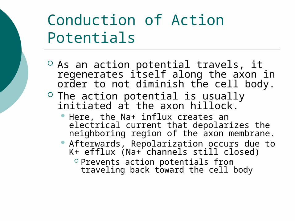
Conduction of Action Potentials
As an action potential travels, it regenerates itself along the axon in order to not diminish the cell body.
The action potential is usually initiated at the axon hillock. Here, the Na+ influx creates an electrical
current that depolarizes the neighboring region of the axon membrane.
Afterwards, Repolarization occurs due to K+ efflux (Na+ channels still closed)
Prevents action potentials from traveling back toward the cell body

Conduction Speed
Factors that affect the speed at which action potentials are conducted Diameter- the larger the faster, the resistance
to the flow of electrical current is inversely proportional to the cross-sectional area of the conductor.
Myelin sheath- by insulating the axon membrane, which causes the depolarizing current associated with an action potential to spread farther along the interior of the axon. This brings more distant regions of the membrane to the threshold sooner.
Saltatory Conduction- action potential appears to jump along the axon from node to node. It speeds it up to 120 m/sec in myelinated axons.

Action Potentials are not transmitted from neurons to other cells, but at the synapses information is transmitted
Electrical synapses- contains gap junctions allowing electrical current to flow directly from cell to cell.
Chemical synapses- involves the release of chemical neurotransmitter by the presynaptic neuron. Presynaptic neuron synthesizes the neurotransmitter
and packages it in synaptic vesicles. Stored in the neuron’s synaptic terminals
Information transfer is more modifiable at chemical synapses than at electrical synapses
LEARN MORE: http://users.rcn.com/jkimball.ma.ultranet/BiologyPages/S/Synapses.html, http://faculty.washington.edu/chudler/synapse.html.

Direct Synaptic Transmission
The binding of the neurotransmitter to a particular part of the channel, the receptor, opens the channel and allows specific ions to diffuse across the postsynaptic membrane. Result- postsynaptic potential- a change in the
membrane potential of the postsynaptic cell Synapses that cause depolarizations bring the
membrane potential toward the threshold are called excitatory postsynaptic potentials.
Na+ and K+ diffuses here Synapses that causes hyperpolarizations are
called inhibitory postsynaptic potentials, they move the membrane potential farther from the threshold.
Channels that are selective for K+ only

Summation of Postsynaptic Potentials
Postsynaptic potentials’ magnitude varies with a number of factors The amount of neurotransmitter released by the presynaptic neuron
They do not regenerate themselves as they spread along the membrane of the cell
Smaller with distance from the synapse Two EPSPs are needed to trigger an action potential in a posynaptic neuron
Two EPSP= temporal summation Two EPSPs produced nearly simultaneously by different synapses on
the same postsynaptic neuron= spatial summation

Indirect Synaptic Transmission
Here, a neurotranmitter binds to a receptor that is not part of an ion channel. Activates a signal transduction pathway
involving a second messenger in the postsynaptic cell
A variety of signal transduction pathways play arole in indirect synaptic transmission. Cyclic AMP- activates protein kinase A
Phosphorylates specific channel proteins in the postsynaptic membrane

Neurotranmitters
Acetylcholine
One of the most common in vertebrates and invertebrates Vertebrates- it
activates a signal transduction pathway
G proteins 1) inhibition of adenylyl cyclase 2) opening of K+ channels in the muscle cell membrane
Biogenic Amines
Derived from amino acids
Includes: epinephrine, norepinephrine, dopamine, and serotonin
Often involved in indirect synaptic transmission- most common in CNS.

Amino Acids and Peptides
Gamma Aminobutyric Acid, Glycine, Glutamate, and Aspartate- all known to function as neurotransmitters.
Several neuropeptides serve as neurotranmitters. Many produced by post
translational modification of much larger protein precursors.
Substance P mediates perception of pain
Endorphins- decreases pain perception
Gases
Nitric oxide(NO), and Carbon Monoxide(CO)- local regulators
CO is synthesized by enzyme heme oxygenase In brain it regulates the
release of hypothalamic hormones
In PNS it acts as inhibitory neurotransmitter that hyperpolarizes intestinal smooth muscle cells
In vertebrates the nervous system shows cephalization and distinct CNS and PNS components CNS
Brain- provides integrative power underling the complex behavior of vertebrates
Spinal Cord- integrates simple responses to certain kinds of stimuli, conveys information to the brain
Derived form dorsal embryonic nerve cord In an adult- this is the narrow central canal of
the spinal cord and the four ventricles of the brain
Called cerebrospinal fluid Axons are often found in well-defined bundles,
or tracts whose myelin sheath give them a whitish
appearance

The Peripheral Nervous System
Transmits information to and from the CNS Plays a large role in regulating vertebrates
movement and internal environment Structure- left-right pairs of cranial and
spinal nerves, they are associated with ganglia
Cranial Nerves- originate in the brain and terminate mostly in the organs of the head and upper body
Spinal nerves- originate in the spinal cord and extend to parts of the body below the head

PNS divided into two parts:
Somatic Nervous System- carries signals to and from skeletal muscles, mainly in response to external stimuli
Autonomic Nervous System- regulates the internal environment by controlling smooth and cardiac muscles and the organs of the digestive, cardiovascular, excretory, and endocrine systems.

Autonomic Nervous System Broken into three part:
Sympathetic Division- corresponds to arousal and energy generation
Parasympathetic Division- causes opposite responses that promote calming and a return to self-maintenance functions
Enteric Division- consists of networks of neurons in the digestive tract, pancreas, and gallbladder Controls these organs’ secretions as well as
activity in the smooth muscles that produce peristalsis
Embryonic Development of the Brain
Vertebrates consist of 3 bilateral symmetrical, anterior bulges of the neural tube Forebrain, midbrain and hindbrain
Fifth week of human embryonic development, five regions have formed from the three primary bulges Forbrain: telencephalon( cerebrum
cerebral cortex) , diencephalon Midbrain- mesencephalon- give rise to brain
stem Hindbrain- metencephalon, myelencephalon-
give rise to brain stem
The Brainstem
Consists of stalk with caplike swellings at the anterior end of the spinal cord
Three parts: medulla oblongata, pons, midbrain Functioning in homeostasis, coordination
and movement, and conduction of information to higher brain centers
Neuron cell bodies send axons to many areas of the cerebral cortex and cerebellum

Medulla oblongata- contains centers that control several visceral functions
The Pons- participates in some of the above activities

The Cerebellum
Develops from part of the metencephalon, important for coordination and error checking during motor, perceptual, and cognitive functions
Involved in learning and remembering motor skills
Receives sensory information about the position of the joints and the length of the muscles

The Diencephalon
Epithalamus- includes the pineal gland and chroid plexus
Thalamus- main input center for sensory information going to the cerebrum and the main output center for motor information leaving the cerebrum
Hypothatlumus- most important brain regions for homeostatic regulation

The Cerebrum
Develops from the telencephalon Divided into right and left cerebral
hemispheres Each consists of an outer covering of
gray matter, the cerebral cortex Internal white matter, groups of
neurons collectively called basal nuclei

Cerebral Cotex
Sensory information is analyzed, motor commands are issued, and language is generated
Neocortex- forms outermost part of mammalian cerebrum, consisting of six parallel layers of neuron arranged tangential to the brain surface

ACTIVITY TIME!!!!!
On the next slide is a picture with colors on it, only there is a catch… the words are colors and the words are written in different colors. Now, try the best you can to repeat the different colors of the words. Instead of reading out the words repeat what color it is written in. Good luck because it is not an easy task!! This game is great to challenge your memory which is developed by the cerebellum.


Concept 48.6 – The cerebral cortex controls voluntary movement and cognitive functions Each side of the cerebral cortex is customarily described as having four lobes, called the frontal, temporal, occipital, and parietal lobes. These areas include primary sensory areas, each of which receives and processes a specific type of sensory information, and association areas, which integrate the information from various parts of the brain.
The human cerebral cortex. Each side of the cerebral cortex is divided into four lobes, and each lobe has specialized functions. Some of the association areas on the left side (shown here) have different functions than those on the right side.

Information Processing in the Cerebral Cortex Most sensory information coming into the cortex is directed via the thalamus
to primary sensory areas within the lobes: visual information to the occipital lobe auditory input to the temporal lobe somatosensory information about touch, pain, pressure, temperature, and
the position of muscles and limbs to the parietal lobe Information about taste goes to a separate sensory region of the parietal lobe
Body representations in the primary motor and primary somatosensory cortices. In these cross–sectional maps of the cortices, the cortical surface area devoted to each body part is represented by the relative size of that part in the cartoons.
(http://www.emc.maricopa.edu/faculty/farabee/BIOBK/BioBookNERV.html)

Lateralization of Cortical Function During brain development after birth, competing functions segregate and
displace each other in the cortex of the left and right cerebral hemispheres, resulting in lateralization of functions.
The left hemisphere becomes more adept at language, math, logical operations, and the serial processing of sequences of information. It has a bias for the detailed, speed–optimized activities required for skeletal muscle control and the processing of fine visual and auditory details.
The right hemisphere is stronger at pattern recognition, face recognition, spatial relations, nonverbal thinking, emotional processing in general, and the simultaneous processing of many kinds of information.
The two hemispheres normally work together harmoniously, trading information back and forth through the fibers of the corpus callosum.
(http://www.kidshealth.org/parent/general/body_basics/brain_nervous_system.html)

Language and Speech The systematic mapping of higher cognitive functions to specific brain
areas began in the 19th century when physicians learned that damage to particular regions of the cortex by injuries, strokes, or tumors can produce distinctive changes in a person’s behavior.
The French physician Pierre Broca conducted postmortem examinations of patients who could understand language but could not speak. He discovered that many of these patients had defects in a small region of the left frontal lobe.
That region, now known as Broca′s area, is located in front of the part of the primary motor cortex that controls muscles in the face.
Mapping language areas in the cerebral cortex. These PET images show regions with different activity levels in one person′s brain during four activities, all related to speech.
(http://www.emc.maricopa.edu/faculty/farabee/BIOBK/BioBookNERV.html)

Emotions Emotions are the result of a complex interplay of many regions of the brain Prominent among these regions is the limbic system, a ring of structures
around the brainstem The limbic system includes three parts of the cerebral cortex—the amygdala,
hippocampus, and olfactory bulb—along with some inner portions of the cortex′s lobes and sections of the thalamus and hypothalamus
These structures interact with sensory areas of the neocortex and other higher brain centers, mediating primary emotions that manifest themselves in behaviors such as laughing and crying
The Limbic System: Structures of the limbic system form early in development and provide a foundation for the higher cognitive functions that appear later, during the development of neocortical areas.
Memory and Learning We hold information, anticipation, or goals for a time in short–term memory
locations in the frontal lobes and then release them if they become irrelevant Should we wish to retain knowledge of a face or a phone number, the
mechanisms of long–term memory are activated in a process that requires the hippocampus.
The transfer of information from short–term to long–term memory is enhanced by rehearsal (“practice makes perfect”), positive or negative emotional states mediated by the amygdala, and the association of new data with data previously learned and stored in long–term memory.
Consciousness Over the past few decades, however, neuroscientists have begun studying
consciousness using brain–imaging techniques such as fMRI It is now possible to compare activity in the human brain during different
states of consciousness These imaging techniques can also be used to compare the conscious and
unconscious processing of sensory information

Concept 48.7 –CNS injuries and diseases are the focus of much
research Unlike the PNS, the mammalian CNS cannot fully repair itself when
damaged or assaulted by disease Surviving neurons in the brain can make new connections and thus
sometimes compensate for damage
Nerve Cell Development To reach their target cells, axons must elongate from a few micrometers
to a meter or more An axon does not follow a straight path to its target cells; rather,
molecular signposts along the way direct and redirect the growing axon in a series of mid–course corrections that result in a meandering, but not random, elongation.
The responsive region at the leading edge of the growing axon is called the growth cone
Signal molecules released by cells along the growth route bind to receptors on the plasma membrane of the growth cone, triggering a signal transduction pathway

Neural Stem Cells Mice that live in stimulating environments and run on exercise wheels have
more new neurons in their hippocampus and perform better on learning tasks than genetically identical caged mice that receive little stimulation
Mature neurons, with their extensive processes and intricate connections with other cells, clearly are not able to undergo cell division
Therefore, the new brain neurons must have come from stem cells Stem cells are relatively unspecialized cells that continually divide
Diseases and Disorders of the Nervous System Schizophrenia - a severe mental disturbance characterized by psychotic
episodes in which patients lose the ability to distinguish reality Depression - Two broad forms of depressive illness are known: bipolar
disorder and major depression. Bipolar disorder, or manic–depressive disorder, involves swings of mood from high to low and affects about 1% of the world′s population. In contrast, people with major depression have a low mood most of the time; they constitute roughly 5% of the population
Alzheimer′s Disease - a mental deterioration, or dementia, characterized by confusion, memory loss, and a variety of other symptoms. Its incidence is age related, rising from about 10% at age 65 to about 35% at age 85
Parkinson′s Disease - a motor disorder characterized by difficulty in initiating movements, slowness of movement, and rigidity
Activity!The nervous system is the master controller of the body. Each thought, each emotion, each action- all result from the activity of
this system! Through its many parts, the nervous system monitors conditions both within and outside the body. The nervous system
processes information and decides what the body should do in response. When the response is needed, the nervous system will
send out electrical signals that will direct the body. Just like the game of telephone, if one neuron (or student) confuses the
message, any neurons (or students) continuing down the chain will receive the incorrect message.
To do this activity, you will need: to get into a circle with your classmates Choose one person to start a message by whispering the message
to the student on her right Continue this message until the same person who started the
message hears it from the student on his/her leftIs the message the same?

If the message isn’t the same, what do you think happened? Did one student confuse the message? What happens in the body, if a neuron
confuses the message?
To further understand the Nervous System check out these links!
http://www.innerbody.com/image/nervov.html
http://www.argosymedical.com/medical_ani_sys/nervous.html
http://health.howstuffworks.com/adam-200011.htm

Any Questions?