Embed Size (px)
Citation preview

ORIGINAL ARTICLE
Electronic supplementary material The online version of this article (doi:10.1007/s11240-016-1072-8) contains supplementary material, which is available to authorized users.
Paula M. [email protected]; [email protected]
Naghi [email protected]
1 Laboratory of Forest Tree Biology and Biotechnology, Department of Forestry, Faculty of Natural Resources, University of Kurdistan, Khanagah Campus, Sanandaj 66177-1-5175, Iran
2 USDA Forest Service, Northern Research Station, Hardwood Tree Improvement and Regeneration Center, 715 West State Street, West Lafayette, IN 47907, USA
Received: 1 June 2016 / Accepted: 25 August 2016 / Published online: 2 September 2016© Springer Science+Business Media Dordrecht (outside the USA) 2016
Protoplast isolation and genetically true-to-type plant regeneration from leaf- and callus-derived protoplasts of Albizia julibrissin
Mohammad-Shafie Rahmani1 · Paula M. Pijut2 · Naghi Shabanian1
Plant Cell Tiss Organ Cult (2016) 127:475–488DOI 10.1007/s11240-016-1072-8
concentrations of NAA plus 4.4 μM BA for further growth. Proliferated leaf- and callus-protoplast-derived calli dif-ferentiated into microshoots on MS medium containing 13.2 μM BA plus 4.6 μM zeatin after 2–3 weeks, with an overall shoot organogenesis efficiency of 78–93 %. Root-ing of microshoots on half-strength MS medium containing 4.9 µM indole-3-butyric acid was successful, and plantlets were acclimatized to the greenhouse with a survival rate of >62 %. Using ten start codon targeted and ten inter-simple sequence repeat primers, the genetic integrity of nine leaf- and six callus-protoplast-based plants was validated along with the mother seedlings.
Keywords Albizia · Cellulase · CPW · Genetic fidelity · Microcolony formation · Protoplast · Silk tree
AbbreviationsBA 6-BenzylaminopurineCPW Cell and protoplast wash solution2,4-D 2,4-Dichlorophenoxyacetic acidFDA Fluorescein diacetateIBA Indole-3-butyric acidISSR Inter-simple sequence repeatsKM Kao and Michayluk mediumMS Murashige and Skoog mediumMSB5 MS medium with Gamborg B5 vitaminsNAA α-Naphthaleneacetic acidPCR Polymerase chain reactionPGR Plant growth regulatorPp ProtoplastPp gfw−1 Protoplasts per gram fresh weightSCoT Start codon targetedTDZ ThidiazuronWPM Woody plant medium
Abstract Protoplast isolation and subsequent plant regeneration of Albizia julibrissin was achieved from leaf and callus explants. Leaf tissue from 4 to 5-week-old in vitro seedlings was the best source for high-yield proto-plast isolation. This approach produced 7.77 × 10⁵ pro-toplasts (Pp) per gram fresh weight with 94 % viability; after 60 min pre-plasmolysis with 0.7 M sorbitol followed by digestion in a solution of cell and protoplast wash plus 0.7 M mannitol, 1.5 % cellulase Onozuka R10, and 1 % pectolyase Y-23 for 6 h. Liquid Kao and Michay-luk medium containing 2.7 μM α-naphthaleneacetic acid (NAA) and 2.2 μM 6-benzylaminopurine (BA) was best for sustained cell division and microcolony formation from both leaf- and callus-derived protoplasts at a density of 3–5 × 10⁵ Pp ml−1. Protoplast-derived microcalli became visible after 3–4 weeks on semi-solid medium of the same composition. Microcalli were then cultured on Murashige and Skoog (MS) medium containing Gamborg B5 vita-mins or woody plant medium supplemented with different
1 3
476 Plant Cell Tiss Organ Cult (2016) 127:475–488
protein localization, as well as many other cellular char-acterizations, such as protein–protein and protein-DNA interactions (Yoo et al. 2007; Faraco et al. 2011). Although advancements in protoplast isolation, culture, and proto-plast-to-plant regeneration have been considerable for her-baceous, agronomic, and fruit-tree species, the development of complete protocols for temperate tree species (exclud-ing conifers) has been limited. Yellow-poplar (Liriodendron tulipifera) plantlets derived from somatic embryos, differ-entiated from embryogenic callus derived from protoplasts, has been reported (Merkle and Sommer 1987). Plantlet regeneration from protoplasts isolated from suspension cul-tures derived from seed-induced calli of a non-hybrid poplar (Populus alba) was successful (Qiao et al. 1998). Camphor tree (Cinnamomum camphora) plantlets were regenerated from protoplasts isolated from embryogenic suspension cultured cells (Du and Bao 2005). Leaf mesophyll-derived protoplasts of mulberry (Morus indica) were regenerated into shoots and a few plants were established in the green-house (Umate et al. 2005). Kanwar et al. (2009) developed a protocol for plant regeneration from callus- and mesophyll-derived protoplasts of the leguminous black locust (Robinia pseudoacacia). The successful regeneration of plants from American elm (Ulmus americana) cell suspension-derived protoplasts has recently been reported (Jones et al. 2015).
Previous investigations on in vitro culture of A. juli-brissin have centered on adventitious shoot regeneration (Sankhla et al. 1993, 1994, 1995, 1996; Zhou et al. 2001; Rahmani et al. 2015) and somatic embryogenesis (Burns and Wetzstein 1998), but there have been no reports on pro-toplast isolation, culture, and plant regeneration of this spe-cies. Therefore, the present study was initiated to develop a method for protoplast isolation, microcalli formation, and subsequent shoot regeneration and rooting of A. julibrissin that could then be the foundation for genetic studies. Inter-simple sequence repeats (ISSR) and start codon targeted (SCoT) polymorphism were used to assess the genetic fidel-ity or variability of in vitro-regenerated plants from proto-plasts of A. julibrissin.
Materials and methods
Plant material
Seeds of A. julibrissin were excised from mature pods col-lected from a 40- to 50-year-old tree growing in the Sisangan area of the Hyrcanian forest, northern Iran. Seeds were rinsed under running tap water for 20 min, immersed in 85–90 °C water (Fordham 1965) that was then allowed to gradually cool to room temperature. Treated seeds were then surface disinfested by soaking in 70 % (v/v) ethanol for 3 min, fol-lowed by immersion in 20 % (v/v) bleach solution (5.25 %
Introduction
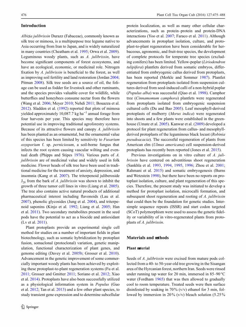
Albizia julibrissin Durazz (Fabaceae), commonly known as silk tree or mimosa, is a multipurpose tree legume native to Asia occurring from Iran to Japan, and is widely naturalized in many countries (Cheatham et al. 1995; Orwa et al. 2009). Leguminous woody plants, such as A. julibrissin, have become significant components of forest ecosystems, and have an ecological, economic, or medicinal role. Nitrogen fixation by A. julibrissin is beneficial to the forest, as well as improving soil fertility and land restoration (Jordan 2004; Pitman 2008). Silk tree seeds are a source of oil, the foli-age can be used as fodder for livestock and other ruminants, and the species provides valuable cover for wildlife, while butterflies and honeybees consume nectar from the flowers (Wang et al. 2006; Meyer 2010; Nehdi 2011; Bouazza et al. 2012). Sladden et al. (1992) reported that plots of mimosa yielded approximately 10,087.7 kg ha−1 annual forage from four harvests per year. This species may therefore have potential use in improving traditional agroforestry systems. Because of its attractive flowers and canopy A. julibrissin has been planted as an ornamental, but the ornamental value of this species has been limited by sensitivity to Fusarium oxysporium f. sp. perniciosum, a soil-borne fungus that infects the root system causing vascular wilting and even-tual death (Phipps and Stipes 1976). Various parts of A. julibrissin are of medicinal value and widely used in folk medicine. Flower heads of silk tree have been used in tradi-tional medicine for the treatment of anxiety, depression, and insomnia (Kang et al. 2007). The triterpenoid juliberoside J28 from the bark of A. julibrissin was shown to inhibit the growth of three tumor cell lines in vitro (Liang et al. 2005). The tree also contains active natural products of additional pharmaceutical interest including flavonoids (Lau et al. 2007), phenolic glycosides (Jung et al. 2004), and triterpe-noid saponins (Kinjo et al. 1992; Liang et al. 2005; Han et al. 2011). Two secondary metabolites present in the seed pods have the potential to act as a biocide and antioxidant (Lv et al. 2011).
Plant protoplasts provide an experimental single cell method for studies on a number of important fields in plant biotechnology, such as somatic hybridization by protoplast fusion, somaclonal (protoclonal) variation, genetic manip-ulation, functional characterization of plant genes, and genome editing (Davey et al. 2005b; Grosser et al. 2010). Advancement in the genetic improvement of some commer-cially important woody plants has been achieved by exploit-ing these protoplast-to-plant regeneration systems (Fu et al. 2011; Grosser and Gmitter 2011; Soriano et al. 2012; Xiao et al. 2014). Protoplasts have also been successfully utilized as a physiological information system in Populus (Guo et al. 2012; Tan et al. 2013) and a few other plant species, to study transient gene expression and to determine subcellular
1 3

477Plant Cell Tiss Organ Cult (2016) 127:475–488
0.7 M [13 % (w/v)] mannitol as osmoticum (CPW13M), and buffered with 5 µM 2-(N-morpholino)ethanesulfonic acid (MES). The pH of the solution was adjusted to 5.5–5.8 and then filter-sterilized through a 0.45 μm filter disc (Millipore, Billerica, MA, USA). Solutions were stored at −20 °C in 5 ml aliquots until used. To inactive protease enzymes, the enzymatic solution was heated at 55 °C for 10 min prior to tissue digestion treatment.
Protoplasts were isolated separately from two types of plant tissue, leaves from 4 to 5-week-old in vitro-seedlings and hypocotyl-derived organogenic callus. Thin strips of leaves (1 g) or callus (1 g) were incubated in sterile 10 cm Petri dishes containing 20 ml CPW solution with 0.7 M man-nitol or sorbitol pre-plasmolysis solution for 30, 60, or 90 min in the dark at 25 ± 2 °C. Each type of tissue (250 mg) was then transferred to a sterile 50-ml Erlenmeyer flask containing 5 ml enzyme solution. Incubation in the enzyme solution was carried out in darkness for different durations (in vitro leaf strips: 3, 6, 9, or 12 h; callus: 8, 12, 16, or 20 h) at 25 ± 2 °C with gentle shaking (40 rpm). Following incubation, the protoplast-enzyme mixture was diluted by adding an equal volume of CPW13M. The protoplast-enzyme solution was then filtered through 0.75 μm nylon mesh (Millipore) and the nylon mesh was rinsed with CPW13M. The filtrate was trans-ferred to 15 ml sterile plastic centrifuge tubes, centrifuged at 100 × g for 5 min, followed by discarding the supernatant without disturbing the protoplast pellet. The protoplast pellet was then washed with 15 ml CPW13M solution, vortexed,
sodium hypochlorite) containing three drops of Tween 20 per 100 ml for 3 min, then rinsed three times (3 min each) in sterile distilled water. Clean seeds were air dried on sterile tissue and were then cultured on plant growth regulator-free Murashige and Skoog (MS) medium (Murashige and Skoog 1962) containing 30 g l−1 sucrose and 7 g l−1 plant agar (Duchefa, Biochemie BV, Netherlands) for germination; which usually occurred within 6–10 days at 26 ± 2 °C under a 16 h photoperiod (40 µmol m−2 s−1).
Expanded leaves from the apex of 4–5-week-old in vitro-germinated seedlings were used as explants for protoplast isolation. For protoplast isolation from callus, additional seeds were cultured on the same medium for germination but were kept in the dark for 10–14 days to obtain elongated and etiolated hypocotyls. Hypocotyl segments (5–7 mm) were excised and cultured horizontally on MS medium con-taining 0.2 g l−1myo-inositol (Duchefa) and supplemented with 10.8 µM α-naphthaleneacetic acid (NAA) and 4.4 µM 6-benzylaminopurine (BA) for induction of white, friable, organogenic callus (Rahmani et al. 2015).
Protoplast isolation
The enzyme solution was a mixture of different concentra-tions and combinations of cellulase Onozuka R-10 and either macerozyme R-10 or pectolyase Y-23 (Duchefa; Tables 1, 2). Enzymes were dissolved in cell and protoplast wash-ing (CPW) salt solution (Frearson et al. 1973) containing
Table 1 Effect of concentration and combination of enzymes in digestion solution and digestion period on protoplast yield and viability from in vitro leaves of Albizia julbrissin
Enzymes (%) Protoplast yield (×105 Pp gfw−1) and viability (%) after digestion period (h)
3 6 9 12 Average
Y V Y V Y V Y V Y V
1.0 C + 0.3 M 0.03u 95 2.26t 86 2.47 s 63 3.35p 62 2.02 771.0 C + 0.5 M 0.05u 93 2.64r 86 3.22p 65 3.53o 63 2.36 771.5 C + 0.3 M 0.05u 92 2.87q 88 3.78n 64 4.07m 60 2.69 761.5 C + 0.5 M 0.06u 87 3.26p 86 4.29l 62 4.36k 60 2.99 742.0 C + 0.3 M 0.06u 88 4.20l 85 4.78i 59 5.44g 52 3.62 712.0 C + 0.5 M 0.08u 92 4.58j 81 5.07h 54 5.88d 50 3.9 691.0 C + 0.5 P 0.12u 93 4.74i 82 5.26h 58 5.57e 58 3.92 731.0 C + 1.0 P 0.12u 92 5.54e 83 6.07c 57 6.89a 50 4.66 701.5 C + 0.5 P 0.15u 91 5.61e 81 5.92d 53 6.19c 52 4.47 691.5 C + 1.0 P 0.25u 92 6.31c 87 6.38b 51 6.72b 48 4.92 672.0 C + 0.5 P 0.20u 90 5.40g 79 6.03c 54 6.37b 52 4.5 682.0 C + 1.0 P 0.21u 87 5.41g 81 6.24c 55 6.78a 49 4.66 68Average 0.11 91 4.4 83 4.95 57.9 5.42 54 3.73 71.58
Means with the same letter within columns are not significantly different at P = 0.05 according to LSD testC cellulase onozuka R-10, M macerozyme R-10, P pectolyase Y-23, Pp gfw−1 protoplasts per gram fresh weight of donor tissue, V viability, Y yield
1 3

478 Plant Cell Tiss Organ Cult (2016) 127:475–488
0.5 g l−1 casein hydrolysate, 2 % (w/v) sucrose, 1 % (w/v) glucose, 0.7 M mannitol, and 2.7 or 5.4 µM NAA or 2.3 or 4.5 µM 2,4-dichlorophenoxyacetic acid (2,4-D) in combina-tion with 2.2 µM BA. The purified protoplasts were cultured in 9 cm (diameter) glass Petri dishes containing 6 ml liquid medium with a plating density of 3–3.5 × 10⁵ Pp ml−1. Prior to culture, the pH of the medium was adjusted to 5.8 using 1 N NaOH followed by autoclaving at 121 °C for 20 min. Culture dishes were placed in the dark at 25 ± 2 °C without agitation. The osmotic strength of the culture medium was lowered by adding 500 μl fresh medium without mannitol to the culture dishes at 4 days intervals. For agarose-embedded culture, KM8p basal medium (pH 5.8) containing either 2.7 or 5.4 µM NAA or 2.3 or 4.5 µM 2,4-D in combination with 2.2 µM BA, 0.2 % (w/v) MES, and 1.4 % (w/v) SeaPlaque™ agarose (Duchefa) was filter-sterilized and maintained at 45 °C. Aliquots of protoplast suspension with a final den-sity of 3.5 × 10⁵ Pp ml−1 were mixed carefully at a ratio of 1:1 with the agarose medium (final agarose concentra-tion 0.7 %) at room temperature. After dispensing (2 ml) and solidifying (60 min incubation at room temperature) the mixture in 60 mm × 12 mm Petri dishes, 5 ml liquid KM8p of the same composition plus 10 ml osmoticum solu-tion (8 % (w/v) sucrose; pH 5.8; filter-sterilized) was added into each Petri dish, followed by gentle swirling to form a thin layer. This medium refreshing step was repeated when browning was just starting to be observed in the develop-ing microcolony formation. The osmoticum solution was
and centrifuged at 100×g for 3 min. Protoplasts were then re-suspended in 3 ml Kao and Michayluk (KM; 1975) plant growth regulator-free liquid medium prior to yield and viabil-ity assessment. Protoplast yield (number of protoplasts per gram of tissue) was determined by using a hemocytometer, and counting the number of protoplasts in 50 µl protoplast suspension. Protoplasts were stained with 0.05 % (w/v) fluo-rescein diacetate (FDA; Sigma) to assess viability (Widholm 1972). A solution of 0.05 % FDA was prepared in CPW13M solution and 100 µl freshly isolated protoplast suspension was placed in 400 µl stain solution for 2–5 min. Viability rate of freshly isolated protoplasts was assessed under ultraviolet light, and expressed as the ratio of the number of viable pro-toplasts to the total number of protoplasts (%). Experiments were carried out three times, and the average number of total protoplasts and viable protoplasts were recorded.
Protoplast culture
Isolated protoplasts were rinsed with 10 ml CPW13M solu-tion and then maintained for 30 min at room temperature to settle. Two different culture protocols were utilized for culture of A. julibrissin leaf- or callus-protoplasts: liquid culture and agarose-embedded culture.
For liquid culture, the rinse solution was replaced with either B5 (Gamborg et al. 1968), MS (minus NH4NO3), or KM8p (Kao and Michayluk 1975; without the nucleic acid bases) basal salt medium and vitamins, supplemented with
Table 2 Effect of concentration and combination of enzymes in digestion solution and digestion period on protoplast yield and viability from hypocotyl callus of Albizia julbrissin
Enzymes (%) Protoplast yield (×105 Pp gfw−1) and viability (%) after digestion period (h)
8 12 16 20 Average
Y V Y V Y V Y V Y V
1.0 C + 0.3 M 0.02r 99 0.06r 85 2.06m 82 4.07h 56 1.55 811.0 C + 0.5 M 0.01r 97 0.31q 85 2.52k 89 4.20h 50 1.76 782.0 C + 0.3 M 0.01r 97 0.31q 86 2.14l 92 4.16h 53 1.66 802.0 C + 0.5 M 0.01r 97 1.18o 86 3.08j 83 4.25h 52 2.13 803.0 C + 0.3 M 0.05r 95 1.26n 85 3.23i 88 5.16e 52 2.43 793.0 C + 0.5 M 0.02r 94 1.35n 87 3.44i 84 5.00f 51 2.45 791.0 C + 0.5 P 0.08r 93 1.51n 86 3.22i 82 5.43c 53 2.56 791.0 C + 1.0 P 0.09r 94 1.46n 94 4.31g 83 5.29d 56 2.79 812.0 C + 0.5 P 0.08r 90 1.11o 92 4.07h 87 6.25c 51 2.88 782.0 C + 1.0 P 0.15q 95 1.81m 89 5.53c 85 6.63a 50 3.53 793.0 C + 0.5 P 0.12q 93 1.07p 89 4.53g 82 6.25b 52 2.99 793.0 C + 1.0 P 0.14q 94 1.07p 91 4.10h 83 5.46c 47 2.69 79Average 0.07 95 1.04 88 3.52 85 5.18 52 2.45 79.33
Means with the same letter within columns are not significantly different at P = 0.05 according to LSD testC cellulase onozuka R-10, M macerozyme R-10, P pectolyase Y-23, Pp gfw−1 protoplasts per gram fresh weight of donor tissue, V viability, Y yield
1 3

479Plant Cell Tiss Organ Cult (2016) 127:475–488
source material. Plants with a genetically homogeneous background (Supplementary Fig. 1) were then selected and used as the original source material. The DNA of these selected open-pollinated seedlings was used as control seedlings in genetic fidelity screening of the protoplast-derived plants.
Developing leaf material from in vitro mother seedlings and 15 regenerated plants from protoplasts (nine leaf-proto-plast based and six callus-protoplast based) were collected (50–100 mg) and ground for genomic DNA extraction using the cetyl trimethyl ammonium bromide protocol of Doyle and Doyle (1990). DNA quality was checked using visual comparative loading in ethidium bromide-stained 0.8 % (w/v) agarose gel with a known concentra-tion of 100 bp DNA ladder (SinaClon, Karaj, Iran). The quantity of isolated DNA was checked using a spectropho-tometer (BioPhotometer, Eppendorf, Hamburg, Germany), and the concentration of DNA samples was adjusted to 20–25 ng μl−1 final concentration using TE buffer, and stored at −20 °C until used.
Among the 20 SCoT and 15 ISSR primer sequences screened, ten and eight primers (Supplementary Table 1), respectively, produced clear and reproducible bands, and were selected for genomic DNA amplification to show possible genetic variability of plants. Polymerase chain reaction (PCR) was performed in a 25 µl reac-tion mixture containing 12.5 µl 2× Master-Mix buffer (0.08 U µl−1 Taq polymerase, 3 mM MgCl2, 0.4 mM of each dNTP) (SinaClon), 0.8 µM of each SCoT or ISSR primer, 40–50 ng genomic DNA, and sterile nuclease-free distilled water. The PCR amplifications were car-ried out in a Bio-Rad C-1000 thermal cycler with an initial denaturation for 5 min at 94 °C, then 36 cycles of 60, 60, and 120 s at 94 °C, 50 °C (for SCoT prim-ers) or 45–60 °C (for ISSR primers), and 72 °C, respec-tively, with a final extension at 72 °C for 10 min; then stored at 4 °C. All PCR reaction mixtures were overlaid with 8 µl mineral oil. In order to visualize possible self-polymerization or DNA contamination, a negative con-trol PCR reaction with sterile distilled water in place of genomic DNA was added to the PCR series. The stability of PCR conditions was checked using a positive PCR reaction with a known DNA template and the specific forward and reverse primers producing a 620 bp product. Two independent PCR reactions were conducted for all amplifications reactions.
Amplified PCR products were visualized and pho-tographed under ultraviolet light (Bio-Rad Molecular Imager XR+ system).after electrophoresis [1.2 % aga-rose (w/v; SinaClon) plus 1 µg ml−1 ethidium bromide; Merck, Germany]. A 1-kb (for SCoTs) or 100-bp (for ISSRs) DNA ladder (SinaClon) was used as molecular weight ruler.
also refreshed at 10 days intervals. The culture plates were sealed with Parafilm and incubated at 25 ± 2 °C in the dark with agitation (30–40 rpm).
Cultures were observed regularly for cell division and microcolony formation for 2 months after initial culture. The protoplast-derived microcolonies and cell clusters inside the agarose beads were gradually released into the liquid phase of the cultures. After browning the liquid phase and microscopic confirmation of microcolonies, half of the liq-uid phase of culture medium was replaced with a fresh equal volume. The liquid medium volume that was removed was transferred to a 5 ml tube followed by centrifugation (40×g). After removal of the supernatant, the pelleted microcolonies were washed with liquid KM8p culture medium and re-sus-pended in 1 ml KM8p liquid medium, and spread on the sur-face of freshly prepared 0.8 % agar medium supplemented with the same components used for the first culture, except that mannitol was eliminated, and cultures were in 100 mm × 12 mm Petri dishes for microcalli formation under 6 h photoperiod (6–8 µmol m−2 s−1). The number of microcolo-nies and microcalli formed per petri dish was recorded.
Callus formation and shoot regeneration
After 3 weeks, microcalli (2–4 mm in diameter) were transferred to petri dishes containing MSB5 medium (MS medium with Gamborg B5 organics) or woody plant medium (WPM; Lloyd and McCown 1981) containing 3 % sucrose, 0.8 % agar (Duchefa), 5.4, 8.1, or 10.8 μM NAA, 4.4 μM BA, and 0.2 g l−1 casein hydrolysate for further growth and proliferation. Callus was sub-cultured to fresh treatment medium every 2 weeks. Shoot organogenesis (MS medium with 13.2 µM BA and 4.6 µM zeatin), in vitro root-ing (half-strength MS medium with 4.9 µM indole-3-butyric acid; IBA), and acclimatization of regenerated plants to ex vitro conditions were achieved following our previously established protocol (Rahmani et al. 2015).
Statistical data analysis
All treatments were replicated three times and three indepen-dent experiments were performed for each treatment. The data were subjected to statistical one-way analysis of variance (ANOVA) using SAS 9.1 (SAS Institute 2004). The mean value and standard error of all treatments were calculated. When the ANOVA of treatment means was statistically sig-nificant, Fisher’s least significant difference (LSD) test was used to distinguish differences between treatments (p = 0.05).
DNA isolation and genetic fidelity screening
Genetic identity of in vitro-germinated seedlings was checked with ISSR primers prior to establishment as
1 3

480 Plant Cell Tiss Organ Cult (2016) 127:475–488
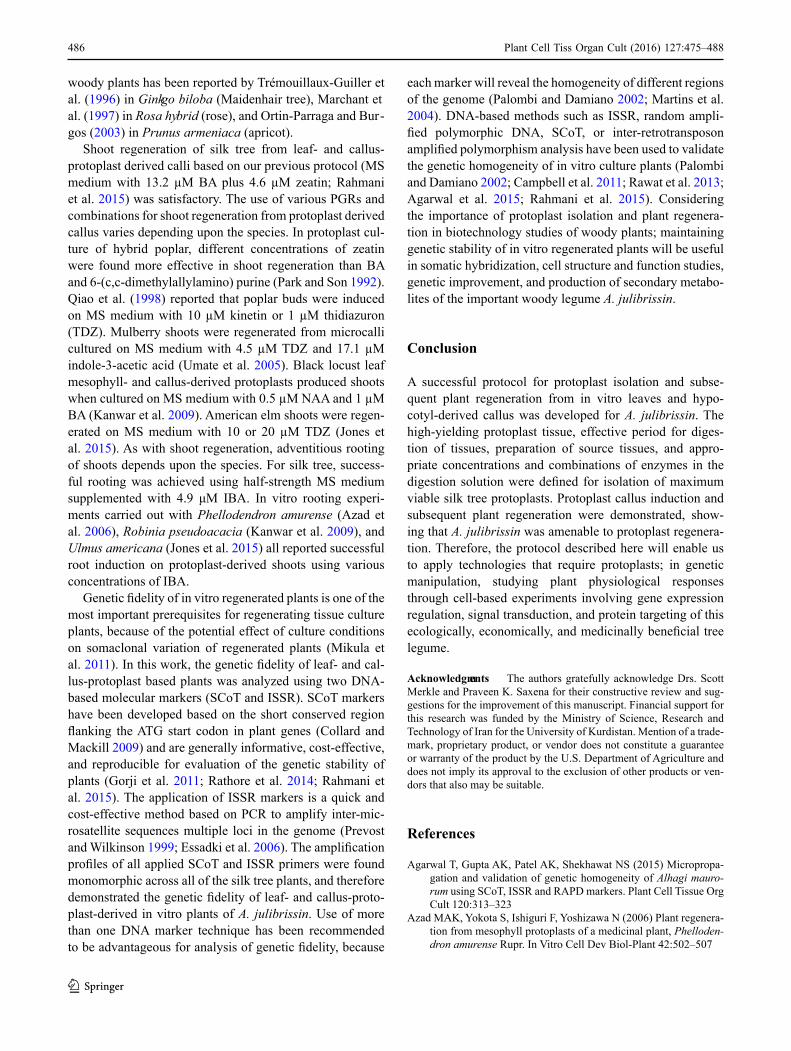
Fig. 1 Protoplast isolation and plant regeneration from leaf- and callus-derived pro-toplasts of Albizia julibrissin. a, b Freshly isolated purified protoplasts from in vitro grown leaves and stained with FDA. c, d Freshly isolated purified protoplasts from callus and stained with FDA. e, f Leaf- and callus-protoplast regenerated adventitious shoots on MS medium with 13.2 μM BA plus 4.6 μM zeatin after 5 weeks. g Rooted shoot regenerated from leaf-derived protoplast; h Acclimatized plants from leaf- (right) and callus- (left) derived protoplasts (Bars 1 cm)
1 3

481Plant Cell Tiss Organ Cult (2016) 127:475–488
cellulase. The inclusion of pectolyase in lieu of macerozyme enhanced the yield of protoplasts in all starting materials. The extension of digestion period increased the protoplast yield from both tissues. However, the mean viability of isolated protoplasts decreased to 54 % and 52 % after 12 h digestion of leaves and 20 h of callus, respectively. Donor tissues were pre-plasmolyzed in CPW with either 0.7 M mannitol or sorbitol for 30, 60, or 90 min. significant differ-ences in protoplast yield and viability were observed among osmotica and pre-plasmolysis durations. Sixty-min pre-plasmolysis of in vitro leaves and 90 min pre-plasmolysis of callus in 0.7 M sorbitol was the most efficient pretreatment for further achievement of protoplasts (Table 3).
Protoplast culture and microcolony formation
Purified protoplasts derived from in vitro leaves and calli were cultured separately at a plating density of 3–3.5 × 10⁵ Pp ml−1 for cell wall regeneration, cell multiplication, and microcolony formation. Leaf- and calli-derived protoplasts cultured in both liquid and agarose systems were able to synthesis cell walls, divide, and form cell colonies. Cells became larger and oval in shape within 30–48 h after cul-ture in medium augmented with auxins (NAA or 2,4-D) plus BA, showing new cell wall synthesis followed by first cell division. Further divisions of the leaf- and callus- derived protoplasts led to the formation of several daughter cells and eventually microcolony development.
Three modified basal media (B5, MS, and KM8p) as liq-uid or semi-solid medium, and NAA or 2,4-D plus 2.2 µM
Results
Protoplast isolation from leaves and callus
Protoplasts were successfully isolated via enzymatic diges-tion from in vitro leaves and callus of silk tree (Fig. 1a, c, respectively), and were stained with FDA (Fig. 1) to deter-mine viability. The yield and viability of protoplasts were significantly different depending on the starting tissue and duration of tissue incubation for digestion (Tables 1, 2). There were also significant differences in protoplast yield and viability between enzyme solutions. Protoplast yield significantly increased with increasing enzyme concentra-tion, but mean viability decreased when concentration of cellulase and macerozyme or pectolyase were increased to 2, 0.5, and 1 %, respectively. With leaf strips, protoplast yield varied between 0.03 and 6.89 × 10⁵ Pp gfw−1 in the presence of different concentrations of cellulase and mac-erozyme or pectolyase (Table 1), while the protoplast yield from callus was 0.01–6.63 × 10⁵ Pp gfw−1. Digestion of leaf strips in 1.5 % cellulase and 1 % pectolyase for 6 h gave the best healthy protoplast yield as it released 6.31 × 10⁵ Pp gfw−1 with 87 % viability (Table 1). The best yield of pro-toplasts from callus (5.53 × 10⁵ Pp gfw−1 with 85 % viabil-ity) was obtained with 2 % cellulase plus 1 % pectolyase for 16 h (Table 2). Between the two tissues, leaf strips yielded on average more protoplasts (3.73 Pp gfw−1 with 71.6 % viability) than callus (2.45 Pp gfw−1 with 79.3 % viability). Macerozyme or pectolyase were compared to evaluate effi-ciency of healthy protoplast isolation in combination with
Donor tissue Osmoticum (0.7 M)
Time (min) Response
Yield × 105 Pp gfw−1 Viability (%)
In vitro leaves (diges-tion in 1.5 % C + 1 % P after 6 h incubation)
Mannitol Control 6.31 ± 0.1cd 87 ± 2.0c30 6.46 ± 0.0c 82 ± 3.7bc60 7.07 ± 0.0b 81 ± 4.3bc90 6.17 ± 0.2cd 86 ± 2.1ab
Sorbitol 30 7.01 ± 0.0b 92 ± 2.5a60 7.77 ± 0.0a 94 ± 2.6a90 6.05 ± 0.0d 92 ± 1.7a
Callus (digestion in 2 % C + 1 % P after 16 h incubation)
Mannitol Control 5.53 ± 0.1ef 85 ± 2.6bc30 5.50 ± 0.0f 80 ± 5.7c60 5.80 ± 0.0d 84 ± 2.8abc90 5.76 ± 0.0de 82 ± 3.1abc
Sorbitol 30 6.17 ± 0.0c 91 ± 3.2ab60 6.53 ± 0.0b 90 ± 2.8abc90 6.92 ± 0.0a 92 ± 2.0a
Values represent means ± SE for three replications in each treatment. Means with the same letter within columns are not significantly different at P = 0.05 according to LSD testC cellulase onozuka R-10, P pectolyase Y-23
Table 3 Effect of osmoticum and pre-plasmolysis time on yield and viability of Albizia julbrissin protoplasts
1 3

482 Plant Cell Tiss Organ Cult (2016) 127:475–488
et al. 2015), in vitro shoots were successfully rooted in half-strength MS medium supplemented with 4.9 μM IBA. On average, 68 and 61 % adventitious root formation was observed for leaf- and callus-protoplast-derived shoots within 4–5 weeks culture, respectively (Fig. 1). Rooted shoots (Fig. 1) were then acclimatized, with over 60 % of the plants showing normal growth and development after 5 weeks after transplanting to soil.
Genetic fidelity screening
Two DNA-based molecular markers, SCoT and ISSR, were applied to demonstrate genetic homogeneity of a total of nine and six regenerated plants from leaf- and callus-derived protoplasts, respectively. Initially 20 SCoT and 15 ISSR primers were screened, and ten and eight primers, respectively, amplified clear and scoreable bands. A total of 53 monomorphic bands were produced from PCR ampli-fication of SCoT primers with amplicons ranging in size from 200 to 1300 bp, giving rise to monomorphic patterns across nine leaf- and six callus-protoplast-derived plants (Supplementary Table 1). The number of scoreable bands from each SCoT primer ranged from four (SCoT-5, SCoT-25, and SCoT-28) to eight (SCoT-24) with an average of 5.3 bands per primer (Supplementary Table 1). All ISSR prim-ers yielded 55 scoreable and reproducible bands ranging from 11 (maximum) to five (minimum) bands from primers UBC-873 and UBC-811, 841, respectively (Supplementary Table 1), with an average of 6.8 bands per primer ranging in size from 300 to 1700 bp.
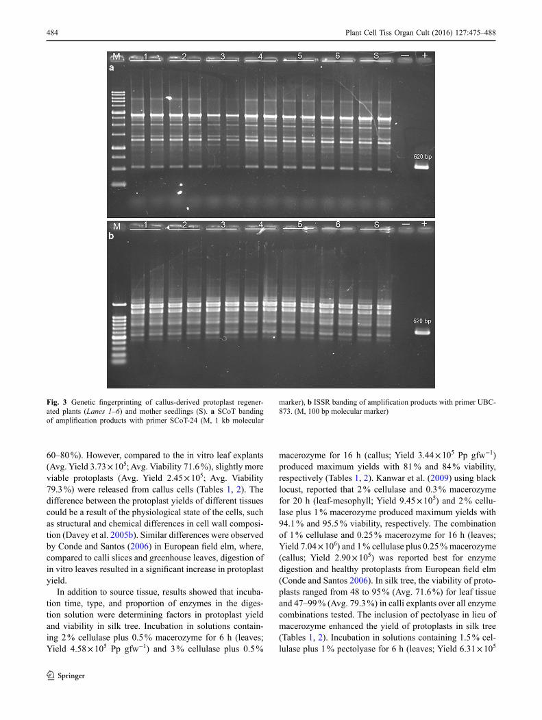
The amplification profiles were found monomorphic across all of the genotypes regenerated from both leaf- and callus-derived protoplasts by all SCoT and ISSR primers used. This finding showed the true-to-type nature of the genetic background of the protoplast-derived in vitro regen-erated plants of A. julibrissin. The monomorphic banding of SCoT-1 and UBC-834 primers representative gels from genotyping nine leaf-protoplast-based regenerated geno-types and the mother seedling are shown in Fig. 2. The monomorphic fingerprinting profiles of the six callus-pro-toplast-derived in vitro plants and the mother seedling using SCoT-24 and UBC-873 are shown in Fig. 3.
Discussion
Since genetically altered organisms are not generally accepted by society, there is an interest in using protoplast-to-plant advancements in the generation of novel germ-plasm for plant improvement (Eeckhaut et al. 2013). One of the most recognized protoplast-based technologies uti-lizes protoplast fusion to produce unique plants with new or improved desirable traits. Successful culture and genetic
BA, were assessed for the effect on microcolony formation and subsequent microcalli growth (Supplementary Table 2). In addition, KM8p medium supplemented with 1.4 % aga-rose was compared. In liquid culture, KM8p medium sup-plemented with 2.7 or 5.4 µM NAA plus 2.2 µM BA were the most effective media for the highest microcolony forma-tion from leaf- and callus-derived protoplasts (Supplemen-tary Table 2) after 3 weeks of culture (52.3 microcolonies for leaf-protoplasts, and 49.3 and 46.0 microcolonies for callus-protoplasts per Petri plate, respectively). When leaf- and callus-derived protoplasts were cultured in MS liquid medium containing 4.5 µM 2,4-D plus 2.2 µM BA, how-ever, microcolony formation was reduced to ten and eight, respectively. The KM8p agarose embedded culture medium containing 2.7 µM NAA plus 2.2 µM BA gave the maxi-mum response of 47 microcolonies (leaf) and 25 microcolo-nies (callus) as compared to the other PGRs tested of this culture system (Supplementary Table 2).
Callus formation and plant regeneration
After 3 weeks, growth of the protoplast derived microcolo-nies on semi-solid medium containing the same components of those used for the first culture (except that mannitol was eliminated), resulted in the initiation of microcalli 2–5 mm in diameter. KM8p medium supplemented with 2.7 or 5.4 µM NAA plus 2.2 µM BA resulted in the highest number of microcallus formation from microcolonies (Supplementary Table 2). The callus formed from both types of protoplast-derived microcolonies was transferred to semi-solid MSB5 medium (MS minerals with Gamborg B5 organics) or WPM containing different concentrations of NAA in combination with 4.4 µM BA (Supplementary Table 3) for further growth and proliferation. MSB5 medium with 10.8 µM NAA plus 4.4 µM BA was best for proliferation of microcalli from both leaf- and callus-derived protoplasts (Supplementary Table 3). In addition, MSB5 medium supplemented with 8.1 µM NAA plus 4.4 µM BA, and WPM with 10.8 µM NAA plus 4.4 µM BA were also suitable media for prolif-eration of microcalli from callus-derived protoplasts. WPM with 5.4 µM NAA plus 4.4 µM BA produced the least amount of callus proliferation from the leaf (35 %) and cal-lus (27.5 %) protoplast-derived microcalli after 4 weeks of culture.
For shoot organogenesis, leaf- and callus-based proto-plast derived calli were separately cultured on MS medium containing 13.2 µM BA plus 4.6 µM zeatin according to our previous protocol (Rahmani et al. 2015). Microshoots started to regenerate after 2–3 weeks culture. The overall shoot organogenesis efficiency of this culture medium was 78–93 % (data not shown). Number of shoots from a sin-gle callus cluster varied from two to four (Fig. 1) after 5 weeks culture. Following our previous protocol (Rahmani
1 3

483Plant Cell Tiss Organ Cult (2016) 127:475–488
to determine a method for silk tree protoplast isolation, a number of parameters including source material, enzymatic solution composition, digestion duration, and plasmoly-sis treatment were assessed. In the present investigation, leaves from in vitro grown seedlings and hypocotyl-derived organogenic callus were used as explant material for proto-plast isolation. Although protoplasts can be isolated from different types of explants, it was reported that only young leaves of in vitro grown shoots, and actively growing callus or cell suspension cultures gave consistent and high yields of healthy protoplasts in woody plants (Liu 2005).
Large numbers of purified viable protoplasts (105–107 Pp gfw−1) are required for protoplast fusion or Agrobacte-rium-mediated genetic modification (Davey et al. 2005a). The yields of protoplasts released from tissues in our study, varied from 0.03 to 7.77 × 10⁵ Pp gfw−1 for leaves and 0.01–6.92 × 10⁵ Pp gfw−1 for calli slices. Suitability of in vitro leaves for protoplast isolation in other woody plant species has been documented by Rezazadeh and Niedz (2015) in guava (Psidium guajava) (Yield 3.7 × 10⁶; Viability > 90 %), Kanwar et al. (2009) in black locust (Yield 9.45 × 10⁵; Viability 94.1 %), and Conde and Santos (2006) in Euro-pean field elm (Ulmus minor) (Yield 3.96 × 10⁷; Viability
transformation of purified protoplasts depends on cell wall re-synthesis and subsequent plant regeneration (Davey et al. 2005b). Establishment of a protoplast-to-plant regenera-tion framework is affected by many components, including the protoplast isolation process, culture density and system, and medium composition (Eeckhaut et al. 2009; Kanwar et al. 2009; Castelblanque et al. 2010; Rezazadeh and Niedz 2015). As the regenerative capacity of protoplasts from different genotypes and tissues varies significantly, such investigations are required (Davey et al. 2005a, b). To our knowledge, this is the first study to investigate protoplast isolation and regeneration capacity of leaf- and callus-derived protoplasts in Albizia julibrissin.
Isolation of a large number of healthy protoplasts is a pre-requisite for successful protoplast culture and regeneration. Several factors including protoplast donor tissue, pretreat-ment of tissue before enzyme maceration, pH and com-position of enzyme solution, temperature and duration of enzyme incubation, gentle agitation, and protoplast isolation method (Frearson et al. 1973; Rao and Prakash 1995; Ortin-Parraga and Burgos 2003; Sinha et al. 2003; Kanwar et al. 2009; Kiełkowska and Adamus 2012) significantly affected the yield and viability of protoplast isolation. Therefore,
Fig. 2 Genetic fingerprinting of leaf-derived protoplast regenerated plants (Lanes 1–9) and mother seedlings (S). a SCoT banding of ampli-fication products with primer SCoT-1 (M, 1 kb molecular marker), b
ISSR banding of amplification products with primer UBC-834. (M, 100 bp molecular marker)
1 3
484 Plant Cell Tiss Organ Cult (2016) 127:475–488
macerozyme for 16 h (callus; Yield 3.44 × 10⁵ Pp gfw−1) produced maximum yields with 81 % and 84 % viability, respectively (Tables 1, 2). Kanwar et al. (2009) using black locust, reported that 2 % cellulase and 0.3 % macerozyme for 20 h (leaf-mesophyll; Yield 9.45 × 10⁵) and 2 % cellu-lase plus 1 % macerozyme produced maximum yields with 94.1 % and 95.5 % viability, respectively. The combination of 1 % cellulase and 0.25 % macerozyme for 16 h (leaves; Yield 7.04 × 10⁶) and 1 % cellulase plus 0.25 % macerozyme (callus; Yield 2.90 × 10⁵) was reported best for enzyme digestion and healthy protoplasts from European field elm (Conde and Santos 2006). In silk tree, the viability of proto-plasts ranged from 48 to 95 % (Avg. 71.6 %) for leaf tissue and 47–99 % (Avg. 79.3 %) in calli explants over all enzyme combinations tested. The inclusion of pectolyase in lieu of macerozyme enhanced the yield of protoplasts in silk tree (Tables 1, 2). Incubation in solutions containing 1.5 % cel-lulase plus 1 % pectolyase for 6 h (leaves; Yield 6.31 × 10⁵
60–80 %). However, compared to the in vitro leaf explants (Avg. Yield 3.73 × 10⁵; Avg. Viability 71.6 %), slightly more viable protoplasts (Avg. Yield 2.45 × 10⁵; Avg. Viability 79.3 %) were released from callus cells (Tables 1, 2). The difference between the protoplast yields of different tissues could be a result of the physiological state of the cells, such as structural and chemical differences in cell wall composi-tion (Davey et al. 2005b). Similar differences were observed by Conde and Santos (2006) in European field elm, where, compared to calli slices and greenhouse leaves, digestion of in vitro leaves resulted in a significant increase in protoplast yield.
In addition to source tissue, results showed that incuba-tion time, type, and proportion of enzymes in the diges-tion solution were determining factors in protoplast yield and viability in silk tree. Incubation in solutions contain-ing 2 % cellulase plus 0.5 % macerozyme for 6 h (leaves; Yield 4.58 × 10⁵ Pp gfw−1) and 3 % cellulase plus 0.5 %
Fig. 3 Genetic fingerprinting of callus-derived protoplast regener-ated plants (Lanes 1–6) and mother seedlings (S). a SCoT banding of amplification products with primer SCoT-24 (M, 1 kb molecular
marker), b ISSR banding of amplification products with primer UBC-873. (M, 100 bp molecular marker)
1 3

485Plant Cell Tiss Organ Cult (2016) 127:475–488
Cell wall formation, mitosis reactivation, and sustained cell divisions are key steps in the formation of microcolo-nies, callus formation, and subsequent plant regeneration from protoplast cultures (Eeckhaut et al. 2013). Improve-ment in protoplast-to-plant protocols have been accom-plished by testing various culture methods and medium formulations (Davey et al. 2005a). Since the first report on regeneration of whole plants from protoplast-derived cells of tobacco (Nagata and Takebe 1971), numerous meth-ods have been developed for a variety of plant species (Davey et al. 2005a, b; Eeckhaut et al. 2013). Plant pro-toplasts are generally cultured at an initial plating density of 5 × 10⁴–1 × 10⁶ Pp ml−1 (Davey et al. 2005b). In the present study, leaf- and calli- derived protoplasts cultured with a plating density of 3–3.5 × 10⁵ Pp ml−1, successfully formed cell walls and then microcolonies when cultured in both liquid and agarose culture medium. A higher rate of colony formation was recorded for protoplasts cultured in KM8p medium supplemented with 2.7 or 5.4 µM NAA plus 2.2 µM BA (Supplementary Table 2). Nutritional require-ments of protoplasts varied according to species and dif-ferent tissues of the same species from which protoplasts were isolated (Davey et al. 2005b). The basal media MS, B5, KM and other modified formulations have been suc-cessfully used to culture protoplasts of a variety of woody species (Conde and Santos 2006; Cai and Kang 2014; Jones et al. 2015; Rezazadeh and Niedz 2015). Different culture systems (liquid or agarose embedded) and PGRs have been successfully applied for protoplast culture of hardwood spe-cies (Kanwar et al. 2009; Jones et al. 2015). In black locust, maximum dividing cells from leaf mesophyll- and callus-derived protoplasts occurred when cultured in Nagata and Takebe (1971) medium and in WPM (without NH4NO3), respectively, containing 5 µM NAA plus 1 µM BA. In American elm, protoplasts initiated and continued cell divi-sion when embedded in agarose beads (37.5 %) versus using liquid KM5/5 and alginate beads (Jones et al. 2015). In silk tree, leaf- and callus-derived protoplasts were more success-ful in microcolony formation during liquid culture than in the agarose KM8p embedded system.
Auxin and cytokinin are two of the main PGRs control-ling plant cell division. In silk tree, microcolonies derived from leaf- and callus-derived protoplasts formed microcalli (2–5 mm in diameter) followed by proliferation in semi-solid MSB5 medium (MS minerals with Gamborg B5 organics) or WPM containing different concentrations of NAA in com-bination with 4.4 µM BA (Supplementary Table 3). Higher callus proliferation efficiency was observed from MSB5 medium with 10.8 µM NAA plus 4.4 µM BA for leaf-proto-plasts, and from MSB5 medium with 8.1 and 10.8 µM NAA plus 4.4 µM BA, and WPM with 10.8 µM NAA plus 4.4 µM BA for callus-protoplasts. Similarly, the key role of NAA and BA in callus formation from protoplast culture of other
Pp gfw−1) and 2 % cellulase plus 1 % pectolyase for 16 h (callus; Yield 5.53 × 10⁵ Pp gfw−1) produced maximum yields with 87 % and 85 % viability, respectively (Tables 1, 2). Similarly, Dorion et al. (1994) reported in Ulmus sp. (elm) that pectolyase showed a high performance in the isolation of protoplasts from in vitro leaves. When suspen-sion cultures of other hardwood tree species were used for protoplast isolation, the type, concentration, and duration of incubation varied considerably; i.e. yellow-poplar: 2 % cellulysin and 1 % macerase, 12 h (Merkle and Sommer 1987); white poplar: 1 % cellulase and 0.1 % pectolyase, 1.5 h (Qiao et al. 1998); camphor tree: 3 % cellulase and 3 % macerozyme, 12 h (Du and Bao 2005); and American elm: 0.2 % cellulase, 0.1 % driselase, and 0.03 % pectolyase for 2 h (Jones et al. 2015). Although the extension of the diges-tion period gradually increased the release of silk tree pro-toplasts, the mean viability of isolated protoplasts decreased considerably with only 54 % after 12 h for leaves and 52 % after 20 h for callus (Tables 1, 2). Similar detrimental effects of increased digestion period on viability of isolated proto-plasts from different explants has been reported for other woody plants such as Quercus acutissima (sawtooth oak) and black locust (Wakita 1997; Wakita et al. 1992; Kanwar et al. 2009). Kanwar et al. (2009) reported a decrease in the viability of black locust callus-derived protoplasts with an increase in digestion period from 24 h (95.5 % viability) to 28 h (63.2 % viability). These results demonstrate that opti-mization of specific enzyme combinations and digestion period is necessary for any species and cell type.
The use of an osmoticum prior to enzyme digestion or included in the enzyme digestion solution to increase proto-plast yield and viability has been well documented. Davey et al. (2005b) suggested that plasmolysis of source material in, for example, a solution containing mannitol or sorbitol prior to enzymatic digestion improved viability and reduced damage and spontaneous fusion of adjacent protoplast. Our results indicated that protoplast isolation based on plasmol-ysis of source tissue with an osmoticum was suitable for releasing a high yield of healthy protoplasts from leaves and callus tissue of A. julibrissin. Sixty- or 90-min-plasmolysis of leaves or callus tissue, respectively, in 0.7 M sorbitol was the most efficient pretreatment for recovering a high yield of viable protoplasts in silk tree (Table 3). Umate et al. (2005) used 0.5 M mannitol in the enzyme solution when isolating protoplasts from leaf tissue of mulberry. Pre-plasmolysis of European field elm leaf strips for 1 h in CPW salts with 0.7 M mannitol and 5 mM MES followed by enzyme diges-tion was reported by Conde and Santos (2006). In black locust, Kanwar et al. (2009) dissolved different concen-trations of cellulase and macerozyme in 0.7 M mannitol. Guo et al. (2012) digested leaves of a Populus clone in an enzyme solution that included 0.4 M mannitol, 3 % cellulase and 0.8 % macerozyme for 5 h.
1 3
486 Plant Cell Tiss Organ Cult (2016) 127:475–488
each marker will reveal the homogeneity of different regions of the genome (Palombi and Damiano 2002; Martins et al. 2004). DNA-based methods such as ISSR, random ampli-fied polymorphic DNA, SCoT, or inter-retrotransposon amplified polymorphism analysis have been used to validate the genetic homogeneity of in vitro culture plants (Palombi and Damiano 2002; Campbell et al. 2011; Rawat et al. 2013; Agarwal et al. 2015; Rahmani et al. 2015). Considering the importance of protoplast isolation and plant regenera-tion in biotechnology studies of woody plants; maintaining genetic stability of in vitro regenerated plants will be useful in somatic hybridization, cell structure and function studies, genetic improvement, and production of secondary metabo-lites of the important woody legume A. julibrissin.
Conclusion
A successful protocol for protoplast isolation and subse-quent plant regeneration from in vitro leaves and hypo-cotyl-derived callus was developed for A. julibrissin. The high-yielding protoplast tissue, effective period for diges-tion of tissues, preparation of source tissues, and appro-priate concentrations and combinations of enzymes in the digestion solution were defined for isolation of maximum viable silk tree protoplasts. Protoplast callus induction and subsequent plant regeneration were demonstrated, show-ing that A. julibrissin was amenable to protoplast regenera-tion. Therefore, the protocol described here will enable us to apply technologies that require protoplasts; in genetic manipulation, studying plant physiological responses through cell-based experiments involving gene expression regulation, signal transduction, and protein targeting of this ecologically, economically, and medicinally beneficial tree legume.
Acknowledgments The authors gratefully acknowledge Drs. Scott Merkle and Praveen K. Saxena for their constructive review and sug-gestions for the improvement of this manuscript. Financial support for this research was funded by the Ministry of Science, Research and Technology of Iran for the University of Kurdistan. Mention of a trade-mark, proprietary product, or vendor does not constitute a guarantee or warranty of the product by the U.S. Department of Agriculture and does not imply its approval to the exclusion of other products or ven-dors that also may be suitable.
References
Agarwal T, Gupta AK, Patel AK, Shekhawat NS (2015) Micropropa-gation and validation of genetic homogeneity of Alhagi mauro-rum using SCoT, ISSR and RAPD markers. Plant Cell Tissue Org Cult 120:313–323
Azad MAK, Yokota S, Ishiguri F, Yoshizawa N (2006) Plant regenera-tion from mesophyll protoplasts of a medicinal plant, Phelloden-dron amurense Rupr. In Vitro Cell Dev Biol-Plant 42:502–507
woody plants has been reported by Trémouillaux-Guiller et al. (1996) in Ginkgo biloba (Maidenhair tree), Marchant et al. (1997) in Rosa hybrid (rose), and Ortin-Parraga and Bur-gos (2003) in Prunus armeniaca (apricot).
Shoot regeneration of silk tree from leaf- and callus-protoplast derived calli based on our previous protocol (MS medium with 13.2 µM BA plus 4.6 µM zeatin; Rahmani et al. 2015) was satisfactory. The use of various PGRs and combinations for shoot regeneration from protoplast derived callus varies depending upon the species. In protoplast cul-ture of hybrid poplar, different concentrations of zeatin were found more effective in shoot regeneration than BA and 6-(c,c-dimethylallylamino) purine (Park and Son 1992). Qiao et al. (1998) reported that poplar buds were induced on MS medium with 10 µM kinetin or 1 µM thidiazuron (TDZ). Mulberry shoots were regenerated from microcalli cultured on MS medium with 4.5 µM TDZ and 17.1 µM indole-3-acetic acid (Umate et al. 2005). Black locust leaf mesophyll- and callus-derived protoplasts produced shoots when cultured on MS medium with 0.5 µM NAA and 1 µM BA (Kanwar et al. 2009). American elm shoots were regen-erated on MS medium with 10 or 20 µM TDZ (Jones et al. 2015). As with shoot regeneration, adventitious rooting of shoots depends upon the species. For silk tree, success-ful rooting was achieved using half-strength MS medium supplemented with 4.9 μM IBA. In vitro rooting experi-ments carried out with Phellodendron amurense (Azad et al. 2006), Robinia pseudoacacia (Kanwar et al. 2009), and Ulmus americana (Jones et al. 2015) all reported successful root induction on protoplast-derived shoots using various concentrations of IBA.
Genetic fidelity of in vitro regenerated plants is one of the most important prerequisites for regenerating tissue culture plants, because of the potential effect of culture conditions on somaclonal variation of regenerated plants (Mikula et al. 2011). In this work, the genetic fidelity of leaf- and cal-lus-protoplast based plants was analyzed using two DNA-based molecular markers (SCoT and ISSR). SCoT markers have been developed based on the short conserved region flanking the ATG start codon in plant genes (Collard and Mackill 2009) and are generally informative, cost-effective, and reproducible for evaluation of the genetic stability of plants (Gorji et al. 2011; Rathore et al. 2014; Rahmani et al. 2015). The application of ISSR markers is a quick and cost-effective method based on PCR to amplify inter-mic-rosatellite sequences multiple loci in the genome (Prevost and Wilkinson 1999; Essadki et al. 2006). The amplification profiles of all applied SCoT and ISSR primers were found monomorphic across all of the silk tree plants, and therefore demonstrated the genetic fidelity of leaf- and callus-proto-plast-derived in vitro plants of A. julibrissin. Use of more than one DNA marker technique has been recommended to be advantageous for analysis of genetic fidelity, because
1 3

487Plant Cell Tiss Organ Cult (2016) 127:475–488
Grosser JW, Gmitter FG (2011) Protoplast fusion for production of tet-raploids and triploids: applications for scion and rootstock breed-ing in citrus. Plant Cell Tiss Org Cult 104:345–357
Grosser JW, Ćalović M, Louzada ES (2010) Protoplast fusion tech-nology—somatic hybridization and cybridization. In: Davey MR, Anthony P (eds) Plant cell culture: essential methods. Wiley, NY, pp 175–198
Guo J, Morrell-Falvey JL, Labbé JL, Muchero W, Kalluri UC, Tus-kan GA, Chen J-G (2012) Highly efficient isolation of Populus mesophyll protoplasts and its application in transient expression assays. PLoS One 7:e44908
Han L, Pan G, Wang Y, Song X, Gao X, Ma B, Kang L (2011) Rapid profiling and identification of triterpenoid saponins in crude extracts from Albizia julibrissin Durazz. by ultra high-perfor-mance liquid chromatography coupled with electrospray ion-ization quadrupole time-of-flight tandem mass spectrometry. J Pharmaceut Biomed Anal 55:996–1009
Jones AMP, Shukla MR, Biswas GCG, Saxena PK (2015) Protoplast-to-plant regeneration of American elm (Ulmus americana). Pro-toplasma 252:925–931
Jordan CF (2004) Organic farming and agroforestry: alleycropping for mulch production for organic farms of southeastern United States. Agroforest Syst 61:79–90
Jung MJ, Kang SS, Jung YJ, Choi JS (2004) Phenolic glycosides from the stem bark of Albizzia julibrissin. Chem Pharm Bull 52:1501–1503
Kang J, Huo CH, Li Z, Li ZP (2007) New ceramides from the flower of Albizia julibrissin. Chin Chem Lett 18:181–184
Kanwar K, Bhardwaj A, Deepika R (2009) Efficient regeneration of plantlets from callus and mesophyll derived protoplasts of Rob-inia pseudoacacia L. Plant Cell Tissue Org Cult 96:95–103
Kao KN, Michayluk MR (1975) Nutritional requirements for growth of Vicia hajastana cells and protoplasts at a very low population density in liquid media. Planta 126:105–110
Kiełkowska A, Adamus A (2012) An alginate-layer technique for cul-ture of Brassica oleracea L. protoplasts. In Vitro Cell Dev Biol-Plant 48:265–273
Kinjo J, Araki K, Fukui K, Higuchi H, Ikeda T, Nohara T, Ida Y, Take-moto N, Miyakoshi M, Shoji J (1992) Six new triterpenoidal gly-cosides including two new sapogenols from Albizziae Cortex. V. Chem Pharm Bull (Tokyo) 40:3269–3273
Lau CS, Carrier DJ, Beitle RR, Bransby DI, Howard LR, Lay JO, Liyanage R, Clausen EC (2007) Identication and quantication of glycoside flavonoids in the energy crop Albizia julibrissin. Biore-source Technol 98:429–435
Liang H, Tong W-Y, Zhao Y-Y, Cui J-R, Tu G-Z (2005) An antitumor compound julibroside J28 from Albizia julibrissin. Bioorg Med Chem Lett 15:4493–4495
Liu J (2005) Protoplast isolation and culture of woody plants. In: Jain SM, Gupta PK (eds) Protocol for somatic embryogenesis in woody plants. Springer, Netherlands, pp 553–566
Lloyd G, McCown B (1981) Commercially feasible micropropagation of mountain laurel, Kalmia latifolia, by use of shoot-tip culture. Proc Int Plant Prop Soc 30:421–427
Lv JS, Zhang LN, Song YZ, Wang XF, Chu XZ (2011) Biological activity exhibited by secondary metabolites of the Albizia juli-brissin Durazz. pod. Intl Biodegrad 65:258–264
Marchant R, Davey MR, Power JB (1997) Isolation and culture of mesophyll protoplasts from Rosa hybrida. Plant Cell Tissue Org Cult 50:131–134
Martins M, Sarmento D, Oliveira MM (2004) Genetic stability of micropropagated almond plantlets, as assessed by RAPD and ISSR markers. Plant Cell Rep 23:492–496
Merkle SA, Sommer HE (1987) Regeneration of Liriodendron tulip-ifera (Family Magnoliaceae) from protoplast culture. Amer J Bot 74:1317–1321
Bouazza L, Bodas R, Boufennara S, Bousseboua H, López S (2012) Nutritive evaluation of foliage from fodder trees and shrubs char-acteristic of Algerian arid and semi-arid areas. J Anim Feed Sci 21:521–536
Burns JA, Wetzstein HY (1998) Embryogenic cultures of the legumi-nous tree Albizia julibrissin and recovery of plants. Plant Cell Tissue Org Cult 54:55–59
Cai X, Kang X-Y (2014) Plant regeneration from cell suspension–derived protoplasts of Populus× beijingensis. In Vitro Cell Dev Biol-Plant 50:92–98
Campbell BC, LeMare S, Piperidis G, Godwin ID (2011) IRAP, a retrotransposon-based marker system for the detection of soma-clonal variation in barley. Mol Breed 27:193–206
Castelblanque L, García-Sogo B, Pineda B, Moreno V (2010) Efficient plant regeneration from protoplasts of Kalanchoe blossfeldiana via organogenesis. Plant Cell Tissue Org Cult 100:107–112
Cheatham S, Johnston MC, Marshall L (1995) Albizia. In: Cheatham S (ed) The useful wild plants of Texas, the southeastern and south-western United States, the southern plains, and northern Mexico. Austin USA, pp 188–190
Collard BCY, Mackill DJ (2009) Start codon targeted (SCoT) poly-morphism: a simple, novel DNA marker technique for generating gene-targeted markers in plants. Plant Mol Biol Rep 27:86–93
Conde P, Santos C (2006) An efficient protocol for Ulmus minor Mill. Protoplast isolation and culture in agarose droplets. Plant Cell Tiss Org Cult 86:359–366
Davey MR, Anthony P, Power JB, Lowe KC (2005a) Plant protoplast technology: current status. Acta Physiol Plant 27:117–129
Davey MR, Anthony P, Power JB, Lowe KC (2005b) Plant proto-plasts: status and biotechnological perspectives. Biotechnol Adv 23:131–171
Dorion N, Jouira HB, Danthu P, Bigot C (1994) Regeneration of plants from protoplasts of Ulmus species (elms). In: Bajaj YPS (ed) Bio-technology in agriculture and forestry 29, Plant protoplasts and genetic engineering V, Springer, Berlin, pp 172–190
Doyle JJ, Doyle JL (1990) Isolation of plant DNA from fresh tissue. Focus 12:13–15
Du L, Bao M (2005) Plant regeneration from protoplasts isolated from embryogenic suspension cultured cells of Cinnamomum cam-phora L. Plant Cell Rep 24:462–467
Eeckhaut T, Duquenne B, Laksmanan P, Van Huylenbroeck J (2009) Development of microcolonies in protoplast culture of Spathip-hyllum wallisii. Acta Hort 829:51–54
Eeckhaut T, Lakshmanan PS, Deryckere D, Van Bockstaele E, Van Huylenbroeck J (2013) Progress in plant protoplast research. Planta 238:991–1003
Essadki M, Ouazzani N, Lumaret R, Moumni M (2006) ISSR varia-tion in olive-tree cultivars from Morocco and other western countries of the Mediterranean Basin. Genet Resour Crop Evol 53:475–482
Faraco M, Di Sansebastiano GP, Spelt K, Koes RE, Quattrocchio FM (2011) One protoplast is not the other! Plant Physiol 156:474–478
Fordham AJ (1965) Germination of woody legume seeds with imper-meable seed coat. Arnoldia 25:1–8
Frearson EM, Power JB, Cocking EC (1973) The isolation, culture and regeneration of Petunia leaf protoplasts. Dev Biol 33:130–137
Fu J, Peng ZJ, Cai XD, Guo WW (2011) Regeneration and molecular characterization of interspecific somatic hybrids between Sat-suma mandarin and two seedy sweet oranges for scion improve-ment. Plant Breed 130:287–290
Gamborg OL, Miller RA, Ojima K (1968) Nutrient requirements of sus-pention cultures of soybean root cells. Exp Cell Res 50:151–158
Gorji AM, Poczai P, Polgar Z, Taller J (2011) Efficiency of arbi-trarily amplified dominant markers (SCoT, ISSR and RAPD) for diagnostic fingerprinting in tetraploid potato. Am J Potato Res 88:226–237
1 3

488 Plant Cell Tiss Organ Cult (2016) 127:475–488
Sankhla D, Davis TD, Sankhla N (1994) Thidiazuron-induced in vitro shoot formation from roots of intact seedlings of Albizzia julibris-sin. Plant Growth Regul 14:267–272
Sankhla D, Sankhla N, Davis TD (1995) Promotion of in vitro shoot formation from excised roots of silktree (Albizzia julibrissin) by an oxime ether derivative and other ethylene inhibitors. Plant Cell Rep 15:143–146
Sankhla D, Davis TD, Sankhla N (1996) In vitro regeneration of silk-tree (Albizzia julibrissin) from excised roots. Plant Cell Tissue Org Cult 44:83–86
SAS Institute Inc (2004) SAS® 9. 0, 9. 1, 9.1.2, and 9.1.3. SAS Insti-tute Inc, Cary
Sinha A, Wetten AC, Caligari PDS (2003) Effect of biotic factors on the isolation of Lupinus albus protoplasts. Austr J Bot 51:103–109
Sladden SE, Bransby DI, Aiken GE, Rose PA (1992) Mimosa could be a new forage legume. In: Highlights agric res 39, p 4, AL Agric Exp Stn, Auburn Univ, Auburn, p 16
Soriano L, Filho FAAM, Camargo LEA, Cristofani-Yaly M, Latado RR, Pacheco CA, Azevedo FA, Mendes BMJ (2012) Regenera-tion and characterization of somatic hybrids combining sweet orange and mandarin/mandarin hybrid cultivars for citrus scion improvement. Plant Cell Tissue Org Cult 111:385–392
Tan B, Xu M, Chen Y, Huang M (2013) Transient expression for func-tional gene analysis using Populus protoplasts. Plant Cell Tissue Org Cult 114:11–18
Trémouillaux-Guiller J, Laurain D, Chénieux JC (1996) Direct embryogenesis in protoplasts of Ginkgo biloba (Maidenhair Tree). In: Bajaj YPS (ed), Biotechnology in agriculture and for-estry 38, Plant protoplasts and genetic engineering VII, Springer, Berlin, pp 33–47
Umate P, Rao KV, Kiranmayee K, Sree TJ, Sadanandam A (2005) Plant regeneration of mulberry (Morus indica) from mesophyll-derived protoplasts. Plant Cell Tissue Org Cult 82:289–293
Wakita Y (1997) Plant regeneration from protoplasts of broad-leaved trees. Bull Utsunomiya Univ For 33:55–108
Wakita Y, Yokota S, Yoshizawa N, Idei T (1992) Isolation and culture of protoplasts from Kunugi (Quercus acutissima Carruth.) callus cultures. Plant Tissue Cult Lett 9:74–80
Wang FQ, Wang ET, Zhang YF, Chen WX (2006) Characterization of rhizobia isolated from Albizia spp. in comparison with micro-symbionts of Acacia spp. and Leucaena leucocephala grown in China. Syst Appl Microbiol 29:502–517
Widholm JM (1972) The use of fluorescein diacetae and phenosafra-nine for determining viability of cultured plant cell. Stain Technol 47:189–194
Xiao S-X, Biswas MK, Li M-Y, Deng X-X, Xu Q, Guo W-W (2014) Production and molecular characterization of diploid and tetra-ploid somatic cybrid plants between male sterile Satsuma manda-rin and seedy sweet orange cultivars. Plant Cell Tissue Org Cult 116:81–88
Yoo S-D, Cho Y-H, Sheen J (2007) Arabidopsis mesophyll protoplasts: a versatile cell system for transient gene expression analysis. Nat Protoc 2:1565–1572
Zhou Y, Zhang ZQ, Zhang JJ (2001) Use of hymexazol (HMI) in rapid propagation of Albizia julibrissin Durazz. Acta Agr Shanghai 17:31–34
Meyer R (2010) Albizia julibrissin. In: Fire effects information sys-tem, [Online] U.S. Dept. Agric., Forest Service, Rocky Mountain Research Station, Fire Sciences Laboaratory (Producer). http://www.fs.fed.us/database/feis/. Accessed 3 Feb 2016
Mikula A, Tomiczak K, Rybczyński JJ (2011) Cryopreservation enhances embryogenic capacity of Gentiana cruciata (L.) sus-pension culture and maintains (epi)genetic uniformity of regener-ants. Plant Cell Rep 30:565–574
Murashige T, Skoog F (1962) A revised medium for rapid growth and bio assays with tobacco tissue cultures. Physiol Plant 15:473–497
Nagata T, Takebe I (1971) Plating of isolated tobacco mesophyll pro-toplasts on agar medium. Planta 99:12–20
Nehdi I (2011) Characteristics, chemical composition and utilisation of Albizia julibrissin seed oil. Ind Crop Prod 33:30–34
Ortín-Párraga F, Burgos L (2003) Isolation and culture of mesophyll protoplast from apricot. J Hort Sci Biotechnol 78:624–628
Orwa C, Mutua A, Kindt R, Jamnadass R, Simons A (2009) Albizia julibrissin. Agroforestry Database: a tree reference and selection guide. Version 4.0. Accessed 4 April 2016 (http://www.worldag-roforestry.org/treedb2/AFTPDFS/Albizia_julibrissin.pdf)
Palombi MA, Damiano C (2002) Comparison between RAPD and SSR molecular markers in detecting genetic variation in kiwifruit (Actinidia deliciosa A. Chev). Plant Cell Rep 20:1061–1066
Park YG, Son SH (1992) In vitro shoot regeneration from leaf meso-phyll protoplasts of hybrid poplar (Populus nigra× P. maximow-iczii). Plant Cell Rep 11:2–6
Phipps PM, Stipes RJ (1976) Histopathology of mimosa (Albizia juli-brissin) infected with Fusarium oxysporum f. sp. perniciosum. Phytopathology 66:839–843
Pitman WD (2008) Establishment and regrowth responses of Albizia julibrissin on Louisiana USA coastal plain soils. Agroforest Syst 74:259–266
Prevost A, Wilkinson MJ (1999) A new system of comparing PCR primers applied to ISSR fingerprinting of potato cultivars. Theor Appl Genet 98:107–112
Qiao J, Kuroda H, Hayashi T, Sakai F (1998) Efficient plantlet regen-eration from protoplasts isolated from suspension cultures of pop-lar (Populus alba L.) Plant Cell Rep 17:201–205
Rahmani M-S, Pijut PM, Shabanian N, Nasri M (2015) Genetic fidel-ity assessment of in vitro-regenerated plants of Albizia julibrissin using SCoT and IRAP fingerprinting. In Vitro Cell Dev Biol-Plant 51:407–419
Rao KS, Prakash AH (1995) A simple method for the isolation of plant protoplasts. J Biosci 20:645–655
Rathore NS, Rai MK, Phulwaria M, Rathore N, Shekhawat NS (2014) Genetic stability in micropropagated Cleome gynandra revealed by SCoT analysis. Acta Physiol Plant 36:555–559
Rawat JM, Rawat B, Mehrotra S, Chandra A, Nautiyal S (2013) ISSR and RAPD based evaluation of genetic fidelity and active ingre-dient analysis of regenerated plants of Picrorhiza kurroa. Acta Physiol Plant 35:1797–1805
Rezazadeh R, Niedz RP (2015) Protoplast isolation and plant regenera-tion of guava (Psidium guajava L.) using experiments in mixture-amount design. Plant Cell Tissue Org Cult 122:585–604
Sankhla D, Davis TD, Sankhla N (1993) Effect of gibberellins biosyn-thesis inhibitors on shoot regeneration from hypocotyl explants of Albizzia julibrissin. Plant Cell Rep 13:115–118
1 3