Embed Size (px)
Citation preview
The Plant Cell Wall Biology Lab., Tohoku University 28 May 2013
Protoplast Isolation and Cell Wall Regeneration.
1. Cell Suspension Culture
1.1 Maintenance of Cell Suspension
Materials:
Suspension-cultured Alex cells of Arabidopsis thaliana L. (Heynh.), ecotype Columbia.
The cultured cells were established by Mathur et al. (1998).
Suspension culture medium (pH5.8, 1L). 4.33g of Murashige and Skoog Plant Salt
Mixture (Wako Co. Ltd., Japan), 3.0% sucrose, 10mg thiamine-HCl, 1mg nicotinic acid,
1mg pyridoxin, 1mg glycine, 100mg myo-inositol, 0.5mg dichlorophenoxyacetic acid
(2,4-D).
1. Transfer 15 mL 0f 7-day-old cell suspension into 35 mL suspension culture
medium in 300-mL Erlenmeyer flask.
2. Place the flask on a rotary shaker at 120 rpm under dark conditions at 23ºC.
3. Culture the cell suspensions every 7 day.
2. Protoplast Isolation and Regeneration
2.1 Protoplast from Cell Suspension
Materials:
5-day-old suspension-cultured Alex cells.
0.45M mannitol solution (pH5.8).
Protoplasting enzyme solution (pH5.8): 1.0% Cellulase Onozuka RS (Yakult
Pharmaceutical Ind. Co. Ltd., Tokyo, Japan) and 0.1% Pectolyase Y-23 (Kyowa Hakko
Kyogo Co. Ltd., Tokyo, Japan) dissolved in mannitol solution.
Modified cell wall regeneration medium (pH5.8, 1L): 3.2g Gamborg’s B-5 Basal
Medium with Minimal Organics (Sigma-Aldrich), 1.0M glucose, 0.25M mannitol, 1µM
NAA.
1. Collect 50 mL of Five-day-old suspension-cultured cells in a sterile Falcon tube at
190×g for 10 min. Remove the supernatant
2. Wash the cells with 0.45M mannitol solution and centrifuge them at 190×g for
5min.
3. Remove the supernanant and resuspend the pelleted cells in a four to eightfold
volume of enzyme solution.
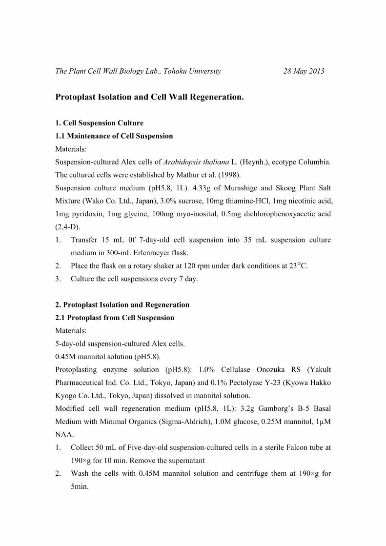
4. Incubate the cells for >1h at 37ºC (Fig.1).
5. Collect the protoplasts at 70×g for 10 min and washed twice with 40 ml of 0.45M
mannitol solution.
6. Resuspend the purified protoplasts gently in cell wall regeneration medium at a
final density of about 105 protoplasts ml–1, and incubated at 25°C to regenerate cell
walls.
3. Ultrastructural and Hisochemical Analysis of Cell Wall Regeneration
3.1 Scanning Electron Microscopic Observation
Material:
2% glutaraldehyde solution (pH 6.0): 2% glutaraldehyde solution with 120 g L–1
sorbitol adjusted to pH 6.0.
0.2% (v/v) ruthenium tetroxide (RuO4).
Ethanol solution series (50%, 60%, 70%, 80%, 95%, 100%): For the100% ethanol
soultion, use 100% bulk ethanol that has Molecular Sieves in the bottom of the bottle.
1. Fix the protoplasts in 2% glutaraldehyde for 16 h.
2. Remove 2% glutaraldehyde and for an additional fixation, incubate 0.2% (v/v)
ruthenium tetroxide for 30 min at room
temperature.
3. Remove 0.2% ruthenium tetroxide and replace
with 50% ethanol. Incubate for 30 min.
4. Repeat for the following ethanol solutions:
60%, 70%, 80%, 95%, 100%.
5. Remove the 100% ethanol, replace with the
Fig. 1. Alex cells treated with protoplasting enzyme solution for 0, 15, 30, 45 min.
0 min 15 min 30 min 45 min
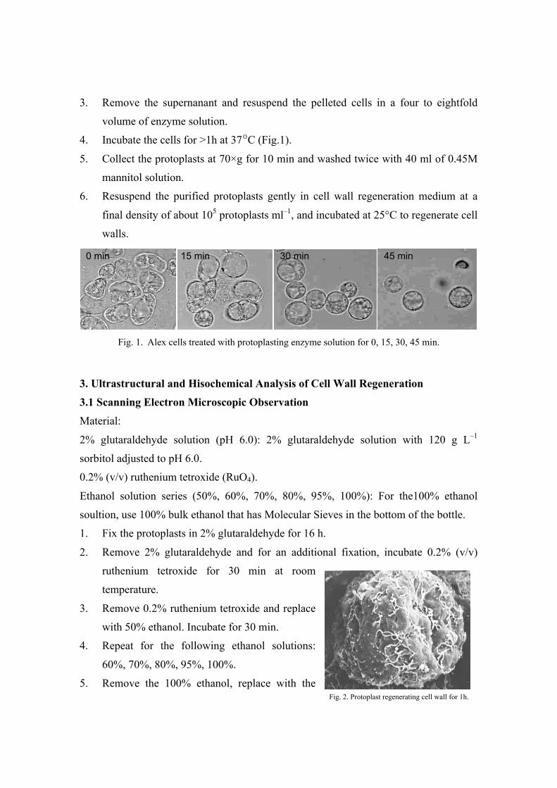
Fig. 2. Protoplast regenerating cell wall for 1h.
100 ethanol and incubate for 30 min.
6. Remove as much as possible for the 100% ethanol, replace with n-butyl alcohol.
7. Lyophilize the specimens with vacuum freeze-drying equipment (ES-2030, Hitachi
Science Systems Ltd., Tokyo, Japan).
8. Suptter-coating the specimens with silver (E-1030, Hitachi Science Systems Ltd.),
and Observe the suptter-coated specimens with an SEM (S-4100, Hitachi Science
Systems Ltd.) at 3 kV (Fig.2).
3.2 Staining with Calcofluor White
Calcofluor White can be used to detect cell wall matrix polysaccharide because it
readily binds to cellulose and other ß-linked glucans.
Material:
1% glutaraldehyde solution (pH 6.0): 1% glutaraldehyde solution with 120 g L–1
sorbitol adjusted to pH 6.0.
0.001% Calcofluor White: 0.001% Calcofluor White M2R (Sigma, St Louis, MO, USA)
with 120 g L–1 sorbitol adjusted to pH 6.0.
1. Fixed the protoplasts in 1% glutaraldehyde.
2. Remove 1% glutaraldehyde, rince three times with sorbitol solution, and incubate
with 0.001% Calcofluor White.
3. Remove Calcofluor White, rince three times with sorbitol solution
4. Observe the specimens under a fluorescence microscope equipped with a UV
fluorescence filter set (excitation filter, 350 nm; barrier filter, 430 nm) (Fig.3).
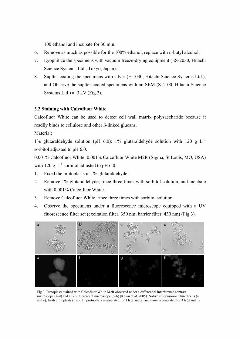
Fig.3. Protoplasts stained with Calcofluor White M2R observed under a differential interference contrast microscope (a–d) and an epifluorescent microscope (e–h) (Kown et al. 2005). Native suspension-cultured cells (a and e), fresh protoplasts (b and f), protoplasts regenerated for 1 h (c and g) and those regenerated for 3 h (d and h).
a b c d
e f g h
3.3 Staining with Aniline Blue
Aniline blue is used to stain the ß-1,3-glucan callose (but not ß-1,4-glucans). Callose is
known to be accumulated on the surface of protoplasts in the early stages of cell wall
regeneration.
Material:
1% glutaraldehyde solution (pH 6.0): 1% glutaraldehyde solution with 120 g L–1
sorbitol adjusted to pH 6.0.
0.005% Aniline Blue: 0.005% Aniline blue fluorochrome 100-1 (Biosupplies Australia),
120 g L–1 sorbitol in 0.15M K2HPO4 (pH8.6).
1. Fixed the protoplasts in 1% glutaraldehyde.
2. Remove 1% glutaraldehyde, rince three times with sorbitol solution, and incubate
with 0.005% Aniline Blue.
3. Observe the specimens under a fluorescence microscope equipped with a UV
fluorescence filter set (excitation filter, 395 nm; barrier filter, 495 nm).
References
1. Mathur, J., Szabados, L., Schaefer, S., Grunenberg, B., Lossow, A., Jonas-Straube,
E., Schell, J., Koncz, C. and Koncz-Kalman, Z. (1998) Gene identification with
sequenced T-DNA tags generated by transformation of Arabidopsis cell
suspension. Plant Journal 13: 707–716.
2. Kown, H-K., Yokoyama, R. and Nishitani, K. (2005) A Proteomic Approach to
Apoplastic Proteins Involved in Cell Wall Regeneration Protoplasts of Arabidopsis
Suspension Cultured Cells. Plant and Cell Physiology 46: 843-857.