Embed Size (px)
Citation preview

Protonation state of E71 in KcsA and its rolefor channel collapse and inactivationManasi P. Bhate and Ann E. McDermott1
Department of Chemistry, Columbia University, 3000 Broadway, New York, NY 10027
Contributed by Ann E. McDermott, July 17, 2012 (sent for review December 15, 2011)
The prototypical prokaryotic potassium channel KcsA alters its poredepending on the ambient potassium; at high potassium, it existsin a conductive form, and at low potassium, it collapses into a non-conductive structurewith reduced ion occupancy. We present solid-state NMR studies of KcsA in which we test the hypothesis that animportant channel-inactivation process, known as C-type inactiva-tion, proceeds via a state similar to this collapsed state. We test thisusing an inactivation-resistant mutant E71A, and show that E71A isunable to collapse its pore at both low potassium and low pH, sug-gesting that the collapsed state is structurally similar to the inacti-vated state. We also show that E71A has a disordered selectivityfilter. Using site-specific Kþ titrations, we detect a local change atE71 that is coupled to channel collapse at low Kþ. To gain more in-sight into this change, we site specifically measure the chemicalshift tensors of the side-chain carboxyls of E71 and its hydrogenbond partner D80, and use the tensors to assign protonation statesto E71 andD80 at high Kþ and neutral pH. Ourmeasurements showthat E71 is protonated at pH 7.5 and must have an unusually per-turbed pKa (>7.5) suggesting that the change at E71 is a structuralrearrangement rather than a protonation event. The results offernew mechanistic insights into why the widely used mutant KcsA–E71A does not inactivate and establish the ambient Kþ level as ameans to populate the inactivated state of KcsA in a controlledway.
membrane proteins ∣ ion channel ∣ chemical-shift anisotropy
Potassium channels constitute a highly conserved family ofintrinsic membrane proteins that are responsible for the pas-
sage of Kþ ions across membranes, and are involved in manyimportant physiological functions. The molecular mechanismsby which these channels are regulated, specifically the controlmechanisms by which they can be closed or inactivated, are ofsignificant interest.
One such control mechanism is C-type inactivation, which is aKþ- and voltage-dependent closure of the outer mouth of thechannel after channel opening. C-type inactivation was first de-scribed as a millisecond timescale process in Shaker channels (1),and a similar process occurs in a wide range of bacterial potas-sium channels, including KcsA and KirBac, and in the mamma-lian channel hERG, albeit at different rates. Understanding themolecular basis for C-type inactivation in mammalian potassiumchannels is of significant clinical interest because of its directrelation to cardiac malfunction and the development of a varietyof pharmaceuticals (2, 3). The inactivated state is a transientlypopulated kinetic intermediate in the Kþ transport cycle and sta-bilizing it on the timescale of structural studies is a challenge.Identifying the molecular structure of this transient intermediatecould yield mechanistic insight into the molecular details of in-activation. In this paper, we test the idea that the C-type inacti-vated state of the channel can be studied by lowering the ambientKþ concentration.
We conduct our studies on the prokaryotic Kþ channel, KcsA,which is native to the soil bacteria Streptomyces lividans and bearsstriking similarities to mammalian potassium channels in termsof its ion selectivity and gating properties. Because of its experi-mental convenience and homology to key regions of mammalian
channels, it has become a model system for mechanistic studiesof potassium channels. KcsA alters the structure of its poredepending on the ambient potassium concentration; at highpotassium, it exists in a conductive form, and at low potassium,it collapses into a nonconductive structure with lowered ion oc-cupancy (4). The channel is gated by pH, and after pH-dependentactivation, it inactivates in a Kþ- and voltage-dependent manneron the timescale of milliseconds to seconds (5).
The different conformations accessed as a function of Kþ andpH illustrate the importance of conformational plasticity forchannel function. NMR is a great tool to study conformationaldynamics. Solid-state NMR offers the unique opportunity tostudy conformational dynamics of membrane proteins in a nativebilayer environment and has recently been used to investigatemechanistic questions in systems like proton channels (6), mem-bers of the rhodopsin family (7), and potassium channels (8, 9).We began our studies of KcsA by reporting the structural andqualitative dynamic characterization of the two limiting states ofthe selectivity filter at high and low potassium, and measuringsite-specific potassium affinities in the pore (9). In this paper,we focus on the low Kþ-induced “collapsed” state of KcsA.
Several experiments have suggested that the C-type inactivatedstate of KcsA may resemble the low Kþ collapsed state. Our ownmeasurements of the slow exchange rate (kf þ kr < 500∕s) of thecollapsed state with respect to the conductive state was oneindication of a possible equivalence because C-type inactivationis also slow and Kþ-dependent (9). Additionally, recent crystal-lographic studies of various KcsAmutants show that when the pHgate of the KcsA is artificially opened, the structure and ionoccupancy of the filter resemble the low Kþ collapsed state(10). We use a mutation of KcsA, E71A, which is known to beinactivation-resistant and has been extensively characterized byelectrophysiology (11), together with our knowledge of the spec-troscopic signatures of the wild-type collapsed state at low Kþ toask the question: Can the inactivation-resistant mutant E71A col-lapse its filter at low Kþ? If it can, then it is unlikely that theinactivated state resembles the low Kþ state; but if it cannot, thenthis result, together with the slow exchange rate and the crystal-lographic results, makes a very good case for structural equiva-lence of the low Kþ state and the inactivated state.
E71A is an important mutant of KcsA. E71 is part of an intri-cate hydrogen bond network involving the side chains of E71,D80, and W67, a water molecule, and also backbone atoms ofG77 and Y78 (Fig. 1 and Table S1). Mutating residues in this net-work affects ion selectivity (12), gating (13), and tetramer stability(14). Because of suppressed inactivation, E71A is a “constitu-tively open” Kþ channel, widely used for kinetic studies (15, 16).Despite its wide use, there is no clear mechanism in the literatureexplaining why E71A does not inactivate. Previous work usingmutations and electrophysiology have suggested that the ionized
Author contributions: M.P.B. and A.E.M. designed research; M.P.B. performed research;M.P.B. and A.E.M. analyzed data; and M.P.B. and A.E.M. wrote the paper.
The authors declare no conflict of interest.1To whom correspondence should be addressed. E-mail: [email protected].
This article contains supporting information online at www.pnas.org/lookup/suppl/doi:10.1073/pnas.1211900109/-/DCSupplemental.
www.pnas.org/cgi/doi/10.1073/pnas.1211900109 PNAS ∣ September 18, 2012 ∣ vol. 109 ∣ no. 38 ∣ 15265–15270
BIOPH
YSICSAND
COMPU
TATIONALBIOLO
GY
CHEM
ISTR
Y
E71 and D80 side chains in close proximity destabilize the con-ductive state of the pore (5). Crystallography, however, showsthat the local structure and hydrogen bond environment are fairlysimilar between the conductive and collapsed/inactivated struc-tures. At the resolution of the available crystal structures, the pro-tonation states of E71 and D80 are tentative (Table S1). NMRstudies of a KcsA–Kv1.3 chimera suggest that a proton is loadedonto the side chain E71 as the Kþis lowered at neutral pH. (8).Electrostatic pKa calculations indicate that the side chain of E71has an unusually high pKa, and at pH 7.5, the side chain of E71must be protonated (17–19). Thus, there is an inconsistency in theliterature regarding the protonation state of E71. In this paper,we use site-specific chemical shift tensor measurements to make amuch less ambiguous measurement of the protonation state ofE71 and its hydrogen bond partner, D80, which is a key pieceof evidence to understand the mechanism of inactivation. We alsocharacterize the structural dynamics of the E71A mutant andshow how it is different from the wild-type channel.
Our studies of wild-type and E71A KcsA lead to unique in-sights into the molecular control of C-type inactivation in KcsA.The results also illustrate the use of solid-state NMR methods toanswer important mechanistic questions about membrane pro-teins while studying them in a native bilayer environment.
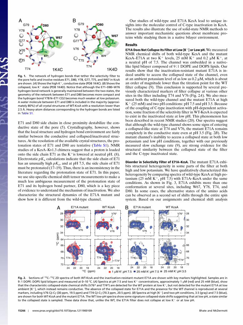
ResultsE71A Does Not Collapse Its Filter at Low [Kþ] or Low pH.WemeasuredNMR chemical shifts of both wild-type KcsA and the mutantKcsA–E71A at two Kþ levels, 25 mM Kþ and 0.2 μM Kþ, ata neutral pH of 7.5. The channel was embedded in a native-like lipid bilayer composed of 9∶1 DOPE and DOPS lipids. Ourresults show that the inactivation-resistant mutant E71A is in-deed unable to access the collapsed state of the channel, evenat an ambient potassium level of as low as 0.2 μM, which is aboutan order of magnitude lower than the titration point for the WTfilter collapse (9). This conclusion is supported by several pre-viously characterized markers of filter collapse at various othersites in the filter including T74 and V76 (Fig. 2A). We also mea-sured both the wild-type channel and the mutant E71A at highKþ (25 mM) and two pH conditions: pH 7.5 and pH 3.5. Becauseof the coupling of C-type inactivation with pH-dependent activa-tion, some fraction of the selectivity filter inWT-KcsA is expectedto exist in the inactivated state at low pH. This phenomenon hasbeen described in recent NMR studies (20). Our spectra suggestthat although the wild-type channel shows some signs of enteringa collapsed-like state at T74 and V76, the mutant E71A remainscompletely in the conductive state even at pH 3.5 (Fig. 2B). Themutant channel’s inability to access a collapsed state at both lowpotassium and low pH conditions, together with our previouslymeasured slow exchange rate (9), are strong evidence for thestructural similarity between the collapsed state of the filterand the C-type inactivated state.
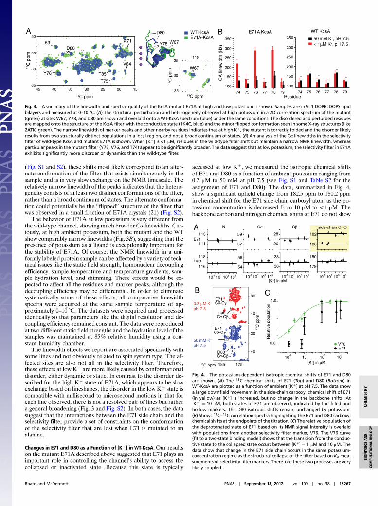
Disorder in Selectivity Filter of E71A–KcsA. The mutant E71A exhi-bits structural heterogeneity in some parts of the filter at bothhigh and low potassium. We have qualitatively characterized thisheterogeneity by comparing spectra of wild-type KcsA at high po-tassium (25 mM Kþ, pH 7.5) with E71A–KcsA under the sameconditions. As shown in Fig. 3, E71A exhibits more than oneconformation at several sites, including W67, Y78, T74, andD80. In some cases, the alternative states of the amino acidscan be observed as a second set of shifts through the entire spinsystem. Based on our assignments and chemical shift analysis
A BD80G79
G77
Y78
V76
T75
T74
E71
W67
D80G79
G77
Y78
V76
T75
T74
E71
W67
1K4C 1K4D
Fig. 1. The network of hydrogen bonds that tether the selectivity filter tothe pore helix and involve residues E71, D80, Y78, G77, T75, and W67 in KcsAare shown. (A) Shows the high Kþ, conductive state (PDB 1K4C). (B) Shows thecollapsed, low Kþ state (PDB 1K4D). Notice that although the E71–D80–W76hydrogen bond network is generally maintained between the two states, thegeometry of the network between E71 and D80 becomes more compact andthe hydrogen bond Y78 N–E71 CO2 becomes much weaker at low potassium.A water molecule between E71 and D80 is included in the majority (approxi-mately 80%) of all crystal structures of WT-KcsA with a resolution lower than2.5 Å. Heavy-atom distances corresponding to the hydrogen bonds are listedin Table S1.
E71A mutant WT KcsA
1 µM K+,pH 7.5 25 mM K+,pH 7.513C (ppm)
13C
(pp
m)
25 mM K+,pH 3.5
A B E71A mutant WT KcsA
34
32
30
28
24 22 20 1872
70
68
66
64
24 22 20 18 24 22 20 1872
70
68
66
24 22 20 18
34
32
30
28V76*
T74* T74*
V76 V76 V76V76
T74 T74 T74 T74
V76*
Fig. 2. Sections of 13C–13C 2D spectra of both WT-KcsA and the inactivation-resistant mutant E71A are shown with key markers highlighted. Samples are in9∶1 DOPE∶DOPS lipid bilayers and measured at 0–10 °C. (A) Spectra at pH 7.5 and two Kþ concentrations, approximately 1 μM (red) and 25 mM (blue), showthat the characteristic collapsed-state chemical shifts (V76* and T74*) are detected for the WT protein at low Kþ, but not detected for the mutant E71A at lowambient [Kþ], which instead remains conductive. The absence of the collapsed state for E71A and the presence for the WT channel is reproduced at severalmarkers, including V76 Cβ-Cγ (30 ppm, 19.5 ppm) and T74 Cβ-Cγ (70.3 ppm, 20.5 ppm). (B) Spectra at high [Kþ] and two pH conditions, 3.5 (gray) and 7.5 (blue),are shown for bothWT-KcsA and themutant E71A. TheWT low-pH spectra show some signature collapsed-state shifts suggesting that at low pH, a state similarto the collapsed state is sampled. These data show that, unlike the WT, the E71A filter does not collapse at low Kþ or at low pH.
15266 ∣ www.pnas.org/cgi/doi/10.1073/pnas.1211900109 Bhate and McDermott
(Fig. S1 and S2), these shifts most likely correspond to an alter-nate conformation of the filter that exists simultaneously in thesample and is in very slow exchange on the NMR timescale. Therelatively narrow linewidth of the peaks indicates that the hetero-geneity consists of at least two distinct conformations of the filter,rather than a broad continuum of states. The alternate conforma-tion could potentially be the “flipped” structure of the filter thatwas observed in a small fraction of E71A crystals (21) (Fig. S2).
The behavior of E71A at low potassium is very different fromthe wild-type channel, showing much broader Cα linewidths. Cur-iously, at high ambient potassium, both the mutant and the WTshow comparably narrow linewidths (Fig. 3B), suggesting that thepresence of potassium as a ligand is exceptionally important forthe stability of E71A. Of course, the NMR linewidth in a uni-formly labeled protein sample can be affected by a variety of tech-nical issues like the static field strength, homonuclear decouplingefficiency, sample temperature and temperature gradients, sam-ple hydration level, and shimming. These effects would be ex-pected to affect all the residues and marker peaks, although thedecoupling efficiency may be differential. In order to eliminatesystematically some of these effects, all comparative linewidthspectra were acquired at the same sample temperature of ap-proximately 0–10 °C. The datasets were acquired and processedidentically so that parameters like the digital resolution and de-coupling efficiency remained constant. The data were reproducedat two different static field strengths and the hydration level of thesamples was maintained at 85% relative humidity using a con-stant humidity chamber.
The linewidth effects we report are associated specifically withsome lines and not obviously related to spin system type. The af-fected sites are also not all in the selectivity filter. Therefore,these effects at low Kþ are more likely caused by conformationaldisorder, either dynamic or static. In contrast to the disorder de-scribed for the high Kþ state of E71A, which appears to be slowexchange based on lineshapes, the disorder in the low Kþ state iscompatible with millisecond to microsecond motions in that foreach line observed, there is not a resolved pair of lines but rathera general broadening (Fig. 3 and Fig. S2). In both cases, the datasuggest that the interactions between the E71 side chain and theselectivity filter provide a set of constraints on the conformationof the selectivity filter that are lost when E71 is mutated to analanine.
Changes in E71 and D80 as a Function of [Kþ] in WT-KcsA.Our resultson the mutant E71A described above suggested that E71 plays animportant role in controlling the channel’s ability to access thecollapsed or inactivated state. Because this state is typically
accessed at low Kþ, we measured the isotropic chemical shiftsof E71 and D80 as a function of ambient potassium ranging from0.2 μM to 50 mM at pH 7.5 (see Fig. S1 and Table S2 for theassignment of E71 and D80). The data, summarized in Fig. 4,show a significant upfield change from 182.5 ppm to 180.2 ppmin chemical shift for the E71 side-chain carboxyl atom as the po-tassium concentration is decreased from 10 μM to <1 μM. Thebackbone carbon and nitrogen chemical shifts of E71 do not show
WT KcsA E71A-KcsA
A
45 40 35 30 25 20 1565
60
55
50
D80
Y78
A71
T85
T75
L59
V84
13C ppm
13C
ppm
CA
line
wid
th (
Hz)
350
300
250
200
150
100
74 75 76 77 78 79Residue
50 mM K+, pH 7.5< 1µM K+, pH 7.5
WT KcsAB350
300
250
200
150
100
74 75 76 77 78 79
E71A KcsA
13C ppm
Y78 W67
D80
110
35
25
30
W67
13C
ppm
Fig. 3. A summary of the linewidth and spectral quality of the KcsA mutant E71A at high and low potassium is shown. Samples are in 9∶1 DOPE∶DOPS lipidbilayers and measured at 0–10 °C. (A) The structural perturbation and heterogeneity observed at high potassium in a 2D correlation spectrum of the mutant(green) at sites W67, Y78, and D80 are shown and overlaid onto a WT-KcsA spectrum (blue) under the same conditions. The disordered and perturbed residuesare mapped onto the structure of the KcsA filter with the conductive state (1K4C, blue) and the minor flipped conformation seen in some X-ray structures (like2ATK, green). The narrow linewidth of marker peaks and other nearby residues indicates that at high Kþ, the mutant is correctly folded and the disorder likelyresults from two structurally distinct populations in a local region, and not a broad continuum of states. (B) An analysis of the Cα linewidths in the selectivityfilter of wild-type KcsA and mutant E71A is shown. When [Kþ] is <1 μM, residues in the wild-type filter shift but maintain a narrow NMR linewidth, whereasparticular peaks in the mutant filter (Y78, V76, and T74) appear to be significantly broader. The data suggest that at low potassium, the selectivity filter in E71Aexhibits significantly more disorder or dynamics than the wild-type filter.
B1.0
0.0
Rel
ativ
e po
pula
tion
118
116
113
111E71
D8056
54
59
57
38
36
28
26
180
178
182
180
10-1 101 103 105
[K+] in µM10-1 101 103 105 10-1 101 103 105 10-1 101 103 105
[K+] in µM10
-110
110
310
5
A N Cα Cβ side-chain C=O
V76E71
185 175
13C
ppm
E71
D80
Cδ-Cγ
Cγ-Cβ
Cδ-Cγ
Cγ-Cβ
50 mM K+
pH 7.5
0.2 µM K+
pH 7.5
13C ppm
40
30
40
30 C
E71
D80
Fig. 4. The potassium-dependent isotropic chemical shifts of E71 and D80are shown. (A) The 13C chemical shifts of E71 (Top) and D80 (Bottom) inWT-KcsA are plotted as a function of ambient [Kþ] at pH 7.5. The data showa large downfield movement in the side-chain carboxyl chemical shift of E71(in yellow) as [Kþ] is increased, but no change in the backbone shifts. At½Kþ� ¼ 10 μM, both states of E71 are observed, indicated by the filled andhollow markers. The D80 isotropic shifts remain unchanged by potassium.(B) Shows 13C–13C correlation spectra highlighting the E71 and D80 carboxylchemical shifts at the endpoints of the titration. (C) The relative population ofthe deprotonated state of E71 based on its NMR signal intensity is overlaidwith populations from another selectivity filter marker, V76. The V76 curve(fit to a two-state binding model) shows that the transition from the conduc-tive state to the collapsed state occurs between ½Kþ� ¼ 1 μM and 10 μM. Thedata show that change in the E71 side chain occurs in the same potassium-concentration regime as the structural collapse of the filter based on Kd mea-surements of selectivity filter markers. Therefore these two processes are verylikely coupled.
Bhate and McDermott PNAS ∣ September 18, 2012 ∣ vol. 109 ∣ no. 38 ∣ 15267
BIOPH
YSICSAND
COMPU
TATIONALBIOLO
GY
CHEM
ISTR
Y
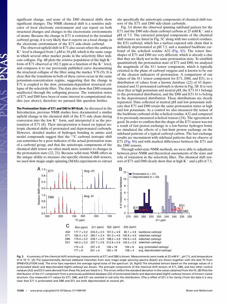
significant change, and none of the D80 chemical shifts showsignificant changes. The NMR chemical shift is a sensitive indi-cator of local electronic environment and can report on bothstructural changes and changes in the electrostatic environmentsof atoms. Because the change in E71 is restricted to the terminalcarboxyl group, it is very likely that it reports on a local change inthe electronic environment of the E71 side-chain carboxyl.
The observed upfield shift in E71 also occurs when the ambientKþ level is changed from 1 μM to 10 μM, which is the same rangein which several other marker peaks in the selectivity filter indi-cate collapse. Fig. 4B plots the relative population of the high Kþ
form of E71 observed at 182.5 ppm as a function of the Kþ level,and compares it to our previously published curve documentingthe structural collapse of the filter using the marker V76 (9). It isclear that the transitions in both of these curves occur in the samepotassium-concentration regime, suggesting that the change inE71 is coupled to the slow, potassium-dependent structural col-lapse of the selectivity filter. The data also show that D80 remainsunaffected through the collapsing process. The ionization statesof E71 and D80 have been of some interest in computational stu-dies (see above); therefore we pursued this question further.
The Protonation State of E71 and D80 in WT-KcsA.As discussed in theIntroduction, previous NMR studies have documented a similarupfield change in the chemical shift of the E71 side chain duringconversion into the low Kþ form, and interpreted it as the pro-tonation of E71 (8). Their interpretation is based on typical iso-tropic chemical shifts of protonated and deprotonated carboxyls.However, detailed studies of hydrogen bonding in amino acidmodel compounds suggest that the 13C carboxyl isotropic shiftcan sometimes be a poor indicator of the actual protonation stateof a carboxyl group, and that the anisotropic components of thechemical shift tensor are often much more sensitive to changes inthe protonation state (22, 23). Because solid-state NMR offers usthe unique ability to measure site-specific chemical shift tensors,we used slow magic-angle-spinning (MAS) experiments to extract
site specifically the anisotropic components of chemical shift ten-sors of the E71 and D80 side-chain carboxyls.
Fig. 5A shows the observed spinning sideband pattern for theE71 and the D80 side-chain carboxyl carbons at 25 mM Kþ and apH of 7.5. The extracted principal components of the chemicalshift tensors are listed in Fig. 5C along with two control residues:the E51 carboxyl, which has a surface-exposed side chain that isdefinitely deprotonated at pH 7.5, and a standard backbone car-bonyl of the α-helical residue A32 (Fig. S3). The tensor line-shapes of E71 and D80 are very different, which is already a cluethat they are likely not in the same protonation state. To establishquantitatively the protonation state of E71 and D80, we analyzedthe magnitude of the δ11 tensor component, which is typicallyoriented in the plane of carboxyl oxygens and is known to be oneof the clearest indicators of protonation. A comparison of ourvalues of the δ11 tensor component for E71, D80, and E51, to adistribution of values from a known database (22) of 43 depro-tonated and 31 protonated carboxyls is shown in Fig. 5B. It is veryclear that at high potassium and neutral pH, the E71 δ11 belongsto the protonated distribution, and the D80 and E51 δ11s belongto the deprotonated distribution. These distributions are clearlyseparated. Data collected at neutral pH and low potassium indi-cate that E71 and D80 retain the same protonation states at highand low potassium. As a control we also measured the tensor ofthe backbone carbonyl of the α-helical residue A32 and comparedit to previously measured α-helical tensors (24). The agreement isgood. In order to confirm that the shape of the E71 tensor was nota result of fast proton exchange in a low-barrier hydrogen bond,we simulated the effects of a fast-limit proton exchange on thesideband patterns of a typical carboxyl carbon. The fast exchangeresults are inconsistent with sideband patterns that we observe atE71 (Fig. S4) and with marked differences between the E71 andthe D80 tensors.
Through solid-state NMRmethods, we were able to adjudicatebetween prior NMR and theoretical assessments of the state androle of ionization in the selectivity filter. The chemical shift ten-sors of E71 and D80 clearly show that at high Kþ and a pH of 7.5,
1.2
0.8
0.4
0.0
Rel
ativ
e N
MR
inte
nsity
E71A12
10
8
6
4
2
0
cnatsni fo #esabatad ni s e
270 265 260 255 250 245 240 235 230 δ11 ppm
Protonated (n = 31) Deprotonated (n = 43)
E71D80
E51
C
D80
A32 177.7 ± 0.2 245.5 ± 2.4 197.5 ± 4.8 90.1 ± 4.8 E71 182.4 ± 0.2 260.1 ± 2.4 181.3 ± 4.6 105.9 ± 4.6 D80 179.4 ± 0.2 239.1 ± 2.5 198.2 ± 4.6 100.9 ± 4.6 E51 184.2 ± 0.2 237.7 ± 2.6 212.8 ± 5.9 102.0 ± 5.9
δiso δ11 δ22 δ33
backbone carbonylsidechain carboxylsidechain carboxylsidechain carboxyl
B
175 ± 6 257 ± 6 158 ± 18 109 ± 8 avg. protonated carboxyl177 ± 6 241 ± 4 183 ± 18 108 ± 4 avg. deprotonated carboxyl
(ppm) (ppm) (ppm) (ppm)
300 200 10013C ppm
1.2
0.8
0.4
0.0
Avg -H
300 200 100
Avg +H
Fig. 5. A summary of the chemical-shift anisotropy measurements at E71 and D80 is shown. Measurements were made at 25 mM Kþ, pH 7.5, and temperatureof 0–10 °C. (A) The experimentally derived sideband intensities from slow magic-angle spinning spectra (black) are shown together with the best fit fromSPINEVOLUTION (red). The error bars represent the average signal:noise of the spectrum. (A, Right) The simulated tensors based on the average values of aprotonated (dark) and deprotonated (light) carboxyl are shown. The principle components of the chemical shift tensors of E71, D80, and two other controlresidues (A32 and E51) were derived from these fits and are listed in C. The errors reflect the standard deviation in the values obtained from the fit. (B) Plots thedistribution of the δ11 component from a previously published database (22) of protonated (dark) and deprotonated (light) carboxyl tensors of known crystalstructure. Our measured δ11 values for E71, D80, and E51 are overlaid in red onto this distribution. (The y-offset of E51 is for clarity.) From the data it is veryclear that E71 is protonated and D80 and E51 are both deprotonated at neutral pH.
15268 ∣ www.pnas.org/cgi/doi/10.1073/pnas.1211900109 Bhate and McDermott
E71 is protonated and D80 is deprotonated. Thus, E71 must havean extremely elevated pKa (>7.5, instead of the expected valueof approximately 4.5), which is consistent with predictions fromvarious electrostatic and molecular dynamics simulations.
DiscussionOur data on the inactivation-resistant mutant E71A show that thecollapsed state accessed at low Kþ is indeed structurally similar tothe C-type inactivated state of the channel. The C-type inacti-vated state is typically populated transiently for a few millise-conds to seconds after the pH gate is opened and is thereforedifficult to study by equilibrium methods. We show that loweringthe ambient Kþ level is an experimentally feasible way of acces-sing the inactivated state. Controlling the Kþ level can be used tomodulate the relative populations of the inactivated state withrespect to the conductive state, which facilitates future biophysi-cal studies of this clinically important process. To gain more me-chanistic insight, we determined the protonation state of E71 andits hydrogen bond partner, D80, and showed that E71 is proto-nated at pH 7.5 and therefore has an unexpectedly high pKa,whereas D80 is deprotonated at pH 7.5. The results are signifi-cant for both scientific and technical reasons. Since description ofthe first crystal structure of KcsA (25), there has been a lot ofinterest in the protonation state of E71 because of its proximityto the selectivity filter. A perturbed pKa for E71 had beenpredicted by multiple electrostatic simulations; specifically,Ranatunga et al. predicted a pKa of >14.5 for E71 (17) andBernache et al. also predicted a perturbation of approximatelyþ10 pKa units for E71 (18). Our own simulations using theX-ray coordinates 1K4C and 1K4D and two different publiclyavailable simulation programs—Hþþ (26) and MCCE (27)—over a range of different protein dielectric constants also quali-tatively support an elevated pKa for E71 (Fig. S5 and Table S3).The data in this paper are previously undescribed unambiguousexperimental measurements that validate these predictions. Weshow that E71 is protonated at neutral pH and high Kþ.
The 2.3-ppm Kþ-dependent upfield change in the carboxylchemical shift of E71, which occurs simultaneously with the struc-tural collapse of the channel and had previously been interpretedas a protonation event (8), is therefore a result of a local struc-tural change in the side chain of E71 upon Kþ depletion and notprotonation. Crystallography (Fig. 1 and Table S1) suggests thatthe E71–D80 interaction is generally conserved between the con-ductive and collapsed states: The carboxyl groups of E71 and D80are engaged in a syn–syn interaction with each other and with aconserved water molecule in both states. However, the E71 car-boxyl looses a key hydrogen bond with the Y78 backbone amideas the channel collapses. We also observe a change in the isotro-pic shift of the Y78 amide 15N at low Kþ, we suspect that the2.3-ppm shift in E71 is reporting on the weakening/loss of a hy-drogen bond to Y78 and on a minor structural rearrangement tocompensate for the loss.
Our results help us understand why the mutant E71A fails toinactivate. Functional studies have shown that replacing E71 witha histidine accelerates C-type inactivation, but most other substi-tutions (including serine, valine, isoleucine, threonine, cysteine,and glutamine) reduce inactivation in KcsA (28). Thus, the sup-pression of C-type inactivation in E71A is unlikely to be an effectof smaller side-chain volume or a specific chemical effect of thealanine. Instead, our data show a significantly more disorderedfilter in the E71A mutant. Molecular dynamics simulations havealso suggested that mutations at E71 can lead to increased dy-namics in the filter (28). Crystallography on the E71A mutanthas indicated disorder in the form of an alternative flipped con-formation of the mutant filter (12). It is intriguing to considerwhether the conductive state of the E71A mutant is preferentiallystabilized by this disorder—i.e., the population of additionalstates entropically stabilizes the conductive state in E71A, and
shifts the Kþ-dependent equilibrium between the conductiveand inactivated/collapsed states into a regime that is beyondour detection (Kd ≪ 1 μM). Such a hypothesis would also ex-plain how a variety of different mutations at E71 could all leadto suppression of C-type inactivation (to various extents), as longas they also lead to increased disorder.
Sequence alignment shows that a glutamic acid in position 71 isnot conserved across all potassium channels. In other channels, itis often substituted by large nonpolar amino acids, like valine andisoleucine, and smaller polar amino acids, like serine (Fig. S6).Mutations at the position equivalent to E71 affect C-type inacti-vation in both hERG and Kv channels (29). We suspect that themanipulation of the contacts and hydrogen bonds at this positionusing different amino acids is a mechanism to tune the relativestability of the conductive state of the channel, and thereby allowfor diversity in the kinetics of C-type inactivation across variouschannels.
From a technical standpoint, our results demonstrate the useof chemical-shift anisotropy to measure functionally importantprotonation states of side chains in a native membrane environ-ment—a measurement that is unique to solid-state NMR. Themagnitude of the chemical-shift anisotropy tensor is typically de-scribed by three parameters—δ11, δ22, and δ33—which measurethe extent of the shielding in three orthogonal directions aroundthe carboxyl carbon of interest, and are averaged to give the iso-tropic shift. The isotropic shift is the most commonly measuredNMR observable, but in the case of the E71 side-chain carboxyl,an analysis based purely on its isotropic chemical shift leads to thewrong protonation state for this residue (Fig. S7). The unusuallyhigh isotropic chemical shift of the protonated E71 (approxi-mately 182.5 ppm versus an average of approximately 179 ppm)is caused by an unusually high value for the δ22 component,which suggests a strong hydrogen bond acceptor positioned nearthe side-chain carbonyl, and is consistent with the positioning ofthe conserved crystallographic water near the E71 carbonyl. Thestrong hydrogen bond indicates that the protons of this water mo-lecule are oriented towards the carbonyl of E71. Our data andsimulations preclude the existence of a low-barrier hydrogenbond between E71 and D80, and suggest that the proton primar-ily resides near the carboxyl of E71 (Fig. S4). It has long beenknown from model compounds that the δ11 and δ22 elementsof the chemical shift tensor are more reliable indicators of pro-tonation than the isotropic shift. Here, we illustrate how such an-isotropic measurements can make a big difference in the contextof mechanistic studies of an important membrane protein.
ConclusionIn this paper, we use a two-pronged approach to investigate therole of a key residue, E71, for KcsA function. First, by using themutant E71A we showed that the low Kþ-induced collapsed stateof KcsA is structurally similar to the C-type inactivated state ofthe channel and that removing the E71 side chain leads to a dis-ordered selectivity filter. Second, we use anisotropic chemicalshifts to make a uniquely direct, conclusive measurement of theside-chain protonation states of E71 and D80 in KcsA and showthat the pKa of E71 is indeed highly perturbed (>7.5) comparedto solution, as was predicted by electrostatic calculations. Theseresults together provide mechanistic insights into the control ofchannel inactivation, which is an important physiological process.
Materials and MethodsNMR Sample Preparation. The WT-KcsA and KcsA–E71A gene cloned into aPASK90 vector were overexpressed in Escherichia coli JM83 cells as describedpreviously (9). The WT-KcsA tetramer is detected as a clean band at approxi-mately 70 kDa in SDS gels at 25 °C. E71A–KcsA is denatured by SDS and runs asa band at approximately 18 kDa. The mutant E71A is unstable when stored indetergent at 4 °C and was therefore stored in liposomes at −80 °C and used asquickly as possible. The protein was reconstituted into 9∶1wt∕wt DOPE/DOPSliposomes as described previously (9). The ionic strength of the dialysis buffer
Bhate and McDermott PNAS ∣ September 18, 2012 ∣ vol. 109 ∣ no. 38 ∣ 15269
BIOPH
YSICSAND
COMPU
TATIONALBIOLO
GY
CHEM
ISTR
Y

was kept constant by replacing Kþ with Naþ. Low Kþ concentrations weredetermined by atomic absorption spectroscopy. The hydration level of sam-ples was maintained at 85% using a home-built hydration chamber. Sampleswere analyzed by cryo-EM to check for the bilayer structure and packed inBruker 4-mm or 3.2-mm rotors.
NMR Spectroscopy. NMR spectra were collected on two spectrometers: aBruker Avance DRX-750MHz spectrometer (with a 4-mm HXYprobe spinningat 14 kHz and a 3.2-mmHXYprobe spinning at 14 KHz) and a Bruker Avance 2900 MHz spectrometer (with a 3.2-mm standard-bore E-free probe). Typical90° pulse lengths for 1H were approximately 2.5 μs on the standard HXYprobes and approximately 3 μs on the E-free probe. Spectra were collectedat a spectrometer-set temperature of 240 K. At 14-KHz spinning, our calibra-tion show that the actual sample temperature was 0–10 °C during signalacquisition. Chemical shift tensors were measured at 5.555-KHz magic-anglespinning and fit using SPINEVOLUTION (SI Text). All 13C chemical shifts were
referenced externally to the adamantane line at 40.48 ppm. Using the recom-mended gyromagnetic ratio from the Biological Magnetic Resonance DataBank, 15N shifts were referenced internally to the carbon shifts.
ACKNOWLEDGMENTS. The authors thank the McDermott group, especiallyWenbo Li, for help with troubleshooting SPINEVOLUTION; Ivan Sergeyevfor help with installing MCCE; and Benjamin Wylie and Segolene Laagefor helpful discussions. We also thank the laboratory of Crina Nimigean forthe E71A plasmid, Marilyn Gunner for helpful advice about electrostaticsimulations, and Dr. Boris Itin at the New York Structural Biology Centerfor support with high-field instrumentation. Professor McDermott is amember of the New York Structural Biology Center, a Strategically TargetedAcademic Research (STAR) center supported by the New York State Office ofScience, Technology, and Academic Research. This work was supported bygrants from the National Institutes of Health: National Institutes of HealthGrant P41 GM66354 supported NMR resources, and this work was supportedby National Institutes of Health Grant R01 GM 88724.
1. Starkus JG, Kuschel L, Rayner MD, Heinemann SH (1997) Ion conduction throughC-type inactivated Shaker channels. J Gen Physiol 110:539–550.
2. Sanguinetti MC, Tristani-Firouzi M (2006) hERG potassium channels and cardiacarrhythmia. Nature 440:463–469.
3. Bowlby M, Peri R, Zhang H, Dunlop J (2008) hERG (KCNH2 or Kv11.1) Kþ channels:Screening for cardiac arrhythmia risk. Curr Drug Metab 9:965–970.
4. Zhou Y, Morais-Cabral JH, Kaufman A, Mackinnon R (2001) Chemistry of ion coordina-tion and hydration revealed by a K channel Fab complex at 2.0 A resolution. Nature414:43–48.
5. Cordero-Morales JF, Cuello LG, Perozo E (2006) Voltage-dependent gating at the KcsAselectivity filter. Nat Struct Mol Biol 13:319–322.
6. Cady SD, et al. (2010) Structure of the amantadine binding site of influenzaM2 protonchannels in lipid bilayers. Nature 463:689–692.
7. Struts AV, Salgado GFJ, Brown MF (2011) Solid-state 2H NMR relaxation illuminatesfunctional dynamics of retinal cofactor in membrane activation of rhodopsin. ProcNatl Acad Sci USA 108:8263–8268.
8. Ader C, et al. (2009) Coupling of activation and inactivation gate in a Kþ channel:Potassium and ligand sensitivity. EMBO J 28:2825–2834.
9. Bhate MP, Wylie BJ, Tian L, McDermott AE (2010) Conformational dynamics in theselectivity filter of KcsA monitored by solid-state NMR. J Mol Biol 401:155–166.
10. Cuello LG, Jogini V, Cortes DM, Perozo E (2010) Structural mechanism of C-type inac-tivation in Kþ channels. Nature 466:203–208.
11. Cordero-Morales JF, et al. (2007) Molecular driving forces determining potassiumchannel slow inactivation. Nat Struct Mol Biol 14:1062–1069.
12. Cheng WWL, McCoy JG, Thompson AN, Nichols CG, Nimigean CM (2011) Mechanismfor selectivity-inactivation coupling in KcsA potassium channels. Proc Natl Acad SciUSA 108:5272–5277.
13. Cordero-Morales JF, Jogini V, Chakrapani S, Perozo E (2011) A multipoint hydrogen-bond network underlying KcsA C-type inactivation. Biophys J 100:2387–2393.
14. Choi H, Heginbotham L (2004) Functional influence of the pore helix glutamate in theKcsA Kþ channel. Biophys J 86:2137–2144.
15. Thompson AN, Posson DJ, Parsa PV, Nimigean CM (2008) Molecular mechanism of pHsensing in KcsA potassium channels. Proc Natl Acad Sci USA 105:6900–6905.
16. Piasta KN, Theobald DL, Miller C (2011) Potassium-selective block of barium permea-tion through single KcsA channels. J Gen Physiol 138:421–436.
17. Ranatunga KM, Shrivastava IH, Smith GR, Sansom MS (2001) Side-chain ionizationstates in a potassium channel. Biophys J 80:1210–1219.
18. Bernèche S, Roux B (2002) The ionization state and the conformation of Glu-71 in theKcsA Kþ channel. Biophys J 82:772–80.
19. Bucher D, Guidoni L, Rothlisberger U (2007) The protonation state of the Glu-71/Asp-80 residues in the KcsA potassium channel: A first-principles QM/MM moleculardynamics study. Biophys J 93:2315–2324.
20. Imai S, OsawaM, Takeuchi K, Shimada I Structural basis underlying the dual gate prop-erties of KcsA. Proc Natl Acad Sci USA 107:6216–6221.
21. Cordero-Morales JF, et al. (2006) Molecular determinants of gating at the potassium-channel selectivity filter. Nat Struct Mol Biol 13:311–318.
22. Gu Z, Zambrano R, McDermott A (1994) Hydrogen bonding of carboxyl groups in solid-state amino acids and peptides: Comparison of carbon chemical shielding, infraredfrequencies, and structures. J Am Chem Soc 116:6368–6372.
23. Gu Z, McDermott A (1993) Chemical shielding anisotropy of protonated and depro-tonated carboxylates in amino acids. J Am Chem Soc 115:4282–4285.
24. Wylie BJ, et al. (2007) Chemical-shift anisotropy measurements of amide and carbonylresonances in a microcrystalline protein with slow magic-angle spinning NMR spectro-scopy. J Am Chem Soc 129:5318–5319.
25. Doyle D, Cabral J, Pfuetzner R, Kuo A, Gulbis J (1998) The structure of the potassiumchannel: Molecular basis of Kþ conduction and selectivity. Science 280:69–77.
26. Gordon J, et al. (2005) Hþþ: A server for estimating pKas and adding missing hydro-gens to macromolecules. Nucleic Acids Res 33:W368–W371.
27. Georgescu RE, Alexov E, Gunner MR (2002) Combining conformational flexibility andcontinuum electrostatics for calculating pKas in proteins. Biophys J 83:1731–1748.
28. Chakrapani S, et al. (2010) On the structural basis of modal gating behavior inKþ channels. Nat Struct Mol Biol 18:67–74.
29. Ferrer T, et al. (2011) Molecular coupling in the human ether-a-go–go-related gene-1(hERG1) Kþ channel inactivation pathway. J Biol Chem 286:39091–39099.
15270 ∣ www.pnas.org/cgi/doi/10.1073/pnas.1211900109 Bhate and McDermott


![Inactivation of the KcsA potassium channel explored with ... · PDF filegating of KcsA at low pH is affected by the applied po-tential (Cuello et al., ... toyl-2-oleoyl-sn-glycero-3-[phospho-rac-(1-glycerol)];](https://img.pdfslide.us/doc/110x75/5a83b0f17f8b9a682c8ef255/inactivation-of-the-kcsa-potassium-channel-explored-with-of-kcsa-at-low-ph-is.jpg)
![Protonation and Muoniation Regiochemistry of …Protonation and Muoniation Regiochemistry of [FeFe]-Hydrogenase Subsite Analogues Jamie N.T. Peck , Joseph A. Wright, Stephen Cottrell,](https://img.pdfslide.us/doc/110x75/5e32c9cbd76e9f08de66e1cf/protonation-and-muoniation-regiochemistry-of-protonation-and-muoniation-regiochemistry.jpg)


![Protonation and solvent effects on a resorcin[4]arene](https://img.pdfslide.us/doc/110x75/625e5da6d862740eeb16be8d/protonation-and-solvent-effects-on-a-resorcin4arene-.jpg)