Embed Size (px)
Citation preview

Proton Exchange and Base Pair Opening in a DNA Triple Helix†
Stephen W. Powell,‡ Lihong Jiang,‡ and Irina M. Russu*,§
Department of Molecular Biology and Biochemistry and Department of Chemistry, Molecular Biophysics Program,Wesleyan UniVersity, Middletown, Connecticut 06459
ReceiVed May 1, 2001; ReVised Manuscript ReceiVed June 20, 2001
ABSTRACT: Nuclear magnetic resonance spectroscopy has been used to characterize opening reactionsand stabilities of individual base pairs in two related DNA structures. The first is the triplex structureformed by the DNA 31-mer 5′-AGAGAGAACCCCTTCTCTCTTTTTCTCTCTT-3′. The structure belongsto the YRY (or parallel) family of triple helices. The second structure is the hairpin double helix formedby the DNA 20-mer 5′-AGAGAGAACCCCTTCTCTCT-3′ and corresponds to the duplex part of theYRY triplex. The rates of exchange of imino protons with solvent in the two structures have been measuredby magnetization transfer from water and by real-time exchange at 10°C in 100 mM NaCl and 5 mMMgCl2 at pH 5.5 and in the presence of two exchange catalysts. The results indicate that the exchange ofimino protons in protonated cytosines is most likely limited by the opening of Hoogsteen C+G base pairs.The base pair opening parameters estimated from imino proton exchange rates suggest that the stabilityof individual Hoogsteen base pairs in the DNA triplex is comparable to that of Watson-Crick base pairsin double-helical DNA. In the triplex structure, the exchange rates of imino protons in Watson-Crickbase pairs are up to 5000-fold lower than those in double-helical DNA. This result suggests that formationof the triplex structure enhances the stability of Watson-Crick base pairs by up to 5 kcal/mol. Thisstabilization depends on the specific location of each triad in the triplex structure.
Triple-helical nucleic acid structures are formed by bindingof a third strand in the major groove of a double-helicalnucleic acid. Formation of these structures generally requiresa tract of purines in one strand of the double helix and atract of pyrimidines in the other strand. The base compositionof the third strand can be purine or pyrimidine rich. In YRY1
triple helices, the third strand is pyrimidine rich and bindsparallel to the homopurine strand of the double helix. Incontrast, in RRY triple helices, the purine-rich third strandis oriented antiparallel to the homopurine strand of the doublehelix (1, 2).
During the past decades there has been a resurgence ofinterest in triple-helical structures of nucleic acids due totheir potential biological roles and applications. A biologicalrole for these structures is suggested by the fact that longtracts of contiguous oligopurines occur in eukaryotic ge-nomes at frequencies 3-6-fold higher than that predictedon a statistical basis (3). Many of these sequences are mirrorrepeats and can form triple-helical H-DNA structures innegatively supercoiled plasmids (4-6). Base sequencescapable of forming H-DNA structures have been implicatedin a variety of biological processes such as transcriptioncontrol (2), lytic replication of the Epstein-Barr virusgenome (7), and homologous recombination (8, 9).
Applications of triple-helical structures in biotechnologyand molecular medicine rely upon the ability of triplex-forming oligonucleotides to target base sequences in double-helical DNA with a degree of specificity comparable to, oreven greater than, that of regulatory DNA-binding proteins.In the antigene strategy, the targeted sites are located withincoding regions or upstream of the genes of interest. Forma-tion of triple-helical structures at these sites inhibits tran-scription in vitro (10-12) and in vivo (13, 14). Triplex-forming oligonucleotides can also be used as sequence-specific endonucleases by covalently linking them to DNA-cleaving agents such as Fe(II)-EDTA or copper(II) phenan-throline (15, 16). Such artificial endonucleases can accessrare sites on the genome with high specificity, thus havinggreat value for physical mapping of chromosomes (17).
The understanding of the possible biological roles ofnucleic acid triple helices, as well as the design and use oftriplex-forming oligonucleotides, requires understanding howthe structure and stability of triple helices depend on theirbase sequences and on solution conditions. High-resolutionstructures for several YRY and RRY DNA triple helices havebeen obtained by nuclear magnetic resonance (NMR)spectroscopy (18, 19). The structures have revealed the basepairing schemes for canonical and noncanonical base triads,the orientation and the conformation of the third strand, andthe conformational changes induced in the duplex by bindingof the third strand in its major groove. The stability oftriple helical structures has been extensively characterizedby UV absorbance spectroscopy, calorimetric techniques, gelelectrophoresis, and quantitative affinity cleavage titration(20-24). These studies have shown that the overall stabilityof DNA triple helices is strongly influenced by the base
† Supported by a grant from the National Science Foundation.* To whom correspondence should be addressed. Phone: (860)-685-
2428. Fax: (860)-685-2211. E-mail: [email protected].‡ Department of Molecular Biology and Biochemistry.§ Department of Chemistry.1 Abbreviations: NMR, nuclear magnetic resonance; ppm, parts per
million; Tris, tris(hydroxymethyl)aminomethane; A, adenine; G, gua-nine; C, cytosine; T, thymine; Y, either pyrimidine nucleotide; R, eitherpurine nucleotide.
11065Biochemistry2001,40, 11065-11072
10.1021/bi010890a CCC: $20.00 © 2001 American Chemical SocietyPublished on Web 08/24/2001

sequence, length of the strands, and presence of tripletmismatches and looped-out bases, as well as solution con-ditions such as temperature, pH, and salt concentration.
The present work extends these previous investigationsby providing a characterization of the stability of a DNAtriple helix at the level of individual base triads. The DNAtriple helix investigated belongs to the YRY family (Figure1B) and is the first intramolecular DNA triplex for which amodel structure was derived from NMR data (25). Moreover,this triple helix has served as a reference for subsequentstructural studies of triple helixes containing noncanonicaltriads (26) and changes in the intramolecular loops (27). Tocompare triple- and double-helical structures directly, wehave also characterized the DNA hairpin corresponding tothe duplex part of the triplex structure (Figure 1C).
MATERIALS AND METHODS
DNA Samples.The two DNA oligonucleotides weresynthesized on an Applied Biosystems 381A automated DNAsynthesizer using the solid-support phosphoramidite method.They were purified by reverse-phase HPLC on a PRP-1column (Hamilton) in 50 mM triethylamine acetate bufferat pH 7 with a gradient of 10-20% acetonitrile in 32 min.The counterions were replaced with Na+ ions by repeatedcentrifugation through Centricon YM-3 tubes (Amicon Inc.).Unless otherwise indicated, the final samples were in 100mM NaCl and 5 mM MgCl2 at pH 5.5 (measured at 10°C).The sample of the triplex contained 230 OD260 units of DNAand that of hairpin 140 OD260 units.
NMR Experiments.The NMR experiments were performedat 10°C on a Varian INOVA 500 spectrometer operating at11.75 T. One-dimensional (1D) NMR spectra were obtainedusing the jump-and-return pulse sequence (28). Protonexchange rates were measured in experiments of transfer ofmagnetization from water and in real-time exchange experi-ments.
In transfer of magnetization experiments, the water protonresonance was selectively inverted using a Gaussian 180°pulse (6.4 ms). The intensity of imino proton resonances was
measured as a function of the exchange delay,τ, followingwater inversion. A weak gradient (0.21 G/cm) was appliedduring the delayτ to prevent the effects of radiation dampingupon the recovery of water magnetization to equilibrium.The observation was with the jump-and-return pulse se-quence. The dependence of the intensity of an exchangeableproton resonance on the delayτ is described by (29)
with
whereI0 is the intensity at equilibrium,R1 is the longitudinalrelaxation rate, andkex is the exchange rate for the proton ofinterest;Rw is the longitudinal relaxation rate of water, andq ) Iw(0)/I0
w expresses the efficiency of water inversion (e.g.,q ) -1 for perfect inversion). In each experiment, 27-30values of the exchange delay were used. To avoid the effectsof spin diffusion, the exchange delays ranged from 2 to 600ms. The intensity of the exchangeable proton resonance ofinterest was fitted as a function of the exchange delayτ toeq 1, using a nonlinear least-squares program. The errorsreported in the paper are those obtained from the fit.Rw andq were measured in separated experiments. The transfer ofmagnetization experiments allowed measurement of ex-change rates faster than∼0.2 s-1. For slower exchange rates,the time dependence of the magnetization (eq 1) is dominatedby the longitudinal relaxation of the imino proton and ofwater protons, and the exchange rates cannot be measuredaccurately.
In real-time exchange experiments, the exchange wasinitiated by adding D2O to a concentrated DNA solution inH2O, to a final volume fraction of D2O of ∼80%. A total of64 transients were accumulated for each spectrum with atotal acquisition time of 4 min per spectrum. The intensityof each resonance was measured using standard deconvo-
FIGURE 1: (A) Structures of C+‚GC and T‚AT triads (5). (B) Base sequence and folded conformation of the DNA 31-mer triplex investigated(25). (C) Base sequence and folded conformation of the DNA 20-mer hairpin investigated. In panels B and C, Watson-Crick hydrogenbonding is indicated by vertical bars, and Hoogsteen hydrogen bonding is indicated by asterisks. The bases shown in bold contain hydrogen-bonded imino groups.
I(τ) ) I0 + [I(0) - I0 - A]e-(R1 + kex)τ + Ae-Rwτ (1)
A ) (q - 1)kex
R1 + kex - RwI0
11066 Biochemistry, Vol. 40, No. 37, 2001 Powell et al.

lution software (Varian) and fitted as a function of theexchange timeτ to the equation
whereI(∞) is the intensity in a fully exchanged sample. Dueto the time elapsed between the initiation of exchange andthe first NMR spectrum (7-12 min), the fastest exchangerate that can be measured in real-time exchange experimentsis ∼10-3 s-1.
The NOESY experiments used to assign the imino protonresonances of the DNA hairpin were performed with amixing time of 150 ms and a regular NOESY pulse sequencein which each pulse was replaced by a jump-and return pulse(30).
Theory of Imino Proton Exchange.In nucleic acids, theexchange of imino protons with solvent protons occurs bytransient opening of the base pairs. In the open state, theimino proton is accessible to proton acceptors, and itshydrogen bonds are broken. The open state is short-livedand thermodynamically unfavorable. Accordingly, the rateof exchange of a given proton is (31)
wherekop is the rate of opening of the base pair,kcl is therate of closing, andkex,open is the rate of proton exchangefrom the open state. The equilibrium constantKop ) kop/kcl
defines the free energy change for the opening reaction (32):
The exchange of the imino proton from the open stateoccurs by two mechanisms. In one, the exchange is catalyzedby external proton acceptors present in solution, e.g., OH-,Tris base, and ammonia. Accordingly, the rate of exchangeby external catalysis is proportional to the acceptor concen-tration (31, 32):
The rate constantkA depends on the pK values of the iminogroup and of the acceptor:
wherekcoll is the rate of collision between the imino groupand the acceptor andF is the fraction of productive transfersof the proton in the transient hydrogen-bonded complexbetween the imino group NH and the acceptor:
with ∆pK ) pK(acceptor)- pK(NH). The rate constantkA
is normally approximated askA ) RkA0, wherekA
0 is therate constant for proton exchange in an isolated nucleoside.R is an empirical coefficient that accounts for any differencesbetween external catalysis in the open state of the base pairand that in free nucleosides. Experimental evidence forvarious catalysts indicates that, in double-helical DNA,Re 1 (33, 34).
The second mechanism for exchange of imino protonsinvolves proton acceptors in the same DNA molecule. The
current model for this intrinsic catalysis (35) assumes that,in the open state, the two bases remain close to each otherand form an outer-sphere complex with a water molecule.The proton acceptors are the nitrogens in the other base ofthe open base pair; for example, for an open Watson-CrickAT base pair, the acceptor of the N3H proton of thymine isthe N1 group in adenine. The rate of exchange resulting fromthis intrinsic catalysis,kex,open
int , is also directly proportionalto the fraction of productive transfers,F, from the NH groupto the acceptors [eq 7 (35)]. Experimental evidence providedby Gueron and co-workers indicates that the same open stateof the base pair is involved in both external and intrinsiccatalysis (35). Accordingly, the total rate of exchange fromthe open state is
The base pair opening parameters that can be obtainedfrom measurements of exchange rates depend on how therate of proton exchange from the open state compares withthe rate of base pair closing. According to eq 3, onedistinguishes two limiting cases:
(i) When kex,open. kcl (EX1 regime)
i.e., exchange is limited by the rate of base pair opening. Inthis case, the exchange rate provides directly the rate ofopening.
(ii) When kex,open, kcl (EX2 regime)
i.e., exchange is slowed relative to that in the open state bya factor equal to the equilibrium constant for opening,Kop.
RESULTS
The two DNA molecules investigated are shown in Figure1. The DNA 31-mer has been first studied by Feigon andco-workers (25). These authors have shown that, in acidicsolutions, the 31-mer folds into an intramolecular YRYtriplex: the pyrimidine strand segment Y (bases 24-31) islocated in the major groove of the hairpin duplex in anorientation parallel to the R strand (bases 1-8). The triplexcontains seven canonical C+‚GC and T‚AT triads (Figure1). The NMR resonance of the imino proton in thymine T24
was not observed in the spectrum. This indicated that T24
does not form Hoogsteen hydrogen bonds with the A1T20
base pair and, thus, is not part of a triad (25). The basesequence of the DNA 20-mer is the same as that in the duplexpart of the triplex. As shown below, in solution, this 20-merforms a double-helical hairpin structure.
The NMR resonances of interest to the present study arethe proton resonances originating from imino groups in thetwo DNA structures. They are shown in Figure 2. In theDNA triplex, these resonances have been assigned toindividual bases by Feigon and co-workers (25). The iminoproton resonances of the hairpin occur in the same spectralregion (i.e., 12.5-14.5 ppm), indicating that the 20-mer isin a double-helical hairpin conformation. We have assignedeach resonance to a specific imino group in the hairpin usingNOESY experiments (results not shown). The resonances
I(τ) ) [I(0) - I(∞)] exp(-kexτ) + I(∞) (2)
kex )kopkex,open
kcl + kex,open(3)
∆Gop ) -RT ln Kop (4)
kex,openext ) kA[acceptor] (5)
kA ) kcollF (6)
F ) (1 + 10-∆pK)-1 (7)
kex,open) kex,openext + kex,open
int (8)
kex ) kop (9)
kex ) Kopkex,open (10)
Proton Exchange in a DNA Triple Helix Biochemistry, Vol. 40, No. 37, 200111067

of the bases located close to the ends of the duplex, i.e., G2,T18, and T13, were identified by their gradual broadening uponincreasing temperature.
The exchange rates of imino protons in both DNAstructures were measured using transfer of magnetizationfrom water and real-time exchange experiments. A repre-sentative series of spectra during real-time exchange is shownin Figure 3. Spectra from transfer of magnetization experi-ments are provided as Supporting Information. The exchangerates are summarized in Table 1. Depending on the exchangerate three classes of protons can be distinguished. In class I,the exchange rates range from 0.4 to 42 s-1. This classcontains the N3H protons of the protonated cytosines (i.e.,C25
+, C27+, and C29
+) and of the terminal thymines (i.e., T13,T20, and T31) in the triplex and G-N1H and T-N3H protonsof the Watson-Crick base pairs in the hairpin. In class II,
the exchange rates are∼10-3 s-1 or slower. This classcontains only triplex protons: G-N1H and T-N3H protonsof Watson-Crick base pairs (i.e., G2, G4, G6, T16, and T18)and T-N3H protons of Hoogsteen base pairs (i.e., T26 andT28). The exchange rates of the imino protons in Watson-Crick base pairs of the triplex are lower than those in thehairpin by factors of up to 5000. In class III, the exchangeis too slow to be measured in experiments of transfer ofmagnetization from water (kex < ∼0.2 s-1) and too fast tobe detectable in real-time exchange measurements (kex >∼10-3 s-1). The imino protons of T14 and T30 in the triplexfall in this class.
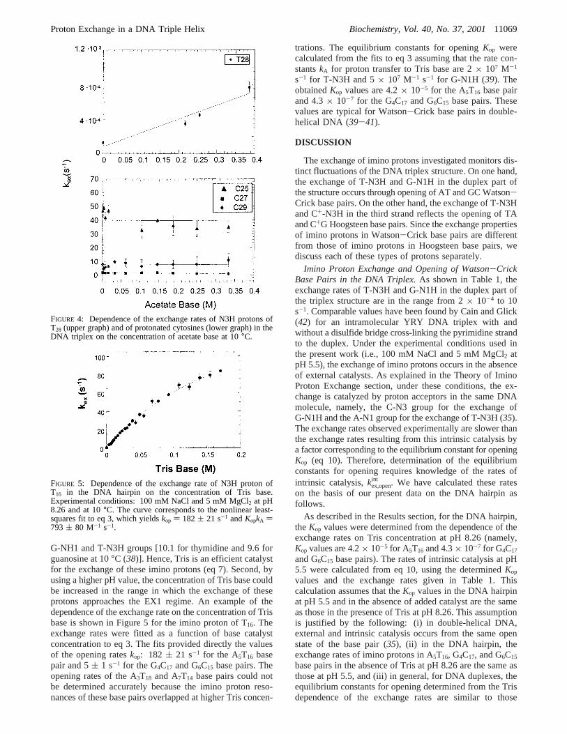
To elucidate the mechanism of exchange of imino protonsin the DNA triplex, we have also investigated the effects ofexternal catalysis by added proton acceptors. The catalystinvestigated was acetate. The choice of acetate was dictatedby its low pK value [pK ) 4.76 at 10°C (36)], which allowslarger concentrations of acetate base to be obtained in thepH range in which the triplex structure is stable. Theconcentration of acetate was varied from 0 to 0.45 M at pH5.5 (corresponding range of the concentration of acetate base0-0.38 M) in the presence of 100 mM NaCl and 5 mMMgCl2. Catalysis by acetate was observed for N3H protonsof the thymines in the third strand, except T31. An exampleof acetate catalysis is shown in Figure 4 for T28. No catalysisby acetate was observed for imino protons of protonated cyto-sines or for those of Watson-Crick base pairs in the DNAtriplex. This result is illustrated in Figure 4, which showsthat the exchange rates of cytosine N3H protons are constantupon increasing the acetate base concentration to 0.34 M.
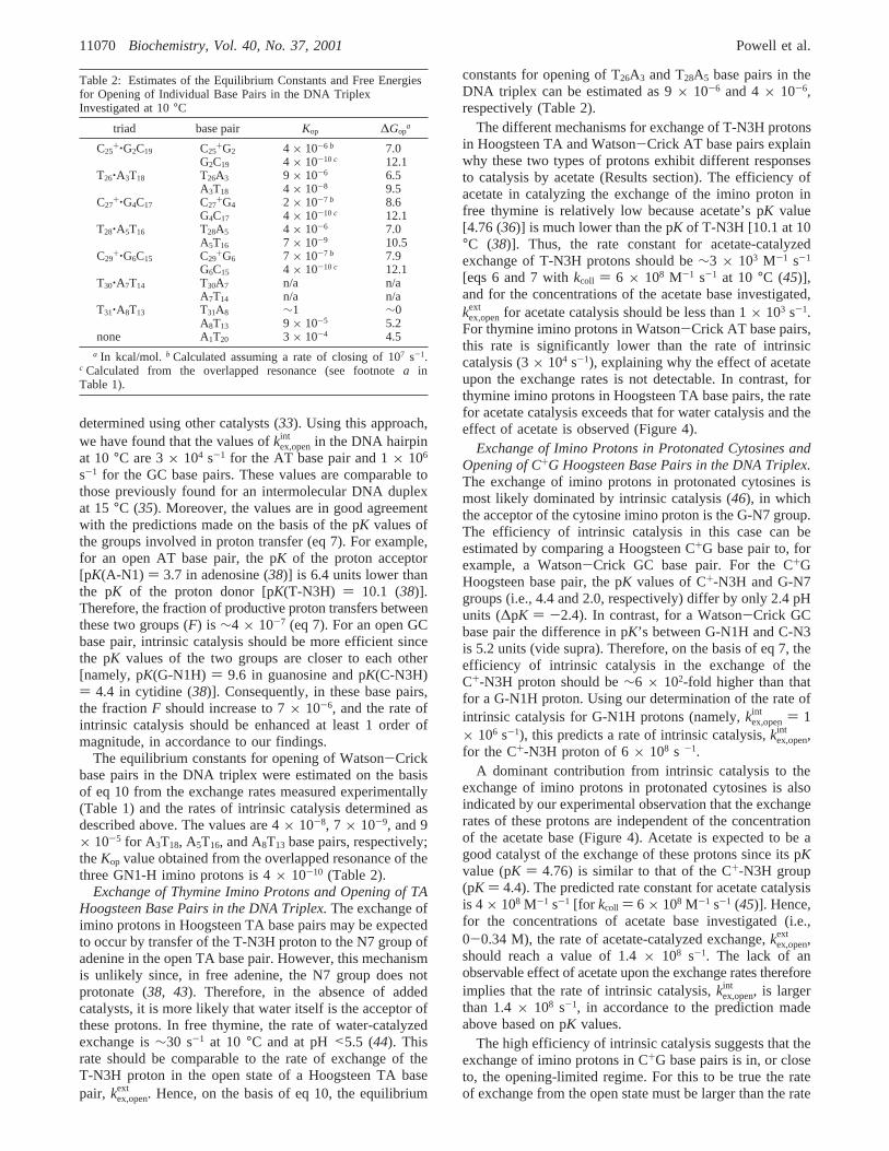
The exchange of imino protons in the DNA hairpin wasfurther characterized in order to obtain the kinetic parametersfor base pair opening in this structure. The exchange rateswere measured in transfer of magnetization experiments atpH 8.26 using Tris as an external catalyst. The rationale forchoosing these experimental conditions was 2-fold. First, thepK value of Tris [8.61 at 10°C (37)] is close to the pK’s of
FIGURE 2: NMR resonances of imino protons in the DNA triplex(A) and the DNA hairpin (B) investigated in 100 mM NaCl and 5mM MgCl2 in 90% H2O/10% D2O at pH 5.5 and at 10°C.
FIGURE 3: Imino proton NMR resonances of the DNA triplexduring real-time exchange measurements. Experimental conditionsare the same as in Figure 2. The exchange time (in minutes) isindicated for each spectrum.
Table 1: Exchange Rates of Imino Protons in the DNA Triplex andHairpin Investigated in 100 mM NaCl and 5 mM MgCl2 at pH 5.5and at 10°C
DNA triplex DNA hairpin
base base pairing kex(s-1) base pairing kex(s-1)
G2 Watson-Crick (4.4( 0.1)× 10-4 a Watson-Crick 1.9( 0.1G4 Watson-Crick (4.4( 0.1)× 10-4 a Watson-Crick 0.45( 0.05G6 Watson-Crick (4.4( 0.1)× 10-4 a Watson-Crick 0.43( 0.07T13 Watson-Crick 2.7( 0.2 Watson-Crick 7.0( 0.7T14 Watson-Crick b Watson-Crick 1.20( 0.07T16 Watson-Crick (2.2( 0.1)× 10-4 Watson-Crick 1.10( 0.06T18 Watson-Crick (1.2( 0.1)× 10-3 Watson-Crick 1.62( 0.08T20 Watson-Crick 10( 2 Watson-Crick cC25
+ Hoogsteen 42( 4T26 Hoogsteen (2.6( 0.1)× 10-4
C27+ Hoogsteen 1.8( 0.1
T28 Hoogsteen (1.30( 0.06)× 10-4
C29+ Hoogsteen 6.6( 0.6
T30 Hoogsteen bT31 Hoogsteen 25( 1
a In the DNA triplex, the imino proton resonances of G2, G4, and G6
overlap (Figure 2). The value ofkex given is obtained from theoverlapped resonance.b The exchange rate is too slow to be measuredby transfer of magnetization from water (kex < ∼0.2 s-1) and too fastto be measured by real-time exchange (kex > ∼10-3 s-1). c Theresonance is not observable due to exchange broadening by fraying atthe ends of the duplex.
11068 Biochemistry, Vol. 40, No. 37, 2001 Powell et al.
G-NH1 and T-N3H groups [10.1 for thymidine and 9.6 forguanosine at 10°C (38)]. Hence, Tris is an efficient catalystfor the exchange of these imino protons (eq 7). Second, byusing a higher pH value, the concentration of Tris base couldbe increased in the range in which the exchange of theseprotons approaches the EX1 regime. An example of thedependence of the exchange rate on the concentration of Trisbase is shown in Figure 5 for the imino proton of T16. Theexchange rates were fitted as a function of base catalystconcentration to eq 3. The fits provided directly the valuesof the opening rateskop: 182 ( 21 s-1 for the A5T16 basepair and 5( 1 s-1 for the G4C17 and G6C15 base pairs. Theopening rates of the A3T18 and A7T14 base pairs could notbe determined accurately because the imino proton reso-nances of these base pairs overlapped at higher Tris concen-
trations. The equilibrium constants for openingKop werecalculated from the fits to eq 3 assuming that the rate con-stantskA for proton transfer to Tris base are 2× 10⁷ M-1
s-1 for T-N3H and 5× 10⁷ M-1 s-1 for G-N1H (39). TheobtainedKop values are 4.2× 10⁻⁵ for the A5T16 base pairand 4.3× 10⁻⁷ for the G4C17 and G6C15 base pairs. Thesevalues are typical for Watson-Crick base pairs in double-helical DNA (39-41).
DISCUSSION
The exchange of imino protons investigated monitors dis-tinct fluctuations of the DNA triplex structure. On one hand,the exchange of T-N3H and G-N1H in the duplex part ofthe structure occurs through opening of AT and GC Watson-Crick base pairs. On the other hand, the exchange of T-N3Hand C+-N3H in the third strand reflects the opening of TAand C+G Hoogsteen base pairs. Since the exchange propertiesof imino protons in Watson-Crick base pairs are differentfrom those of imino protons in Hoogsteen base pairs, wediscuss each of these types of protons separately.
Imino Proton Exchange and Opening of Watson-CrickBase Pairs in the DNA Triplex.As shown in Table 1, theexchange rates of T-N3H and G-N1H in the duplex part ofthe triplex structure are in the range from 2× 10⁻⁴ to 10s-1. Comparable values have been found by Cain and Glick(42) for an intramolecular YRY DNA triplex with andwithout a disulfide bridge cross-linking the pyrimidine strandto the duplex. Under the experimental conditions used inthe present work (i.e., 100 mM NaCl and 5 mM MgCl2 atpH 5.5), the exchange of imino protons occurs in the absenceof external catalysts. As explained in the Theory of IminoProton Exchange section, under these conditions, the ex-change is catalyzed by proton acceptors in the same DNAmolecule, namely, the C-N3 group for the exchange ofG-N1H and the A-N1 group for the exchange of T-N3H (35).The exchange rates observed experimentally are slower thanthe exchange rates resulting from this intrinsic catalysis bya factor corresponding to the equilibrium constant for openingKop (eq 10). Therefore, determination of the equilibriumconstants for opening requires knowledge of the rates ofintrinsic catalysis,kex,open
int . We have calculated these rateson the basis of our present data on the DNA hairpin asfollows.
As described in the Results section, for the DNA hairpin,theKop values were determined from the dependence of theexchange rates on Tris concentration at pH 8.26 (namely,Kop values are 4.2× 10⁻⁵ for A5T16 and 4.3× 10⁻⁷ for G4C17
and G6C15 base pairs). The rates of intrinsic catalysis at pH5.5 were calculated from eq 10, using the determinedKop
values and the exchange rates given in Table 1. Thiscalculation assumes that theKop values in the DNA hairpinat pH 5.5 and in the absence of added catalyst are the sameas those in the presence of Tris at pH 8.26. This assumptionis justified by the following: (i) in double-helical DNA,external and intrinsic catalysis occurs from the same openstate of the base pair (35), (ii) in the DNA hairpin, theexchange rates of imino protons in A5T16, G4C17, and G6C15
base pairs in the absence of Tris at pH 8.26 are the same asthose at pH 5.5, and (iii) in general, for DNA duplexes, theequilibrium constants for opening determined from the Trisdependence of the exchange rates are similar to those
FIGURE 4: Dependence of the exchange rates of N3H protons ofT28 (upper graph) and of protonated cytosines (lower graph) in theDNA triplex on the concentration of acetate base at 10°C.
FIGURE 5: Dependence of the exchange rate of N3H proton ofT16 in the DNA hairpin on the concentration of Tris base.Experimental conditions: 100 mM NaCl and 5 mM MgCl2 at pH8.26 and at 10°C. The curve corresponds to the nonlinear least-squares fit to eq 3, which yieldskop ) 182 ( 21 s-1 andKopkA )793 ( 80 M-1 s-1.
Proton Exchange in a DNA Triple Helix Biochemistry, Vol. 40, No. 37, 200111069
determined using other catalysts (33). Using this approach,we have found that the values ofkex,open
int in the DNA hairpinat 10°C are 3× 10⁴ s-1 for the AT base pair and 1× 10⁶
s-1 for the GC base pairs. These values are comparable tothose previously found for an intermolecular DNA duplexat 15°C (35). Moreover, the values are in good agreementwith the predictions made on the basis of the pK values ofthe groups involved in proton transfer (eq 7). For example,for an open AT base pair, the pK of the proton acceptor[pK(A-N1) ) 3.7 in adenosine (38)] is 6.4 units lower thanthe pK of the proton donor [pK(T-N3H) ) 10.1 (38)].Therefore, the fraction of productive proton transfers betweenthese two groups (F) is ∼4 × 10⁻⁷ (eq 7). For an open GCbase pair, intrinsic catalysis should be more efficient sincethe pK values of the two groups are closer to each other[namely, pK(G-N1H) ) 9.6 in guanosine and pK(C-N3H)) 4.4 in cytidine (38)]. Consequently, in these base pairs,the fractionF should increase to 7× 10⁻⁶, and the rate ofintrinsic catalysis should be enhanced at least 1 order ofmagnitude, in accordance to our findings.
The equilibrium constants for opening of Watson-Crickbase pairs in the DNA triplex were estimated on the basisof eq 10 from the exchange rates measured experimentally(Table 1) and the rates of intrinsic catalysis determined asdescribed above. The values are 4× 10⁻⁸, 7 × 10⁻⁹, and 9× 10⁻⁵ for A3T18, A5T16, and A8T13 base pairs, respectively;theKop value obtained from the overlapped resonance of thethree GN1-H imino protons is 4× 10⁻¹⁰ (Table 2).
Exchange of Thymine Imino Protons and Opening of TAHoogsteen Base Pairs in the DNA Triplex.The exchange ofimino protons in Hoogsteen TA base pairs may be expectedto occur by transfer of the T-N3H proton to the N7 group ofadenine in the open TA base pair. However, this mechanismis unlikely since, in free adenine, the N7 group does notprotonate (38, 43). Therefore, in the absence of addedcatalysts, it is more likely that water itself is the acceptor ofthese protons. In free thymine, the rate of water-catalyzedexchange is∼30 s-1 at 10 °C and at pH<5.5 (44). Thisrate should be comparable to the rate of exchange of theT-N3H proton in the open state of a Hoogsteen TA basepair, kex,open
ext . Hence, on the basis of eq 10, the equilibrium
constants for opening of T26A3 and T28A5 base pairs in theDNA triplex can be estimated as 9× 10⁻⁶ and 4× 10⁻⁶,respectively (Table 2).
The different mechanisms for exchange of T-N3H protonsin Hoogsteen TA and Watson-Crick AT base pairs explainwhy these two types of protons exhibit different responsesto catalysis by acetate (Results section). The efficiency ofacetate in catalyzing the exchange of the imino proton infree thymine is relatively low because acetate’s pK value[4.76 (36)] is much lower than the pK of T-N3H [10.1 at 10°C (38)]. Thus, the rate constant for acetate-catalyzedexchange of T-N3H protons should be∼3 × 10³ M-1 s-1
[eqs 6 and 7 withkcoll ) 6 × 10⁸ M-1 s-1 at 10 °C (45)],and for the concentrations of the acetate base investigated,kex,open
ext for acetate catalysis should be less than 1× 10³ s-1.For thymine imino protons in Watson-Crick AT base pairs,this rate is significantly lower than the rate of intrinsiccatalysis (3× 10⁴ s-1), explaining why the effect of acetateupon the exchange rates is not detectable. In contrast, forthymine imino protons in Hoogsteen TA base pairs, the ratefor acetate catalysis exceeds that for water catalysis and theeffect of acetate is observed (Figure 4).
Exchange of Imino Protons in Protonated Cytosines andOpening of C+G Hoogsteen Base Pairs in the DNA Triplex.The exchange of imino protons in protonated cytosines ismost likely dominated by intrinsic catalysis (46), in whichthe acceptor of the cytosine imino proton is the G-N7 group.The efficiency of intrinsic catalysis in this case can beestimated by comparing a Hoogsteen C+G base pair to, forexample, a Watson-Crick GC base pair. For the C+GHoogsteen base pair, the pK values of C+-N3H and G-N7groups (i.e., 4.4 and 2.0, respectively) differ by only 2.4 pHunits (∆pK ) -2.4). In contrast, for a Watson-Crick GCbase pair the difference in pK’s between G-N1H and C-N3is 5.2 units (vide supra). Therefore, on the basis of eq 7, theefficiency of intrinsic catalysis in the exchange of theC+-N3H proton should be∼6 × 10²-fold higher than thatfor a G-N1H proton. Using our determination of the rate ofintrinsic catalysis for G-N1H protons (namely,kex,open
int ) 1× 10⁶ s-1), this predicts a rate of intrinsic catalysis,kex,open
int ,for the C+-N3H proton of 6× 10⁸ s -1.
A dominant contribution from intrinsic catalysis to theexchange of imino protons in protonated cytosines is alsoindicated by our experimental observation that the exchangerates of these protons are independent of the concentrationof the acetate base (Figure 4). Acetate is expected to be agood catalyst of the exchange of these protons since its pKvalue (pK ) 4.76) is similar to that of the C+-N3H group(pK ) 4.4). The predicted rate constant for acetate catalysisis 4× 10⁸ M-1 s-1 [for kcoll ) 6 × 10⁸ M-1 s-1 (45)]. Hence,for the concentrations of acetate base investigated (i.e.,0-0.34 M), the rate of acetate-catalyzed exchange,kex,open
ext ,should reach a value of 1.4× 10⁸ s-1. The lack of anobservable effect of acetate upon the exchange rates thereforeimplies that the rate of intrinsic catalysis,kex,open
int , is largerthan 1.4× 10⁸ s-1, in accordance to the prediction madeabove based on pK values.
The high efficiency of intrinsic catalysis suggests that theexchange of imino protons in C+G base pairs is in, or closeto, the opening-limited regime. For this to be true the rateof exchange from the open state must be larger than the rate
Table 2: Estimates of the Equilibrium Constants and Free Energiesfor Opening of Individual Base Pairs in the DNA TriplexInvestigated at 10°C
triad base pair Kop ∆Gopa
C25+‚G2C19 C25
+G2 4 × 10⁻⁶ b 7.0G2C19 4 × 10⁻¹⁰c 12.1
T26‚A3T18 T26A3 9 × 10⁻⁶ 6.5A3T18 4 × 10⁻⁸ 9.5
C27+‚G4C17 C27
+G4 2 × 10⁻⁷ b 8.6G4C17 4 × 10⁻¹⁰c 12.1
T28‚A5T16 T28A5 4 × 10⁻⁶ 7.0A5T16 7 × 10⁻⁹ 10.5
C29+‚G6C15 C29
+G6 7 × 10⁻⁷ b 7.9G6C15 4 × 10⁻¹⁰c 12.1
T30‚A7T14 T30A7 n/a n/aA7T14 n/a n/a
T31‚A8T13 T31A8 ∼1 ∼0A8T13 9 × 10⁻⁵ 5.2
none A1T20 3 × 10⁻⁴ 4.5a In kcal/mol. b Calculated assuming a rate of closing of 107 s-1.
c Calculated from the overlapped resonance (see footnotea inTable 1).
11070 Biochemistry, Vol. 40, No. 37, 2001 Powell et al.
of base pair closing (eqs 3 and 9). The rates of closing ofC+G base pairs are expected to be similar to those in double-helical DNA [i.e., 107-108 s -1 (40, 41)]. Hence, it is likelythatkex,openg kcl and the exchange rates of C+-N3H protonsmeasured experimentally (Table 1) are close to the rates ofopening C+G base pairs in C+‚GC triads. On the basis ofthese arguments, we have estimated the equilibrium constantsfor opening of C+G base pairs by assuming a value of 107
s -1 for the rates of closing of these base pairs. The valuesof Kop and opening free energies obtained with this assump-tion are shown in Table 2. Higher values of the closing rateswould result in lowerKop values; hence, our estimates mostlikely provide upper limits for the equilibrium constants foropening for these bases.
Site-ResolVed Structural Energetics in the DNA Triplex.The results obtained in the present work allow us to mapthe stability of the DNA triplex structure at the level ofindividual base pairs and distinct base pairing interactions.The exchange of imino protons reflects those conformationalfluctuations in the DNA triplex which yield an open,exchange-competent state of the imino group. Accordingly,the local stabilization energies derived from exchange ratesrepresent the free energy changes associated with theseconformational fluctuations (∆Gop) (32).
One important result of the present investigation is thatthe equilibrium constants for opening of individual base pairsin the duplex part of the triplex structure are much lowerthan those in a DNA double helix. In the double-helicalhairpin investigated, the equilibrium constants for openingare 4.2× 10⁻⁵ for A5T16 and 4.3× 10⁻⁷ for G4C17 and G6C15
(Results section). Formation of the triple-helical structurereduces these equilibrium constants up to 6000-fold (Table2). Hence, relative to DNA double helices, the Watson-Crick base pairs in the triplex are further stabilized by extrafree energies of up to 5 kcal/mol (eq 4). This stabilization issignificant only for Watson-Crick base pairs located in thecentral part of the triplex structure. For terminal base pairs,such as T20 and T13, the exchange rates and the openingequilibrium constants are comparable to those in double-helical DNA (Tables 1 and 2). For the AT base pairs locatedin the central part of the triplex the exchange rates alsodepend on the exact location of the base pair in the structure.As shown in Table 1, the exchange rate for T18 is five timeslarger than that for T16. This difference clearly reflects adestabilization of the A3T18 base pair relative to the A5T16
base pair (Table 2). This effect may be related to the loca-tion of the A3T18 base pair toward the 5′-end of the tri-plex structure (this directionality is defined relative to thatof the purine Watson-Crick strand). As shown by Feigonand co-workers (25), in this part of the structure the T24A1T20
triad does not form. Hence, these observations suggest thatthe 5′-end part of the triplex structure is less stable than therest, possibly due to conformational constraints imposed bythe short, three-base loop connecting the two pyrimidinestrands.
In the DNA triplex, the energetic cost of the structuralfluctuations which bring the imino protons of Watson-Crickbase pairs into the open, solvent-accessible state is expectedto be higher than that in double-helical DNA. For ex-ample, formation of the open state for the guanine in aC+‚GC triad should perturb the hydrogen bonds involved inboth Watson-Crick and Hoogsteen base pairing (Figure 1).
Thus, one expects the free energy change for opening thesebases to be higher than in double-helical DNA. However, itis interesting to note that, in Watson-Crick AT base pairsof T‚AT triads, the hydrogen-bonding pattern for thyminesis the same as that in a DNA double helix. Moreover, analysisof NMR-derived triplex structures (19) shows that placingthe third strand in the major groove of the double helix doesnot introduce significant steric hindrance for opening ofthymines in either the major or the minor groove. Despitethese structural characteristics, the stabilization free energiesfor AT base pairs in the triplex are significantly higher thanthose in double-helical DNA (Table 2). This suggests thatthe opening transitions probed by the exchange of thymineimino protons in Watson-Crick base pairs may involvelarger perturbations of the triplex structure at or near theexchanging proton.
Our present results also provide new insights into theopening kinetics and stabilization free energies of Hoogsteenbase pairs in the DNA triplex. As shown in Table 2, theequilibrium constants for opening of Hoogsteen base pairsin the central part of the triplex range from 2× 10⁻⁷ to9 × 10-6. These values are comparable to those normallyfound for Watson-Crick base pairs in double-helical DNA[4.2 × 10⁻⁵ and 4.3× 10⁻⁷ in the DNA hairpin investigatedand other results (40, 41)]. This finding implies that, in acanonical YRY triplex such as the one investigated here,the stability of Hoogsteen base pairs is comparable to thatof Watson-Crick base pairs in duplex DNA. As in duplexDNA, the base pairs formed by Hoogsteen interactionsappear to open one at the time, independently of each other.The exchange rates of Hoogsteen imino protons, however,depend on the location of the base pairs in the structure. Asin the case of double-helical DNA, the exchange of the lasttwo bases, T30 and T31, is faster, being affected by frayingat the end of the strand. Moreover, for Hoogsteen iminoprotons located in the 5′-end part of the structure theexchange rates are higher than those in central triads. Forinstance, the exchange rate of T26 is approximately 2-foldhigher than that of T28. Similarly, among the three C+‚GCtriads present in the structure, the highest exchange rate isobserved for C+25 (Table 1). These observations provideadditional support to the suggestion that the triads locatedat the 5′-end of the structure are less stable than the othercentral triads in the triplex.
In summary, the results presented in this paper demonstratethat, in a YRY triplex, the binding of a third strand in themajor groove of double-helical DNA significantly stabilizesthe Watson-Crick base pairs against the structural fluctua-tions responsible for imino proton exchange. This stabiliza-tion appears to be dependent on the location of the base triadrelative to the ends of the triplex and on steric constraintsimposed by the loop connecting the third strand to the restof the structure. Similar dependencies are observed for thestabilization free energies of Hoogsteen base pairs. The DNAinvestigated in the present work is one of only a few DNAcanonical triplexes for which proton exchange has beencharacterized at the level of individual triads (42). Thus, noinferences can be drawn yet on the dependence of the localstabilization energy on base sequence. Characterization ofnucleic acid triple helices containing different base sequencesand selected noncanonical base triads will provide thisimportant information.
Proton Exchange in a DNA Triple Helix Biochemistry, Vol. 40, No. 37, 200111071

ACKNOWLEDGMENT
We thank Dr. Ryszard Michalczyk for help with the DNAsynthesis.
SUPPORTING INFORMATION AVAILABLE
Figure 1S showing representative spectra from experimentsof transfer of magnetization from water on the DNA triplexinvestigated. This material is available free of charge viathe Internet at http://pubs.acs.org.
REFERENCES
1. Frank-Kamenetskii, M. D., and Mirkin, S. M. (1995)Annu.ReV. Biochem. 64, 65-95.
2. Soyfer, V. N., and Potaman, V. N. (1995)Triple-HelicalNucleic Acids, Springer, New York.
3. Behe, M. J. (1995)Nucleic Acids Res. 23, 689-695.4. Mirkin, S. M., Lyamichev, V. I., Drushlyak, K. N., Dobrynin,
V. N., Filippov, S. A., and Frank-Kamenetskii, M. D. (1987)Nature (London) 330, 495-497.
5. Mirkin, S. M., and Frank-Kamenetskii, M. D. (1994)Annu.ReV. Biophys. Biomol. Struct. 23, 541-576.
6. Schroth, G. P., and Ho, P. S. (1995)Nucleic Acids Res. 23,1977-1983.
7. Portes-Sentis, S., Sergeant, A., and Gruffat, H. (1997)NucleicAcids Res. 25, 1347-1354.
8. Kohwi, Y., and Panchenko, Y. (1993)Genes DeV. 7, 1766-1778.
9. Rooney, S. M., and Moore, P. D. (1995)Proc. Natl. Acad.Sci. U.S.A. 92, 2141-2144.
10. Cooney, M., Czernuszewicz, G., Postel, E. H., Flint, S. J., andHogan, M. E. (1988)Science 241, 456-459.
11. Duval-Valentin, G., Thuong, N. T., and Helene, C. (1992)Proc. Natl. Acad. Sci. U.S.A. 89, 504-508.
12. Maher, L. J., Dervan, P. B., and Wold, B. (1992)Biochemistry31, 70-81.
13. Orson, F. M., Thomas, D. W., McShan, W. M., Kessler, D.J., and Hogan, M. E. (1991)Nucleic Acids Res. 19, 3435-3441.
14. Postel, E. H., Flint, S. J., Kessler, D. J., and Hogan, M. E.(1991)Proc. Natl. Acad. Sci. U.S.A. 88, 8227-8231.
15. Francois, J. C., Saison-Behmoaras, T., Barbier, C., Chassignol,M., Thuong, N. T., and Helene, C. (1989)Proc. Natl. Acad.Sci. U.S.A. 86, 9702-9706.
16. Moser, H. E., and Dervan, P. B. (1987)Science 238, 645-650.
17. Strobel, S. A., Doucette-Stamm, L. A., Riba, L., Housman,D. E., and Dervan, P. B. (1991)Science 254, 1639-1642.
18. Radhakrishnan, I., and Patel, D. J. (1993)Structure 1, 135-152.
19. Wang, E., and Feigon, J. (1999) inOxford Handbook ofNucleic Acid Structure(Neidle, S., Ed.) pp 355-388, OxfordUniversity Press, New York.
20. Plum, G. E., Park, Y. W., Singleton, S. F., Dervan, P. B., andBreslauer, K. J. (1990)Proc. Natl. Acad. Sci. U.S.A. 87, 9436-9440.
21. Plum, G. E., Pilch, D. S., Singleton, S. F., and Breslauer, K.J. (1995)Annu. ReV. Biophys. Biomol. Struct. 24, 319-350.
22. Plum, G. E. (1997)Biopolymers 44, 241-256.23. Roberts, R. W., and Crothers, D. M. (1991)Proc. Natl. Acad.
Sci. U.S.A. 88, 9397-9401.24. Singleton, S. F., and Dervan, P. B. (1992)J. Am. Chem. Soc.
114, 6957-6965.25. Macaya, R., Wang, E., Schultze, P., Sklenar, V., and Feigon,
J. (1992)J. Mol. Biol. 225, 755-773.26. Wang, E., Malek, S., and Feigon, J. (1992)Biochemistry 31,
4838-4846.27. Tarkoy, M., Phipps, A. K., Schultze, P., and Feigon, J. (1998)
Biochemistry 37, 5810-5819.28. Plateau, P., and Gueron, M. (1982)J. Am. Chem. Soc. 104,
7310-7311.29. Ernst, R. R., Bodenhausen, G., and Wokaun, A. (1987)
Principles of Nuclear Magnetic Resonance in One and TwoDimensions, Clarendon Press, Oxford.
30. Michalczyk, R., and Russu, I. M. (1993)FEBS Lett. 331, 217-222.
31. Hvidt, A., and Nielsen, S. O. (1966)AdV. Protein Chem. 21,287-386.
32. Englander, S. W., and Kallenbach, N. R. (1984)Q. ReV.Biophys. 16, 521-655.
33. Gueron, M., Charretier, E., Hagerhorst, J., Kochoyan, M.,Leroy, J. L., and Moraillon, A. (1990) inStructure & Methods(Sarma, R. H., and Sarma, M. H., Eds.) Vol. 3, pp 113-137,Adenine Press, New York.
34. Nonin, S., Leroy, J.-L., and Gueron, M. (1995)Biochemistry34, 10652-10659.
35. Gueron, M., Kochoyan, M., and Leroy, J. L. (1987)Nature(London) 328, 89-92.
36. Weast, R. C. (1987) inCRC Handbook of Chemistry andPhysics, CRC Press, Boca Raton, FL.
37. Gueffroy, D. E. (1975) inCALBIOCHEM: Buffers, Calbio-chem-Novabiochem Co., San Diego, CA.
38. Ts’o, P. O. P. (1974)Basic Principles in Nucleic AcidChemistry, Academic Press, New York.
39. Moe, J. G., and Russu, I. M. (1992)Biochemistry 31, 8421-8428.
40. Folta-Stogniew, E. J., and Russu, I. M. (1994)Biochemistry33, 11016-11024.
41. Gueron, M., and Leroy, J. L. (1995)Methods Enzymol. 261,383-413.
42. Cain, R. J., and Glick, G. D. (1998)Biochemistry 37, 1456-1464.
43. Buchner, P., Maurer, W., and Ruterjans, H. (1978)J. Magn.Reson. 29, 45-63.
44. Nonin, S., Leroy, J.-L., and Gueron, M. (1996)Nucleic AcidsRes. 24, 586-595.
45. Benight, A. S., Schurr, J. M., Flynn, P. F., Reid, B. R., andWemmer, D. E. (1988)J. Mol. Biol. 200, 377-399.
46. Leroy, J. L., Gehring, K., Kettani, A., and Gueron, M. (1993)Biochemistry 32, 6019-6031.
BI010890A
11072 Biochemistry, Vol. 40, No. 37, 2001 Powell et al.