Embed Size (px)
Citation preview
1
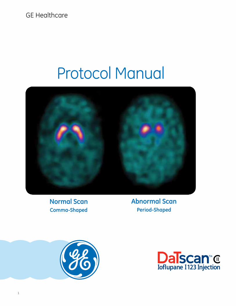
Protocol Manual
Normal ScanComma-Shaped
Abnormal ScanPeriod-Shaped
GE Healthcare
2
Contents
Section 1: Introduction ......................................................................... 4 1.1 Scope
1.2 What is DaTscan?
Section 2: Image Acquisition, Interpretation, and Reporting
Imaging ................................................................................................... 6 2.1 Imaging Guidelines for DaTscan 2.2 Patient Positioning 2.3 Recommended Image Acquisition Parameters 2.4 Image Processing Parameters 2.4.1 Camera-Specific Parameters 2.5 Quality Checks 2.6 Examples of Deficient Image Quality Causes 2.6.1 Radius of Rotation 2.6.2 Lateral Head Tilt
Interpretation and Reporting ........................................................... 19 2.7 Normal vs Abnormal Images 2.8 Normal Uptake 2.9 Abnormal Uptake 2.10 Unexpected Results 2.11 Reporting Reminders
Section 3: Step-by-Step Guide to DaTscan
Patient Preparation ............................................................................ 26 3.1 Patient Protocols 3.2 Patient Counseling
Dosing.................................................................................................... 28 3.3 Thyroid Blocking 3.4 Dose Administration and Calibration
Administration and Storage ............................................................. 29 3.5 Administration of DaTscan 3.6 Storage of DaTscan 3.7 Schedule II Radiopharmaceutical Requirements
Section 4: Important Risk and Safety Information ...................... 32
Section 5: GE Healthcare’s Commitment to You .......................... 33
Full Prescribing Information ............................................................. 34

Introduction
Section 1
4
Please see Important Risk and Safety Information on page 32 and Full Prescribing Information at the end of this document prior to administration.
Introduction
1.1 Scope
This document describes the procedure and parameters for visual interpretation of single-photon emission computed tomography (SPECT) images acquired after administration of DaTscan. It is assumed that the person reading the images is a clinician with expertise in the interpretation of nuclear medicine studies and has a detailed knowledge of brain anatomy. In addition, for this document to be applicable, DaTscan must have been prescribed and administered according to the Full Prescribing Information, and the images must have been acquired and processed with the GE Healthcare-recommended protocols specified in sections 2.3 and 2.4.
1.2 What is DaTscan?
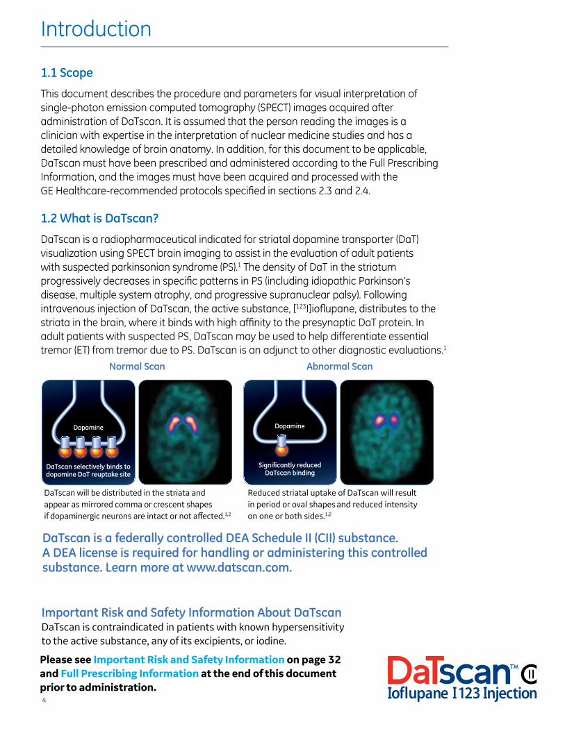
DaTscan is a radiopharmaceutical indicated for striatal dopamine transporter (DaT) visualization using SPECT brain imaging to assist in the evaluation of adult patients with suspected parkinsonian syndrome (PS).1 The density of DaT in the striatum progressively decreases in specific patterns in PS (including idiopathic Parkinson’s disease, multiple system atrophy, and progressive supranuclear palsy). Following intravenous injection of DaTscan, the active substance, [123I]ioflupane, distributes to the striata in the brain, where it binds with high affinity to the presynaptic DaT protein. In adult patients with suspected PS, DaTscan may be used to help differentiate essential tremor (ET) from tremor due to PS. DaTscan is an adjunct to other diagnostic evaluations.1
Important Risk and Safety Information About DaTscanDaTscan is contraindicated in patients with known hypersensitivity to the active substance, any of its excipients, or iodine.
Reduced striatal uptake of DaTscan will result in period or oval shapes and reduced intensity on one or both sides.1,2
DaTscan will be distributed in the striata and appear as mirrored comma or crescent shapes if dopaminergic neurons are intact or not affected.1,2
Abnormal Scan
Dopamine
DaTscan selectively binds to dopamine DaT reuptake site
Dopamine
Significantly reduced DaTscan binding
Normal Scan
Dopamine
DaTscan selectively binds to dopamine DaT reuptake site
Dopamine
Significantly reduced DaTscan binding
DaTscan is a federally controlled DEA Schedule II (CII) substance. A DEA license is required for handling or administering this controlled substance. Learn more at www.datscan.com.

5
Image Acquisition, Interpretation, and Reporting
Section 2
6
Imaging
2.1 Imaging Guidelines for DaTscan1
Begin SPECT imaging three to six hours following DaTscan administration. Acquire images using a gamma camera fitted with low-energy, high-resolution (LEHR) collimators and set to a photopeak of 159 keV with a ±10% energy window. Angular sampling should not be less than 120 views over 360 degrees. Position the patient supine with the head on an off-the-table headrest. A flexible head restraint such as a strip of tape across the chin or forehead can also be used to help avoid movement. Set a circular orbit for the detector heads with the radius as small as possible (11 to a maximum of 15 cm).
Experimental studies with a striatal phantom suggest that optimal images are obtained with matrix size and zoom factors selected to give a pixel size of 3.5 to 4.5 mm. Collect a minimum of 1.5 million counts for optimal images.
2.2 Patient Positioning
The following factors in patient positioning are critical to acquiring interpretable images with DaTscan.
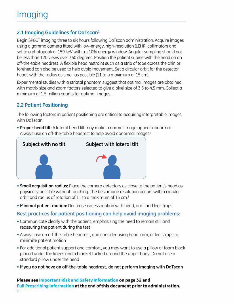
• Proper head tilt: A lateral head tilt may make a normal image appear abnormal. Always use an off-the-table headrest to help avoid abnormal images1
• Small acquisition radius: Place the camera detectors as close to the patient’s head as physically possible without touching. The best image resolution occurs with a circular orbit and radius of rotation of 11 to a maximum of 15 cm.1
• Minimal patient motion: Decrease excess motion with head, arm, and leg straps
Best practices for patient positioning can help avoid imaging problems:• Communicate clearly with the patient, emphasizing the need to remain still and
reassuring the patient during the test
• Always use an off-the-table headrest, and consider using head, arm, or leg straps to minimize patient motion
• For additional patient support and comfort, you may want to use a pillow or foam block placed under the knees and a blanket tucked around the upper body. Do not use a standard pillow under the head
• If you do not have an off-the-table headrest, do not perform imaging with DaTscan
Subject with no tilt Subject with lateral tilt
Example of the effect of lateral tilt on a subject with normal uptake of DaTscan™
Example of the effect of lateral tilt on a subject with normal uptake of DaTscan™
Please see Important Risk and Safety Information on page 32 and Full Prescribing Information at the end of this document prior to administration.
7
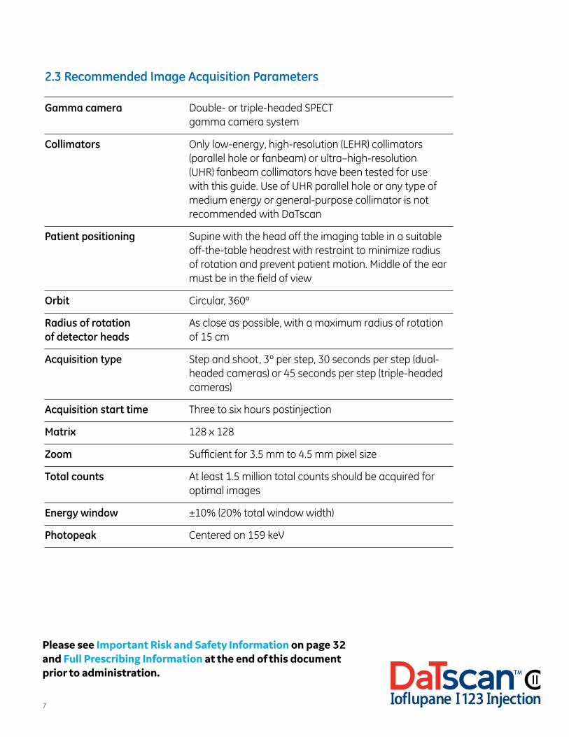
2.3 Recommended Image Acquisition Parameters
Gamma camera Double- or triple-headed SPECT gamma camera system
Collimators Only low-energy, high-resolution (LEHR) collimators (parallel hole or fanbeam) or ultra–high-resolution (UHR) fanbeam collimators have been tested for use with this guide. Use of UHR parallel hole or any type of medium energy or general-purpose collimator is not recommended with DaTscan
Patient positioning Supine with the head off the imaging table in a suitable off-the-table headrest with restraint to minimize radius of rotation and prevent patient motion. Middle of the ear must be in the field of view
Orbit Circular, 360º
Radius of rotation As close as possible, with a maximum radius of rotationof detector heads of 15 cm
Acquisition type Step and shoot, 3º per step, 30 seconds per step (dual-headed cameras) or 45 seconds per step (triple-headed cameras)
Acquisition start time Three to six hours postinjection
Matrix 128 x 128
Zoom Sufficient for 3.5 mm to 4.5 mm pixel size
Total counts At least 1.5 million total counts should be acquired for optimal images
Energy window ±10% (20% total window width)
Photopeak Centered on 159 keV
Please see Important Risk and Safety Information on page 32 and Full Prescribing Information at the end of this document prior to administration.
8
2.4 Image Processing Parameters
Reconstruction algorithm Filtered back projection (FBP) or iterative reconstruction (eg, OSEM)
Filtering (pre-2D or post-3D) Butterworth (or other low-pass linear filter)
Filter power factor 8 to 10 (system-dependent)
Cutoff Changing the filter cutoff will affect image resolution. 0.5 to 0.6 cycles per cm nominally (or as appropriate to achieve approximately the same level of smoothing as the images shown throughout this guide)
Attenuation correction Not necessary. If desired, can use Chang (also known as linear or zero-order)
Attenuation correction A locally calculated attenuation correction coefficient coefficient (if used) from a phantom measurement should be used if available. Otherwise, use nominal value of 0.11 cm-1
Background subtraction No background subtraction
Pixel (voxel) size 3.5 mm to 4.5 mm isotropic
Presentation Transaxial (transverse) slices parallel to the anterior commissure-posterior commissure (AC-PC) line with single-pixel thickness
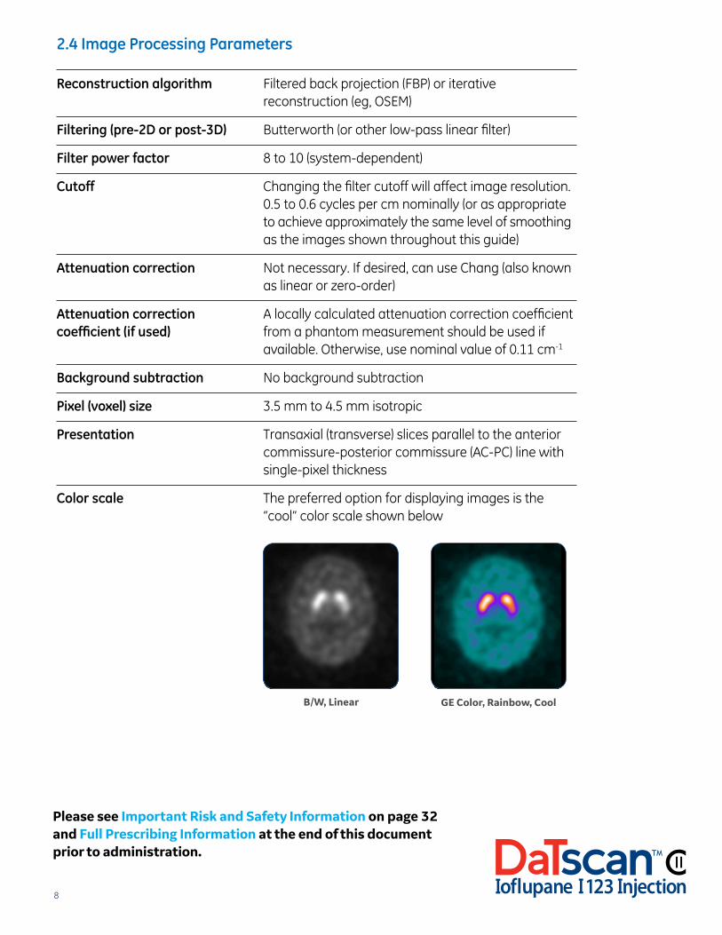
Color scale The preferred option for displaying images is the “cool” color scale shown below
Please see Important Risk and Safety Information on page 32 and Full Prescribing Information at the end of this document prior to administration.
GE Color, Rainbow, CoolB/W, Linear
9
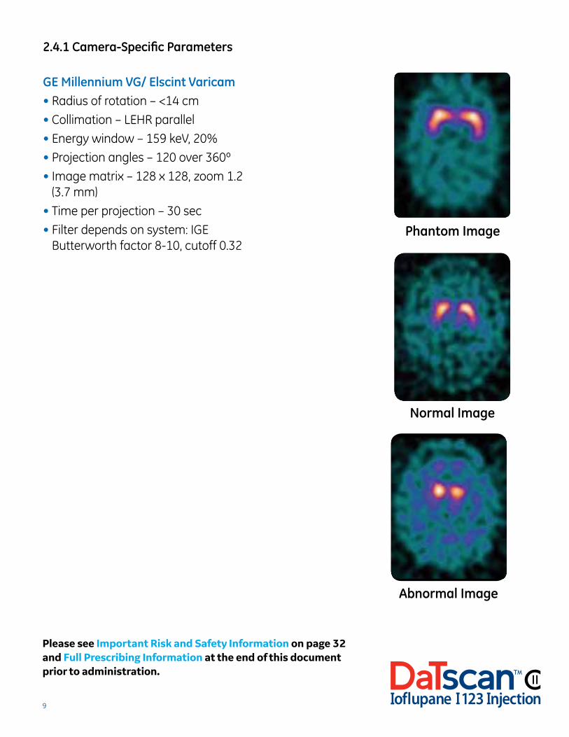
2.4.1 Camera-Specific Parameters
GE Millennium VG/ Elscint Varicam• Radius of rotation – <14 cm
• Collimation – LEHR parallel
• Energy window – 159 keV, 20%
• Projection angles – 120 over 360º
• Image matrix – 128 x 128, zoom 1.2 (3.7 mm)
• Time per projection – 30 sec
• Filter depends on system: IGE Butterworth factor 8-10, cutoff 0.32
Phantom Image
Normal Image
Abnormal Image
Please see Important Risk and Safety Information on page 32 and Full Prescribing Information at the end of this document prior to administration.
10
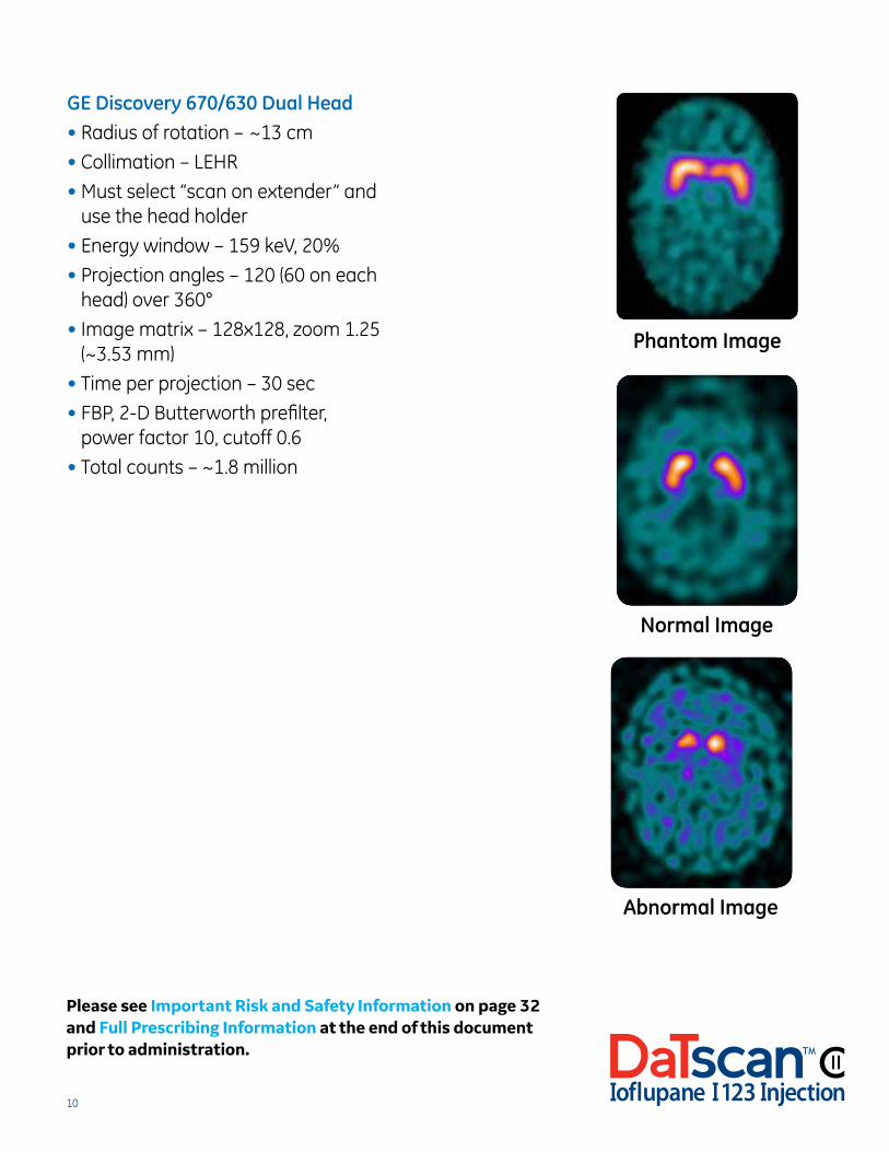
GE Discovery 670/630 Dual Head• Radius of rotation – ~13 cm
• Collimation – LEHR
• Must select “scan on extender” and use the head holder
• Energy window – 159 keV, 20%
• Projection angles – 120 (60 on each head) over 360°
• Image matrix – 128x128, zoom 1.25 (~3.53 mm)
• Time per projection – 30 sec
• FBP, 2-D Butterworth prefilter, power factor 10, cutoff 0.6
• Total counts – ~1.8 million
Phantom Image
Normal Image
Abnormal Image
Please see Important Risk and Safety Information on page 32 and Full Prescribing Information at the end of this document prior to administration.
11
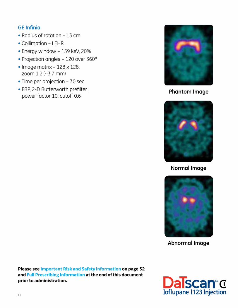
GE Infinia• Radius of rotation – 13 cm
• Collimation – LEHR
• Energy window – 159 keV, 20%
• Projection angles – 120 over 360º
• Image matrix – 128 x 128, zoom 1.2 (~3.7 mm)
• Time per projection – 30 sec
• FBP, 2-D Butterworth prefilter, power factor 10, cutoff 0.6
Phantom Image
Normal Image
Abnormal Image
Please see Important Risk and Safety Information on page 32 and Full Prescribing Information at the end of this document prior to administration.
12
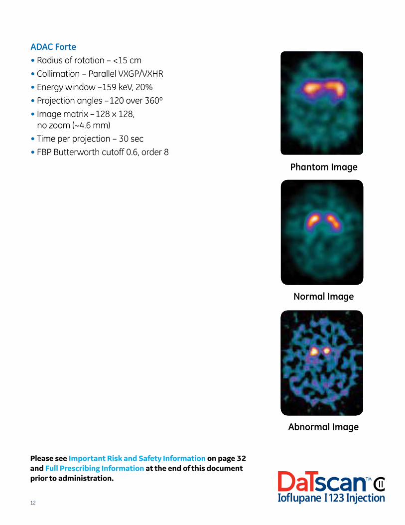
ADAC Forte• Radius of rotation – <15 cm
• Collimation – Parallel VXGP/VXHR
• Energy window –159 keV, 20%
• Projection angles –120 over 360º
• Image matrix – 128 x 128, no zoom (~4.6 mm)
• Time per projection – 30 sec
• FBP Butterworth cutoff 0.6, order 8
Phantom Image
Normal Image
Abnormal Image
Please see Important Risk and Safety Information on page 32 and Full Prescribing Information at the end of this document prior to administration.
13
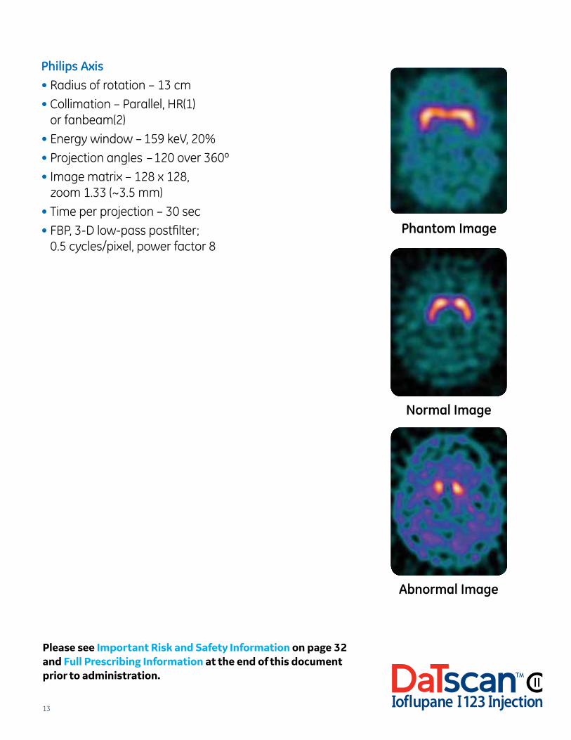
Philips Axis• Radius of rotation – 13 cm
• Collimation – Parallel, HR(1) or fanbeam(2)
• Energy window – 159 keV, 20%
• Projection angles – 120 over 360º
• Image matrix – 128 x 128, zoom 1.33 (~3.5 mm)
• Time per projection – 30 sec
• FBP, 3-D low-pass postfilter; 0.5 cycles/pixel, power factor 8
Phantom Image
Normal Image
Abnormal Image
Please see Important Risk and Safety Information on page 32 and Full Prescribing Information at the end of this document prior to administration.

14
Philips Prism 2000• Radius of rotation – 14 cm
• Collimation – UHR
• Energy window – 159 keV, 20%
• Projection angles – 120 over 360º
• Image matrix – 128 x 128, zoom 1.33 (~3.5 mm)
• Time per projection – 30 sec
• FBP, 3-D low-pass postfilter; 0.6 cycles/pixel, power factor 8 Phantom Image
Normal Image
Abnormal Image
Please see Important Risk and Safety Information on page 32 and Full Prescribing Information at the end of this document prior to administration.
15
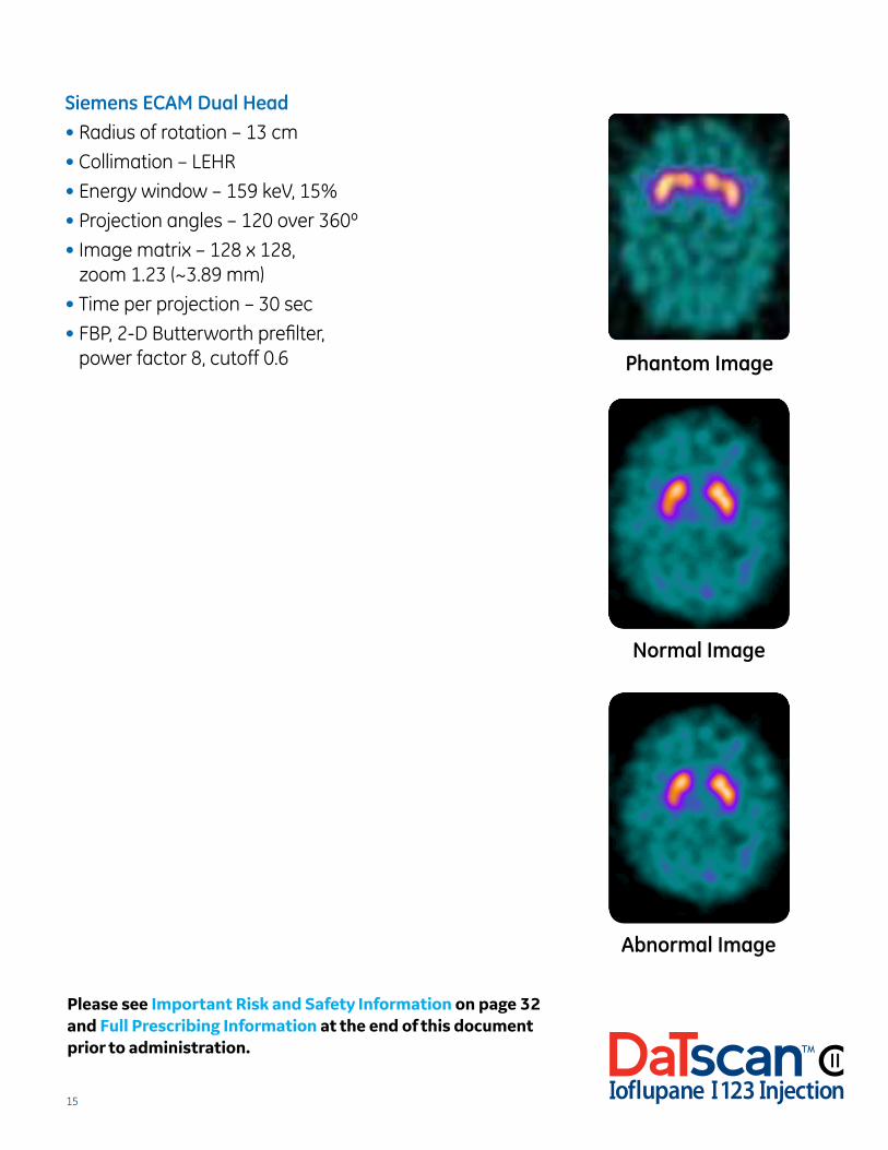
Siemens ECAM Dual Head• Radius of rotation – 13 cm
• Collimation – LEHR
• Energy window – 159 keV, 15%
• Projection angles – 120 over 360º
• Image matrix – 128 x 128, zoom 1.23 (~3.89 mm)
• Time per projection – 30 sec
• FBP, 2-D Butterworth prefilter, power factor 8, cutoff 0.6 Phantom Image
Normal Image
Abnormal Image
Please see Important Risk and Safety Information on page 32 and Full Prescribing Information at the end of this document prior to administration.
16
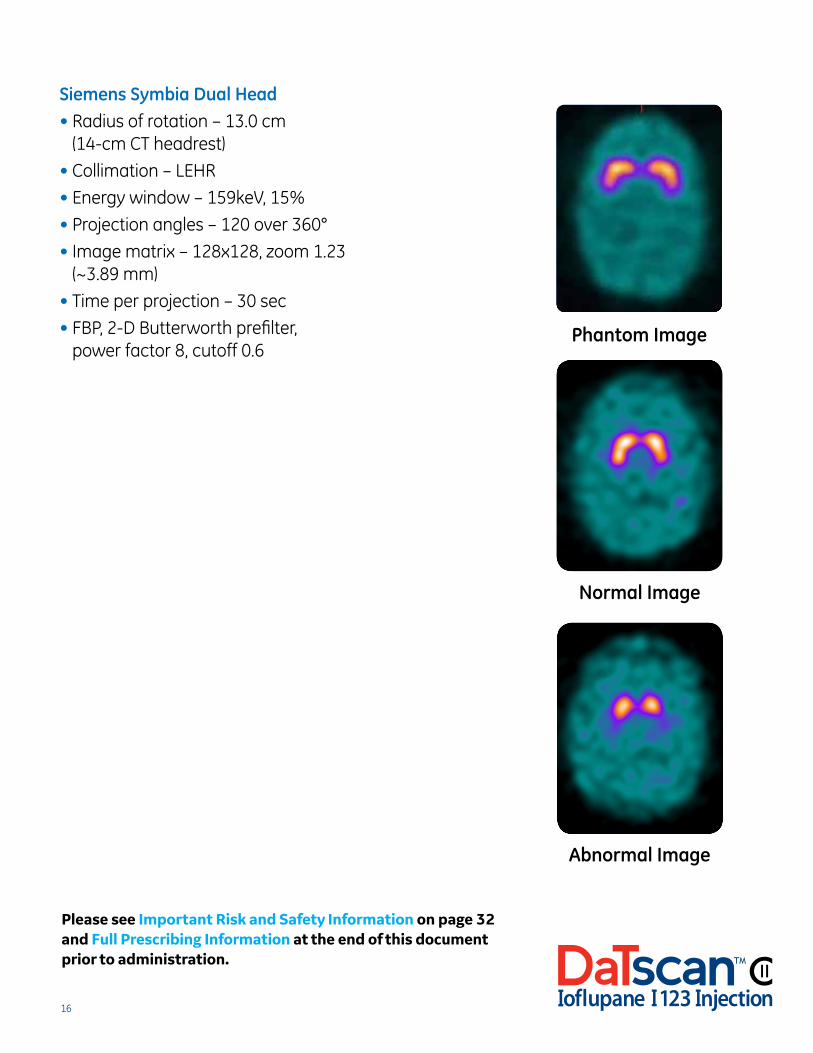
Siemens Symbia Dual Head• Radius of rotation – 13.0 cm
(14-cm CT headrest)
• Collimation – LEHR
• Energy window – 159keV, 15%
• Projection angles – 120 over 360°
• Image matrix – 128x128, zoom 1.23 (~3.89 mm)
• Time per projection – 30 sec
• FBP, 2-D Butterworth prefilter, power factor 8, cutoff 0.6
Phantom Image
Normal Image
Abnormal Image
Please see Important Risk and Safety Information on page 32 and Full Prescribing Information at the end of this document prior to administration.
17
2.5 Quality Checks
Prior to interpretation, images should be reviewed for correct acquisition and processing procedures:
• All parameters stated in sections 2.3 and 2.4 should be followed
• Radius of rotation must be 11 to a maximum of 15 cm
• FOV should extend from the top of the head to the level of the middle ear
• Minimum 1.5 M total counts
• Pixel size must be between 3.5 mm and 4.5 mm
• No visible patient motion or camera-head misalignment in the rotating tomograms
• No more than two dropped frames are allowed in the study, and they cannot be consecutive (adjacent)
• The patient’s head should not show a lateral (left or right ear toward shoulder) tilt. A slight caudal tilt (chin up or down) or rotation of the head is acceptable
• Transaxial slices should be oriented with the front of the head pointing up
Images should be reviewed for quality problems immediately after acquisition. If image quality problems are detected (such as patient motion, incorrect pixel size, dropped frames, or low total counts), it is preferable to reimage the patient (while still within the three- to six-hour window postinjection) than to apply a software corrective solution. The start time for the second acquisition should occur no later than six hours postinjection.
2.6 Examples of Deficient Image Quality Causes
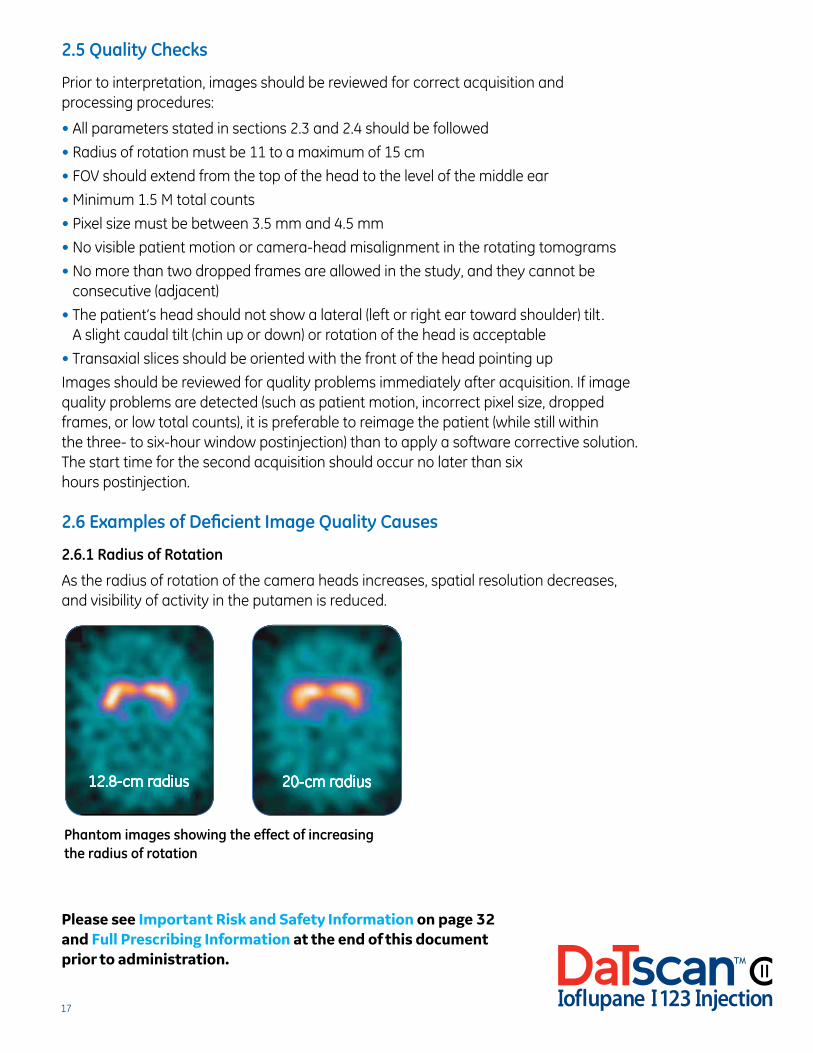
2.6.1 Radius of Rotation
As the radius of rotation of the camera heads increases, spatial resolution decreases, and visibility of activity in the putamen is reduced.
Phantom images showing the effect of increasing the radius of rotation
12.8-cm radius 20-cm radius
Please see Important Risk and Safety Information on page 32 and Full Prescribing Information at the end of this document prior to administration.
18
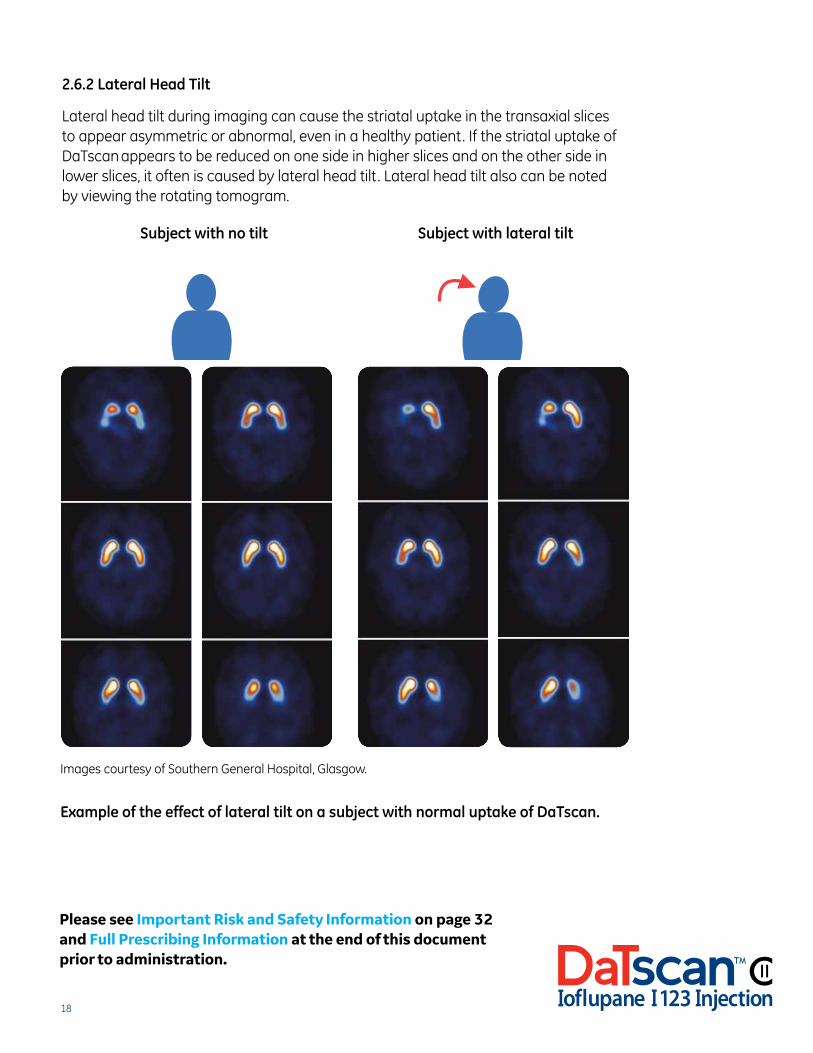
2.6.2 Lateral Head Tilt
Lateral head tilt during imaging can cause the striatal uptake in the transaxial slices to appear asymmetric or abnormal, even in a healthy patient. If the striatal uptake of DaTscan appears to be reduced on one side in higher slices and on the other side in lower slices, it often is caused by lateral head tilt. Lateral head tilt also can be noted by viewing the rotating tomogram.
Images courtesy of Southern General Hospital, Glasgow.
Example of the effect of lateral tilt on a subject with normal uptake of DaTscan.
Subject with no tilt Subject with lateral tilt
Example of the effect of lateral tilt on a subject with normal uptake of DaTscan™
Example of the effect of lateral tilt on a subject with normal uptake of DaTscan™
Please see Important Risk and Safety Information on page 32 and Full Prescribing Information at the end of this document prior to administration.
19
Interpretation and Reporting2.7 Normal vs Abnormal Images
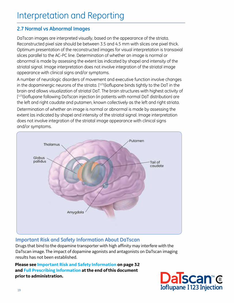
DaTscan images are interpreted visually, based on the appearance of the striata. Reconstructed pixel size should be between 3.5 and 4.5 mm with slices one pixel thick. Optimum presentation of the reconstructed images for visual interpretation is transaxial slices parallel to the AC-PC line. Determination of whether an image is normal or abnormal is made by assessing the extent (as indicated by shape) and intensity of the striatal signal. Image interpretation does not involve integration of the striatal image appearance with clinical signs and/or symptoms.
A number of neurologic disorders of movement and executive function involve changes in the dopaminergic neurons of the striata. [123I]ioflupane binds tightly to the DaT in the brain and allows visualization of striatal DaT. The brain structures with highest activity of [123I]ioflupane following DaTscan injection (in patients with normal DaT distribution) are the left and right caudate and putamen, known collectively as the left and right striata.
Determination of whether an image is normal or abnormal is made by assessing the extent (as indicated by shape) and intensity of the striatal signal. Image interpretation does not involve integration of the striatal image appearance with clinical signs and/or symptoms.
Important Risk and Safety Information About DaTscanDrugs that bind to the dopamine transporter with high affinity may interfere with the DaTscan image. The impact of dopamine agonists and antagonists on DaTscan imaging results has not been established.
Please see Important Risk and Safety Information on page 32 and Full Prescribing Information at the end of this document prior to administration.
20
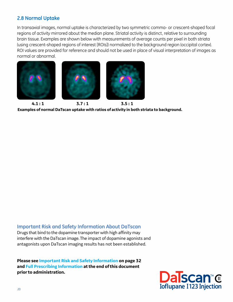
2.8 Normal Uptake
In transaxial images, normal uptake is characterized by two symmetric comma- or crescent-shaped focal regions of activity mirrored about the median plane. Striatal activity is distinct, relative to surrounding brain tissue. Examples are shown below with measurements of average counts per pixel in both striata (using crescent-shaped regions of interest [ROIs]) normalized to the background region (occipital cortex). ROI values are provided for reference and should not be used in place of visual interpretation of images as normal or abnormal.
Important Risk and Safety Information About DaTscanDrugs that bind to the dopamine transporter with high affinity may interfere with the DaTscan image. The impact of dopamine agonists and antagonists upon DaTscan imaging results has not been established.
Please see Important Risk and Safety Information on page 32 and Full Prescribing Information at the end of this document prior to administration.
Examples of normal DaTscan uptake with ratios of activity in both striata to background. 4.1 : 1 3.7 : 1 3.5 : 1
21
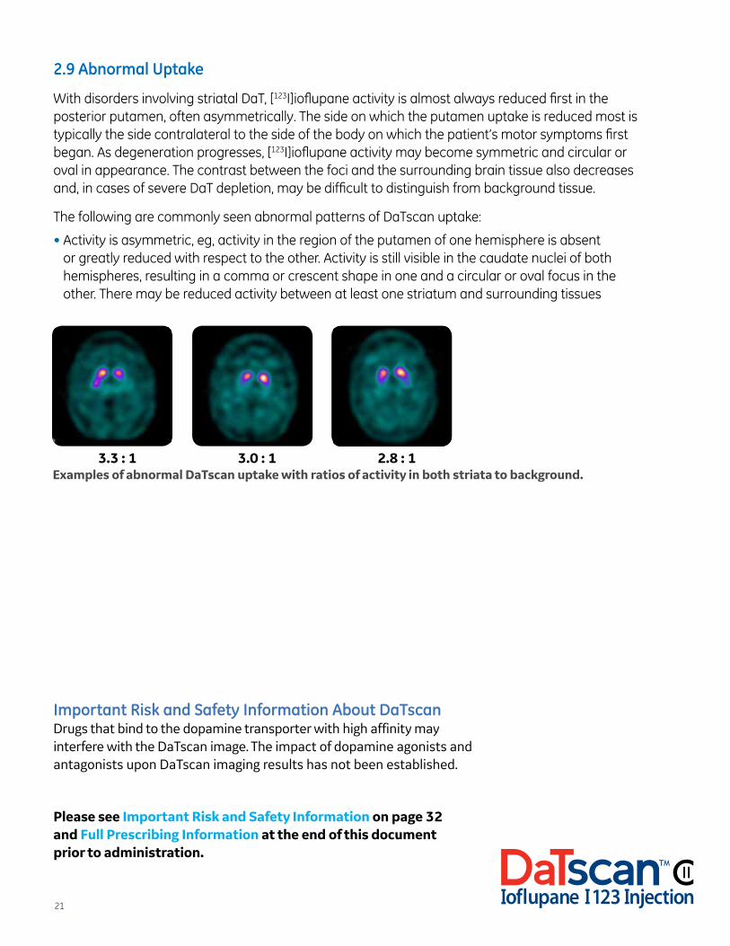
2.9 Abnormal Uptake
With disorders involving striatal DaT, [123I]ioflupane activity is almost always reduced first in the posterior putamen, often asymmetrically. The side on which the putamen uptake is reduced most is typically the side contralateral to the side of the body on which the patient’s motor symptoms first began. As degeneration progresses, [123I]ioflupane activity may become symmetric and circular or oval in appearance. The contrast between the foci and the surrounding brain tissue also decreases and, in cases of severe DaT depletion, may be difficult to distinguish from background tissue.
The following are commonly seen abnormal patterns of DaTscan uptake:
• Activity is asymmetric, eg, activity in the region of the putamen of one hemisphere is absent or greatly reduced with respect to the other. Activity is still visible in the caudate nuclei of both hemispheres, resulting in a comma or crescent shape in one and a circular or oval focus in the other. There may be reduced activity between at least one striatum and surrounding tissues
Important Risk and Safety Information About DaTscanDrugs that bind to the dopamine transporter with high affinity may interfere with the DaTscan image. The impact of dopamine agonists and antagonists upon DaTscan imaging results has not been established.
Please see Important Risk and Safety Information on page 32 and Full Prescribing Information at the end of this document prior to administration.
3.3 : 1 3.0 : 1 2.8 : 1Examples of abnormal DaTscan uptake with ratios of activity in both striata to background.
22
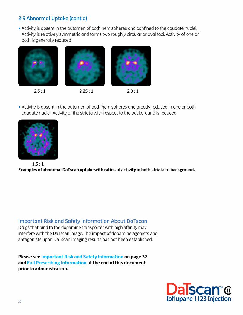
2.9 Abnormal Uptake (cont’d)
• Activity is absent in the putamen of both hemispheres and confined to the caudate nuclei. Activity is relatively symmetric and forms two roughly circular or oval foci. Activity of one or both is generally reduced
• Activity is absent in the putamen of both hemispheres and greatly reduced in one or both caudate nuclei. Activity of the striata with respect to the background is reduced
2.5 : 1 2.25 : 1 2.0 : 1
1.5 : 1 Examples of abnormal DaTscan uptake with ratios of activity in both striata to background.
Important Risk and Safety Information About DaTscanDrugs that bind to the dopamine transporter with high affinity may interfere with the DaTscan image. The impact of dopamine agonists and antagonists upon DaTscan imaging results has not been established.
Please see Important Risk and Safety Information on page 32 and Full Prescribing Information at the end of this document prior to administration.
23
Please see Important Risk and Safety Information on page 32 and Full Prescribing Information at the end of this document prior to administration.
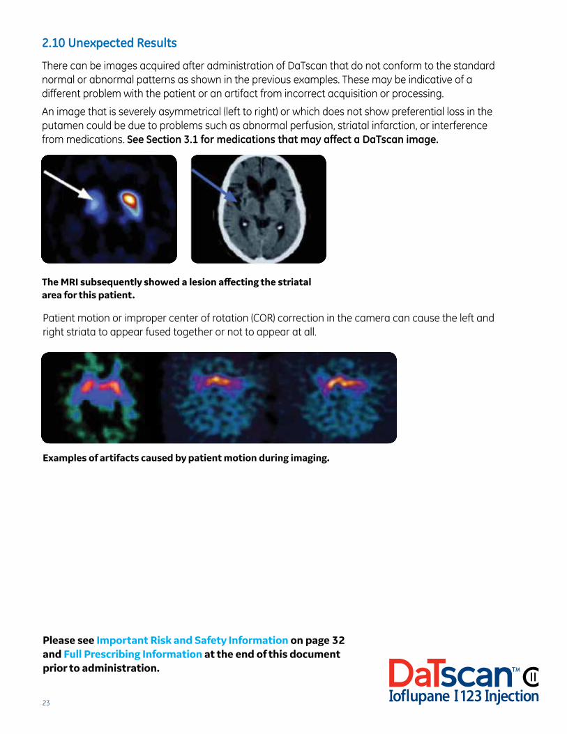
2.10 Unexpected Results
There can be images acquired after administration of DaTscan that do not conform to the standard normal or abnormal patterns as shown in the previous examples. These may be indicative of a different problem with the patient or an artifact from incorrect acquisition or processing.
An image that is severely asymmetrical (left to right) or which does not show preferential loss in the putamen could be due to problems such as abnormal perfusion, striatal infarction, or interference from medications. See Section 3.1 for medications that may affect a DaTscan image.
The MRI subsequently showed a lesion affecting the striatal area for this patient.
Patient motion or improper center of rotation (COR) correction in the camera can cause the left and right striata to appear fused together or not to appear at all.
Examples of artifacts caused by patient motion during imaging.
24
2.11 Reporting RemindersBe sure to consider the following points when reporting the results of DaTscan imaging to the referring physician:
• Results of visual examination
• Results of quantification (if performed). Please note that quantification is not necessary for the interpretation of DaTscan SPECT images
• Accuracy of acquisition
• How much DaTscan was injected
• Time point of acquisition
Please see Important Risk and Safety Information on page 32 and Full Prescribing Information at the end of this document prior to administration.
25
Step-by-Step Guide to DaTscan
Section 3
26
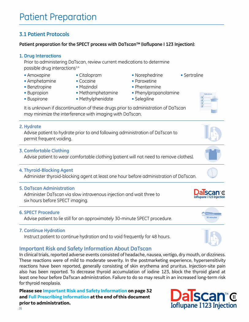
Patient Preparation
3.1 Patient Protocols
Patient preparation for the SPECT process with DaTscan™ (Ioflupane I 123 Injection):
1. Drug InteractionsPrior to administering DaTscan, review current medications to determine possible drug interactions1,4
• Amoxapine • Citalopram • Norephedrine • Sertraline• Amphetamine • Cocaine • Paroxetine• Benztropine • Mazindol • Phentermine• Bupropion • Methamphetamine • Phenylpropanolamine • Buspirone • Methylphenidate • Selegiline
It is unknown if discontinuation of these drugs prior to administration of DaTscan may minimize the interference with imaging with DaTscan.
2. HydrateAdvise patient to hydrate prior to and following administration of DaTscan to permit frequent voiding.
3. Comfortable ClothingAdvise patient to wear comfortable clothing (patient will not need to remove clothes).
4. Thyroid-Blocking AgentAdminister thyroid-blocking agent at least one hour before administration of DaTscan.
5. DaTscan AdministrationAdminister DaTscan via slow intravenous injection and wait three to six hours before SPECT imaging.
6. SPECT ProcedureAdvise patient to lie still for an approximately 30-minute SPECT procedure.
7. Continue HydrationInstruct patient to continue hydration and to void frequently for 48 hours.
Important Risk and Safety Information About DaTscanIn clinical trials, reported adverse events consisted of headache, nausea, vertigo, dry mouth, or dizziness. These reactions were of mild to moderate severity. In the postmarketing experience, hypersensitivity reactions have been reported, generally consisting of skin erythema and pruritus. Injection-site pain also has been reported. To decrease thyroid accumulation of iodine 123, block the thyroid gland at least one hour before DaTscan administration. Failure to do so may result in an increased long-term risk for thyroid neoplasia.
Medications
30 minutes
Medications
30 minutes
Medications
30 minutes
Medications
30 minutes
Medications
30 minutes
Medications
30 minutes
Please see Important Risk and Safety Information on page 32 and Full Prescribing Information at the end of this document prior to administration.
27
3.2 Patient Counseling
Instruct patients to inform you if they1:
• Have reduced renal or hepatic function
• Are sensitive to DaTscan or iodine
• Are sensitive to potassium iodide oral solution or Lugol’s Solution
• May be pregnant, are trying to become pregnant, or are breastfeeding
Instruct patients to increase their level of hydration prior to and after receiving DaTscan and to void frequently for the first 48 hours following administration of DaTscan.
Please see Important Risk and Safety Information on page 32 and Full Prescribing Information at the end of this document prior to administration.
28
Dosing
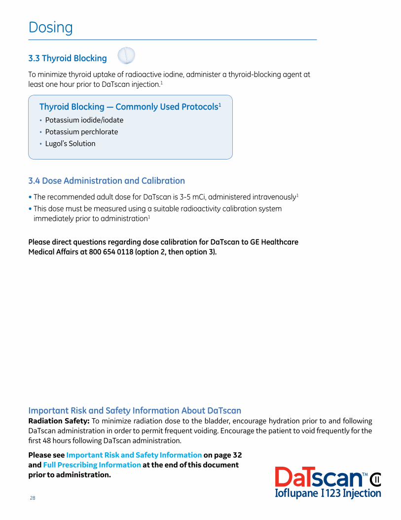
3.3 Thyroid Blocking
To minimize thyroid uptake of radioactive iodine, administer a thyroid-blocking agent at least one hour prior to DaTscan injection.1
3.4 Dose Administration and Calibration
• The recommended adult dose for DaTscan is 3-5 mCi, administered intravenously1
• This dose must be measured using a suitable radioactivity calibration system immediately prior to administration1
Please direct questions regarding dose calibration for DaTscan to GE Healthcare Medical Affairs at 800 654 0118 (option 2, then option 3).
Thyroid Blocking — Commonly Used Protocols1
• Potassium iodide/iodate
• Potassium perchlorate
• Lugol’s Solution
Important Risk and Safety Information About DaTscanRadiation Safety: To minimize radiation dose to the bladder, encourage hydration prior to and following DaTscan administration in order to permit frequent voiding. Encourage the patient to void frequently for the first 48 hours following DaTscan administration.
Medications
30 minutes
Please see Important Risk and Safety Information on page 32 and Full Prescribing Information at the end of this document prior to administration.
29
Administration and Storage
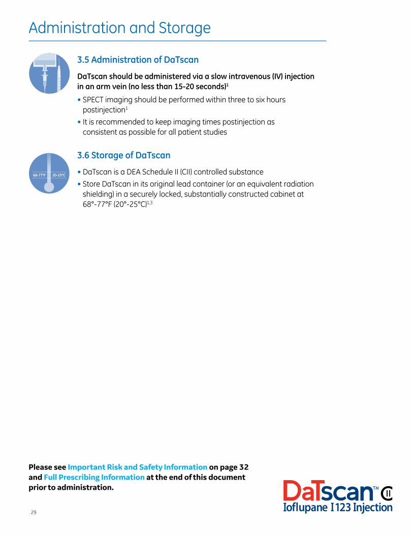
3.5 Administration of DaTscan
DaTscan should be administered via a slow intravenous (IV) injection in an arm vein (no less than 15-20 seconds)1
• SPECT imaging should be performed within three to six hours postinjection1
• It is recommended to keep imaging times postinjection as consistent as possible for all patient studies
3.6 Storage of DaTscan
• DaTscan is a DEA Schedule II (CII) controlled substance
• Store DaTscan in its original lead container (or an equivalent radiation shielding) in a securely locked, substantially constructed cabinet at 68°-77°F (20°-25°C)1,3
140
120
100
80
60
40
20
0
°F °C
60
50
40
30
20
10
0
-1068-77°F 20-25°C
140
120
100
80
60
40
20
0
°F °C
60
50
40
30
20
10
0
-1068-77°F 20-25°C
Please see Important Risk and Safety Information on page 32 and Full Prescribing Information at the end of this document prior to administration.
30
3.7 Schedule II Radiopharmaceutical Requirements
The following checklist summarizes the licensing requirements and GE Healthcare’s shipping requirements for a Schedule II product:
Current and Valid Drug Enforcement Administration (DEA) and Radioactive Materials (RAM) Licenses • In order for GE Healthcare to deliver DaTscan, the address on both licenses must be the same
and must be the address to which the product will be shipped
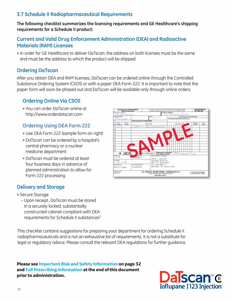
Ordering DaTscan After you obtain DEA and RAM licenses, DaTscan can be ordered online through the Controlled Substance Ordering System (CSOS) or with a paper DEA Form 222. It is important to note that the paper form will soon be phased out and DaTscan will be available only through online orders.
Ordering Online Via CSOS • You can order DaTscan online at
http://www.orderdatscan.com
Ordering Using DEA Form 222 • Use DEA Form 222 (sample form on right)
• DaTscan can be ordered by a hospital’s central pharmacy or a nuclear medicine department
• DaTscan must be ordered at least four business days in advance of planned administration to allow for Form 222 processing
Delivery and Storage • Secure Storage – Upon receipt, DaTscan must be stored
in a securely locked, substantially constructed cabinet compliant with DEA requirements for Schedule II substances3
SMITH, JOHN MD ANYTOWN HOSPITAL DEPT OF RADIOLOGY 123 ANYTOWN ST ANYTOWN, PA 12345-6789
See Reverse of PURCHASER'S Copy for Instructions
OMB APPROVALNo. 0000-000
No order form may be issued for Schedule I and II substances unless a completed application form has been received, (21 CFR 1305,04).
TO: (Name of Supplier) STREET ADDRESS
City and STATE DATE TO BE FILLED IN BY SUPPLIER SUPPLIERS DEA REGISTRATION No.
TO BE FILLED IN BY PURCHASERLINE
No.
1
2
3
4
5
6
7
8
9
10
No. of Size of Packages Date Packages Package Name of Item National Drug Code Shipped Shipped
Date Issued DEA Registration No. Name and Address of Registrant
Scheduless
Registered as a No. of this Order Form
DEA Form 222(May 2008)
SIGNATURE OF PURCHASEROR ATTORNEY OR AGENT(MUST BE 10 OR LESS)LAST LINE
COMPLETED
U.S. OFFICIAL ORDER FORMS - SCHEDULES I & IIDRUG ENFORCEMENT ADMINISTRATION
SUPPLIER’S COPY 1
2/1/11
2, 2N, 3, 3N
XX1234567
123456789PRACTITIONER
123456789
SAMPLE
This checklist contains suggestions for preparing your department for ordering Schedule II radiopharmaceuticals and is not an exhaustive list of requirements. It is not a substitute for legal or regulatory advice. Please consult the relevant DEA regulations for further guidance.
Please see Important Risk and Safety Information on page 32 and Full Prescribing Information at the end of this document prior to administration.

31
Important Risk and Safety Information
Section 4
32
Section 4
Important Risk and Safety Information About DaTscan™ (Ioflupane I 123 Injection)INDICATIONS AND USE: DaTscan is a radiopharmaceutical indicated for striatal dopamine transporter visualization using single-photon emission computed tomography (SPECT) brain imaging to assist in the evaluation of adult patients with suspected parkinsonian syndromes (PSs). DaTscan may be used to help differentiate essential tremor from tremor due to PS (idiopathic Parkinson’s disease [PD], multiple system atrophy [MSA], and progressive supranuclear palsy [PSP]). DaTscan is an adjunct to other diagnostic evaluations. DaTscan was not designed to distinguish among PD, MSA, and PSP. The effectiveness of DaTscan as a screening or confirmatory test and for monitoring disease progression or response to therapy has not been established. CONTRAINDICATIONS: DaTscan is contraindicated in patients with known hypersensitivity to the active substance, any of the excipients, or iodine. WARNINGS AND PRECAUTIONS — Hypersensitivity Reactions: Hypersensitivity reactions, generally consisting of skin erythema and pruritus, have been reported following DaTscan administration. Thyroid Accumulation: The DaTscan injection may contain up to 6% of free iodide (iodine 123 or I-123). To decrease thyroid accumulation of I-123, block the thyroid gland at least one hour before administration of DaTscan; failure to do so may increase the long-term risk for thyroid neoplasia. ADVERSE REACTIONS: In clinical trials, headache, nausea, vertigo, dry mouth, or dizziness of mild to moderate severity were reported. In postmarketing experience, hypersensitivity reactions and injection-site pain have been reported. DRUG INTERACTIONS: Drugs that bind to the dopamine transporter with high affinity may interfere with the DaTscan image. The impact of dopamine agonists and antagonists on DaTscan imaging results has not been established. SPECIFIC POPULATIONS — Pregnancy: It is unknown whether DaTscan can cause fetal harm or increase the risk of pregnancy loss in pregnant women. DaTscan should be given to pregnant women only if clearly needed. Like all radiopharmaceuticals, DaTscan may cause fetal harm, depending on the stage of fetal development and the magnitude of the radionuclide dose. Radioactive iodine products cross the placenta and can permanently impair fetal thyroid function. Nursing Mothers: It is not known whether DaTscan is excreted into human milk; however, I-123 is excreted into human milk. Because many drugs are excreted into human milk and because of the potential for serious adverse reactions in nursing infants, a decision should be made whether to interrupt nursing after administration of DaTscan or not to administer DaTscan at all. Nursing women may consider interrupting nursing and pump and discard breast milk for six days after DaTscan administration to minimize risks to a nursing infant. Pediatric Use: The safety and efficacy of DaTscan have not been established in pediatric patients. Geriatric Use: There were no differences in responses between the elderly and younger patients that would require a dose adjustment. Renal and Hepatic Impairment: The effect of renal or hepatic impairment on DaTscan imaging has not been established. The kidney excretes DaTscan; patients with severe renal impairment may have increased radiation exposure and altered DaTscan images. DRUG ABUSE AND DEPENDENCE: Ioflupane I 123 Injection is a DEA Schedule II controlled substance. A DEA license is required for handling or administering this controlled substance. OVERDOSAGE: It is unknown whether or not ioflupane is dialyzable. The major risks of overdose relate to increased radiation exposure and long-term risk for neoplasia. In case of radioactivity overdosage, frequent urination and defecation should be encouraged to minimize radiation exposure to the patient. PROCEDURE — Radiation Safety: DaTscan emits radiation and must be handled with safety measures to minimize radiation exposure to clinical personnel and patients.
27
Please read Full Prescribing Information at the end of this document prior to administration.
TABLE OF CONTENTS
33
Section 5
GE Healthcare’s Commitment to You
If you have a question regarding DaTscan, please refer to the numbers below for assistance:
Customer Service To place an order, call 800 292 8514
Medical Affairs For technical or product-related questions, call 800 654 0118 (option 2, then option 3)
Reimbursement Hotline For reimbursement-related questions (eg, appropriate coding), call our Hotline at 800 767 6664
For more information about DaTscan, visit www.datscan.com.
Please see Important Risk and Safety Information on page 32 and Full Prescribing Information at the end of this document prior to administration.
© 2015 General Electric Company — All rights reserved.GE and the GE Monogram are trademarks of General Electric Company.
DaTscan is a trademark of General Electric Company or one of its subsidiaries.
April 2015 73-JB14728USa(1)
References: 1. DaTscan [prescribing information]. Arlington Heights, IL: GE Healthcare; 2011. 2. Colloby SJ, Williams ED, Burn DJ, Lloyd JJ, McKeith IG, O’Brien JT. Progression of dopaminergic degeneration in dementia with Lewy bodies and Parkinson’s disease with and without dementia assessed using 123I-FP-CIT SPECT. Eur J Nucl Med Mol Imaging. 2005;32:1176-1185. 3. Drug Enforcement Administration. Section 1301.75 Physical security controls for practitioners. In: Code of Federal Regulations. http://www.deadiversion.usdoj.gov/21cfr/cfr/1301/1301_75.htm. Accessed March 5, 2015. 4. Booij J, Kemp P. Dopamine transporter imaging with [123I]FP-CIT SPECT: potential effects of drugs. Eur J Nucl Med Mol Imaging. 2008;35:424-438.

34
HIGHLIGHTS OF PRESCRIBING INFORMATIONThese highlights do not include all the information needed to use DaTscansafely and effectively. See full prescribing information for DaTscan.
DaTscan (Ioflupane I 123 Injection) for Intravenous Use, CIIInitial U.S. Approval: 2011
INDICATIONS AND USAGEDaTscan (Ioflupane I 123 Injection) is a radiopharmaceutical indicated for striataldopamine transporter visualization using single photon emission computedtomography (SPECT) brain imaging to assist in the evaluation of adult patients withsuspected Parkinsonian syndromes (PS). In these patients, DaTscan may be used tohelp differentiate essential tremor from tremor due to PS (idiopathic Parkinson'sdisease, multiple system atrophy and progressive supranuclear palsy). DaTscan is anadjunct to other diagnostic evaluations. (1)
DOSAGE AND ADMINISTRATION• DaTscan emits gamma radiation and must be handled with safety measures.
(2.1)• Measure patient dose by a suitable radioactivity calibration system immediately
prior to administration. (2.1)• Administer a thyroid-blocking agent at least one hour before the dose of
DaTscan. (2.2)• The recommended DaTscan dose is 111 to 185 MBq (3 to 5 mCi). (2.4)• Begin SPECT imaging between 3 and 6 hours post-injection. (2.6)
DOSAGE FORMS AND STRENGTHS2.5 mL of sterile solution for intravenous injection in a single-use vial [74 MBq (2mCi)/mL at calibration time]. (3)
CONTRAINDICATIONSKnown hypersensitivity to the active substance or to any of the excipients, or toiodine. (4)
WARNINGS AND PRECAUTIONS• Hypersensitivity reactions have been reported following DaTscan administration.
Have anaphylactic and hypersensitivity treatment measures available prior toDaTscan administration. (5.1)
• Administer a thyroid-blocking agent before DaTscan administration. (5.2)
ADVERSE REACTIONSHypersensitivity and injection site reactions have been reported following DaTscanadministration. (6.2) In clinical trials, the most common adverse reactions,headache, nausea, vertigo, dry mouth or dizziness occurred in < 1% of subjects.(6.1)
To report SUSPECTED ADVERSE REACTIONS, contact GE Healthcare at 1-800-654-0118 or FDA at 1-800-FDA-1088 or www.fda.gov/medwatch.
DRUG INTERACTIONSAmoxapine, amphetamine, benztropine, bupropion, buspirone, cocaine, mazindol, methamphetamine, methylphenidate, norephedrine, phentermine,phenylpropanolamine, selegiline, sertraline, citalopram and paroxetine mayinterfere with DaTscan imaging. (7) The effects of dopamine agonists andantagonists on DaTscan imaging have not been established.
USE IN SPECIFIC POPULATIONS• Pregnancy: No human or animal data. Any radiopharmaceutical, including
DaTscan, may cause fetal harm. Use only if clearly needed. (8.1)• Nursing Mothers: A decision should be made whether to interrupt nursing after
DaTscan administration or not to administer DaTscan, taking into considerationthe importance of the drug to the mother. (8.3)
• Pediatric: Safety and effectiveness have not been established. (8.4)
See 17 for PATIENT COUNSELING INFORMATION.
Revised: 4/2011
FULL PRESCRIBING INFORMATION: CONTENTS*
1 INDICATIONS AND USAGE2 DOSAGE AND ADMINISTRATION
2.1 Radiation Safety2.2 Thyroid Blockade Before DaTscan Injection2.3 Preparation and Administration2.4 Recommended Dose 2.5 Radiation Dosimetry2.6 Imaging Guidelines2.7 Image Interpretation
3 DOSAGE FORMS AND STRENGTHS4 CONTRAINDICATIONS5 WARNINGS AND PRECAUTIONS
5.1 Hypersensitivity Reactions5.2 Thyroid Accumulation
6 ADVERSE REACTIONS6.1 Clinical Study Experience 6.2 Postmarketing Experience
7 DRUG INTERACTIONS8 USE IN SPECIFIC POPULATIONS
8.1 Pregnancy8.3 Nursing Mothers8.4 Pediatric Use8.5 Geriatric Use8.6 Renal and Hepatic Impairment
9 Drug Abuse and Dependence9.1 Controlled Substance
10 OVERDOSAGE11 DESCRIPTION
11.1 Physical Characteristics11.2 External Radiation
12 CLINICAL PHARMACOLOGY12.1 Mechanism of Action12.2 Pharmacodynamics12.3 Pharmacokinetics
13 NONCLINICAL TOXICOLOGY13.1 Carcinogenesis, Mutagenesis, Impairment of Fertility13.2 Animal Toxicology and/or Pharmacology
14 CLINICAL STUDIES16 HOW SUPPLIED/STORAGE AND HANDLING17 PATIENT COUNSELING INFORMATION*Sections or subsections omitted from the full prescribing information are notlisted.
FULL PRESCRIBING INFORMATION
1 INDICATIONS AND USAGEDaTscan is a radiopharmaceutical indicated for striatal dopamine transportervisualization using single photon emission computed tomography (SPECT) brainimaging to assist in the evaluation of adult patients with suspected Parkinsoniansyndromes (PS). In these patients, DaTscan may be used to help differentiateessential tremor from tremor due to PS (idiopathic Parkinson’s disease, multiplesystem atrophy and progressive supranuclear palsy). DaTscan is an adjunct to otherdiagnostic evaluations.
2 DOSAGE AND ADMINISTRATION
2.1 Radiation Safety DaTscan emits radiation and must be handled with safety measures to minimizeradiation exposure to clinical personnel and patients. Radiopharmaceuticals shouldbe used by or under the control of physicians who are qualified by specific trainingand experienced in the safe use and handling of radionuclides, and whoseexperience and training have been approved by the appropriate government agencyauthorized to license the use of radionuclides. DaTscan dosing is based upon theradioactivity determined using a suitably calibrated instrument immediately prior toadministration.
To minimize radiation dose to the bladder, encourage hydration prior to andfollowing DaTscan administration in order to permit frequent voiding. Encouragethe patient to void frequently for the first 48 hours following DaTscanadministration [see Dosage and Administration (2.5)].
2.2 Thyroid Blockade Before DaTscan InjectionBefore administration of DaTscan, administer Potassium Iodide Oral Solution orLugol’s Solution (equivalent to 100 mg iodide) or potassium perchlorate (400 mg) toblock uptake of iodine 123 by the patient's thyroid. Administer the blocking agent atleast one hour before the dose of DaTscan [see Warnings and Precautions (5.2)].
2.3 Preparation and AdministrationUse aseptic procedures and radiation shielding during preparation andadministration. Inspect the DaTscan vial prior to administration and do not use it ifthe vial contains particulate matter or discoloration [see Description (11)]. AdministerDaTscan as a slow intravenous injection (administered over a period of not less than15 to 20 seconds) via an arm vein.
2.4 Recommended DoseThe recommended dose is 111 to 185 MBq (3 to 5 mCi) administered intravenously[see Clinical Studies (14)].
2.5 Radiation DosimetryThe estimated radiation absorbed doses to an average adult from intravenousinjection of DaTscan are shown in Table 1. The values are calculated assumingurinary bladder emptying at 4.8-hour intervals and appropriate thyroid blocking(iodine 123 is a known Auger electron emitter).
DaTscan™
Ioflupane I 123 InjectionCII
TABLE OF CONTENTS
35
Table 1Estimated Radiation Absorbed Doses from DaTscan
ABSORBED DOSE PERORGAN / TISSUE UNIT ADMINISTERED
ACTIVITY(μGy / MBq)
Adrenals 12.9Brain 17.8Striata 230.0Breasts 7.8Esophagus 10.0Gallbladder Wall 26.4Stomach Wall 11.2Small Intestine Wall 21.2Colon Wall a 39.8Upper Large Intestine Wall 38.1Lower Large Intestine Wall 42.0Heart Wall 12.9Kidneys 10.9Liver 27.9Lungs 41.2Muscle 9.4Osteogenic Cells 28.2Ovaries 16.8Pancreas 13.0Red Marrow 9.2Skin 6.0Spleen 10.4Testes 8.5Thymus 10.0Thyroid 9.0Urinary Bladder Wall 53.1Uterus 16.1Total Body 11.3EFFECTIVE DOSE PER UNITADMINISTERED ACTIVITY (µSv/MBq) 21.3
a The absorbed dose to the colon wall is the mass-weighted sum of the absorbeddoses to the upper and lower large intestine walls, Dcolon = 0.57DULI + 0.43DLLI[Publication 80 of the ICRP (International Commission on Radiological Protection);Annals of the ICRP 28 (3). Oxford: Pergamon Press; 1998]
The Effective Dose resulting from a DaTscan administration with an administeredactivity of 185 MBq (5 mCi) is 3.94 mSv in an adult.
2.6 Imaging GuidelinesBegin SPECT imaging 3 to 6 hours following DaTscan administration. Acquire imagesusing a gamma camera fitted with high-resolution collimators and set to aphotopeak of 159 keV with a ± 10% energy window. Angular sampling should be notless than 120 views over 360 degrees. Position the subject supine with the head onan off-the-table headrest, a flexible head restraint such as a strip of tape across thechin or forehead may be used to help avoid movement, and set a circular orbit forthe detector heads with the radius as small as possible (typically 11 to 15 cm).
Experimental studies with a striatal phantom suggest that optimal images areobtained with matrix size and zoom factors selected to give a pixel size of 3.5 to 4.5mm. Collect a minimum of 1.5 million counts for optimal images.
2.7 Image InterpretationDaTscan images are interpreted visually, based upon the appearance of the striata.Reconstructed pixel size should be between 3.5 and 4.5 mm with slices 1 pixel thick.Optimum presentation of the reconstructed images for visual interpretation istransaxial slices parallel to the anterior commissure-posterior commissure (AC-PC)line. Determination of whether an image is normal or abnormal is made by assessingthe extent (as indicated by shape) and intensity of the striatal signal. Imageinterpretation does not involve integration of the striatal image appearance withclinical signs and/or symptoms.
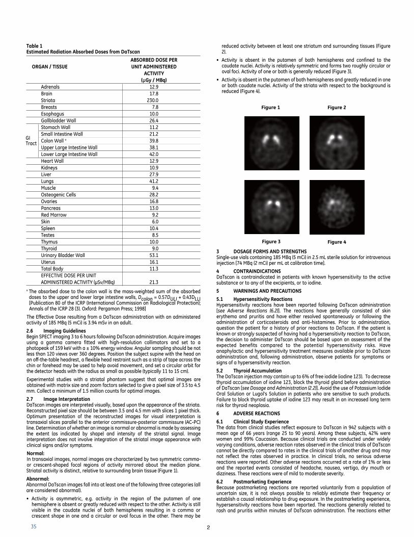
Normal: In transaxial images, normal images are characterized by two symmetric comma-or crescent-shaped focal regions of activity mirrored about the median plane.Striatal activity is distinct, relative to surrounding brain tissue (Figure 1).
Abnormal:Abnormal DaTscan images fall into at least one of the following three categories (allare considered abnormal).
• Activity is asymmetric, e.g. activity in the region of the putamen of onehemisphere is absent or greatly reduced with respect to the other. Activity is stillvisible in the caudate nuclei of both hemispheres resulting in a comma orcrescent shape in one and a circular or oval focus in the other. There may be
2
reduced activity between at least one striatum and surrounding tissues (Figure2).
• Activity is absent in the putamen of both hemispheres and confined to thecaudate nuclei. Activity is relatively symmetric and forms two roughly circular oroval foci. Activity of one or both is generally reduced (Figure 3).
• Activity is absent in the putamen of both hemispheres and greatly reduced in oneor both caudate nuclei. Activity of the striata with respect to the background isreduced (Figure 4).
3 DOSAGE FORMS AND STRENGTHSSingle-use vials containing 185 MBq (5 mCi) in 2.5 mL sterile solution for intravenousinjection [74 MBq (2 mCi) per mL at calibration time].
4 CONTRAINDICATIONSDaTscan is contraindicated in patients with known hypersensitivity to the activesubstance or to any of the excipients, or to iodine.
5 WARNINGS AND PRECAUTIONS
5.1 Hypersensitivity ReactionsHypersensitivity reactions have been reported following DaTscan administration[see Adverse Reactions (6.2)]. The reactions have generally consisted of skinerythema and pruritis and have either resolved spontaneously or following theadministration of corticosteroids and anti-histamines. Prior to administration,question the patient for a history of prior reactions to DaTscan. If the patient isknown or strongly suspected of having had a hypersensitivity reaction to DaTscan,the decision to administer DaTscan should be based upon an assessment of theexpected benefits compared to the potential hypersensitivity risks. Haveanaphylactic and hypersensitivity treatment measures available prior to DaTscanadministration and, following administration, observe patients for symptoms orsigns of a hypersensitivity reaction.
5.2 Thyroid AccumulationThe DaTscan injection may contain up to 6% of free iodide (iodine 123). To decreasethyroid accumulation of iodine 123, block the thyroid gland before administrationof DaTscan [see Dosage and Administration (2.2)]. Avoid the use of Potassium IodideOral Solution or Lugol’s Solution in patients who are sensitive to such products.Failure to block thyroid uptake of iodine 123 may result in an increased long termrisk for thyroid neoplasia.
6 ADVERSE REACTIONS
6.1 Clinical Study ExperienceThe data from clinical studies reflect exposure to DaTscan in 942 subjects with amean age of 66 years (range 25 to 90 years). Among these subjects, 42% werewomen and 99% Caucasian. Because clinical trials are conducted under widelyvarying conditions, adverse reaction rates observed in the clinical trials of DaTscancannot be directly compared to rates in the clinical trials of another drug and maynot reflect the rates observed in practice. In clinical trials, no serious adversereactions were reported. Other adverse reactions occurred at a rate of 1% or lessand the reported events consisted of headache, nausea, vertigo, dry mouth ordizziness. These reactions were of mild to moderate severity.
6.2 Postmarketing ExperienceBecause postmarketing reactions are reported voluntarily from a population ofuncertain size, it is not always possible to reliably estimate their frequency orestablish a causal relationship to drug exposure. In the postmarketing experience,hypersensitivity reactions have been reported. The reactions generally related torash and pruritis within minutes of DaTscan administration. The reactions either
Figure 1 Figure 2
Figure 3 Figure 4
GI Tract
36
resolved spontaneously or following the administration of corticosteroids andantihistamines. Injection site pain has also been reported.
7 DRUG INTERACTIONSThe ioflupane within DaTscan binds to the dopamine transporter. Drugs that bindto the dopamine transporter with high affinity may interfere with the imageobtained following DaTscan administration. These potentially interfering drugsconsist of: amoxapine, amphetamine, benztropine, bupropion, buspirone, cocaine,mazindol, methamphetamine, methylphenidate, norephedrine, phentermine,phenylpropanolamine, selegiline, and sertraline. Selective serotonin reuptakeinhibitors (paroxetine and citalopram) may increase or decrease ioflupane bindingto the dopamine transporter. Whether discontinuation of these drugs prior toDaTscan administration may minimize the interference with a DaTscan image isunknown.
The impact of dopamine agonists and antagonists upon DaTscan imaging resultshas not been established.
8 USE IN SPECIFIC POPULATIONS
8.1 Pregnancy Pregnancy Category C: It is not known whether DaTscan can cause fetal harm orincrease the risk of pregnancy loss when administered to a pregnant woman.Animal reproductive and developmental toxicity studies have not been conductedwith DaTscan. Prior to the administration of DaTscan to women of childbearingpotential, assess for the presence of pregnancy. DaTscan should be given to apregnant woman only if clearly needed.
Like all radiopharmaceuticals, DaTscan has a potential to cause fetal harm. Thelikelihood of fetal harm depends on the stage of fetal development, and themagnitude of the radionuclide dose. Administration of DaTscan at a dose of 185MBq (5 mCi) results in an absorbed radiation dose to the uterus of 0.3 rad (3.0 mGy).Radiation doses greater than 15 rad (150 mGy) have been associated withcongenital anomalies but doses under 5 rad (50 mGy) generally have not.Radioactive iodine products cross the placenta and can permanently impair fetalthyroid function.
8.3 Nursing MothersIt is not known whether DaTscan is excreted into human milk. However, iodine 123is excreted into human milk. Because many drugs are excreted into human milk andbecause of the potential for serious adverse reactions in nursing infants, a decisionshould be made whether to interrupt nursing after administration of DaTscan or notto administer DaTscan, taking into account the importance of the drug to themother. Based on the physical half-life of iodine 123 (13.2 hours), nursing womenmay consider interrupting nursing and pumping and discarding breast milk for 6days after DaTscan administration in order to minimize risks to a nursing infant.
8.4 Pediatric UseDaTscan is not indicated for use in children. The safety and efficacy of DaTscanhave not been established in pediatric patients.
8.5 Geriatric UseIn the two principal clinical studies, 45% of the subjects were aged 65 and over.There were no differences in response compared to younger subjects that wouldrequire a dose adjustment. Other reported clinical experience has not identifieddifferences in responses between the elderly and younger patients.
8.6 Renal and Hepatic ImpairmentThe effect of renal or hepatic impairment upon DaTscan imaging has not beenestablished. DaTscan is excreted by the kidney and patients with severe renalimpairment may have increased radiation exposure and altered DaTscan images.
9 DRUG ABUSE AND DEPENDENCE
9.1 Controlled SubstanceIoflupane I 123 Injection is a Schedule II controlled substance under the ControlledSubstances Act. A DEA license is required for handling or administering thiscontrolled substance.
10 OVERDOSAGEThe clinical consequence of overdose with DaTscan has not been reported. It isunknown whether or not ioflupane is dialyzable. Due to the small quantity ofioflupane in each vial, overdosage with ioflupane is not expected to result inpharmacologic effects. The major risks of overdose relates predominantly toincreased radiation exposure, with the long-term risks for neoplasia. In case ofoverdosage of radioactivity, frequent urination and defecation should beencouraged to minimize radiation exposure to the patient; care should be taken toavoid contamination from the radioactivity eliminated by the patient.
11 DESCRIPTIONDaTscan [Ioflupane I 123 Injection] is a sterile, pyrogen-free radiopharmaceutical forintravenous injection. The clear and colorless solution is supplied in single-use vialsin which each milliliter contains 0.07 to 0.13 µg ioflupane, 74 MBq (2 mCi) of iodine123 (as ioflupane I 123) at calibration time, 5.7 mg acetic acid, 7.8 mg sodium acetateand 0.05 mL (5%) ethanol. The pH of the solution is between 4.2 and 5.2. IoflupaneI 123 has the following structural formula:
11.1 Physical CharacteristicsIodine 123 is a cyclotron-produced radionuclide that decays to 123Te by electroncapture and has a physical half-life of 13.2 hours. The photon that is useful fordetection and imaging studies is listed in Table 2.
Table 2Principal Radiation Emission Data – Iodine-123
Radiation Energy Level (keV) Abundance (%)
Gamma 159 83
11.2 External RadiationThe specific gamma-ray constant for iodine 123 is 1.6 R/mCi-hr at 1 cm. The firsthalf-value thickness of lead (Pb) for iodine 123 is 0.04 cm. The relative transmissionof radiation emitted by the radionuclide that results from interposition of variousthicknesses of Pb is shown in Table 3 (e.g., the use of 2.16 cm Pb will decrease theexternal radiation exposure by a factor of about 1,000).
Table 3Reduction in In-air Collision Kerma Caused by Lead Shielding a
Shield Thickness Reduction in In-air cm of lead (Pb) Collision Kerma
0.04 0.50.13 10-1
0.77 10-2
2.16 10-3
3.67 10-4
a Calculation based on attenuation and energy-transfer coefficients obtained fromNational Institute of Standards & Technology Internal Report NISTIR 5632.
12 CLINICAL PHARMACOLOGY
12.1 Mechanism of ActionThe active drug substance in DaTscan is N-ω-fluoropropyl-2β-carbomethoxy-3β-(4-[123I]iodophenyl)nortropane or ioflupane I 123. In vitro, ioflupane binds reversibly tothe human recombinant dopamine transporter (DaT) (Ki = 0.62 nM; IC50 = 0.71 nM).Autoradiography of post-mortem human brain slices exposed to radiolabeledioflupane shows concentration of the radiolabel in striatum (caudate nucleus andputamen). The specificity of the binding of ioflupane I 125 to dopamine transporterwas demonstrated by competition studies with the DaT inhibitor GBR 12909 (adopamine reuptake inhibitor), the serotonin reuptake inhibitor citalopram, and thenorepinephrine reuptake inhibitor desipramine in post-mortem human brain slicesexposed to radiolabeled ioflupane. Citalopram reduced binding in the neocortex andthalamus with only minor effects in the striatum. This indicated that the binding inthe cortex and thalamus is mainly to the serotonin reuptake sites. Desipramineshowed no effect on the level of striatal binding of ioflupane I 125, but reducedextrastriatal binding by 60 to 85%. The binding of ioflupane I 125 to the striatum wasabolished in the presence of high concentrations of GBR 12909, indicating selectivityof ioflupane binding for the pre-synaptic DaT.
Following administration of DaTscan to humans, radioactive decay of the iodine 123emits gamma radiation which can be detected externally using gamma detectors,allowing visualization of the brain striata through SPECT imaging [see ClinicalPharmacology (12.3)].
12.2 PharmacodynamicsAs DaTscan contains a very small quantity of ioflupane, no ioflupanepharmacologic effects are expected [see Description (11)].
12.3 PharmacokineticsThe pharmacokinetics of ioflupane I 123 were studied by monitoring radioactivityfollowing intravenous injection; only 5% of the administered radioactivity remainedin whole blood at 5 minutes post-injection. Uptake in the brain reachedapproximately 7% of injected radioactivity at 10 minutes post-injection anddecreased to 3% after 5 hours; striata to background ratios were relativelyconstant between 3 and 6 hours post-injection. About 30% of the whole brainradioactivity was attributed to striatal uptake. By 48 hours post-injection,
3
N
I123
F
CO2Me
H
H
37
approximately 60% of the injected radioactivity has been excreted in the urine, withfecal excretion estimated to be approximately 14%.
13 NONCLINICAL TOXICOLOGY
13.1 Carcinogenesis, Mutagenesis, Impairment of FertilityStudies on reproductive toxicity have not been conducted. Ioflupane showed noevidence of mutagenic potential in in vitro or in vivo mutagenicity studies. Studiesto assess the carcinogenic potential of ioflupane have not been performed.
13.2 Animal Toxicology and/or PharmacologySingle- and repeated-dose intravenous toxicity studies have been performed usingioflupane in rats, rabbits, and dogs. Additionally, single-dose acute toxicity studieshave been performed in cynomolgus monkeys. No mortality or other toxicity wasobserved at doses up to 5,500 times the maximum clinical dose of DaTscan; at dosesgreater than 1,500 times the maximum clinical dose, pharmacological responsessuch as mydriasis and hyperactivity were seen in some species.
14 CLINICAL STUDIESThe safety and efficacy of DaTscan were evaluated in two multicenter, single-armstudies (Study 1 and Study 2) that evaluated 284 adult patients with tremor. In thestudies, DaTscan image outcomes were compared to a reference clinical diagnosticstandard of "PS" or “non-PS”. The reference clinical diagnostic standard for "PS"consisted of the following diagnoses: Parkinson’s disease (PD), multiple systematrophy (MSA), and progressive supranuclear palsy (PSP). These three conditionshave been associated with dopaminergic neurodegeneration and DaTscan imagingwas not designed to distinguish among the conditions. The reference clinicaldiagnostic standard for "non-PS" consisted of an essential tremor (ET) diagnosis orother non-PS diagnosis. Three to 6 hours after DaTscan administration, subjectsunderwent SPECT imaging with a variety of multi-headed cameras or a multi-detector single-slice systems. The median administered activity evaluated in clinicalstudies was 173 MBq (4.7 mCi) [range, 88 to 287 MBq (2.4 to 7.8 mCi)].
DaTscan images were evaluated by readers blinded to clinical information. Study 1readers had no other role in patient assessment; Study 2 readers included siteinvestigators. The reference clinical diagnostic standards were the clinicaldiagnoses established by a consensus panel of movement disorder specialists thatevaluated data inclusive through 36 months of follow-up (Study 1) or theinvestigator-determined baseline clinical diagnosis (Study 2). Study 1 consisted ofpatients with early features of Parkinsonism; patients with features suggestive ofMSA or PSP were excluded. Study 2 consisted of patients with clinically establisheddiagnosis of PS (PD, MSA, PSP) or ET.
Among the 99 patients in Study 1, 44% were female, 42% were aged 65 or over andall were Caucasian; among the 185 patients in Study 2, 35% were female, 48% wereaged 65 or over and 99% were Caucasian. Among the patients in Study 1, thebaseline clinical diagnoses consisted of: probable PD (44%), possible PD (31%),“benign” PD (6%), possible ET (11%), and other diagnoses (7%). Among the patientsin Study 2, the baseline clinical diagnoses consisted of: PD (70%), ET (15%), MSA(10%), and PSP (5%).
Table 4 shows the positive percent agreement and negative percent agreement ofthe DaTscan image results with the reference clinical diagnostic standard. Positivepercent agreement represents the percent of patients with abnormal DaTscanimages among all the patients with a clinical diagnostic reference standard of PS.The negative percent agreement represents the percent of patients with normalDaTscan images among the patients with a non-PS clinical diagnostic referencestandard.
Table 4: Positive and Negative Percent Agreements for Studies 1 and 2
Positive percent agreement Negative percent agreement(95 % CI) (% patients with an (95 % CI) (% patients with a
abnormal DaTscan image normal DaTscan image amongamong patients with PS) patients with non-PS)
Study 1 (patients with early signs and/or symptoms of PS)
Reader A, n = 99 77 (66, 87) 96 (82, 100)
Reader B, n = 96 78 (66, 87) 96 (82, 100)
Reader C, n = 98 79 (67, 87) 96 (82, 100)
Study 2 (patients with established diagnoses of PS or ET)
Reader A, n = 185 93 (88, 97) 96 (81, 100)
Reader B, n = 185 97 (93, 99) 74 (54, 89)
Reader C, n = 185 96 (92, 99) 85 (66, 96)
Reader D, n = 185 92 (87, 96) 93 (76, 99)
Reader E, n = 185 94 (90, 97) 93 (76, 99)
The effectiveness of DaTscan as a screening or confirmatory test and for monitoringdisease progression or response to therapy has not been established.
16 HOW SUPPLIED/STORAGE AND HANDLINGDaTscan is supplied in 10-mL glass vials containing a total volume of 2.5 mL ofsolution with a total radioactivity of 185 MBq (5 mCi) at calibration time. Each vial isenclosed in a lead container of appropriate thickness.
NDC 17156-210-01
Storage
Store DaTscan at 20° to 25°C (68° to 77°F). This product does not contain apreservative. Store DaTscan within the original lead container or equivalentradiation shielding.
Do not use DaTscan (Ioflupane I 123 Injection) preparations after the expiration dateand time stated on the label.
HandlingThis preparation is approved for use by persons licensed by the Illinois EmergencyManagement Agency pursuant to 32 IL. Adm. Code Section 330.260(a) and 335.4010or equivalent licenses of the Nuclear Regulatory Commission or an AgreementState.
17 PATIENT COUNSELING INFORMATIONInstruct patients to inform their physician or healthcare provider if they:1. have reduced renal or hepatic function.2. are sensitive to DaTscan.3. are sensitive to Potassium Iodide Oral Solution or Lugol’s Solution.4. may be pregnant, are trying to become pregnant, or are breast feeding.
Instruct patients to increase their level of hydration prior to and after receivingDaTscan and to void frequently for the first 48 hours following DaTscanadministration.
Manufactured and Distributed by GE Healthcare, Medi-Physics, Inc. Arlington Heights, IL 60004 U.S.A.
DaTscan is a trademark of GE Healthcare.
GE and the GE Monogram are trademarks of General Electric Company.
© 2011 General Electric Company – All rights reserved.
BK-43-2010B Revised April 2011
TABLE OF CONTENTS