Embed Size (px)
Citation preview
BIOMARKERS IN BRAIN DISEASE
Proteomics for Brain Disorders—ThePromise for Biomarkers
Malcolm Ward,a Andreas Guntert,b James Campbell,a
and Ian Pikea
aProteome Sciences plc, London, United KingdombInstitute of Psychiatry, Kings College, London, United Kingdom
Biomarkers of brain disorders are urgently needed to aid diagnosis, monitor diseaseprogression, and, as new medicines are introduced, detect the patient’s response totreatment. Proteomics provides the opportunity to discover novel biochemical markersbased on protein or peptide changes, either in concentration levels or post-translationalmodification status. There are many challenges associated with proteomics studies,and this article represents a review of the issues discussed during the proteomicsbreakout sessions held at the Biomarkers for Brain Disorders conference in Ox-ford in January 2009. Although to date, there are very few qualified biomarkers thathave arisen as a result of proteomics efforts, we remain optimistic that proteomicswill deliver biomarkers for brain disorders. To be successful, we need to recognizethat such endeavors are likely to require multidisciplinary teams and continued col-laboration between academia, the biotechnology industry, and the pharmaceuticalsector.
Key words: biomarker; mass spectrometry; Tandem Mass Tag
Introduction
Proteomics is generally regarded as the studyof proteins in complex biological systems forexample tissues and biological fluids, such asplasma, cerebral spinal fluid (CSF), and urine.The overall protein content within these ma-trices is referred to as the proteome, and thisrepresents a discrete collection of biomolecules,which are governed by both cellular and envi-ronmental conditions. Unlike the genome, theproteome is dynamic, with protein changes in-fluenced by temporal and spatial arrangements.Any laboratory specimen therefore is simply asnapshot in time and is likely to be completelyunique.
The challenge of proteomics is therefore todetect the changes in protein and peptide an-alytes, which are associated with disease onset,
Address for correspondence: Dr. Malcolm Ward, PO 045 Institute ofPsychiatry, London SE5 8AF, UK. [email protected]
progression, or response to therapy. Normally,the desire is to detect as many proteins as pos-sible to increase the chance of observing thesechanges. However, it should be noted that dueto inherent technical limitations to date, no pro-teomics experiment can be expected to capturethe entire proteome, but clearly separation andanalytical sensitivity is important. The atten-tion to overall study design is important too,and it is essential to define the overall aims ofthe biomarker study paying particular attentionto the nature and number of samples requiredand the approach to data analysis, statisticaltesting, and data integration.
The Biomarker DevelopmentPipeline
As with biomarker studies involving the other“omics” technologies, such as genomics andmetabolomics, the development of a proteinor peptide molecule toward final qualification
Biomarkers in Brain Disease: Ann. N.Y. Acad. Sci. 1180: 68–74 (2009).doi: 10.1111/j.1749-6632.2009.05018.x c© 2009 New York Academy of Sciences.
68
Ward et al.: Biomarkers for Brain Disorders 69
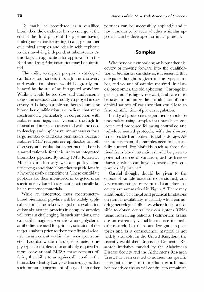
Figure 1. The biomarker development pipeline. (In color in Annals online.)
as a biomarker has several stages (Fig. 1).The biomarker development pipeline beginswith the discovery phase experiments. Thereare many different technology platforms avail-able for protein separation, identification, andquantitation, each of which introduces its ownbias into the data. While it is not always neces-sary to standardize proteomics experiments, inthe context of biomarker research the qualityof the measurements will be significantly en-hanced by the use of an appropriate biologicalstandard or reference material. The inclusionof such a well-defined biological material acrossexperiments enables normalization of all dataand hence the continuous calibration of theanalyte(s).
One embodiment of such a procedure is fa-cilitated by the use of isobaric mass tags.1 Hereone could consider labeling a large quantityof a biological sample, such as plasma, withthe isobaric Tandem Mass Tag (TMT) reagentTMT6-126 (Pierce Biotechnology, Rockford,IL) to create a distinct proteome reference.Each study sample would then be labeled withthe related isobaric tag TMT6-127. Subsequentto labeling with TMT, each sample would bespiked with an equal aliquot of the tagged ref-erence and analyzed individually by tandemmass spectrometry. By using isobaric tags, eachpeptide present in the sample and the referenceco-elute and are analyzed in the same MS/MSexperiment yielding both peptide identity andquantitation. Because each sample contains thesame quantitative reference, the level of all pep-tides can be related across the whole study co-hort via normalization and cross-referencing.We are currently developing global TMT Ref-erence Materials suitable for proteomics inves-tigations involving human plasma and otherbiological fluids and tissues.
An alternative strategy to biomarker discov-ery is to adopt a targeted approach, which fo-
cuses on the identification of changes within adefined subset of analytes. Here, discrete pan-els of analytes can be tested using multiplexedimmunoassays, such as those employing bead-based or planar arrays where tens up to thou-sands of proteins are quantitatively detectedusing antibodies. One of the most popular ap-proaches currently employed is a bead arraybased on the XMAP technology developed byLuminex, which is offered by several compa-nies, including Bio-Rad, Millipore, and RulesBased Medicine, who each have panels for in-flammatory proteins, such as chemokines andcytokines, as well as neurology panels basedon protein content developed in collaborationwith several research groups.
Once interesting changes in protein expres-sion or post-translational modification statushave been observed and considered to war-rant further investigation, additional experi-ments are undertaken to evaluate the candidateand provide extra data to support the origi-nal discovery. Hence, the second stage of thebiomarker development pipeline is an evalua-tion phase. Typically, this involves measuringthe target analyte(s) using an orthogonal ap-proach, either Western blotting or a suitable im-munoassay. However, switching to such orthog-onal technologies is not without drawbacks, andrecently, mass spectrometry methods involvingmultiple reaction monitoring or multiple selec-tive reaction monitoring have begun to emergeas an alternative means of testing. These lat-ter methods have the advantage of measuringthe same biomarker event as was seen in dis-covery and may avoid the pitfalls of switchingto an immunoassay that measures an inferredbiomarker based on the parent protein. Thisis increasingly important where the biomarkeris a change in post-translational modifica-tion rather than a change in total proteinconcentration.
70 Annals of the New York Academy of Sciences
To finally be considered as a qualifiedbiomarker, the candidate has to emerge at theend of the third phase of the pipeline havingundergone extensive testing in a large numberof clinical samples and ideally with replicatestudies involving independent laboratories. Atthis stage, an application for approval from theFood and Drug Administration may be submit-ted.
The ability to rapidly progress a catalog ofcandidate biomarkers through the discoveryand evaluation phases would be greatly en-hanced by the use of an integrated workflow.While it would be too slow and cumbersometo use the methods commonly employed in dis-covery to the large sample numbers required forbiomarker qualification, we believe that massspectrometry, particularly in conjunction withisobaric mass tags, can overcome the high fi-nancial and time costs associated with the needto develop and implement immunoassays for alarge number of candidate biomarkers. Becauseisobaric TMT reagents are applicable to bothdiscovery and evaluation experiments, there isa sound rationale for their use in an integratedbiomarker pipeline. By using TMT ReferenceMaterials in discovery, we can quickly iden-tify strong candidate biomarker peptide ions ina hypothesis-free experiment. These candidatepeptides are then monitored in targeted massspectrometry-based assays using isotopically la-beled reference materials.
While an integrated mass spectrometry-based biomarker pipeline will be widely appli-cable, it must be acknowledged that evaluationof low abundance proteins in complex sampleswill remain challenging. In such situations, onecan easily imagine a scenario where polyclonalantibodies are used for primary selection of thetarget analytes prior to their specific and selec-tive measurement within the mass spectrom-eter. Essentially, the mass spectrometer sim-ply replaces the detection antibody required inmore conventional ELISA measurements of-fering the ability to unequivocally confirm thebiomarker identity. Early evidence suggests thatsuch immune enrichment of target biomarker
peptides can be successfully applied,2 and itnow remains to be seen whether a similar ap-proach can be developed for intact proteins.
Samples
Whether one is embarking on biomarker dis-covery or moving forward into the qualifica-tion of biomarker candidates, it is essential thatadequate thought is given to the type, num-ber, and volume of samples required. In clini-cal proteomics, the old aphorism “Garbage in,garbage out” is highly relevant, and care mustbe taken to minimize the introduction of non-clinical sources of variance that could lead tofalse identification of protein regulation.
Ideally, all proteomics experiments should beundertaken using samples that have been col-lected and processed following controlled andwell-documented protocols, with the shortesttime possible from patient to stable storage. Af-ter procurement, the samples need to be care-fully curated. For biofluids, such as those de-rived from blood, attention should be given topotential sources of variation, such as freeze–thawing, which can have a drastic effect on anumber of proteins.3
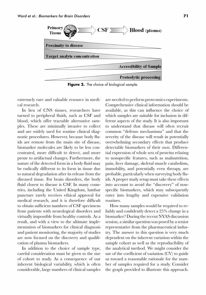
Careful thought should be given to thechoice of sample material to be studied, andkey considerations relevant to biomarker dis-covery are summarized in Figure 2. There mayadditionally be ethical and practical limitationson sample availability, especially when consid-ering neurological diseases where it is not pos-sible to obtain central nervous system (CNS)tissue from living patients. Postmortem brainsare an extremely valuable resource in medi-cal research, but there are few good reposi-tories and as a consequence, material is notwidely available. In the United Kingdom, therecently established Brains for Dementia Re-search initiative, funded by the Alzheimer’sDisease Society and the Alzheimer’s ResearchTrust, has been created to address this specificissue, but, in the short-to-medium term, humanbrain-derived tissues will continue to remain an
Ward et al.: Biomarkers for Brain Disorders 71
Figure 2. The choice of biological sample.
extremely rare and valuable resource in medi-cal research.
In lieu of CNS tissues, researchers haveturned to peripheral fluids, such as CSF andblood, which offer tractable alternative sam-ples. These are minimally invasive to collectand are widely used for routine clinical diag-nostic procedures. However, because body flu-ids are remote from the main site of disease,biomarker molecules are likely to be less con-centrated, more difficult to detect, and moreprone to artifactual changes. Furthermore, thenature of the detected form in a body fluid maybe radically different to its form in tissue dueto natural degradation after its release from thediseased tissue. For brain disorders, the bodyfluid closest to disease is CSF. In many coun-tries, including the United Kingdom, lumbarpuncture rarely receives ethical approval formedical research, and it is therefore difficultto obtain sufficient numbers of CSF specimensfrom patients with neurological disorders andvirtually impossible from healthy controls. As aresult, and with a view to the ultimate imple-mentation of biomarkers for clinical diagnosisand patient monitoring, the majority of studiesare now focused on the discovery and qualifi-cation of plasma biomarkers.
In addition to the choice of sample type,careful consideration must be given to the sizeof cohort to study. As a consequence of ourinherent biological variability, which is oftenconsiderable, large numbers of clinical samples
are needed to perform proteomics experiments.Comprehensive clinical information should beavailable, as this can influence the choice ofwhich samples are suitable for inclusion in dif-ferent aspects of the study. It is also importantto understand that disease will often recruitcommon “defense mechanisms” and that theseverity of the disease will result in potentiallyoverwhelming secondary effects that producedetectable biomarkers of their own. Differen-tial expression of whole sets of proteins relatingto nonspecific features, such as malnutrition,pain, liver damage, skeletal muscle catabolism,immobility, and potentially even therapy, areprobable, particularly when surveying body flu-ids. A proper study setup must take these effectsinto account to avoid the “discovery” of non-specific biomarkers, which may subsequentlyenter into lengthy and expensive validationroutines.
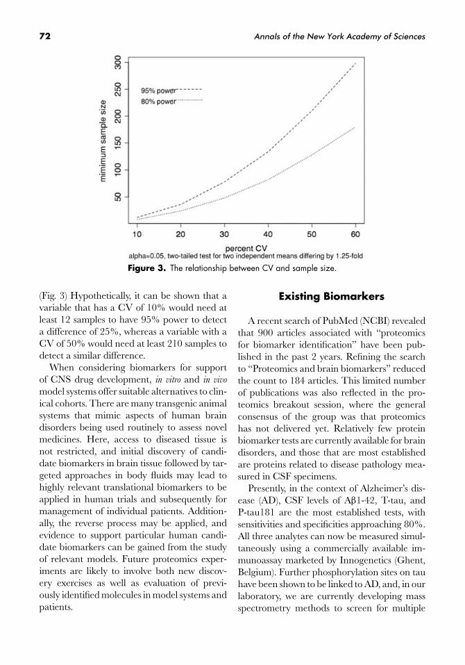
How many samples would be required to re-liably and confidently detect a 25% change in abiomarker? During the recent NYAS discussionsession, a similar question was posed by a seniorrepresentative from the pharmaceutical indus-try. The answer to this question is very muchdependent on the inherent variation within thesample cohort as well as the reproducibility ofthe analytical method. We might consider theuse of the coefficient of variation (CV) to guideus toward a reasonable rationale for the num-ber of samples required for a study. Considerthe graph provided to illustrate this approach.
72 Annals of the New York Academy of Sciences
Figure 3. The relationship between CV and sample size.
(Fig. 3) Hypothetically, it can be shown that avariable that has a CV of 10% would need atleast 12 samples to have 95% power to detecta difference of 25%, whereas a variable with aCV of 50% would need at least 210 samples todetect a similar difference.
When considering biomarkers for supportof CNS drug development, in vitro and in vivo
model systems offer suitable alternatives to clin-ical cohorts. There are many transgenic animalsystems that mimic aspects of human braindisorders being used routinely to assess novelmedicines. Here, access to diseased tissue isnot restricted, and initial discovery of candi-date biomarkers in brain tissue followed by tar-geted approaches in body fluids may lead tohighly relevant translational biomarkers to beapplied in human trials and subsequently formanagement of individual patients. Addition-ally, the reverse process may be applied, andevidence to support particular human candi-date biomarkers can be gained from the studyof relevant models. Future proteomics exper-iments are likely to involve both new discov-ery exercises as well as evaluation of previ-ously identified molecules in model systems andpatients.
Existing Biomarkers
A recent search of PubMed (NCBI) revealedthat 900 articles associated with “proteomicsfor biomarker identification” have been pub-lished in the past 2 years. Refining the searchto “Proteomics and brain biomarkers” reducedthe count to 184 articles. This limited numberof publications was also reflected in the pro-teomics breakout session, where the generalconsensus of the group was that proteomicshas not delivered yet. Relatively few proteinbiomarker tests are currently available for braindisorders, and those that are most establishedare proteins related to disease pathology mea-sured in CSF specimens.
Presently, in the context of Alzheimer’s dis-ease (AD), CSF levels of Aβ1-42, T-tau, andP-tau181 are the most established tests, withsensitivities and specificities approaching 80%.All three analytes can now be measured simul-taneously using a commercially available im-munoassay marketed by Innogenetics (Ghent,Belgium). Further phosphorylation sites on tauhave been shown to be linked to AD, and, in ourlaboratory, we are currently developing massspectrometry methods to screen for multiple
Ward et al.: Biomarkers for Brain Disorders 73
sites in tau isolated from preclinical materials,where particular regions of the brain can beexamined, as an adjunct to the development ofnew medicines.
Also in AD, a number of plasma pro-teins including Complement Factor H (CFH),alpha-2-macroglobulin (A2M), ApolipoproteinE, and Complement C1 inhibitor, are showingpromise. The initial reports of these candidatebiomarkers4,5 have been extended by perform-ing proteomics analysis of plasma from patientswith AD enrolled in a phase IIb clinical trialof the PPARγ agonist rosiglitazone.6 Further-more, plasma levels of CFH and A2M showsignificant positive correlation with the hip-pocampal metabolite ratio N-acetylaspartate/myo-inositol (NAA/mI), a biochemical mea-sure that is associated with cognitive declinein early AD, suggesting that these proteins mayreflect disease progression in early AD.7
Data Integration and Sharing
We are pleased to report that applying pro-teomics to the search for biomarkers of braindisorders is starting to demonstrate readily ac-cessible measures of the presence, stage, andtreatment response. However, it should beclearly stated that further important insightsinto the relevance of these candidate biomark-ers to disease is required. Such insights will onlybe gained by combining proteomics measure-ments with additional parameters, such as thosederived from other “omics” approaches, imag-ing, or clinical observations. Vigorous statisticaltesting of combined data is required, and the ex-tent of bioinformatics support necessary shouldnot be underestimated. Armed with sophisti-cated software packages, or indeed the wizardryto create bespoke data processing scripts, in-dividuals skilled in the art of data analysis arevaluable assets to any organization. The manip-ulation of large data sets is often necessary, andwith information derived from many disparatesources, careful attention is needed to ensurethat variables are appropriately correlated. Al-
though various aspects of a data analysis canbe automated, there is ultimately always a needfor manual intervention somewhere within theprocess.
As is the case with existing consortia, suchas for example AddNeuroMed and ADNI, ap-propriate mechanisms for data integration andsharing among established groups working ona global scale need to be acceptable to all par-ticipants and stakeholders. While presenting aconsiderable challenge to academia and indus-try alike, the authors were encouraged by thenumber of stakeholders attending the meetingwho supported proposals for just such data in-tegration during a breakout session hosted byADNI.
Closing Remarks
To reiterate the statement made publicly atthe end of the conference, we remain optimisticthat proteomics will deliver new biomarkermolecules. These biomarkers will be used tosupport the diagnosis of a variety of brain dis-orders and to help detect progression and re-sponse to therapies as they are introduced intomainstream clinical practice during the comingyears.
Conflicts of Interest
The authors declare no conflicts of interest.
References
1. Thompson, A., J. Schafer, K. Kuhn, et al. 2003. Tan-dem Mass Tags: a novel quantification strategy forcomparative analysis of complex protein mixtures byMS/MS. Anal. Chem. 75: 1895–1904.
2. Anderson, N.L., N.G. Anderson, L.R. Haines, et al.2004. Mass spectrometric quantitation of peptides andproteins using Stable Isotope Standards and Captureby Anti-Peptide Antibodies (SISCAPA). J. Proteome Res.
3: 235–244.3. Rogers, M., P. Clarke, J. Noble, et al. 2003. Pro-
teomic profiling of urinary proteins in renal cancer bysurface enhanced laser desorption ionization andneural-network analysis: identification of key issues

74 Annals of the New York Academy of Sciences
affecting potential clinical utility. Cancer Res. 63: 6971–6983.
4. Hye, A., S. Lynham, M. Thambisetty, et al. 2006.Proteome-based plasma biomarkers for Alzheimer’sdisease. Brain 129: 3042–3050.
5. Cutler, P., E. Akuffo, W. Bodnar, et al. 2008. Proteomicidentification and early validation of complement 1inhibitor and pigment epithelium-derived factor: twonovel biomarkers of Alzheimer’s disease in humanplasma. Proteomics Clin. Appl. 2: 467–477.
6. Akuffo, E.L., J.B. Davis, S.M. Fox, et al. 2008. The dis-covery and early validation of novel plasma biomark-ers in mild-to-moderate Alzheimer’s disease patientsresponding to treatment with rosiglitazone. Biomarkers
13: 618–636.7. Thambisetty, M., A. Hye, C. Foy, et al.
2008. Proteome-based identification of plasma pro-teins associated with hippocampal metabolism inearly Alzheimer’s disease. J. Neurol. 255: 1712–1720.