Embed Size (px)
Citation preview
Instructions for use
Title Proteomics analysis of heterogeneous flagella in brown algae (Stramenopiles)
Author(s) Fu, Gang; Nagasato, Chikako; Oka, Seiko; Cock, J. Mark; Motomura, Taizo
Citation Protist, 165(5): 662-675
Issue Date 2014-09
Doc URL http://hdl.handle.net/2115/57666
Type article (author version)
Additional Information There are other files related to this item in HUSCAP. Check the above URL.
File Information Text (Fu et al.).pdf
Hokkaido University Collection of Scholarly and Academic Papers : HUSCAP
1
Proteomics analysis of heterogeneous flagella in brown algae (Stramenopiles) 1
Gang Fu1, Chikako Nagasato1, Seiko Oka2, J. Mark Cock3 and Taizo Motomura1,* 2
3
1Muroran Marine Station, Field Science Center for Northern Biosphere, Hokkaido 4
University, Muroran 051-0013, Hokkaido, Japan 5
2Instrumental Analysis Division, Equipment Management Center, Creative Research 6
Institution, Hokkaido University, Sapporo 001-0021, Hokkaido, Japan 7
3University Pierre et Marie Curie and Centre National de la Recherche Scientifique, 8
Unité Mixte de Recherche 7139, Laboratoire International Associé Dispersal and 9
Adaptation in Marine Species, Station Biologique de Roscoff, 29682 Roscoff Cedex, 10
France 11
12
*Correspondence author: Taizo Motomura 13
Email: [email protected] 14
Tel: +81 143 22 2846 15
Fax: +81 143 22 4135 16
17
A running title: Heterogeneous flagella in brown algae 18
19
20
2
Abstract 1
Flagella are conserved organelles among eukaryotes and they are composed of many 2
proteins, which are necessary for flagellar assembly, maintenance and function. 3
Stramenopiles, which include brown algae, diatoms and oomycetes, possess two 4
laterally inserted flagella. The anterior flagellum (AF) extends forward and bears 5
tripartite mastigonemes, whilst the smooth posterior flagellum (PF) often has a 6
paraflagellar body structure. These heterogeneous flagella have served as crucial 7
structures in algal studies especially from a viewpoint of phylogeny. However, the 8
protein compositions of the flagella are still largely unknown. Here we report a 9
LC-MS/MS based proteomics analysis of brown algal flagella. In total, 495 flagellar 10
proteins were identified. Functional annotation of the proteome data revealed that 11
brown algal flagellar proteins were associated with cell motility, signal transduction and 12
various metabolic activities. We separately isolated AF and PF and analyzed their 13
protein compositions. This analysis led to the identification of several AF- and 14
PF-specific proteins. Among the PF-specific proteins, we found a candidate novel blue 15
light receptor protein involved in phototaxis, and named it HELMCHROME because of 16
the steering function of PF. Immunological analysis revealed that this protein was 17
localized along the whole length of the PF and concentrated in the paraflagellar body. 18
19
20
Key words: blue light receptor; brown algae; creatine kinase; flagella; phototaxis; 21
proteomics. 22
3
Introduction 1
Flagella or cilia are almost ubiquitous organelles in a diverse range of eukaryotic 2
cells and substantial studies have revealed their versatile roles in cellular motility and 3
signal perception (Cavalier-Smith 2002; Davenport and Yoder 2005). The structure of 4
the flagellum is evolutionarily conserved (Carvalho-Santos et al. 2011; Mitchell 2007) 5
and for a motile flagellum, its core structure is a “9+2” axoneme comprising nine outer 6
doublet microtubules and central pair of microtubules. Various macromolecular 7
components such as outer and inner dynein arms, radial spokes and central pair 8
projections, are periodically organized along the microtubule-based axoneme and 9
responsible for flagellar beating (Nicastro et al. 2005). The axoneme extends from the 10
basal bodies beyond the cell surface and is covered by flagellar membrane, which is 11
continuous with the plasma membrane but has different protein and lipid compositions 12
relating to the sensory function of flagella (Pazour and Bloodgood 2008). In addition to 13
the axoneme structure, an intraflagellar transport (IFT) system is also present in the 14
compartment enclosed by the flagellar membrane and the outer doublet microtubules 15
(Kozminski et al. 1993). The IFT consists of anterograde and retrograde protein 16
complexes responsible for flagellar assembly and maintenance (Rosenbaum and 17
Witman 2002). Given the functional diversity and structural complexity, it is not 18
surprising that hundreds of proteins are required for the correct assembly, maintenance 19
and functioning of flagella. As a result of a combination of bioinformatics, genomics 20
and proteomics analyses of the flagella of model organisms, our understanding of the 21
4
flagellar proteins has greatly advanced in the last decade (Gherman et al. 2006; Inglis et 1
al. 2006). 2
The stramenopiles constitute a large independent group among eight eukaryotic 3
lineages, and possess diverse organisms from unicellular parasitic flagellates to giant 4
kelp, including five non-photosynthetic subgroups (such as oomycetes and 5
labyrinthulomycetes) and eleven photosynthetic ones (such as brown algae and diatoms) 6
(Baldauf 2008). Stramenopiles are characterized by possessing an anterior flagellum 7
(AF) with tripartite mastigonemes in the motile stage (gametes and zoospores). An 8
alternative name for the stramenopiles is the heterokonts, because most species within 9
this group often have a shorter, smooth posterior flagellum (PF) in addition to the AF. 10
Functionally, the AF generates propulsive force through waveform bending to power 11
the cell forward swimming motility and the PF exhibits rapid lateral beating to steer the 12
swimming direction (Geller and Müller 1981; Matsunaga et al. 2010). 13
The brown algae (Phaeophyceae) are a major group within the stramenopiles 14
that are mostly found in marine habitats, and distributed worldwide. During a typical 15
brown algal life cycle, the two heterogeneous flagella (AF and PF) are observed in the 16
unicellular reproductive cells (gametes and zoospores). As one of the most important 17
organelles in brown algae, a great deal of attention has been paid to flagella and their 18
ultrastructural characters have been studied extensively (Henry and Cole 1982a, b; 19
Maier 1997a, b; Manton and Clarke 1951, Manton et al.1953). The flagella are also 20
critical structures when considering phylogenetic relationships within the brown algae 21
(Clayton 1989; O’Kelly 1989). However, the protein composition of brown algal 22
5
flagella remain largely unknown, probably owing to the difficulty in isolating high 1
quality brown algal flagella in sufficient quantity and, until recently, the lack of a brown 2
algal genome database to underpin the flagellar protein analysis. 3
Although the genome sequence of the model brown alga Ectocarpus siliculosus 4
has become available (Cock et al. 2010), proteomics analysis of its flagella is hampered 5
by limitations in the amount of this organelle that can be isolated. In this study, we used 6
plurizoids of Colpomenia bullosa collected from the field to carry out flagellar 7
proteomics analysis. Light and electron microscopy showed that plurizoids of 8
Colpomenia and Ectocarpus are structurally similar. Moreover, the two species both 9
belong to the Ectocarpales according to the traditional brown algal classification criteria 10
(Guiry and Guiry 2014), and a multi-marker based phylogenetic analysis of 72 brown 11
algal taxa revealed that Colpomenia is phylogenetically close to Ectocarpus (Silberfeld 12
et al. 2010). These data encouraged us to use the genome sequence of E. siliculosus as a 13
database to identify C. bullosa flagellar proteins. Here, we present the result of 14
LC-MS/MS based proteomics analysis of isolated flagella from the latter species. We 15
also identified several AF- and PF-specific proteins. A candidate novel blue light 16
receptor protein responsible for phototaxis of brown algal swarmers was also discussed. 17
18
Results and Discussions 19
Heterogeneity and isolation of brown algal flagella 20
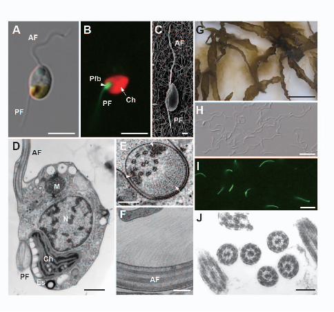
The flagellated cells of C. bullosa and E. siliculosus are around 5x7 µm in size 21
and bear two heterogeneous flagella in different lengths (Fig. 1A). Under blue-violet 22
6
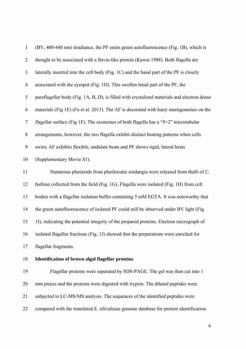
(BV, 400-440 nm) irradiance, the PF emits green autofluorescence (Fig. 1B), which is 1
thought to be associated with a flavin-like protein (Kawai 1988). Both flagella are 2
laterally inserted into the cell body (Fig. 1C) and the basal part of the PF is closely 3
associated with the eyespot (Fig. 1D). This swollen basal part of the PF, the 4
paraflagellar body (Fig. 1A, B, D), is filled with crystalized materials and electron dense 5
materials (Fig 1E) (Fu et al. 2013). The AF is decorated with hairy mastigonemes on the 6
flagellar surface (Fig 1F). The axonemes of both flagella has a “9+2” microtubular 7
arrangements, however, the two flagella exhibit distinct beating patterns when cells 8
swim; AF exhibits flexible, undulate beats and PF shows rigid, lateral beats 9
(Supplementary Movie S1). 10
Numerous plurizoids from plurilocular zoidangia were released from thalli of C. 11
bullosa collected from the field (Fig. 1G). Flagella were isolated (Fig. 1H) from cell 12
bodies with a flagellar isolation buffer containing 5 mM EGTA. It was noteworthy that 13
the green autofluorescence of isolated PF could still be observed under BV light (Fig. 14
1I), indicating the potential integrity of the prepared proteins. Electron micrograph of 15
isolated flagellar fractions (Fig. 1J) showed that the preparations were enriched for 16
flagellar fragments. 17
Identification of brown algal flagellar proteins 18
Flagellar proteins were separated by SDS-PAGE. The gel was then cut into 1 19
mm pieces and the proteins were digested with trypsin. The diluted peptides were 20
subjected to LC-MS/MS analysis. The sequences of the identified peptides were 21
compared with the translated E. siliculosus genome database for protein identification. 22
7
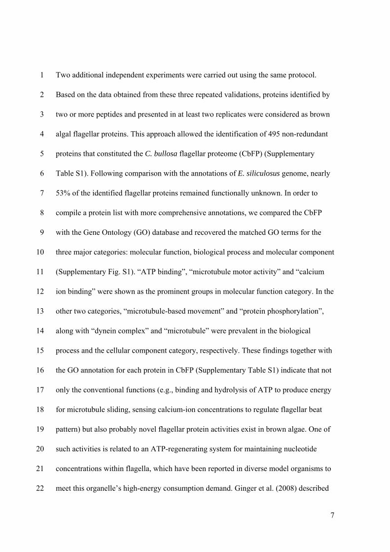
Two additional independent experiments were carried out using the same protocol. 1
Based on the data obtained from these three repeated validations, proteins identified by 2
two or more peptides and presented in at least two replicates were considered as brown 3
algal flagellar proteins. This approach allowed the identification of 495 non-redundant 4
proteins that constituted the C. bullosa flagellar proteome (CbFP) (Supplementary 5
Table S1). Following comparison with the annotations of E. siliculosus genome, nearly 6
53% of the identified flagellar proteins remained functionally unknown. In order to 7
compile a protein list with more comprehensive annotations, we compared the CbFP 8
with the Gene Ontology (GO) database and recovered the matched GO terms for the 9
three major categories: molecular function, biological process and molecular component 10
(Supplementary Fig. S1). “ATP binding”, “microtubule motor activity” and “calcium 11
ion binding” were shown as the prominent groups in molecular function category. In the 12
other two categories, “microtubule-based movement” and “protein phosphorylation”, 13
along with “dynein complex” and “microtubule” were prevalent in the biological 14
process and the cellular component category, respectively. These findings together with 15
the GO annotation for each protein in CbFP (Supplementary Table S1) indicate that not 16
only the conventional functions (e.g., binding and hydrolysis of ATP to produce energy 17
for microtubule sliding, sensing calcium-ion concentrations to regulate flagellar beat 18
pattern) but also probably novel flagellar protein activities exist in brown algae. One of 19
such activities is related to an ATP-regenerating system for maintaining nucleotide 20
concentrations within flagella, which have been reported in diverse model organisms to 21
meet this organelle’s high-energy consumption demand. Ginger et al. (2008) described 22
8
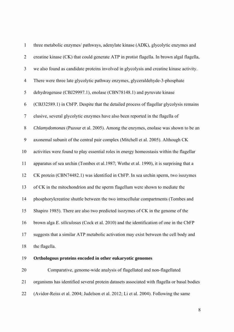
three metabolic enzymes/ pathways, adenylate kinase (ADK), glycolytic enzymes and 1
creatine kinase (CK) that could generate ATP in protist flagella. In brown algal flagella, 2
we also found as candidate proteins involved in glycolysis and creatine kinase activity. 3
There were three late glycolytic pathway enzymes, glyceraldehyde-3-phosphate 4
dehydrogenase (CBJ29997.1), enolase (CBN78148.1) and pyruvate kinase 5
(CBJ32589.1) in CbFP. Despite that the detailed process of flagellar glycolysis remains 6
elusive, several glycolytic enzymes have also been reported in the flagella of 7
Chlamydomonas (Pazour et al. 2005). Among the enzymes, enolase was shown to be an 8
axonemal subunit of the central pair complex (Mitchell et al. 2005). Although CK 9
activities were found to play essential roles in energy homeostasis within the flagellar 10
apparatus of sea urchin (Tombes et al.1987; Wothe et al. 1990), it is surprising that a 11
CK protein (CBN74482.1) was identified in CbFP. In sea urchin sperm, two isozymes 12
of CK in the mitochondrion and the sperm flagellum were shown to mediate the 13
phosphorylcreatine shuttle between the two intracellular compartments (Tombes and 14
Shapiro 1985). There are also two predicted isozymes of CK in the genome of the 15
brown alga E. siliculosus (Cock et al. 2010) and the identification of one in the CbFP 16
suggests that a similar ATP metabolic activation may exist between the cell body and 17
the flagella. 18
Orthologous proteins encoded in other eukaryotic genomes 19
Comparative, genome-wide analysis of flagellated and non-flagellated 20
organisms has identified several protein datasets associated with flagella or basal bodies 21
(Avidor-Reiss et al. 2004; Judelson et al. 2012; Li et al. 2004). Following the same 22
9
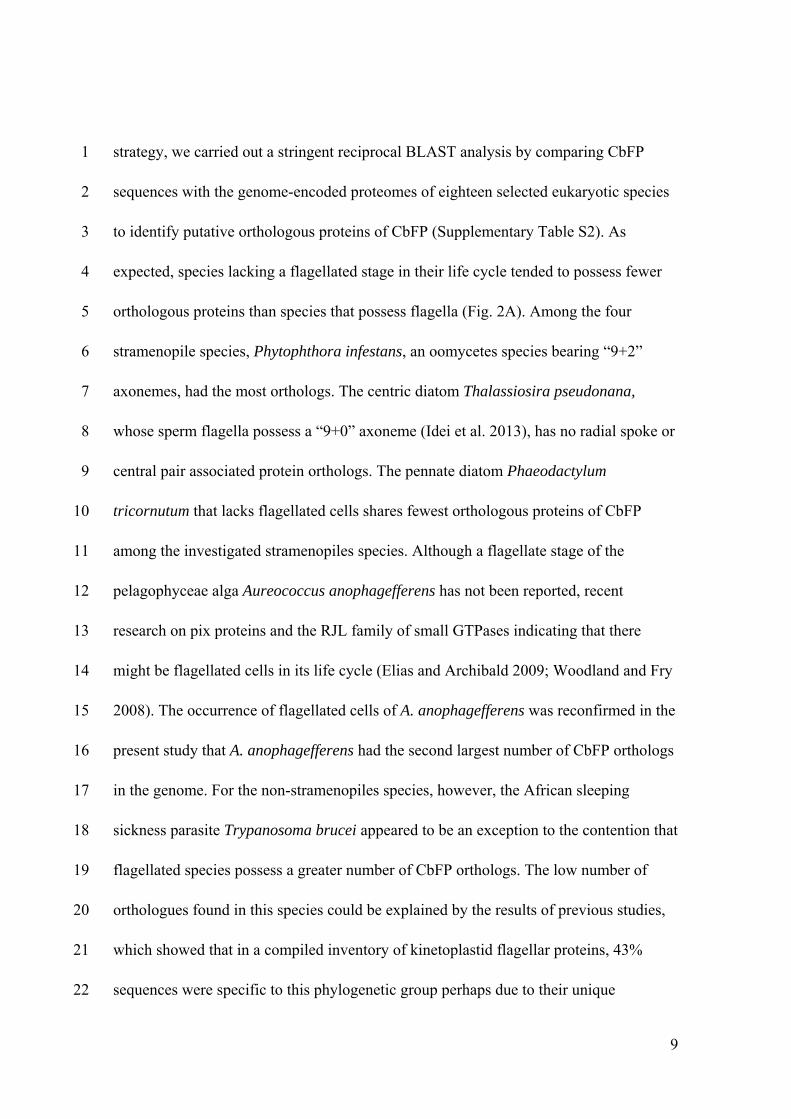
strategy, we carried out a stringent reciprocal BLAST analysis by comparing CbFP 1
sequences with the genome-encoded proteomes of eighteen selected eukaryotic species 2
to identify putative orthologous proteins of CbFP (Supplementary Table S2). As 3
expected, species lacking a flagellated stage in their life cycle tended to possess fewer 4
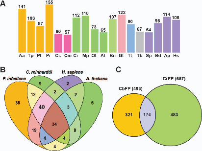
orthologous proteins than species that possess flagella (Fig. 2A). Among the four 5
stramenopile species, Phytophthora infestans, an oomycetes species bearing “9+2” 6
axonemes, had the most orthologs. The centric diatom Thalassiosira pseudonana, 7
whose sperm flagella possess a “9+0” axoneme (Idei et al. 2013), has no radial spoke or 8
central pair associated protein orthologs. The pennate diatom Phaeodactylum 9
tricornutum that lacks flagellated cells shares fewest orthologous proteins of CbFP 10
among the investigated stramenopiles species. Although a flagellate stage of the 11
pelagophyceae alga Aureococcus anophagefferens has not been reported, recent 12
research on pix proteins and the RJL family of small GTPases indicating that there 13
might be flagellated cells in its life cycle (Elias and Archibald 2009; Woodland and Fry 14
2008). The occurrence of flagellated cells of A. anophagefferens was reconfirmed in the 15
present study that A. anophagefferens had the second largest number of CbFP orthologs 16
in the genome. For the non-stramenopiles species, however, the African sleeping 17
sickness parasite Trypanosoma brucei appeared to be an exception to the contention that 18
flagellated species possess a greater number of CbFP orthologs. The low number of 19
orthologues found in this species could be explained by the results of previous studies, 20
which showed that in a compiled inventory of kinetoplastid flagellar proteins, 43% 21
sequences were specific to this phylogenetic group perhaps due to their unique 22
10
paraflagellar rod structure (Baron et al. 2007; Broadhead et al. 2006; Ralston et al. 1
2009). 2
The orthologs found in non-flagellated organisms indicated that these proteins, 3
which include kinases and metabolic enzymes, might play roles in both flagella and the 4
cytoplasm. To identify the proteins that are highly conserved only in flagella, we 5
analyzed 40 CbFP orthologous proteins shared in flagellated P. infestans, C. reinhardtii 6
and human, but not in A. thaliana (Fig. 2B). Of these proteins, 19 (48%) were essential 7
components of the flagellar axoneme or the IFT system, and 10 (25%) had putative 8
functions in signal transduction or metabolism pathways. However, the function of 11 9
(27%) of these proteins still remained unclear (Supplementary Table S3). It is 10
interesting that a molecular chaperon protein, Hsp70 (CBJ32839.1), was conserved in 11
the flagellated species but not in A. thaliana. The first identification of Hsp70 in flagella 12
was reported in Chlamydomonas (Bloch and Johnson 1995), after which a further 13
exploration of molecular chaperons suggested their wide distributions as flagellar 14
components (Stephens and Lemieux 1999). Despite the large number of HSP family 15
members, the only CbFP Hsp70 ortholog in Chlamydomonas was HSP70A 16
(XP_001701326) (Supplementary Table S2), which was reported localizing in flagella 17
(Shapiro et al. 2005). This indicates that some flagellar Hsp chaperons might have 18
unique functions in flagellar assembly and maintenance. 19
In addition, we subjected the CbFP to a BLAST (E-value was set as ≤ 1e-10) 20
analysis using 657 C. reinhardtii flagellar proteins (Pazour et al. 2005, available at 21
http://labs.umassmed.edu/chlamyfp/) as query sequences to compare the two flagellar 22
11
proteomes (Fig. 2C). One hundred and seventy-four of the CbFP proteins had homologs 1
in the C. reinhardtii flagellar proteome (CrFP), most of them being known structural 2
and motor proteins (Supplementary Table S4), indicating the common characters of 3
flagella in these two algal species. However, the large proportion (321) of the CbFP that 4
lacks homologs in the CrFP reflects the significant differences between them. These 5
include obvious structural differences, such as flagellar length, mastigoneme and 6
paraflagellar body, and perhaps distinct sensory functions of the flagella. For example, 7
transduction of light stimuli in C. reinhardtii is triggered from cytoplasmic 8
channelrhodopsins to flagellar axonemes (Govorunova et al. 2004; Okita et al. 2005), 9
while in brown algae the photoreceptor protein is likely to be located at the PF 10
(Flores-Moya et al. 2002; Kawai 1988; Kawai et al. 1990; Müller 1987), which implies 11
a different light-induced signaling cascade within the flagella. Therefore, the list of 321 12
proteins in the CbFP that lack CrFP homologs provide potential candidates to explore 13
the proteins associated with structural and functional differences between brown and 14
green algal flagella. 15
Conservation of flagellar disease proteins in CbFP 16
Increasing evidence supports that flagellar dysfunction causes severe vertebrate 17
diseases (Bisgrove and Yost 2006; Fliegauf et al. 2007; Marshall 2008; Marshall and 18
Nonaka 2006). Since flagella are highly conserved organelles among eukaryotes, 19
proteins involved in many of these flagella-related disorders could also be detected in 20
non-vertebrate organisms such as C. reinhardtii (Pazour et al. 2005). Likewise, we 21
identified homologs of such proteins in CbFP and all of them had a matched sequence 22
12
in the human genome when analyzed by BLAST (Table 1). Defects in these proteins 1
result in a broad range of vertebrate diseases, including dysfunction of the male 2
reproductive system, hydrocephalus, Bardet-Biedl Syndrome, polycystic kidney disease 3
and so on (see references in Table 1). The comparison between CbFP and CrFP 4
(Supplementary Table S4) showed that homologues of the disease-related proteins 5
identified in CbFP were also conserved in Chlamydomonas flagella. Conservation of 6
flagellar disease proteins in non-vertebrate organisms indicates that these proteins are 7
essential flagellar components relating to its fundamental structure and function. For 8
example, in addition to IFT and dynein arm proteins, homologues of hydin and Spag16 9
are also axonemal proteins localized at central pair apparatus (Lechtreck and Witman 10
2007; Zhang et al. 2002). 11
Independent proteomics analysis of the AF and the PF 12
It would be intriguing and important to identify AF- and PF-specific proteins to 13
explain their morphological and functional heterogeneities at the molecular level in 14
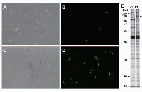
brown algae. We have developed an effective procedure to separately isolate AF and PF 15
(Fig. 3A-D). One-dimensional gel electrophoresis confirmed that several bands limited 16
to AF or PF could be detected (Fig. 3E). We also performed 2-D analysis of the isolated 17
flagellar proteins, which yielded over 20 specific spots associated with each flagellar 18
preparation (Supplementary Fig. S2). Utilizing these flagellar fractions, we attempted to 19
identify AF- and PF-specific proteins by LC-MS/MS. 20
Three and fourteen proteins which were specific to AF and PF, respectively, 21
were identified by two or more peptides (Supplementary Table S5). The identification 22
13
of a mastigoneme-associated protein (CBJ28331.1) in the AF proteome but not in the 1
PF indicates that these proteins are likely to be bona fide flagella-specific proteins. A 2
giant protein (CBJ30163.1) annotated as “similar to connectin/titin isoform N2-B” was 3
identified in the AF-specific dataset. It has an extraordinary huge predicted molecular 4
weight of 1822 kDa and at the N-terminal, a signal peptide and one transmembrane 5
helix could be predicted. The domain architecture of this protein includes multiple 6
copies of the “Fibronectin type III” domain, three “PA14” domains and three 7
“Filamin/ABP280 repeat” domains, which are widely distributed in scaffold proteins 8
involved in ligand binding activity (de Groot and Klis 2008; Feng and Walsh 2004; 9
Koide et al. 1998). This connenctin-like protein is likely to be associated with 10
mastigoneme structures in the AF, and this is consistent with our previous study (Fu et 11
al. 2013) that connecting structures linking mastigoneme and flagellar axoneme could 12
be observed with electron tomography. 13
Existence of PF-specific, signal transduction proteins might be likely related to 14
locomotive reaction of PF upon stimulations. For example, pheromone-simulated male 15
gametes of Ectocarpus increased the beat frequency of PF, which subsequently changed 16
the swimming direction of the male gamete for approaching to the female gamete 17
(Geller and Müller 1981). PF also performed one to several rapid lateral beats upon 18
light stimulation (Matsunaga et al. 2010), indicating that PF also plays essential roles in 19
phototaxis of brown algal swarmers. 20
HELMCHROME, a candidate novel blue light receptor for phototaxis 21
14
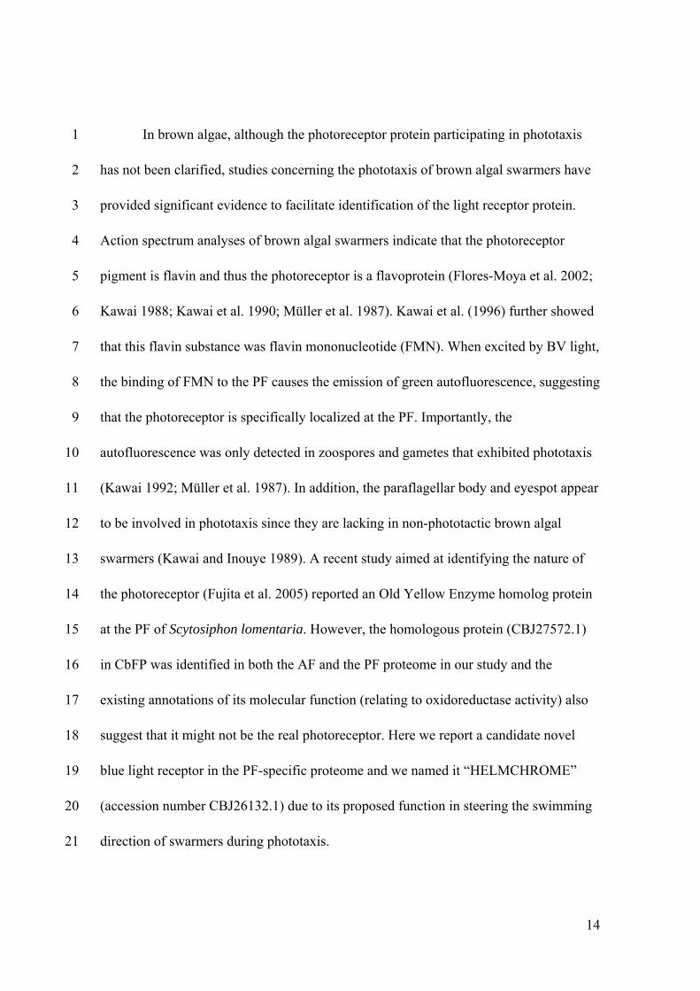
In brown algae, although the photoreceptor protein participating in phototaxis 1
has not been clarified, studies concerning the phototaxis of brown algal swarmers have 2
provided significant evidence to facilitate identification of the light receptor protein. 3
Action spectrum analyses of brown algal swarmers indicate that the photoreceptor 4
pigment is flavin and thus the photoreceptor is a flavoprotein (Flores-Moya et al. 2002; 5
Kawai 1988; Kawai et al. 1990; Müller et al. 1987). Kawai et al. (1996) further showed 6
that this flavin substance was flavin mononucleotide (FMN). When excited by BV light, 7
the binding of FMN to the PF causes the emission of green autofluorescence, suggesting 8
that the photoreceptor is specifically localized at the PF. Importantly, the 9
autofluorescence was only detected in zoospores and gametes that exhibited phototaxis 10
(Kawai 1992; Müller et al. 1987). In addition, the paraflagellar body and eyespot appear 11
to be involved in phototaxis since they are lacking in non-phototactic brown algal 12
swarmers (Kawai and Inouye 1989). A recent study aimed at identifying the nature of 13
the photoreceptor (Fujita et al. 2005) reported an Old Yellow Enzyme homolog protein 14
at the PF of Scytosiphon lomentaria. However, the homologous protein (CBJ27572.1) 15
in CbFP was identified in both the AF and the PF proteome in our study and the 16
existing annotations of its molecular function (relating to oxidoreductase activity) also 17
suggest that it might not be the real photoreceptor. Here we report a candidate novel 18
blue light receptor in the PF-specific proteome and we named it “HELMCHROME” 19
(accession number CBJ26132.1) due to its proposed function in steering the swimming 20
direction of swarmers during phototaxis. 21
15
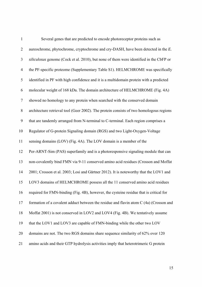
Several genes that are predicted to encode photoreceptor proteins such as 1
aureochrome, phytochrome, cryptochrome and cry-DASH, have been detected in the E. 2
siliculosus genome (Cock et al. 2010), but none of them were identified in the CbFP or 3
the PF-specific proteome (Supplementary Table S1). HELMCHROME was specifically 4
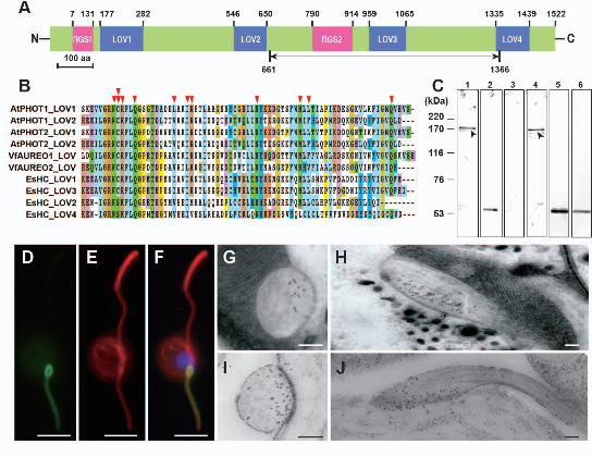
identified in PF with high confidence and it is a multidomain protein with a predicted 5
molecular weight of 168 kDa. The domain architecture of HELMCHROME (Fig. 4A) 6
showed no homology to any protein when searched with the conserved domain 7
architecture retrieval tool (Geer 2002). The protein consists of two homologous regions 8
that are tandemly arranged from N-terminal to C-terminal. Each region comprises a 9
Regulator of G-protein Signaling domain (RGS) and two Light-Oxygen-Voltage 10
sensing domains (LOV) (Fig. 4A). The LOV domain is a member of the 11
Per-ARNT-Sim (PAS) superfamily and is a photoresponsive signaling module that can 12
non-covalently bind FMN via 9-11 conserved amino acid residues (Crosson and Moffat 13
2001; Crosson et al. 2003; Losi and Gärtner 2012). It is noteworthy that the LOV1 and 14
LOV3 domains of HELMCHROME possess all the 11 conserved amino acid residues 15
required for FMN-binding (Fig. 4B), however, the cysteine residue that is critical for 16
formation of a covalent adduct between the residue and flavin atom C (4a) (Crosson and 17
Moffat 2001) is not conserved in LOV2 and LOV4 (Fig. 4B). We tentatively assume 18
that the LOV1 and LOV3 are capable of FMN-binding while the other two LOV 19
domains are not. The two RGS domains share sequence similarity of 62% over 120 20
amino acids and their GTP hydrolysis activities imply that heterotrimeric G protein 21
16
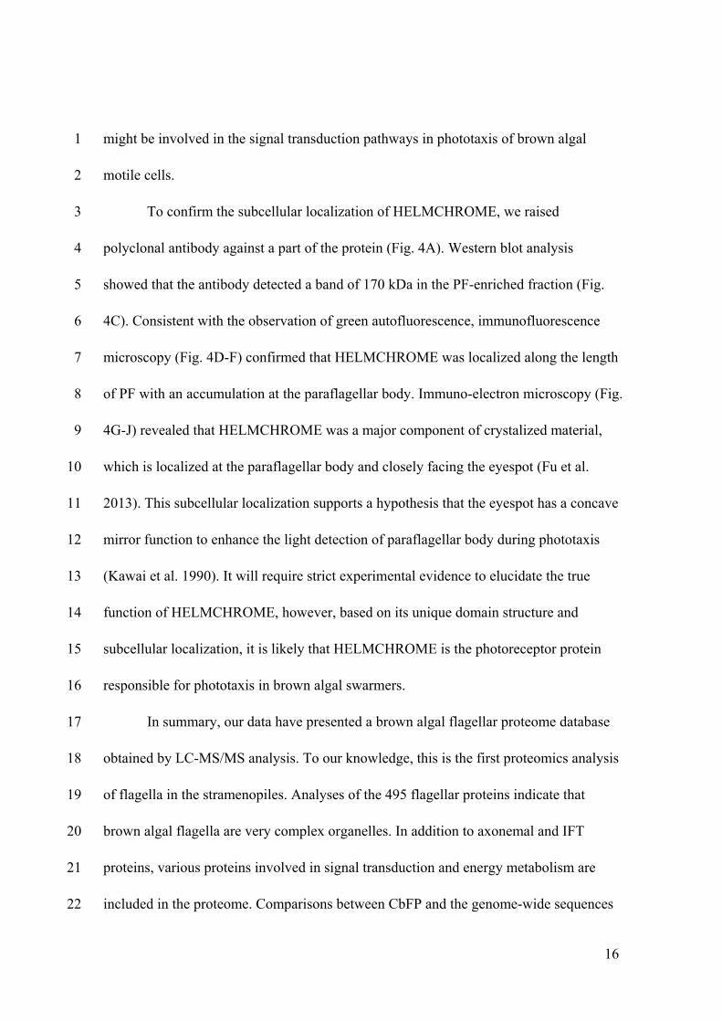
might be involved in the signal transduction pathways in phototaxis of brown algal 1
motile cells. 2
To confirm the subcellular localization of HELMCHROME, we raised 3
polyclonal antibody against a part of the protein (Fig. 4A). Western blot analysis 4
showed that the antibody detected a band of 170 kDa in the PF-enriched fraction (Fig. 5
4C). Consistent with the observation of green autofluorescence, immunofluorescence 6
microscopy (Fig. 4D-F) confirmed that HELMCHROME was localized along the length 7
of PF with an accumulation at the paraflagellar body. Immuno-electron microscopy (Fig. 8
4G-J) revealed that HELMCHROME was a major component of crystalized material, 9
which is localized at the paraflagellar body and closely facing the eyespot (Fu et al. 10
2013). This subcellular localization supports a hypothesis that the eyespot has a concave 11
mirror function to enhance the light detection of paraflagellar body during phototaxis 12
(Kawai et al. 1990). It will require strict experimental evidence to elucidate the true 13
function of HELMCHROME, however, based on its unique domain structure and 14
subcellular localization, it is likely that HELMCHROME is the photoreceptor protein 15
responsible for phototaxis in brown algal swarmers. 16
In summary, our data have presented a brown algal flagellar proteome database 17
obtained by LC-MS/MS analysis. To our knowledge, this is the first proteomics analysis 18
of flagella in the stramenopiles. Analyses of the 495 flagellar proteins indicate that 19
brown algal flagella are very complex organelles. In addition to axonemal and IFT 20
proteins, various proteins involved in signal transduction and energy metabolism are 21
included in the proteome. Comparisons between CbFP and the genome-wide sequences 22
17
of flagellated and non-flagellated species, and between CbFP and CrFP, have provided 1
datasets to explore evolutionarily conserved flagellar proteins and brown algal-specific 2
proteins. Identification of AF- and PF-specific proteins has provided candidate proteins 3
for investigating the heterogeneity of the two flagella at the molecular level. Finally, 4
HELMCHROME is considered to be a novel blue light receptor protein which is 5
involved in phototaxis of brown algal swarmers. 6
7
Methods 8
Flagella isolation:Mature thalli of Colpomenia bullosa (Saunders) Yamada were 9
collected at Charatsunai (42.03 N, 140.99 E), Muroran, Japan. They were thoroughly 10
washed with autoclaved seawater and placed at 10˚C under dark condition for one night. 11
When the thalli were submerged in chilled seawater next day, a large number of motile 12
cells were released into the seawater. The motile cells were collected by centrifugation 13
at 3000 rpm for 3 min. Two ml of flagellar isolation buffer (30 mM HEPES, 5 mM 14
MgSO4, 5 mM EGTA, 25 mM KCl, 1M sorbitol, pH 7.0) were added to the swarmer 15
pellet, and the cells were resuspended in the buffer by pipetting up and down. The 16
suspension was then transferred into a 15 ml glass tube and vortexed vigorously for 17
60-90 sec. In order to remove the cell bodies, the suspension was firstly centrifuged for 18
3 times (2000 rpm, 3 min for each). The supernatant obtained was transferred to a new 19
tube and centrifuged for another 3 times at a higher speed (3000 rpm, 3min for each). 20
Finally, the supernatant was collected into a 1.5 ml centrifuge tube and centrifuged at 21
15000 rpm for 60 min. The flagellar pellet was obtained after removing the supernatant. 22
18
For isolating AF and PF separately, isolation buffer was added to the collected cell 1
pellets and gently pipetted up and down for 60 sec without vortexing. After this 2
treatment, the AF was readily detached from the cell body while the PF was still 3
remained undetached. The supernatant containing AF was transferred to a new glass 4
tube and treated with the same flagellar isolation procedure as described above. Another 5
2 ml of flagellar isolation buffer was added to the remained pellets, which contained the 6
cells with only PF. Followed the same procedure as used for isolating both flagella, 7
PF-enriched fractions could be obtained after the final centrifugation. All the 8
centrifugations were performed at 4˚C. The isolated flagella were stored at -80˚C until 9
use. 10
Transmission electron microscopy (TEM): Isolated flagella were fixed with 2% 11
glutaradehyde and 2% tannic acid in 0.1 M cacodylate buffer (pH 7.2) for 90 min on ice. 12
After washing with 0.1 M cacodylate buffer, these samples were post-fixed with 2% 13
OsO4 in 0.1 M cacodylate buffer on ice for 1 h. Samples were dehydrated in an acetone 14
series and embedded in Suppr’s epoxy resin. Thin sections were stained with 4% uranyl 15
acetate and lead citrate and observed with a JEM-1011 electron microscope (JEOL, 16
Tokyo, Japan). Preparation for motile cell samples was done by freeze fixation and 17
substitution protocols essentially as previously described (Fu et al. 2013; Nagasato and 18
Motomura 2002). 19
Scanning electron microscopy (SEM): Male gametes liberated from gameteophytes of 20
Ectocarpus siliculosus 32m strain which were cultured in half-strength PESmedium 21
(Provasoli 1968) under cool white fluorescent lamps (30-40 µmol photons m-2·s-1) at 22
19
15˚C under long-day conditions (14 hr light: 10 hr dark), were simultaneously fixed 1
with 3 ml glutaradehyde fixative (2% glutaradehyde, 0.1% CaCl2, 2% NaCl in 0.1 M 2
cacodylate buffer) and 1 ml OsO4 fixative (2% in H2O) for 15 min. Fixed gametes were 3
put on Omnipore membrane (0.1 µm) (Merck Millipore, MA, USA) and washed with 4
cacodylate buffer (2% NaCl, 0.1% CaCl2 in 0.1 M cacodylate buffer). Samples were 5
dehydrated in an acetone series, critical point-dried with HCP-2 (Hitachi, Tokyo, Japan), 6
and finally coated with Au-Pd with E101 ion sputter (Hitachi, Tokyo, Japan). Images 7
were taken by a JSM-6301F field-emission scanning electron microscope (JEOL, Tokyo, 8
Japan) operating at 5 kV. 9
High-speed videography: Flagellar beat patterns of Ectocarpus siliculosus plurizoids 10
were observed using a Zeiss microscope (Axio Vert.A1) under 40x objective. Videos 11
were recorded with a frame rate of 600 frames per sec (fps) by a high-speed digital 12
camera HAS220 (DITECT, Tokyo, Japan). 13
SDS-PAGE and 2-D gel electrophoresis: Isolated flagella were solubilized in SDS 14
sample buffer (50 mM Tris-HCl, pH 6.5, 2% SDS, 10% glycerol, 3% 15
β-mercaptoethanol, 0.2% bromophenol blue) and a protein assay was carried out using 16
the 2D Quant kit (GE Healthcare, Buckinghamshire HP7 9NA, England). Twenty µg of 17
flagellar protein were loaded to 10% polyacrylamide gel and the gel was stained with 18
Coomassie Brilliant Blue. For 2D analysis, flagellar proteins were rehydrated with 200 19
µl of rehydration buffer (Urea, 8M, CHAPS, 2%, 0.5% IPG buffer pH 4-7, 65 mM DTT, 20
0.2% bromophenol blue) and subsequently used for isoelectric focusing with an 21
Immobiline DryStrip gels (IPG strips, pH 4-7, 11cm) (GE Healthcare). The IPGphor 22
20
IEF System (GE Healthcare) was applied according to the manufacture protocol. Before 1
the second dimension of the electrophoresis, the strip was equilibrated with 2
equilibration buffer (50 mM Tris-HCl, pH 6.8, 6 M Urea, 30% glycerol, 1% DTT and 3
0.2% bromophenol blue) for 30 min. Gels were stained by silver staining method after 4
the second dimension electrophoresis and protein spots were analyzed using PDQuest 5
software (Bio-Rad, Hercules, CA, USA). 6
In-gel digestion and LC-MS/MS: One-dimensional gels were excised and cut into 7
1-mm pieces and the proteins were reduced with 10 mM dithiothreitol in 25 mM 8
ammonium bicarbonate (ABC) solution and subsequently alkylated with 55 mM 9
iodoacetamide in 25 mM ABC solution. Trypsin digestion was carried out for 14 h at 10
37˚C and peptides were extracted with solutions of 50% acetonitrile (ACN)/0.1% 11
formic acid (FA), 100% ACN/0.1% FA and 0.1% FA in H2O. The final elution volume 12
was adjusted to 100 µl by evaporation. All regents used were HPLC grade. Four μl of 13
tryptic digests were separated on a Paradigm MS2 HPLC (Bruker-Michrom, Aubum, 14
CA, USA) equipped with an HTS-PAL auto-sample injection system (LEAP, Carrboro, 15
NC, USA) on a nanocapillary column (0.1 mm- Inner Diameter × 50 mm, Chemicals 16
Evaluation and Research Institute, Tokyo, Japan). Solvent A consisted of 2% AN/0.1% 17
FA in H2O, and solvent B consisted of 90% AN/0.1% FA in H2O. The column elute 18
was subjected into an LTQ-Orbitrap XL mass spectrometer (Thermo Scientific, 19
Waltham, MA, USA) with nano-electrospray using a gradient system. During gradient 20
analysis, a lock mass (m/z445) function was applied to obtain constant mass accuracy. 21
MS/MS spectral data were analyzed using Thermo Proteome Discoverer version 22
21
1.2.0.208 with a Mascot search engine (Matrix Science, London W1U 7GB�UK), 1
using a translated dataset of the Ectocarpus genome. The parameters for data searching 2
were set as following: peptide tolerance as 10 ppm, MS/MS tolerance as 0.8 Da, 3
dynamic modification of methionine oxidation and static modification of cysteine 4
carbamidomethylation. Proteins were identified under FDR criteria of P<0.05. To 5
reduce the number of redundant proteins, proteins that had shared peptides were 6
grouped. Using three isolated flagellar preparations, the identifications were carried out 7
separately for 3 times. Proteins identified by two or more peptides and found in 2 8
experiments were assigned to the Colpomenia bullosa flagellar proteome (CbFP). 9
Irrelevant proteins such as trypsin and keratins were excluded before analysis. 10
Protein analysis: Gene Ontology annotation and analysis were performed using the 11
Blast2Go software (http://www.blast2go.com/b2ghome). Prediction of transmembrane 12
helices was performed using the TMHMM Server 13
(http://www.cbs.dtu.dk/services/TMHMM). 14
The strategy for searching orthologous proteins of brown algae flagellar proteome was 15
modified from a previously described method (Broadhead et al. 2006). The genome 16
translation data of 17 selected organisms (Aureococcus anophagefferens, Thalassiosira 17
pseudonana, Phaeodactylum tricornutum, Phytophthora infestans, Chondrus crispus, 18
Cyanidioschyzon merolae, Chlamydomonas reinhardtii, Micromonas pusilla, 19
Ostreococcus tauri, Arabidopsis thaliana, Guillardia theta, Tetrahymena thermophila, 20
Trypanosoma brucei, Schizosaccharomyces pombe, Batrachochytrium dendrobatidis, 21
Strongylocentrotus purpuratus and Homo sapiens) were retrieved from the NCBI 22
22
database (http://www.ncbi.nlm.nih.gov/genome). The genome data of Bigelowiella 1
natans was retrieved from the JGI database 2
(http://genome.jgi.doe.gov/Bigna1/Bigna1.home.html). A local reciprocal blast 3
application was developed using the BLAST+ version 2.2.26 obtained from NCBI 4
(ftp://ftp.ncbi.nlm.nih.gov/blast/executables/blast+/LATEST). First round blast was 5
carried out to screen 495 CbFP proteins against each organism’s genome with an 6
e-value cut off of ≤ 1e-10 and percentage of identical matches ≥ 30%. If a best-hit 7
sequence was found in the genome, the corresponding protein in CbFP was labeled in 8
red in Supplementary Table 2; otherwise, query sequences that did not pass the e-value 9
entry test were indicated in blue. The subsequent round of blast was performed with the 10
same parameters using genome proteins as query sequences and CbFP as the subject 11
sequences. Only the sequences of CbFP that passed the reciprocal blast, with query 12
coverage ≥ 30% in the first round blast, were accepted as proteins with orthologs in one 13
other genome, and these proteins were indicated in yellow. 657 flagellar protein 14
sequences were obtained from the Chlamydomonas Flagellar Proteome Database 15
(http://labs.umassmed.edu/chlamyfp/index.php), the data of which was derived from 16
another flagellar proteomics study (Pazour et al. 2005). The CbFP was compared with 17
the 657 sequences by BLASTP with an e-value cut off of ≤ 1e-10. 18
Antibody generation: A polyclonal antibody was raised against HELMCHROME in 19
two rabbits by injecting the antigen proteins expressed in E. coli. To prepare the antigen, 20
axenic gametophytes of Ectocarpus siliculosus 32-strain were cultured in half-strength 21
PES medium under cool white fluorescent lamps (30-40 mol photons m-2·s-1), 22
23
long-day condition (14 hr light: 10 hr dark) at 15˚C. When thalli became sexually 1
mature, total RNA of fresh algal material (100 mg) was extracted using the RNeasy 2
Mini Kit (Qiagen, Venlo, Netherlands). RT-PCR was performed using the RNA PCR 3
kit (AMV) Ver.3.0 (Takara, Ohtsu, Japan). The open reading frame of HELMCHROME 4
was amplified using nested PCR with primers HelmcF1 5
(5’GACCAGTATGGTCGGAAATTCG3’) and HelmcR1 (5’GTT TTC CCA GTC 6
ACG AC3’) for first PCR, and HelmcF2 (5’ GACCAGTATGGTCGGAAATTCG3’) 7
and HelmcR2 (5’ CGTAGAGATGAGGTTATCCAGG3’) for the second PCR. The 8
nested PCR product was subcloned into a pT7Blue T-vector (Merck Millipore, MA, 9
USA) and sequenced. A large fragment (2052 bp) of the gene encoding the protein 10
region as indicated in Fig. 4A was amplified using primers HelmcF3 (5’ 11
CGGGGTACCATGGAACGAGTCCTG3’) and HelmcR3 (5’ 12
CAATAGAGTCGACGAACATGTCGCTGA3’). Recombinant plasmid for protein 13
expression was constructed by ligating the PCR product into pCold II DNA vector 14
(TaKaRa), and both were digested with KpnI and SalI enzymes. BL 21 cells (TaKaRa) 15
were transformed with the recombinant plasmid. Protein expression and purification 16
(His TALON Gravity Column purification kit, Clontech Laboratories, Mountain View, 17
CA 94043�USA) were carried out according to the manufacture's protocols. 18
Western blot analysis: For Western Blot analysis, flagellar proteins were separated by 19
SDS-PAGE (7.5 % gel), and the gel was electrotransferred onto a polyvinylidene 20
fluoride (PVDF) membrane (ATTO, Tokyo, Japan). The membrane was cut into strips 21
and blocked for 1 h with 1% BSA and 5% skim milk in TTBS buffer (0.1% Tween 20 22
24
in TBS). The rabbit polyclonal anti-HELMCHROME (diluted 1:5000) and mouse 1
monoclonal anti-α-tubulin (DM1A, diluted 1:3000, Sigma-Aldrich, St Louis, MO, USA) 2
antibodies were used as primary antibodies. Secondary antibodies were anti-rabbit or 3
-mouse IgG (Fc), alkaline phosphatase-conjugated antibodies (1:5000) (Promega, 4
Madison, WS, USA). The color reaction was detected by a ProtoBlot II AP System kit 5
(Promega) according to the manufacture's instructions. 6
Immunofluorescence microscopy: C. bullosa plurizoids were preliminarily fixed in 7
3% paraformaldehyde, 0.1% glutaradehyde and 2% NaCl in PHEM buffer (60 mM 8
PIPES, 25 mM HEPES, 10 mM EGTA, 2 mM MgCl2, pH 7.5) for 30 min on ice. 9
Samples were mounted on poly-L-lysine coated cover glasses, treated with PBS 10
containing 5% Triton X-100 for 30 min at room temperature and washed with PBS for 11
three times. After an incubation in PBS containing 0.1% NaBH4 for 20 min at room 12
temperature, the samples were washed 3 times with PBS and treated in blocking 13
solution (2.5% skim milk, 5% normal goat serum and 0.05% NaN3 in PBS) for 30 min 14
at 37˚C. Then they were incubated with the polyclonal anti-HELMCHROME antibody 15
(diluted 1:800 with PBS) and an anti-tubulin antibody (DM1A, diluted 1:200 with PBS, 16
Sigma-Aldrich) overnight at 20˚C. Next, they were incubated with fluorescein 17
isothiocyanate (FITC)-conjugated goat anti-rabbit IgG (diluted 1:50, Sigma-Aldrich) 18
and rhodamine-B-conjugated goat anti-mouse IgG (diluted 1:50, Sigma-Aldrich) for 60 19
min at 37˚C. The nuclei were stained with 0.5 µg/ml 4’-6-diamidino-2-phenylindole 20
(DAPI) for 10 min at room temperature. Finally, samples were mounted with Mowiol 21

25
4-88 mounting medium (Osborn and Weber 1982) containing 0.2% p-phenylenediamine, 1
and observed with a BX50 fluorescence microscope (Olympus, Tokyo, Japan). 2
Immuno-electron microscopy: The plurilocular zoidangia of E. siliculosus and motile 3
cells of C. bullosa were rapidly freezed in liquid propane and liquid nitrogen for 10 sec 4
in each, and then transferred to previously cooled ethanol and kept at -80˚C for 4 d. The 5
samples were kept at -40˚C for 2 h and subsequently infiltrated with 25%, 50%, 75% 6
and 100% of Lowicryl HM20 resin (Electron Microscopy Sciences, Hatfield, PA, USA) 7
in ethanol (v/v) for 1 h in each concentration at 4˚C. Finally, they were kept in fresh 100% 8
HM20 overnight at -40˚C. Polymerization was carried out at -40˚C for 2 d and room 9
temperature for 1 d under UV light. Thin sections were cut and mounted on a nickel slot 10
grid, treated with blocking solution (2.5% skim milk, 5% normal goat serum and 0.1% 11
NaN3 in PBS) for 1 h at room temperature, and subsequently incubated in the blocking 12
solution containing 1:1000 diluted anti-HELMCHROME antibody for 12 h at 20˚C. 13
After rinsing with PBS and water for several times, they were incubated with 1:50 14
diluted 10 nm colloidal gold-conjugated goat anti-rabbit IgG (BBI Solutions) in PBS for 15
1 h at room temperature. Finally, the thin sections were stained with TI blue (Nisshin 16
EM, Tokyo, Japan) and lead citrate, and observed with a JEM-1011 electron microscope 17
(JEOL, Tokyo, Japan). 18
19
Acknowledgements 20
We would like to thank Dr. Kazuo Inaba, Shimoda Marine Research Center, 21
Tsukuba University, for his helpful advice on the flagellar proteomics analysis. This 22
26
study was supported by a Grant-in-Aid for Scientific Research on Innovative Areas 1
from the Ministry of Education, Culture, Sports, Science and Technology of Japan 2
(24112701). 3
4
Rerefences 5
Avidor-Reiss T, Maer AM, Koundakjian E, Polyanovsky A, Keil T, Subramaniam 6
S, Zuker CS (2004) Decoding cilia function: defining specialized genes required 7
for compartmentalized cilia biogenesis. Cell 117: 527–539 8
Baldauf SL (2008) An overview of the phylogeny and diversity of eukaryotes. J Syst 9
Evol 46: 263–273 10
Baron DM, Ralston KS, Kabututu ZP, Hill KL (2007) Functional genomics in 11
Trypanosoma brucei identifies evolutionarily conserved components of motile 12
flagella. J Cell Sci 120: 478–491 13
Bisgrove BW, Yost HJ (2006) The roles of cilia in developmental disorders and 14
disease. Development 133: 4131–4143 15
Bloch MA, Johnson KA (1995) Identification of a molecular chaperone in the 16
eukaryotic flagellum and its localization to the site of microtubule assembly. J Cell 17
Sci 108: 3541–3545 18
Broadhead R, Dawe HR, Farr H, Griffiths S, Hart SR, Portman N, Shaw MK, 19
Ginger ML, Gaskell SJ, McKean PG, et al (2006) Flagellar motility is required 20
for the viability of the bloodstream trypanosome. Nature 440: 224–227 21
27
Carvalho-Santos Z, Azimzadeh J, Pereira-Leal J B, Bettencourt-Dias M (2011) 1
Tracing the origins of centrioles, cilia, and flagella. J Cell Biol 194: 165–175 2
Cavalier-Smith T (2002) The phagotrophic origin of eukaryotes and phylogenetic 3
classification of Protozoa. Int J Syst Evol Microbiol 52: 297–354 4
Clayton MN (1989) Brown algae and chromophyte phylogeny. In: Green JC, 5
Leadbeater BSC and Diver WL (eds) The chromophyte alge: problems and 6
perspectives. Clarendon Press, Oxford, pp 229–254 7
Cock JM, Sterck L, Rouzé P, Scornet D, Allen AE, Amoutzias G, Anthouard V, 8
Artiguenave F, Aury J-M, Badger JH, et al (2010) The Ectocarpus genome and 9
the independent evolution of multicellularity in brown algae. Nature 465: 617–621 10
Crosson S, Moffat K (2001) Structure of a flavin-binding plant photoreceptor domain: 11
insights into light-mediated signal transduction. Proc Natl Acad Sci USA 98: 12
2995–3000 13
Crosson S, Rajagopal S, Moffat K (2003) The LOV domain family: photoresponsive 14
signaling modules coupled to diverse output domains. Biochemistry 42: 2–10 15
Davenport JR, Yoder BK (2005) An incredible decade for the primary cilium: a look 16
at a once-forgotten organelle. Am J Physiol-Renal 289: F1159–69 17
Davy BE, Robinson ML (2003) Congenital hydrocephalus in hy3 mice is caused by a 18
frameshift mutation in Hydin, a large novel gene. Hum Mol Genet 12:1163-1170 19
de Groot PWJ, Klis FM (2008) The conserved PA14 domain of cell wall-associated 20
fungal adhesins governs their glycan-binding specificity. Mol Microbiol 68: 21
535–537 22
28
Elias M, Archibald JM (2009) The RJL family of small GTPases is an ancient 1
eukaryotic invention probably functionally associated with the flagellar apparatus. 2
Gene 442: 63–72 3
Fan Y, Esmail MA, Ansley SJ, Blacque OE, Boroevich K, Ross AJ et al. (2004) 4
Mutations in a member of the Ras superfamily of small GTP-binding proteins 5
causes Bardet-Biedl syndrome. Nat Genet 36: 989–993 6
Feng Y, Walsh CA (2004) The many faces of filamin: a versatile molecular scaffold 7
for cell motility and signalling. Nat Cell Bio 6: 1034–1038 8
Fliegauf M, Benzing T, Omran H (2007) When cilia go bad: cilia defects and 9
ciliopathies. Nat Rev Mol Cell Bio 11: 880–893 10
Flores-Moya A, Posudin YI, Fernández JA, Figueroa FL, Kawai H (2002) 11
Photomovement of the swarmers of the brown algae Scytosiphon lomentaria and 12
Petalonia fascia: effect of photon irradiance, spectral composition and UV dose. J 13
Photochem Photobiol B 66: 134–140 14
Fu G, Nagasato C, Ito T, Müller DG, Motomura T (2013) Ultrastructural analysis of 15
flagellar development in plurilocular sporangia of Ectocarpus siliculosus 16
(Phaeophyceae). Protoplasma 250: 261–272 17
Fujita S, Iseki M, Yoshikawa S, Makino Y, Watanabe M, Motomura T, Kawai H, 18
Murakami A (2005) Identification and characterization of a fluorescent flagellar 19
protein from the brown alga Scytosiphon lomentaria (Scytosiphonales, 20
Phaeophyceae): A flavoprotein homologous to Old Yellow Enzyme. Eur J Phycol 21
40: 159–167 22
29
Geer LY (2002) CDART: protein homology by domain architecture. Genome Res 12: 1
1619–1623 2
Geller A, Müller DG (1981) Analysis of the flagellar beat pattern of male Ectocarpus 3
siliculosus gametes (Phaeophyta) in relation to chemotactic stimulation by female 4
cells. J Exp Biol 92: 53–66 5
Gherman A, Davis EE, Katsanis N (2006) The ciliary proteome database: an 6
integrated community resource for the genetic and functional dissection of cilia. 7
Nat Genet 38: 961–962 8
Ginger ML, Portman N, McKean PG (2008) Swimming with protists: perception, 9
motility and flagellum assembly. Nat Rev Micro 6: 838-850 10
Govorunova EG, Jung KH, Sineshchekov OA, Spudich JL (2004) Chlamydomonas 11
sensory rhodopsins A and B: cellular content and role in photophobic responses. 12
Biophys J 86: 2342–2349 13
Guiry MD, Guiry GM (2014) AlgaeBase. World-wide electronic publication, National 14
University of Ireland, Galway. http://www.algaebase.org 15
Henry EC, Cole KM (1982a) Ultrastructure of swarmers in the Laminariales 16
(Phaeophyceae). I. Zoospores. J Phycol 18: 550–569 17
Henry EC, Cole KM (1982b) Ultrastructure of swarmers in the Laminariales 18
(Phaeophyceae). II. Sperms. J Phycol 18: 570–579 19
Idei M, Osada K, Sato S, Nakayama T, Nagumo T, Mann DG (2013) Sperm 20
ultrastructure in the diatoms Melosira and Thalassiosira and the significance of the 21
9 + 0 configuration. Protoplasma 250: 833-850 22
30
Inglis P, Boroevich K, Leroux M (2006) Piecing together a ciliome. Trends Genet 22: 1
491–500 2
Judelson HS, Shrivastava J, Manson J (2012) Decay of genes encoding the oomycete 3
flagellar proteome in the downy mildew Hyaloperonospora arabidopsidis. PLoS 4
One 7: e47624 5
Kawai H (1988) A flavin-like autofluorescent substance in the posterior flagellum of 6
golden and brown algae. J Phycol 24: 114–117 7
Kawai H (1992) Green flagellar autofluorescence in brown algal swarmers and their 8
phototactic responses. Bot Mag Tokyo 105: 171–184 9
Kawai H, Inouye I (1989) Flagellar autofluorescence in forty-four chlorophyll 10
c-containing algae. Phycologia 28: 222–227 11
Kawai H, Müller D, Fölster E, Häder D-P (1990) Phototactic responses in the 12
gametes of the brown alga, Ectocarpus siliculosus. Planta 182: 292–297 13
Kawai H, Nakamura S, Mimuro M, Furuya M, Watanabe M (1996) 14
Microspectrofluorometry of the autofluorescent flagellum in phototactic brown 15
algal zoids. Protoplasma 191: 172–177 16
Koide A, Bailey CW, Huang X, Koide S (1998) The fibronectin type III domain as a 17
scaffold for novel binding proteins. J Mol Biol 284: 1141–1151 18
Kozminski KG, Johnson KA, Forscher P, Rosenbaum JL (1993) A motility in the 19
eukaryotic flagellum unrelated to flagellar beating. Proc Natl Acad Sci USA 90: 20
5519–5523 21
31
Lechtreck K-F, Witman GB (2007) Chlamydomonas reinhardtii hydin is a central pair 1
protein required for flagellar motility. J Cell Biol 176: 473–482 2
Li JB, Gerdes JM, Haycraft CJ, Fan Y, Teslovich TM, May-Simera H, Li H, 3
Blacque OE, Li L, Leitch CC, et al (2004) Comparative genomics identifies a 4
flagellar and basal body proteome that includes the BBS5 human disease gene. Cell 5
117: 541–552 6
Lorenzetti D, Bishop CE, Justice MJ (2004) Deletion of the Parkin coregulated gene 7
causes male sterility in the quakingviable mouse mutant. Proc Natl Acad Sci USA 8
101: 8402–8407 9
Losi A, Gärtner W (2012) The evolution of flavin-binding photoreceptors: an ancient 10
chromophore serving trendy blue-light sensors. Annu Rev Plant Biol 63: 49–72 11
Mahjoub MR, Trapp ML, Quarmby LM (2005) NIMA-related kinases defective in 12
murine models of polycystic kidney diseases localize to primary cilia and 13
centrosomes. J Am Soc Nephrol 16: 3485-3489 14
Maier I (1997a) The fine structure of the male gamete of Ectocarpus siliculosus 15
(Ectocarpales, Phaeophyceae). I. General structure of the cell. Eur J Phycol 32: 16
241–25 17
Maier I (1997b) The fine structure of the male gamete of Ectocarpus siliculosus 18
(Ectocarpales, Phaeophyceae). II. The flagellar apparatus. Eur J Phycol 32: 19
255–266 20
Manton I, Clarke B (1951) Electron microscope observations on the zoospores of 21
Pylaiella and Laminaria. J Exp Bot 2: 242–243 22
32
Manton I, Clarke B, Greenwood AD (1953) Further observations with the electron 1
microscope on spermatozoids in the brown algae. J Exp Bot 4: 319–329 2
Marshall WF (2008) The cell biological basis of ciliary disease. J Cell Bio 180: 17–21 3
Marshall WF, Nonaka S (2006) Cilia: Tuning in to the cell's antenna. Currt Biol 16: 4
R604–R614 5
Matsunaga S, Uchida H, Iseki M, Watanabe M, Murakami A (2010) Flagellar 6
motions in phototactic steering in a brown algal swarmer. Photochem Photobiol 86: 7
374–381 8
Mitchell BF, Pedersen LB, Feely M, Rosenbaum JL, Mitchell DR (2005) ATP 9
production in Chlamydomonas reinhardtii flagella by glycolytic enzymes. Mol 10
Biol Cell 16: 4509–4518 11
Mitchell DR (2007) The evolution of eukaryotic cilia and flagella as motile and sensory 12
organelles. Adv Exp Med Biol 607: 130–140 13
Müller DG, Maier I, Müller H (1987) Flagellum autofluorescence and 14
photoaccumulation in heterokont algae. Photochem Photobiol 46: 1003–1008 15
Nagarkatti-Gude DR, Jaimez R, Henderson SC, Teves ME, Zhang Z, Strauss JF 16
(2011) Spag16, an axonemal central apparatus gene, encodes a male germ cell 17
nuclear speckle protein that regulates SPAG16 mRNA expression. PLoS ONE 6: 18
e20625 19
Nagasato C, Motomura T (2002) Influence of the centrosome in cytokinesis of brown 20
algae: polyspermic zygotes of Scytosiphon lomentaria (Scytosiphonales, 21
Phaeophyceae). J Cell Sci 115: 2541–2548 22
33
Nicastro D, McIntosh JR, Baumeister W (2005) 3D structure of eukaryotic flagella in 1
a quiescent state revealed by cryo-electron tomography. Proc Nat Acad Sci USA 2
102: 15889-15894 3
O’Kelly CJ (1989) The evolutionary origin of the brown algae: information from 4
studies of motile cell ultrastructure. In: Green JC, Lead- beater BSC and Diver WL 5
(eds) The chromophyte alge: problems and perspectives. Oxford University Press, 6
Oxford, pp. 255–278 7
Okita N, Isogai N, Hirono M, Kamiya R, Yoshimura K (2005) Phototactic activity in 8
Chlamydomonas “ non-phototactic” mutants deficient in Ca2+-dependent control of 9
flagellar dominance or in inner-arm dynein. J Cell Sci 118: 529–537 10
Osborn M, Weber K (1982) Immunofluorescence and immunocytochemical 11
procedures with affinity purified antibodies: tubulin-containing structures. In 12
Wilson L (ed) Methods in Cell Biology. Academic Press, New York 24: 97-132 13
Pazour GJ, Agrin N, Leszyk J, Witman GB (2005) Proteomic analysis of a 14
eukaryotic cilium. J Cell Biol 170: 103-113. 15
Pazour GJ, Bloodgood RA (2008) Targeting proteins to the ciliary membrane. Curr 16
Top Dev Biol 85: 115-149 17
Provasoli L (1968) Media and Prospects for the Cultivationof Marine Algae. In 18
Watanabe A, Hattori A (eds) Culture andCollection of Algae. Proc US - Japan 19
Conf, Hakone, Sep 1996, Jan Soc Plant Physiol, pp 63–75 20
34
Ralston KS, Kabututu ZP, Melehani JH, Oberholzer M, Hill KL (2009) The 1
Trypanosoma brucei flagellum: moving parasites in new directions. Annu Rev 2
Microbiol 63: 335–362 3
Rosenbaum JL, Witman GB (2002) Intraflagellar transport. Nat Revs Mol Cell Biol 4
3: 813–825 5
Shapiro J, Ingram J, Johnson KA (2005) Characterization of a molecular chaperone 6
present in the eukaryotic flagellum. Eukaryotic Cell 4: 1591–1594 7
Silberfeld T, Leigh JW, Verbruggen H, Cruaud C, De Reviers B, Rousseau F (2010) 8
A multi-locus time-calibrated phylogeny of the brown algae (Heterokonta, 9
Ochrophyta, Phaeophyceae): Investigating the evolutionary nature of the "brown 10
algal crown radiation". Mol Phylogenet Evol 56: 659–674 11
Stephens RE, Lemieux NA (1999) Molecular chaperones in cilia and flagella: 12
implications for protein turnover. Cell Motil Cytoskeleton 44: 274–283 13
Tombes, RM, Shapiro, BM (1985) Metabolite channeling: a phosphorylcreatine 14
shuttle to mediate high energy phosphate transport between sperm mitochondrion 15
and tail. Cell 41: 325–334 16
Tombes RM, Brokaw C, Shapiro BM (1987) Creatine kinase-dependent energy 17
transport in sea urchin spermatozoa. Flagellar wave attenuation and theoretical 18
analysis of high energy phosphate diffusion. Biophys J 52: 75–86 19
Tuson M, He M, Anderson KV (2011) Protein kinase A acts at the basal body of the 20
primary cilium to prevent Gli2 activation and ventralization of the mouse neural 21
tube. Development 138: 4921-4930 22
35
Varmuza S, Jurisicova A, Okano K, Hudson J, Boekelheide K, Shipp EB (1999) 1
Spermiogenesis is impaired in mice bearing a targeted mutation in the protein 2
phosphatase 1cgamma gene. Devel Biol 205: 98–110 3
Woodland HR, Fry AM (2008) Pix proteins and the evolution of centrioles. PLoS One 4
3: e3778 5
Wothe DD, Charbonneau H, Shapiro BM (1990) The phosphocreatine shuttle of sea 6
urchin sperm: flagellar creatine kinase resulted from a gene triplication. Proc Natl 7
Acad Sci USA 87: 5203–5207 8
Zhang Z, Kostetskii I, Moss SB, Jones BH, Ho C, Wang H, et al. (2004) 9
Haploinsufficiency for the murine orthologue of Chlamydomonas PF20 disrupts 10
spermatogenesis. Proc Natl Acad Sci USA 101: 12946-12951 11
Zhang Z, Sapiro R, Kapfhamer D, Bucan M, Bray J, Chennathukuzhi V et al. 12
(2002) A sperm-associated WD repeat protein orthologous to Chlamydomonas 13
PF20 associates with Spag6, the mammalian orthologue of Chlamydomonas PF16. 14
Mol Cell Biol 22: 7993–8004 15
16
17
18
36
Legends of Figures 1
Figure 1. Swarmers and heterogeneous flagella of Colpomenia bullosa and Ectocarpus 2
siliculosus. A: DIC image of C. bullosa plurizoid bearing a long anterior flagellum (AF) 3
and a short posterior flagellum (PF). B: Fluorescent image of the same cell in A under 4
BV light (400-440 nm). Green autofluorescence of PF and red autofluorescence of 5
chloroplast (arrow) are observed. Note that green autofluorescence accumulates in the 6
paraflagellar body (arrowhead) of the PF. C: SEM image of an E. siliculosus gamete. 7
Note that two flagella are laterally inserted into the cell body. D: TEM image of C. 8
bullosa plurizoid. Note that paraflagellar body is closely associated with eyespots. E: 9
Cross-section view of the paraflagellar body of the PF. Crystalized material (arrow) and 10
electron dense material (arrowheads) could be observed around the axoneme. F: TEM 11
image of mastigonemes on the surface of the AF. G: Release of numerous plurizoids 12
from thalli of C. bullosa. H: DIC image of isolated flagella. I: Fluorescent image of the 13
same sample as H. Note that the green autofluorescence of PF can still be observed. J: 14
TEM image of isolated flagella. Both ruptured flagella and intact ones can be observed. 15
AF, anterior flagellum; Ch, chloroplast; Es, eyespot; M, mitochondria; N, nucleus; PF, 16
posterior flagellum; Pfb, paraflagellar body. Scale bars represent 5 µm in (A) and (B); 1 17
µm in (C); 500 nm in (D); 200 nm in (E), (F) and (J); 5 cm in (G); 10 µm in (H) and (I). 18
Figure 2. Conservation of CbFP in flagellated and non-flagellated organisms. A: 19
Number of CbFP orthologous proteins found in the genome of eighteen organisms by 20
reciprocal BLAST. Aa, Aureococcus anophagefferens; Tp, Thalassiosira pseudonana; 21
37
Pt, Phaeodactylum tricornutum; Pi, Phytophthora infestans; Cc, Chondrus crispus; Cm, 1
Cyanidioschyzon merolae; Cr, Chlamydomonas reinhardtii; Mp, Micromonas pusilla; 2
Ot, Ostreococcus tauri; At, Arabidopsis thaliana; Bn, Bigelowiella natans; Gt, 3
Guillardia theta; Tt, Tetrahymena thermophila; Tb, Trypanosoma brucei; Sp, 4
Schizosaccharomyces pombe; Bd, Batrachochytrium dendrobatidis; Sp, 5
Strongylocentrotus purpuratus; Hs, Homo sapiens. B: Comparison of CbFP 6
orthologous proteins in three flagellated organisms P. infestans, C. reinhardtii , H. 7
sapiens and one non-flagellated organism A. thaliana. The forty proteins that are 8
conserved only in flagellated organisms (see Supplementary Table S3). C: Comparison 9
of CbFP with C. reinhardtii flagella proteome (CrFP). 174 proteins of CbFP have 10
homologs in CrFP (see Supplementary Table S4). 11
12
Figure 3. Separate isolation of the AF and the PF from C. bullosa plurizoids. A: DIC 13
image of isolated AF. B: Fluorescent image of the same sample as in A. A few (15%) 14
PF contaminants that emit fluorescence can be observed. C: DIC image of isolated PF. 15
D: Fluorescent image of the same sample as in C. All the isolated flagella were PF. E: 16
SDS-PAGE of AF- and PF- specific proteins. Arrowhead indicates one PF-specific 17
band, which corresponds to the HELMCHROME protein. Scale bars represent 10 µm. 18
19
Figure 4. HELMCHROME, a candidate novel blue light receptor protein. A: 20
Architecture of the conserved domains of HELMCHROME. The residue number from 21
N-terminus to C-terminus shows the alignment for each domain. A part of the protein, 22
38
from 661 to 1366 amino acids (indicated by the two-way arrow), was expressed in E. 1
coli to raise the polyclonal antibody. B: Alignments of LOV domains of 2
HELMCHROME, PHOTOTROPIN and AUREOCHROME. Conserved residues in 3
each column are indicated by the same color. Arrowheads at the top show eleven 4
residues necessary for FMN binding. Double arrowhead shows the cysteine residue, 5
which is conserved in EsHC_LOV1 and EsHC_LOV3 but not in EsHC_LOV2 and 6
EsHC_LOV4. AtPHOT, Arabidopsis thaliana PHOTOCHROPIN; VfAUREO, 7
Vaucheria frigida AUREOCHROME; EsHC, Ectocarpus siliculosus HELMCHROME. 8
C: Western blot analysis. Lane 1, both flagella (AF and PF) and anti-HELMCHROME 9
antibody; Lane 2, both flagella (AF and PF) and anti-tubulin antibody; Lane 3, AF and 10
anti-HELMCHROME antibody; Lane 4, PF and anti-HELMCHROME antibody; Lane 11
5, AF and anti-tubulin antibody; Lane 6, PF and anti-tubulin antibody. Arrowheads in 12
Lane 1 and 4 indicate the detected HELMCHROME band. D-F: Immunofluorescence 13
microscopy images of a plurizoid of C. bullosa treated with anti-HELMCHROME 14
antibody (D), anti-tubulin antibody (E) and merged with DAPI (F). Note that 15
HELMCHROME is distributed along the entire length of the PF with an obvious 16
accumulation at paraflagellar body. G-J: Immuno-electron micrographs of paraflagellar 17
body of PF in Ectocarpus siliculosus (G, H) and Colpomenia bullosa (I, J). 18
Anti-HELMCHROME antibody conjugated to 10 nm gold particles labels the 19
crystalized material zone in paraflagellar body. Scale bars represent 5 µm in (D-F) and 20
200 nm in (G-J). 21
22
39
Supplementary Material 1
2
Movie S1. High-speed video of a swimming plurizoid of Ectocarpus siliculosus (32m). 3
4
Figure S1 Gene Ontology (GO) analysis of identified 495 brown algal flagellar proteins. 5
The categories are sorted in multiple level (cutoff = 6.0) charts, which represent 6
molecular functions (A), biological process (B) and cellular component (C). 7
8
Figure S2 Two-dimensional electrophoresis of AF and PF proteins. Red and black 9
arrowheads indicate AF and PF-specific spots, respectively. 10
11
Table S1 Colpomenia bullosa flagellar proteome (CbFP) identified by LC-MS/MS. 12
13
Table S2 Conservation of brown algal flagellar proteins in the genomes of eighteen 14
eukaryotic organisms. 15
16
Table S3 Forty CbFP proteins, orthologs of which are shared in the flagellated 17
organisms P. infestans, C. reinhardtii and H. sapiens but not in the non-flagellated 18
organism A. thaliana. 19
20
Table S4 Homologous proteins of CbFP in the Chlamydomonas reinhardtii flagellar 21
proteome (CrFP). 22

40
1
Table S5 AF- and PF-specific proteins. 2
3
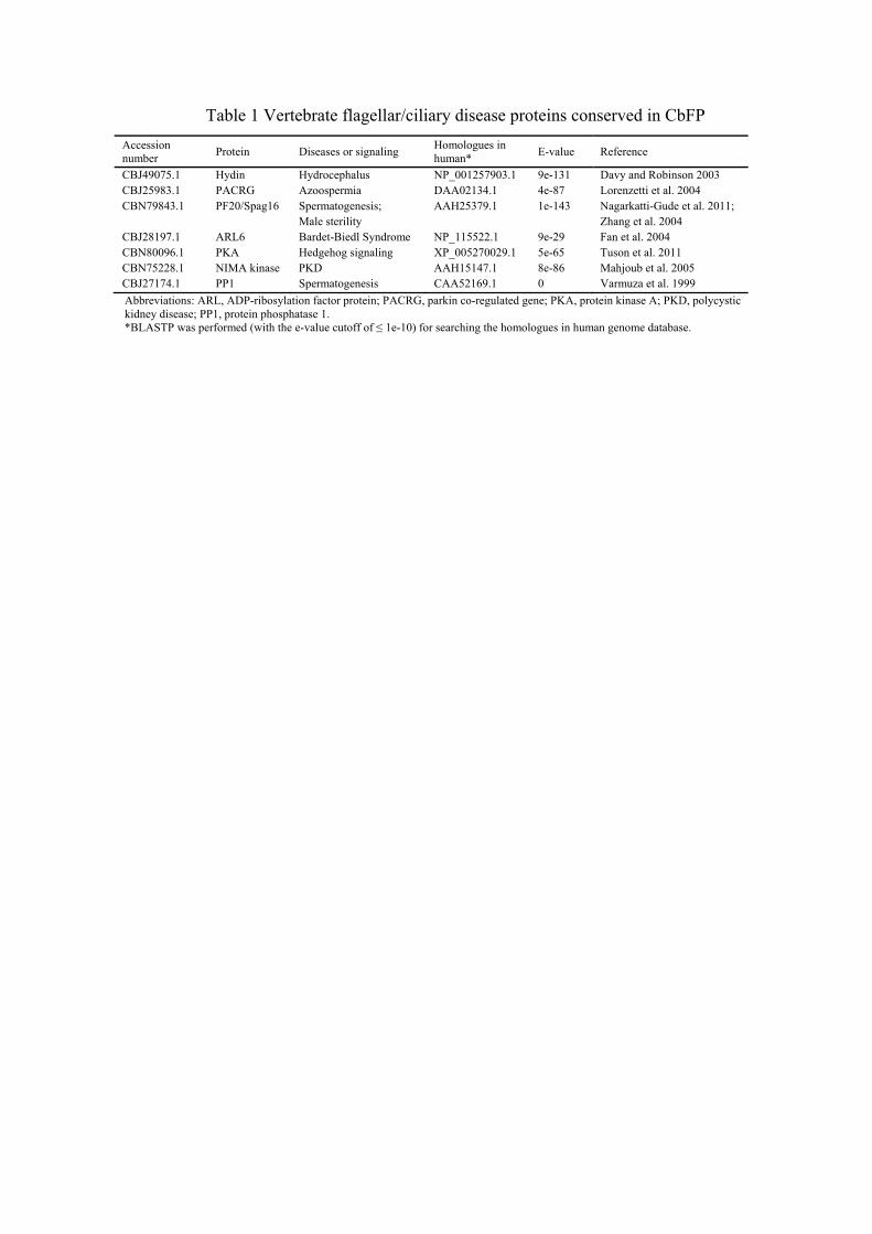
Table 1 Vertebrate flagellar/ciliary disease proteins conserved in CbFP
Accession number
Protein Diseases or signaling Homologues in human*
E-value Reference
CBJ49075.1 Hydin Hydrocephalus NP_001257903.1 9e-131 Davy and Robinson 2003 CBJ25983.1 PACRG Azoospermia DAA02134.1 4e-87 Lorenzetti et al. 2004 CBN79843.1
PF20/Spag16 Spermatogenesis; Male sterility
AAH25379.1 1e-143 Nagarkatti-Gude et al. 2011; Zhang et al. 2004
CBJ28197.1 ARL6 Bardet-Biedl Syndrome NP_115522.1 9e-29 Fan et al. 2004 CBN80096.1 PKA Hedgehog signaling XP_005270029.1 5e-65 Tuson et al. 2011 CBN75228.1 NIMA kinase PKD AAH15147.1 8e-86 Mahjoub et al. 2005 CBJ27174.1 PP1 Spermatogenesis CAA52169.1 0 Varmuza et al. 1999
Abbreviations: ARL, ADP-ribosylation factor protein; PACRG, parkin co-regulated gene; PKA, protein kinase A; PKD, polycystic kidney disease; PP1, protein phosphatase 1. *BLASTP was performed (with the e-value cutoff of ≤ 1e-10) for searching the homologues in human genome database.