Embed Size (px)
Citation preview
790 Electrophoresis 2016, 37, 790–794
Andrea J. Hanson1
Andrzej J. Paszczynski2Erik R. Coats3
1Microbiology, MolecularBiology, and BiochemistryGraduate Program, Departmentof Biological Sciences,University of Idaho, Moscow,ID, USA
2University of Idaho andWashington State UniversitySchool of Food Science, FoodResearch Center, Moscow, ID,USA
3Department of CivilEngineering, University ofIdaho, Moscow, ID, USA
Received August 26, 2015Revised December 8, 2015Accepted January 4, 2016
Short Communication
Proteomic profiling of an undefinedmicrobial consortium cultured in fermenteddairy manure: Methods development
The production of polyhydroxyalkanoates (PHA; bioplastics) from waste or surplus feed-stocks using mixed microbial consortia (MMC) and aerobic dynamic feeding (ADF) is agrowing field within mixed culture biotechnology. This study aimed to optimize a 2DEworkflow to investigate the proteome dynamics of an MMC synthesizing PHA from fer-mented dairy manure. To mitigate the challenges posed to effective 2DE by this complexsample matrix, the bacterial biomass was purified using Accudenz gradient centrifuga-tion (AGC) before protein extraction. The optimized 2DE method yielded high-quality gelssuitable for quantitative comparative analysis and subsequent protein identification by LC-MS/MS. The optimized 2DE method could be adapted to other proteomic investigationsinvolving MMC in complex organic or environmental matrices.
Keywords:
2DE / Accudenz gradient centrifugation / Mixed culture biotechnology / Mixedmicrobial consortia DOI 10.1002/elps.201500400
� Additional supporting information may be found in the online version of thisarticle at the publisher’s web-site
The production of polyhydroxyalkanoates (PHA) using mixedmicrobial consortia (MMC) and fermented waste/surplusfeedstocks has attracted attention as an inexpensive alter-native to current commercial production which relies onpure cultures and refined substrates [1]. To be successful,the MMC must be highly enriched for PHA-producing bac-teria. This is commonly achieved through aerobic dynamicfeeding (ADF) [2], which imposes transient exogenous car-bon availability (i.e., “feast-famine” conditions). However, ad-vancement of this process has been impeded by an incom-plete understanding of the physiology and biochemistry of theMMC response to ADF conditions. 2DE-based proteomics of-fers a powerful approach to elucidate functional responses;however, optimal 2DE procedures have not been establishedfor MMC cultured in fermented waste feedstocks. To addressthis need, the research herein focused on the application of2DE to an MMC subjected to ADF conditions and culturedon fermented dairy manure.
Correspondence: Dr. Erik R. Coats, Associate Professor of CivilEngineering, University of Idaho, 875 Perimeter Drive, MS1022,Moscow, ID 83844-1022E-mail: [email protected]: +1-208 885 6608
Abbreviations: ADF, aerobic dynamic feeding; AGC, Accud-enz gradient centrifugation; MMC, mixed microbial consortia;PHA, polyhydroxyalkanoates
Like other 2DE-based investigations involving mixedculture biotechnology (e.g., activated sludge processes andanaerobic digestion), the fermented dairy manure samplematrix added to the inherent complexity of MMC proteinmixtures. Direct protein extraction from MMC biomass andsubsequent electrophoretic separation is challenging due tothe abundance of non-bacterial protein impurities and non-bacterial solids (including volatile fatty acids, salts, crude fat,non-structural carbohydrates, inert particulates, lignocellu-lose degradation products, and pigmented compounds).
To combat similar interferences, indirect protein extrac-tion approaches utilizing gradient centrifugation that first iso-late/purify bacterial cells from complex matrices have beenapplied in MMC proteome studies of soil and freshwatersamples using 1D SDS PAGE [3, 4]. Drawing from these in-vestigations, the aim of this study was to optimize an MMCsample processing procedure using Accudenz gradient cen-trifugation (AGC) and sequential protein extraction to im-prove the recovery of bacterial proteins from reactor biomassand ensure effective protein separation by 2DE.
Sample source: MMC biomass samples were obtainedfrom a sequencing batch reactor operated under ADF con-ditions conducive for PHA synthesis. Briefly, the reactor wasfully aerobic, constantly mixed, had a volume of 1.8 L, solidsand hydraulic retention time of 4 d, and cycle length of 24 h.
Colour Online: See the article online to view Figs. 1-3 in colour
C© 2016 WILEY-VCH Verlag GmbH & Co. KGaA, Weinheim www.electrophoresis-journal.com

Electrophoresis 2016, 37, 790–794 Proteomics and 2 - DE 791
Figure 1. Outline of the optimized “AGC 2DE”workflow. See text for additional details.
The fermented dairy manure used for substrate was preparedin the following manner: effluent from a lab-scale manurefermenter was screened to remove large solids, centrifugedat 8000 rpm for 5 min to remove fine solids, autoclaved, anddiluted with tap water prior to addition to the reactor. The sus-pended solids concentration (including microbial biomass) inthe reactor was maintained above 2000 mg/L.
Optimized “AGC 2DE” method: The following proce-dures are outlined in Fig.1. All reagents used were ofelectrophoresis-grade. Initial biomass collection and washingwas adapted from Wilmes and Bond [5] with minor modifi-cations. Briefly, 80 mL MMC sample was centrifuged at5000 rpm for 15 min at 4°C in an SS-34 rotor (Sorvall,Waltham, MA). The supernatant was discarded; the pelletwas resuspended in 40 mL of 0.9% NaCl and washed by cen-trifugation at 5000 rpm for 15 min at 4°C. After discardingthe wash, the pellet was resuspended in 25 mL of 25 mMTris, pH 7.4.
The AGC procedures were adapted from published pro-tocols [3, 6] with modifications. Briefly, to the 25 mL washedbiomass suspension, 8 mL of 1.3 g/mL Accudenz (AccurateChemical Company, Westbury, NY) solution was added be-neath the sample and incubated for 30 min at 4°C. The sample
was centrifuged at 8000 rpm for 40 min at 4°C in a JS13.1swing-out rotor (Beckman Coulter, Pasadena, CA). Separatedbacterial cells at the gradient interface (a visible turbid layer)were collected with a 5 mL syringe fitted with an 18-gaugeneedle bent at 90° (this size permits bacterial cell flow). Col-lected cells were washed twice in 15 mL of double-DI H2O bycentrifugation at 8000 rpm for 10 min at 4°C using the samerotor; washes were discarded and the bacterial cell pellet wassubjected to sequential protein extraction.
For sequential protein extraction, the bacterial cell pelletwas resuspended in 650 �L Lysis Buffer 1 (25 mM Tris pH 7.4,0.1 mM EDTA, and 0.5 mM 4-(2-aminoethyl)benzenesulfonylfluoride hydrochloride (AEBSF)), vigorously mixed, andtransferred to a 1.5 mL self-standing screw cap tube. Thebacterial cell suspension was sonicated using a Model 100Sonic Dismembrator (Thermo Fisher, Waltham, MA) on icewith 15 s pulses up to 2 min with the power setting at 3,after which 0.1 mm zirconia/silica beads (Biospec ProductsInc., Bartlesville, OK) were added to the suspension to ap-proximately half the sample volume. The suspension was ho-mogenized in a bead mill beater (Biospec Products Inc.) for1 min, followed by 30 s pulses for 6 min, and then centrifugedat 13 000 rpm for 15 min at 4°C, and the supernatant (protein
C© 2016 WILEY-VCH Verlag GmbH & Co. KGaA, Weinheim www.electrophoresis-journal.com

792 A. J. Hanson et al. Electrophoresis 2016, 37, 790–794
Figure 2. Bacterial cell sepa-ration and disruption. Phasecontrast images (1000x) show-ing representative images of(A) biomass directly fromthe MMC reactor, (B) bacte-rial cells isolated using AGC,(C) bacterial suspension afterbrief sonication and first beadmill beating in Lysis Buffer 1,and (D) bacterial suspensionafter 30 min incubation andsecond bead mill beating in Ly-sis Buffer 2. Images were ac-quired using a Nikon Eclipse55i phase contrast microscopewith NIS-147 Elements Br. 3.0.Bars in the bottom right cornerrepresent 1 �m.
fraction #1) was stored on ice. The pellet was resuspendedin 600 �L Lysis Buffer 2 (7 M urea, 2 M thiourea, 4% w/vCHAPS, 10 mM Tris-1 mM EDTA, 50 mM DTT, and 0.5 mMAEBSF; DTT, CHAPS and AEBSF were added immediatelybefore use), then incubated on ice for 30 min with vigorousmixing every 10 min. The aforementioned bead mill beat-ing procedure was repeated, along with centrifugation andsupernatant retention (protein fraction #2). A nuclease treat-ment was performed separately on each protein fraction byadding 50U of Benzonase nuclease (Sigma-Aldrich, St. Louis,MO) and incubating at room temperature for 10 min withlight mixing every 2 min. Suspensions were centrifuged at13 000 rpm for 25 min at 4°C; the supernatants were com-bined into one sample and vigorously mixed. When required,protein samples were stored at –80°C.
Protein concentration of the combined fractions was de-termined by the RCDC Protein Assay (Bio-Rad, Hercules, CA)following the manufacturer’s instructions and using BSA asthe protein standard. Protein precipitation and clean-up pro-cedures were performed using a ReadyPrep 2-D Cleanup Kit(Bio-Rad) following the manufacturer’s instructions. The pro-tein pellet was resuspended in 175 �L Rehydration Buffer(7 M urea, 2 M thiourea, 3% (w/v) CHAPS, 50 mM DTT,and 0.2% (w/v) Bio-Lyte 3/10 ampholytes; DTT, CHAPS, andampholytes were added immediately before use). The sam-ple was mixed for 1 h at 4°C. Protein concentration in therehydration solution was determined using the RCDC Assay,
and the sample was diluted in Rehydration Buffer to nor-malize the concentration to 3.2 mg/mL; a trace amount ofbromophenol blue was added, followed by vigorous mixingand centrifugation at 13 000 rpm for 10 min at 4°C prior toIPG strip loading.
For IEF, 400 �g protein in 125 �L was loaded onto a 7 cmReadyStrip IPG Strip pH 4–7 (Bio-Rad), equilibrated for 1 hat room temperature, covered with mineral oil, and passivelyrehydrated for 16 h at 20°C. The IPG strip was focused in aPROTEAN IEF Cell (Bio-Rad) at 250 V for 15 min, followedby linear ramping to 4000 V in 2.5 h, after which 4,000 Vwas held for approximately 3.25 h (for a total of 13,000 Vh).Immediately following IEF, the IPG strip was equilibratedtwice for 20 min each in Equilibration Buffer A (6 M urea,30% (w/v) glycerol, 2% (w/v) SDS in 0.05 M Tris pH 8.8, and2% (w/v) DTT) and Equilibration Buffer B (same as A onlyDTT was replaced by 2.5% (w/v) iodoacedamide) with gentleshaking; for each equilibration, fresh buffer was exchangedafter 10 min.
For SDS PAGE separation, the IPG strip was rinsed inTris-Glycine-SDS buffer (25 mM Tris, 192 mM glycine, 0.1%SDS), loaded onto an 8 × 10 cm 12% Mini-PROTEAN TGXpre-cast gel (Bio-Rad), sealed with overlay agarose, and elec-trophoresed at 70 V for 40 min, followed by 150 V untilthe tracking dye reached the bottom of the gel; molecularweight markers were applied to an electrode wick and in-serted into the gel before the application of overlay agarose.
C© 2016 WILEY-VCH Verlag GmbH & Co. KGaA, Weinheim www.electrophoresis-journal.com

Electrophoresis 2016, 37, 790–794 Proteomics and 2 - DE 793
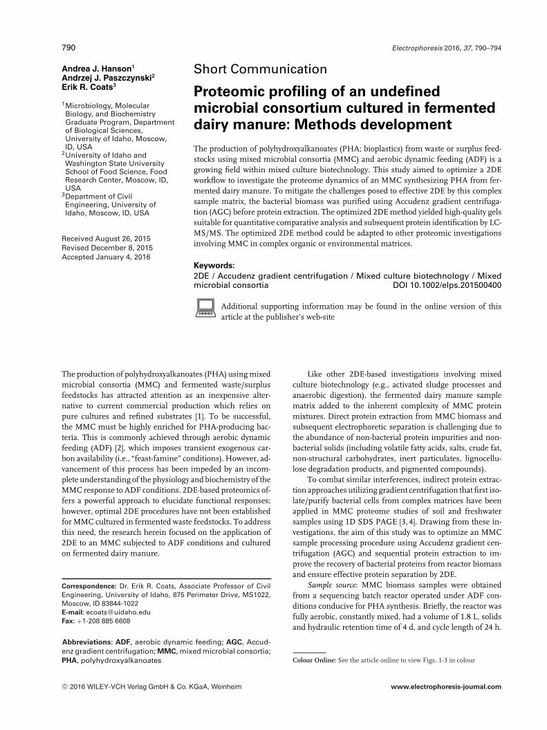
Figure 3. Representative 2DEgel image for the optimized“AGC 2DE” method. Molecu-lar weight markers (in kDa) arelisted to the left of the im-age, and the pH range of theIPG strip is depicted below theimage.
The gel was washed twice in double-DI H2O for 5 min, fixed inethanol/acetic acid/water (40:10:50 (v/v)) for 30 min, stainedwith pre-made Coomassie Brilliant Blue G-250 (CBB; Bio-Rad) for 20 h, and de-stained with double-DI H2O for sev-eral hours. The gel was scanned using an Odyssey ImagingSystem (Li-COR Biosciences, Lincoln, NE) with the followingsettings which generated 16 bit, 600 DPI TIFF files: reso-lution, 42 �m; quality, medium; focus offset, 0.5; detectionchannel, 700 nm. REDFIN 3 Gel Image Analysis Software(Ludesi, Malmo, Sweden) was used for gel image analysis fol-lowing the manufacturer’s recommendations related to im-age quality control, gel warping, spot matching, spot intensitymeasurement, background correction, and spot volume nor-malization.
The primary objective during the development of the“AGC 2DE” workflow (Fig. 1) was to increase the reliabilityand reproducibility of 2DE for investigating MMC operatedunder ADF conditions. Initial 2DE attempts indicated signif-icant interference from the sample matrix and biased proteinrecovery, in addition to poor-quality gels (data not shown).AGC and sequential protein extraction were incorporated intothe method to address these shortcomings.
AGC was used to mitigate the interference from the fer-mented dairy manure matrix on the bacterial protein extrac-tion by separating the bacterial cells from the non-bacterialsolids. While AGC may introduce bias into the bacterial re-covery [7], priority was given to achieving successful 2DE.To that end, AGC removed the non-bacterial solids from thesample, reducing the potential for the co-extraction of im-purities and partitioning of proteins during extraction. Thesuspended solids concentration in the MMC reactor was 2394± 736 mg/L, and as shown in Fig. 2A and B, AGC effectively
isolated bacterial cells from other solids. The bacterial cellpellet was typically 1/3 of the initial biomass pellet wet mass,reinforcing the value of AGC as a purification tool to separatebacterial cells from matrix debris.
Sequential protein extraction was adopted in an effort tomaximize bacterial protein recovery while reducing extractionbias. As shown in Fig. 2B, cell morphology was diverse in theMMC; the brief sonication, coupled with two rounds of beadmill beating applied in the “AGC 2DE” method aided cell dis-ruption (Fig. 2C and D) to help maximize protein recovery. Noprotein loss was observed following protein fraction combina-tion, allaying concerns that protein precipitation might occurdue to buffer dissimilarity. Throughout “AGC 2DE” devel-opment, the protein concentration in the combined proteinsample was 12.8 ± 5.5 mg/mL, which was sufficiently highfor the 2DE clean-up and purification procedures. Togetherwith the identification of both cytoplasmic and hydrophobicmembrane-bound proteins (excised from the 2DE gel andidentified by LC-MS/MS using the procedures adapted fromChecinska et al. [8]; data not shown), the effectiveness of thesequential protein extraction was reinforced.
The “AGC 2DE” method was repeated multiple timesand consistently yielded gels with spot patterns similar tothat shown in Fig. 3. Throughout “AGC 2DE” optimization,the number of detected protein spots ranged from 585 to639, with an average of 608 ± 26; spot numbers for the“AGC 2DE” method were higher compared to 2DE attemptsinvolving direct extraction (data not shown). Other positiveresults of the “AGC 2DE” method included well-resolvedspots, minimal horizontal streaking, reduced vertical stack-ing, and nominal background interference (Fig. 3). Proteinsfrom the “AGC 2DE” method exhibited an array of isoelectric
C© 2016 WILEY-VCH Verlag GmbH & Co. KGaA, Weinheim www.electrophoresis-journal.com

794 A. J. Hanson et al. Electrophoresis 2016, 37, 790–794
points across the pH 4 to 7 range, and an apparent molec-ular weight range spanning from less than 15 kDa up to190 kDa. IPG strips with pH 3–10 and 7–10 ranges were eval-uated, in addition to active IPG strip rehydration; however,protein resolution was not improved (data not shown). CBBG-250 was ultimately chosen over other evaluated stains (CBBR-250, Silver Stain for MS, and SYPRO Ruby) for its low back-ground interference, reproducibility, sensitivity when used incombination with infrared scanning, and compatibility withLC-MS/MS.
The high-quality 2DE gels resulting from the “AGC2DE” method enabled protein spot identification via LC-MS/MS, which complimented quantitative comparativeassessments. As an example, LC-MS/MS analysis of one pro-tein spot and subsequent MS/MS ions search using MAS-COT (Matrix Science) resulted in the assignment of four pro-teins to Meganema perideroedes (the MASCOT search criteriaused and corresponding protein identification informationis provided in Supporting Information Tables S1–S3). Thehighest scoring protein was an amino acid ABC transportersubstrate-binding protein with 17 peptide matches represent-ing 74% amino acid sequence coverage. The other candidateproteins associated with the excised protein spot includedtwo hypothetical proteins and LacI transcriptional regulator,each assigned to M. perideroedes. The MS proteomics datahave been deposited to the ProteomeXchange Consortium [9]via the PRIDE partner repository with the dataset identifierPXD003004 and 10.6019/PXD003004; the MASCOT resultfile, peak list file, and all raw LC-MS/MS data can be accessedthrough the PRIDE repository.
The optimized “AGC 2DE” procedures presented hereinwere effective as a biomass processing method for MMCprotein profiling in an engineered system laden with 2DE-interfering impurities and non-bacterial solids. The “AGC2DE” method yielded reproducible, high-quality 2DE gelsfrom which protein spots have been excised and identifiedvia LC-MS/MS. Notably, the presented workflow could bemodified based on available laboratory equipment; for ex-ample, a similar make or model of swing-out rotor could besubstituted, different cell disruption methods could be imple-mented, or alternative gel imaging systems could be used inplace of an infrared scanner. As such, the “AGC 2DE” work-flow could be adapted to other MMC in complex matrices
often encountered in mixed culture biotechnology studies aspart of a gel-based or gel-free proteomics approach.
The authors acknowledge Nicholas M. Guho at the Univer-sity of Idaho for assistance with gel image processing and figureediting. This material is based upon work supported by the Na-tional Science Foundation under Grant Number CBET-0950498,and Environmental Protection Agency Science to Achieve ResultsFellowship. Any opinions, findings, and conclusions or recommen-dations expressed in this material are those of the authors and donot necessarily reflect the views of the funding agency.
The authors declare no conflicts of interest.
1 References
[1] Dias, J. M. L., Lemos, P. C., Serafim, L. S., Oliveira, C.,Eiroa, M., Albuquerque, M. G. E., Ramos, A. M., Oliveira,R., Reis, M. A. M., Macromol. Biosci. 2006, 6, 885–906.
[2] Majone, M., Massanisso, P., Carucci, A., Lindrea, K.,Tandoi, V., Water Sci. Technol. 1996, 34, 223–232.
[3] Williams, M. A., Taylor, E. B., Mula, H. P., Soil Biol.Biochem. 2010, 42, 1148–1156.
[4] Pierre-Alain, M., Christophe, M., Severine, S., Houria,A., Philippe, L., Lionel, R., Microb. Ecol. 2007, 53, 426–434.
[5] Wilmes, P., Bond, P. L., Environ. Microbiol. 2004, 6,911–920.
[6] Courtois, S., Frostegard, A., Goransson, P., Depret, G.,Jeannin, P., Simonet, P., Environ. Microbiol. 2001, 3,431–439.
[7] Holmsgaard, P. N., Norman, A., Hede, S. C., Poulsen, P.H. B., Al-Soud, W. A., Hansen, L. H., Sørensen, S. J., SoilBiol. Biochem. 2011, 43, 2152–2159.
[8] Checinska, A., Burbank, M., Paszczynski, A. J., Appl. Env-iron. Microbiol. 2012, 78, 6413–6422.
[9] Vizcaino, J. A., Deutsch, E. W., Wang, R., Csordas, A.,Reisinger, F., Rios, D., Dianes, J. A., Sun, Z., Farrah, T., Ban-deira, N., Binz, P.-A., Xenarios, I., Eisenacher, M., Mayer,G., Gatto, L., Campos, A., Chalkley, R. J., Kraus, H.-J., Al-bar, J. P., Martinez-Bartolome, S., Apweiler, R., Omenn, G.S., Martens, L., Jones, A. R., Hermjakob, H., Nat. Biotech.2014, 32, 223–226.
C© 2016 WILEY-VCH Verlag GmbH & Co. KGaA, Weinheim www.electrophoresis-journal.com