Embed Size (px)
Citation preview
Phytochemistry 71 (2010) 351–362
Contents lists available at ScienceDirect
Phytochemistry
journal homepage: www.elsevier .com/locate /phytochem
Review
Proteomic approaches to study plant–pathogen interactions
B.F. Quirino a,b,*, E.S. Candido a, P.F. Campos a, O.L. Franco a, R.H. Krüger c
a Universidade Católica de Brasília, Genomic Sciences and Biotechnology Program, Brasília, DF, Brazilb Embrapa-Agroenergia, Brasília, DF, Brazilc Universidade de Brasília, Brasília, DF, Brazil
a r t i c l e i n f o
Article history:Received 22 August 2008Received in revised form 23 June 2009Available online 11 December 2009
Keywords:ProteomicsProteomePlantPathogenPathosystem
0031-9422/$ - see front matter � 2009 Elsevier Ltd. Adoi:10.1016/j.phytochem.2009.11.005
* Corresponding author. Address: Universidade CSciences and Biotechnology Program, SGAN 916 AvBrasília, DF, Brazil. Tel.: +55 61 3448 7115; fax: +55 6
E-mail addresses: [email protected], betania.quiri
a b s t r a c t
The analysis of plant proteomes has drastically expanded in the last few years. Mass spectrometry tech-nology, stains, software and progress in bioinformatics have made identification of proteins relativelyeasy. The assignment of proteins to particular organelles and the development of better algorithms topredict sub-cellular localization are examples of how proteomic studies are contributing to plant biology.Protein phosphorylation and degradation are also known to occur during plant defense signaling cas-cades. Despite the great potential to give contributions to the study of plant–pathogen interactions, onlyrecently has the proteomic approach begun to be applied to this field. Biological variation and complexityin a situation involving two organisms in intimate contact are intrinsic challenges in this area, however,for proteomics studies yet, there is no substitute for in planta studies with pathogens, and ways toaddress these problems are discussed. Protein identification depends not only on mass spectrometry,but also on the existence of complete genome sequence databases for comparison. Although the numberof completely sequenced genomes is constantly growing, only four plants have their genomes completelysequenced. Additionally, there are already a number of pathosystems where both partners in the inter-action have genomes fully sequenced and where functional genomics tools are available. It is thus tobe expected that great progress in understanding the biology of these pathosystems will be made overthe next few years. Cheaper sequencing technologies should make protein identification in non-modelspecies easier and the bottleneck in proteomic research should shift from unambiguous protein identifi-cation to determination of protein function.
� 2009 Elsevier Ltd. All rights reserved.
Contents
1. Introduction . . . . . . . . . . . . . . . . . . . . . . . . . . . . . . . . . . . . . . . . . . . . . . . . . . . . . . . . . . . . . . . . . . . . . . . . . . . . . . . . . . . . . . . . . . . . . . . . . . . . . . . . . 3512. Proteomic work involving plants . . . . . . . . . . . . . . . . . . . . . . . . . . . . . . . . . . . . . . . . . . . . . . . . . . . . . . . . . . . . . . . . . . . . . . . . . . . . . . . . . . . . . . . . 3533. Plant–pathogen interactions and proteomics . . . . . . . . . . . . . . . . . . . . . . . . . . . . . . . . . . . . . . . . . . . . . . . . . . . . . . . . . . . . . . . . . . . . . . . . . . . . . . . 354
3.1. A late blossoming . . . . . . . . . . . . . . . . . . . . . . . . . . . . . . . . . . . . . . . . . . . . . . . . . . . . . . . . . . . . . . . . . . . . . . . . . . . . . . . . . . . . . . . . . . . . . . . 3543.2. Approaches to analyze an interaction involving two organisms . . . . . . . . . . . . . . . . . . . . . . . . . . . . . . . . . . . . . . . . . . . . . . . . . . . . . . . . . . 354
4. Genomics helping proteomics . . . . . . . . . . . . . . . . . . . . . . . . . . . . . . . . . . . . . . . . . . . . . . . . . . . . . . . . . . . . . . . . . . . . . . . . . . . . . . . . . . . . . . . . . . . 3565. Concluding remarks . . . . . . . . . . . . . . . . . . . . . . . . . . . . . . . . . . . . . . . . . . . . . . . . . . . . . . . . . . . . . . . . . . . . . . . . . . . . . . . . . . . . . . . . . . . . . . . . . . . 358
Acknowledgements . . . . . . . . . . . . . . . . . . . . . . . . . . . . . . . . . . . . . . . . . . . . . . . . . . . . . . . . . . . . . . . . . . . . . . . . . . . . . . . . . . . . . . . . . . . . . . . . . . . 358References . . . . . . . . . . . . . . . . . . . . . . . . . . . . . . . . . . . . . . . . . . . . . . . . . . . . . . . . . . . . . . . . . . . . . . . . . . . . . . . . . . . . . . . . . . . . . . . . . . . . . . . . . . 358
ll rights reserved.
atólica de Brasília, Genomic. W5 norte, Cep 70.790-160,1 3347 4797.
[email protected] (B.F. Quirino).
1. Introduction
Over the last few years, much attention has been given tomicroarray studies of mRNA expression, and next generationDNA sequencers now permit a truly global analysis of the mRNAcomplement (via cDNA) of any cell. However, it has long been
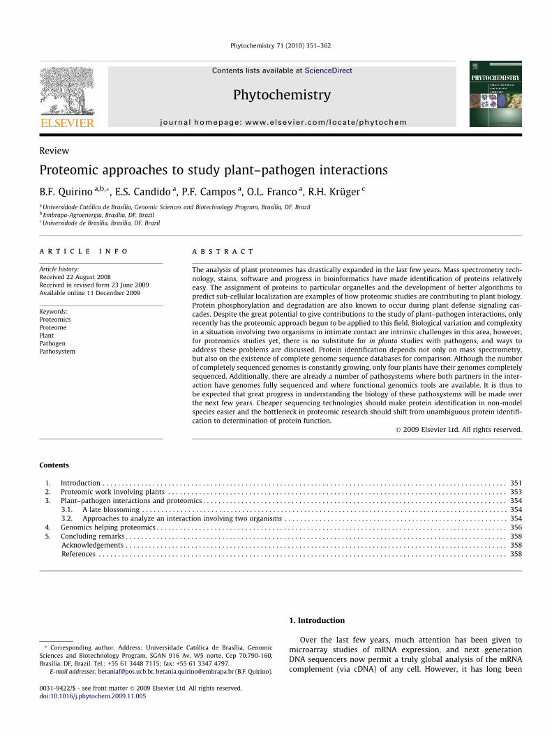
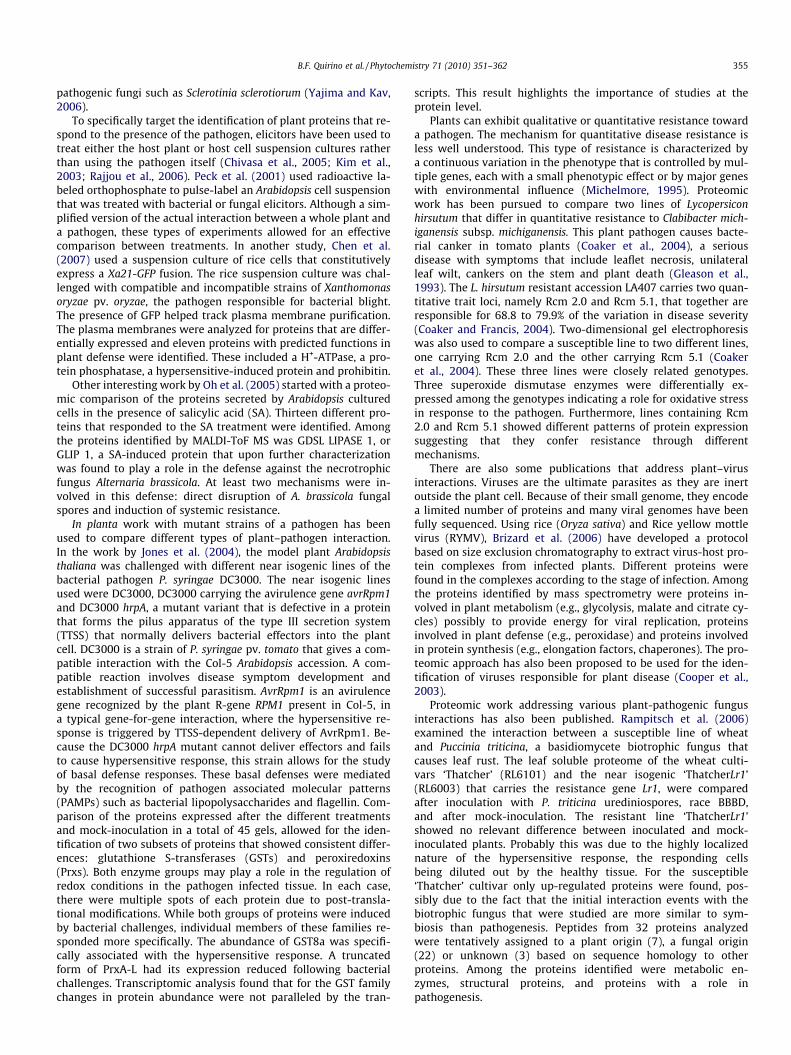
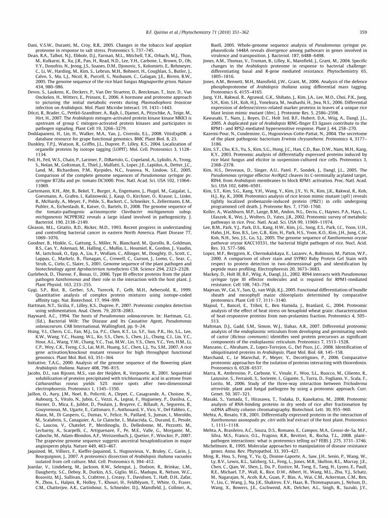
Fig. 1. Schematic representation of the different approaches used in proteomicstudies.
352 B.F. Quirino et al. / Phytochemistry 71 (2010) 351–362
known that gene expression is regulated at different levels, and anumber of heritable characteristics are not encoded by DNA. Forexample, changes in mRNA abundance are not always mirroredby corresponding protein levels and early rapid changes in cellbehavior often rely on pre-existing proteins that are either post-translationally modified, have changed their sub-cellular locationand/or are degraded. Other nuances that can be biologically rele-vant include genes encoding differently spliced mRNAs that cangive rise to more than one protein. Thus, as more layers of generegulation, such as epigenetic DNA modifications and the plethoraof small non-coding RNAs are uncovered, it becomes clear thatmany biological questions can only be addressed at the protein le-vel as the presence of either a gene or its mRNA are no guarantee ofa role in cellular activity. Therefore, technologies aimed at studyingproteins in a cell are a welcome complement. Proteomics is theglobal study of the protein content of a cell, but this presentsnew technical challenges due to their greater structural complexitywhen compared to that of nucleic acids. Technological advancesare, however allowing for today’s progress in this area. In thisway, new and exciting questions can now be tackled, which to-gether with other ‘omics’ such as genomics and metabolomics, al-lowed for a new perspective towards understanding how a cellworks.
Examples of the types of proteomic studies are varied. Whileyeast-two-hybrid and yeast-three-hybrid systems (Quirino et al.,2004) can be used to identify cellular proteins able to interact withcertain proteins under investigation, mass spectrometry is used toidentify cellular proteins and two-dimensional (2-D) gel electro-phoresis and thus allows comparison of the types of protein andabundance present in different biological situations.
Contrasting samples in respect to time (e.g., proteins expressedearly vs. late during development), space (e.g., proteins exclusivelypresent in roots) or treatment (e.g. dehydrated vs. non-dehydratedtissue) have also been studied using 2-D gel electrophoresis, withdifferently expressed proteins being observed in early proteomicstudies decades ago (Orrick et al., 1973; Nagabhushan et al., 1974).
Today, the advances in mass spectrometry (MS) and their syn-ergism with genomics are revolutionizing biochemistry. A massspectrometer can now be used to identify either a single separatedprotein or hundreds of proteins present in a complex mixture withrelative ease. In this context, mass spectrometry is an analyticaltechnique that identifies the chemical composition of a compoundor sample (Sparkman, 2000). This methodology employs chemicalfragmentation of a protein into charged particles (ions) and mea-surements of charge and mass of the resulting particles. Ionizationof molecules is obtained through techniques such as either electro-spray (ESI) or matrix-assisted laser desorption/ionization (MALDI)(Siuzdak, 1996). Ionized molecules, which gain or lose their chargeby protonation, deprotonation or electron ejection, are electrostat-ically propelled inside the instrument and detected according totheir mass to charge ratio. Furthermore, there are a number of dat-abases with information about protein sequences obtained by thetranslation of open reading frames. This allows for the comparisonof data obtained by mass spectrometry to these protein databasesto identify the proteins or peptides present in a sample. In addition,the popularity of traditional as well as the development of newhigh throughput DNA sequencing technologies enables completegenome sequencing to become possible to many more species.
Another development in biochemistry has been the use of tan-dem mass spectrometry (MS/MS). In MS/MS, a particular ion is se-lected with a mass filter/analyzer and then the selected ion isfurther fragmented and analyzed. Fragmentation can be inducedby introducing the ion into a chamber with a collision gas suchas argon where collision of ions and argon atoms results in frag-mentation. The daughter ion spectrum is then analyzed (Siuzdak,1996). MS/MS is extensively used in ‘de novo’ peptide sequencing.
Many methods for protein analysis, using either gel-based ornon-gel based protein separation followed by MS or MS/MS analy-sis, have also been developed (Fig. 1). As the 2-D PAGE is still themost commonly used method, here we will only cite as examplesthe analysis of proteomes by MudPIT, DIGE, iTRAQ, SILAC, ICATand LOPIT. In MudPIT (multidimensional protein identificationtechnology), liquid chromatography (i.e., a strong cation exchangephase followed by a reversed phase chromatography) is directlycoupled to tandem mass spectrometry (Washburn et al., 2001).Independent protein separation technologies (i.e., non-gel based)are complementary to gel-based ones and together the differenttechniques allow for improved proteomic coverage (Washburnet al., 2001; Koller et al., 2002).
There has also been much progress in technologies that allowfor quantitative measurements of proteins such as DIGE. In DIGE,a gel-based technology, two protein samples labeled with differentfluorescent dyes are run on the same gel and directly compared. IniTRAQ, peptides derived from each sample are derivatized withamine-specific isobaric tags which are indistinguishable by MSbut exhibit MS/MS signature ions (Patton, 2002; Ross et al.,2004). This allows for both relative and absolute quantitation ofpeptides from different samples simultaneously. In SILAC, stableisotopes, such as 13C, are used to label amino acids which are madeavailable to cells in culture (Ong et al., 2003). Mass spectrometrycan be used for quantitation of proteins as ‘light’ and ‘heavy’ formsof a protein can be distinguished. ICAT is similar to SILAC in that italso uses different isotopes to label proteins of different samples.However, in ICAT, the isotopes are in affinity tags. The ICAT tag iscomposed of an affinity tag such as biotin which is used to purifythe labeled proteins, a linker that will harbor either heavy or lightisotopes and a reactive group with specificity toward thiol groups
B.F. Quirino et al. / Phytochemistry 71 (2010) 351–362 353
(Gygi et al., 1999). MS/MS analysis allows for determination of theratio between ‘light’ and ‘heavy’ forms of each peptide thereby pro-viding quantitative comparisons between samples. LOPIT or Local-ization of Organelle Proteins by Isotope Tagging uses an initialpartial separation of organelles by density gradient centrifugationfollowed by analysis of protein distribution in the gradient by ICATand mass spectrometry. Analysis of the protein distribution in thegradient and grouping with proteins of known cellular localizationallows assignment of a protein to an organelle (Dunkley et al.,2004).
The new techniques available for proteomic studies are varied,but this will not be the focus of this review. Those interested in fur-ther reading can refer to other reviews (Chen, 2008; Carpentieret al., 2008; Ong et al., 2003). Recent reviews cover plant proteo-mics in general (Rossignol et al., 2006) and catalogue the proteinsthat are being identified in plant–pathogen studies through a pro-teomics approach (Mehta et al., 2008).
Herein, we address the challenges of using a proteomics ap-proach to study plant–pathogen interactions, a biological situationthat involves at least two organisms in close contact. We also dis-cuss how genomics is impacting proteomics and future prospects.
2. Proteomic work involving plants
A typical proteomic experiment starts with protein extractionfrom cells, although such studies with plants can be particularlychallenging. This is because plant cells are not only rich in otherconstituents, such as cell wall polysaccharides and polyphenols,but also have a number of proteases that can degrade samples. Inaddition, the dominance of certain proteins can make it difficultto study other less abundant proteins. Rubisco (ribulose bisphos-phate carboxylase–oxygenase) is the predominant protein inleaves, and storage proteins are also highly abundant in seeds(Chen and Harmon, 2006; Jones et al., 2004). There are various pro-tocols available, each most suited to a specific tissue. Most proto-cols are based on a trichloroacetic acid (TCA) and/or acetoneprecipitation of proteins or on phenol extraction, where proteinsare solubilized in the phenolic phase and then are precipitatedwith methanol and ammonium sulfate (Carpentier et al., 2005; Ja-cobs et al., 2001). The protocols that use phenol have been em-ployed successfully for different plant species.
Two-dimensional gel electrophoresis is currently the most com-mon method used in proteomic studies. In this methodology, theprotein samples of interest are initially separated via the isoelectricfocusing point (based on protein pI) on a strip that has a gradient ofpHs (IPG strip) in the first dimension. This is then followed by sep-aration by mass on a SDS–polyacrylamide gel (PAGE) in the seconddimension.
Proteins on gels are visualized by staining, this being performedwith Coomasie, silver nitrate, or a number of new fluorescent dyes.Although popular, Coomassie Brilliant Blue (R-250) is not as sensi-tive as silver staining. Colloidal Coomassie (G-250) is an improve-ment over the standard Coomassie as it is more sensitive. Silvernitrate staining gives excellent results but can interfere withdownstream identification of proteins by mass spectrometry. Thenew fluorescent dyes such as SYPRO-Ruby and Flamingo Pink™and Krypton™ are very sensitive and compatible with MS technol-ogy. SYPRO-Ruby can detect 1–2 ng protein in a band which is adetection limit similar to that of silver staining (Lopez et al.,2000). Unfortunately though, the price of these new dyes stillneeds to become more competitive. There are also other dyes ableto detect specifically modified proteins on SDS–PAGE gels such asphosphoproteins (e.g., Pro-Q™ Diamond) or glycoproteins (e.g.,Pro-Q™ Emerald). These dyes allow for a new host of experimentsto be performed.
For greater reliability of data, biological (i.e., independently ob-tained samples) as well as technical replicates (i.e., the same sam-ple run on a different gel) are compared using various software(e.g., PDQuest, Bionumerics, etc.). Gel spots deemed differentiallyexpressed, based on statistical analysis of the gels, are excisedand processed for identification by MS analysis. This processing in-volves a fragmentation process called ‘in-gel digestion’ in whichthe protein, still in the matrix, is digested with an enzyme thatcleaves at specific points. Trypsin, for instance, cleaves the peptidechain at the carboxyl side of lysine and arginine, except when theseare followed by proline. The collection of peptide products is thenintroduced into the mass analyzer, with the protein either identi-fied through a technique known as peptide mass fingerprinting(PMF) or via tandem MS analysis by ‘de novo’ sequencing. InPMF, the absolute masses of the peptides originating from the un-known protein are accurately measured with a mass spectrometer(Clauser et al., 1999). These masses are then compared by bioinfor-matics to a database containing known protein sequences or thegenome of the organism. This is achieved by using computer pro-grams, such as MASCOT (http://www.matrixscience.com/search_form_select.html), Phenyx (http://phenyx.vital-it.ch/pwi/login/login.jsp) and OMSSA (http://pubchem.ncbi.nlm.nih.gov/omssa/).These programs translate genomes into proteins and then theoret-ically cleave proteins into peptides and calculate the absolutemasses of the peptides from each protein. They then comparepeptide masses of the unknown target protein to the theoreticalpeptide masses of each protein deposited in database. PMF is ahigh-throughput technique, however, it will only work if the pro-tein sequence is present in the database utilized. Therefore, manylaboratories prefer to use MS/MS to sequence the peptides.
Although popular, 2-DE gel analysis coupled to MS has manylimitations. Overall, it is a laborious technique. Low abundanceproteins are particularly hard to detect in complex mixtures of pro-teins and contaminants present in plant protein samples such aspolysaccharides often interfere with gel resolution. Also, on anyexperiment, one has to make choices of the range of pH gradientto be used in the first dimension, as well as the percentage of acryl-amide used for the second dimension gel which limits which pro-teins will separate well. Therefore, the 2-D gel electrophoresisproteomics approach will always fail to detect certain proteins.
One way to minimize some of the problems of 2-D gel electro-phoresis coupled to MS approach is to reduce the complexity of theprotein sample. Because certain organelles can be isolated in ahighly pure state, this approach is called sub-cellular proteomics.A number of studies have been devoted to examine proteins thatare resident in different plant cell compartments, such as the chlo-roplast (Arai et al., 2008), mitochondria (Brugiere et al., 2004),nucleus (Pandey et al., 2008), peroxisomes (Reumann et al.,2007), vacuole (Jaquinod et al., 2007), tonoplast (Schmidt et al.,2007), endoplasmic reticulum (Maltman et al., 2007) and plantcell-wall (Chivasa et al., 2002). One consequence of the experimen-tal data that sub-cellular proteomics provides is that the signalstargeting proteins to particular organelles can be determined withmore precision. These studies should also help in development ofmore accurate programs for the in silico prediction of a protein’slocalization within a cell (Reumann et al., 2007). Proteomics stud-ies at the sub-cellular level are also contributing to the elucidationof new metabolic pathways as well as to the observation of func-tional differentiation of cells being observed (Majeran et al.,2005; Reumann et al., 2007). Even new roles for organelles areemerging from sub-cellular proteomic studies (Chivasa et al.,2002; Reumann et al., 2007). Based on the presence of proteins,such as ß-glucosidases and myrosinases, previously unknown tobe localized to leaf peroxisomes, a new role for these organellesin defense against pathogens and herbivores has been proposed(Reumann et al., 2007).
354 B.F. Quirino et al. / Phytochemistry 71 (2010) 351–362
Other studies have been able to reduce the complexity of pro-tein samples, by focusing attention on the biology of particularproteins. For example, in two studies the authors proposed to iden-tify proteins able to interact with thioredoxin, a small protein witha disulfide active site involved in redox regulation. To this end,chromatography using a column that contained a mutated thiore-doxin was used to trap potential protein targets (Marchand et al.,2006; Wong et al., 2004). Proteins with bound ubiquitin have beenidentified using a column that contained the ubiquitin binding do-main (UBA) polypeptide from the P62 protein bound to agarosebeads (Manzano et al., 2008). Proteins that bind RNA were affinitypurified with a single stranded DNA column (Masaki et al., 2008)followed by 2-D gel electrophoresis. Other than affinity purifica-tion methods, sample complexity was also reduced based on thephysical chemical properties of proteins. Hartman et al. (2007)separated proteins present in complexes by differential sedimenta-tion through a rate zonal centrifugation gradient (Hartman et al.,2007). Another approach that has been used with success (in bac-teria) is fractionation of proteins by ammonium sulfate precipita-tion (Park et al., 2008).
3. Plant–pathogen interactions and proteomics
3.1. A late blossoming
Modification of proteins, such as by phosphorylation, have longbeen known to be important in the signal transduction cascadesthat trigger plant defenses (Asai et al., 2002; Dóczi et al., 2007;Jones et al., 2006). Both MAP kinases and calcium-dependent pro-tein kinases (CDPKs) can play a part in pathogen recognition and inthe downstream events that lead to plant defense (Romeis, 2001).The signaling cascade downstream from the flagellin receptor FLS2,a leucine-rich repeat receptor (LRR) kinase involved in signal rec-ognition in the plant innate immune response, involves a numberof phosphorylation events associated with a MAP-kinase cascade(Asai et al., 2002). In other work, Romeis et al. (2001) investigatedthe role of the calcium-dependent protein kinase NtCDPK2 byvirus-induced gene silencing (VIGS) in N. benthamiana. CDPK-si-lenced plants showed a reduced and delayed hypersensitive re-sponse in a gene-for-gene response (Romeis et al., 2001).Furthermore, the Pseudomonas syringae type III effectors AvrRpm1and AvrB, that are recognized by the Arabidopsis resistance proteinRPM1, induce phosphorylation of the protein RIN4, a negative reg-ulator of plant defense (Mackey et al., 2002). Despite the fact thatphosphorylation events are central in the cascades involved inplant defense, knowledge about the targets of the phosphorylationis mostly still lacking. Protein cleavage and degradation have alsobeen shown to play a key role in the early events of the hypersen-sitive response. The type III effector avrRpt2 from P. syringae is ableto trigger plant defenses through a cascade of events that involvesproteasome-mediated RIN4 disappearance (Kawasaki et al., 2005;Kim et al., 2005; Takemoto and Jones, 2005). These examples high-light the need for large-scale methods to identify the proteins thatundergo phosphorylation, as well as proteins that disappear whena plant is responding to the presence of a pathogen. However, theseare excellent examples of situations where only a proteomics ap-proach (i.e., as compared to other genomics approaches) will bestprovide useful biological information. A proteomics-based ap-proach will thus facilitate the identification of such proteins andreveal the details about the signaling cascades involved in theinteraction between plants and pathogens.
Despite the potential contribution that proteomics can give toplant–pathogen interaction studies, including a number of studiesto address how plants react to abiotic stresses (Bindschedler et al.,2008; Dani et al., 2005; Majoul et al., 2004; Yan et al., 2006; Yang
et al., 2007), it is only now that proteomics is blossoming in thisarea. This ‘late’ blossoming may have several reasons. In planta,studies with pathogens can have considerable biological variationand this can be a problem for the proteomics studies as well. Fur-thermore, proteomics studies of plant–pathogen interactions faceintrinsic difficulties because by definition the plant–pathogeninteraction is a complex one, involving the protein complementof two organisms. Because of intimate physical contact, it can behard to distinguish proteins that are differentially expressed bythe plant in response to the pathogen from those of the pathogenitself. Despite these problems, there is currently no substitute forin planta studies with pathogens. To minimize biological variabil-ity, particular attention should be paid to the experimental design.Inoculation in a block design, pooling tissue samples and indepen-dent replication of the experiment are all important measures(Jones et al., 2006).
3.2. Approaches to analyze an interaction involving two organisms
As discussed below, there are papers where researchers have fo-cused on the pathogen side of the plant–pathogen interaction andothers where the focus was on the plant side. A simplified model ofthe actual plant–pathogen interaction has been used while othersdevised interesting experimental set-ups where the partners ofthe interaction can be separated. Furthermore, there are studieswhere the proteins were identified but a host or pathogen origincould not be assigned with any degree of certainty. In addition,there are studies of plant defense that use mutant plants, such aslesion-mimic mutants, which do not directly involve a pathogenat all (Jung et al., 2006; Kim et al., 2008; Tsunezuka et al., 2005).
To learn how a pathogen changes its protein expression in re-sponse to the plant, many researchers have used host tissue ex-tracts instead of the host itself. This strategy has been used tostudy how Xanthomonas axonopodis pv. citri responds to the pres-ence of leaf extracts from a susceptible host plant (sweet orange)as well as a resistant plant (ponkan) and a non-host plant (passionfruit) (Mehta and Rosato, 2001). In other work, Tahara et al. (2003)examined X. axonopodis pv. passiflora proteins induced in the pres-ence of host plant (passion fruit) leaf extracts. A putative mem-brane-related protein and a hypothetical protein were novelproteins induced specifically by the host plant extract and an inor-ganic pyrophosphatase and a hypothetical protein that showedsimilarity to the yciF gene of Salmonella thyphimurium were up-regulated by the host plant extract. The function these proteinsplay in the plant–pathogen interaction are, however, unknown asno functional data was presented in the paper.
Separation of the pathogen from the host is sometimes possible,for instance when the host plant has sturdy leaves. To study pro-tein expression of Xanthomonas campestris pv. campestris in closeinteraction with the host plant Brassica oleracea, Andrade et al.(2008) used a protocol where bacteria were syringe infiltrated intothe leaves of the host and later recovered for protein extraction andseparation by 2-D gel electrophoresis for proteomic analysis. Thispaper showed that it was possible, in certain cases, to study theproteins expressed by the bacterial pathogen using live interactionpartners. Here again no functional data about the proteins identi-fied was presented.
Reference maps of secreted proteins of different plant patho-gens are a benchmark for future studies involving these pathogensand their hosts. Eighty seven proteins secreted by X. campestris pv.campestris cultivated in minimal medium were identified (Wattet al., 2005). In another study with the soft-rot causing bacteriumErwinia chrysanthemi, among the proteins secreted were a cellu-lase, proteases, endopectate lyases, pectin acetylesterases, a pectinmethylesterase, and a polygalacturonase (Kazemi-Pour et al.,2004). Secretome studies have also been performed with plant
B.F. Quirino et al. / Phytochemistry 71 (2010) 351–362 355
pathogenic fungi such as Sclerotinia sclerotiorum (Yajima and Kav,2006).
To specifically target the identification of plant proteins that re-spond to the presence of the pathogen, elicitors have been used totreat either the host plant or host cell suspension cultures ratherthan using the pathogen itself (Chivasa et al., 2005; Kim et al.,2003; Rajjou et al., 2006). Peck et al. (2001) used radioactive la-beled orthophosphate to pulse-label an Arabidopsis cell suspensionthat was treated with bacterial or fungal elicitors. Although a sim-plified version of the actual interaction between a whole plant anda pathogen, these types of experiments allowed for an effectivecomparison between treatments. In another study, Chen et al.(2007) used a suspension culture of rice cells that constitutivelyexpress a Xa21-GFP fusion. The rice suspension culture was chal-lenged with compatible and incompatible strains of Xanthomonasoryzae pv. oryzae, the pathogen responsible for bacterial blight.The presence of GFP helped track plasma membrane purification.The plasma membranes were analyzed for proteins that are differ-entially expressed and eleven proteins with predicted functions inplant defense were identified. These included a H+-ATPase, a pro-tein phosphatase, a hypersensitive-induced protein and prohibitin.
Other interesting work by Oh et al. (2005) started with a proteo-mic comparison of the proteins secreted by Arabidopsis culturedcells in the presence of salicylic acid (SA). Thirteen different pro-teins that responded to the SA treatment were identified. Amongthe proteins identified by MALDI-ToF MS was GDSL LIPASE 1, orGLIP 1, a SA-induced protein that upon further characterizationwas found to play a role in the defense against the necrotrophicfungus Alternaria brassicola. At least two mechanisms were in-volved in this defense: direct disruption of A. brassicola fungalspores and induction of systemic resistance.
In planta work with mutant strains of a pathogen has beenused to compare different types of plant–pathogen interaction.In the work by Jones et al. (2004), the model plant Arabidopsisthaliana was challenged with different near isogenic lines of thebacterial pathogen P. syringae DC3000. The near isogenic linesused were DC3000, DC3000 carrying the avirulence gene avrRpm1and DC3000 hrpA, a mutant variant that is defective in a proteinthat forms the pilus apparatus of the type III secretion system(TTSS) that normally delivers bacterial effectors into the plantcell. DC3000 is a strain of P. syringae pv. tomato that gives a com-patible interaction with the Col-5 Arabidopsis accession. A com-patible reaction involves disease symptom development andestablishment of successful parasitism. AvrRpm1 is an avirulencegene recognized by the plant R-gene RPM1 present in Col-5, ina typical gene-for-gene interaction, where the hypersensitive re-sponse is triggered by TTSS-dependent delivery of AvrRpm1. Be-cause the DC3000 hrpA mutant cannot deliver effectors and failsto cause hypersensitive response, this strain allows for the studyof basal defense responses. These basal defenses were mediatedby the recognition of pathogen associated molecular patterns(PAMPs) such as bacterial lipopolysaccharides and flagellin. Com-parison of the proteins expressed after the different treatmentsand mock-inoculation in a total of 45 gels, allowed for the iden-tification of two subsets of proteins that showed consistent differ-ences: glutathione S-transferases (GSTs) and peroxiredoxins(Prxs). Both enzyme groups may play a role in the regulation ofredox conditions in the pathogen infected tissue. In each case,there were multiple spots of each protein due to post-transla-tional modifications. While both groups of proteins were inducedby bacterial challenges, individual members of these families re-sponded more specifically. The abundance of GST8a was specifi-cally associated with the hypersensitive response. A truncatedform of PrxA-L had its expression reduced following bacterialchallenges. Transcriptomic analysis found that for the GST familychanges in protein abundance were not paralleled by the tran-
scripts. This result highlights the importance of studies at theprotein level.
Plants can exhibit qualitative or quantitative resistance towarda pathogen. The mechanism for quantitative disease resistance isless well understood. This type of resistance is characterized bya continuous variation in the phenotype that is controlled by mul-tiple genes, each with a small phenotypic effect or by major geneswith environmental influence (Michelmore, 1995). Proteomicwork has been pursued to compare two lines of Lycopersiconhirsutum that differ in quantitative resistance to Clabibacter mich-iganensis subsp. michiganensis. This plant pathogen causes bacte-rial canker in tomato plants (Coaker et al., 2004), a seriousdisease with symptoms that include leaflet necrosis, unilateralleaf wilt, cankers on the stem and plant death (Gleason et al.,1993). The L. hirsutum resistant accession LA407 carries two quan-titative trait loci, namely Rcm 2.0 and Rcm 5.1, that together areresponsible for 68.8 to 79.9% of the variation in disease severity(Coaker and Francis, 2004). Two-dimensional gel electrophoresiswas also used to compare a susceptible line to two different lines,one carrying Rcm 2.0 and the other carrying Rcm 5.1 (Coakeret al., 2004). These three lines were closely related genotypes.Three superoxide dismutase enzymes were differentially ex-pressed among the genotypes indicating a role for oxidative stressin response to the pathogen. Furthermore, lines containing Rcm2.0 and Rcm 5.1 showed different patterns of protein expressionsuggesting that they confer resistance through differentmechanisms.
There are also some publications that address plant–virusinteractions. Viruses are the ultimate parasites as they are inertoutside the plant cell. Because of their small genome, they encodea limited number of proteins and many viral genomes have beenfully sequenced. Using rice (Oryza sativa) and Rice yellow mottlevirus (RYMV), Brizard et al. (2006) have developed a protocolbased on size exclusion chromatography to extract virus-host pro-tein complexes from infected plants. Different proteins werefound in the complexes according to the stage of infection. Amongthe proteins identified by mass spectrometry were proteins in-volved in plant metabolism (e.g., glycolysis, malate and citrate cy-cles) possibly to provide energy for viral replication, proteinsinvolved in plant defense (e.g., peroxidase) and proteins involvedin protein synthesis (e.g., elongation factors, chaperones). The pro-teomic approach has also been proposed to be used for the iden-tification of viruses responsible for plant disease (Cooper et al.,2003).
Proteomic work addressing various plant-pathogenic fungusinteractions has also been published. Rampitsch et al. (2006)examined the interaction between a susceptible line of wheatand Puccinia triticina, a basidiomycete biotrophic fungus thatcauses leaf rust. The leaf soluble proteome of the wheat culti-vars ‘Thatcher’ (RL6101) and the near isogenic ‘ThatcherLr1’(RL6003) that carries the resistance gene Lr1, were comparedafter inoculation with P. triticina urediniospores, race BBBD,and after mock-inoculation. The resistant line ‘ThatcherLr1’showed no relevant difference between inoculated and mock-inoculated plants. Probably this was due to the highly localizednature of the hypersensitive response, the responding cellsbeing diluted out by the healthy tissue. For the susceptible‘Thatcher’ cultivar only up-regulated proteins were found, pos-sibly due to the fact that the initial interaction events with thebiotrophic fungus that were studied are more similar to sym-biosis than pathogenesis. Peptides from 32 proteins analyzedwere tentatively assigned to a plant origin (7), a fungal origin(22) or unknown (3) based on sequence homology to otherproteins. Among the proteins identified were metabolic en-zymes, structural proteins, and proteins with a role inpathogenesis.
356 B.F. Quirino et al. / Phytochemistry 71 (2010) 351–362
Proteomic analysis during a plant-pathogenic fungus interac-tion in parts other than leaves has also been studied. The xylemsap of tomato plants infected with the vascular wilt fungus Fusar-ium oxysporum was investigated. While the PR-5 protein was foundin both compatible and incompatible interactions, other PR pro-teins were associated with compatible interactions only (Repet al., 2002). Clubroot is a disease that affects different Brassicasand is caused by the obligate biotrophic fungus Plasmodiophorabrassicae. Using the A. thaliana–P. brassicae pathosystem, the pro-tein complement of roots and stems of infected and non-infectedplants was analyzed (Devos et al., 2006). Proteins associated withcell defense, metabolism and cell differentiation showed alteredabundance compared to non-infected controls. Together withother data, the authors suggest that upon P. brassicae infection, anew meristematic area is established in the roots and this mayact as a sink for host auxin, carbohydrates, nitrogen and energyto maintain the pathogen and start gall development.
Marra et al. (2006) published interesting proteomic workinvolving a plant–pathogen interaction and a third element, a bio-control organism. The three-way interaction involved bean plants,a fungal pathogen (Botrytis cinerea or Rhizoctonia solani) and anantagonistic strain of the fungus Trichoderma atrviridae. The prote-omes of the partners involved were analyzed alone, in all combina-tions of two and the complete interaction involving the threepartners. To do so the authors grew each fungus on agar plates cov-ered with a cellophane membrane, such that the fungus could beremoved from the plates and arranged in a Petri dish over the planttissue alone or in a plant–fungus–fungus ‘‘sandwich”. This interest-ing experimental set up permitted the partners to interact, as thecellophane membrane allowed the diffusion of micro- andmacro-molecules, at the same time that the partners could be sep-arated and have their proteins extracted from each separately.
4. Genomics helping proteomics
The first plant to have its genome fully sequenced was A. thali-ana and this was accomplished in the year 2000 (Initiative, 2000).In addition to that of A. thaliana (�120 Mb), three other plant gen-omes have now been fully sequenced: rice (�450 Mb) (Project,2005), poplar (�550 Mb)(Tuskan et al., 2006) and grape(�500 Mb)(Jaillon et al., 2007; Velasco et al., 2007). Rice is a staplefood in many countries and was adopted as a model organism formonocot plant species. In the rice genome, 37,544 protein-codinggenes were identified (Project, 2005). Of these protein-codinggenes, 71% had a putative homologue in Arabidopsis. Poplar (Popu-lus trichocarpa), also known as black cottonwood, is the model spe-cies for trees and can be used to make paper, plywood and lumber(Stokstad, 2006). The poplar genome has 45,555 nuclear genes(Tuskan et al., 2006). It can be transformed, regenerated and vege-tatively propagated. Experiments involving addition or knockingout of genes can be performed (Brunner et al., 2004). Of the fourfully sequenced plant genomes, the grape genome was the last tobecome available through the efforts of two separate groups.Grapes can be consumed fresh or dried and also can be used tomake juice or wine. Grapes and its derived products have largemarkets worldwide. The grape genome sequenced was that ofthe variety Pinot Noir containing over 29,000 predicted genes(Velasco et al., 2007). Genomic resources are recently beginningto be developed for this species (Doddapaneni et al., 2008).
From the four plant genomes already sequenced, Arabidopsisand rice have the most well studied biology and the greatest num-ber of tools developed. There are available a number of T-DNAinsertional mutant collections for both species (e.g., Hsing et al.,2007; Sussman et al., 2000). However, the availability of the poplarand grape genomes should be an incentive for the development of
tools to study these plants. In addition to these four completely se-quenced plant genomes, there are many ongoing large scale gen-ome sequencing projects (e.g., papaya already released as a draft(Ming et al., 2008), soybean, corn, tomato, potato). Furthermore,there are Expressed sequenced tags (ESTs) available for many moreplant species (http://www.ncbi.nlm.nih.gov).
From the pathogen side of the plant–pathogen interaction, anumber of bacterial and fungal genomes have been completely se-quenced, assembled, annotated and published (Table 1). Many oth-ers are in progress and should be added to the list in the nearfuture. The first plant pathogen to have its genome completely se-quenced was Xyllela fastidiosa (Simpson et al., 2000). This genomesequencing project of the strain 9a5c was undertaken by a Brazil-ian consortium of labs. Brazil is a great producer and exporter oforanges and X. fastidiosa is responsible for citrus variegated chloro-sis (CVC). X. fastidiosa is transmitted by sharpshooter leafhoppers(Roberto et al., 1996) and affected oranges lose commercial valuefor they are small and hard. As its name implies, X. fastidiosa is afastidious bacterium and localizes to the xylem. Other strains ofX. fastidiosa are known to cause disease in other economicallyimportant plants such as Pierce’s disease that affect grapevines.
Pseudomonas and Xanthomonas are the two bacterial genusesfor which there is the greatest amount of genome information.The P. syringae/Arabidopsis pathosystem has given great contribu-tions to the field of plant–pathogen interactions (Quirino and Bent,2003). P. syringae patovars phaseolicola, syringae and tomato thatcause halo blight in bean, brown spot disease in bean and bacterialspeck in tomato, respectively, have all had their genomes se-quenced (Table 1). Recently, a fourth related genome, P. syringaepv. oryzae that is a pathogen of rice has also been sequenced (Rein-hardt et al., 2009). In the literature there is an enormous amount ofinformation on the P. syringae pv. tomato DC3000 interaction withA. thaliana, despite the fact that Arabidopsis is not a natural host ofthis pathogen. Different xanthomonads that are pathogenic to cit-rus, crucifers, tomato and rice among other plants have also beensequenced: X. axonopodis pv. citri, X. campestris pv. campestris, X.campestris pv. vesicatoria and X. oryzae pv. oryzae. The literatureinvolving Xanthomonas is prolific and there is quite a lot knownabout its type III virulence factors (Gurlebeck et al., 2006).
Another bacterium whose genome was fully sequenced is Rals-tonia solanacerum, a soilborne plant pathogen that has a wide hostrange including potato, tomato, tobacco, banana and geranium(Hayward, 1994). Arabidopsis can also be infected by R. solanacea-rum to produce wilt symptoms that are similar to those found inits natural hosts (Yang and Ho, 1998).
Magnaporthe oryzae (M. grisea) is a fungus responsible for riceblast, a serious plant disease worldwide that is difficult to control.It has been estimated that enough rice to feed 60 million people isdestroyed by rice blast disease each year (Zeigler et al., 1994). Itwas the first plant-pathogenic fungus to have its genome com-pletely sequenced (Dean et al., 2005). The genome of two oomyce-tes Phytophthora sojae and P. ramorum have also become availablein draft form (Tyler et al., 2006). P. sojae is a soybean pathogen andP. ramorum is responsible for sudden oak death. There are variousefforts to sequence the genomes of many other plant pathogenicfungi (e.g., Fungal Genome Initiative, www.broad.mit.edu). Beyondthe scope of this review, many other plant pathogenic organisms,such as viruses, viroids and phytoplasma, all of which have smallgenomes have also been completely sequenced.
Pathogens usually have small genomes compared to theirplant hosts and different plant pathogenic bacteria and fungihave been genetically manipulated for a long time. Therefore,for most pathosystems, it is the sequencing of the plant genomeand development of tools to study the biology of the plant thatlimits progress. Fortunately, there are now a number of patho-systems for which the genomes of both interaction partners
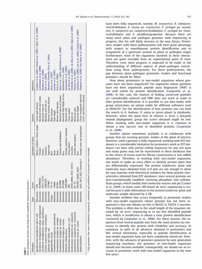
Tabl
e1
Plan
tpa
thog
enic
bact
eria
and
fung
iw
ith
com
plet
ely
sequ
ence
dge
nom
es.a
Path
ogen
Type
Stra
inEx
ampl
esof
hos
tsD
isea
seR
efer
ence
Agr
obac
teri
umtu
mef
acie
nsan
dA
.vit
isB
acte
riu
mC
58M
aize
,soy
bean
,cot
ton
,gra
peC
row
nga
lldi
seas
eG
oodn
eret
al.(
2001
),W
ood
etal
.(20
01),
Slat
eret
al.(
2009
)Cl
avib
acte
rm
ichi
gane
nsis
subs
p.m
ichi
gane
nsis
Bac
teri
um
NC
PPB
382
Tom
ato
Bac
teri
alw
ilt
and
can
ker
Gar
tem
ann
etal
.(20
08)
Clav
ibac
ter
mic
higa
nens
issu
bsp.
sepe
doni
cus
Bac
teri
um
ATC
C33
113
Pota
toB
acte
rial
rin
gro
tB
entl
eyet
al.(
2008
)Er
win
iaca
rato
vora
subs
p.at
rose
ptic
aB
acte
riu
mSC
RI1
043
Pota
toes
Bla
ckle
gan
dso
ftro
tB
ell
etal
.(20
04)
Leif
soni
axy
lisu
bsp.
xyli
Bac
teri
um
CTC
B07
Suga
rcan
eR
atoo
nst
un
tin
gdi
seas
eM
onte
iro-
Vit
orel
loC
Bet
al.(
2004
)M
agna
port
heor
yzae
Fun
gus
70–1
5R
ice
Bla
stdi
seas
eD
ean
etal
.(20
05)
Pseu
dom
onas
syri
ngae
pv.p
hase
olic
ola
Bac
teri
um
1448
AB
ean
Hal
obl
igh
tJo
arda
ret
al.(
2005
)Ps
eudo
mon
assy
ring
aepv
.syr
inga
eB
acte
riu
mB
728a
Bea
nB
row
nsp
otdi
seas
eFe
ilet
al.(
2005
)Ps
eudo
mon
assy
ring
aepv
.tom
ato
Bac
teri
um
DC
3000
Tom
ato
and
Ara
bido
psis
Bac
teri
alsp
eck
ofto
mat
oB
uel
let
al.,
(200
3),R
ein
har
dtet
al.(
2009
)Ps
eudo
mon
assy
ring
aepv
.ory
zae
Bac
teri
um
DC
3000
Ric
eB
acte
rial
hal
obl
igh
tof
rice
Rei
nh
ardt
etal
.(20
09)
Ral
ston
iaso
lana
cear
umB
acte
riu
mG
MI1
000
Tom
ato,
pota
toes
,ban
anas
Bac
teri
alw
ilt
Sala
nou
bat
etal
.(20
02)
Xan
thom
onas
axon
opod
ispv
.cit
riB
acte
riu
m30
6C
itru
spl
ants
Cit
rus
can
ker
daSi
lva
etal
.(20
02)
Xan
thom
onas
cam
pest
ris
pv.c
ampe
stri
sB
acte
riu
mB
100,
8004
,ATC
C33
913
Cru
cife
rsB
lack
rot
Vor
hol
ter
etal
.(20
08),
daSi
lva
etal
.(20
02),
Qia
net
al.(
2005
)X
anth
omon
asca
mpe
stri
spv
.ves
icat
oria
Bac
teri
um
85–1
0To
mat
oan
dpe
pper
sB
acte
rial
spot
Thie
me
etal
.(20
05)
Xan
thom
onas
oryz
aepv
.ory
zae
Bac
teri
um
PXO
99a ,M
AFF
,KA
CC
Ric
eB
acte
rial
blig
ht
Salz
berg
etal
.(20
08),
Lee
etal
.(20
05),
Och
iai
etal
.(20
05)
Xyl
lela
fast
idio
saB
acte
riu
m9a
5c,M
12,M
23,T
emec
ula
1O
ran
getr
ees
and
grap
evin
esC
itru
sva
rieg
ated
chlo
rosi
s(C
VC
)an
dPi
erce
’sdi
seas
eSi
mps
onet
al.(
2000
),V
anSl
uys
etal
.(20
03)
aO
nly
gen
omes
that
wer
eco
mpl
etel
yse
quen
ced,
ann
otat
edan
dpu
blis
hed
injo
urn
als
wer
ein
clu
ded.
B.F. Quirino et al. / Phytochemistry 71 (2010) 351–362 357
have been fully sequenced, namely, M. oryzae/rice, R. solanacea-rum/Arabidopsis, X. oryzae pv. oryzae/rice, P. syringae pv. oryzae/rice, X. campestris pv. campestris/Arabidopsis, P. syringae pv. toma-to/Arabidopsis and X. fastidiosa/grapevine. Because there aremany more plant and pathogen genomes with sequencing inprogress, this list will likely increase in the near future. Proteo-mics studies with these pathosystems will have great advantagewith respect to unambiguous protein identification and toassignment of a particular protein to plant or pathogen origin.Furthermore, most of the organisms involved in these interac-tions are quite tractable from an experimental point of view.Therefore, even more progress is expected to be made in theunderstanding of different aspects of plant–pathogen interac-tions using these pathosystems. For these pathosystems, thegap between plant–pathogen proteomic studies and functionalgenomics should be filled.
How about proteomics in non-model organisms whose gen-omes have not been sequenced? For organisms whose genomeshave not been sequenced, peptide mass fingerprint (PMF) isnot well suited for protein identification (Carpentier et al.,2008). In this case, the chances of finding conserved peptidesare considerably reduced and PMF does not work or leads tofalse protein identification. It is possible to use data banks withgroup restrictions, an option viable for different softwares suchas MASCOT. For the identification of host proteins one can limitthe search to A. thaliana, O. sativa or green plants in databanks.However, when the plant host of interest is from a distantlyrelated phylogenetic group the scores obtained might be low.When working with non-model organisms it is common toobtain a low success rate of identified proteins (Carpentieret al., 2008).
Another option sometimes available is to collaborate withgroups that are carrying genomic studies of the plant of interest.However, until a genome is fully sequenced, working with EST dat-abases is a considerable limitation for proteomics work as EST dat-abases can have only partial coding sequences for any one geneand many genes may not be represented in these databases dueto the choice of tissue used for library construction or low mRNAabundance. Therefore, in working with non-model organisms,one needs to make an extra effort to identify protein spots thatare differentially expressed. The protein isolelectric point andmolecular mass obtained from 2-D gels are not enough to allowfor easy matches with theoretical numbers for these protein char-acteristics obtained from EST databases, since several proteins arepost-translationally modified, receiving phosphate and carbohy-drate groups, which modify their molecular masses and pIs (Coakeret al., 2004). In these cases, MS-based ‘de novo’ sequencing is cru-cial because it adds information to the protein isoelectric point andmolecular weight obtained by 2-DE.
Another problem that occurs frequently in proteomic studieswith non-model organisms whose genome has not been se-quenced is that one obtains no hits in BLAST or FASTA 3 searches.This problem is often due to the small length of the sequence ob-tained by ‘de novo’ sequencing or to too few identified peptideions, which is insufficient to obtain a clear protein identification(reviewed by Carpentier et al., 2008). For these reasons, the se-quences from several peptide ions from the same protein are nec-essary to identify this protein with reliability and accuracy. Insummary, in spite of all advances obtained in proteomics andMS, several limitations, especially in peptide identification innon-model organisms have not been completely solved yet. How-ever, with the advances of genomics powered by next generationsequencing machines, the genomes of non-model organismsshould also become available. Consequently, we should see an in-crease in proteomic work with non-model organisms in the nextfew years.
358 B.F. Quirino et al. / Phytochemistry 71 (2010) 351–362
5. Concluding remarks
To understand the cellular biology and biochemistry underlyingplant–pathogen interactions there is no substitute for studyingproteins which are directly responsible for cellular activity. There-fore, despite the enormous amount of data generated by transcrip-tome analysis, the picture is still incomplete and the proteomicapproach offers a new perspective that so far has been lacking.The technical advances in mass spectrometry now allow for rela-tively easy identification of proteins, particularly for organismswith fully sequenced genomes. In this scenario, the use of a proteo-mic approach to study plant–pathogen interactions is now becom-ing more popular. The most commonly used approach inproteomics studies of plant–pathogen interactions has been 2-Dgel electrophoresis followed by mass spectrometry. A consensusabout the need for biological and technical replicates and statisticalanalysis is emerging. The new technological advances such asMudPIT, ICAT, DIGE and others will eventually be used to studyhow plants and pathogens interact. Hitherto, most proteomics pa-pers are still descriptive and end with speculations about the roleof the differentially expressed proteins in the plant–pathogeninteraction. At least for model organisms this will change withtime and speculation will be substituted for hard data from func-tional genomics experiments.
The number of organisms with a sequenced genome is con-stantly growing. Today over 1,000 complete genomes includingbacteria, archebacteria and eukaryotes have been published(www.genomesonline.org). Next generation high throughputsequencing techniques, such as pyrosequencing, that allow fastersequencing at low cost, should give a boost to ever more ambitioussequencing projects. In the next few years, the number of com-pletely sequenced plant genomes as well as plant pathogenicmicroorganisms should drastically increase. This should have afavorable impact in protein identification allowing peptide massfingerprinting to be used for more plant and pathogen species.Therefore, for non-model plants, the bottleneck in proteomicsshould shift from unambiguous protein identification to determi-nation of protein function. Information from functionally studiedArabidopsis and rice genes will continue to benefit studies withother plant species and help the annotation of other plant gen-omes. The amount of information about different aspects of thebiology of these plant models, as well as the many tools availablefor them and the number of scientists dedicated to their researchcreates a synergism that puts them at great advantage over otherplant species (Quirino et al., 2004). However, an effort must bemade to develop easy transformation protocols for different cropplants that will allow the establishment of knockout collectionsand silencing experiments in other species. Particularly for thefield of plant–pathogen interactions, there will be unique featuresto each pathosystem and work with model organisms will not re-veal the whole story.
Acknowledgements
This work has been funded by Conselho Nacional de Desen-volvimento Científico e Tecnológico (CNPq) and FAP-DF. We aregrateful ro Rinaldo Pereira for critical comments.
References
Andrade, A.E., Silva, L.P., Pereira, J.L., Noronha, E.F., Reis Jr., F.B., Bloch Jr., C., dosSantos, M.F., Domont, G.B., Franco, O.L., Mehta, A., 2008. In vivo proteomeanalysis of Xanthomonas campestris pv. campestris in the interaction with thehost plant Brassica oleracea. FEMS Microbiol. Lett. 281, 167–174.
Arai, Y., Hayashi, M., Nishimura, M., 2008. Proteomic analysis of highly purifiedperoxisomes from etiolated soybean cotyledons. Plant Cell Physiol. 49, 526–539.
Asai, T., Tena, G., Plotnikova, J., Willmann, M.R., Chiu, W.L., Gomez-Gomez, L., Boller,T., Ausubel, F.M., Sheen, J., 2002. MAP kinase signalling cascade in Arabidopsisinnate immunity. Nature 415, 977–983.
Bell, K.S., Sebaihia, M., Pritchard, L., Holden, M.T., Hyman, L.J., Holeva, M.C.,Thomson, N.R., Bentley, S.D., Churcher, L.J., Mungall, K., Atkin, R., Bason, N.,Brooks, K., Chillingworth, T., Clark, K., Doggett, J., Fraser, A., Hance, Z., Hauser, H.,Jagels, K., Moule, S., Norbertczak, H., Ormond, D., Price, C., Quail, M.A., Sanders,M., Walker, D., Whitehead, S., Salmond, G.P., Birch, P.R., Parkhill, J., Toth, I.K.,2004. Genome sequence of the enterobacterial phytopathogen Erwiniacarotovora subsp. atroseptica and characterization of virulence factors. Proc.Natl. Acad. Sci. USA 101, 11105–11110.
Bentley, S.D., Corton, C., Brown, S.E., Barron, A., Clark, L., Doggett, J., Harris, B.,Ormond, D., Quail, M.A., May, G., Francis, D., Knudson, D., Parkhill, J., Ishimaru,C.A., 2008. Genome of the actinomycete plant pathogen Clavibactermichiganensis subsp. sepedonicus suggests recent niche adaptation. J. Bacteriol.190, 2150–2160.
Bindschedler, L.V., Palmblad, M., Cramer, R., 2008. Hydroponic isotope labelling ofentire plants (HILEP) for quantitative plant proteomics; an oxidative stress casestudy. Phytochemistry 69, 1962–1972.
Brizard, J.P., Carapito, C., Delalande, F., Van Dorsselaer, A., Brugidou, C., 2006.Proteome analysis of plant–virus interactome: comprehensive data for virusmultiplication inside their hosts. Mol. Cell. Proteomics 5, 2279–2297.
Brugiere, S., Kowalski, S., Ferro, M., Seigneurin-Berny, D., Miras, S., Salvi, D., Ravanel,S., d’Herin, P., Garin, J., Bourguignon, J., Joyard, J., Rolland, N., 2004. Thehydrophobic proteome of mitochondrial membranes from Arabidopsis cellsuspensions. Phytochemistry 65, 1693–1707.
Brunner, A.M., Busov, V.B., Strauss, S.H., 2004. Poplar genome sequence: functionalgenomics in an ecologically dominant plant species. Trends Plant Sci. 9, 49–56.
Buell, C.R., Joardar, V., Lindeberg, M., Selengut, J., Paulsen, I.T., Gwinn, M.L., Dodson,R.J., Deboy, R.T., Durkin, A.S., Kolonay, J.F., Madupu, R., Daugherty, S., Brinkac, L.,Beanan, M.J., Haft, D.H., Nelson, W.C., Davidsen, T., Zafar, N., Zhou, L., Liu, J.,Yuan, Q., Khouri, H., Fedorova, N., Tran, B., Russell, D., Berry, K., Utterback, T.,Van Aken, S.E., Feldblyum, T.V., D’Ascenzo, M., Deng, W.L., Ramos, A.R., Alfano,J.R., Cartinhour, S., Chatterjee, A.K., Delaney, T.P., Lazarowitz, S.G., Martin, G.B.,Schneider, D.J., Tang, X., Bender, C.L., White, O., Fraser, C.M., Collmer, A., 2003.The complete genome sequence of the Arabidopsis and tomato pathogenPseudomonas syringae pv. tomato DC3000. Proc. Natl. Acad. Sci. USA 100, 10181–10186.
Carpentier, S.C., Witters, E., Laukens, K., Deckers, P., Swennen, R., Panis, B., 2005.Preparation of protein extracts from recalcitrant plant tissues: an evaluation ofdifferent methods for two-dimensional gel electrophoresis analysis. Proteomics5, 2497–2507.
Carpentier, S.C., Panis, B., Vertommen, A., Swennen, R., Sergeant, K., Renaut, J.,Laukens, K., Witters, E., Samyn, B., Devreese, B., 2008. Proteome analysis of non-model plants: a challenging but powerful approach. Mass. Spectrom. Rev. 27,354–377.
Chen, C.H., 2008. Review of a current role of mass spectrometry for proteomeresearch. Anal. Chim. Acta 624, 16–36.
Chen, S., Harmon, A.C., 2006. Advances in plant proteomics. Proteomics 6, 5504–5516.
Chen, F., Yuan, Y., Li, Q., He, Z., 2007. Proteomic analysis of rice plasma membranereveals proteins involved in early defense response to bacterial blight.Proteomics 7, 1529–1539.
Chivasa, S., Ndimba, B.K., Simon, W.J., Robertson, D., Yu, X.L., Knox, J.P., Bolwell, P.,Slabas, A.R., 2002. Proteomic analysis of the Arabidopsis thaliana cell wall.Electrophoresis 23, 1754–1765.
Chivasa, S., Simon, W.J., Yu, X.L., Yalpani, N., Slabas, A.R., 2005. Pathogen elicitor-induced changes in the maize extracellular matrix proteome. Proteomics 5,4894–4904.
Clauser, K.R., Baker, P., Burlingame, A.L., 1999. Role of accurate mass measurement(±10 ppm) in protein identification strategies employing MS or MS/MS anddatabase searching. Anal. Chem. 71, 2871–2882.
Coaker, G., Francis, D., 2004. Mapping, genetic effects, and epistatic interaction oftwo bacterial canker resistance QTLs from Lycopersicon hirsutum. Theor. Appl.Genet. 108, 1047–1055.
Coaker, G.L., Willard, B., Kinter, M., Stockinger, E.J., Francis, D.M., 2004. Proteomicanalysis of resistance mediated by Rcm 2.0 and Rcm 5.1, two loci controllingresistance to bacterial canker of tomato. Mol. Plant Microbe Interact. 17, 1019–1028.
Cooper, B., Eckert, D., Andon, N.L., Yates, J.R., Haynes, P.A., 2003. Investigativeproteomics: identification of an unknown plant virus from infected plants usingmass spectrometry. J. Am. Soc. Mass Spectrom. 14, 736–741.
da Silva, A.C., Ferro, J.A., Reinach, F.C., Farah, C.S., Furlan, L.R., Quaggio, R.B.,Monteiro-Vitorello, C.B., Van Sluys, M.A., Almeida, N.F., Alves, L.M., do Amaral,A.M., Bertolini, M.C., Camargo, L.E., Camarotte, G., Cannavan, F., Cardozo, J.,Chambergo, F., Ciapina, L.P., Cicarelli, R.M., Coutinho, L.L., Cursino-Santos, J.R.,El-Dorry, H., Faria, J.B., Ferreira, A.J., Ferreira, R.C., Ferro, M.I., Formighieri, E.F.,Franco, M.C., Greggio, C.C., Gruber, A., Katsuyama, A.M., Kishi, L.T., Leite, R.P.,Lemos, E.G., Lemos, M.V., Locali, E.C., Machado, M.A., Madeira, A.M., Martinez-Rossi, N.M., Martins, E.C., Meidanis, J., Menck, C.F., Miyaki, C.Y., Moon, D.H.,Moreira, L.M., Novo, M.T., Okura, V.K., Oliveira, M.C., Oliveira, V.R., Pereira, H.A.,Rossi, A., Sena, J.A., Silva, C., de Souza, R.F., Spinola, L.A., Takita, M.A., Tamura,R.E., Teixeira, E.C., Tezza, R.I., Trindade dos Santos, M., Truffi, D., Tsai, S.M.,White, F.F., Setubal, J.C., Kitajima, J.P., 2002. Comparison of the genomes oftwo Xanthomonas pathogens with differing host specificities. Nature 417,459–463.
B.F. Quirino et al. / Phytochemistry 71 (2010) 351–362 359
Dani, V.S.W., Duranti, M., Croy, R.R., 2005. Changes in the tobacco leaf apoplastproteome in response to salt stress. Proteomics 5, 737–745.
Dean, R.A., Talbot, N.J., Ebbole, D.J., Farman, M.L., Mitchell, T.K., Orbach, M.J., Thon,M., Kulkarni, R., Xu, J.R., Pan, H., Read, N.D., Lee, Y.H., Carbone, I., Brown, D., Oh,Y.Y., Donofrio, N., Jeong, J.S., Soanes, D.M., Djonovic, S., Kolomiets, E., Rehmeyer,C., Li, W., Harding, M., Kim, S., Lebrun, M.H., Bohnert, H., Coughlan, S., Butler, J.,Calvo, S., Ma, L.J., Nicol, R., Purcell, S., Nusbaum, C., Galagan, J.E., Birren, B.W.,2005. The genome sequence of the rice blast fungus Magnaporthe grisea. Nature434, 980–986.
Devos, S., Laukens, K., Deckers, P., Van Der Straeten, D., Beeckman, T., Inze, D., VanOnckelen, H., Witters, E., Prinsen, E., 2006. A hormone and proteome approachto picturing the initial metabolic events during Plasmodiophora brassicaeinfection on Arabidopsis. Mol. Plant Microbe Interact. 19, 1431–1443.
Dóczi, R., Brader, G., Pettkó-Szandtner, A., Rajh, I., Djamei, A., Pitzschke, A., Teige, M.,Hirt, H., 2007. The Arabidopsis mitogen-activated protein kinase kinase MKK3 isupstream of group C mitogen-activated protein kinases and participates inpathogen signaling. Plant Cell 19, 3266–3279.
Doddapaneni, H., Lin, H., Walker, M.A., Yao, J., Civerolo, E.L., 2008. VitisExpDB: adatabase resource for grape functional genomics. BMC Plant Biol. 8, 23.
Dunkley, T.P.J., Watson, R., Griffin, J.L., Dupree, P., Lilley, K.S., 2004. Localization oforganelle proteins by isotope tagging (LOPIT). Mol. Cell. Proteomics 3, 1128–1134.
Feil, H., Feil, W.S., Chain, P., Larimer, F., DiBartolo, G., Copeland, A., Lykidis, A., Trong,S., Nolan, M., Goltsman, E., Thiel, J., Malfatti, S., Loper, J.E., Lapidus, A., Detter, J.C.,Land, M., Richardson, P.M., Kyrpides, N.C., Ivanova, N., Lindow, S.E., 2005.Comparison of the complete genome sequences of Pseudomonas syringae pv.syringae B728a and pv. tomato DC3000. Proc. Natl. Acad. Sci. USA 102, 11064–11069.
Gartemann, K.H., Abt, B., Bekel, T., Burger, A., Engemann, J., Flugel, M., Gaigalat, L.,Goesmann, A., Grafen, I., Kalinowski, J., Kaup, O., Kirchner, O., Krause, L., Linke,B., McHardy, A., Meyer, F., Pohle, S., Ruckert, C., Schneiker, S., Zellermann, E.M.,Puhler, A., Eichenlaub, R., Kaiser, O., Bartels, D., 2008. The genome sequence ofthe tomato-pathogenic actinomycete Clavibacter michiganensis subsp.michiganensis NCPPB382 reveals a large island involved in pathogenicity. J.Bacteriol. 190, 2138–2149.
Gleason, M.L., Gitaitis, R.D., Ricker, M.D., 1993. Recent progress in understandingand controlling bacterial cancer in eastern North America. Plant Disease 77,1069–1076.
Goodner, B., Hinkle, G., Gattung, S., Miller, N., Blanchard, M., Qurollo, B., Goldman,B.S., Cao, Y., Askenazi, M., Halling, C., Mullin, L., Houmiel, K., Gordon, J., Vaudin,M., Iartchouk, O., Epp, A., Liu, F., Wollam, C., Allinger, M., Doughty, D., Scott, C.,Lappas, C., Markelz, B., Flanagan, C., Crowell, C., Gurson, J., Lomo, C., Sear, C.,Strub, G., Cielo, C., Slater, S., 2001. Genome sequence of the plant pathogen andbiotechnology agent Agrobacterium tumefaciens C58. Science 294, 2323–2328.
Gurlebeck, D., Thieme, F., Bonas, U., 2006. Type III effector proteins from the plantpathogen Xanthomonas and their role in the interaction with the host plant. J.Plant Physiol. 163, 233–255.
Gygi, S.P., Rist, B., Gerber, S.A., Turecek, F., Gelb, M.H., Aebersold, R., 1999.Quantitative analysis of complex protein mixtures using isotope-codedaffinity tags. Nat. Biotechnol. 17, 994–999.
Hartman, N.T., Sicilia, F., Lilley, K.S., Dupree, P., 2007. Proteomic complex detectionusing sedimentation. Anal. Chem. 79, 2078–2083.
Hayward, A.C., 1994. The hosts of Pseudomonas solanacearum. In: Hartman, G.L.(Ed.), Bacterial Wilt: The Disease and Its Causative Agent, Pseudomonassolanacearum. CAB International, Wallingford, pp. 9–24.
Hsing, Y.I., Chern, C.G., Fan, M.J., Lu, P.C., Chen, K.T., Lo, S.F., Sun, P.K., Ho, S.L., Lee,K.W., Wang, Y.C., Huang, W.L., Ko, S.S., Chen, S., Chen, J.L., Chung, C.I., Lin, Y.C.,Hour, A.L., Wang, Y.W., Chang, Y.C., Tsai, M.W., Lin, Y.S., Chen, Y.C., Yen, H.M., Li,C.P., Wey, C.K., Tseng, C.S., Lai, M.H., Huang, S.C., Chen, L.J., Yu, S.M., 2007. A ricegene activation/knockout mutant resource for high throughput functionalgenomics. Plant Mol. Biol. 63, 351–364.
Initiative, T.A.G., 2000. Analysis of the genome sequence of the flowering plantArabidopsis thaliana. Nature 408, 796–815.
Jacobs, D.I., van Rijssen, M.S., van der Heijden, R., Verpoorte, R., 2001. Sequentialsolubilization of proteins precipitated with trichloroacetic acid in acetone fromCatharanthus roseus yields 52% more spots after two-dimensionalelectrophoresis. Proteomics 1, 1345–1350.
Jaillon, O., Aury, J.M., Noel, B., Policriti, A., Clepet, C., Casagrande, A., Choisne, N.,Aubourg, S., Vitulo, N., Jubin, C., Vezzi, A., Legeai, F., Hugueney, P., Dasilva, C.,Horner, D., Mica, E., Jublot, D., Poulain, J., Bruyere, C., Billault, A., Segurens, B.,Gouyvenoux, M., Ugarte, E., Cattonaro, F., Anthouard, V., Vico, V., Del Fabbro, C.,Alaux, M., Di Gaspero, G., Dumas, V., Felice, N., Paillard, S., Juman, I., Moroldo,M., Scalabrin, S., Canaguier, A., Le Clainche, I., Malacrida, G., Durand, E., Pesole,G., Laucou, V., Chatelet, P., Merdinoglu, D., Delledonne, M., Pezzotti, M.,Lecharny, A., Scarpelli, C., Artiguenave, F., Pe, M.E., Valle, G., Morgante, M.,Caboche, M., Adam-Blondon, A.F., Weissenbach, J., Quetier, F., Wincker, P., 2007.The grapevine genome sequence suggests ancestral hexaploidization in majorangiosperm phyla. Nature 449, 463–467.
Jaquinod, M., Villiers, F., Kieffer-Jaquinod, S., Hugouvieux, V., Bruley, C., Garin, J.,Bourguignon, J., 2007. A proteomics dissection of Arabidopsis thaliana vacuolesisolated from cell culture. Mol. Cell. Proteomics 6, 394–412.
Joardar, V., Lindeberg, M., Jackson, R.W., Selengut, J., Dodson, R., Brinkac, L.M.,Daugherty, S.C., Deboy, R., Durkin, A.S., Giglio, M.G., Madupu, R., Nelson, W.C.,Rosovitz, M.J., Sullivan, S., Crabtree, J., Creasy, T., Davidsen, T., Haft, D.H., Zafar,N., Zhou, L., Halpin, R., Holley, T., Khouri, H., Feldblyum, T., White, O., Fraser,C.M., Chatterjee, A.K., Cartinhour, S., Schneider, D.J., Mansfield, J., Collmer, A.,
Buell, 2005. Whole-genome sequence analysis of Pseudomonas syringae pv.phaseolicola 1448A reveals divergence among pathovars in genes involved invirulence and transposition. J. Bacteriol. 187, 6488–6498.
Jones, A.M., Thomas, V., Truman, B., Lilley, K., Mansfield, J., Grant, M., 2004. Specificchanges in the Arabidopsis proteome in response to bacterial challenge:differentiating basal and R-gene mediated resistance. Phytochemistry 65,1805–1816.
Jones, A.M., Bennett, M.H., Mansfield, J.W., Grant, M., 2006. Analysis of the defencephosphoproteome of Arabidopsis thaliana using differential mass tagging.Proteomics 6, 4155–4165.
Jung, Y.H., Rakwal, R., Agrawal, G.K., Shibato, J., Kim, J.A., Lee, M.O., Choi, P.K., Jung,S.H., Kim, S.H., Koh, H.J., Yonekura, M., Iwahashi, H., Jwa, N.S., 2006. Differentialexpression of defense/stress-related marker proteins in leaves of a unique riceblast lesion mimic mutant (blm). J. Proteome Res. 5, 2586–2598.
Kawasaki, T., Nam, J., Boyes, D.C., Holt 3rd, B.F., Hubert, D.A., Wiig, A., Dangl, J.L.,2005. A duplicated pair of Arabidopsis RING-finger E3 ligases contribute to theRPM1- and RPS2-mediated hypersensitive response. Plant J. 44, 258–270.
Kazemi-Pour, N., Condemine, G., Hugouvieux-Cotte-Pattat, N., 2004. The secretomeof the plant pathogenic bacterium Erwinia chrysanthemi. Proteomics 4, 3177–3186.
Kim, S.T., Cho, K.S., Yu, S., Kim, S.G., Hong, J.C., Han, C.D., Bae, D.W., Nam, M.H., Kang,K.Y., 2003. Proteomic analysis of differentially expressed proteins induced byrice blast fungus and elicitor in suspension-cultured rice cells. Proteomics 3,2368–2378.
Kim, H.S., Desveaux, D., Singer, A.U., Patel, P., Sondek, J., Dangl, J.L., 2005. ThePseudomonas syringae effector AvrRpt2 cleaves its C-terminally acylated target,RIN4, from Arabidopsis membranes to block RPM1 activation. Proc. Natl. Acad.Sci. USA 102, 6496–6501.
Kim, S.T., Kim, S.G., Kang, Y.H., Wang, Y., Kim, J.Y., Yi, N., Kim, J.K., Rakwal, R., Koh,H.J., Ky, K., 2008. Proteomics analysis of rice lesion mimic mutant (spl1) revealstightly localized probenazole-induced protein (PBZ1) in cells undergoingprogrammed cell death. J. Proteome Res. 7, 1750–1760.
Koller, A., Washburn, M.P., Lange, B.M., Andon, N.L., Deciu, C., Haynes, P.A., Hays, L.,Ulaszek, R., Wei, J., Wolters, D., Yates, J.R., 2002. Proteomic survey of metabolicpathways in rice. Proc. Natl. Acad. Sci. USA 99, 11969–11974.
Lee, B.M., Park, Y.J., Park, D.S., Kang, H.W., Kim, J.G., Song, E.S., Park, I.C., Yoon, U.H.,Hahn, J.H., Koo, B.S., Lee, G.B., Kim, H., Park, H.S., Yoon, K.O., Kim, J.H., Jung, C.H.,Koh, N.H., Seo, J.S., Go, S.J., 2005. The genome sequence of Xanthomonas oryzaepathovar oryzae KACC10331, the bacterial blight pathogen of rice. Nucl. Acid.Res. 33, 577–586.
Lopez, M.F., Berggren, K., Chernokalskaya, E., Lazarev, A., Robinson, M., Patton, W.F.,2000. A comparison of silver stain and SYPRO Ruby Protein Gel Stain withrespect to protein detection in two-dimensional gels and identification bypeptide mass profiling. Electrophoresis 20, 3673–3683.
Mackey, D., Holt III, B.F., Wiig, A., Dangl, J.L., 2002. RIN4 interacts with Pseudomonassyringae type III effector molecules and is required for RPM1-mediatedresistance. Cell 108, 743–754.
Majeran, W., Cai, Y., Sun, Q., van Wijk, K.J., 2005. Functional differentiation of bundlesheath and mesophyll maize chloroplasts determined by comparativeproteomics. Plant Cell 17, 3111–3140.
Majoul, T., Bancel, E., Triboï, E., Ben Hamida, J., Branlard, G., 2004. Proteomicanalysis of the effect of heat stress on hexaploid wheat grain: characterizationof heat-responsive proteins from non-prolamins fraction. Proteomics 4, 505–513.
Maltman, D.J., Gadd, S.M., Simon, W.J., Slabas, A.R., 2007. Differential proteomicanalysis of the endoplasmic reticulum from developing and germinating seedsof castor (Ricinus communis) identifies seed protein precursors as significantcomponents of the endoplasmic reticulum. Proteomics 7, 1513–1528.
Manzano, C., Abraham, Z., Lopez-Torrejon, G., Del Pozo, J.C., 2008. Identification ofubiquitinated proteins in Arabidopsis. Plant Mol. Biol. 68, 145–158.
Marchand, C., Le Marechal, P., Meyer, Y., Decottignies, P., 2006. Comparativeproteomic approaches for the isolation of proteins interacting with thioredoxin.Proteomics 6, 6528–6537.
Marra, R., Ambrosino, P., Carbone, V., Vinale, F., Woo, S.L., Ruocco, M., Ciliento, R.,Lanzuise, S., Ferraioli, S., Soriente, I., Gigante, S., Turra, D., Fogliano, V., Scala, F.,Lorito, M., 2006. Study of the three-way interaction between Trichodermaatroviride, plant and fungal pathogens by using a proteomic approach. Curr.Genet. 50, 307–321.
Masaki, S., Yamada, T., Hirasawa, T., Todaka, D., Kanekatsu, M., 2008. Proteomicanalysis of RNA-binding proteins in dry seeds of rice after fractionation byssDNA affinity column chromatography. Biotechnol. Lett. 30, 955–960.
Mehta, A., Rosato, Y.B., 2001. Differentially expressed proteins in the interaction ofXanthomonas axonopodis pv. citri with leaf extract of the host plant. Proteomics1, 1111–1118.
Mehta, A., Brasileiro, A.C., Souza, D.S., Romano, E., Campos, M.A., Grossi-de-Sa, M.F.,Silva, M.S., Franco, O.L., Fragoso, R.R., Bevitori, R., Rocha, T.L., 2008. plant–pathogen interactions: what is proteomics telling us? FEBS J. 275, 3731–3746.
Michelmore, R., 1995. Molecular approaches to manipulation of disease resistancegenes. Annu. Rev. Phytopathol. 33, 393–427.
Ming, R., Hou, S., Feng, Y., Yu, Q., Dionne-Laporte, A., Saw, J.H., Senin, P., Wang, W.,Ly, B.V., Lewis, K.L., Salzberg, S.L., Feng, L., Jones, M.R., Skelton, R.L., Murray, J.E.,Chen, C., Qian, W., Shen, J., Du, P., Eustice, M., Tong, E., Tang, H., Lyons, E., Paull,R.E., Michael, T.P., Wall, K., Rice, D.W., Albert, H., Wang, M.L., Zhu, Y.J., Schatz,M., Nagarajan, N., Acob, R.A., Guan, P., Blas, A., Wai, C.M., Ackerman, C.M., Ren,Y., Liu, C., Wang, J., Na, J.K., Shakirov, E.V., Haas, B., Thimmapuram, J., Nelson, D.,Wang, X., Bowers, J.E., Gschwend, A.R., Delcher, A.L., Singh, R., Suzuki, J.Y.,
360 B.F. Quirino et al. / Phytochemistry 71 (2010) 351–362
Tripathi, S., Neupane, K., Wei, H., Irikura, B., Paidi, M., Jiang, N., Zhang, W.,Presting, G., Windsor, A., Navajas-Perez, R., Torres, M.J., Feltus, F.A., Porter, B., Li,Y., Burroughs, A.M., Luo, M.C., Liu, L., Christopher, D.A., Mount, S.M., Moore, P.H.,Sugimura, T., Jiang, J., Schuler, M.A., Friedman, V., Mitchell-Olds, T., Shippen,D.E., dePamphilis, C.W., Palmer, J.D., Freeling, M., Paterson, A.H., Gonsalves, D.,Wang, L., Alam, M., 2008. The draft genome of the transgenic tropical fruit treepapaya (Carica papaya Linnaeus). Nature 452, 991–996.
Monteiro-Vitorello CB, C.L., Van Sluys, M.A., Kitajima, J.P., Truffi, D., do Amaral, A.M.,Harakava, R., de Oliveira, J.C., Wood, D., de Oliveira, M.C., Miyaki, C., Takita, M.A.,da Silva, A.C., Furlan, L.R., Carraro, D.M., Camarotte, G., Almeida Jr, N.F., Carrer,H., Coutinho, L.L., El-Dorry, H.A., Ferro, M.I., Gagliardi, P.R., Giglioti, E., Goldman,M.H., Goldman, G.H., Kimura, E.T., Ferro, E.S., Kuramae, E.E., Lemos, E.G., Lemos,M.V., Mauro, S.M., Machado, M.A., Marino, C.L., Menck, C.F., Nunes, L.R., Oliveira,R.C., Pereira, G.G., Siqueira, W., de Souza, A.A., Tsai, S.M., Zanca, A.S., Simpson,A.J., Brumbley, S.M., Setúbal, J.C., 2004. The genome sequence of the gram-positive sugarcane pathogen Leifsonia xyli subsp. xyli. Mol. Plant MicrobeInteract. 17, 827–836.
Nagabhushan, N., Gulyas, A., Zalik, S., 1974. Comparison of plant cytoplasmicribosomal proteins by two-dimensional polyacrylamide gel electrophoresis.Plant Physiol. 53, 516–518.
Ochiai, H., Inoue, Y., Takeya, M., Sasaki, A., Kaku, H., 2005. Genome sequence ofXanthomonas oryzae pv. oryzae suggests contribution of large numbers ofeffector genes and insertion sequences to its race diversity. Jpn. Agr. Res. Q. 39,275–287.
Oh, I.S., Park, A.R., Bae, M.S., Kwon, S.J., Kim, Y.S., Lee, J.E., Kang, N.Y., Lee, S., Cheong,H., Park, O.K., 2005. Secretome analysis reveals an Arabidopsis lipase involved indefense against Alternaria brassicicola. Plant Cell 17, 2832–2847.
Ong, S., Foster, L.J., Mann, M., 2003. Mass spectrometric-based approaches inquantitative proteomics. Methods 29, 124–130.
Orrick, L.R., Olson, M.O.J., Busch, H., 1973. Comparison of nucleolar proteins ofnormal rat liver and Novikoff hepatoma ascites cells by two-dimensionalpolyacrylamide gel electrophoresis. Proc. Natl. Acad. Sci. USA 70, 1316–1320.
Pandey, A., Chakraborty, S., Datta, A., Chakraborty, N., 2008. Proteomics approach toidentify dehydration responsive nuclear proteins from chickpea (Cicer arietinumL.). Mol. Cell. Proteomics 7, 88–107.
Park, J.W., Lee, S.G., Song, J.Y., Joo, J.S., Chung, M.J., Kim, S.C., Youn, H.S., Kang, H.L.,Baik, S.C., Lee, W.K., Cho, M.J., Rhee, K.H., 2008. Proteomic analysis ofHelicobacter pylori cellular proteins fractionated by ammonium sulfateprecipitation. Electrophoresis 29, 2891–2903.
Patton, W.F., 2002. Detection technologies in proteome analysis. J. Chromatogr. B –Anal. Technol. Biomed. Life Sci. 771, 3–31.
Peck, S.C., Nuhse, T.S., Hess, D., Iglesias, A., Meins, F., Boller, T., 2001. Directedproteomics identifies a plant-specific protein rapidly phosphorylated inresponse to bacterial and fungal elicitors. Plant Cell 13, 1467–1475.
Project, International Rice Genome Sequencing, 2005. The map-based sequence ofthe rice genome. Nature 436, 793–800.
Qian, W., Jia, Y., Ren, S.X., He, Y.Q., Feng, J.X., Lu, L.F., Sun, Q., Ying, G., Tang, D.J., Tang,H., Wu, W., Hao, P., Wang, L., Jiang, B.L., Zeng, S., Gu, W.Y., Lu, G., Rong, L., Tian,Y., Yao, Z., Fu, G., Chen, B., Fang, R., Qiang, B., Chen, Z., Zhao, G.P., Tang, J.L., He, C.,2005. Comparative and functional genomic analyses of the pathogenicity ofphytopathogen Xanthomonas campestris pv. campestris. Genome Res. 15, 757–767.
Quirino, B.F., Bent, A.F., 2003. Deciphering host resistance and pathogen virulence:the Arabidopsis/Pseudomonas interaction as a model. Mol. Plant Pathol. 4, 517–530.
Quirino, B.F., Genger, R., Ham, J.H., Zabala, G., Bent, A.F., 2004. Identification andfunctional analysis of Arabidopsis proteins that interact with resistance geneproduct RPS2 in yeast. Physiol. Mol. Plant Pathol. 65, 257–267.
Rajjou, L., Belghazi, M., Huguet, R., Robin, C., Moreau, A., Job, C., Job, D., 2006.Proteomic investigation of the effect of salicylic acid on Arabidopsis seedgermination and establishment of early defense mechanisms. Plant Physiol.141, 910–923.
Rampitsch, C., Bykova, N.V., McCallum, B., Beimcik, E., Ens, W., 2006. Analysis of thewheat and Puccinia triticina (leaf rust) proteomes during a susceptible host-pathogen interaction. Proteomics 6, 1897–1907.
Reinhardt, J.A., Baltrus, D.A., Nishimura, M.T., Jeck, W.R., Jones, C.D., Dangl, J.L., 2009.De novo assembly using low-coverage short read sequence data from the ricepathogen Pseudomonas syringae pv. oryzae. Genome Res. 19, 294–305.
Rep, M., Dekker, H.L., Vossen, J.H., de Boer, A.D., Houterman, P.M., Speijer, D., Back,J.W., de Koster, C.G., Cornelissen, B.J., 2002. Mass spectrometric identification ofisoforms of PR proteins in xylem sap of fungus-infected tomato. Plant Physiol.130, 904–917.
Reumann, S., Babujee, L., Ma, C., Wienkoop, S., Siemsen, T., Antonicelli, G.E., Rasche,N., Luder, F., Weckwerth, W., Jahn, O., 2007. Proteome analysis of Arabidopsisleaf peroxisomes reveals novel targeting peptides, metabolic pathways, anddefense mechanisms. Plant Cell 19, 3170–3193.
Roberto, S.R., Coutinho, A., De Lima, J.E.O., Miranda, V.S., Carlos, E.F., 1996.Transmissão de Xyllela fastidiosa pelas cigarrinhas Dilobopterus costalimai,Acrogonia terminalis e Oncometopia facialis em citros. Fitopatol. Bras. 21, 517–518.
Romeis, T., 2001. Protein kinases in the plant defence response. Curr. Opin. PlantBiol. 4, 407–414.
Romeis, T., Ludwig, A.A., Martin, R., Jones, J.D., 2001. Calcium-dependent proteinkinases play an essential role in a plant defence response. EMBO J. 20, 5556–5567.
Ross, P.L., Huang, Y.N., Marchese, J.N., Williamson, B., Parker, K., Hattan, S.,Khainovski, N., Pillai, S., Dey, S., Daniels, S., Purkayastha, S., Juhasz, P., Martin,S., Bartlet-Jones, M., He, F., Jacobson, A., Pappin, D.J., 2004. Multiplexed proteinquantitation in Saccharomyces cerevisiae using amine-reactive isobaric taggingreagents. Mol. Cell. Proteomics 3, 1154–1169.
Rossignol, M., Peltier, J.B., Mock, H.P., Matros, A., Maldonado, A.M., Jorrin, J.V., 2006.Plant proteome analysis: a 2004–2006 update. Proteomics 6, 5529–5548.
Salanoubat, M., Genin, S., Artiguenave, F., Gouzy, J., Mangenot, S., Arlat, M., Billault,A., Brottier, P., Camus, J.C., Cattolico, L., Chandler, M., Choisne, N., Claudel-Renard, C., Cunnac, S., Demange, N., Gaspin, C., Lavie, M., Moisan, A., Robert, C.,Saurin, W., Schiex, T., Siguier, P., Thebault, P., Whalen, M., Wincker, P., Levy, M.,Weissenbach, J., Boucher, C.A., 2002. Genome sequence of the plant pathogenRalstonia solanacearum. Nature 415, 497–502.
Salzberg, S.L., Sommer, D.D., Schatz, M.C., Phillippy, A.M., Rabinowicz, P.D., Tsuge,S., Furutani, A., Ochiai, H., Delcher, A.L., Kelley, D., Madupu, R., Puiu, D.,Radune, D., Shumway, M., Trapnell, C., Aparna, G., Jha, G., Pandey, A., Patil,P.B., Ishihara, H., Meyer, D.F., Szurek, B., Verdier, V., Koebnik, R., Dow, J.M.,Ryan, R.P., Hirata, H., Tsuyumu, S., Won Lee, S., Ronald, P.C., Sonti, R.V., VanSluys, M.A., Leach, J.E., White, F.F., Bogdanove, A.J., 2008. Genome sequenceand rapid evolution of the rice pathogen Xanthomonas oryzae pv. oryzaePXO99A. BMC Genom. 9, 204.
Schmidt, U.G., Endler, A., Schelbert, S., Brunner, A., Schnell, M., Neuhaus, H.E., Marty-Mazars, D., Marty, F., Baginsky, S., Martinoia, E., 2007. Novel tonoplasttransporters identified using a proteomic approach with vacuoles isolatedfrom cauliflower buds. Plant Physiol. 145, 216–229.
Simpson, A.J., Reinach, F.C., Arruda, P., Abreu, F.A., Acencio, M., Alvarenga, R., Alves,L.M., Araya, J.E., Baia, G.S., Baptista, C.S., Barros, M.H., Bonaccorsi, E.D., Bordin, S.,Bove, J.M., Briones, M.R., Bueno, M.R., Camargo, A.A., Camargo, L.E., Carraro,D.M., Carrer, H., Colauto, N.B., Colombo, C., Costa, F.F., Costa, M.C., Costa-Neto,C.M., Coutinho, L.L., Cristofani, M., Dias-Neto, E., Docena, C., El-Dorry, H.,Facincani, A.P., Ferreira, A.J., Ferreira, V.C., Ferro, J.A., Fraga, J.S., Franca, S.C.,Franco, M.C., Frohme, M., Furlan, L.R., Garnier, M., Goldman, G.H., Goldman,M.H., Gomes, S.L., Gruber, A., Ho, P.L., Hoheisel, J.D., Junqueira, M.L., Kemper,E.L., Kitajima, J.P., Krieger, J.E., Kuramae, E.E., Laigret, F., Lambais, M.R., Leite, L.C.,Lemos, E.G., Lemos, M.V., Lopes, S.A., Lopes, C.R., Machado, J.A., Machado, M.A.,Madeira, A.M., Madeira, H.M., Marino, C.L., Marques, M.V., Martins, E.A.,Martins, E.M., Matsukuma, A.Y., Menck, C.F., Miracca, E.C., Miyaki, C.Y.,Monteriro-Vitorello, C.B., Moon, D.H., Nagai, M.A., Nascimento, A.L., Netto, L.E.,Nhani Jr., A., Nobrega, F.G., Nunes, L.R., Oliveira, M.A., de Oliveira, M.C., deOliveira, R.C., Palmieri, D.A., Paris, A., Peixoto, B.R., Pereira, G.A., Pereira Jr., H.A.,Pesquero, J.B., Quaggio, R.B., Roberto, P.G., Rodrigues, V., de, M.R.A.J., de Rosa Jr.,V.E., de Sa, R.G., Santelli, R.V., Sawasaki, H.E., da Silva, A.C., da Silva, A.M., daSilva, F.R., da Silva Jr., W.A., da Silveira, J.F., Silvestri, M.L., Siqueira, W.J., deSouza, A.A., de Souza, A.P., Terenzi, M.F., Truffi, D., Tsai, S.M., Tsuhako, M.H.,Vallada, H., Van Sluys, M.A., Verjovski-Almeida, S., Vettore, A.L., Zago, M.A., Zatz,M., Meidanis, J., Setubal, J.C., 2000. The genome sequence of the plant pathogenXylella fastidiosa. The Xylella fastidiosa Consortium of the Organization forNucleotide Sequencing and Analysis. Nature 406, 151–159.
Siuzdak, G., 1996. Mass Spectrometry for Biotechnology. Academic Press, SanDiego.
Slater, S.C., Goldman, B.S., Goodner, B., Setubal, J.C., Farrand, S.K., Nester, E.W., Burr,T.J., Banta, L., Dickerman, A.W., Paulsen, I., Otten, L., Suen, G., Welch, R., Almeida,N.F., Arnold, F., Burton, O.T., Du, Z., Ewing, A., Godsy, E., Heisel, S., Houmiel, K.L.,Jhaveri, J., Lu, J., Miller, N.M., Norton, S., Chen, Q., Phoolcharoen, W., Ohlin, V.,Ondrusek, D., Pride, N., Stricklin, S.L., Sun, J., Wheeler, C., Wilson, L., Zhu, H.,Wood, D.W., 2009. Genome sequences of three agrobacterium biovars helpelucidate the evolution of multichromosome genomes in bacteria. J. Bacteriol.191 (8), 2501–2511.
Sparkman, O.D. (Ed.), 2000. Mass Spectrometry Desk Reference. Global ViewPublications, Pittsburgh.
Stokstad, E., 2006. Poplar tree sequence yields genome double take. Science 5793,1556.
Sussman, M.R., Amasino, R.M., Young, J.C., Krysan, P.J., Austin-Phillips, S., 2000. TheArabidopsis knockout facility at the University of Wisconsin-Madison. PlantPhysiol. 124, 1465–1467.
Tahara, S.T., Mehta, A., Rosato, Y.B., 2003. Proteins induced by Xanthomonasaxonopodis pv. Passiflorae with leaf extract of the host plant (Passiflorae edulis).Proteomics 3, 95–102.
Takemoto, D., Jones, D.A., 2005. Membrane release and destabilization of ArabidopsisRIN4 following cleavage by Pseudomonas syringae AvrRpt2. Mol. Plant MicrobeInteract. 18, 1258–1268.
Thieme, F., Koebnik, R., Bekel, T., Berger, C., Boch, J., Buttner, D., Caldana, C., Gaigalat,L., Goesmann, A., Kay, S., Kirchner, O., Lanz, C., Linke, B., McHardy, A.C., Meyer, F.,Mittenhuber, G., Nies, D.H., Niesbach-Klosgen, U., Patschkowski, T., Ruckert, C.,Rupp, O., Schneiker, S., Schuster, S.C., Vorholter, F.J., Weber, E., Puhler, A., Bonas,U., Bartels, D., Kaiser, O., 2005. Insights into genome plasticity andpathogenicity of the plant pathogenic bacterium Xanthomonas campestris pv.Vesicatoria revealed by the complete genome sequence. J. Bacteriol. 187, 7254–7266.
Tsunezuka, H., Fujiwara, M., Kawasaki, T., Shimamoto, K., 2005. Proteome analysisof programmed cell death and defense signaling using the rice lesion mimicmutant cdr2. Mol. Plant Microbe Interact. 18, 52–59.
Tuskan, G.A., Difazio, S., Jansson, S., Bohlmann, J., Grigoriev, I., Hellsten, U., Putnam,N., Ralph, S., Rombauts, S., Salamov, A., Schein, J., Sterck, L., Aerts, A., Bhalerao,R.R., Bhalerao, R.P., Blaudez, D., Boerjan, W., Brun, A., Brunner, A., Busov, V.,
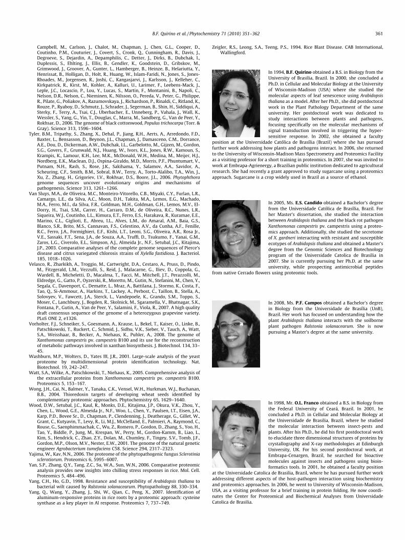
B.F. Quirino et al. / Phytochemistry 71 (2010) 351–362 361
Campbell, M., Carlson, J., Chalot, M., Chapman, J., Chen, G.L., Cooper, D.,Coutinho, P.M., Couturier, J., Covert, S., Cronk, Q., Cunningham, R., Davis, J.,Degroeve, S., Dejardin, A., Depamphilis, C., Detter, J., Dirks, B., Dubchak, I.,Duplessis, S., Ehlting, J., Ellis, B., Gendler, K., Goodstein, D., Gribskov, M.,Grimwood, J., Groover, A., Gunter, L., Hamberger, B., Heinze, B., Helariutta, Y.,Henrissat, B., Holligan, D., Holt, R., Huang, W., Islam-Faridi, N., Jones, S., Jones-Rhoades, M., Jorgensen, R., Joshi, C., Kangasjarvi, J., Karlsson, J., Kelleher, C.,Kirkpatrick, R., Kirst, M., Kohler, A., Kalluri, U., Larimer, F., Leebens-Mack, J.,Leple, J.C., Locascio, P., Lou, Y., Lucas, S., Martin, F., Montanini, B., Napoli, C.,Nelson, D.R., Nelson, C., Nieminen, K., Nilsson, O., Pereda, V., Peter, G., Philippe,R., Pilate, G., Poliakov, A., Razumovskaya, J., Richardson, P., Rinaldi, C., Ritland, K.,Rouze, P., Ryaboy, D., Schmutz, J., Schrader, J., Segerman, B., Shin, H., Siddiqui, A.,Sterky, F., Terry, A., Tsai, C.J., Uberbacher, E., Unneberg, P., Vahala, J., Wall, K.,Wessler, S., Yang, G., Yin, T., Douglas, C., Marra, M., Sandberg, G., Van de Peer, Y.,Rokhsar, D., 2006. The genome of black cottonwood, Populus trichocarpa (Torr. &Gray). Science 313, 1596–1604.
Tyler, B.M., Tripathy, S., Zhang, X., Dehal, P., Jiang, R.H., Aerts, A., Arredondo, F.D.,Baxter, L., Bensasson, D., Beynon, J.L., Chapman, J., Damasceno, C.M., Dorrance,A.E., Dou, D., Dickerman, A.W., Dubchak, I.L., Garbelotto, M., Gijzen, M., Gordon,S.G., Govers, F., Grunwald, N.J., Huang, W., Ivors, K.L., Jones, R.W., Kamoun, S.,Krampis, K., Lamour, K.H., Lee, M.K., McDonald, W.H., Medina, M., Meijer, H.J.,Nordberg, E.K., Maclean, D.J., Ospina-Giraldo, M.D., Morris, P.F., Phuntumart, V.,Putnam, N.H., Rash, S., Rose, J.K., Sakihama, Y., Salamov, A.A., Savidor, A.,Scheuring, C.F., Smith, B.M., Sobral, B.W., Terry, A., Torto-Alalibo, T.A., Win, J.,Xu, Z., Zhang, H., Grigoriev, I.V., Rokhsar, D.S., Boore, J.L., 2006. Phytophthoragenome sequences uncover evolutionary origins and mechanisms ofpathogenesis. Science 313, 1261–1266.
Van Sluys, M.A., de Oliveira, M.C., Monteiro-Vitorello, C.B., Miyaki, C.Y., Furlan, L.R.,Camargo, L.E., da Silva, A.C., Moon, D.H., Takita, M.A., Lemos, E.G., Machado,M.A., Ferro, M.I., da Silva, F.R., Goldman, M.H., Goldman, G.H., Lemos, M.V., El-Dorry, H., Tsai, S.M., Carrer, H., Carraro, D.M., de Oliveira, R.C., Nunes, L.R.,Siqueira, W.J., Coutinho, L.L., Kimura, E.T., Ferro, E.S., Harakava, R., Kuramae, E.E.,Marino, C.L., Giglioti, E., Abreu, I.L., Alves, L.M., do Amaral, A.M., Baia, G.S.,Blanco, S.R., Brito, M.S., Cannavan, F.S., Celestino, A.V., da Cunha, A.F., Fenille,R.C., Ferro, J.A., Formighieri, E.F., Kishi, L.T., Leoni, S.G., Oliveira, A.R., Rosa Jr.,V.E., Sassaki, F.T., Sena, J.A., de Souza, A.A., Truffi, D., Tsukumo, F., Yanai, G.M.,Zaros, L.G., Civerolo, E.L., Simpson, A.J., Almeida Jr., N.F., Setubal, J.C., Kitajima,J.P., 2003. Comparative analyses of the complete genome sequences of Pierce’sdisease and citrus variegated chlorosis strains of Xylella fastidiosa. J. Bacteriol.185, 1018–1026.
Velasco, R., Zharkikh, A., Troggio, M., Cartwright, D.A., Cestaro, A., Pruss, D., Pindo,M., Fitzgerald, L.M., Vezzulli, S., Reid, J., Malacarne, G., Iliev, D., Coppola, G.,Wardell, B., Micheletti, D., Macalma, T., Facci, M., Mitchell, J.T., Perazzolli, M.,Eldredge, G., Gatto, P., Oyzerski, R., Moretto, M., Gutin, N., Stefanini, M., Chen, Y.,Segala, C., Davenport, C., Dematte, L., Mraz, A., Battilana, J., Stormo, K., Costa, F.,Tao, Q., Si-Ammour, A., Harkins, T., Lackey, A., Perbost, C., Taillon, B., Stella, A.,Solovyev, V., Fawcett, J.A., Sterck, L., Vandepoele, K., Grando, S.M., Toppo, S.,Moser, C., Lanchbury, J., Bogden, R., Skolnick, M., Sgaramella, V., Bhatnagar, S.K.,Fontana, P., Gutin, A., Van de Peer, Y., Salamini, F., Viola, R., 2007. A high qualitydraft consensus sequence of the genome of a heterozygous grapevine variety.PLoS ONE 2, e1326.
Vorholter, F.J., Schneiker, S., Goesmann, A., Krause, L., Bekel, T., Kaiser, O., Linke, B.,Patschkowski, T., Ruckert, C., Schmid, J., Sidhu, V.K., Sieber, V., Tauch, A., Watt,S.A., Weisshaar, B., Becker, A., Niehaus, K., Puhler, A., 2008. The genome ofXanthomonas campestris pv. campestris B100 and its use for the reconstructionof metabolic pathways involved in xanthan biosynthesis. J. Biotechnol. 134, 33–45.
Washburn, M.P., Wolters, D., Yates III, J.R., 2001. Large-scale analysis of the yeastproteome by multidimensional protein identification technology. Nat.Biotechnol. 19, 242–247.
Watt, S.A., Wilke, A., Patschkowski, T., Niehaus, K., 2005. Comprehensive analysis ofthe extracellular proteins from Xanthomonas campestris pv. campestris B100.Proteomics 5, 153–167.
Wong, J.H., Cai, N., Balmer, Y., Tanaka, C.K., Vensel, W.H., Hurkman, W.J., Buchanan,B.B., 2004. Thioredoxin targets of developing wheat seeds identified bycomplementary proteomic approaches. Phytochemistry 65, 1629–1640.
Wood, D.W., Setubal, J.C., Kaul, R., Monks, D.E., Kitajima, J.P., Okura, V.K., Zhou, Y.,Chen, L., Wood, G.E., Almeida Jr., N.F., Woo, L., Chen, Y., Paulsen, I.T., Eisen, J.A.,Karp, P.D., Bovee Sr., D., Chapman, P., Clendenning, J., Deatherage, G., Gillet, W.,Grant, C., Kutyavin, T., Levy, R., Li, M.J., McClelland, E., Palmieri, A., Raymond, C.,Rouse, G., Saenphimmachak, C., Wu, Z., Romero, P., Gordon, D., Zhang, S., Yoo, H.,Tao, Y., Biddle, P., Jung, M., Krespan, W., Perry, M., Gordon-Kamm, B., Liao, L.,Kim, S., Hendrick, C., Zhao, Z.Y., Dolan, M., Chumley, F., Tingey, S.V., Tomb, J.F.,Gordon, M.P., Olson, M.V., Nester, E.W., 2001. The genome of the natural geneticengineer Agrobacterium tumefaciens C58. Science 294, 2317–2323.
Yajima, W., Kav, N.N., 2006. The proteome of the phytopathogenic fungus Sclerotiniasclerotiorum. Proteomics 6, 5995–6007.
Yan, S.P., Zhang, Q.Y., Tang, Z.C., Su, W.A., Sun, W.N., 2006. Comparative proteomicanalysis provides new insights into chilling stress responses in rice. Mol. Cell.Proteomics 5, 484–496.
Yang, C.H., Ho, G.D., 1998. Resistance and susceptibility of Arabidopsis thaliana tobacterial wilt caused by Ralstonia solanacearum. Phytopathology 88, 330–334.
Yang, Q., Wang, Y., Zhang, J., Shi, W., Qian, C., Peng, X., 2007. Identification ofaluminum-responsive proteins in rice roots by a proteomic approach: cysteinesynthase as a key player in Al response. Proteomics 7, 737–749.
Zeigler, R.S., Leong, S.A., Teeng, P.S., 1994. Rice Blast Disease. CAB International,Wallingford.
In 1994, B.F. Quirino obtained a B.S. in Biology from theUniversity of Brasilia, Brazil. In 2000, she concluded aPh.D. in Cellular and Molecular Biology at the Universityof Wisconsin-Madison (USA) where she studied themolecular aspects of leaf senescence using Arabidopsisthaliana as a model. After her Ph.D., she did postdoctoralwork in the Plant Pathology Department of the sameuniversity. Her postdoctoral work was dedicated tostudy interactions between plants and pathogens,focusing specifically on the molecular mechanisms ofsignal transduction involved in triggering the hyper-sensitive response. In 2002, she obtained a faculty
position at the Universidade Católica de Brasília (Brazil) where she has pursuedfurther work addressing how plants and pathogens interact. In 2006, she returnedto the University of Wisconsin-Madison Mass Spectrometry and Proteomics Facility
as a visiting professor for a short training in proteomics. In 2007, she was invited towork at Embrapa-Agrienergy, a Brazilian public institution dedicated to agriculturalresearch. She had recently a grant approved to study sugarcane using a proteomicsapproach. Sugarcane is a crop widely used in Brazil as a source of ethanol.In 2005, Ms. E.S. Candido obtained a Bachelor’s degreefrom the Universidade Católica de Brasília, Brazil. Forher Master’s dissertation, she studied the interactionbetween Arabidopsis thaliana and the black rot pathogenXanthomonas campestris pv. campestris using a proteo-mics approach. Additionally, she studied the secretomeof X. gardneri interacting with resistant and susceptibleecotypes of Arabidopsis thaliana and obtained a Master’sdegree from the Genomic Sciences and Biotechnologyprogram of the Universidade Catolica de Brasilia in2007. She is currently pursuing her Ph.D. at the sameuniversity, while prospecting antimicrobial peptides
from native Cerrado flowers using proteomic tools.
In 2008, Ms. P.F. Campos obtained a Bachelor’s degreein Biology from the Universidade de Brasília (UnB),Brazil. Her work has focused on understanding how theplant Arabidopsis thaliana interacts with the soilborneplant pathogen Ralstonia solanacearum. She is nowpursuing a Master’s degree at the same university.
In 1998, Mr. O.L. Franco obtained a B.S. in Biology fromthe Federal University of Ceará, Brazil. In 2001, heconcluded a Ph.D. in Cellular and Molecular Biology atthe Universidade de Brasilia, Brazil, where he studiedthe molecular interaction between insect-pests andplants. After his Ph.D., he did his first postdoctoral workto elucidate three dimensional structures of proteins bycrystallography and X-ray methodologies at EdinburghUniversity, UK. For his second postdoctoral work, atEmbrapa-Cenargen, Brazil, he searched for bioactivemolecules against insects and pathogens using bioin-formatics tools. In 2001, he obtained a faculty position
at the Universidade Catolica de Brasilia, Brazil, where he has pursued further workaddressing different aspects of the host-pathogen interaction using biochemistryand proteomics approaches. In 2006, he went to University of Wisconsin-Madison,
USA, as a visiting professor for a brief training in protein folding. He now coordi-nates the Center for Proteomical and Biochemical Analyses from UniversidadeCatolica de Brasilia.
362 B.F. Quirino et al. / Phytochemistry 71 (2010) 351–362
In 1993, Mr. R.H. Krüger obtained a B.S. in Biology fromthe University of Brasilia, Brazil. In 1995, he obtained aMaster’s degree from the same university. In 2002, hecompleted a Ph.D. in Microbiology at the University ofWisconsin-Madison (USA) where he studied plasmidbiology focusing on the interactions between proteinsand DNA. After his Ph.D., he accepted a faculty positionat the Universidade Católica de Brasília (Brazil) GenomicSciences and Biotechnology Program. In 2008, heaccepted a faculty position at Cellular Biology Depart-ment of the Universidade de Brasilia. He is also one ofthe leaders for the implementation of the multiinstitu-
tional Federal District Next Generation Sequencing Facility, the most completesequencing facility in South America, to be inaugurated in Brasilia this year. One ofthe main goals of this facility will be to study plant–pathogen interactions
employing ultra-deep sequencing technologies (transcriptome) and how it corre-lates with proteomic studies.