Embed Size (px)
Citation preview

1
Proteoglycan Detection KitCat. No. 280560-N
INTRODUCTIONGlycosaminoglycans (GAGs) are a major component of the extracellular matrix (ECM) of tissue and are long, unbranched polysaccharides containing a repeating disaccharide unit. GAGs consist of N- and/or O-sulfate groups and are covalently linked to core proteins to form proteoglycans. The most common GAGs are hyaluronic acid, dermatan sulfate, chondroitin sulfate, heparin, heparan sulfate, and keratan sulfate. Hyaluronic acid is unique among the GAGs in that it does not contain any sulfate and is not found covalently attached to proteins as a proteoglycan (it is, however, a component of non-covalently formed complexes with proteoglycans in the ECM).
GAGs are highly negatively charged molecules that impart high viscosity to tissues and liquids. Along with the high viscosity comes low compressibility, which makes these molecules ideal; for example,as a lubricant (synovial fluid) for joints. GAGs and proteoglycans play an important role in cellular adhesion, growth, migration, and differentiation. They are also involved in regulation of enzymesand tissue remodeling in response to injury, or in tissue destruction in diseases such as rheumatoid arthritis. Certain types of proteoglycans are also upregulated in other diseases, such as osteoarthritis.
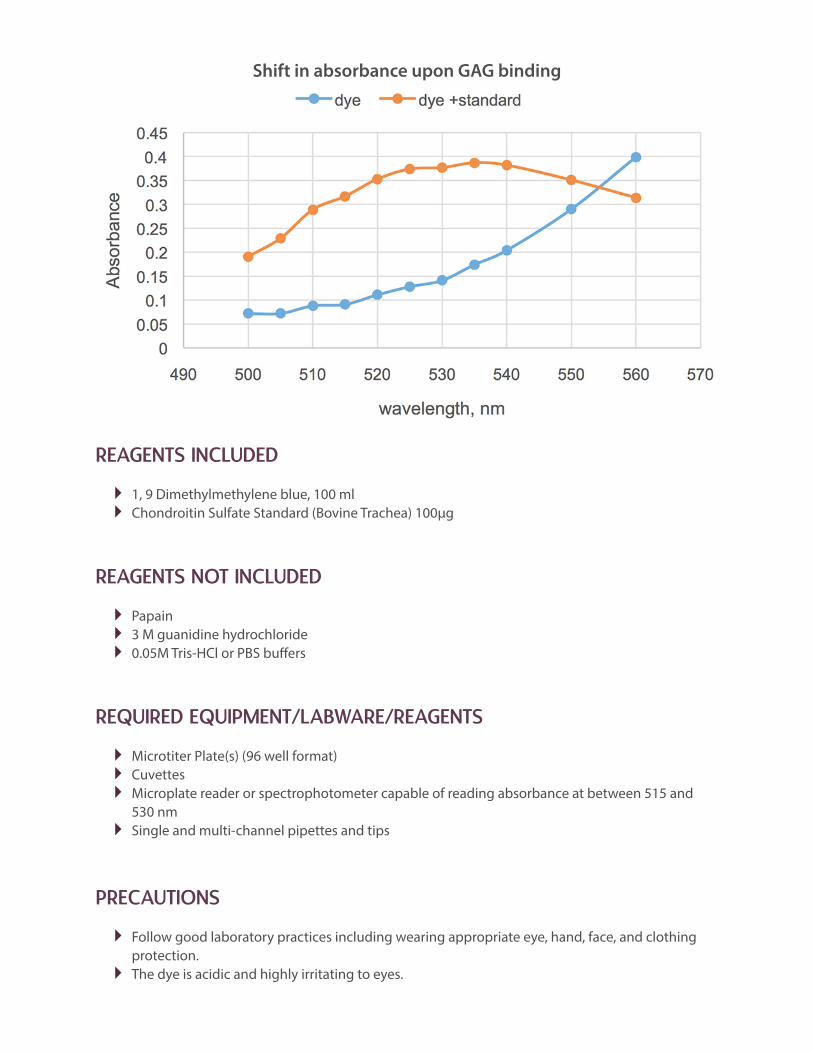
Sulfated GAGs can be measured directly by use of a metachromatic dye, 1, 9 Dimethylmethylene blue. The GAG-dye complex results in an absorption spectrum shift which can be measured at between 515and 530 nm.
PRINCIPLE OF THE ASSAYThe Proteoglycan Assay uses the metachromatic dye 1, 9‐dimethylmethylene blue to quantifythe amount of sulfated glycosaminoglycans in the standard and test samples. The binding of the sulfated glycosaminoglycans to the dye induces a shift in the absorption spectrum which is directly proportional to the amount of sulfated glycosaminoglycans. The sample values, µg/ml of sulfated glycosaminoglycans, are determined by the standard curve. The assay detects chondroitin 4 and 6 sulfates, and heparin, keratin, and dermatan sulfates. Hyaluronic acid will not be detected and will not interfere with the assay.
Various biologic fluids, such as synovial fluid, serum, amniotic fluid and urine can be tested following papain digestion. Tissue culture medium can be tested directly. Tissue samples such as cartilage, skin, and other organs must first be digested with papain or extracted with 3 M guanidinium-HCl before testing (see Sample Preparation).
2
REAGENTS INCLUDED
� 1, 9 Dimethylmethylene blue, 100 ml � Chondroitin Sulfate Standard (Bovine Trachea) 100µg
REAGENTS NOT INCLUDED
� Papain � 3 M guanidine hydrochloride � 0.05M Tris-HCl or PBS buffers
REQUIRED EQUIPMENT/LABWARE/REAGENTS
� Microtiter Plate(s) (96 well format) � Cuvettes � Microplate reader or spectrophotometer capable of reading absorbance at between 515 and 530 nm � Single and multi-channel pipettes and tips
PRECAUTIONS
� Follow good laboratory practices including wearing appropriate eye, hand, face, and clothing protection. � The dye is acidic and highly irritating to eyes.
Shift in absorbance upon GAG binding

3
SAMPLE PREPARATION
The kit can detect both newly synthesized GAGs excreted from cells in culture medium and GAGs incorporated into the extracellular matrix. To measure soluble GAGs, varying dilutions of culture media can be directly applied to the dye (see data below). To measure GAG content in tissue the sample must first be extracted with high salt such as 3 M guanidinium-HCl or digested with papain. The papain digestion method can also be used to measure GAGs in fluids where contaminating proteins might interfere with the assay. The investigator must determine the best method(s) to use for any sample.
The following methods below serve as a guideline:
Cells grown in tissue culture
Important: perform a count of the cells so that the amount of GAG measured can be expressed relative to the number of cells. For best results with cell culture supernatant use medium without phenol red. If medium with phenol red is used prepare the standard curve in medium to account for the interference. After the desired period of culture, remove the media and proceed directly to testing the sample using varying dilutions. To assay the cell matrix, the following papain digestion protocolcan be used:
1. Prepare 20 mM sodium phosphate buffer (pH 6.8) containing 1 mM EDTA, 2 mM dithiothreitoland 300 µg/mL Papain. This solution can be placed directly on the cell layer using sufficient volume to just cover the cell monolayer.
2. Incubate at 60°C for 30-60 minutes or longer if necessary until the layer is soluble.3. The above reagents are available from AMSBIO in our Tissue Digestion Kit for Glycosaminoglycan
Isolation (280560-N).
Tissue Samples
Important: measure the dry or wet weight of the sample so that the amount of GAG measuredcan be expressed relative to the mg of tissue sample.
Method 1: High Salt Extraction
1. Mince or homogenize 1-3 mg of tissue.2. Add 0.5 ml of 3 M guanidine hydrochloride/0.05 M Tris-HCl buffer, pH 7.5 and agitate on a
rotator/rocker at 4°C overnight.3. Dialyze 3 M guanidine extracts against 1X TBS and store at -20°C until measured
Method 2: Papain DigestionUse the Tissue Digestion kit available from AMSBIO (280560-TDK)
1. Mince or homogenize 1-3 mg of tissue.2. Place tissue in 1.0 ml of 20mM sodium phosphate buffer (pH 6.8) containing 1 mM EDTA, 2 mM
dithiothreitol and 300 µg Papain.3. Incubate at 60°C for 60 minutes or longer if necessary until tissue is soluble (larger tissue samples
will require more time). Add iodoacetic acid to a final concentration, of 10 mM. Add 5 ml of 50 mM Tris/HCl (pH 8.0). The sample is ready to test

4
REAGENT PREPARATION
DMB dye. Ready to use.Chondroitin Sulfate (Bovine Trachea) Standard 100 µg. Add 1.0 ml of water to dissolve the chondroitin sulfate. This standard solution is now 100 µg/ml. Any remaining standard can be stored at 2-8°C for up to 2 months. For longer storage prepare aliquots and freeze at minus 18-22°C.
For the spectrophotometer method: prepare dilutions of the 100 µg/ml standard to create working standards between 0.5 and 20 µg/ml. Zero or blank is buffer alone, plus an equal volume of dye.
For the microplate method: prepare dilutions of the 100 µg/ml standard to create working standards between 0.5 and 20 µg/ml. Zero is buffer alone.
ASSAY PROCEDURE
It is recommended that test samples and positive controls be run in duplicate. Prepare Samples and Reagents as described above
For the spectrophotometer method:1. Samples. Add 1.0 ml of sample to 1.0 ml dye in a tube.2. Standards. Add 1.0 ml of each standard to 1.0ml dye in separate tubes. 3. Measure. Add the 2.0 ml of samples or standards to cuvettes, and record absorbance at
between 515 and 530 nm.
For the microplate method:1. Samples. Add 0.1 ml of sample to 0.1 ml dye in a microplate well.2. Standards. Add 0.1 ml of each standard to 0.1 ml dye in separate microplate wells. 3. Measure. Read the plate in a microplate reader at between 515 and 530 nm.
Figure Legend. Bovine chondrocytes were cultured for 2 or 4 days with varying amounts of TGF-β. Culture supernatants were measured for the presence of proteoglycan by testing for GAG content using the Proteoglycan Detection Kit (Cat# 280560-N).
Effect of TGFb on proteoglycan synthesis
5
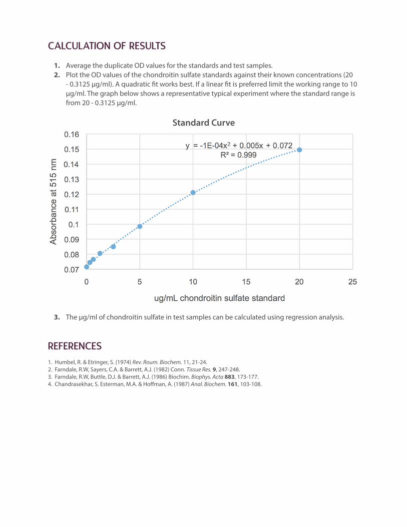
CALCULATION OF RESULTS
1. Average the duplicate OD values for the standards and test samples.2. Plot the OD values of the chondroitin sulfate standards against their known concentrations (20
- 0.3125 µg/ml). A quadratic fit works best. If a linear fit is preferred limit the working range to 10 µg/ml. The graph below shows a representative typical experiment where the standard range is from 20 - 0.3125 µg/ml.
3. The µg/ml of chondroitin sulfate in test samples can be calculated using regression analysis.
REFERENCES1. Humbel, R. & Etringer, S. (1974) Rev. Roum. Biochem. 11, 21‐24.2. Farndale, R.W, Sayers, C.A. & Barrett, A.J. (1982) Conn. Tissue Res. 9, 247‐248.3. Farndale, R.W, Buttle, D.J. & Barrett, A.J. (1986) Biochim. Biophys. Acta 883, 173‐177.4. Chandrasekhar, S. Esterman, M.A. & Hoffman, A. (1987) Anal. Biochem. 161, 103‐108.
Standard Curve

6
RECENT CITATIONS
Anitua, E., Zalduendo, M., & Troya, M. (2019). Autologous plasma rich in growth factors technology for isolation and ex vivo expansion of human dental pulp stem cells for clinical translation. Regenerative medicine, 14(2) 97-111 Tamburaci, S., Cecen, B., Ustun, O., Ergur, B., Havitcioglu, H., & Tihminlioglu, F. (2019). Production and Characterization of Novel Bilayer Nanocomposite Scaffold Composed of
Chitosan/Si-nHap and Zein/POSS Structures for Osteochondral Tissue Regeneration. ACS Applied Bio Materials, 2(4), 1440-1455. Wang, Z., Winsor, K., Nienhaus, C., Hess, E., & Blackmore, M. G. (2017). Combined chondroitinase and KLF7 expression reduce net retraction of sensory and CST axons from sites of spinal injury. Neurobiology of disease, 99, 24-35. Also cites our Chondroitinase ABC enzyme and 2B6 CS “stub” antibody (see www.amsbio.com/Chondroitinase-ABC.aspx) Zayas-Santiago, A., Cross, S. D., Stanton, J. B., Marmorstein, A. D., & Marmorstein, L. Y. (2017). Mutant Fibulin-3 Causes Proteoglycan Accumulation and Impaired Diffusion Across Bruch's Membrane. Investigative Ophthalmology & Visual Science, 58(7), 3046-3054.