Embed Size (px)
Citation preview

Protein-coated nanoparticles embedded in films asdelivery platformsJavier O. Moralesa,c, Alistair C. Rossd and Jason T. McConvilleb
aCollege of Pharmacy, The University of Texas at Austin, Austin, TX, bUniversity of New Mexico, Albuquerque, NM, USA, cSchool of Chemical andPharmaceutical Sciences, University of Chile, Santiago, Chile and dFerring Controlled Therapeutics Ltd, East Kilbride, Scotland, UK
Keywordsantisolvent precipitation; buccal delivery;enzyme activity; films; protein-coatednanoparticles
CorrespondenceJason T. McConville, 2705 Frontier NE,Albuquerque, NM 87131, USA.E-mail: [email protected]
Received November 16, 2012Accepted January 29, 2013
doi: 10.1111/jphp.12046
Abstract
Objectives This work aimed to evaluate the performance of nanoparticle-loadedfilms based on matrices of polymethacrylates and hydroxypropylmethylcellulose(HPMC) intended for delivery of macromolecules.Methods Lysozyme (Lys)-loaded nanoparticles were manufactured by antisolventco-precipitation. After size, loading efficiency and stability characterization, theselected batch of particles was further formulated into films. Films were character-ized for mechanical properties, mucoadhesion, Lys release and activity aftermanufacture.Key findings We found that protein-coated nanoparticles could be obtained inUSP phosphate buffer pH 6.8. Particles obtained at pH 6.8 had a z-average of347.2 nm, a zeta-potential of 21.9 mV and 99.2% remaining activity after manu-facture. This formulation was further studied for its application in films for buccaldelivery. Films loaded with nanoparticles that contained Eudragit RLPO (ERL)exhibited excellent mechanical and mucoadhesive properties. Due to its higherwater-swelling and solubility compared with ERL, the use of HPMC allowed us totailor the release of Lys from films. The formulation composed of equal amountsof ERL and HPMC revealed a sustained release over 4 h, with Lys remaining fullyactive at the end of the study.Conclusions Mucoadhesive films containing protein-coated nanoparticles arepromising carriers for the buccal delivery of proteins and peptides in a stableform.
Introduction
The recent increase in the number of products under reviewby the US Food and Drug Administration (FDA) or under-going late-phase clinical trials demonstrates that proteinand peptide therapeutics is a rapidly growing field in thepharmaceutical industry.[1] However, it is widely reported inthe literature that the efficacious delivery of these therapeu-tic agents is the determinant factor in product develop-ment.[2,3] Therefore, to achieve the real potential of proteinand peptide therapeutics, effective smart delivery platformsand strategies to overcome the formulation and deliverychallenges need to be developed. The conventionalapproach for the delivery of macromolecules is throughinjections.[4] This method of delivery is largely associatedwith drawbacks in patient compliance and acceptance; the
start of therapy can be delayed and patients can developneedle anxiety.[5] In addition, the number of injections maylead to compliance issues[6] in therapies that rely on thisroute of administration. Therefore, alternative routes ofdelivery are vital to achieve a broad platform of successfulproduct development.
Among alternative routes of delivery for proteins and pep-tides, the oral route has long been widely investigated.[7,8]
However, several drawbacks associated with the gastroin-testinal tract make the development of novel delivery plat-forms for macromolecules very challenging. Instability ingastric pH, proteolytic enzyme content in the upper gastroin-testinal tract and insufficient permeation and bioavailabilityhas limited the success that has been achieved.[9] These
bs_bs_banner
And PharmacologyJournal of Pharmacy
Research Paper
© 2013 Royal Pharmaceutical Society, Journal of Pharmacy and Pharmacology, 65, pp. 827–838 827

limitations have led to the exploration of other routes ofdelivery, such as pulmonary, nasal and transdermal routes.Particularly, the buccal route of delivery offers interestingadvantages in comparison with the oral route and its associ-ated gastrointestinal-tract limitations for the delivery ofprotein and peptides.[9] In bypassing absorption via the gut,the stability of macromolecules may not be compromisedbefore reaching the circulation. Other advantages of thisroute of delivery, such as its good vascular drainage, ease ofadministration and relatively low enzyme levels, make it agood candidate for the delivery of proteins and peptides.[10]
Mucoadhesive films as dosage forms for the buccal routeof delivery have been investigated in the past decade butlittle effort has been made with regards to the delivery ofproteins and peptides as particulate forms.[11,12] From a for-mulation standpoint, actives are usually added to the filmby their inclusion in the casting solution, then allowing thesolution to dry into the solid form. However, in general thepolymers used in formulations containing proteins aremore hydrophobic in comparison with the hydrophilicnature of proteins.[8] This could potentially lead to precipi-tation of proteins during storage, or in vivo, leading topossible instability.[13] Additionally, strategies such as incor-porating insulin as a solid solution into poly(lactic-co-glycolic acid) (PLGA) microspheres, to prevent chemicalreactions in the solid state and to control the peptiderelease, have been unsuccessful. During PLGA erosion, themicroenvironmetal pH drops and deamidation has beenfound to be the main reaction that causes insulin instabil-ity.[14] For the delivery of insulin, chitosan seems to bea more suitable candidate as a polymer vehicle. Cui et al.have developed chitosan–ethylenediaminetetracetic acid(chitosan–EDTA) films containing insulin for buccal deliv-ery and have demonstrated the retention of the physicalstructure of the peptide upon release.[15] However, there isno mention of the uniformity of the drug in the film uponsolidification and this prohibits any conclusion about drugdistribution homogeneity. More recently, Giovino et al. havedeveloped chitosan films for the buccal sustained delivery ofinsulin in polyethylene glycol-b-polylactic acid (PEG-b-PLA) nanoparticles as a model for buccal macromoleculardelivery.[16] Although adequate physico-mechanical proper-ties were achieved, very high heterogeneity was revealed bythe mechanical variables studied (time to break, tensilestrength, Young’s modulus and work done to break). Thistherefore raises concern over the tight control of manufac-ture necessary to prepare films with homogeneous particledistribution, adequate physico-mechanical properties, highloading efficiency and retention of macromolecule activity.
In recent years, investigations of enzyme immobilizationin organic solvents have opened the door for the manufac-ture of particulate-containing films with enhanced activity.Especially, the antisolvent co-precipitation method has been
shown to produce particles coated with a variety of biologi-cals including nucleic acids, proteins, enzymes and otherparticulate systems.[17,18] However, most of these investiga-tions led to particles in the range of 1–5 mm or higher. Toguarantee physical stability of the films in terms of bothmechanical and mucoadhesive properties, such large parti-cles are undesirable due to the potential for aggregation andloss in active distribution homogeneity.[19] Our group hasrecently described the manufacture of submicron andnanosized particles of lysozyme (Lys)-loaded d,l-valine(Val), also known as protein-coated nanoparticles (PCNPs),and the advantages of this method of manufacture toprovide high loading efficiency and enzymatic stability.[20]
Based on our previous investigations, it is known that acombination of high mixing energy provided by a probesonicator, the addition of the aqueous phase by means of anebulizer and the use of surfactant as a stabilizer can alto-gether yield relatively narrowly distributed PCNPs. Here, wesought to study the performance of PCNP-containing filmsbased on polymer matrices of polymethacrylates andhydroxypropylmethylcellulose (HPMC) that are ultimatelyintended for buccal delivery of macromolecules.
Materials and Methods
Materials
Val and Lys were obtained from Sigma-Aldrich (St Louis,MO, USA). Sorbitan monostearate (Span 60) was obtainedfrom Spectrum Chemical (New Brunswick, NJ, USA).Eudragit RSPO and RLPO (ERS and ERL) were kindlydonated by Evonik Industries (Darmstadt, Germany). Car-bopol 974P (C974P) and Noveon AA-1 Polycarbophil(PCP) were donated by Lubrizol Advanced Materials(Cleveland, OH, USA). HPMC (Methocel E50 PremiumLV) was donated by Colorcon (Harleysville, PA, USA). Tri-ethylcitrate (TEC; Vertellus Specialties Inc., Indianapolis,IN, USA), mucin (Spectrum Chemical) and Micrococcuslysodeikticus (Worthington Biochemical Corp., Lakewood,NJ, USA) were purchased and used as received. HPLC-gradeisopropanol (IPA) was obtained from Fisher Scientific (FairLawn, NJ, USA) and de-ionized water was procured inhouse (Milli-Q Direct; Millipore, Billerica, MA, USA). Allother chemicals used were of analytical or reagent grade.
Protein-coated nanoparticle manufacture
The manufacturing process for PCNPs was based on anti-solvent co-precipitation and our approach has been recentlypublished elsewhere.[20] Briefly, the co-precipitant Val andthe amount of Lys to be precipitated were dissolved in oneof the buffers (all with a concentration of 50 mm) and solu-tions studied to observe the effect of pH on the manufac-turing process (Table 1). First, Val was dissolved in the
Javier O. Morales et al.PCNP-loaded films as delivery platforms
© 2013 Royal Pharmaceutical Society, Journal of Pharmacy and Pharmacology, 65, pp. 827–838828

aqueous phase at a concentration of 61.2 mg/ml (or 90% ofits saturation concentration) and then Lys was dissolved inthis solution to yield a protein content of 40% w/w basedon solid content. By means of an Aeroneb Pro vibratingmesh nebulizer (Aerogen, Galway, Ireland), the aqueousphase was then added to the antisolvent organic phase. Theorganic solvent must be miscible with water to promote thefast dehydration of the precipitant and co-precipitant. Wehave shown previously that IPA containing Span60 was themost effective antisolvent yielding smaller particle sizes;[20]
therefore, a 0.008 mm Span 60 solution was used. Finally,during the addition of the aqueous phase, high-energymixing was provided by means of a Branson Sonifier 450probe sonicator (Branson Ultrasonics, Danbury, CT, USA).After addition of the total volume of aqueous phase, sonica-tion was maintained for 20 more minutes to further stopparticle growth during the early stages of coagulation.[20]
Particle sizing
To determine the particle size of the slurries obtained inIPA a Zetasizer Nano ZS (Malvern Intruments Ltd,Malvern, UK) was used. Using the Mie theory and a scatter-ing angle of 173°, mean particle size was obtained as az-average, which corresponded to the intensity weightedmean hydrodynamic size measured by dynamic light scat-tering (DLS). Additionally, an estimate of the width of thedistribution was obtained from the instrument as a poly-dispersity index (PdI). Approximately 1 ml of IPA slurrydirectly obtained from the manufacturing process was ana-lysed by DLS in disposable polystyrene cuvettes (1 cm pathlength). A total of three to five 5 determinations of 15–20runs each were conducted. For these determinations, thereal and imaginary refractive indices used were 1.590 and0.010, respectively.
Zeta-potential determination
Zeta-potentials (ZP) of slurries were obtained by laserDoppler micro-electrophoresis using a Malvern ZetasizerNano ZS (Malvern Instruments Ltd). Approximately 1 ml ofslurry directly obtained from the manufacturing processwas added to a polycarbonate capillary cell for determina-tion of zeta-potential. A total of five determinations of
14–20 runs each were conducted at 150 V to obtain theaverage zeta-potential of the slurries.
Lysozyme quantification by reverse-phasehigh-performance liquid chromatography
Chromatography was performed using a Zorbax 300SBC18 Rapid Resolution column (3.5 mm, 4.6 mm innerdiameter ¥ 150 mm length; Agilent Technologies, SantaClara, CA, USA).The mobile phase consisted of two solventswith different polarities: solvent A consisted of water with5% v/v acetonitrile and 0.1% v/v trifluoroacetic acid;solvent B consisted of acetonitrile with 5% v/v water and0.085% v/v trifluoroacetic acid. The mobile phase consistedinitially of 10% v/v solvent B and was maintained for 3 minfollowed by a solvent gradient of 60% v/v solvent B for16 min and then a drop back to 10% v/v maintained for1 min, for a total run time of 20 min. The flow rate was setto 1 ml/min and temperature remained constant at 25 °C.The injection volume was 50 ml and the UV detector was setto 215 nm. Under these conditions, Lys eluted at about11.1 min. The reverse-phase high-performance liquid chro-matography (RP-HPLC) method was validated and exhib-ited adequate linearity, accuracy and reproducibility(relative standard deviation < 0.1%). For the determinationof Lys loading efficiency, particles were separated by cen-trifugation at 18 000g (Avanti J 25; Beckman, Fullerton, CA,USA) then dried overnight at room temperature with apositive air flow. The solids were then resuspended inpH 6.8 phosphate buffer (50 mm) and Lys was quantifiedusing the RP-HPLC method. To compute Lys loading effi-ciency, the percent mass ratio of Lys in the formulation tothe Lys initially added to the manufacturing process wascalculated. To determine the content of Lys in films, releaseof Lys was allowed to occur over 24 h at 37 °C in pH 6.8phosphate buffer (50 mm) in an orbital shaker (EnvironShaker 3527; Lab-Line Instruments, Melrose Park, IL, USA)and then the media assayed by RP-HPLC.
Lysozyme activity with Micrococcuslysodeikticus
The enzymatic activity of Lys after manufacture of particleswas determined turbidimetrically based on the Shugarmethod.[21] Activity was correlated with a decrease inabsorbance at 450 nm of solutions containing Micrococcuslysodeikticus due to the lytic activity of Lys on the cell walls.A 0.3 mg/ml cell suspension (0.9 ml) was mixed with astock lysozyme pH 6.2 phosphate buffer solution contain-ing 0.1 mg/ml (0.1 ml) to determine the maximum lyticeffect. After separation and drying of particles, the solid wasdissolved in a pH 6.2 phosphate buffer (50 mm) to a con-centration of 0.1 mg/ml. Following the same procedure,sample solutions were assayed against a suspension of
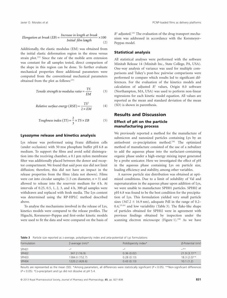
Table 1 Formulations prepared to study the effect of pH in themanufacturing process of Lys PCNPs
Formulation Protein model Buffer/solution pH
SPH01 Lys N Phthalate 5.4SPH02 Lys Phosphate 6.8SPH03 Lys Borate 10SPH04 Lys NaOH 13
Javier O. Morales et al. PCNP-loaded films as delivery platforms
© 2013 Royal Pharmaceutical Society, Journal of Pharmacy and Pharmacology, 65, pp. 827–838 829
M. lysodeikticus and absorbance was measured at 450 nm todetermine maximum activity of solutions. Relative activitywas calculated considering the absorbance measured for afresh Lys stock solution as 100% activity. To determine theremaining relative activity of Lys in films, the same mediaobtained after overnight shaking described in the abovesection on Lys quantification by RP-HPLC, was used.
Preparation of particle-containing films
Casting solutions were prepared by combining two organicsolutions and cast overnight in polytetrafluoroethylenemolds. Acetone was used to dissolve or suspend the polymercombinations as depicted in Table 2. This solution wascombined in a solvent mixture of acetone–IPA (4 : 6) withsuitable amounts of SPH02 (pH 6.8) of Lys-containing IPA(for the control formulation, FPH06) to yield the finalcasting solution. Control formulations were manufacturedutilizing C974P and PCP as mucoadhesive models, wellknown for their mucoadhesive character in the literature(Table 2). After 24 hours, films were peeled off andstored in aluminium foil sachets in a dessicator untilcharacterization.
Morphology of particles and films
A scanning electron microscope (Quanta 650 FEG; FEICompany, Hillsboro, OR, USA) was used for imaging andultrastructure analysis of both particles and particle-containing films. After separation and drying of slurries,samples were mounted onto aluminium stubs using con-ductive carbon tape for coating. For the imaging of films,cross-sections were obtained by a freeze-fracture method toensure clean-cut edges and to avoid plastic deformation(often resulting from mechanical cutting). Fragments of thesurface of the film were frozen by submerging in liquidnitrogen and then cracked. Pieces of the films were fixed onaluminium stubs by means of conductive carbon tape forcoating. Coating was performed, using a 208 HR Cressing-ton sputter coater (Cressington Scientific Instruments Ltd,
Watford, UK) with Pt/Pd, to a thickness of 10–15 nm in ahigh-vacuum evaporator. To avoid structural deformationduring imaging, the electron beam voltage was kept at2–5 kV.
Mucoadhesive and mechanical properties offilms in vitro
Mucoadhesion tests were conducted on a TA.XTPlus textureanalyser (Stable Micro Systems, Godalming, UK) equippedwith a 5 kg load cell. Briefly, films were held in the horizon-tal position and 5 ml of model mucus (a freshly made 2%w/v mucin solution) was placed on top of the film. Thisamount was sufficient to mimic the average saliva thickness.A stainless-steel cylindrical probe (7 mm diameter) wasattached to the mobile arm of the texture analyser and itwas brought into contact with the film and mucin solution,held at an applied force of 50 mN for 15 s and then with-drawn at a rate of 0.5 mm/s. The mucoadhesive force(MAF) and work of adhesion (WoA) were obtained fromthe peak and the area under the curve in the force-versus-distance profile, respectively.
For the determination of mechanical properties, rectan-gular strips of 1 ¥ 5 cm2 were cut and 1 cm on each endwas held between clamps attached to the texture analyser,leaving a testing area of 1 ¥ 3 cm2. The upper clamp (con-nected to the mobile arm of the texture analyser) wasmoved upwards at a rate of 0.5 mm/s until film failure.Stress was determined from the force measurementsobtained from the instrument divided by the cross-sectional area of the film, while strain was computed bydividing the increase in length by the initial film length.From the plot, the tensile strength (TS) and the elongationat break (EB) were obtained from the peak stress and themaximum strain, respectively, also represented by the fol-lowing equations:[12]
Tensile strength TSPeak stress
area of filmCross sectional( ) =
-(1)
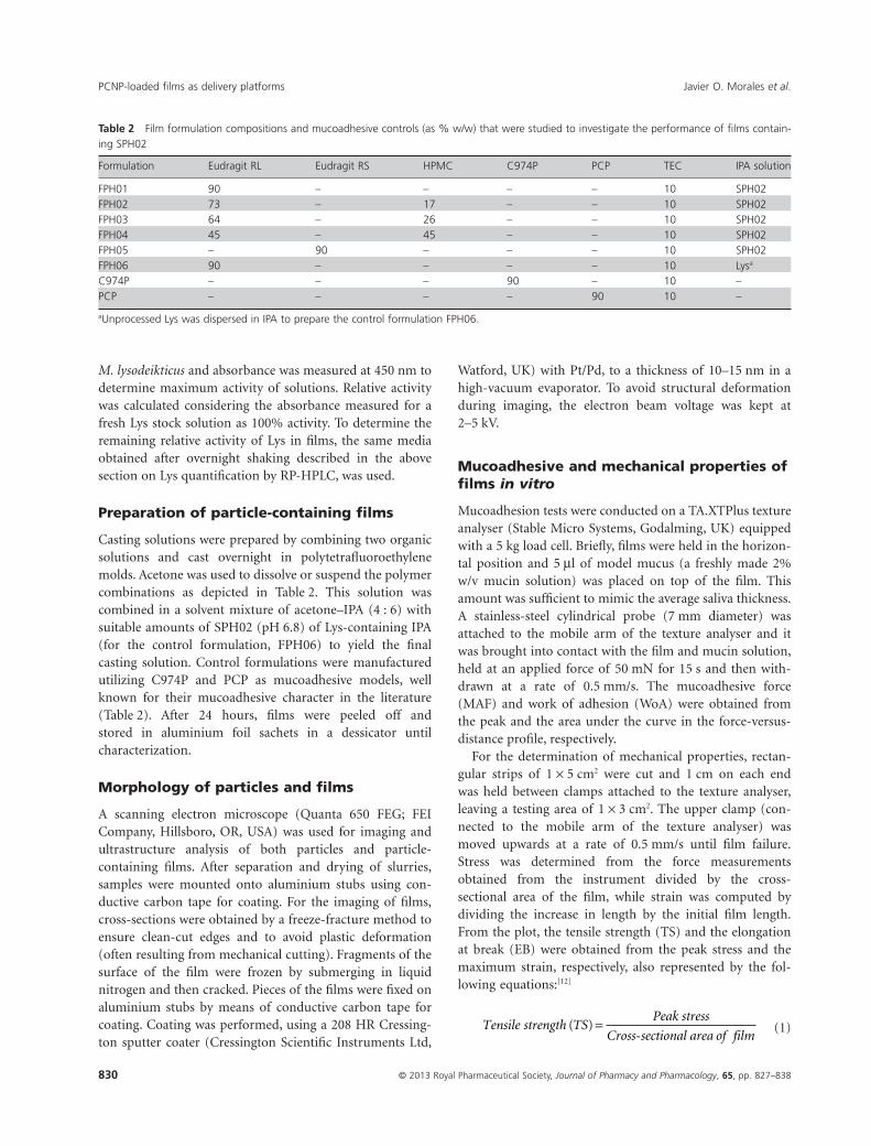
Table 2 Film formulation compositions and mucoadhesive controls (as % w/w) that were studied to investigate the performance of films contain-ing SPH02
Formulation Eudragit RL Eudragit RS HPMC C974P PCP TEC IPA solution
FPH01 90 – – – – 10 SPH02FPH02 73 – 17 – – 10 SPH02FPH03 64 – 26 – – 10 SPH02FPH04 45 – 45 – – 10 SPH02FPH05 – 90 – – – 10 SPH02FPH06 90 – – – – 10 Lysa
C974P – – – 90 – 10 –PCP – – – – 90 10 –
aUnprocessed Lys was dispersed in IPA to prepare the control formulation FPH06.
Javier O. Morales et al.PCNP-loaded films as delivery platforms
© 2013 Royal Pharmaceutical Society, Journal of Pharmacy and Pharmacology, 65, pp. 827–838830
Elongation at break EBIncrease in length at break
Initial film le( ) =
nngth×100
(2)
Additionally, the elastic modulus (EM) was obtained fromthe initial elastic deformation region in the stress versusstrain plot.[22] Since the rate of the mobile arm extensionwas constant for all samples tested, direct comparison ofthe slope in this region can be done. To further evaluatemechanical properties three additional parameters werecomputed from the conventional mechanical parametersobtained from the plot as follows:[23]
Tensile strength to modulus ratioTS
EM= (3)
Relative surface energy RSETS
EM( ) =
×
2
2(4)
Toughness index TI TS EB( ) = × ×2
3(5)
Lysozyme release and kinetics analysis
Lys release was performed using Franz diffusion cells(under occlusion) with 50 mm phosphate buffer pH 6.8 asmedium. To support the films and avoid solid disintegra-tion into the receiving chamber, a 0.1 mm nylon membranefilter was additionally placed between the donor and recep-tor compartment. We found that said pore size did not limitdiffusion; therefore, this did not have an impact in therelease properties from the films (data not shown). Filmswere cut into circular samples (1.5 cm diameter, n = 3) andallowed to release into the reservoir medium for 4 h. Atintervals of 0.25, 0.5, 1, 2, 3, and 4 h, 300-ml samples werewithdrawn and replaced with fresh media. The Lys contentwas determined using the RP-HPLC method describedabove.
To analyse the mechanisms involved in the release of Lys,kinetics models were compared to the release profiles. TheHiguchi, Korsmeyer–Peppas and first-order kinetic modelswere used to fit the data and were compared on the basis of
R2 adjusted.[24] The evaluation of the drug transport mecha-nism was addressed in accordance with the Korsmeyer–Peppas model.
Statistical analysis
All statistical analyses were performed with the softwareMinitab Release 14 (Minitab Inc., State College, PA, USA).One-way analysis of variance was used for multiple com-parisons and Tukey’s post-hoc pairwise comparisons wereperformed to compare which results led to significant dif-ferences. For the evaluation of the kinetics models andcalculation of adjusted R2 values, Origin 8.0 software(Northampton, MA, USA) was used to perform non-linearregressions for each kinetic model equation. All values arereported as the mean and standard deviation of the mean(SD) is shown in parenthesis.
Results and Discussion
Effect of pH on the particlemanufacturing process
We previously reported a method for the manufacture ofsubmicron and nanosized particles containing Lys by anantisolvent co-precipitation method.[20] The optimizedmethod of manufacture consisted of the use of a nebulizerto add the aqueous phase into the surfactant-containingorganic phase under a high-energy mixing input generatedby a probe sonicator. Here we investigated the effect of pHin the aqueous phase containing Lys on particle size,loading efficiency and stability, among other variables.
A narrow particle size distribution was obtained at opti-mized conditions. Due to a limit of solubility of Val andsupersaturation in the aqueous phase upon addition of Lys,we were unable to manufacture SPH01 particles. SPH02 atpH 6.8 was found to be the best condition for the precipita-tion of Lys. This formulation yielded very small particlesizes (347.2 � 16.9 nm), adequate PdI in the range of 0.2–0.4,[25,26] and low variability (Table 3). The flake-like shapeof particles obtained for SPH02 were in agreement withprevious findings obtained by inspection under thescanning electron microscope (Figure 1).[20] As we have
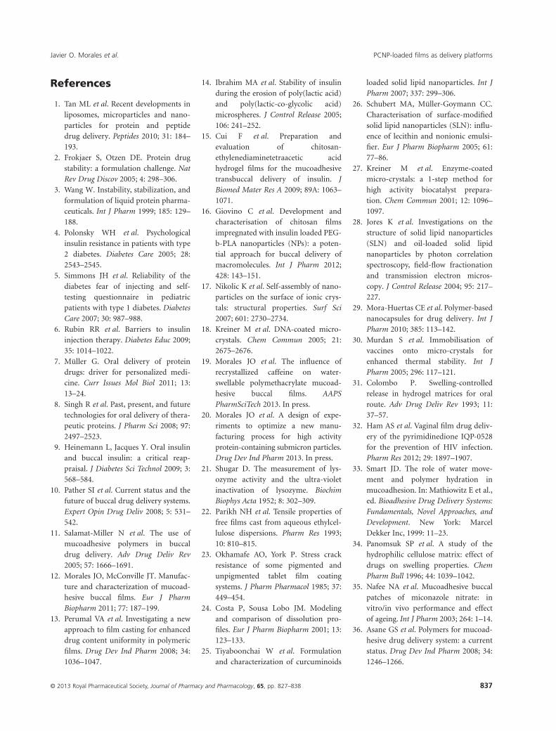
Table 3 Particle size reported as z-average, polydispersity index and zeta-potential of Lys formulations
Formulation Z-average (nm)* Polidispersity index* Ζ-Potential (mV)
SPH01 –a –a –**SPH02 347.2 (16.9) 0.36 (0.02) 21.9 (3.7)**SPH03 1384.0 (152.7) 0.28 (0.13) 18.3 (2.0)**SPH04 1220.2 (426.6) 0.43 (0.13) 10.1 (1.2)
Results are represented as the mean (SD). *Among parameters, all differences were statistically significant (P < 0.05). **Non-significant differences(P < 0.05). aCo-precipitant and Lys did not dissolve at pH 5.4.
Javier O. Morales et al. PCNP-loaded films as delivery platforms
© 2013 Royal Pharmaceutical Society, Journal of Pharmacy and Pharmacology, 65, pp. 827–838 831

previously shown, during the antisolvent precipitationprocess and due to the very high concentration ofco-precipitant (i.e. Val), the particle takes on the shape ofthe core-forming material.[20] The active then precipitateson the surface of the growing nanoparticle and, much like asurfactant, hinders further growth by coagulation or con-densation.[27] The asymmetrical shape of the flake-like par-ticles also has an impact on the resulting particle size andPdI obtained by DLS.[28] Having very high length-to-thickness ratio, homogeneity measured by the PdI isincreased. Jores et al. reported similar findings after manu-facturing platelet-like solid lipid nanoparticles and nanos-tructured lipid carriers. Asymmetrical particle shape wasfound to increase PdI values to the range of 0.1–0.3.[28] TheZP found for SPH02 is well correlated with particles in thenano size and the magnitude indicates good dispersion sta-bility of the formulation.[29] Even though slurries obtainedhere were rapidly needed in the process of manufacturingfilms embedded with particles (typically 5–10 min lapsedafter particle manufacture and the start of the film manu-facturing process), visual observations hinted at the greaterdispersion stability of SPH02. During a one-week period,particles in the SPH02 slurry remained in suspension incomparison with any of the other formulations which sedi-mented shortly after manufacture.
Regardless of the pH, excellent Lys loading efficiency andstability was achieved. With loading efficiencies in the rangeof 70.5–73.4 (no statistical differences found, P < 0.05) andremaining relative activity in the range of 91.4–101.1%, wecorroborated that the method of manufacture of nanopar-ticles by the antisolvent co-precipitation method is success-ful in rendering functional particles (Figure 2). This also
indicates that the pH of the buffer solution containing Lysbefore manufacture had little effect on the resulting stabilityafter manufacture. Investigations on the manufacture ofmicroparticles in the range of 1–10 mm obtained through asimilar process of antisolvent co-precipitation have shownpositive results regarding the stability of the macromol-ecules coating an inert core.[27,30] Our findings here consti-tute an improvement over the particles obtained previouslywhere the optimized conditions allowed for a z-average of439 nm with a loading efficiency around 50% and remain-ing activity of 98.7%.[20] We were also able to obtain nano-particles with a much higher enzyme load (40% instead of10% w/w), which represents an advantage in terms ofdosing in the final dosage form.
Development of lysozymeparticle-containing films
Films were successfully manufactured and their surfaceappeared homogeneous to the eye. A close inspectionrevealed a smooth surface in films without HPMC, whilefilms that contained HPMC presented with a roughersurface (resulting from the composite nature of the ERL-HPMC matrix). Regardless of the roughness observed inHPMC-containing films, the area-normalized weight wasaround 100 mg/cm2 with no statistically significant differ-ence between formulations (P > 0.05, Table 4). The incor-poration of HPMC in ERL films resulted in a tendency toincreasing thickness, being significantly different only at thehighest content of HPMC. The hydrophilic character ofHPMC results in swelling upon dispersion in the solventmixture, and after drying remains in a less-dense state thanERL.[31] A similar trend for increase in thickness and surfaceroughness with an increase in HPMC content has beendescribed by Ham et al. in films for the vaginal delivery of a
Figure 1 SEM micrograph of protein-coated nanoparticles from for-mulation SPH02. Scale bar =1 mm (Data are means � SD, n = 5).
100.0**
*80.0
60.0
40.0
20.0
0.0SPH02 SPH03 SPH04
100.0
80.0
60.0
40.0
20.0
0.0
Lys load efficiency (%)
Lyso
zym
e lo
adin
g e
ffic
ien
cy (
%)
Lys activity (%)
Lyso
zym
e ac
tivi
ty (
%)
Figure 2 Lys loading efficiency (�) and relative activity (�) of Lys-containing particle formulations. *P < 0.05, no significant differenceswere found among Lys loading efficiency results. **P < 0.05, all theactivity results were significantly different from each other (Data aremeans � SD, n = 3).
Javier O. Morales et al.PCNP-loaded films as delivery platforms
© 2013 Royal Pharmaceutical Society, Journal of Pharmacy and Pharmacology, 65, pp. 827–838832

pyrimidinedione.[32] Scanning electron microscopy (SEM)observation of cross sections of selected film formulationsobtained by freeze-fracture revealed a uniform distributionof the flake-like particles throughout the polymeric matrix(Figure 3). A closer look (Figure 4) showed that mostlyindividual particles were separately enclosed resulting in ahigh drug content uniformity in the films. As mentionedabove, agglomerates of HPMC can also be observed homo-geneously distributed under SEM, indicating the compositenature of the film that is responsible for the rough charac-teristic of the HPMC-ERL matrix.
Mucoadhesion and mechanical properties oflysozyme-containing films
ERS and ERL are more commonly known for their applica-tions in sustained drug delivery by controlling drug releaserate of dosage forms. However, more recently we have
reported on the high mucoadhesive properties exhibited bypolymethacrylates and, more specifically, ERL.[19] In accord-ance with our previous findings, films containing ERLshowed high or very high mucoadhesive properties.Figure 5 shows that FPH01 had a much higher WoA thanany of the other formulations studied and more than themucoadhesive controls studied, namely C974P and PCP(P < 0.05). Additionally, among the series of formulationsstudied (FPH01–06), FPH01 exhibited a significantly higherMAF (P < 0.05). We believe that the presence of water-soluble particles homogeneously distributed among the filmsurface allows for a more rapid and homogeneous waterpenetration. According to the theories of mucoadhesionbased on diffusion and water penetration,[33] the presence ofwater in the interface is paramount for the establishment ofthe mucoadhesive bond. The water rapidly driven in by thewater-soluble molecules allows for polymer chain mobilityresulting in entanglement with the mucin molecules in the
Table 4 Area-normalized weight, thickness and mechanical properties for Lys-containing films. Results are represented as the mean and standarddeviation in parenthesis
Formulation Weight* (mg/cm2) Thickness (mm) Tensile strength (N/mm2) Elongation at break (%) Elastic modulus (N/mm2/%)
FPH01 98.3 (4.0) 428.7 (14.4) 1.653 (0.160)a 197.9 (26.7)a 0.318 (0.110)a,b
FPH02 100.5 (4.2) 449.0 (18.1) 2.783 (0.133) 50.0 (11.0)b,c 0.831 (0.048)FPH03 93.0 (4.9) 433.3 (26.7) 5.169 (0.462)b 25.6 (6.5)b,d 1.554 (0.191)FPH04 101.9 (8.7) 556.6 (29.4)** 5.005 (0.464)b 18.0 (4.0)c,d 1.228 (0.129)FPH05 103.8 (4.7) 367.4 (16.6)** 0.580 (0.075)c 233.6 (43.9)a 0.153 (0.038)a
FPH06 101.9 (0.5) 439.7 (13.8) 1.273 (0.124)a,c 124.7 (12.9) 0.465 (0.093)b
Results are represented as the mean (SD). *No significant differences were found among area-normalized weight of formulations. **Among thick-ness variation, only FPH03 and FPH04 are different from each other and all the rest. a–dAmong parameters, non-significant differences are indicatedin pairs of letters (P < 0.05).
Figure 3 SEM micrographs of cross-sections of films obtained byfreeze-fracture. (a) FPH01, (b) FPH03, (c) FPH04, and (d) FPH05. Scalebar represents 20 mm.
Figure 4 SEM micrographs of cross-sections of films obtained byfreeze-fracture. (a) FPH01, (b) FPH03, (c) FPH04, and (d) FPH05. Thebar represents 5 mm.
Javier O. Morales et al. PCNP-loaded films as delivery platforms
© 2013 Royal Pharmaceutical Society, Journal of Pharmacy and Pharmacology, 65, pp. 827–838 833

mucus layer and establishment of the mucoadhesive bond.Similar effects have been found in systems wherein the drugwas incorporated as particulate material. Panomsuk et al.have described that the inclusion of a water-soluble drug,such as theophylline, increases the amount of water associ-ated with the polymer, favouring gelling and swelling.[34] Ithas also been shown that the presence of particulate mate-rial interrupts the polymer matrix continuum; this allowsthe polymer chains to move more freely, leading to anincrease in water penetration.[35] The absence of particulatematerial in the formulations containing only C974P andPCP leads to their inherent mucoadhesion resulting fromthe capacity of the polymer to absorb water and plasticizethe polymer chains to interact with mucin. It should benoted that the addition of HPMC hinders the full extent ofmucoadhesion enhancement possibly by capturing the par-ticles (that are more hydrophilic) in HPMC-rich domains.This results in slower hydration and thus a weaker mucoad-hesive bond that mostly depends on the mucoadhesion ofHPMC. HPMC has been used in the past as a mucoadhesivematerial but its mucoadhesive power is lower than thatobserved for PCP and C974P.[36] A similar trend was foundin investigations by Wong et al. performed on films com-posed of a polymethacrylate and HPMC.[37] The authorsfound that an increase in the HPMC concentration resultedin a decrease in mucoadhesion. We believe that higher con-tents than 30% make a substantial change in the materialproperties and the inherent mucoadhesivity of HPMCstarts to play a role at high contents. The polymer matrix ofFPH04 was equally composed of HPMC and ERL (% w/w)resulting in an increase in mucoadhesion compared with
FPH03 (Figure 5). At this higher HPMC content, theinherent mucoadhesion becomes more dominant in theinteraction resulting in an overall higher mucoadhesion,overriding the detrimental effect of encapsulating thewater-soluble particles. However, the mucoadhesive proper-ties of FPH04 were still below those observed for FPH01.Finally, ERS exhibited higher MAF than the ERL films withdrug in solid solution.[19] This is interesting consideringthat ERS is the more hydrophobic material due to its lowercontent of quaternary ammonium groups. We have previ-ously shown that ERS consistently exhibits lower MAF andWoA than ERL.[19] However, we believe that the enhancingeffect of the water-soluble particulate material discussedearlier is responsible for the higher extent of mucoadhesiveproperties.
Films as dosage forms for the buccal route of deliveryneed to withstand the stress originating from the mechani-cal activity of the mouth. Adequate mechanical andmucoadhesive characteristics are needed for the films toremain in contact with the mucosa for the desired amountof time of release.[38] Another source of mechanical stressoriginates from the processes of manufacturing, handlingand administration.[39] Thus, to successfully develop films asdosage forms for buccal delivery, a relatively high TS and EBand a low EM are desirable.[38] Additionally, derived fromthe conventional parameters extracted from stress-versus-strain curves, a relatively high TS/EM, RSE and TI arerequired.[23,40]
Table 4 shows that adequate control over TS, EB and EMwas achieved for FPH01, FPH05 and FPH06; all of whichonly contained either ERL or ERS and no other polymer.Quaternary ammonium polymethacrylates have been previ-ously described to have suitable properties as film-formingmaterial for dosage forms for the buccal route.[19] In thatstudy we showed that film formulations containing 10% tri-ethylcitrate as plasticizer rendered films with medium TS,high EB and low EM. Here, we have found similar condi-tions for films that did not contain HPMC as a releasemodifier polymer. The addition of HPMC was correlatedwith an increase in TS, decrease in EB and a slight increasein EM (Table 4). This is indicative of less ductile yet moreresistant films. The effect of HPMC over the mechanicalproperties of films is clearer after analysis of the derivedmechanical parameters. TS/EM is an indicator of the levelof internal stress in a film, the larger its value the higher thefilm crack resistance. RSE is also used to estimate crackresistance and is approximated from the surface energy ofthe film. Finally, TI is an estimation of energy absorbed perunit volume of film under stress.[23] FPH01 is the formula-tion that possessed the largest TS/EM indicating high resist-ance to cracking (Table 5). The addition of HPMC reducedthis value significantly, except for FPH04; however, TS/EMvalues remained high and acceptable. In the same vein, the
600
Max
imu
m a
dh
esiv
e fo
rce
(mN
)
Wo
rk o
f ad
hes
ion
(µJ
)
Maximum adhesive forceWork of adhesion
*
ac
FPH01
FPH02
FPH03
FPH04
FPH05
FPH06
C974P PC
P
500
400
300
200
100
0
600
700
500
400
300
200
100
0
de
fg
dhefh
g
abbc
Figure 5 Mucoadhesive properties of Lys-containing films. The samevalues for conventional mucoadhesive polymers, such as C974P andPCP, are depicted for comparing the performance of films developedhere. a–h. Non-significant differences among maximum adhesive forceare indicated in pairs of letters (P > 0.05). *Only work of adhesion ofFPH01 was significantly different from other formulations (P < 0.05).All other values were not statistically different (P > 0.05) (Data aremeans � SD, n = 5).
Javier O. Morales et al.PCNP-loaded films as delivery platforms
© 2013 Royal Pharmaceutical Society, Journal of Pharmacy and Pharmacology, 65, pp. 827–838834

RSE of films increased with increase in the content ofHPMC, being highest for FPH04 at 10.32 N/mm2·%, indi-cating crack resistance. Comparison of TI indicates that,except for FPH01 which was shown to be the toughest for-mulation, the TI of all other formulations varied within anacceptable range (Table 5).
Lysozyme release and kinetics
From the drug release profiles we observed an increase in therelease rate and extent of release as the concentrationof HPMC increased in the formulations (FPH01–04,Figure 6). HPMC is a water-swellable and water-erodiblepolymer that will dissolve from the dosage form; therefore,increasing concentrations of HPMC in formulations allowfor domains in the film that will release Lys faster than ERL-rich domains. In accordance with the similarity value, f2,[41]
FPH03 and FPH04 were the only formulations that rendereda similar Lys release profile (Table 6). Therefore, an increasein the HPMC content from 30 to 50% w/w of polymer didnot elicit significant differences in the release profile. Accord-ing to the Korsmeyer–Peppas model, even though FPH04 hasa higher constant (k = 0.2800 for FPH04 and 0.2255 forFPH03) contributing to faster release at earlier times, the
higher exponential term of FPH03 (n = 0.6604 for FPH03and 0.4875 for FPH04) allows for faster release at later times.Similar effects have been described before in films combiningHPMC and ERL.[42] Among the various materials studied,Hassan et al. found that the combination of HPMC and ERLresulted in a lower burst release (< 20% drug released in thefirst 15 min) and in formulations that only contained HPMCa more rapid release was found to be associated with theswellable soluble matrix that HPMC constitutes in water.[31]
Another study conducted by Averineni et al. showed theeffect of increasing concentrations of HPMC in chitosan-containing film formulations.[43] Over a 210-min period,drug release increased from 52.52 to 73.23% for the formula-tions containing the lowest and highest amounts of HPMC,respectively.
In the Kormeyer–Peppas release kinetics model, n is therelease exponent, and is an indicator of the drug releasemechanism.[44] In the particular case of n = 0.5, the drugrelease mechanism is purely Fickian diffusion (the particu-lar solution that constitutes the Higuchi model equation).When n = 1 the equation describes a zero-order releasemechanism, and the region in the range of 0.5 < n < 1 rep-resents the so-called anomalous transport. First-orderkinetics applies to dosage forms that normally containwater-soluble drugs and porous polymer matrices. In saidsystems, drug release is proportional to the amount of drugremaining inside; therefore, the rate of drug releasedecreases with time. From Table 7 we can observe that,except for FPH04, all formulations exhibit an anomalous
Table 5 Derived mechanical parameters calculated from conventional mechanical properties derived from a stress vs strain plot
Formulation TS : EM (%-1)Relative surface energy(N/mm2·%)
Toughness index(N/mm2·%)
FPH01 5.69 (1.94)i,ii,iii 4.59 (1.06)a 216.51 (21.32)i,ii,iii,iv,v
FPH02 3.36 (0.32)i 4.69 (0.63)a 92.08 (15.55)i
FPH03 3.34 (0.22)ii 8.62 (0.77)b 88.10 (22.67)ii
FPH04 4.11 (0.50) 10.32 (1.93)b 60.32 (15.70)iii,vi
FPH05 3.88 (0.50) 1.11 (0.08)c 89.91 (17.96)iv
FPH06 2.77 (0.29)iii 1.75 (0.08)c 106.33 (20.12)v,vi
Results are represented as the mean (SD). EM, elastic modulus; TS, tensile strength. i–viAmong parameters, statistically significant differences indi-cated in pairs of roman numerals (P < 0.05). a–cAmong parameters, non-significant differences are indicated in pairs of letters.
Lys
rele
ased
(%
)
FPH01
FPH02
FPH03
FPH04
FPH05
FPH06
70
60
50
40
30
20
10
00 1 2
Time (h)3 4
Figure 6 Lys release profiles from particle-containing films FPH01,FPH02, FPH03, FPH04, FPH05 and the control (FPH06) (Data aremeans � SD, n = 6).
Table 6 Differences among FPH series of formulations based on thesimilarity factor, f2
f2 FPH01 FPH02 FPH03 FPH04 FPH05 FPH06
FPH01 – 40.76 22.47 20.73 27.23 19.40FPH02 – 34.46 31.64 18.11 12.65FPH03 – 56.79 9.75 5.89FPH04 – 9.00 5.29FPH05 – 45.23FPH06 –
Release profiles are similar if f2 � 50.
Javier O. Morales et al. PCNP-loaded films as delivery platforms
© 2013 Royal Pharmaceutical Society, Journal of Pharmacy and Pharmacology, 65, pp. 827–838 835

release of Lys. This is a consequence of systems that arewater-swellable, where drug release occurs by a combinationof diffusion and case-II transport. For FPH04, the release ismore adequately modelled by the Higuchi model (evidencedby the higher R2). This indicates that drug release in thissystem follows Fickian diffusion through the polymer matrix.In addition, all formulations are better adjusted to the first-order kinetics model (according to the R2). This modeldescribes drug release from porous matrices, such as thoseformed in a water-swollen polymethacrylate film, containinga water-soluble drug, such as the Lys-containing particles. Inthis system, drug release is proportional to the amount ofdrug remaining in the interior of the dosage form.[24] Fromthe release profile we can also observe that when Lys wasadded to the film formulation as a solid solution very littlerelease was achieved over the 4-h period. Molecules in solidsolution are completely surrounded by the polymeric matrixand a higher number of interactions between polymer andLys can be achieved. This results in a very slow release overthe time period (below limit of quantification).
Lysozyme activity remaining afterfilm manufacture
After 24 h of release in dissolution media, the activity of theLys released was evaluated to measure any decrease in activ-ity, as an indicator of enzyme stability. As depicted inFigure 7, Lys remaining activity was excellent for all the for-mulations studied revealing that the processing of manufac-turing particles into films for buccal delivery did not renderthe enzyme inactive. As shown above in the characterizationof nanoparticles, enzyme activity is not compromisedduring the manufacturing process, and the results obtainedin this section show that further processing into polymericfilms does not render the enzyme inactive. FPH05 exhibiteda slightly lower activity, which we believe was due to partialrelease of Lys over the 24-hours period (data not shown).
Conclusions
We have successfully developed PCNP-containing filmsbased on polymer matrices of polymethacrylates and
HPMC as delivery vehicles for buccal delivery. Lys-loadednanoparticles manufactured by our antisolvent precipita-tion process were incorporated in the films. By controllingthe pH and increasing the loading we were able to opti-mize conditions previously described. The new conditionsfor the method of manufacture allowed for the productionof small and narrowly distributed particles with highloading efficiency and excellent remaining activity. Parti-cles from formulation SPH02 were used in the manufac-ture of films for buccal delivery. All films containing Lys-coated nanoparticles had acceptable mechanical propertiesand Eudragit RL was shown to have excellent mucoadhe-sive properties. Additionally, films were able to sustain therelease of Lys over 4 h, modulated to faster release rates bythe use of HPMC. Finally, we were able to achieve excel-lent enzyme activity maintained in films containingEudragit RL.
Declarations
Conflict of interest
The Author(s) declare(s) that they have no conflicts ofinterest to disclose.
Table 7 Model parameters and adjusted R2 values for the FPH series of formulations
Formulation
Korsmeyer–PeppasQ = k ¥ tna
HiguchiQ = k ¥ t0.5a
First orderQ = k ¥ (1 - e-nt)a
k n Adj R2 k Adj R2 k n Adj R2
FPH01 0.1092 0.7702 0.9980 0.1407 0.9391 0.4956 0.2474 0.9995FPH02 0.1728 0.6287 0.9955 0.1943 0.9791 0.4702 0.4672 0.9965FPH03 0.2255 0.6604 0.9874 0.2612 0.9659 0.6579 0.4335 0.9998FPH04 0.2800 0.4875 0.9751 0.2769 0.9881 0.5340 0.8316 0.9966FPH05 0.0457 0.5837 0.9961 0.0492 0.9894 0.1114 0.5380 0.9911
aWhere Q is the amount of drug released in time t, k is a constant and n is an exponential constant.
Lys
acti
vity
(%
)
a a100.0
80.0
60.0
40.0
20.0
0.0FPH01 FPH02 FPH03 FPH04 FPH05 FPH06
Figure 7 Lys relative activity obtained from infinity release studiesfrom film formulations. a: non-significant difference is indicated in pairof letters.
Javier O. Morales et al.PCNP-loaded films as delivery platforms
© 2013 Royal Pharmaceutical Society, Journal of Pharmacy and Pharmacology, 65, pp. 827–838836
References
1. Tan ML et al. Recent developments inliposomes, microparticles and nano-particles for protein and peptidedrug delivery. Peptides 2010; 31: 184–193.
2. Frokjaer S, Otzen DE. Protein drugstability: a formulation challenge. NatRev Drug Discov 2005; 4: 298–306.
3. Wang W. Instability, stabilization, andformulation of liquid protein pharma-ceuticals. Int J Pharm 1999; 185: 129–188.
4. Polonsky WH et al. Psychologicalinsulin resistance in patients with type2 diabetes. Diabetes Care 2005; 28:2543–2545.
5. Simmons JH et al. Reliability of thediabetes fear of injecting and self-testing questionnaire in pediatricpatients with type 1 diabetes. DiabetesCare 2007; 30: 987–988.
6. Rubin RR et al. Barriers to insulininjection therapy. Diabetes Educ 2009;35: 1014–1022.
7. Müller G. Oral delivery of proteindrugs: driver for personalized medi-cine. Curr Issues Mol Biol 2011; 13:13–24.
8. Singh R et al. Past, present, and futuretechnologies for oral delivery of thera-peutic proteins. J Pharm Sci 2008; 97:2497–2523.
9. Heinemann L, Jacques Y. Oral insulinand buccal insulin: a critical reap-praisal. J Diabetes Sci Technol 2009; 3:568–584.
10. Pather SI et al. Current status and thefuture of buccal drug delivery systems.Expert Opin Drug Deliv 2008; 5: 531–542.
11. Salamat-Miller N et al. The use ofmucoadhesive polymers in buccaldrug delivery. Adv Drug Deliv Rev2005; 57: 1666–1691.
12. Morales JO, McConville JT. Manufac-ture and characterization of mucoad-hesive buccal films. Eur J PharmBiopharm 2011; 77: 187–199.
13. Perumal VA et al. Investigating a newapproach to film casting for enhanceddrug content uniformity in polymericfilms. Drug Dev Ind Pharm 2008; 34:1036–1047.
14. Ibrahim MA et al. Stability of insulinduring the erosion of poly(lactic acid)and poly(lactic-co-glycolic acid)microspheres. J Control Release 2005;106: 241–252.
15. Cui F et al. Preparation andevaluation of chitosan-ethylenediaminetetraacetic acidhydrogel films for the mucoadhesivetransbuccal delivery of insulin. JBiomed Mater Res A 2009; 89A: 1063–1071.
16. Giovino C et al. Development andcharacterisation of chitosan filmsimpregnated with insulin loaded PEG-b-PLA nanoparticles (NPs): a poten-tial approach for buccal delivery ofmacromolecules. Int J Pharm 2012;428: 143–151.
17. Nikolic K et al. Self-assembly of nano-particles on the surface of ionic crys-tals: structural properties. Surf Sci2007; 601: 2730–2734.
18. Kreiner M et al. DNA-coated micro-crystals. Chem Commun 2005; 21:2675–2676.
19. Morales JO et al. The influence ofrecrystallized caffeine on water-swellable polymethacrylate mucoad-hesive buccal films. AAPSPharmSciTech 2013. In press.
20. Morales JO et al. A design of expe-riments to optimize a new manu-facturing process for high activityprotein-containing submicron particles.Drug Dev Ind Pharm 2013. In press.
21. Shugar D. The measurement of lys-ozyme activity and the ultra-violetinactivation of lysozyme. BiochimBiophys Acta 1952; 8: 302–309.
22. Parikh NH et al. Tensile properties offree films cast from aqueous ethylcel-lulose dispersions. Pharm Res 1993;10: 810–815.
23. Okhamafe AO, York P. Stress crackresistance of some pigmented andunpigmented tablet film coatingsystems. J Pharm Pharmacol 1985; 37:449–454.
24. Costa P, Sousa Lobo JM. Modelingand comparison of dissolution pro-files. Eur J Pharm Biopharm 2001; 13:123–133.
25. Tiyaboonchai W et al. Formulationand characterization of curcuminoids
loaded solid lipid nanoparticles. Int JPharm 2007; 337: 299–306.
26. Schubert MA, Müller-Goymann CC.Characterisation of surface-modifiedsolid lipid nanoparticles (SLN): influ-ence of lecithin and nonionic emulsi-fier. Eur J Pharm Biopharm 2005; 61:77–86.
27. Kreiner M et al. Enzyme-coatedmicro-crystals: a 1-step method forhigh activity biocatalyst prepara-tion. Chem Commun 2001; 12: 1096–1097.
28. Jores K et al. Investigations on thestructure of solid lipid nanoparticles(SLN) and oil-loaded solid lipidnanoparticles by photon correlationspectroscopy, field-flow fractionationand transmission electron micros-copy. J Control Release 2004; 95: 217–227.
29. Mora-Huertas CE et al. Polymer-basednanocapsules for drug delivery. Int JPharm 2010; 385: 113–142.
30. Murdan S et al. Immobilisation ofvaccines onto micro-crystals forenhanced thermal stability. Int JPharm 2005; 296: 117–121.
31. Colombo P. Swelling-controlledrelease in hydrogel matrices for oralroute. Adv Drug Deliv Rev 1993; 11:37–57.
32. Ham AS et al. Vaginal film drug deliv-ery of the pyrimidinedione IQP-0528for the prevention of HIV infection.Pharm Res 2012; 29: 1897–1907.
33. Smart JD. The role of water move-ment and polymer hydration inmucoadhesion. In: Mathiowitz E et al.,ed. Bioadhesive Drug Delivery Systems:Fundamentals, Novel Approaches, andDevelopment. New York: MarcelDekker Inc, 1999: 11–23.
34. Panomsuk SP et al. A study of thehydrophilic cellulose matrix: effect ofdrugs on swelling properties. ChemPharm Bull 1996; 44: 1039–1042.
35. Nafee NA et al. Mucoadhesive buccalpatches of miconazole nitrate: invitro/in vivo performance and effectof ageing. Int J Pharm 2003; 264: 1–14.
36. Asane GS et al. Polymers for mucoad-hesive drug delivery system: a currentstatus. Drug Dev Ind Pharm 2008; 34:1246–1266.
Javier O. Morales et al. PCNP-loaded films as delivery platforms
© 2013 Royal Pharmaceutical Society, Journal of Pharmacy and Pharmacology, 65, pp. 827–838 837

37. Wong CF et al. Formulation andevaluation of controlled releaseEudragit buccal patches. Int J Pharm1999; 178: 11–22.
38. Peh KK, Wong CF. Polymeric films asvehicle for buccal delivery: swelling,mechanical, and bioadhesive proper-ties. J Pharm Pharm Sci. 1999; 2:53–61.
39. Perumal VA et al. Formulation ofmonolayered films with drug and
polymers of opposing solubilities. Int JPharm 2008; 358: 184–191.
40. Omari DM et al. Lactic acid-inducedmodifications in films of Eudragit RLand RS aqueous dispersions. Int JPharm 2004; 274: 85–96.
41. Moore JW, Flanner HH. Mathematicalcomparison of dissolution profiles.Pharm Tech 1996; 20: 64–74.
42. Hassan MA et al. Formulation and invitro/in vivo evaluation of naproxen
mucoadhesive buccal patches for localeffect. J Drug Deliv Sci Technol 2011;21: 423–431.
43. Averineni RK et al. Development ofmucoadhesive buccal films for thetreatment of oral sub-mucous fibrosis:a preliminary study. Pharm DevTechnol 2009; 14: 199–207.
44. Korsmeyer RW et al. Mechanisms ofsolute release from porous hydrophilicpolymers. Int J Pharm 1983; 15: 25–35.
Javier O. Morales et al.PCNP-loaded films as delivery platforms
© 2013 Royal Pharmaceutical Society, Journal of Pharmacy and Pharmacology, 65, pp. 827–838838