Embed Size (px)
Citation preview

andrologia 18 (5): 474-482 (1986) Received Juli 17,1985
Department of Zoology, University of Delhi, Delhi/India
Protein Synthesis in the Rat Seminiferous Epithelium During Onset of Spermatogenesis - an Autoradiographic Study
M E E N A KUMARI and S. DURAISWAMI
Summary: The pattern of protein synthesis in Sertoli and germ cells during the onto- geny of spermatogenesis in the rat was followed by autoradiography using (3H)-Leucine as precursor. The results suggest that the protein synthetic potential of Sertoli cells is greater than that of germ cells at any stage of differentiation. Significantly enough, the ontogeny-related protein synthetic capabilities of the germ cell compartment find a paral- lel in those reported for the adult rodent. Furthermore, the data support the suggestion that proteins may be transported from Sertoli cells to germ cells.
Proteinsynthese wahrend der Spermatogenese in den Tubuli seminiferi- Epithelien der Ratte - eine autoradiographische Studie
Zusammenfassung: Die Struktur der Proteinsynthese in Sertoli- und Keimzellen der Ratte wahrend der Spermatogenese wurde unter Verwendung von 3H-Leucin als Vorstufe autoradiographisch verfolgt. Die Ergebnisse legen nahe, daB das ProteinsynthesePotential von Sertolizellen in allen Differenzierungsstadn groi3er als das von Keimzellen ist. Die Proteinsynthesefahigkeiten der Ontogenese in den Keimzellen zeigen signifikante Paralle- len zu Untersuchungen an ausgewachsenen Nagetieren. Dariiber hinaus stiitzen die Ergeb- nisse den Hinweis, dai3 die Proteine moglicherweise von den Sertoliiellen in die Keimzel- len transportiert werden.
Introduction
The Sertoli cells, constituting the sole non-germinal cell type within the seminiferous tubule, have been attributed an important role in germ cell development and differentia- tion (Fawcett - 1976). The close association of the germ cells with the Sertoli cells signifies that for an understanding of the molecular events during spermatogenesis, it is necessary to examine the interaction between these two compartments* specifically with reference to synthesis of macromolecules. The technique of autoradiography renders it feasible to investigate spatial and temporal relationships in RNA and protein synthesis during sperma- togenesis. Beginning with the early studies of Monesi (1965) on protein synthesis in the
* In view of the presumed interrelationships between these two cell elements, they will be referred to, on occasion, as the “germ cell compartment” and the “Sertoli cell compartment”. These terms are not to be confused with two others already extant in the literature, namely, the “basal compartment” and the “adluminal compartment”.
Key words: Spermatogenesis, rat - protein synthesis, seminiferous epithelium - autoradiography, protein synthesis
andrologia 18 (1986)
Protein Synthesis 475
testicular cells of the adult mouse, many reports, based on autoradiography, have been published (Davis and Langford - 1970; Nagy - 1974; Kierszenbaum - 1977; Davies and Lawrence - 1980; Dadoune et al. - 1981). Virtually all of the available information on protein synthesis in the testis is from adult animals. Our interest was to examine the pro- tein synthesizing capability of Sertoli and germ cells during the onset of sexual maturity in the colony-bred albino rat.
In the light of observations (Clermont et al. - 1959; Clermont and Harvey - 1965; Huckins - 1965; Davis and Schuetz - 1975; Condos - 1977; Aleman et al. - 1978; Hoche- reau and Courot - 1978) suggesting that local housing conditions as well as strain differ- ences may affect the timing of initiation and establishment of spermatogenesis as well as its duration in different colonies, a histological study of the ontogeny of spermatogenesis was carried out on our Holtzman-derived rat colony maintained under standard animal house conditions (24 f 1' C; 14 hr. light - 10 hr. dark schedule; free access to water and commercial pelleted rat diet). Based on information so obtained, days 8, 14, 18,23 and 30 (post-natum) were chosen for the present investigation in as much as each of them represents a crucial stage in the progression of the first spermatogenic cycle.
Materials and Methods
Chemicals: (3H)-Leucine, L-[4,S3 H(N)]- specific activity, 56.5 Ci/mMol. and Omni- fluor were purchased from New England Nuclear Co., Boston, MA, USA. Osmium tetroxide was purchased from Johnson Matthey and Co. Ltd., England; a, a'-Azoisobuty- ronitrile from Koch-Light Lab. Ltd., England and 2-Hydroxyethyl methacrylate from Fluka, Switzerland. Nuclear track emulsion, NTB-3, was purchased from Eastman Kodak, Rochester/N.Y. USA.
All other reagents and chemicals were purchased locally and were of analytical grade
Buffers: (1) Modified mammalian Krebs-Ringer medium (MMKRB): This buffer was a purity.
modification of Krebs improved Ringer 111 medium and had the following composition (expressed as millimoles of each ingredient per liter of final solution): sodium chloride (384.4 mM), potassium chloride (80 mM), calcium chloride (42.8 mM), potassium dihy- drogen phosphate (19.9 mM), magnesium sulphate (19.9 mM), sodium bicarbonate 60 mM), sodium pyruvate (83.2 mM), sodium lactate (4.9 mM), sodium fumarate (90 mM), sodium L-glutamate (83.1 mM), glucose (194.8 mM), disodium hydrogen phos- phate (19.5 mM), sodium dihydrogenphosphate (16.8 mM), glutamine (5 mM), HEPES (25 mM), and Phenol red (0.008 mM). Streptomycin (50 pglml) and penicillin (50 units/ ml) were also added. The pH of the buffer was adjusted to 7.3 with 1 N NaOH.
(2) MMKRB with unlabeled L-Leucine: To the buffer was added unlabeled L-Leucine to l00mM.
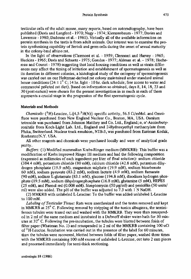
Labeling of Testicular Tissue: Rats were anesthesized and the testes removed and kept in MMKRB at 25" C. Following removal by stripping of the tunica albuginea, the semini- ferous tubules were teased out and washed with the MMKRB. They were then resuspend- ed in 2 ml of the same medium and incubated in a Dubnoff shaker water-bath for 30 min- utes at 32" C. Following the pre-incubation, the tubules were blotted between folds of filter paper (Whatman No. 1) and resuspended in 2 ml of the MMKRB containing 100 uCi of 3H-Leucine. Incubation was carried out in the presence of the label for 60 minutes, then the tubules were recovered, blotted between folds of filter paper, washed three times with the MMKRB containing 100 mM excess of unlabeled L-Leucine, cut into 2 mm pieces and processed immediately for semi-thick sectioning.
andrologia 18 (1986)
416 Meena Kumari and S. Duraiswami
Preparation of semi-thick sections: The seminiferous tubule pieces were fixed by im- mersion for six hours at room temperature in cacodylate buffer (0.1 M; pH 7.3) contain- ing glutaraldehyde-paraformaldehyde prepared according to Karnovsky (1 965) and post- fixed in 1% Os04 in 1.5% potassium ferrocyanate (Kamovsky - 1971). The material was washed several times with distilled water and dehydrated through methoxy-ethanol, etha- nol, n-propanol and n-butanol, and finally embedded in glycol methacrylate (see: Sharma and Sharma - 1980).
The blocks were trimmed and 2 pm sections cut on a Spencer microtome using glass knives. Sections were picked up individually and dropped onto water on a clean micro- scope slide where they flattened immediately. After drying in air, the sections held firmly to the slide without recourse to an adhesive.
Autoradiography: Autoradiography was performed according to the dipping method of Kopriwa and Leblond (1962). The slides were left in chilled (4" C) 5% trichloro-acetic acid for 30 minutes, washed under running tap water and dried at room temperature. They were then dipped in Kodak NTB-3 nuclear track emulsion, diluted in 1 : 1 with distil- led water, at 42" C, dried vertically, dipped in chilled 0.4% Omnifluor in toluene for 2 minutes (Mukherjee et al. - 1975), dried once more and packed in black plastic boxes containing silica gel.
Following 15 days of exposure at 4" C, the slides were developed in IPC 19b developer for 10 minutes at 4" C and fixed for 10 minutes at 4" Ci in IPC acid fixer with hardner. The processed slides were stained with 1% toluidine blue in 1% of sodium tetraborate and examined under the light microscope. On completion of photomicrography, the silver grains were removed by the method of Frgland (1 965), the slides restained with toluidine blue and the same areas as were photographed were reexamined.
Results
The histological study revealed that on day 8, the still dividing presumptive Sertoli cells constitute the major population of the seminiferous tubules. The germ cell types en- countered on this day, viz, gonocytes I1 (Condos - 1977), transitional cells and type A sper- matogonia (dormant as well as active), represent the minor population. As for the other
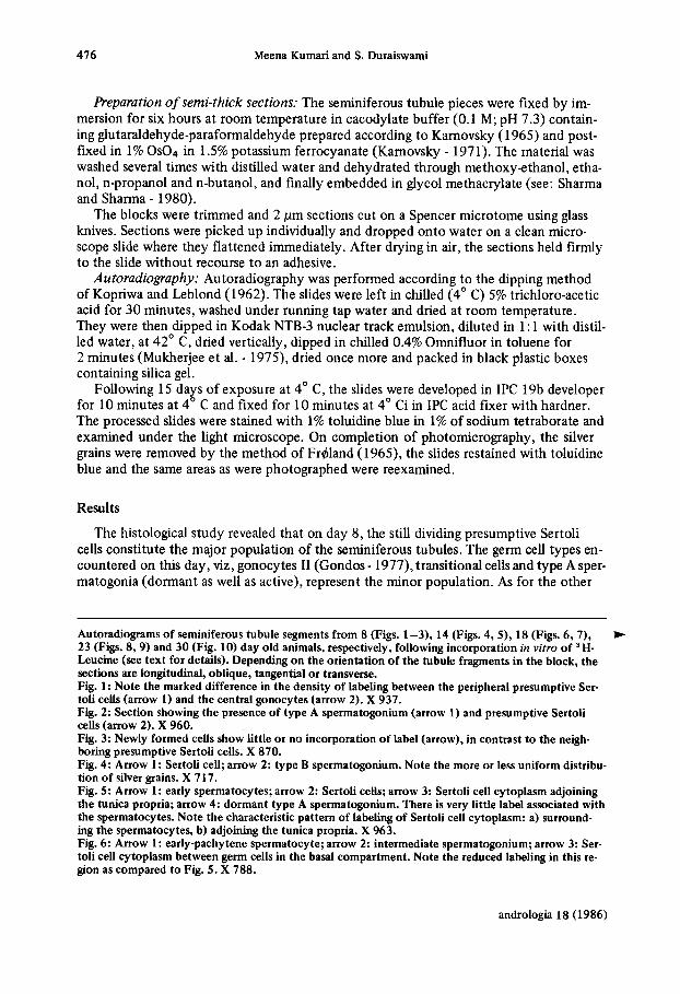
Autoradiograms of seminiferous tubule segments from 8 (Figs. 1 -3), 14 (Figs. 4, S), 18 (Figs. 6,7), 23 (Figs. 8, 9) and 30 (Fig. 10) day old animals, respectively, following incorporation in uitro of ' H- Leucine (see text for details). Depending on the orientation of the tubule fragments in the block, the sections are longitudinal, oblique, tangential or transverse. Fig. 1: Note the marked difference in the density of labeling between the peripheral presumptive Ser- toli cells (arrow 1) and the central gonocytes (arrow 2). X 937. Fig. 2: Section showing the presence of type A spermatogonium (arrow 1) and presumptive Sertoli cells (arrow 2). X 960. Fig. 3: Newly formed cells show little or no incorporation of label (arrow), in contrast t o the neigh- boring presumptive Sertoli cells. X 870. Fig. 4: Arrow 1: Sertoli cell; arrow 2: type B spermatogonium. Note the more or less uniform distribu- tion of silver grains. X 7 17. Fig. 5 : AROW 1: early spermatocytes; arrow 2: Sertoli cells; arrow 3: Sertoli cell cytoplasm adjoining the tunica propria; arrow 4: dormant type A spermatogonium. There is very little label associated with the spermatocytes. Note the characteristic pattern of labeling of Sertoli cell cytoplasm: a) surround- ing the spermatocytes, b) adjoining the tunica propria. X 963. Fig. 6: Arrow 1: early-pachytene spermatocyte; arrow 2: intermediate spermatogonium; arrow 3: Ser- toli cell cytoplasm between germ cells in the basal compartment. Note the reduced labeling in this re- gion as compared to Fig. 5 . X 788.
*
andrologia 18 (1986)

Protein Synthesis 411
andrologia 18 (1986)

478 Meena Kumari and S. Duraiswami
Table 1 Distribution of the various cell types' in the seminiferous
epithelium on specified days* of ontogeny - ~
Cell types Day 14 Day 18 Day23 Day 30
Sertoli cells
In Spg.
Type A dividing Type B dividing Resting Spc.' Leptotene Spc.' Zygotene Spc.' Pachytene Spc.' Round Spermatid
Type AISpg.'
Type B SP~..'
70.42 12.03 3.82 2.1 0.84
- 4.67 6.09
49.76 3.71
15.19
6.84
0.11
24.36
-
-
-
-
39.66 2.42
2.7
2.51 1.21
10.42 20.11 20.94
-
-
35.05 1.80 4.5 1 1.59
2.84 2.63
15.76 28.47
7.29
-
-
'expressed as percentage of total cell population *Day 8 has been omitted since there is little differentiation of germ
' Spg. - spermatogonia 'Spc. - spermatocytes
cells
age groups, details of the cell types present as well as their distribution, percentage-wise, are given in Table 1. On day 8, the peripheral presumptive Sertoli cells show heavy label- ing, suggesting that they are actively engaged in protein synthesis. The centrally located gonocytes, on the other hand, exhibit a distinctly attenuated density of the silver grains (Fig. 1). This constrasting situation between the Sertoli cell compartment and the germ cell compartment is maintained through the days of ontogeny examined.
Thegerm cell compartment: Of the germ cell types present on day 8, the active type A spermatogonia showed significant incorporation of labeled amino acid into protein (Fig. 2) in contrast to the prespermatogonial cells and dormant type A spermatogonia. Not sur- prisingly, dividing germ cells were found not to incorporate label into their cytoplasm (Fig. 3). The pattern of labeling of the different types of spermatogonia turned out to be identical in all the age groups and, in terms of decreasing grain density, could be arranged in the following order: active gype A spermatogonia (Figs. 7, 8) > intermediate type spermatogonia (Figs. 6 , 10) > type B spermatogonia (Figs. 4,9).
There was significant incorporation of label into all types of spermatocytes, but this was invariably less than that encountered in type A spermatogonia. The order of decreas- ing grain density was: mid-pachytene (Figs. 7 ,9 ) >late pachytene (Fig. 10) >early sper- matocytes (Figs. 5 , 6 ) > secondary spermatocytes.
On day 30, round spermatids were present in some of the seminiferous tubules. The following gradation in the distribution of grains amongst the different germ cell types was observed: spermatogonial cells > spermatocytes > spermatids (Fig. 10).
The Sertoli cell compartment: The Sertoli cells, while highly active in incorporating the labeled amino acid into protein, nevertheless showed an age-dependent decline in the extent of protein synthetic capability, as judged by the density of distribution of silver grains in the autoradiograms (compare Fig. 1 with 10). Nevertheless, it should be reiterat- ed that incorporation into Sertoli cells was consistently greater than into germ cells on
andrologia 18 (1986)

Protein Synthesis 479
Fig. 7: Arrow I: mid-pachytene spermatocyte; arrow 2: active type A spermatogonium; arrow 3: Ser- toli cell cytoplasm adjoining tunica propria; arrow 4: Sertoli cell cytoplasm surrounding spermato- cytes. X 742. Fig. 8: Note the characteristic gradient in the density of distribution of silver grains from the basal to adluminal region of the seminiferous tubule. L: Lumen of the tubule, arrow: type A spermatogonium. X 819. Fig. 9: Shows that labeling is not confined to Sertoli cells. Arrow I: type B spermatogonium; arrow 2: mid-pachytene spermatocyte. X 845. Fig. 10: Note gradient in density of grain distribution from the periphery to the centre, i.e. from the basal to the adluminal region. Arrow 1: intermediate spermatogonium; arrow 2: Sertoli cell nucleus; arrow 3: late-pachytene spermatocyte; arrow 4: round spermatid. X 1024.
any given day of ontogeny. As is well known, the appearance of the more advanced stages of germ cell differentiation is coeval with the establishment of the basal and adluminal compartments within the seminiferous epithelium. Concomitantly, there is also a charac- teristic intra-cytoplasmic regionalization of grain distribution in the Sertoli cells. On day 14, the silver grains were found uniformly distributed through the Sertoli cell cytoplasm
andrologia 18 (1986)

480 Meena Kumari and S. Duraiswami
in those seminiferous tubules in which the sole representatives of the germ cell compart- ment were spermatogonial cells (Fig. 4). On the other hand, in those tubules in which early spermatocytes were also present, there was a characteristic accumulation of silver grains in the immediate vicinity of the lamina propria as well as in the cytoplasmic proces- ses surrounding the early spermatocytes (Fig. 5). At 18 days of age, with the progression of germ cell differentiation to include pachytene spermatocytes, there was a marked de- cline in the sequestration of silver grains in the vicinity of the lamina propria and an ex- aggeration of grain accumulation in the cytoplasmic processes surrounding the sperma- tocytes (Figs. 6,7). On day 23, wherever type A spermatogonia were encountered, the cytoplasm of the adjoining Sertoli cell was heavily labeled in the immediate vicinity of the lamina propria (Fig. 8) which was not the case when intermediate and/or type B sper- matogonia were present (Fig. 9). Heavy labeling was encountered in Sertoli cell cytoplas- mic processses surrounding spermatocytes (Figs. 8,9) and, on day 30, spermatids as well (Fig. 10).
Discussion
It is significant that the present observations on ontogeny-related protein synthetic capabilities of the constituents of the germ cell compartment find a parallel in those re- ported for the adult rodent. The labeling characteristics of the spermatogonial cells are in conformity with those recorded by Monesi (1965) in the case of the adult mouse. The same worker further reported that mid-pachytene spermatocytes at tubule stages I1 to IV show maximum protein synthesis (Monesi - 1965). For our part, we observed that while primary spermatocytes incorporated the label into protein at all stages of the meiotic cycle, mid-pachytene spermatocytes showed the heaviest labeling. Uptake of label by the constituents of the adluminal compartment was consistently less than that by the sper- matogonial cells of the basal compartment. With the appearance of the more advanced stages of germ cell differentiation on days 23 and 30, there was a noticeable gradient in the density of grain distribution, decreasing from the periphery of the seminiferous tubules to the lumen. These features correspond to those described by Dadoune et al. (1981) based on an autoradiographic study of in vivo and in vitro protein synthesis in the adult mouse testis. Using type B spermatogonia as reference standard, they noted a de- crease in the extent of labeling progressing from spermatogonial cells through spermato- cytes to spermatids. The pattern of labeling of the Sertoli cells as a function of ontogeny showed, in its turn, some interesting features. The distribution of silver grains appeared to depend on the kind(s) of associated germ cells. On day 14, for instance, the Sertoli cell cytoplasm was uniformly labeled in those segments of the seminiferous tubules where spermatogonial cells were the sole constituents of the germ cell compartment. Where pri- mary spermatocytes were also present, the grains were localized in: (a) that part of the Sertoli cell cytoplasm found between the spermatogonial cells and (b) in the cytoplasmic processes surrounding the primary spermatocytes. With the appearance of mid-pachytene spermatocytes (day 18), the cytoplasm of the Sertoli cells surrounding them was heavily labeled. These characteristics of labeling persisted through day 23 into day 30.
It is of interest that the above patterns affecting protein synthesis, parallel the dif- ferential 3H-uridine labeling in the adult mouse testis (Kierszenbaum - 1974). Further- more, it was noted in a follow-up study (Kierszenbaum - 1977) that after a short pulse with labeled amino acid, silver grains were associated with cytoplasmic processses of the Sertoli cells extending among neighbouring spermatocytes and spermatids. Our results thus lend strong support to Kierszenbaum’s suggestion based on a similar approach that
andrologia 18 (1986)

Protein Synthesis 481
proteins and other products may be transported from Sertoli cells to these germinal cells (Kierszenbaum - 1977). In any event, localization of Sertoli cell protein synthesis to those portions of its cytoplasm lying in close proximity to differentiating germ cells is strong evidence for some kind of intercellular communication between the two compartments during ontogeny.
Acknowledgement: This work was done as part of a project receiving financial support from the Department of Science and Technology, Government of India. We thank MI. E.A. Daniels for help in the preparation of the photomicrographs.
References Alemb, V., R. Trejo, E. Morales, P. Hernbdez-Jburegui and G. Delhumeau-Ongay. 1978. A simple
and rapid technique to isolate enriched populations of spermatocytes and spermatids from the im- mature rat testis. J. Reprod. Fertil. 54,67-75.
Clermont, Y., C.P. Leblond and B. Messier. 1959. Durie du cycle de 1’8pithilium seminal du rat. Arch. Anat. micr. Morph. exp. Suppl. 48, 37-56.
Clermont, Y. and S.C. Harvey. 1965. Duration of the cycle of the seminiferous epithelium of normal, hypophysectomized and hypophysectomized-hormone treated albino rats. Endocrinology 76,80- 89.
Dadoune, J.P., M.A. Fain-Maurel, M.F. Alfonsi and G. Katsanis. 1981. In vivo and in vitro radioauto- graphic investigation of amino acid incorporation into male germ cells (mouse). Biol. Reprod. 24, 153- 162.
Davies, A.G. and N.R. Lawrence. 1980. Autoradiographic study of Lysine and Arginine incorporation stimulated by follicle stimulating hormone in the mouse testis in vivo. J. Endocrinol. 84,43-48.
Davis, J.R. and G.A. Langford. Testicular Proteins. In: A.D. Johnson, W.R. Comes and N.L. Van De- mark (Eds.). 1970. The testis Vol. 11. New York: Academic Press, pp. 259-306.
Davis, J.C. and A.W. Schuetz. 1975. Separation of germinal cells from immature rat testes by sedimen- tation at unit gravity. Exp. Cell Res. 91,79-86.
Fawcett, D.W. The male reproductive system. In: R.O. Creep, M.A. Koblinsky and F.S. Jaffe (Eds.). 1976. Reproduction and Human Welfare: A challenge to research. Cambridge: The MIT Press, pp.
Frdland, A. 1965. Photographic recording and dye staining of chromosomes for autoradiography and morphology. Stain Techn. 40,41-43.
Condos, B. Testicular development. In: A.D. Johnson and W.R. Comes (Eds.). 1977. The Testis Vol. IV. New York: Academic Press, pp. 1-37.
Hochereaude Reviers, M.-T. and M. Courot. 1978. Sertoli cells and development of seminiferous epi- thelium. Ann. Biol. anim. 18 (2B), 573-583.
Huckins, C. 1965. Duration of spermatogenesis in pre- and postpuberal Wistar rats. Anat. Rec. 15 1, 364.
Kamovsky, M.J. 1965. A formaldehyde-glutaraldehyde fixative of high osmolality for use in electron microscopy. J. Cell Biol. 27,137A.
Karnovsky, M.J. 197 1. Use of fenocyanide-reduced osmium tetroxide in electron microscopy. In: Proc. of 11th Am. SOC. of Cell Biol., Nov. 17-20,1971. New Orleans, Louisiana, Abstract 284, p. 146.
165 -277.
Kierszenbaum, A.L. 1974. RNA synthetic activities of Sertoli cells in the mouse testis. Biol. Reprod.
Kierszenbaum, A.L. Distribution of newly snythesized proteins in Sertoli cells as traced by electron 11,365-376.
microscope autoradiography. In: P. Troen and H.R. Nankin (Eds.), 1977. The Testis in normal and infertile men. New York: Raven Press, pp. 125-136.
Kopriwa, B.M. and C.P. Leblond. 1962. Improvements in the coating technique of radioautography. J. Histochem. Cytochem. 10,269-284.
Monesi, V. 1965. Synthetic activities during spermatogenesis in the mouse. Exp. Cell Res. 39,197- 224.
Mukherjee, A.S., R.N. Chatterjee, S.N. Chatterjee, S.N. Mandal, A. Nag and D. Majumdar. 1975. A rapid autoradiographic technique for chromosomal squash preparations. Indian J. exp. Biol. 13, 261-263.
andrologia 18 (1986)

482 Meena Kumari and S . Duraiswami
Nagy, F. 1974. Autoradiographic study of the postnatal changes in ' H-Leucine incorporation by Ser-
Sharma, A.K. and A. Sharma. 1980. Chromosome Techniques - Theory and Practice. London: But- toli cell nuclei in rat testis. J. Reprod. Fertil. 37, 353-359.
terworths, pp. 263-294.
Address: Prof. S . DURAISWAMI, Department of Zoology, University of Delhi, Delhi-l10007/India.
F. Kudlien: h z t e im Nationalsozialismus. 3 12 Seiten. Koln: Kiepenheuer & Witsch, 1985. Leinen rnit Schutzumschlag DM 48,-
Hier wird ein Buch vorgelegt, das sich mit den Arzten im nationalsozialistischen Staat aus- einandersetzt. In drei groDe Teile zerfallt dieser Band, der von Kudlien und acht weiteren Mitarbeitem verfaat worden ist:
I. vor 1933; 11. Das Dritte Reich und seine arztlichen Helfer; 111. Arzte als Helfer von Verfolgten, Kritikern und NS-Mafinahmen, Gegner des Dritten Reiches.
Bedauerlich ist, daD die von den Autoren selbst gesehene Notwendigkeit von Gespra- chen mit Zeitzeugen zwecks Erweiterung der schriftlichen Aktenunterlagen ausgeklammert werden muate. Gerade hier hatten die Autoren unmittelbar Erlebtes erfahren konnen, selbst wenn es - wenigstens teilweise - ein ,,verzerrtes (verklartes?) Bild" geboten hatte. Der Vergleich rnit den in den Akten vorhandenen Belegen hatte dann dazu beigetragen, das Gesamtbild plastischer darzustellen. Die nicht benutzbaren Unterlagen des Sicher- heitsdienstes (S.D.) der SS hatte da ein mindestens ebenso verzerrtes Bild ergeben.
Fur den Historiker ist die Suche nach der Wahrheit oberstes Gesetz und jede Art von Spekulation verwerflich. Leider klingt derartiges an einigen Stellen dennoch durch. Un- Mar bleibt mir, wieso die Aussagen des Hartmann-Bundes zur Situation der Arzte anla&- lich der Wirtschaftskrise als ,,mit hochst unglaubwurdigen Zahlen manipuliert" dargestellt werden konnen; die eigene Erinnerung an diese Zeit und die Erlebnisse mit den um die nackte Existenz ringenden Eltern geben da ein anderes Bild, welches keineswegs ein Ein- zelfall war.
Bemerkenswert die Daten zur Gleichschaltung der Arzte 1933, zum ,,Lebensborn" ei- ner Institution, die weitestgehend als sog. Zuchtanstalt galt, was sie jedoch nicht war, so- wie zur Leistungsmedizin (Pervitin), um nur einige Schwerpunkte zu nennen.
Der Begriff ,,Intemistik" ist im allgemeinen Sprachgebrauch fur ,,Innere Medizin" nicht ublich. Das Namensregister ist leider sehr unvollstandig. Ein Sachregister fehlt vollig. Beides ist aber gerade fur ein derartiges Sachbuch, mag es auch nur fur Laien gedacht sein, dringend erforderlich. Das Buch wird in dieser Form fur den Laien sicher dem Anspruch einer erstmaligen Gesamtdarstellung des Themas gerecht. Es ist zu wiinschen, daD dieser Kreis von Autoren, der sich in muhseliger Kleinarbeit durch die vorhandenen Dokumente, Schriften und Selbstzeugnisse durcharbeiten muate, auch den nachsten, logischen Schritt tut, namlich ein wissenschaftliches Gesamtwerk zur Thematik vorzulegen. Von der Seite der Wissenschaft ware es wohl richtiger gewesen, dieses wissenschaftlich ausgerichtete Buch vorab zu verfassen und sich erst dann einem popularwissenschaftlichen Werk zuzu- wenden. C. Schirren (Hamburg)
andrologia 18 (1986)