Embed Size (px)
Citation preview

Protein Kinase A in Postmortem Brain of DepressedSuicide Victims: Altered Expression of SpecificRegulatory and Catalytic SubunitsYogesh Dwivedi, Hooriyah S. Rizavi, Pradeep K. Shukla, Jennifer Lyons, Gabor Faludi, Miklos Palkovits,Andrea Sarosi, Robert R. Conley, Rosalinda C. Roberts, Carol A. Tamminga, and Ghanshyam N. Pandey
Background: We recently reported reduced [3H]cyclic adenosine monophosphate binding and catalytic activity of protein kinase Ain prefrontal cortex of depressed suicide victims. Here we examined the molecular basis of these alterations and whether these findingscan be replicated in another cohort.Methods: Prefrontal cortex from depressed suicide victims and nonpsychiatric controls were obtained from the Lenhossek HumanBrain Program, Budapest and the Maryland Brain Collection Program. [3H]cyclic adenosine monophosphate binding and proteinkinase A activity were determined by radioligand binding and enzymatic assay, respectively. Expression of catalytic and regulatorysubunits was determined by quantitative reverse transcription polymerase chain reaction and Western blot, respectively.Results: [3H]cyclic adenosine monophosphate binding and total and endogenous protein kinase A activity were significantlydecreased in membrane and cytosol fractions of prefrontal cortex of depressed suicide victims from the Budapest cohort, with a similarmagnitude (33%–40% reduction) as reported for the Maryland cohort. In both cohorts, selective reduction (36%–41%) in mRNA andprotein expression of the regulatory RII� and the catalytic C� was observed.Conclusions: Our results suggest abnormalities in [3H]cyclic adenosine monophosphate binding and catalytic activity kinase A inbrain of depressed suicide victims, which could be due to reduced expression of RII� and C�. These abnormalities in PKA may becritical in the pathophysiology of depression.
Key Words: Protein kinase A, human, postmortem brain, depres-sion, cAMP signal transduction
Protein phosphorylation and dephosphorylation are key tosignal transduction mechanisms and mediation of physio-logic functions in the central nervous system (CNS). In the
cyclic adenosine monophosphate (cAMP) signaling system, pro-tein phosphorylation is mediated by the enzyme protein kinase A(PKA), which is a tetrameric holoenzyme composed of a regu-latory (R) subunit dimer with a catalytic (C) subunit bound toeach regulatory subunit. When two molecules of cAMP bind toeach regulatory subunit, PKA dissociates, releasing two catalyticsubunits and a regulatory subunit dimer. The free catalyticsubunits can then influence a range of diverse cellular events byphosphorylating an array of cytoplasmic and nuclear proteinsubstrates on serine/threonine residues (reviewed in Skålheggand Tasken 2000). Protein phosphorylation by PKA has beenimplicated in a variety of cellular events in the brain, includingcell growth and differentiation, ion-channel regulation, neuro-transmitter synthesis and release, synaptic plasticity, and genetranscription (Nestler and Greengard 1994).
Many earlier studies have demonstrated that certain compo-nents of the cAMP pathway are altered in postmortem brain ofpeople with depressive symptoms or those who committedsuicide. For example, we and other investigators have reportedhigher expression of stimulatory G protein (GS�) and lowexpression of inhibitory G protein (Gi�) subunits (Pacheco et al
1996; Dwivedi et al 2002b) and decreased GTP�S-stimulated orforskolin-stimulated cAMP formation (Cowburn et al 1994) inpostmortem brain of suicide victims. Reduced catalytic activityand expression levels of adenylyl cyclase have been reported inpostmortem brain of depressed suicide victims (Reiach et al1999). Because of the major role played by PKA in the mediationof cAMP signaling, recent studies have examined the role of thisphosphorylating enzyme in the pathophysiology of depression(Lowther et al 1997; Odagaki et al 2001; Shelton et al 1996, 1999).
Very recently, we demonstrated that [3H]cAMP binding to PKAregulatory subunits and catalytic activity of PKA are significantlyreduced in the prefrontal cortex (PFC) of suicide victims. Inter-estingly, these changes were present only in those suicide victimswho had major depression at the time of death (Dwivedi et al2002a). The molecular nature of these abnormalities, however, isnot clear. Several studies show that cAMP-mediated regulation ofPKA subunits acts through gene transcription (Tasken et al 1991),messenger RNA (mRNA) stability (Knutsen et al 1991), as well asthrough altered stability of the regulatory and catalytic proteinsafter dissociation of the holoenzyme (Houge et al 1990). Thepresent study examines whether the observed changes in[3H]cAMP binding to regulatory subunits and in the catalyticactivity of PKA in depressed suicide victims are associated withalterations in the expression of regulatory and/or catalytic sub-units and whether these changes occur at the transcriptionaland/or the translational level.
On the basis of the elution profile on diethylaminoethyl(DEAE) exchange chromatography, two major forms of PKAhave been identified, i.e., type I and type II. These two typesdiffer in their structure and in the regulatory subunits incorpo-rated, termed RI or RII, whereas their catalytic subunits are eitheridentical or very similar. Cloning studies have revealed multipleisoforms for each regulatory and catalytic subunit. Two RIsubunits, termed RI� and RI�, and two RII subunits, termed RII�and RII�, have been identified. Furthermore, three distinctcatalytic subunits have been identified, termed C�, C�, and C�.Each regulatory and catalytic subunit is a separate gene product
From the Psychiatric Institute, Department of Psychiatry (YD, HSR, PKS, JL,GNP), University of Illinois at Chicago, Chicago, Illinois; Laboratory ofMorphology (GF, MP, AS), Hungarian Academy of Sciences and Semmel-weis University, Budapest, Hungary; and Maryland Psychiatric ResearchCenter (RRC, RCR, CAT), Baltimore, Maryland.
Address reprint requests to Yogesh Dwivedi, Ph.D., Psychiatric Institute,University of Illinois at Chicago, 1601 W. Taylor Street, Chicago IL 60612.
Received April 16, 2003; revised October 21, 2003; accepted November 5, 2003.
BIOL PSYCHIATRY 2004;55:234–2430006-3223/04/$30.00doi:10.1016/j.biopsych.2003.11.003 © 2004 Society of Biological Psychiatry

and has a distinct expression pattern in different tissues (Skål-hegg and Tasken 2000). Besides expression in other tissues, allPKA regulatory and catalytic subunits are expressed in the brain,except C�, which is present only in testis.
Recently, we procured postmortem brain tissues of depressedsuicide victims and nonpsychiatric control subjects from anadditional brain collection program. The present study wasundertaken to examine: 1) whether the decrease in [3H]cAMPbinding and PKA catalytic activity previously observed in the PFCof depressed suicide victims can be replicated in another cohortof brain samples, and 2) the molecular basis of altered [3H]cAMPbinding to regulatory subunits and PKA catalytic activity byexamining the expression of individual regulatory (RI�, RII�,RI�, RII�) and catalytic (C�, C�) PKA subunits.
Methods and Materials
Subjects, Tissue Collection, Dissection, and StorageThe study was performed in prefrontal cortex (Brodmann’s
area 9) obtained from the right hemisphere of depressed suicidevictims (n � 28) and nonpsychiatric control subjects (n � 28),hereafter referred to as control subjects. Brain tissues werecollected from: 1) Brain Collection Program of the MarylandPsychiatric Research Center, Baltimore, Maryland (Maryland co-hort; 11 depressed suicide victims and 11 control subjects) and 2)Lenhossek Human Brain Program, Semmelweis University,Budapest, Hungary (Budapest cohort; 17 depressed suicidevictims and 17 control subjects). Tissues were collected only aftera family member gave informed consent. After removal from thecranium, the brains were cut into six major pieces (four cerebralcortical lobes, basal ganglia-diencephalon, and lower brainstem-cerebellum), rapidly frozen on dry ice, and stored at �70°Cuntil dissection. During dissection, the frontal lobes were slicedinto 1-mm to 1.5-mm thick coronal sections at a temperaturebetween 0°C to 10°C. To keep the samples frozen, the dissec-tions were performed on a metal plate over a container filledwith dry ice. The prefrontal cortical samples were cut out of thecoronal sections by a fine microdissecting (Graefe) knife under astereomicroscope with low magnification. The dorsomedial pre-frontal cortex (Brodmann’s area 9) was taken just dorsal to thefrontopolar area including the most polar portion of the superiorand partly the middle frontal gyrus between the superior andintermediate frontal sulci. In the sections of the dissected corticalarea, the gray and white matters were separated. The tissueswere chopped into smaller pieces and stored at �80°C until use.
All tissues from control subjects and suicide victims werescreened for evidence of neuropathology by experienced neu-ropathologists at each brain collection program. The tissues wereexamined histologically. Fixed sections of PFC were screenedwith hematoxylin and eosin (H&E) staining and an antibody toglial fibrillary acid protein. The presence of Alzheimer disease,infarcts, demyelinating diseases, or atrophy disqualified subjectsfrom the study. In addition, in each case, screening for thepresence of human immunodeficiency virus (HIV) was done inblood samples and all HIV-positive cases were excluded. Toxi-cology data were obtained by the analysis of urine and bloodsamples.
Diagnostic MethodAt least one family member, after giving written informed
consent, underwent an interview based on the Diagnostic Eval-uation After Death (DEAD) (Salzman et al 1983) and the Struc-tured Clinical Interview for the DSM-IV (SCID) (Spitzer et al
1995). The interviews were done by a trained psychiatric socialworker. Two psychiatrists independently reviewed the write-upfrom this interview, as well as the SCID that was completed fromit, as part of their diagnostic assessment of the case. Diagnoseswere made from the data obtained in this interview, medicalrecords from the case, and records obtained from the MedicalExaminer’s office. The two diagnoses were compared and dis-crepancies were resolved by means of a consensus conference.Control subjects were verified as free from mental illnesses usingthese consensus diagnostic procedures. This study was approvedby the Institutional Review Board (IRB) of the University ofIllinois at Chicago.
Determination of Bmax and KD of [3H]cAMP Binding and PKAActivity
Specific [3H]cAMP binding and basal and cAMP-stimulatedPKA activity were determined in membrane and cytosol fractionsof PFC obtained from the Budapest cohort essentially by aprocedure described earlier (Dwivedi et al 2002a). The maximumnumber of binding sites (Bmax) and apparent dissociation con-stant (KD) of [3H]cAMP binding were calculated by Scatchardplots using the EBDA program (Elsevier-Biosoft, Cambridge,United Kingdom) (McPherson 1985). Protein kinase A activitywas determined using [�-32P] adenosine triphosphate (ATP) andKemptide (Calbiochem, La Jolla, California) in the presence(total) and the absence (endogenous) of 100 �mol/L cAMP. Thephosphorylated substrate was separated from residual [�-32P]ATP and 32P incorporation was determined.
Quantitation of Catalytic and Regulatory Subunit Isoforms ofPKA by Western Blot
Immunolabeling of individual catalytic and regulatory sub-units of PKA in membrane and cytosol fractions of PFC obtainedfrom both cohorts was determined by Western blot as describedearlier (Dwivedi et al 2002c). Equal volumes of membrane orcytosol fractions (20 �L containing 15 �g protein) were resolvedonto 10% (wt/vol) sodium dodecyl sulfate (SDS)-polyacrylamidegel and blotted on enhanced chemiluminescence (ECL) mem-brane (Amersham, Arlington Heights, Illinois). Each gel con-tained samples from four control subjects and four depressedsuicide victims, and all the membranes were processed simulta-neously starting from gel loading to development of the film. Apooled brain sample from control and suicide victims was run oneach gel to reduce the interblot variability. Membranes wereincubated with polyclonal antibodies for PKA RI�, RI�, RII�,RII�, C�, or C� (Santa Cruz Biotechnology, Santa Cruz, Califor-nia) overnight at 4°C. The dilution for each antibody was asfollows: PKA RI�, RI� (1:2500); RII�, RII� (1:2000); and C�, C�(1:5000). The membranes were then incubated with horseradish-peroxidase-linked secondary antibody (antirabbit immunoglob-ulin G [IgG]; 1:2000) for 5 hours at room temperature andexposed to ECL autoradiography film. The same membraneswere stripped and reprobed with multiple PKA or �-actin (SigmaChemical Co., St. Louis, Missouri) antibodies. �-actin was used asa housekeeping protein. The optical densities (OD) of the bandswere quantified using Inquiry Program (Loats Associates, Inc.,Westminster, Maryland), and the OD of each band was correctedby the OD of the corresponding �-actin band.
The specificity of each antiserum was checked by using ahundredfold excess of blocking peptide (relative to the molarityof the antiserum) corresponding to the epitope used to generateeach PKA subunit. Western blots showing the immunolabeling ofthe various PKA subunits are provided in Figure 1. RI� migrated
Y. Dwivedi et al BIOL PSYCHIATRY 2004;55:234–243 235
www.elsevier.com/locate/biopsych
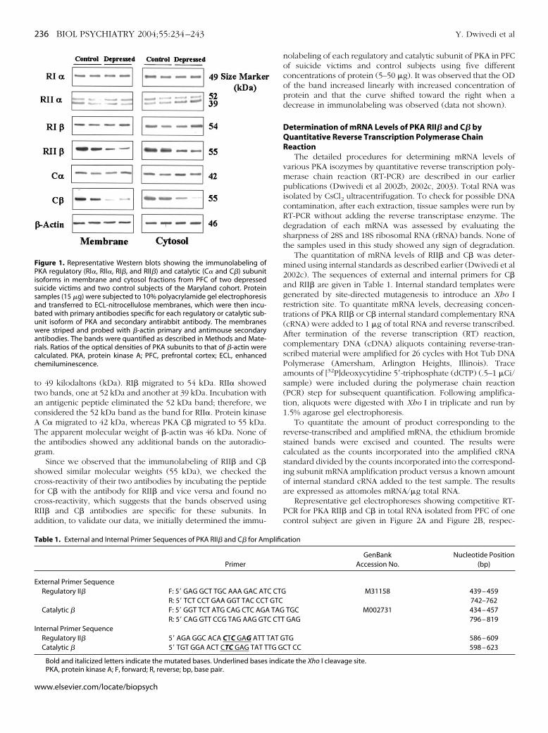
to 49 kilodaltons (kDa). RI� migrated to 54 kDa. RII� showedtwo bands, one at 52 kDa and another at 39 kDa. Incubation withan antigenic peptide eliminated the 52 kDa band; therefore, weconsidered the 52 kDa band as the band for RII�. Protein kinaseA C� migrated to 42 kDa, whereas PKA C� migrated to 55 kDa.The apparent molecular weight of �-actin was 46 kDa. None ofthe antibodies showed any additional bands on the autoradio-gram.
Since we observed that the immunolabeling of RII� and C�showed similar molecular weights (55 kDa), we checked thecross-reactivity of their two antibodies by incubating the peptidefor C� with the antibody for RII� and vice versa and found nocross-reactivity, which suggests that the bands observed usingRII� and C� antibodies are specific for these subunits. Inaddition, to validate our data, we initially determined the immu-
nolabeling of each regulatory and catalytic subunit of PKA in PFCof suicide victims and control subjects using five differentconcentrations of protein (5–50 �g). It was observed that the ODof the band increased linearly with increased concentration ofprotein and that the curve shifted toward the right when adecrease in immunolabeling was observed (data not shown).
Determination of mRNA Levels of PKA RII� and C� byQuantitative Reverse Transcription Polymerase ChainReaction
The detailed procedures for determining mRNA levels ofvarious PKA isozymes by quantitative reverse transcription poly-merase chain reaction (RT-PCR) are described in our earlierpublications (Dwivedi et al 2002b, 2002c, 2003). Total RNA wasisolated by CsCl2 ultracentrifugation. To check for possible DNAcontamination, after each extraction, tissue samples were run byRT-PCR without adding the reverse transcriptase enzyme. Thedegradation of each mRNA was assessed by evaluating thesharpness of 28S and 18S ribosomal RNA (rRNA) bands. None ofthe samples used in this study showed any sign of degradation.
The quantitation of mRNA levels of RII� and C� was deter-mined using internal standards as described earlier (Dwivedi et al2002c). The sequences of external and internal primers for C�and RII� are given in Table 1. Internal standard templates weregenerated by site-directed mutagenesis to introduce an Xho Irestriction site. To quantitate mRNA levels, decreasing concen-trations of PKA RII� or C� internal standard complementary RNA(cRNA) were added to 1 �g of total RNA and reverse transcribed.After termination of the reverse transcription (RT) reaction,complementary DNA (cDNA) aliquots containing reverse-tran-scribed material were amplified for 26 cycles with Hot Tub DNAPolymerase (Amersham, Arlington Heights, Illinois). Traceamounts of [32P]deoxycytidine 5�-triphosphate (dCTP) (.5–1 �Ci/sample) were included during the polymerase chain reaction(PCR) step for subsequent quantification. Following amplifica-tion, aliquots were digested with Xho I in triplicate and run by1.5% agarose gel electrophoresis.
To quantitate the amount of product corresponding to thereverse-transcribed and amplified mRNA, the ethidium bromidestained bands were excised and counted. The results werecalculated as the counts incorporated into the amplified cRNAstandard divided by the counts incorporated into the correspond-ing subunit mRNA amplification product versus a known amountof internal standard cRNA added to the test sample. The resultsare expressed as attomoles mRNA/�g total RNA.
Representative gel electrophoreses showing competitive RT-PCR for PKA RII� and C� in total RNA isolated from PFC of onecontrol subject are given in Figure 2A and Figure 2B, respec-
Figure 1. Representative Western blots showing the immunolabeling ofPKA regulatory (RI�, RII�, RI�, and RII�) and catalytic (C� and C�) subunitisoforms in membrane and cytosol fractions from PFC of two depressedsuicide victims and two control subjects of the Maryland cohort. Proteinsamples (15 �g) were subjected to 10% polyacrylamide gel electrophoresisand transferred to ECL-nitrocellulose membranes, which were then incu-bated with primary antibodies specific for each regulatory or catalytic sub-unit isoform of PKA and secondary antirabbit antibody. The membraneswere striped and probed with �-actin primary and antimouse secondaryantibodies. The bands were quantified as described in Methods and Mate-rials. Ratios of the optical densities of PKA subunits to that of �-actin werecalculated. PKA, protein kinase A; PFC, prefrontal cortex; ECL, enhancedchemiluminescence.
Table 1. External and Internal Primer Sequences of PKA RII� and C� for Amplification
PrimerGenBank
Accession No.Nucleotide Position
(bp)
External Primer SequenceRegulatory II� F: 5� GAG GCT TGC AAA GAC ATC CTG M31158 439 – 459
R: 5� TCT CCT GAA GGT TAC CCT GTC 742–762Catalytic � F: 5� GGT TCT ATG CAG CTC AGA TAG TGC M002731 434 – 457
R: 5� CAG GTT CCG TAG AAG GTC CTT GAG 796 – 819Internal Primer Sequence
Regulatory II� 5� AGA GGC ACA CTC GAG ATT TAT GTG 586 – 609Catalytic � 5� TGT GGA ACT CTC GAG TAT TTG GCT CC 598 – 623
Bold and italicized letters indicate the mutated bases. Underlined bases indicate the Xho I cleavage site.PKA, protein kinase A; F, forward; R, reverse; bp, base pair.
236 BIOL PSYCHIATRY 2004;55:234–243 Y. Dwivedi et al
www.elsevier.com/locate/biopsych
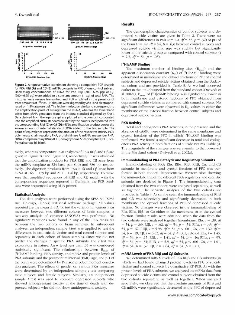
tively, whereas competitive PCR analyses of PKA RII� and C� aregiven in Figure 2C and Figure 2D, respectively. It was observedthat the amplification products for PKA RII� and C� arise fromthe mRNA template at 324 base pair (bp) and 386 bp, respec-tively. The digestion products from PKA RII� and C� arise fromcRNA at 165 � 159 bp and 210 � 176 bp, respectively. To makesure that amplified sequences of RII� and C� match with thecorresponding sequences reported in GenBank, the PCR prod-ucts were sequenced using M13 primer.
Statistical AnalysisThe data analyses were performed using the SPSS 8.0 (SPSS
Inc., Chicago, Illinois) statistical software package. All valuesreported are the mean SD. To test the variation in various PKAmeasures between two different cohorts of brain samples, atwo-way analysis of variance (ANOVA) was performed. Nosignificant variations were found in any of the PKA measuresbetween the two cohorts of brain samples. In subsequentanalyses, an independent sample t test was applied to test thedifferences in total suicide victims and total control subjects andseparately in each cohort of brain samples. Since we did notpredict the changes in specific PKA subunits, the t test wasexploratory in nature. An � level less than .05 was consideredstatistically significant. The relationships between Bmax of[3H]cAMP binding, PKA activity, and mRNA and protein levels ofPKA subunits and the postmortem interval (PMI), age, and pH ofthe brain were determined by Pearson product moment correla-tion analyses. The effects of gender on various PKA measureswere determined by an independent sample t test comparingmale subjects and female subjects. Similarly, an independentsample t test was used to compare depressed subjects whoshowed antidepressant toxicity at the time of death with de-pressed subjects who did not show antidepressant toxicity.
Results
The demographic characteristics of control subjects and de-pressed suicide victims are given in Table 2. There were nosignificant differences in PMI (t � 1.0, df � 53, p � .32) or pH ofthe brain (t � .49, df � 54, p � .63) between control subjects anddepressed suicide victims. Age was slightly but significantlylower in the suicide group as compared with control subjects (t� 2.3, df � 54, p � .03).
[3H]cAMP BindingThe maximum number of binding sites (Bmax) and the
apparent dissociation constant (KD) of [3H]cAMP binding weredetermined in membrane and cytosol fractions of PFC of controlsubjects and depressed suicide victims obtained from the Budap-est cohort and are provided in Table 3. As we had observedearlier in the PFC obtained from the Maryland cohort (Dwivedi etal 2002a), Bmax of [3H]cAMP binding was significantly lower inboth membrane and cytosol fractions of PFC obtained fromdepressed suicide victims as compared with control subjects. Nosignificant differences were observed in KD values in either themembrane or the cytosol fraction between control subjects anddepressed suicide victims.
PKA ActivityTotal and endogenous PKA activities, in the presence and the
absence of cAMP, were determined in the same membrane andcytosol fractions of the PFC in which [3H]cAMP binding wasperformed. We found a significant decrease in total and endog-enous PKA activity in both fractions of suicide victims (Table 3).The magnitude of the changes was very similar to that observedin the Maryland cohort (Dwivedi et al 2002a).
Immunolabeling of PKA Catalytic and Regulatory SubunitsImmunolabeling of PKA RI�, RII�, RI�, RII�, C�, and C�
subunits in membrane and cytosol fractions of PFC was per-formed in both cohorts. Representative Western blots showingthe immunolabeling of the different PKA regulatory and catalyticsubunits are depicted in Figure 1. The immunolabeling dataobtained from the two cohorts were analyzed separately, as wellas together. The separate analyses of the two cohorts areprovided in Table 4. As can be seen, the immunolabeling of RII�and C� was selectively and significantly decreased in bothmembrane and cytosol fractions of PFC of depressed suicidevictims. No changes were observed in the immunolabeling ofRI�, RII�, RI�, or C� either in the membrane or in the cytosolfraction. Similar results were obtained when the data from thetwo cohorts were analyzed together (membrane: RI�, t � .39, df� 54, p � .69; RI�, t � .62, df � 54, p � .54; RII�, t � .43, df �54, p � .67; RII�, t � 5.98, df � 54, p .001; C�, t � 1.32, df �54, p � .19; C�, t � 6.02, df � 54, p .001; cytosol: RI�, t � 1.45,df � 54, p � .15; RI�, t � 1.41, df � 54, p � .16; RII�, t � .95,df � 54, p � .34; RII�, t � 5.9, df � 54, p .001; C�, t � 1.01,df � 54, p � .32; C�, t � 7.64, df � 54, p .001).
mRNA Levels of PKA RII� and C� SubunitsWe determined mRNA levels of PKA RII� and C� subunits (in
which we had found changed protein levels) in PFC of suicidevictims and control subjects by quantitative RT-PCR. As with theprotein levels of PKA subunits, we analyzed the mRNA data fromdepressed suicide victims and control subjects obtained from thetwo cohorts separately, as well as together. When analyzedseparately, we observed that the absolute amounts of RII� andC� mRNA were significantly decreased in the PFC of depressed
Figure 2. A representative experiment showing a competitive PCR analysisfor PKA RII� (A) and C� (B) mRNA contents in PFC of one control subject.Decreasing concentrations of cRNA for PKA RII� (200�6.25 pg) or C�(200�6.25 pg) were added to a constant amount (1 �g) of total RNA. Themixtures were reverse transcribed and PCR amplified in the presence oftrace amounts of [32P]dCTP; aliquots were digested by Xho I and electropho-resed on 1.5% agarose gel. The higher molecular size band corresponds tothe amplification product arising from the mRNA, whereas the lower bandarises from cRNA generated from the internal standard digested by Xho I.Data derived from the agarose gel are plotted as the counts incorporatedinto the amplified cRNA standard divided by the counts incorporated intothe corresponding RII� (C) or C� (D) mRNA amplification product versus theknown amount of internal standard cRNA added to the test sample. Thepoint of equivalence represents the amount of the respective mRNA. PCR,polymerase chain reaction; PKA, protein kinase A; mRNA, messenger RNA;cRNA, complementary RNA; dCTP, deoxycytidine 5�-triphosphate; PFC, pre-frontal cortex; bl, blank.
Y. Dwivedi et al BIOL PSYCHIATRY 2004;55:234–243 237
www.elsevier.com/locate/biopsych

Table 2. Characteristics of Depressed Suicide Victims and Control Subjects
GroupAge(y) Gender
PMI(h)
BrainpH Cause of Death
Drug Toxicity(At the Time of Death)
Suicide1a 22 Female 16 5.3 Drug overdose Propranolol2a 24 Male 7 5.6 GSW None3a 21 Male 17 6.1 GSW None4a 27 Male 24 6.4 GSW None5a 38 Male 24 6.3 Drug overdose Ethanol, Diphenhydramine6a 36 Female 10 6.5 GSW Butalbital, Diphenhydramine, Acetaminophen7a 41 Female 27 5.9 Drug overdose Amitriptyline, Desipramine, Diphenhydramine, Nortriptyline,
Pseudophedrine, Salicylate, Ethanol8a 44 Female 11 5.6 Drug overdose Nortriptyline9a 46 Female 16 6.1 Drug overdose Nortriptyline
10a 46 Female 21 5.3 Drug overdose Amitriptyline, Desipramine, Ethanol11a 53 Male 23 6.1 Jumped None12b 72 Female 4 5.9 Poisoning Barbiturate, Secondary-alcohol: 1%13b 45 Male 5 5.7 Poisoning Benzodiazepines � Barbiturate14b 48 Male 4 5.9 Poisoning Barbiturate � Benodiazepine � Secondary-alcohol15b 47 Male 6 5.8 Hanging NA16b 66 Male 6 5.9 Hanging Secondary-alcohol17b 42 Male 3 6.0 Hanging NA18b 28 Female 6 6.0 Poisoning Meprobamate, Benzodiazepine, Propranolol, Secondary-alcohol19b 60 Male 3.5 6.5 Hanging Secondary-alcohol20b 44 Male 4 6.4 Hanging Secondary-alcohol21b 77 Male 4 5.9 Hanging NA22b 36 Male 6 6.6 Hanging NA23b 52 Male 3 6.8 Hanging Secondary-alcohol24b 49 Male 6 6.7 Hanging Secondary-alcohol25b 45 Female 6 7.0 Poisoning Clonazepam, Metoprolol, Meprobamate26b 49 Male 5 6.2 Hanging NA27b 43 Male 4 6.6 Hanging Secondary-alcohol28b 42 Male 4 6.7 Hanging NA
Mean 44 9 Female/19 Male 9.7 6.14SD 13 7.7 .44
Controls1a 22 Male 19 6.2 GSW None2a 63 Female 30 5.7 Ovarian cancer None3a 31 Male 8 5.6 GSW None4a 33 Male 15 6.0 GSW Acetaminophen5a 37 Male 5 6.6 ASCVD None6a 65 Female 23 5.6 ASCVD None7a 38 Male 16 5.8 Lung sarcoidosis None8a 40 Female 7 6.5 ASCVD None9a 23 Male 15 6.7 GSW None
10a 37 Male 9.5 6.1 ASCVD None11a 42 Female 23 6.2 Pneumonia None12b 64 Female 4 6.0 Myocardial infarction None13b 52 Male 4 6.2 Myocardial infarction None14b 33 Female 3 6.2 Myocardial infarction None15b 60 Female 3 6.0 Circulatory failure None16b 76 Female 3 5.98 Accident None17b 50 Male NA 6.3 Acute heart failure None18b 64 Male 1.5 6.4 Myocardial infarction None19b 78 Male 1.5 6.5 Myocardial infarction None20b 84 Male 1.5 6.3 Myocardial infarction None21b 78 Female 1.5 6.2 Acute heart failure None22b 65 Male 1 6.5 Acute heart failure None23b 58 Female 1 5.8 Acute heart failure None24b 51 Male 1 6.4 Acute heart failure None25b 74 Male 3 6.6 Acute heart failure None26b 71 Male 2 6.6 Acute heart failure None27b 52 Male 1.5 5.8 Acute heart failure None28b 80 Female 1 6.5 Acute heart failure None
Mean 54 11 Female/17 Male 7.5 6.19SD 18 8.3 .32
PMI, postmortem interval; GSW, gunshot wound; NA, not available; ASCVD, atherosclerotic cardiovascular disease; SD, standard deviation.aMaryland cohort.bBudapest cohort.
238 BIOL PSYCHIATRY 2004;55:234–243 Y. Dwivedi et al
www.elsevier.com/locate/biopsych

suicide victims as compared with control subjects in both cohortswith a similar magnitude (Table 5). When analyzed together,decreases of a similar extent were observed in mRNA levels ofRII� and C� in depressed suicide victims as compared withcontrol subjects (RII�, t � 5.73, df � 54; p .001; C�, t � 5.12,df � 54, p .001).
Correlations Between [3H]cAMP Binding and PKA Activity andBetween mRNA and Protein Levels of PKA RII� and C�Subunits
Since PKA catalytic activity depends on the binding of cAMPto regulatory subunits and the dissociation of catalytic subunitsfrom regulatory subunits, we examined the relationship ofcAMP-dependent PKA activity and Bmax of [3H]cAMP binding incontrol and suicide victims. We found a significant relationship
between PKA activity and [3H]cAMP binding in both membrane(r � .44, p � .009) and cytosol (r � .53, p � .001) fractions.
Similarly, significant correlations between mRNA and proteinlevels of RII� and C� were observed in both membrane andcytosol fractions (membrane: RII�, r � .29, p � .025; C�, r � .37,p � .005; cytosol: RII�, r � .39, p � .002; C�, r � .32, p � .016).
Effects of Confounding VariablesThe effects of potential confounding variables, namely age,
gender, PMI, and method of suicide, were evaluated with respectto mRNA and protein levels of PKA RII� and C� subunits, inwhich we had found differences between control subjects andsuicide victims. We found no significant effects of age on proteinlevels of RII� or C� either in the membrane (RII�, r � .20, p �.14; C�, r � .23, p � .09) or in the cytosol (RII�, r � .17, p � .20;C�, r � .19, p � .15) fraction. Similarly, age had no significant
Table 3. [3H]cAMP Binding and Catalytic Activity of PKA in PFC of Depressed Suicide Victims and Control SubjectsObtained from Budapest Cohort
Measures
Membrane Cytosol
Controls(n � 17)
Depressed Suicide(n � 17)
Controls(n � 17)
Depressed Suicide(n � 17)
[3H]cAMP Bindinga
Bmax 142.76 39.20 85.11 25.17d 268.23 91.28 174.25 48.66d
KDb .77 .38 .69 .17 .67 .19 .71 .18
PKA Activityc
Endogenous 123.41 32.66 82.17 28.14d 323.82 95.62 238.70 66.54e
Total 375.35 80.63 250.11 64.28d 803.88 194.83 490.52 130.16d
Values are the mean standard deviation (SD).cAMP, cyclic adenosine monophosphate; PKA, protein kinase A; PFC, prefrontal cortex; Bmax, maximum number of
binding sites; KD, apparent dissociation constant.afmol/mg protein.bnM.cpmol/min/mg protein.dp .001.ep � .005.
Table 4. Immunolabeling of PKA Subunits in PFC of Depressed Suicide Victims and Control Subjects in TwoDifferent Cohorts of Brain Samples
Immunolabelinga
Membrane Cytosol
Controls Depressed Suicide Controls Depressed Suicide
Budapest Cohort n � 17 n � 17 n � 17 n � 17RI� .88 .19 .83 .18 .85 .19 .76 .20RII� 1.03 .24 1.08 .21 .88 .18 .81 .14RI� .88 .25 .86 .18 1.05 .23 .96 .26RII� 1.40 .30 .88 .35b 1.34 .26 .85 .31b
C� 1.10 .20 1.06 .17 .98 .18 .96 .17C� 1.21 .19 .78 .26b 1.4 .28 .84 .25b
Maryland Cohort n � 11 n � 11 n � 11 n � 11RI� .90 .17 .92 .23 .80 .20 .75 .14RII� 1.20 .32 1.19 .17 .80 .14 .79 .14RI� .90 .18 .84 .20 1.10 .29 1.00 .18RII� 1.30 .35 .81 .27c 1.28 .38 .81 .28d
C� .97 .18 .85 .17 1.20 .30 1.08 .18C� 1.30 .35 .83 .34e 1.24 .14 .81 .26b
Values are the mean standard deviation (SD).PKA, protein kinase A; PFC, prefrontal cortex; R, regulatory; C, catalytic.aRatio to �-actin.bp .001.cp � .001.dp � .004.ep � .005.
Y. Dwivedi et al BIOL PSYCHIATRY 2004;55:234–243 239
www.elsevier.com/locate/biopsych

effects on mRNA levels of RII� (r � .03, p � .79) or C� (r � .14,p � .27). Since the PMI of the Budapest cohort was lower (3.33 1.59 hours) than that of the Maryland cohort (16.65 7.31hours), the effect of PMI on various measures of PKA in bothcohorts was analyzed separately. We did not find significanteffects of PMI on protein or mRNA levels of RII� or C� either inthe Maryland cohort (protein: membrane, RII�, r � .39, p � .07;C�, r � .01, p � .96; cytosol, RII�, r � .03, p � .89; C�, r � .05,p � .83; mRNA: RII�, r � .26, p � .25; C�, r � .38, p � .08) orin the Budapest cohort (protein: membrane, RII�, r � .13, p �.48; C�, r � .07, p � .69; cytosol, RII�, r � .08, p � .65; C�, r �.018, p � .63; mRNA: RII�, r � .16, p � .37; C�, r � .3, p � .06).Furthermore, we found no significant effects of pH of the brainon mRNA levels of either RII� (r � .2, p � .14) or C� (r � .06,p � .64). In addition, these confounding variables had nosignificant effects on [3H]cAMP binding or on endogenous ortotal PKA activity either in the membrane ([3H]cAMP binding:age, r � .21, p � .23; PMI, r � .13, p � .47; endogenous PKAactivity: age, r � .07, p � .69; PMI, r � .28, p � .10; total PKAactivity: age, r � .09, p � .61; PMI, r � .30, p � .08) or in thecytosol ([3H]cAMP binding: age, r � .27, p � .12; PMI, r � .05, p� .77; endogenous PKA activity: age, r � .23, p � .17; PMI, r �.24, p � .17; total PKA activity: age, r � .26, p � .13; PMI, r � .30,p � .08) fraction. We did not find a significant correlationbetween any of the above-described confounding variables andprotein levels of RI�, RII�, RI�, or C� either in membrane or incytosol (data not shown).
There were 12 male subjects and 10 female subjects in theMaryland cohort and 24 male subjects and 10 female subjects inthe Budapest cohort. Comparison studies showed no significantdifferences in any of the PKA measures between male subjectsand female subjects either in the Maryland cohort (protein levelsof RII�: membrane, t � .59, df � 20, p � .56; cytosol, t � .08, df� 20, p � .08; C�: membrane, t � 1.25, df � 20, p � .22; cytosol,t � 1.14, df � 20, p � .27; mRNA levels of RII�: t � .87, df � 20,p � .39; C�: t � .19, df � 20, p � .85) or in the Budapest cohort([3H]cAMP binding: membrane, t � .38, df � 32, p � .71; cytosol,t � .29, df � 32, p � .24; endogenous PKA activity: membrane,t � .45, df � 32, p � .66; cytosol, t � 1.48, df � 32, p � .15; totalPKA activity: membrane, t � .62, df � 32, p � .54; cytosol, t �1.63, df � 32, p � .11; protein levels of RII�: membrane, t � 1.53,df � 32, p � .14; cytosol, t � .46, df � 32, p � .65; C�:membrane, t � .46, df � 32, p � .65; cytosol, t � 1.61, df � 32,p � .12; mRNA levels of RII�: t � .38, df � 32, p � .71; C�: t �.99, df � 32, p � .33).
To examine whether the method of suicide had any effect onthe measures of PKA in which we found changes, we comparedsuicide victims who died by violent means (n � 16) with thosewho died by drug overdose/poisoning (n � 11). No significantdifferences in [3H]cAMP binding (membrane, t � .24, df � 15, p� .81; cytosol, t � .69, df � 15, p � .50), endogenous PKAactivity (membrane, t � .09, df � 15, p � .93; cytosol, t � .16, df� 15, p � .88), total PKA activity (membrane, t � 1.93, df � 15,p � .07; cytosol, t � .68, df � 15, p � .51), protein levels of RII�(membrane: t � .29, df � 25, p � .78; cytosol: t � .36, df � 25,p � .72) protein levels of C� (membrane: t � .73, df � 25, p �.47; cytosol: t � .83, df � 25, p � .41), or mRNA levels of RII� (t� .14, df � 25, p � .89) or C� (t � .71, df � 25, p � .48) wereobserved between these two groups.
Those depressed suicide victims who showed the presence ofantidepressant(s) in plasma at the time of death had been treatedwith antidepressant(s) at least 1 month before death. We exam-ined whether antidepressant treatment had any significant effectson the various measures of PKA. Out of 28 depressed suicidevictims, 4 showed antidepressant toxicology (all in the Marylandcohort), whereas in 6 cases (cases 15, 17, 21, 22, 26, and 28) asindicated in Table 2, the antidepressant treatment informationwas not available. No significant differences were observed inprotein levels of RII� (membrane: t � 1.27, df � 24, p � .21;cytosol: t � .05, df � 24, p � .96) or C� (membrane: t � .84, df� 24, p � .41; cytosol: t � 1.8, df � 24, p � .25) or mRNA levelsof RII� (t � .66, df � 24, p � .51) or C� (t � .86, df � 24, p �.39) between those depressed suicide victims who were treatedwith antidepressants and those who were not treated withantidepressants.
In summary, we observed significant and selective decreasesin [3H]cAMP binding and PKA activity, as well as in protein andmRNA expression of RII� and C� in depressed suicide victims.These decreases were similar across the two different braincohorts. Although the demographic characteristics of depressedsuicide victims were slightly different in these two cohorts,because we did not find any significant correlations between thevarious demographic characteristics and PKA measures, ourfindings clearly demonstrate that the changes in PKA are notsecondary to the demographic characteristics of the two cohortsbut are related to depression.
Discussion
In a previous study, we reported that [3H]cAMP binding andtotal and endogenous catalytic PKA activity were significantlyreduced in the PFC of suicide victims with major depression(Dwivedi et al 2002a). In the present study, we replicated thesefindings in a different cohort of brain samples obtained from theLenhossek Human Brain Program, Semmelweiss University,Budapest, Hungary. The magnitudes of the decreases in[3H]cAMP binding and in total and endogenous catalytic activityof PKA are similar to those observed previously. These resultssuggest that our finding of abnormalities in [3H]cAMP bindingand PKA activity in depressed suicide victims is consistent acrossdifferent cohorts of brain samples.
An interesting observation of our present and previous studiesis that the reductions in [3H]cAMP binding and in total andendogenous catalytic activity of PKA occur in both membraneand cytosol fractions of the PFC. On activation by cAMP, PKAtranslocates from the cytosol to the membrane. It has beenreported that the translocation of PKA occurs through differentsubcellular compartments, with consequent phosphorylation of
Table 5. Messenger RNA Levels of PKA Subunits in Total RNA Isolatedfrom PFC of Depressed Suicide Victims and Control Subjects Obtainedfrom Two Different Cohorts
mRNA Levelsa Controls Depressed Suicide
Budapest Cohort n � 17 n � 17RII� 170 42 106 38b
C� 125 28 84 29b
Maryland Cohort n � 11 n � 11RII� 167 37 116 33c
C� 148 40 99 28d
Values are the mean standard deviation (SD).PKA, protein kinase A; PFC, prefrontal cortex; mRNA, messenger RNA.aAttomoles/�g total RNA.bp .001.cp � .003.dp � .004.
240 BIOL PSYCHIATRY 2004;55:234–243 Y. Dwivedi et al
www.elsevier.com/locate/biopsych

selective substrates (Dell’Acqua and Scott 1997). In our study, wedid not find such a translocation, since lower [3H]cAMP binding,as well as lower PKA activity, were seen in both membrane andcytosol fractions. This suggests that abnormalities in PKA are notspecific to certain compartments but rather are generalized.Furthermore, our observations of a reduction in [3H]cAMP bind-ing but no apparent change in [3H]cAMP binding affinity indicatethe possibility of a lesser abundance of regulatory subunits. It hasbeen shown that one of the functions of the regulatory subunitsis to protect the catalytic subunits from proteolytic degradationby binding and keeping them in the holoenzyme state (Steinbergand Agard 1981). Since we observed significantly reduced cata-lytic activity of PKA in the PFC of suicide victims and since thisdecrease was present even in the absence of cAMP (endoge-nous), it is highly possible that this decrease could be related toregulation at the transcriptional and/or translational level. Wetherefore investigated the expression of not only the differentPKA regulatory subunits but also the catalytic subunits in PFC ofdepressed suicide victims from both cohorts. We found thatprotein levels of RII� and C� were selectively reduced in boththe membrane and the cytosol fraction of PFC of depressedsuicide victims with a similar magnitude in both cohorts. Toexamine whether these changes occur at the transcription level,we determined the mRNA levels of these two specific regulatoryand catalytic subunits. As we found with protein levels, themRNA levels of RII� and C� were significantly decreased in thePFC of depressed suicide victims in both cohorts. The changes inprotein levels of RII� and C� were significantly correlated withtheir respective mRNA levels. Our results thus suggest that thedecreases in [3H]cAMP binding and PKA activity could be relatedto decreases in these specific regulatory and catalytic subunits.
Similar to our findings, other studies found decreased[3H]cAMP binding (Manier et al 2000) and lower �-adrenergicreceptor-stimulated PKA activity (Shelton et al 1996; 1999) infibroblasts of depressed patients. Interestingly, it has been shownthat chronic treatment with imipramine, tranylcypromine, orelectroconvulsive shock causes the translocation of PKA in ratbrain (Nestler et al 1989). In addition, long-term treatment withserotonin (5-HT) or norepinephrine (NE) reuptake inhibitorsincreases the binding of cAMP to 52-kDa RII subunits of PKA(reviewed in Popoli et al 2000). These findings thus suggest thatin depression, functions of PKA may be reduced and antidepres-sants may be alleviating depressive symptoms by increasing thefunctioning of PKA. In this regard, we did not find any effects ofantidepressant treatment on the expression of C� or RII� sub-units; we could not draw any meaningful conclusions becausethe number of subjects in our total study group who were treatedwith antidepressants was too small (n � 4).
Earlier, Fields et al (1999) reported increased catalytic activityof PKA, whereas Rahman et al (1997) reported lower [3H]cAMPbinding in postmortem brain of bipolar patients. Very recently,increased protein levels of cytosolic C� and RII� were reportedin postmortem brain of the same bipolar patients in which adecrease in [3H]cAMP binding was reported (Chang et al 2003).The finding of decreased [3H]cAMP binding is contrary to that ofincreased levels of RII� in these patients; however, as discussedby Chang et al (2003), since no saturation experiments wereperformed for [3H]cAMP binding, the observed decrease couldreflect either changes in net cyclic AMP binding affinity or in theabundance of PKA regulatory subunits. Since we found theopposite results in the expression of RII� and a decrease in C�instead of increased C� as reported in bipolar patients, it is quite
possible that PKA may be differentially regulated in depressionand bipolar disorder.
The mechanism responsible for the selectively altered expres-sion of PKA subunits is not clear at the present time; however, ithas been demonstrated in rat Sertoli cells, activation of the cAMPpathway stimulates mRNA levels of RI�, RII�, C�, and RII�, theincrease in RII� being the greatest (Landmark et al 1993; Oyen etal 1988; Tasken et al 1991); however, prolonged stimulation withcAMP results in a decrease in RII� mRNA (Oyen et al 1988). Inthe postmortem brain of suicide victims, it has been shown thatthe expression of GS� is increased and of Gi� is decreased(Dwivedi et al 2002b; Pacheco et al 1996), which suggests greaterstimulation of the cAMP pathway. It is quite possible that thissustained stimulation may cause an adaptive change in expres-sion of the RII� subunit. Another possible explanation of re-duced expression of the RII� subunit could be related to proteinkinase C (PKC). It has been shown that activation of PKC inhibitscAMP induction of RII� mRNA and that this inhibition persistseven after PKC down-regulation (Tasken et al 1992). Interest-ingly, alterations in PKC have been reported in postmortem brainof suicide victims (Pandey et al 1997). The explanation of theselective decrease in the C� subunit appears to be complex. Ithas been shown that regulatory subunits associate with catalyticsubunits and protect catalytic subunits from degradation byproteolytic enzymes (Steinberg and Agard 1981). Furthermore, inbrain tissues of RII�-mutant mice, there is a profound decrease inC� and C� subunit levels due to increased degradation (Brandonet al 1998); however, we found decreased expression of only theC� subunit, and this decrease was present at both the transcrip-tional and the translocational level, eliminating the possibility ofany degradation. It appears that additional factors or mechanismsmay be involved in the regulation of the C� gene in postmortembrain of depressed suicide victims.
At the functional level, PKA is involved in myriads of physi-ologic functions in the brain, including neurotransmitter synthe-sis and release, gene expression, synaptic plasticity, memory,and cell growth and differentiation. The major mechanism ofPKA-mediated function is through the phosphorylation of spe-cific substrates, which include cAMP response element bindingprotein (CREB), nuclear receptors, and high-mobility group-containing proteins, thus influencing their dimerization or DNAbinding properties (Fimia and Sassone-Corsi 2001). Recent stud-ies demonstrate that CREB could be involved in depressive andsuicidal behavior (Dwivedi et al 2003; D’Sa and Duman 2002).Another important aspect of PKA is its involvement in thecross-talk between different signaling mechanisms. For example,PKA interferes at various levels with other signaling pathways,including inactivation of phospholipase C� (Liu and Simon1996); phosphorylation of IP3 receptors, thereby modulatingCa2� influx (Bugrim 1999); phosphorylation of G proteins, withconsequent decrease in phosphoinositide hydrolysis and Ca2�
release (Wen et al 1992); and phosphorylation of calcium-calmodulin kinase (Matsushita and Nairn 1999). Furthermore,PKA participates in neurite outgrowth (Song and Poo 1999),neuronal differentiation (Liesi et al 1983), and cell survival (Rydeland Greene 1988; Li et al 2000). The specific functions of RII�and C� are not clearly known; however, tissue distributionstudies suggest that RII� is predominantly expressed in brain,adrenal, and adipose tissues and is the principal mediator ofcAMP activity in the mammalian CNS (Sarkar et al 1984), whereasC� is expressed primarily in the brain (Uhler et al 1986). Ludviget al (1990) showed that RII� immunolabeling is associated withpostsynaptic structures, suggesting that this subunit is involved in
Y. Dwivedi et al BIOL PSYCHIATRY 2004;55:234–243 241
www.elsevier.com/locate/biopsych

several postsynaptic neuronal functions. Also, many studies havedemonstrated that RII� and C� subunits may be specificallyinvolved in neuronal and behavioral functions. For example,Constantinescu et al (2002) showed that the RII� subunit cantranslocate to the nucleus and induce CREB phosphorylation.Additionally, RII�-mutant mice exhibit defective motor behavior(Brandon et al 1998) and C�-mutant mice show impaired hip-pocampal plasticity (Qi et al 1996). Whether these abnormalitiesare relevant to depression is not clear at the present; however,given the significance of PKA in many biological actions in thebrain, together with emerging studies demonstrating specificroles for its RII� and C� subunits in physiologic and behavioralmanifestations, our replicated observations of decreased catalyticand selectively decreased regulatory activities and expression ofRII� and C� in PFC of depressed suicide victims suggest thatabnormalities in PKA may be of critical importance in thepathophysiology of depressive behavior.
Supported by a Career Development Award (KO1 MH 01836)from the National Institute of Mental Health (NIMH) and aYoung Investigator Award from the American Foundation forSuicide Prevention to Dr. Yogesh Dwivedi and by RO1 MH48153from the NIMH to Dr. G.N. Pandey. We acknowledge with thanksthe cooperation of John Smialek, M.D., Chief Medical Examiner,and Dennis Chute, M.D., Assistant Medical Examiner, in thecollection of brain samples, Ms. Terri U’Prichard for performingthe psychological autopsies, Boris Lapidus, M.D., for the dissec-tions, and Ms. Barbara Brown and Ms. Miljana Petkovich fororganizing the brain tissues.
Brandon EP, Logue SF, Adams MR, Qi M, Sullivan SP, Matsumoto AM, et al(1998): Defective motor behavior and neural gene expression in RII�-protein kinase A mutant mice. J Neurosci 18:3639 –3649.
Bugrim AE (1999): Regulation of Ca2� release by cAMP-dependent proteinkinase. A mechanism for agonist-specific calcium signaling? Cell Calcium25:219 –226.
Chang A, Li PP, Warsh JJ (2003): Altered cAMP-dependent protein kinase Asubunit immunolabeling in postmortem brain from patients with bipo-lar affective disorder. J Neurochem 84:781–791.
Constantinescu A, Gordon AS, Diamond I (2002): cAMP-dependent proteinkinase types I and II differentially regulate cAMP response element-mediated gene expression. J Biol Chem 277:18810 –18816.
Cowburn RF, Marcusson JO, Eriksson A, Wiehager B, O’Neill C (1994): Ade-nylyl cyclase activity and G-protein subunit levels in postmortem frontalcortex of suicide victims. Brain Res 633:297–304.
Dell’Acqua ML, Scott JD (1997): Protein kinase A anchoring. J Biol Chem272:12881–12884.
D’Sa C, Duman RS (2002): Antidepressants and neuroplasticity. Bipolar Dis-ord 4:183.
Dwivedi Y, Conley RC, Roberts RC, Tamminga CA, Pandey GN (2002a):[3H]CyclicAMP binding sites and PKA activity in the prefrontal cortex ofsuicide victims. Am J Psychiatry 159:66 –73.
Dwivedi Y, Rao JS, Rizavi HS, Kotawski J, Conley RR, Roberts RC, et al (2003):Abnormal expression and functional characteristics of cyclic adenosinemonophosphate response element binding protein in postmortembrain of suicide subjects. Arch Gen Psychiatry 60:273–282.
Dwivedi Y, Rizavi HS, Conley RR, Roberts RC, Tamminga CA, Pandey GN(2002b): mRNA and protein expression of selective alpha subunits of Gproteins are abnormal in prefrontal cortex of suicide victims. Neuropsy-chopharmacology 27:499 –517.
Dwivedi Y, Rizavi HS, Pandey GN (2002c): Differential effects of haloperidoland clozapine on [3H]cAMP binding, protein kinase A (PKA) activity, andmRNA and protein expression of selective regulatory and catalytic sub-unit isoforms of PKA in rat brain. J Pharmacol Exp Ther 301:197–209.
Fields A, Li PP, Kish SJ, Warsh J (1999): Increased cyclic AMP-dependentprotein kinase activity in postmortem brain from patients with bipolaraffective disorder. J Neurochem 73:1704 –1710.
Fimia GM, Sassone-Corsi P (2001): Cyclic AMP signaling. J Cell Sci 114:1971–1972.
Houge G, Vintermyr OK, Doskeland SO (1990): The expression of cAMP-dependent protein kinase subunits in primary rat hepatocyte cultures.Cyclic AMP downregulates its own effector system by decreasing theamount of catalytic subunit and increasing the mRNAs for the inhibitory(R) subunits of cAMP-dependent protein kinase. Mol Endocrinol 4:481–488.
Knutsen HK, Tasken KA, Eskild W, Jahnsen T, Hansson V (1991): Adenosine3�,5�-monophosphate-dependent stablization of messenger ribonu-cleic acids (mRNAs) for protein kinase A (PKA) subunits in rat Sertoli cells:Rapid degradation of mRNAs for PKA subunits is dependent on ongoingRNA and protein synthesis. Endocrinology 129:2496 –2502.
Landmark BF, Oyen O, Skalhegg BS, Fauske B, Jahnsen T, Hansson V (1993):Cellular localization and age-dependent changes of the regulatory sub-units of cAMP-dependent protein kinase in rat testis. J Reprod Fertil99:323–334.
Li M, Wang X, Meintzer MK, Laessig T, Birnbaum MJ, Heidenreich KA (2000):Cyclic AMP promotes neuronal surival by phosphorylation of glycogensynthase kinase 3�. Mol Cell Biol 20:9356 –9363.
Liesi P, Rechardt L, Wartiovaara J (1983): Nerve growth factor induces adren-ergic neuronal differentiation in F9 teratocarcinoma cells. Nature306:265–267.
Liu M, Simon MI (1996): Regulation by cAMP-dependent protein kinease of aG-protein-mediated phospholipase C. Nature 382:83–87.
Lowther S, Katona CLE, Crompton MR, Horton RW (1997): Brain [3H]cAMPbinding sites are unaltered in depressed suicides, but decreased byantidepressants. Brain Res 758:223–228.
Ludvig N, Ribak CE, Scott JD, Rubin CS (1990): Immunocytochemical local-ization of the neural-specific regulatory subunit of the type II cyclicAMP-dependent protein kinase to postsynaptic structures in the ratbrain. Brain Res 520:90 –102.
Manier DH, Shelton RC, Ellis TC, Peterson CS, Eiring A, Sulser F (2000): Humanfibroblasts as a relevant model to study signal transduction in affectivedisorders. J Affect Disord 61:51–58.
Matsushita M, Nairn AC (1999): Inhibition of the Ca2�/calmodulin-depen-dent protein kinase I cascade by cAMP-dependent protein kinase. J BiolChem 274:10086 –10093.
McPherson GA (1985): Analysis of radioligand binding experiments: A col-lection of computer programs for the IBM PC. J Pharmacol Methods14:213–228.
Nestler EJ, Greengard P (1994): Protein phosphorylation and the regulationof neuronal function. In: Siegel GH, Albers RW, Agranoff BW, Molinoff P,editors. Basic Neurochemistry: Molecular, Cellular, and Medical Aspects.Boston: Little, Brown, and Company, 449 –474.
Nestler EJ, Terwilliger RZ, Duman RS (1989): Chronic antidepressant admin-istration alters the subcellular distribution of cyclic AMP-dependent pro-tein kinase in rat frontal cortex. J Neurochem 53:1644 –1647.
Odagaki Y, Garcia-Sevilla JA, Huguelet P, La Harpe R, Koyama T, Guimon J(2001): Cyclic AMP-mediated signaling components are upregulated inthe prefrontal cortex of depressed suicide victims. Brain Res 898:224 –231.
Oyen O, Sandberg M, Eskild W, Levy FO, Knutsen G, Beebe S, et al (1988):Differential regulation of messenger ribonucleic acids for specific sub-units of cyclic adenosine 3, 5�-monophosphate (cAMP)-dependent pro-tein kinase by cAMP in rat Sertoli cells. Endocrinology 122:2658 –2666.
Pacheco MA, Stockmeier C, Meltzer HY, Overholser JC, Dilley GE, Jope RS(1996): Alterations in phosphoinositide signaling and G protein levels indepressed suicide brain. Brain Res 723:37–45.
Pandey GN, Dwivedi Y, Pandey SC, Conley RR, Roberts RC, Tamminga CA(1997): Protein kinase C in the postmortem brain of teenage suicidevictims. Neurosci Lett 228:111–114.
Popoli M, Brunello N, Perez J, Racagni G (2000): Second messenger-regu-lated protein kinases in the brain: Their functional role and the action ofantidepressant drugs. J Neurochem 74:21–33.
Qi M, Zhuo M, Skalhegg BS, Brandon EP, Kandel ER, McKnight GS, et al (1996):Impaired hippocampal plasticity in mice lacking the C� catalytic subunitof cAMP-dependent protein kinase. Proc Natl Acad Sci U S A 93:1571–1576.
Rahman S, Li PP, Young LT, Kofman O, Kish SJ, Warsh JJ (1997): Reduced[3H]cyclic AMP binding in postmortem brain from subjects with bipolaraffective disorder. J Neurochem 68:297–304.
242 BIOL PSYCHIATRY 2004;55:234–243 Y. Dwivedi et al
www.elsevier.com/locate/biopsych

Reiach JS, Li, PP, Warsh JJ, Kish SJ, Young LT (1999): Reduced adenylyl cyclaseimmunolabeling and activity in postmortem temporal cortex of de-pressed suicide victims. J Affect Disord 56:141–151.
Rydel RE, Greene LA (1988): CAMP analogs promote survival and neuriteoutgrowth in cultures of rat sympathetic and sensory neurons inde-pendently of nerve growth factor. Proc Natl Acad Sci U S A 85:1257–1261.
Salzman S, Clayton P, Winokur G (1983): Diagnostic Evaluation After Death(DEAD). Rockville, MD: National Institute of Mental Health, NeuroscienceResearch Branch.
Sarkar D, Erlichman J, Rubin CS (1984): Identification of a calmodulin-bind-ing protein that co-purifies with the regulatory subunit of brain proteinkinase II. J Biol Chem 259:9840 –9846.
Shelton RC, Manier DH, Peterson CS, Ellis TC, Sulser F (1999): Cyclic AMP-dependent protein kinase in subtypes of major depression and normalvolunteers. Int J Neuropsychopharmacol 2:187–192.
Shelton RC, Mainer DH, Sulser F (1996): CAMP-dependent protein kinaseactivity in major depression. Am J Psychiatry 153:1037–1042.
Skålhegg BS, Tasken K (2000): Specificity in the cAMP/PKA signaling path-way. Differential expression, regulation, and subcellular localization ofsubunits of PKA. Front Biosci 5:D678 –D693.
Song HJ, Poo MM (1999): Signal transduction underlying growth cone guid-ance by diffusible factors. Curr Opin Neurobiol 9:355–363.
Spitzer RL, Williams JBW, Gibbon M, First MB (1995): Structured Clinical Inter-view for DSM-IV (SCID). New York: New York State Psychiatric Institute,Biometrics Research.
Steinberg RA, Agard DA (1981): Turnover of regulatory subunit of cyclicAMP-dependent protein kinase in S49 mouse lymphoma cells. Regula-tion by catalytic subunit and analogs of cyclic AMP. J Biol Chem256:10731–10734.
Tasken KA, Knutsen HK, Attramadal H, Tasken K, Jahnsen T, Hanson, et al(1991): Different mechanisms are involved in cAMP-mediated inductionof mRNAs for subunits of cAMP-dependent protein kinases. Mol Endocri-nol 5:21–28.
Tasken KA, Knutsen HK, Eikvar L, Tasken K, Eskild W, Jahnsen T, et al (1992):Protein kinase C activation by 12-O-tetradecanoylphorbol 13-acetatemodulates messenger ribonucleic acid levels for two of the regulatorysubunits of 3�,5�-cyclic adenosine monophosphate-dependent proteinkinases (RII alpha and RI alpha) via multiple and distinct mechanisms.Endocrinology 130:1271–1280.
Uhler MD, Chrivia JC, McKnight GS (1986): Evidence for a second isoform ofthe catalytic subunit of cAMP-dependent protein kinase. J Biol Chem261:15360 –15363.
Wen Y, Anwer K, Singh SP, Sanborn BM (1992): Protein kinase-A inhibitsphospholipase-C activity and alters protein phosphorylation in rat myo-metrial plasma membranes. Endocrinology 131:13777–13782.
Y. Dwivedi et al BIOL PSYCHIATRY 2004;55:234–243 243
www.elsevier.com/locate/biopsych