Embed Size (px)
Citation preview
German Edition: DOI: 10.1002/ange.201508968Protein FoldingInternational Edition: DOI: 10.1002/anie.201508968
Microtubule-Binding R3 Fragment from Tau Self-Assembles into GiantMultistranded Amyloid RibbonsJozef Adamcik, Antoni S�nchez-Ferrer, Nadine Ait-Bouziad, Nicholas P. Reynolds,Hilal A. Lashuel,* and Raffaele Mezzenga*
Abstract: Tau protein and its fragments self-assemble intoamyloid fibrils in the presence of polyanions, such as heparin.By combining microscopy, scattering, and spectroscopy tech-niques, we studied the aggregation of the 26-mer Tau-derivedpeptide alone, Tau306–327, the third repeat fragment (R3) of themicrotubule-binding domain. We show that: i) the sole Tau306–
327 can self-assemble into amyloid fibrils without the need ofaggregation-promoting polyanions; ii) the resulting structuresconsist of surprisingly large, well-ordered 2D laminated flatribbons, with a log-normal distribution of the lateral width,reaching the unprecedented lateral size of 350 nm and/or 45individual protofilaments, that is, the largest amyloid laminatedstructures ever observed for Tau or any other amyloidogenicsequence. Our results provide insight into the moleculardeterminants of Tau aggregation and open new perspectivesin the understanding of the assembly of amyloid fibrils and b-sheet-based biomaterials.
The self-assembly of peptides and proteins, such as a-synuclein, Ab peptide, and Tau protein, into b-sheet-richfibrillar amyloid aggregates has been implicated in neuro-degeneration and the pathogenesis of several devastatingneurodegenerative disorders including ParkinsonÏs and Alz-heimerÏs diseases.[1, 2] Advances in understanding the mecha-nism of amyloid formation has inspired the design of
biomaterials with a wide-range of applications in medicineand biotechnology.[3]
Tau is a microtubule-binding protein and is the primaryconstituents of the neurofibrillary tangles found in the brainof AlzheimerÏs disease patients. Tau misfolding and aggrega-tion is also thought to play a central role in the pathogenesisof several other human neurodegenerative diseases, knowncollectively as “tauopathies”.[4] In vitro, the Tau protein doesnot aggregate spontaneously and adopts predominantlydisordered conformations, but undergoes rapid aggregationin the presence of polyanions, such as heparin.[5]
Previous experimental studies have shown that theaggregation of Tau protein is strongly associated with twoshort amino acid sequences: one located in the third repeatfragment (R3, that is, VQIVYKPVDLSKVTSKCGSLG-NIHHK) of the microtubule-binding domain of Tau,306VQIVYK311, and its homologous sequence, 275VQIINK280,in the second repeat fragment. Both of these fragments havebeen shown to be critical in nucleating the self-assembly andformation of b-sheet-rich fibrillar aggregates of Tau.[6] The X-ray crystallographic structure of the sequence VQIVYKprovided insight into the aggregation of Tau protein, showingpaired layers of parallel, in-register b-sheets similar to thoseof many other amyloids.[7] Interestingly, the aggregation of theR3 fragment containing the 306VQIVYK311 sequence, in thepresence of heparin, leads to the formation of fibrillarstructures that exhibit similar structural and morphologicalproperties as those assembled from the full-length Tauprotein.[8]
While investigating the self-assembly properties of the 26amino acid peptide fragment that comprises the R3 domain ofTau (Tau306-327), we observed that this peptide aggregates inthe absence of heparin and forms giant, flat, multistrandedamyloid ribbons generated from the lateral assembly of up to45 individual protofilaments. This unparalleled lateral growthof protofilaments leads to an effective 2D laminated mor-phology, as confirmed by both atomic force microscopy(AFM) and small-angle X-ray scattering (SAXS), andsuggests a potential role in artificial biomaterials design.
The occurrence of laminated ribbon structures haspreviously been observed during the self-assembly of severalmolecules, including a simple peptidomimetic,[9] the Ab(16–22) peptide,[10] 20-mer peptides,[11] an amphiphilic dipeptideincorporated with an azobenzene moiety,[12] and hydrolyzedlysozyme and b-lactoglobulin proteins.[13] However, theselaminated structures only reached a relatively low number ofprotofilaments, usually less than 20, maintaining the highaspect ratio typical of fibrous morphologies. Furthermore,and of more immediate significance, a lateral aggregation
[*] Dr. J. Adamcik, Dr. A. S�nchez-Ferrer, Prof. Dr. R. MezzengaDepartment of Health Sciences and Technology, ETH ZírichSchmelzbergstrasse 9, LFO E23, 8092 Zírich (Switzerland)E-mail: [email protected]
N. Ait-Bouziad, Prof. Dr. H. A. LashuelLaboratory of Molecular and Chemical Biology of NeurodegenerationBrain Mind InstituteEcole Polytechnique F¦d¦rale de Lausanne (EPFL)1015 Lausanne (Switzerland)andQatar Biomedical Research InstituteHamad Bin Khalifa UniversityDoha, P.O. Box 5825 (Qatar)E-mail: [email protected]
Dr. N. P. ReynoldsARC Training Centre for BiodevicesFaculty of Science Engineering and TechnologySwinburne University of TechnologyJohn Street, Melbourne, Vic 3122 (Australia)andManufacturing Flagship, CSIROBayview Avenue, Clayton, Vic 3169 (Australia)
Supporting information for this article is available on the WWWunder http://dx.doi.org/10.1002/anie.201508968.
AngewandteChemieCommunications
618 Ó 2016 Wiley-VCH Verlag GmbH & Co. KGaA, Weinheim Angew. Chem. Int. Ed. 2016, 55, 618 –622
process leading to highly laminated structures has never beenobserved during the aggregation process of the Tau protein orfragments derived from it, neither in the presence nor absenceof heparin.
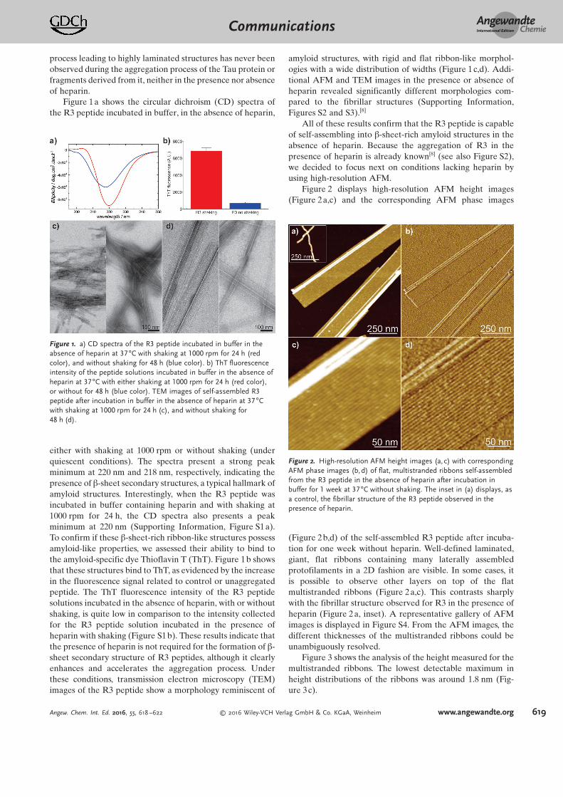
Figure 1a shows the circular dichroism (CD) spectra ofthe R3 peptide incubated in buffer, in the absence of heparin,
either with shaking at 1000 rpm or without shaking (underquiescent conditions). The spectra present a strong peakminimum at 220 nm and 218 nm, respectively, indicating thepresence of b-sheet secondary structures, a typical hallmark ofamyloid structures. Interestingly, when the R3 peptide wasincubated in buffer containing heparin and with shaking at1000 rpm for 24 h, the CD spectra also presents a peakminimum at 220 nm (Supporting Information, Figure S1 a).To confirm if these b-sheet-rich ribbon-like structures possessamyloid-like properties, we assessed their ability to bind tothe amyloid-specific dye Thioflavin T (ThT). Figure 1b showsthat these structures bind to ThT, as evidenced by the increasein the fluorescence signal related to control or unaggregatedpeptide. The ThT fluorescence intensity of the R3 peptidesolutions incubated in the absence of heparin, with or withoutshaking, is quite low in comparison to the intensity collectedfor the R3 peptide solution incubated in the presence ofheparin with shaking (Figure S1 b). These results indicate thatthe presence of heparin is not required for the formation of b-sheet secondary structure of R3 peptides, although it clearlyenhances and accelerates the aggregation process. Underthese conditions, transmission electron microscopy (TEM)images of the R3 peptide show a morphology reminiscent of
amyloid structures, with rigid and flat ribbon-like morphol-ogies with a wide distribution of widths (Figure 1 c,d). Addi-tional AFM and TEM images in the presence or absence ofheparin revealed significantly different morphologies com-pared to the fibrillar structures (Supporting Information,Figures S2 and S3).[8]
All of these results confirm that the R3 peptide is capableof self-assembling into b-sheet-rich amyloid structures in theabsence of heparin. Because the aggregation of R3 in thepresence of heparin is already known[8] (see also Figure S2),we decided to focus next on conditions lacking heparin byusing high-resolution AFM.
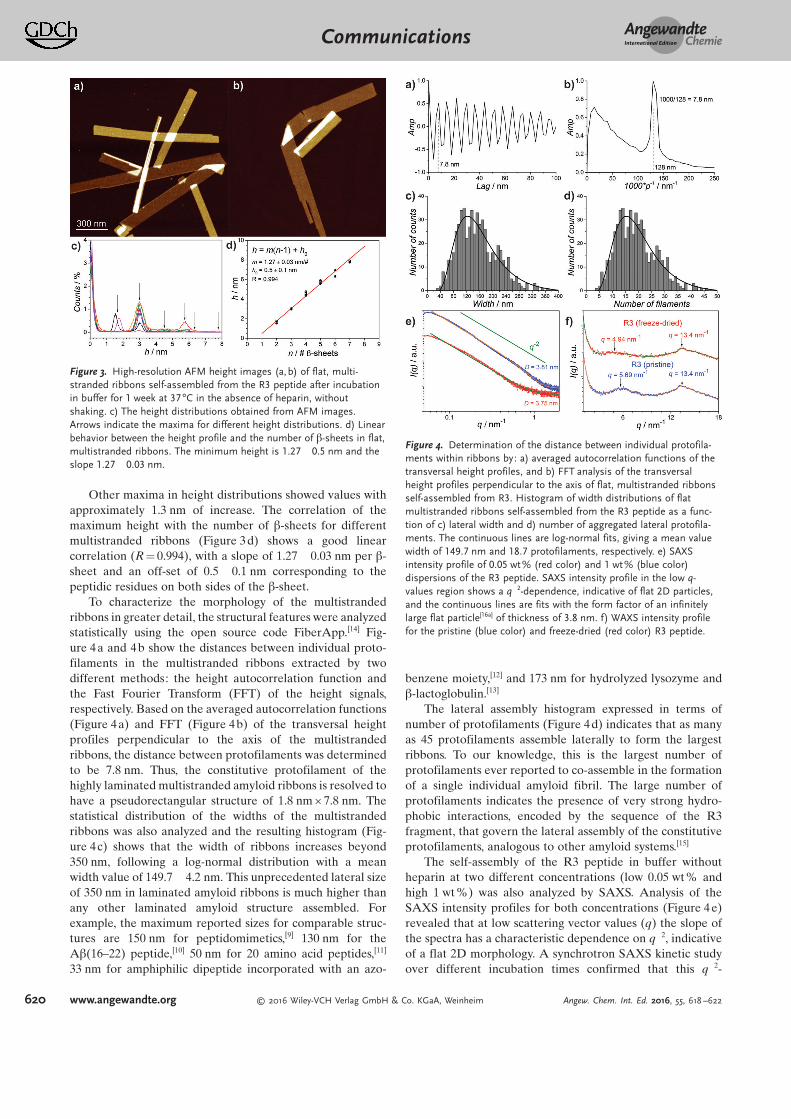
Figure 2 displays high-resolution AFM height images(Figure 2a,c) and the corresponding AFM phase images
(Figure 2b,d) of the self-assembled R3 peptide after incuba-tion for one week without heparin. Well-defined laminated,giant, flat ribbons containing many laterally assembledprotofilaments in a 2D fashion are visible. In some cases, itis possible to observe other layers on top of the flatmultistranded ribbons (Figure 2a,c). This contrasts sharplywith the fibrillar structure observed for R3 in the presence ofheparin (Figure 2 a, inset). A representative gallery of AFMimages is displayed in Figure S4. From the AFM images, thedifferent thicknesses of the multistranded ribbons could beunambiguously resolved.
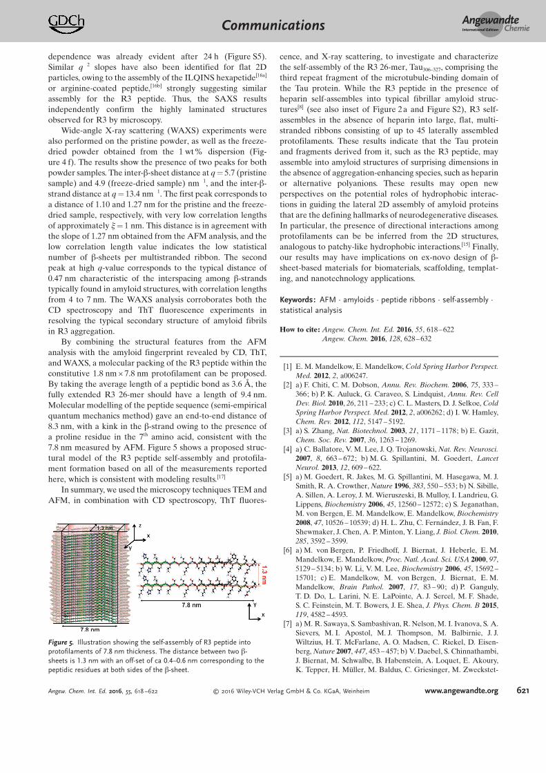
Figure 3 shows the analysis of the height measured for themultistranded ribbons. The lowest detectable maximum inheight distributions of the ribbons was around 1.8 nm (Fig-ure 3c).
Figure 1. a) CD spectra of the R3 peptide incubated in buffer in theabsence of heparin at 37 88C with shaking at 1000 rpm for 24 h (redcolor), and without shaking for 48 h (blue color). b) ThT fluorescenceintensity of the peptide solutions incubated in buffer in the absence ofheparin at 37 88C with either shaking at 1000 rpm for 24 h (red color),or without for 48 h (blue color). TEM images of self-assembled R3peptide after incubation in buffer in the absence of heparin at 37 88Cwith shaking at 1000 rpm for 24 h (c), and without shaking for48 h (d).
Figure 2. High-resolution AFM height images (a, c) with correspondingAFM phase images (b,d) of flat, multistranded ribbons self-assembledfrom the R3 peptide in the absence of heparin after incubation inbuffer for 1 week at 37 88C without shaking. The inset in (a) displays, asa control, the fibrillar structure of the R3 peptide observed in thepresence of heparin.
AngewandteChemieCommunications
619Angew. Chem. Int. Ed. 2016, 55, 618 –622 Ó 2016 Wiley-VCH Verlag GmbH & Co. KGaA, Weinheim www.angewandte.org
Other maxima in height distributions showed values withapproximately 1.3 nm of increase. The correlation of themaximum height with the number of b-sheets for differentmultistranded ribbons (Figure 3d) shows a good linearcorrelation (R = 0.994), with a slope of 1.27� 0.03 nm per b-sheet and an off-set of 0.5� 0.1 nm corresponding to thepeptidic residues on both sides of the b-sheet.
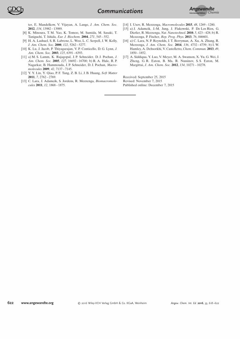
To characterize the morphology of the multistrandedribbons in greater detail, the structural features were analyzedstatistically using the open source code FiberApp.[14] Fig-ure 4a and 4b show the distances between individual proto-filaments in the multistranded ribbons extracted by twodifferent methods: the height autocorrelation function andthe Fast Fourier Transform (FFT) of the height signals,respectively. Based on the averaged autocorrelation functions(Figure 4a) and FFT (Figure 4b) of the transversal heightprofiles perpendicular to the axis of the multistrandedribbons, the distance between protofilaments was determinedto be 7.8 nm. Thus, the constitutive protofilament of thehighly laminated multistranded amyloid ribbons is resolved tohave a pseudorectangular structure of 1.8 nm × 7.8 nm. Thestatistical distribution of the widths of the multistrandedribbons was also analyzed and the resulting histogram (Fig-ure 4c) shows that the width of ribbons increases beyond350 nm, following a log-normal distribution with a meanwidth value of 149.7� 4.2 nm. This unprecedented lateral sizeof 350 nm in laminated amyloid ribbons is much higher thanany other laminated amyloid structure assembled. Forexample, the maximum reported sizes for comparable struc-tures are 150 nm for peptidomimetics,[9] 130 nm for theAb(16–22) peptide,[10] 50 nm for 20 amino acid peptides,[11]
33 nm for amphiphilic dipeptide incorporated with an azo-
benzene moiety,[12] and 173 nm for hydrolyzed lysozyme andb-lactoglobulin.[13]
The lateral assembly histogram expressed in terms ofnumber of protofilaments (Figure 4d) indicates that as manyas 45 protofilaments assemble laterally to form the largestribbons. To our knowledge, this is the largest number ofprotofilaments ever reported to co-assemble in the formationof a single individual amyloid fibril. The large number ofprotofilaments indicates the presence of very strong hydro-phobic interactions, encoded by the sequence of the R3fragment, that govern the lateral assembly of the constitutiveprotofilaments, analogous to other amyloid systems.[15]
The self-assembly of the R3 peptide in buffer withoutheparin at two different concentrations (low 0.05 wt% andhigh 1 wt %) was also analyzed by SAXS. Analysis of theSAXS intensity profiles for both concentrations (Figure 4 e)revealed that at low scattering vector values (q) the slope ofthe spectra has a characteristic dependence on q¢2, indicativeof a flat 2D morphology. A synchrotron SAXS kinetic studyover different incubation times confirmed that this q¢2-
Figure 3. High-resolution AFM height images (a, b) of flat, multi-stranded ribbons self-assembled from the R3 peptide after incubationin buffer for 1 week at 37 88C in the absence of heparin, withoutshaking. c) The height distributions obtained from AFM images.Arrows indicate the maxima for different height distributions. d) Linearbehavior between the height profile and the number of b-sheets in flat,multistranded ribbons. The minimum height is 1.27�0.5 nm and theslope 1.27�0.03 nm.
Figure 4. Determination of the distance between individual protofila-ments within ribbons by: a) averaged autocorrelation functions of thetransversal height profiles, and b) FFT analysis of the transversalheight profiles perpendicular to the axis of flat, multistranded ribbonsself-assembled from R3. Histogram of width distributions of flatmultistranded ribbons self-assembled from the R3 peptide as a func-tion of c) lateral width and d) number of aggregated lateral protofila-ments. The continuous lines are log-normal fits, giving a mean valuewidth of 149.7 nm and 18.7 protofilaments, respectively. e) SAXSintensity profile of 0.05 wt% (red color) and 1 wt% (blue color)dispersions of the R3 peptide. SAXS intensity profile in the low q-values region shows a q¢2-dependence, indicative of flat 2D particles,and the continuous lines are fits with the form factor of an infinitelylarge flat particle[16a] of thickness of 3.8 nm. f) WAXS intensity profilefor the pristine (blue color) and freeze-dried (red color) R3 peptide.
AngewandteChemieCommunications
620 www.angewandte.org Ó 2016 Wiley-VCH Verlag GmbH & Co. KGaA, Weinheim Angew. Chem. Int. Ed. 2016, 55, 618 –622
dependence was already evident after 24 h (Figure S5).Similar q¢2 slopes have also been identified for flat 2Dparticles, owing to the assembly of the ILQINS hexapetide[16a]
or arginine-coated peptide,[16b] strongly suggesting similarassembly for the R3 peptide. Thus, the SAXS resultsindependently confirm the highly laminated structuresobserved for R3 by microscopy.
Wide-angle X-ray scattering (WAXS) experiments werealso performed on the pristine powder, as well as the freeze-dried powder obtained from the 1 wt% dispersion (Fig-ure 4 f). The results show the presence of two peaks for bothpowder samples. The inter-b-sheet distance at q = 5.7 (pristinesample) and 4.9 (freeze-dried sample) nm¢1, and the inter-b-strand distance at q = 13.4 nm¢1. The first peak corresponds toa distance of 1.10 and 1.27 nm for the pristine and the freeze-dried sample, respectively, with very low correlation lengthsof approximately x = 1 nm. This distance is in agreement withthe slope of 1.27 nm obtained from the AFM analysis, and thelow correlation length value indicates the low statisticalnumber of b-sheets per multistranded ribbon. The secondpeak at high q-value corresponds to the typical distance of0.47 nm characteristic of the interspacing among b-strandstypically found in amyloid structures, with correlation lengthsfrom 4 to 7 nm. The WAXS analysis corroborates both theCD spectroscopy and ThT fluorescence experiments inresolving the typical secondary structure of amyloid fibrilsin R3 aggregation.
By combining the structural features from the AFManalysis with the amyloid fingerprint revealed by CD, ThT,and WAXS, a molecular packing of the R3 peptide within theconstitutive 1.8 nm × 7.8 nm protofilament can be proposed.By taking the average length of a peptidic bond as 3.6 è, thefully extended R3 26-mer should have a length of 9.4 nm.Molecular modelling of the peptide sequence (semi-empiricalquantum mechanics method) gave an end-to-end distance of8.3 nm, with a kink in the b-strand owing to the presence ofa proline residue in the 7th amino acid, consistent with the7.8 nm measured by AFM. Figure 5 shows a proposed struc-tural model of the R3 peptide self-assembly and protofila-ment formation based on all of the measurements reportedhere, which is consistent with modeling results.[17]
In summary, we used the microscopy techniques TEM andAFM, in combination with CD spectroscopy, ThT fluores-
cence, and X-ray scattering, to investigate and characterizethe self-assembly of the R3 26-mer, Tau306–327, comprising thethird repeat fragment of the microtubule-binding domain ofthe Tau protein. While the R3 peptide in the presence ofheparin self-assembles into typical fibrillar amyloid struc-tures[8] (see also inset of Figure 2a and Figure S2), R3 self-assembles in the absence of heparin into large, flat, multi-stranded ribbons consisting of up to 45 laterally assembledprotofilaments. These results indicate that the Tau proteinand fragments derived from it, such as the R3 peptide, mayassemble into amyloid structures of surprising dimensions inthe absence of aggregation-enhancing species, such as heparinor alternative polyanions. These results may open newperspectives on the potential roles of hydrophobic interac-tions in guiding the lateral 2D assembly of amyloid proteinsthat are the defining hallmarks of neurodegenerative diseases.In particular, the presence of directional interactions amongprotofilaments can be be inferred from the 2D structures,analogous to patchy-like hydrophobic interactions.[15] Finally,our results may have implications on ex-novo design of b-sheet-based materials for biomaterials, scaffolding, templat-ing, and nanotechnology applications.
Keywords: AFM · amyloids · peptide ribbons · self-assembly ·statistical analysis
How to cite: Angew. Chem. Int. Ed. 2016, 55, 618–622Angew. Chem. 2016, 128, 628–632
[1] E. M. Mandelkow, E. Mandelkow, Cold Spring Harbor Perspect.Med. 2012, 2, a006247.
[2] a) F. Chiti, C. M. Dobson, Annu. Rev. Biochem. 2006, 75, 333 –366; b) P. K. Auluck, G. Caraveo, S. Lindquist, Annu. Rev. CellDev. Biol. 2010, 26, 211 – 233; c) C. L. Masters, D. J. Selkoe, ColdSpring Harbor Perspect. Med. 2012, 2, a006262; d) I. W. Hamley,Chem. Rev. 2012, 112, 5147 – 5192.
[3] a) S. Zhang, Nat. Biotechnol. 2003, 21, 1171 – 1178; b) E. Gazit,Chem. Soc. Rev. 2007, 36, 1263 – 1269.
[4] a) C. Ballatore, V. M. Lee, J. Q. Trojanowski, Nat. Rev. Neurosci.2007, 8, 663 – 672; b) M. G. Spillantini, M. Goedert, LancetNeurol. 2013, 12, 609 – 622.
[5] a) M. Goedert, R. Jakes, M. G. Spillantini, M. Hasegawa, M. J.Smith, R. A. Crowther, Nature 1996, 383, 550 – 553; b) N. Sibille,A. Sillen, A. Leroy, J. M. Wieruszeski, B. Mulloy, I. Landrieu, G.Lippens, Biochemistry 2006, 45, 12560 – 12572; c) S. Jeganathan,M. von Bergen, E. M. Mandelkow, E. Mandelkow, Biochemistry2008, 47, 10526 – 10539; d) H. L. Zhu, C. Fern�ndez, J. B. Fan, F.Shewmaker, J. Chen, A. P. Minton, Y. Liang, J. Biol. Chem. 2010,285, 3592 – 3599.
[6] a) M. von Bergen, P. Friedhoff, J. Biernat, J. Heberle, E. M.Mandelkow, E. Mandelkow, Proc. Natl. Acad. Sci. USA 2000, 97,5129 – 5134; b) W. Li, V. M. Lee, Biochemistry 2006, 45, 15692 –15701; c) E. Mandelkow, M. von Bergen, J. Biernat, E. M.Mandelkow, Brain Pathol. 2007, 17, 83 – 90; d) P. Ganguly,T. D. Do, L. Larini, N. E. LaPointe, A. J. Sercel, M. F. Shade,S. C. Feinstein, M. T. Bowers, J. E. Shea, J. Phys. Chem. B 2015,119, 4582 – 4593.
[7] a) M. R. Sawaya, S. Sambashivan, R. Nelson, M. I. Ivanova, S. A.Sievers, M. I. Apostol, M. J. Thompson, M. Balbirnie, J. J.Wiltzius, H. T. McFarlane, A. O. Madsen, C. Riekel, D. Eisen-berg, Nature 2007, 447, 453 – 457; b) V. Daebel, S. Chinnathambi,J. Biernat, M. Schwalbe, B. Habenstein, A. Loquet, E. Akoury,K. Tepper, H. Mîller, M. Baldus, C. Griesinger, M. Zweckstet-
Figure 5. Illustration showing the self-assembly of R3 peptide intoprotofilaments of 7.8 nm thickness. The distance between two b-sheets is 1.3 nm with an off-set of ca 0.4–0.6 nm corresponding to thepeptidic residues at both sides of the b-sheet.
AngewandteChemieCommunications
621Angew. Chem. Int. Ed. 2016, 55, 618 –622 Ó 2016 Wiley-VCH Verlag GmbH & Co. KGaA, Weinheim www.angewandte.org
ter, E. Mandelkow, V. Vijayan, A. Lange, J. Am. Chem. Soc.2012, 134, 13982 – 13989.
[8] K. Minoura, T. M. Yao, K. Tomoo, M. Sumida, M. Sasaki, T.Taniguchi, T. Ishida, Eur. J. Biochem. 2004, 271, 545 – 552.
[9] H. A. Lashuel, S. R. Labrenz, L. Woo, L. C. Serpell, J. W. Kelly,J. Am. Chem. Soc. 2000, 122, 5262 – 5277.
[10] K. Lu, J. Jacob, P. Thiyagarajan, V. P. Conticello, D. G. Lynn, J.Am. Chem. Soc. 2003, 125, 6391 – 6393.
[11] a) M. S. Lamm, K. Rajagopal, J. P. Schneider, D. J. Pochan, J.Am. Chem. Soc. 2005, 127, 16692 – 16700; b) R. A. Hule, R. P.Nagarkar, B. Hammouda, J. P. Schneider, D. J. Pochan, Macro-molecules 2009, 42, 7137 – 7145.
[12] Y. Y. Lin, Y. Qiao, P. F. Tang, Z. B. Li, J. B. Huang, Soft Matter2011, 7, 2762 – 2769.
[13] C. Lara, J. Adamcik, S. Jordens, R. Mezzenga, Biomacromole-cules 2011, 12, 1868 – 1875.
[14] I. Usov, R. Mezzenga, Macromolecules 2015, 48, 1269 – 1280.[15] a) J. Adamcik, J.-M. Jung, J. Flakowski, P. De Los Rios, G.
Dietler, R. Mezzenga, Nat. Nanotechnol. 2010, 5, 423 – 428; b) R.Mezzenga, P. Fischer, Rep. Prog. Phys. 2013, 76, 046601.
[16] a) C. Lara, N. P. Reynolds, J. T. Berryman, A. Xu, A. Zhang, R.Mezzenga, J. Am. Chem. Soc. 2014, 136, 4732 – 4739; b) I. W.Hamley, A. Dehsorkhi, V. Castelletto, Chem. Commun. 2013, 49,1850 – 1852.
[17] A. Siddiqua, Y. Luo, V. Meyer, M. A. Swanson, X. Yu, G. Wei, J.Zheng, G. R. Eaton, B. Ma, R. Nussinov, S. S. Eaton, M.Margittai, J. Am. Chem. Soc. 2012, 134, 10271 – 10278.
Received: September 25, 2015Revised: November 7, 2015Published online: December 7, 2015
AngewandteChemieCommunications
622 www.angewandte.org Ó 2016 Wiley-VCH Verlag GmbH & Co. KGaA, Weinheim Angew. Chem. Int. Ed. 2016, 55, 618 –622
![Heterocycle Synthesis German Edition:DOI:10.1002/ange ... · related metal alkoxides failed to catalyze the reaction.[10] A further survey of alternate chiral diols in combination](https://img.pdfslide.us/doc/110x75/603fd2e943a1456a6d06b96c/heterocycle-synthesis-german-editiondoi101002ange-related-metal-alkoxides.jpg)


![German Edition:DOI:10.1002/ange.201600706 All-Metal ...partitioning (AdNDP) method.[18] Detailed AdNDP results can be found in the Supporting Information. In brief,the AdNDP analyses](https://img.pdfslide.us/doc/110x75/60a8e4c86d81a46b580b2ed3/german-editiondoi101002ange201600706-all-metal-partitioning-adndp-method18.jpg)


![German Edition:DOI:10.1002/ange.201910916 APyrazine-Based ...€¦ · scale application of LIBs is constrained by the limited and unevenly distributed lithium resources in the Earth’scrust.[2]](https://img.pdfslide.us/doc/110x75/603007578a4d5c50d84db191/german-editiondoi101002ange201910916-apyrazine-based-scale-application.jpg)
![Activity-Based Probes German Edition:DOI:10.1002/ange ...med.stanford.edu/content/dam/sm/bogyolab/documents/...[9a] Selective release of the leaving group adds additional versatility](https://img.pdfslide.us/doc/110x75/5f926c06722aa625ac671fd1/activity-based-probes-german-editiondoi101002ange-med-9a-selective.jpg)