Embed Size (px)
Citation preview
PROTEIN CHROMATOGRAPHY *Lavanya.G, Mubarak.SK, Vishali , Sujana.K, Pramela Rani, B. Syama Sundar
ANU college of Pharmaceutical sciences, Acharya Nagarjuna University, Guntur dist, Andhra Pradesh, India
Abstract: Proteins, peptides, lipids, amino acids, carbohydrates, vitamins and drugs can be separated using chromatography . Chromatography is the separation of a mixture in to individual components using a stationary phase and mobile phase. Proteins can be separated by using chromatographic techniques like size exclusion chromatography, ion exchange chromatography, affinity chromatography, fast protein liquid chromatography, high performance liquid chromatography, reversed phase chromatography, electrophoresis. Separation techniques rely on the differences in the solubility, size, charge, and adsorption characteristics of protein molecules. Ion-exchange chromatography is used to separate proteins on the basis of charge. Affinity chromatography utilises ligands, such as enzyme inhibitors, coenzymes, or antibodies, to specifically bind proteins to a solid support. Size of the Protein can be separated by using size exclusion chromatography. Electrophoresis can be used to separate proteins from complex mixtures on the basis of size and charge. Keywords: affinity chromatography, Chromatography, electrophoresis,fast protein liquid chromatographyhigh performance liquid chromatography, ion exchange chromatography, reversed phase chromatography, size exclusion chromatography. Introduction: Chromatography Chromatography is the separation of a mixture in to individual components using a stationary phase and mobile phase. These include proteins, peptides, lipids, amino acids, carbohydrates, vitamins and drugs. History The credit for the discovery of chromatography goes to Russian botanist Mikhail Tswett in 1906. Tswett described the separation of plant leaf pigments in solution by passing through a column of solid adsorbents. He coined the term chromatography (Greek = Chroma-colour; graphin = to write), since the technique dealt with the separation of colour compounds. Principle The principle of separation can be either adsorption or partition. Hence can be called as adsorption chromatography (or) partition chromatography. The stationary phase can be a solid or liquid, while the moving phase is a liquid or gas. When the stationary phase is solid, the basis is adsorption and when it is liquid, the basis is partition.
Theory Plate theory and Rate theory are two theories that are applicable to chromatography. 1) Plate Theory: Plate theory describes a chromatography system as being in equilibrium between the stationary and mobile phases. This views the column as divided into a number of imaginary theoretical plate. This is significant because as the number of plates in a column increases (or) HETP increases, so does the separation of components it also provides as equation that describes the elution curve (or) the chromatogram of a solute it can also be used to find the volume and the column efficiency. HETP = L / N Where : L = Column length N = Number of theoretical plates 2) Rate Theory: The Rate theory describes the migration of molecules in a column. This included band shape, broadening and the diffusion of a solute.
Lavanya G et al / J Biomed Sci and Res., Vol 3 (3), 2011,424-438
424

3) Rate theory follows the Van Deemter equation, using this equation, it is possible to find the optimum velocity and a minimum plate height. B H = A + --- + C. v. v Where: A = Eddy-Diffusion B = Longitudinal Diffusion C = mass transfer v = linear velocity PROTEINS Introduction: Proteins were first described by the Dutch chemist Garhaedus Johannes Mulder and named by the Swedish chemist Jons Jakob Beezetlins in 1838. Proteins are the most abundant organic molecules of the living system. They occur in every part of the cell and constitute about 50% of the cellular dry weight.
Proteins form the fundamental basis of structure and function of life. Proteins are an important class of biological macro molecules present in all organisms. Each protein polymer also known as polypeptide. Proteins are Polymers of amino acids Proteins on complete hydrolysis yield L- amino acids. This is a common property of proteins. Therefore, proteins are the polymers of L--amino acids. Functions: These functions may be broadly divided in two groups: 1) Structural functions There are primarily responsible for structure and strength of body. These include collagen and elastin found in the bone matrix, vascular system and other organs and -keratin present in epidermal tissues. 2) Dynamic functions These include proteins acting as enzymes, hormones, blood clotting factors.
Lavanya G et al / J Biomed Sci and Res., Vol 3 (3), 2011,424-438
425

Immunoglobulins, membrane receptors, storage proteins, besides their function in genetic control, muscle contraction, respiration. Classification I. According to chemical composition, proteins are divided into two classes. 1) Simple proteins: They are made up of chains of amino acid units only upon hydrolysis they yield mixture of amino acids. Ex: Albumins : Egg albumin, Serum albumin Globulins : Tissue globulin, Serum globulin Glutenins : Glutenin in wheat 2) Conjugated proteins: They are made of a simple protein united covalently with non-protein factor called a prosthetic group. Ex: Glycoproteins : Mucin in saliva. Phosphoproteins : Casein in milk Chromoprotein : Hb in red blood cells II. According to molecular shape Proteins are classified as follows: 1) Fibrous proteins: Fibre like and they are polypeptide chains coiled about one another and held together by strong inter-chain hydrogen bonds. They are insoluble in water. Ex: Keratin : Present in skin, hair and nails Myosin : Present in muscles Collagen : Present in tendons, cartilages and bones 2) Globular proteins: Spherical in shape (or) globe like. They are polypeptide chains coiled back and held by intra-chain hydrogen bonds. They are soluble in water.
Ex: Enzymes : Pepsin from stomach; helps digestion Insulin : Secreted by pancreas; regulates glucose metabolism Antibodies: Protect the body from infection AMINO ACIDS Introduction: Amino acids are the building block of the proteins. Amino acids are a group of organic compounds containing two functional groups amino (-NH2) is basic while the carboxyl group (-COOH) is acidic in nature. H H | | _ R–C-COOH R-C-COO | | NH2 NH3
+ General structure Exists as ion About 20 amino acids are commonly found in proteins. Essential amino acids: Essential amino acids are those which can not be produced in sufficient quantity by the human body. They must be induced in diet. Ex: Valine, phenyl alanine, tryptophan Non-essential amino acids: Non-essential amino acids are those which can be synthesized from other compounds by the body. Ex: glycine, alanine, serine Some commonly used amino acids. Different techniques for amino acids and proteins: 1) Physical techniques: Such as electrophoresis, chromatography, Immuno electrophoresis are widely used for the recovery of amino acids and proteins.
Lavanya G et al / J Biomed Sci and Res., Vol 3 (3), 2011,424-438
426

Lavanya G et al / J Biomed Sci and Res., Vol 3 (3), 2011,424-438
427

Fig: Paper Electrophoresis
Electrophoresis: The movement of charged particles in an electric field resulting in their migration towards the oppositely charged electrode is known as electrophoresis. The rate of migration of ions depends on several factors that include shape, size, net charge and solvation of the ions, viscosity of the solution and magnitude of current employed. Mainly paper electrophoresis is used. (a) Paper electrophoresis: In this technique, the sample is applied on a strip of filter paper wetted with desired buffer solution. The ends of the strip are dipped in the buffer reservoirs in which the
electrodes are placed. The electric current is applied allowing the molecules to migrate for sufficient time. The paper is removed, dried and stained with a dye that specifically colours, the substances to be detected the colour spots can be identified by comparing with a set of standards even simultaneously. (b) Immuno electrophoresis: This technique involves combination of the principles of electrophoresis and immunological reactions. The complex proteins of biological samples (human serum) are subjected to electrophoresis. The antibody is then applied in a trough parallel to the electrophoretic separation.
Lavanya G et al / J Biomed Sci and Res., Vol 3 (3), 2011,424-438
428

The antibodies diffuse and when they come in contact with antigens, precipitation occurs, resulting in the form of precipitin bands. 2) Chemical techniques: Precipitation Proteins are easily precipitated: a) By heat b) By ethyl alcohol c) By concentrated inorganic acids d) By silver nitrate e) By picric acid / tannic acid f) By UV rays & cosmic rays Hydrolysis: Simple proteins can be hydrolysed with acids (HCl), alkalis (NaOH), or enzymes to give component amino acids. Oxidation: Proteins are oxidised on burning and putrefaction. The products include nitrogen, CO2 and water. Reaction with ninhydrin: The -amino acid react with ninhydrin to form a purple, blue or pink colour complex (Ruhemann’s principle). Amino acid + Ninhydrin Keto acid + NH3 + CO2 + Hydrindantin Hydrindantin + NH3 + Ninhydrin Ruhemann’s purple
Ninhydrin reaction is used for quantitative determination of amino acids and proteins. PROTEIN CHROMATOGRAPHIC METHODS Chromatographic methods: Usually a protein separation contains one or more chromatographic steps. The basic procedure in the chromatography is to flow the solution containing the protein through a column packed with various materials. Different proteins interact differently with the column material, and thus can be separated by the time required to pass the column, or the conditions requires to elute the protein from the column. Usually proteins are detected as they are coming off the column by their absorbance at 280 nm. Different chromatographic methods are classified based on one of the following: 1) Size of protein Ex: Size exclusion chromatography 2) Net charge of protein Ex: Ion exchange chromatography 3) Specific liquid binding properties of protein. Size exclusion chromatography: Also called as molecular sieve chromatography (or) gel filtration chromatography.
Lavanya G et al / J Biomed Sci and Res., Vol 3 (3), 2011,424-438
429

Table: Proteins separated using size exclusion chromatography
S.No. Name of Protein St. Phase M. Phase Temp. 1
Native soluble protein complex
Superose polyacrylamide
NaOH 4oC
2
Immuno precipitation soluble proteins
Polyacrylamide gel
NaOH 4oC
Principle The principle is that smaller molecules have to traverse a larger volume in a porous matrix. Stationary phase (column matrix) = “beads” of a polysacchacide material that separates proteins based on size and shape. Mobile phase = buffer Method It separates larger proteins from small ones, since the larger molecules travel faster
through the cross-linked polymer in the chromatography column. Large molecules can’t get into the smaller pores in the beads and move rapidly through the column, eluting sooner. Smaller molecules and ions can enter all the pores in the beads with the buffer and thus have more space to “explore” on their way down the column and elute later. Applications: The main application is the fractionation of proteins and other water-
Lavanya G et al / J Biomed Sci and Res., Vol 3 (3), 2011,424-438
430
soluble polymers. It is used to analyse the molecular weight distribution of organic- soluble polymers. It is widely used technique for the purification and analysis of synthetic and biological polymers, such as proteins, polysaccharides and nucleic acid. Biochemical applications: This can determine the quaternary structure of purified proteins that have slow exchange times. Sec can also assay protein tertiary structure, as it measures three hydrodynamic volume, allowing folded and unfolded versions of the same protein to be distinguished. Ex: Hydrodynamic radius of folded form is 14oA. and the unfolded form is 36oA. Here folded form will elute much later due to it smaller size. Sec can be used as a measure of both the size and the polydispersity of the synthesised polymer. Ion-Exchange Chromatography Ion-Exchange Chromatography is the process by which a mixture of similar charged ions can be separated according to the nature and degree of their ionic charge. Principle: The principle of separation is by reversible exchange of ions between the ions present in the solution and those present in the ion exchange resin. Stationary phase (matrix, resin) Mobile phase (buffer e.g. O. IN HCl, IN NaOH Phosphate buffer, acetate buffer) The column to be used is selected according to its type and strength of charge. Anion exchange resins have a positive charge and are used to retain and separate negatively charged compounds, while cation exchange resins have a negative
charge and are used to separate positively charged molecules. Method of separation Elution is done by changing the pH in the column, which results in a change or neutralisation of the charged functional groups of each protein. Proteins bind to the matrix by electrostatic interactions. Before the separation begins a buffer is pumped through the column to equilibrate the opposing charged ions. Upon injection of the sample, solute molecules will exchange with the buffer ions as each competes for the binding sites on the resin. The length of extension of each solute depends upon the strength of its charge. The most weakly charged compounds will elute first, followed by those with successively stronger charges. Ex: Bovine plasma protein fractionation by ion exchange chromatography. An ion exchange chromatography process was developed to separate the main protein fractions of bovine blood plasma using a composite material, Q-Hyper Dresin and a gel material, DEAE-sepharose. Applications: Ion exchange chromatography is used to separate proteins on the basis of their net charge at particular pH. This technique is used extensively in the biotechnology industry and is particularly useful when used as a polishing step following an affinity capture column. It is used for the protease purification. It is used for the alkaline phosphate purification. It is used to capture the biotinylated proteins. Separation of inorganic ions: cations and anions. Organic separations: Most of the pharmaceutical compounds are either
Lavanya G et al / J Biomed Sci and Res., Vol 3 (3), 2011,424-438
431

strongly or weakly acidic or basic in nature. Hence a mixture of those compounds can
be separated by using ion exchange resins. Some classes of compounds which can be
Lavanya G et al / J Biomed Sci and Res., Vol 3 (3), 2011,424-438
432

separated are amino acids, proteins. Affinity chromatography Affinity chromatography is a very useful technique for “polishing” or completing the purification process. Beads in the chromatography column are cross-linked to ligands that bind specifically to the target protein. The protein is then removed from the column by rinsing with a solution containing free ligands. This method generally gives the purest results and highest specific activity compared to other techniques. Principle: The basic principle is that a biospecific ligand is immobilised to a solid support (or) resin to which a solution containing the protein of interest is passed over. Ligands are often based on biological function pairs such as enzymes and substrates (or) antigens and antibodies. The specific ligand binds the protein of interest and all non-specific molecules are washed away. Stationary phase: Gel matrix (agarose) Mobile phase: Buffer Method: Inject a sample into an initially equilibrated affinity chromatography column. Only the substances with affinity for the ligand are retained in the column. Other substances with no affinity for the ligand are eluted from the column. The substances retained in the column can be eluted from the column by changing pH (or) salt (or) organic solvent concentration of the eluent. Ex: Recombinant proteins can be separated by using affinity chromatography. Proteins with a known affinity are protein tagged in order to aid their purification. The protein may have been genetically modified so as to follow it to be selected for affinity binding, this is known as a fusion protein.
During elution excess glutathione is used to displace the tagged protein. APPLICATIONS Clinical applications: These include the use of Bolonate, lectin, protein A (or) protein G and immunosupports for the direct quantification of solutions. Other applications that are reviewed include affinity base chiral separation and the use of affinity, Chromatography for the study of drug (or) hormone interactions with binding proteins. Some areas of possible future developments are considered such as the use of synthetic dyes, immobilised metal ions, molecular imprints (or) aptamers as affinity ligands for clinical analytes. Affinity chromatography can be used in a number of applications, including nucleic acid protein purification, protein purification from cell free extracts and purification from blood. It is used to purify and concentrate a substance from a mixture into a buffering solution. It is used to reduce the amount of a substance in a mixture. It can be used to purify and concentrate an enzyme solution. The most common use of affinity chromatography is for the purification of recombinant proteins. Fast Protein Liquid Chromatography (FPLC) It is a form of liquid chromatography similar to HPLC that is used to separate or purify proteins and other polymers from complex mixture. FPLC system is a complete system for laboratory scale chromatographic separations of proteins and other biomolecules.
Lavanya G et al / J Biomed Sci and Res., Vol 3 (3), 2011,424-438
433

Figure: FPLC Principle FPLC is a type of liquid chromatography where the pumped solvent velocity is microprocessor-controlled through a software interface to ensure the constant flow rate of solvents. Stationary phase: A resin composed of cross-linked agarose beads. Mobile Phase: Liquid. Method: The Columns used in FPLC are large tubes that contain small () particles or gel beads that are known as stationary phase. The chromatographic bed is composed by gel beads in side the column and the sample is introduced into the injector and carried into the column by the flowing solvent. As a result of different components adhering (or) diffusing through the gel the sample mixture gets separated. Proteins separated Ex: Identification of zinc proteins in rat parotid saliva.
Rat pancreatic juice proteins were separated by FPLC. Applications FPLC is most often used in the enzymology and biology fields. FPLC can be used in pancreatic juice to determine the presence of certain proteins in the juice that potentially lead to disease. Using the anion – exchange column in the FPLC process, plasma proteins in the cerebrospinal fluid are profiled. FPLC is used in cases of tubular protein urea to isolate specific proteins from urine to measure the levels of excreted proteins. High Performance Liquid Chromatography (HPLC) High performance liquid chromatography is a form of column chromatography used frequently in biochemistry and analytical chemistry. It is used to analyse, identify, purify and quantify compounds.
Lavanya G et al / J Biomed Sci and Res., Vol 3 (3), 2011,424-438
434

Figure: HPLC Principle: HPLC separates mixtures of compounds on the basis of polarity. Separation is based on adsorption. Stationary Phase: Silica gel. Mobile Phase: Buffer. Method: It has a mobile phase, a stationary phase, detector. The mobile phase is continuously pumped at a fixed flow rate through the system and mixed by the pump. The injector is used to introduce a plug of a sample in to the mobile phase without having to stop the mobile phase flow and without introducing air into the system. Some of compounds in the sample mixture will have greater preference for stationary phase than the mobile phase and will be retained in the column longer. Proteins can be separated using HPLC Proteins were extracted from mantles and hepatopancreases of rock oysters and
fractioned by size-exclusion (HPLC). HPLC profiles of Fe, Cu, Zn and Ag indicate that those elements are bound to proteins extracted from mantles and hepatopancreases. Applications: HPLC can separate the double-stranded DNA molecules. Isolation and identification of drugs (or) metabolites in urine, plasma, serum etc., can be carried out. Purification of some compounds of natural or synthetic origin on preparative scale. It is used in quality control to ensure the purity of raw materials, to control and improve process yields, to quantify assay; of final products, or to evaluate product stability and monitor degradation. It is used for analysing air and water pollutants for monitoring pesticide levels in the environment.
Lavanya G et al / J Biomed Sci and Res., Vol 3 (3), 2011,424-438
435
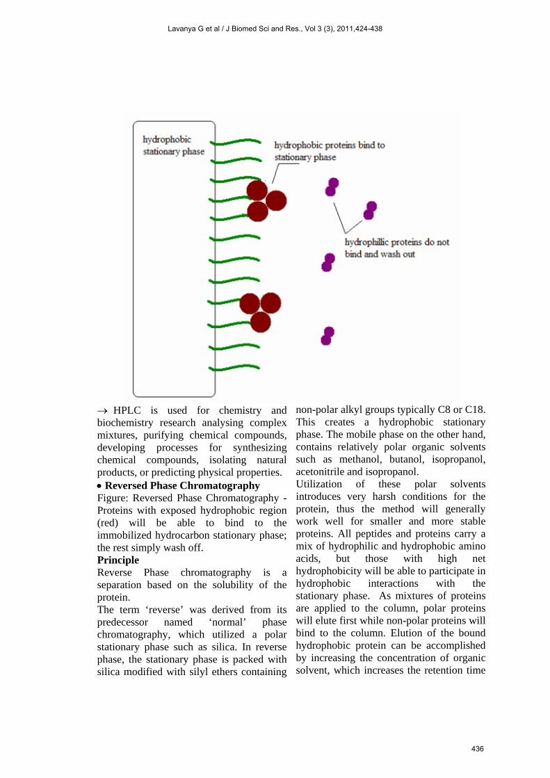
HPLC is used for chemistry and biochemistry research analysing complex mixtures, purifying chemical compounds, developing processes for synthesizing chemical compounds, isolating natural products, or predicting physical properties. Reversed Phase Chromatography Figure: Reversed Phase Chromatography - Proteins with exposed hydrophobic region (red) will be able to bind to the immobilized hydrocarbon stationary phase; the rest simply wash off. Principle Reverse Phase chromatography is a separation based on the solubility of the protein. The term ‘reverse’ was derived from its predecessor named ‘normal’ phase chromatography, which utilized a polar stationary phase such as silica. In reverse phase, the stationary phase is packed with silica modified with silyl ethers containing
non-polar alkyl groups typically C8 or C18. This creates a hydrophobic stationary phase. The mobile phase on the other hand, contains relatively polar organic solvents such as methanol, butanol, isopropanol, acetonitrile and isopropanol. Utilization of these polar solvents introduces very harsh conditions for the protein, thus the method will generally work well for smaller and more stable proteins. All peptides and proteins carry a mix of hydrophilic and hydrophobic amino acids, but those with high net hydrophobicity will be able to participate in hydrophobic interactions with the stationary phase. As mixtures of proteins are applied to the column, polar proteins will elute first while non-polar proteins will bind to the column. Elution of the bound hydrophobic protein can be accomplished by increasing the concentration of organic solvent, which increases the retention time
Lavanya G et al / J Biomed Sci and Res., Vol 3 (3), 2011,424-438
436
of a particular component. Reverse phase chromatography is commonly coupled with mass spectrometry in an effort to quantify the protein that is eluted from the column. Stages in Reversed Phase Chromatography The stages in Reversed Phase Chromatography are very similar to the stages found in Ion exchange Chromatography. Once again, the process can be summarized in four steps. 1. Equilibration 2. Sample Application 3. Elution 4. Regeneration Equilibration In this step the hydrophobic column is primed by applying the specific sample buffer. Because the column is hydrophobic, water molecules tend to be ordered at the junction between the column and the buffer. Application of the Sample In this step, the sample protein is injected into the system. Proteins in the mixture that have a high percentage of exposed hydrophobic amino acid residues will be adsorbed to the hydrophobic stationary phase. Other proteins in the mixture will be washed out. Elution This stage in the process involves changing the buffer conditions to elute the bound hydrophobic protein. The most common way to do this is to use a gradient that slowly increases the hydrophobicity using an organic buffer. Regeneration This stage involves washing off any remaining protein from the stationary phase and returning the conditions back to the way they were at the start of the process. This involves creating a less hydrophobic environment.
Conclusion: There are a variety of techniques used to separate and characterise proteins. Separation techniques rely on the differences in the solubility, size, charge, and adsorption characteristics of protein molecules. Ion-exchange chromatography is used to separate proteins on the basis of charge. Affinity chromatography utilises ligands, such as enzyme inhibitors, coenzymes, or antibodies, to specifically bind proteins to a solid support. Size of the Protein can be separated by using size exclusion chromatography. Electrophoresis can be used to separate proteins from complex mixtures on the basis of size and charge. From the above, we can conclude that HPLC and FPLC are widely used techniques for the separation of proteins. Acknowledgements: I express my sincere thanks to my guide Smt. K. Sujana, Assistant professor, College of Pharmaceutical Sciences, Acharya Nagarjuna University for her guidance and encouragement for this seminar. I take this opportunity to express my gratitude to Acharya Nagarjuna University for providing the necessary facilities and infrastructure. I am also grateful to Prof. B. Syama Sundar, Principal, College of Pharmaceutical Sciences, Acharya Nagarjuna University, Prof. A. Prameela Rani and other Teaching, Non-Teaching staff for their support. I thank all my Classmates for their cooperation. References: [1] A text book of Organic Chemistry- Arun bahl
Bs bahl, Edn. 2006. Pg.no:1, 21,389. [2] A text book of Biochemistry – U.
Satyanarayana, Edn. 2008. Pg.no:569-579. [3] Voet and Voet, Biochemsitry John Wiley and
Sons, 1995. [4] Nelson & Cox, Lehinger. Principles of
Biochemistry, 3rd ed. [5] http://GE Healthcare Life Sciences.
Lavanya G et al / J Biomed Sci and Res., Vol 3 (3), 2011,424-438
437
[6] http://books.google.co.in/books?id=JM [7] http://en.wikipedia.org/wiki/Affinity_chromato
graphy [8] http://www.biochem.arizona.edu/classes/bioc46
2/462a/462a.html [9] www.museumstuff.com/learn/topics/Protein_pu
rification::sub::Chromatographic Methods [10] www.siemens.com/answers
[11] https://mail.google.com/mail/h/qshfwobfxvu7/?view=att&th=12db368bfa5c9fle&attid=. . .
[12] https://mail.google.com/mail/?ui=2&ik=e287648c98&view=att&th=1
[13] https://mail.google.com/mail/h/11z7prfplyscq/?view=att&th=12dbc5b33709fe63&attid=0.1….
[14] mhtml:file://E:\Documents and Settings\Guest\My Documents\nature of aa_docx.mht
Lavanya G et al / J Biomed Sci and Res., Vol 3 (3), 2011,424-438
438