Embed Size (px)
Citation preview

1
Protein BioinformaticsPart I: Access to information
Jonathan Pevsner, [email protected]
260.655 April 6, 2006
[1] Proteins at NCBI• RefSeq accession numbers• Cn3D to visualize structures
[2] The Protein Data Bank (PDB)
[3] UniProt
[4] ExPASy (Expert Protein Analysis System)• DeepView, the Swiss-Pdb Viewer.
Outline
DNA RNA protein
Central dogma of molecular biology
genome transcriptome proteome
Central dogma of bioinformatics and genomics

2
DNA RNA
cDNAESTsUniGene
phenotype
genomicDNAdatabases
protein sequence databases
protein
[1] NCBI
0
10
20
30
40
50
60
1985
Growth of GenBank
Bas
e pa
irs o
f DN
A (b
illio
ns)
Sequ
ence
s (m
illio
ns)
200019951990
December1982
September2005

3
GenBankEMBL DDBJHousedat EBI
EuropeanBioinformatics
Institute
There are three major public DNA databases
Housed at NCBINational
Center forBiotechnology
Information
Housed in Japan
www.ncbi.nlm.nih.gov
Accession numbers are labels for sequences
NCBI includes databases (such as GenBank) that contain information on DNA, RNA, or protein sequences. You may want to acquire information beginning with a query such as the name of a protein of interest, or theraw nucleotides comprising a DNA sequence of interest.
DNA sequences and other molecular data are tagged with accession numbers that are used to identify a sequenceor other record relevant to molecular data.

4
What is an accession number?
An accession number is label that used to identify a sequence. It is a string of letters and/or numbers that corresponds to a molecular sequence.
Examples (all for retinol-binding protein, RBP4):
X02775 GenBank genomic DNA sequenceNT_030059 Genomic contigRs7079946 dbSNP (single nucleotide polymorphism)
N91759.1 An expressed sequence tag (1 of 328)NM_006744 RefSeq DNA sequence (from a transcript)
NP_007635 RefSeq proteinAAC02945 GenBank proteinQ28369 SwissProt protein1KT7 Protein Data Bank structure record
protein
DNA
RNA
Accessing protein sequences via Entrez
Entrez Gene with RefSeq
Entrez Gene is a great starting point: it collectskey information on each gene/protein from major databases. It covers all major organisms.
RefSeq provides a curated, optimal accession number for each DNA (NM_006744) or protein (NP_007635)
Example #1. Sean mentioned silk fibroin. How do you find its sequence?
From the NCBI home page, type “silk fibroin” and hit “Go”

5

6
FASTA format
Example #2. Find the sequence of myoglobin
From the NCBI home page, type “myoglobin” and hit “Go”
12
3
4

7
1. Entrez Gene entries offer a wealth of information, including links to RefSeq entries and to the Human Protein Reference Database (HPRD; Akhilesh)
2. HomoloGene provides access to RefSeq identifiers of a protein family, and offers links (domains, pairwise alignments)
3. Entrez Protein shows 922 myoglobins (too many),including 47 RefSeq (still a lot).

8
You can try scrolling through the RefSeq list, or apply “Limits”
As another approach, click “TaxBrowser”…
Enter the name of the organism you are interested in…
Follow a link of interest…
Now click protein…
You now can view all sperm whale proteins…

9
…and restrict the output to sperm whale myoglobin.
4. Structure provides links to myoglobin structures
Access to PDB through NCBI
Molecular Modeling DataBase (MMDB)
Cn3D (“see in 3D” or three dimensions):structure visualization software
Vector Alignment Search Tool (VAST):view multiple structures

10
You can limit the output to particular species, e.g. with the command human[organism]
Click 2MM1 to enter MMDB, the Molecular Modeling Database
11
Overlay two or more structures with VAST at NCBI
Click “Chain”…
Click one or more boxesthen “View 3D Structure”…

12
Overlay two or more structures with VAST at NCBI
Overlay two or more structures with VAST at NCBI
Click “globin”…
Access the Conserved Domain database at NCBI(http://www.ncbi.nlm.nih.gov/Structure/cdd/cdd.shtml)

13
[2] PDB
The Protein Data Bank (PDB)
• PDB is the principal repository for protein structures• Established in 1971• Accessed at http://www.rcsb.org/pdb or simply
http://www.pdb.org• Currently contains over 35,000 structure entities
Updated 3/06

14
PDB content growth
PDB holdings (September, 2005)
29,876 proteins, peptides1,338 protein/nucl. complexes1,500 nucleic acids13 carbohydrates32,727 total
Search for keyword DNACyields mouse zinc finger binding proteins

15
Protein Data Bank
Swiss-Prot, NCBI, EMBL
CATH, Dali, SCOP, FSSP
gateways to access PDB files
databases that interpret PDB files
[3] UniProt
UniProt combines information in Swiss-Prot, TrEMBL, and PIR. UniProt is comprised of three components
• The UniProt Knowledgebase (UniProtKB) is the central access point for extensive curated protein information.
• The UniProt Reference Clusters (UniRef) databases combine closely related sequences into a single record.
• The UniProt Archive (UniParc) is a comprehensive repository, reflecting the history of all protein sequences.
UniProt (Universal Protein Resource) at www.uniprot.org
16
www.uniprot.org
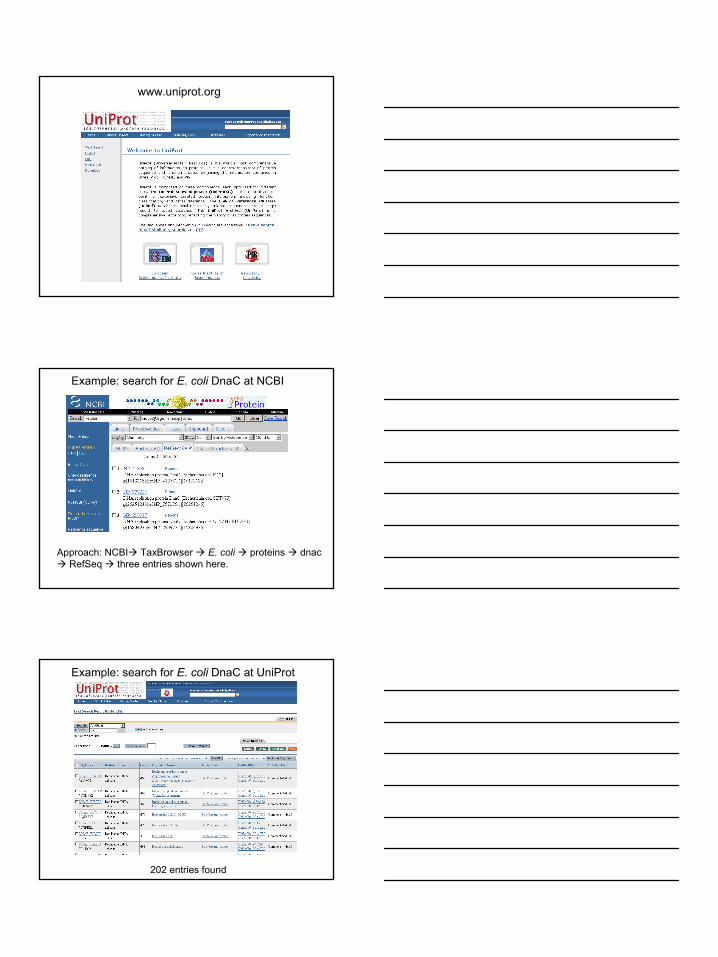
Example: search for E. coli DnaC at NCBI
Approach: NCBI TaxBrowser E. coli proteins dnacRefSeq three entries shown here.
Example: search for E. coli DnaC at UniProt
202 entries found

17
Example: search for E. coli DnaC at UniProt
Add input box (organism, coli) and 20 entries found
Example: search for E. coli DnaC at UniProt
The UniProt entry links to Entrez Gene (but not RefSeq)
[4] ExPASy / DeepView

18
ExPASy to access protein and DNA sequences
ExPASy sequence retrieval system(ExPASy = Expert Protein Analysis System)
Visit http://www.expasy.ch/
ExPASy to access protein and DNA sequences
When you search the ExPASy database, you are now querying the UniProt Knowledgebase.
► UniProtKB/Swiss-Prot; a curated protein sequence database which strives to provide a high level of annotation (such as the description of the function of a protein, its domains structure, post-translational modifications, variants, etc.), a minimal level of redundancy and high level of integration with other databases.
UniProtKB/Swiss-Prot Release 49.3 of 21-Mar-2006: 212,425 entries (More statistics)

19
ExPASy to access protein and DNA sequences
When you search the ExPASy database, you are now querying the UniProt Knowledgebase.
► UniProtKB/TrEMBL; a computer-annotated supplement of Swiss-Prot that contains all the translations of EMBL nucleotide sequence entries not yet integrated in Swiss-Prot.
UniProtKB/TrEMBL Release 32.3 of 21-Mar-2006: 2,666,963 entries
Example: find human tyrosinase at ExPASy
From the ExPASy home,click Swiss-Prot SRS
Start Continue
Example: find human tyrosinase at ExPASy

20
Example: find human tyrosinase at ExPASy
Note the lack of RefSeq and multiple accession numbersChoose the right protein by inspection (P14679)
Example: find human tyrosinase at ExPASy
Example: find human tyrosinase at ExPASy

21
Example: find human tyrosinase at ExPASy
Example: find human tyrosinase at NCBI
Human tyrosinase: blastp against pdb to find known structures

22
Human tyrosinase: blastp against pdb
Human tyrosinase: best blastp pdb match to structure 2AHL
Go to www.pdb.org and enter 2AHL…
…click SwissPDB viewer

23
View protein structures with DeepView
centermove (translate)zoom in/outrotate
measure dihedral angles (ω, φ, ϕ) (omega, phi, psi) from a selected atom.
measure bond angles (pick center atom, then two more atoms)
click, select two atoms, determine distance in angstroms
identify an atom (and the group to which it belongs). Type: CA, CB, OGroup: LYS116, etc. x,y,z atom coordinates
display groups within a particular distance (e.g. 10A) from a selected atom. Note the selection on the control and graphics panels.
center display on a selected atom
mutate a selected atom

24
► Go to window control panel.
►Shift/click to select the first five amino acid residues of myoglobin.They should appear red.► Click “labl” (i.e. label)(see arrow, above right). Those five residuesnow have a “v”. ►Inspect the display panel; those five residues are labeled.
Download and practice using DeepView!Try using myoglobin.
The ExPASy download site includes a helpfulweb-based tutorialhttp://www.usm.maine.edu/~rhodes/SPVTut/