Embed Size (px)
Citation preview

© 2003 Hindawi Publishing Corporation
Journal of Biomedicine and Biotechnology • 2003:5 (2003) 257–266 • PII. S1110724303209220 • http://jbb.hindawi.com
REVIEW ARTICLE
Protein and Chemical Microarrays—Powerful Toolsfor Proteomics
Qingchai Xu and Kit S. Lam∗
Division of Hematology and Oncology, Department of Internal Medicine, UC Davis Cancer Center,University of California, Davis, 4501 X Street, Sacramento, CA 95817, USA
Received 11 September 2002; accepted 10 December 2002
In the last few years, protein and chemical microarrays have emerged as two important tools in the field of proteomics. Specificproteins, antibodies, small molecule compounds, peptides, and carbohydrates can now be immobilized on solid surfaces to formhigh-density microarrays. Depending on their chemical nature, immobilization of these molecules on solid support is accomplishedby in situ synthesis, nonspecific adsorption, specific binding, nonspecific chemical ligation, or chemoselective ligation. These arraysof molecules can then be probed with complex analytes such as serum, total cell extracts, and whole blood. Interactions between theanalytes and the immobilized array of molecules are evaluated with a number of different detection systems. In this paper, variouscomponents, methods, and applications of the protein and chemical microarray systems are reviewed.
INTRODUCTION
Chemical array is a form of the combinatorial li-brary method that was first described by Geysen et al in1984 [1]. Peptides were synthesized on polyethylene pinsin a 96-well footprint and used for B-cell epitope map-ping. Enzyme-linked immunosorbent assays were usedfor such analysis. In 1991, Foder et al of Affymax, Inc(Palto Alto, Calif) [2] reported the use of photolithog-raphy in conjunction with light-directed peptide synthe-sis to generate 1024 peptide on a 1.6-cm2 glass surfaceand the use of fluorescent microscopy to analyze interac-tions between the peptides and fluorescent-labeled anti-bodies. In 1992, Frank described the synthesis of a pep-tide array as spots on paper [3]. These three techniquesall use parallel synthesis methods to generate arrays ofpeptides that are spatially separable and addressable. Theidentity of each peptide spot is known prior to any biolog-ical assay. In 1991, we reported the use of the highly effi-cient “split-mix” synthesis method to generate “one-beadone-compound” (OBOC) combinatorial peptide library(millions of peptides), in which each 80-µm bead dis-played only one peptide entity [4]. This spatially separa-ble but nonaddressable peptide microarray or library wasscreened with an enzyme-linked colorimetric assay, andindividual color beads were then physically isolated formicrosequencing. These early studies paved the road forthe microarray field. In 1994, scientists from Affymetrix(Santa Clara, Calif), a spin-off company from Affymax,reported the use of photolithography/photochemistry ap-proaches to synthesize the first DNA chip [5]. Microarraysof oligonucleotides were synthesized in situ on glass sur-
face, and fluorescent-labeled octanucleotide probes wereused to identify the complementary oligonucleotides onthis 256 array. In 1995, Brown et al introduced a differ-ent form of a DNA microarray chip by using high-speedrobot to spot array of different cDNAs on the glass surface.Fluorescent-labeled cDNA derived from mRNA of wholecell lysate was then used to probe the DNA microarray,allowing determination of expression levels of thousandsof genes simultaneously [6, 7]. Several automatic array-ers have since become commercially available. The suc-cessful application of DNA microarrays to gene expres-sion analysis [8, 9, 10], genetic diagnosis [11], and drugtarget identification [12], and the rapid development ofthe proteomics field have propelled many to think aboutthe use of protein or chemical microarray as an efficienttool to evaluate the function of complex protein mixtures.In the last few years, several groups independently devel-oped different chemical and protein microarray methodsand applied them to study various biological and chemicalproblems [13, 14]. In this review, we will give an overviewin the field and highlight recent developments.
TYPES OF MICROARRAY
Microarray, sometimes referred to as a chip or arrayedlibrary, can be classified into two general types: biochips(biomicroarray) and chemical microarray. Biochips areusually generated from biochemical or biological com-ponents, such as, protein (including enzymes and anti-bodies), DNA, cell [15], and tissue [16]. Chemical mi-croarrays consist of arrays of organic compounds includ-ing small organic molecules, peptides, and sugars. Based
258 Q. Xu and K. S. Lam 2003:5 (2003)
on how chemical microarrays are constructed, they canalso be categorized as in situ synthesis array and spottingarray. The chemistry of the in situ synthesis approach ismore limited, particularly when photochemical reactionis a required synthetic step. As a result, only oligomericmolecules such as oligonucleotides or peptides are usedin the in situ synthesis array. A spotting array refers to anarray of compounds that are presynthesized and directlytransferred and immobilized on a solid surface. This ap-proach is more versatile and can be applied to generate amicroarray of almost any molecules.
IN SITU SYNTHESIS MICROARRAY APPROACH
Two typical in situ synthesis approaches are SPOT-synthesis [3] and light-directed parallel synthesis [2]. TheSPOT-synthesis method involves parallel peptide synthe-sis on membrane or paper. In this method, a small vol-ume of solutions containing Fmoc-amino acids plus cou-pling reagents is dispensed onto the designated spot onthe membrane. After the coupling reaction is complete,the whole membrane is washed and the N-terminal pro-tecting group is deprotected prior to the next coupling cy-cle. Limited by possibility of contamination by reagentsfrom adjacent spots, the distance between each spot can-not be too small. This results in a rather low-density array(eg, 25 spots/cm2). Production of peptide arrays with thistechnology has been reviewed recently [17]. AutomatedSPOT-synthesizers, such as the Auto-Spot Robot ASP222(Intavis Inc, Germany), are now commercially available.
The light-directed in situ synthesis was initially devel-oped for peptide synthesis [2] but has now been widelyused for the synthesis of DNA microarrays. The commer-cially available Affymetrix chips are prepared by this ap-proach. Figure 1 depicts the deprotection chemistries oflight-directed parallel synthesis. The original method usesamino acids with a photolabile protecting group (eg nitro-veratryloxycarbonyl, NVOC) as building blocks, and pho-tolithographic method with appropriate masks is used tospatially deprotect the N-terminal protecting group dur-ing peptide synthesis. This approach was later adapted tooligonucleotide synthesis [5, 6]. In 1996, McGall et al re-ported a new photolithographic masking method that in-volves the use of a polymeric photoresistant film to con-struct a pattern onto the glass surface [18]. The patternedphotoresistant film is used to mask selected regions ofthe substrate from exposure to standard chemical reagentsduring synthesis. The main advantage of this approach isthat the resolution is superior, and therefore the densityof the microarray can increase significantly.
Instead of using photolithographic masks, Singh-Gasson et al [19], reported a maskless light-directed ar-ray synthesizer, which utilizes a digital micromirror de-vice consisting of a 600 × 800 array of micromirrors toform virtual masks. With 1 : 1 imaging, the synthesizercan produce 480,000 pixels of synthetic oligomers in a10×14 mm area. A similar maskless microarray setup wasalso reported by Gao et al to synthesize oligonucleotide
[20] and peptide [21] microarrays. However, this methodis unique in that it uses light to generate acid in situ todeprotect the acid labile protecting groups of the grow-ing chain (4,4′-dimethoxytrityl (DMT) for nucleotides orBoc for amino acids, Figure 1) instead of using photola-bile protecting groups. As a result, commercially availablestandard building blocks for both peptide and oligonu-cleotide synthesis can be used with this method.
SPOTTING MICROARRAY APPROACH
As previously indicated, in situ synthesis methodsare mainly used to generate arrays of oligomeric com-pounds such as oligonucleotide and peptides. In contrast,the spotting microarray approach, in conjunction with aproper immobilization method, can generate many copiesof the same chip more efficiently since the compoundsneed to be synthesized only once. Although macro-molecules such as proteins, DNA, and larger peptides caneasily be adsorbed onto solid surface through noncovalentinteractions, immobilization of small organic molecules,short peptides, or simple sugars will require covalent at-tachment, preferably through chemoselective site-specificligation.
IMMOBILIZATION VIA PHYSICAL ADSORPTION
The first and simplest type of immobilization isthrough surface adsorption. This approach is particu-larly useful for proteins, and it has been used in stan-dard ELISA, dot blot, and Western blot for many years.The commonly used solid supports are hydrophobic plas-tics such as polystyrene. Protein microarrays have alsobeen generated on an aminosilane cationic surface [22],nitrocellulose membrane [23], and Hybond ECL mem-brane [24, 25]. Through adsorption, Wang et al haveprepared carbohydrate arrays on nitrocellulose bondedslides [26]. They discovered that the efficiency of ad-sorption of dextran to nitrocellulose increases with size.The 2000-kd dextran bound significantly stronger thanthe 20-kd dextran. Wang et al [27] have used HydroGel-coated slides for arraying 43 monoclonal antibodies(mAb) against cytokines and chemokines, and used afluorescence-based multiplexed immunoassays to quanti-tate the level of these proteins. We spotted peptide-proteinand peptide-agarose conjugates on polystyrene slide asa way to display peptides (Xu and Lam et al, unpub-lished data, May 2003). Fang et al [28] have developeda membrane protein microarray by spotting cell mem-brane preparations containing G-protein-coupled recep-tors on γ-aminopropylsilane-derivatized glass or gold-coated glass surface.
IMMOBILIZATION VIA SPECIFIC SURFACEINTERACTION
In addition to immobilization via nonspecific physi-cal adsorption, molecules can be tagged and immobilized

2003:5 (2003) Protein and Chemical Microarrays—Powerful Tools for Proteomics 259
NO2
CH3O
CH3O
CH2
O
O
NH
Rhν
CH3O
CH3O
COOH
N N
HOOC
CH3O + H2N
CH3O
R
6-nitroveratryloxycarbonyl (NVOC)
(a) The photolabile protecting group (NVOC) is removed by light illumination.
PGA precursor
DMT O R
R
or
Boc NH
R
PGAHO R
or
H2N
PhSS+
SbF−6hν
DCMH+ SbF−6 + PhSPh + Other photoproducts
(PGA)
(b) Acid labile protecting groups (DMT and Boc) are removed by a photogenerated acid (PGA).
Figure 1. Deprotection chemistry in two approaches of light-directed parallel synthesis.
through specific noncovalent interactions between the tagand the already immobilized capturing molecule for thetag. A typical example is the biotin-streptavidin systemfor immobilizing biotinylated proteins onto streptavidin-coated surfaces [29]. Likewise, a small molecule canalso be biotinylated and printed onto a surface thathas been precoated with a monolayer of streptavidinor neutravidin. Neutravidin is sometimes preferred asit has less nonspecific interaction with other proteins.We were able to successfully print a microarray of bi-otinylated synthetic peptides onto a neutravidin-coatedpolystyrene microscope slide using a Wittech 03 arrayer(Wittech, Inc, Taiwan) [30]. Lesaicherre et al have re-cently reported [31] the derivatization of glass slides withavidin for immobilization of biotinylated peptides. Thepoly-His-Ni2+ system has also been reported for pro-tein microarray [32]. In this method, proteins contain-ing a poly-histidine tag were printed onto Ni2+-chelatingsurfaces. Another approach is to use anti-GST anti-body or glutathione-coated slides to capture a series ofGST-fusion proteins. Boronic acid groups are knownto form very stable complex with some moieties suchas cis or coaxial 1,2-diol. Immobilized phenylboronicacids have been used in chromatography [33] and pro-tein immobilization [34]. Similar strategies can, in prin-ciple, be applied to protein or chemical microarrays aswell.
IMMOBILIZATION VIA COVALENT ATTACHMENT
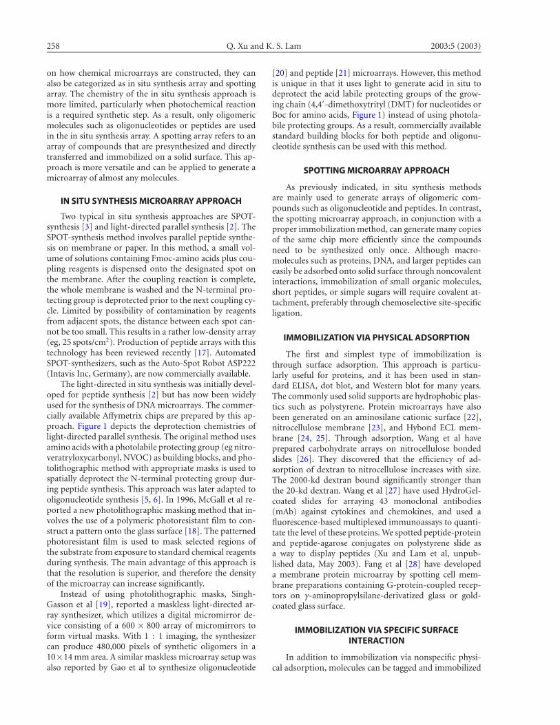
Although nonspecific physical adsorption has beenused successfully for generating a microarray of macro-molecules, this approach is less useful for the prepa-ration of small molecule or small peptide microarrays.These small molecules can be conjugated to a tag whichin turn binds to the immobilized capturing agent (seeabove). Alternatively, they can be immobilized via cova-lent attachment to a functional group on the solid sur-face. Figures 2 and 3 summarize some of the commonchemistries used in generating microarrays by covalentattachment. Chemical modification of the solid surfaceis necessary to create functional groups for covalent im-mobilization and to achieve homogeneous immobiliza-tion. Commercially available aldehyde-derivatized glassslides that have been used for DNA immobilization canalso be used for protein microarrays [35]. The aldehydegroups on the glass surface react with primary amines onthe protein to form Schiff ’s base linkages. BSA is used toblock the remaining unreacted aldehyde groups or othernonspecific binding sites. Zhu et al [32, 36] have de-scribed the use of a 3-glycidoxypropyltrimethoxysilane(GPTS) to activate polydimethylsiloxane (PDMS) on theslide surface prior to protein immobilization. Lin et alhave reported [37] the printing of protein microarrays onan aminopropyltrimethoxysilane surface activated with
260 Q. Xu and K. S. Lam 2003:5 (2003)
NHROR NH2
O
NOO
O
(a) NHS/NH2 [37, 38].
RNHR NH2
NaBH4
O
H
(b) Aldehyde/NH2 [35].
RNH
O
NHR NH2
N C O
(c) Isocyanate/NH2 [38].
OSiO
OR
R OHOSiO
Cl
(d) Chlorinated glass/OH [40].
RSN
O
O
R SH
O
N
O
(e) Maleimide/thiol [39].
NHRHO
R NH2
O
(f) Glycidoxy/NH2 [36].
Figure 2. Chemistries of covalent immobilization (nonselectiveligation).
bis-sulfosuccinimidyl suberate. Furthermore, Benters etal [38] have demonstrated the use of succinimidyl ester-or isocyanate-functionalized dendrimer on a solid surfacefor nucleic acid and protein microarrays.
Immobilization of small molecules or short peptidesoften requires covalent linkage of the compounds ontothe solid support. Michael addition has been used bySchreiber’s group to ligate thiol-containing compounds tomaleimide-derivatized glass slides to form a microarray
RON
O
R ONH2
CHO
O
RCys
OS
N
OR
(a) Glyoxylyl/NH2O, N-terminal Cys [42, 43].
OO
OSNH2CysR
NH2CysS
R
OOO
(b) Quinine/cyclodiene [44].
R
SH
O
N
ORCysS
O
(c) Thioester/N-terminal Cys [31].
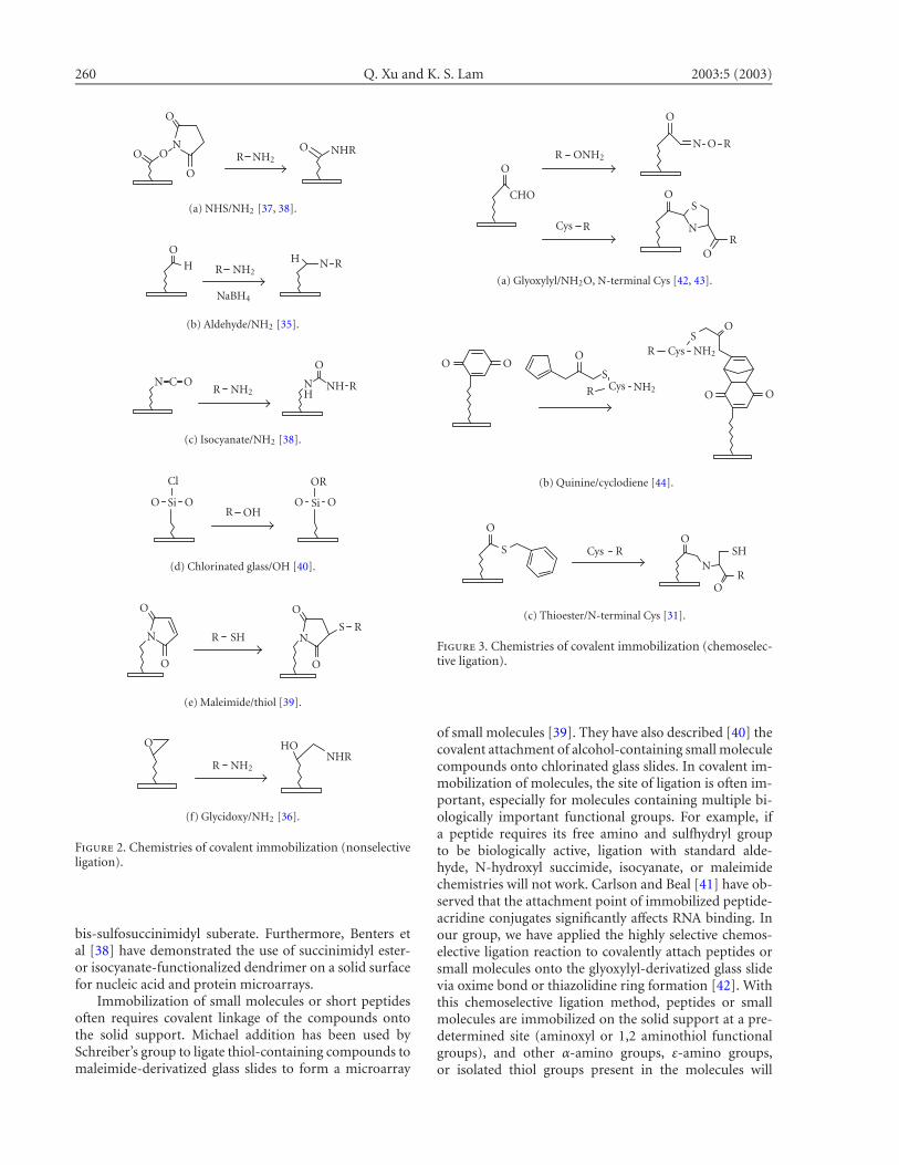
Figure 3. Chemistries of covalent immobilization (chemoselec-tive ligation).
of small molecules [39]. They have also described [40] thecovalent attachment of alcohol-containing small moleculecompounds onto chlorinated glass slides. In covalent im-mobilization of molecules, the site of ligation is often im-portant, especially for molecules containing multiple bi-ologically important functional groups. For example, ifa peptide requires its free amino and sulfhydryl groupto be biologically active, ligation with standard alde-hyde, N-hydroxyl succimide, isocyanate, or maleimidechemistries will not work. Carlson and Beal [41] have ob-served that the attachment point of immobilized peptide-acridine conjugates significantly affects RNA binding. Inour group, we have applied the highly selective chemos-elective ligation reaction to covalently attach peptides orsmall molecules onto the glyoxylyl-derivatized glass slidevia oxime bond or thiazolidine ring formation [42]. Withthis chemoselective ligation method, peptides or smallmolecules are immobilized on the solid support at a pre-determined site (aminoxyl or 1,2 aminothiol functionalgroups), and other α-amino groups, ε-amino groups,or isolated thiol groups present in the molecules will
2003:5 (2003) Protein and Chemical Microarrays—Powerful Tools for Proteomics 261
not react. This eliminates the risk of inactivating thecompounds during immobilization. We have recently re-ported a new strategy to significantly improve the load-ing of glyoxylyl groups onto glass surfaces by using acrylicacid as a starting material [43]. Further improvement ofloading can be accomplished by adding hydrophilic link-ers and bifurcating amino acids such as lysine between theglyoxylyl group and the glass surface (Xu and Lam, May2002). Lesaicherre et al [31] have recently reported thederivatization of glass slides with thioester for chemos-elective ligation of peptides with an N-terminal cysteine(1,2 aminothiol group) to form an amide bond. House-man et al [44] have described a peptide chip prepared bythe Diels-Alder-mediated immobilization of peptides onthe quinone-functionalized surfaces. In this method, a cy-clodiene moiety is incorporated into peptides for the pur-pose of immobilization. As an alternative to immobilizingpeptide-protein conjugates onto plastic slides via physicaladsorption, we have used a chemically derivatized proteinscaffold (eg, glyoxylyl-functionalized BSA) to first coat thepolystyrene slide via nonspecific adsorption. Peptides orsmall molecules are then printed onto the functionalizedprotein-coated slides. After incubation, these compoundsare immobilized onto the coating surface via a site-specificligation reaction. In some applications, immobilization ofthe ligand onto the glass surface via a long hydrophiliclinker or a protein may be beneficial for biological activity.For example, we have found that a long linker is neededfor phosphorylation of a peptide substrate by p60c-src pro-tein tyrosine kinase [42].
CD, MICROFLUIDICS, MULTIPLEX-BEADS, ANDFIBER-OPTIC MICROARRAYS
Kido et al [45] have developed a compact disc-basedmicroarray system for immunoassays, which uses a piezo-electric inkjet applicator to generate a high-density anti-gen microarray (75-µm spot) on a polycarbonate disc.Competitive inhibition immunoassays with fluorescentantibodies are performed on the disc, and the final read-out is accomplished with a commercially available fluo-rescence scanner.
Microfluidics is another format of microarray that isbased on microchannel systems. Shi et al [46] have de-veloped a radial capillary array electrophoresis microplatesystem, which consists of 96 separate microfabricated sep-aration channels that connect a central common anodereservoir to 96 injectors at the perimeter of the 10-cmdiameter wafer. A laser-excited rotary confocal scannerwith four-color detection channels is used for detection.A high-quality restriction map of the 96 samples can beobtained in less than 120 seconds with this device.
Han et al have described [47] the development ofquantum-dot-tagged microbeads for multiplexed opticalcoding of biomolecules. This group immobilizes differ-ent peptides, proteins, or nucleic acids on various sam-ples of microbeads into which different-size hydrophobicquantum dots are embedded. These quantum-dot-tagged
microbeads are used in suspension in conjunction withflow cytometry to track the identity of the immobilizedmolecules when mixed with the analytes. These investiga-tors have used DNA hybridization studies to demonstratethat the coding and target signals can be read at a singlebead level simultaneously. Similar technology using tradi-tional fluorophores to encode microbeads has been com-mercialized by Luminex (Austin, Tex).
Walt has pioneered the fiber-optic microarray biosen-sor technology, which is commercially known as IlluminaBeadArray [48]. It involves the patterning of an array ofwells at the tip of an optical imaging fiber bundle. Thedepth of each well can be controlled by etching time andthe concentration of hydrofluoric acid used. The fiber-optic tip is then dipped into a suspension of a libraryof OBOC expressing microbeads (3 micron diameter).The chemicals on the microbeads can be synthesized by“split-mix” synthesis or through parallel synthesis. Alter-natively, proteins can be adsorbed on the beads in parallel.Through surface tension, each of the microwells is filledwith just one microbead. This random microbead array isdecoded [49] for a specific application, prior to analysis ofunknown samples. This technology has been applied forthe detection of a variety of analytes ranging from DNAto organic vapors [50, 51, 52]. One major advantage ofthis method is that the packing density of the array is ex-tremely high with center-to-center distance of each beadat 5 µm. However, the necessary decoding step is a majordrawback.
PRINTING METHODS AND INSTRUMENTATION
Photolithography, mechanical microspotting, andinkjet application are the three general methods for mi-croarray printing. The mask and maskless photolithog-raphy methods [3, 19, 20, 21] that involve in situ light-directed synthesis of oligomers can generate high-quality,high-density microarrays. The mechanical spotting (orsurface contact) technique uses a pin to transfer liq-uid samples to the slides by direct surface contact. Thepin-ring technique (eg, GSM 417 arrayer, Affymetrix)and the stamp microcontact technique (eg, SpotBot Per-sonal Microarrayer, TeleChem International, Inc, Sunny-vale, Calif) allow for fairly reproducibly sized spots. Theinkjet method employs an electric current to dispense theliquid sample onto the solid support. This noncontactpiezoelectric technique accurately and rapidly dispensesmonodisperse droplets. Moerman et al [53] have reportedthe use of electrospraying in a stable cone-jet mode togenerate a highly reproducible spot of biological mate-rial 130–350 µm in diameter, and as small as 50 pL. Morerecently, Avseenko et al [54] have used the electrospraymethod to deposit dry protein onto a dextran-grafted sur-face, followed by incubation in a 100% humidity cham-ber. This method was able to generate a 0.6× 0.6 mm2 ar-ray with each spot 30–40 µm in diameter and consisting of28 different protein antigens and allergens. Ringeisen et al[55] have utilized a laser transfer technique, which allows

262 Q. Xu and K. S. Lam 2003:5 (2003)
accurate deposition of picoliter volumes of active proteinsonto standard microarray substrates.
MICROARRAY APPLICATION
Table 1 summarizes the various aspects of the mi-croarray technology: attachment chemistries, immobi-lized molecules, analytes, and detection methods. In mostcases, the analytes that have been used to probe the mi-croarray are complex mixtures of molecules: for exam-ple, mRNA or cDNA preparations, total cell extracts orintact cells, complex protein mixtures, body fluids suchas serum or whole blood, fermentation broths, environ-mental samples, or microorganisms. In some cases, a pureprotein, such as a protein kinase or a protease, is usedto probe the peptide microarray to determine the sub-strate profile of these enzymes. Protein microarrays arefrequently used to study different protein functions suchas protein-protein interactions. Zhu et al [32] have identi-fied many new calmodulin- and phospholipid-interactingproteins through assaying a microarray of 5800 yeast pro-teins using known proteins and phospholipids as probes.Similar technique was also used to evaluate protein sub-strate profile of 119 yeast protein kinases [36]. Sreeku-mar et al [56] have applied protein microarrays to dis-cover novel radiation-regulated proteins. Huang et al [25]have used antibody arrays to quantitate a large number ofdifferent cytokines from sera and culture media. Kneze-vic et al [57] have reported antibody microarrays to an-alyze protein expression in cancer tissue of the oral cav-ity. Ziauddin and Sabatini have reported the use of plas-mid DNA microarray to transfect cells in situ to form anew cell microarray with newly expressed proteins en-coded by the plasmid DNA [15]. The use of protein mi-croarrays for serum marker detection and discovery us-ing prostate cancer as a model disease has recently beenreviewed [58]. Mezzasoma et al [59] have reported theuse of microbial antigen arrays to detect serum anti-bodies against the ToRCH antigens in a panel of char-acterized human sera. Antibody microarrays have beenused for proteomic profiling of the cancer microenviron-ment [57]. Paweletz et al [60] have reported the devel-opment of the so-called reverse-phase protein microar-rays to quantitate proteins derived from microdissectedtissues. They have immobilized serial dilution of totalcell lysates from microdissected tissues on nitrocellulose-coated glass slides and have used different enzyme-linkedantibodies to probe specific proteins or phosphopro-teins.
Park and Clark [61] have described a sol-gel-encap-sulated enzyme array to screen biocatalytic activity orenzyme inhibition. Rakow and Suslick [62] have devel-oped a colorimetric sensor array for detection of volatilechemicals at a concentration below two parts per million.A chemical microarray technique has also been appliedto monitor chemical reactions by determining the enan-tiomeric excess of thousands of samples [63].
All the assay methods developed for on-bead screen-ing of OBOC combinatorial libraries [4, 64] are appli-cable to chemical microarrays. In our laboratory, we usethe OBOC combinatorial library method (100,000 to afew million different compounds per library) to identifyligands or substrates that are of biological interest. Thepositive hits are then resynthesized and spotted in a mi-croarray format, in multiple replicate sets for subsequentprobing with a number of different analytes and underdifferent conditions. In principal, this stepwise approachwill enable us to focus our attention on a finite num-ber of ligands that can subsequently be characterized anddeveloped into a diagnostic chip. For example, we haveused whole cell binding assay to screen random peptidelibraries for leukemia cell binding ligands. The positiveligands are then resynthesized, immobilized on plastic orglass slides, and these peptide microarrays can be used toprobe whole blood derived from patients with leukemia[65]. Our ultimate goal is to develop microarrays of can-cer targeting peptides that can be probed, allowing physi-cians to rapidly identify the therapeutic peptide cocktaileffective for a specific patient. Similarly, we have used anon-bead functional assay to screen OBOC combinatoriallibraries for protein kinase substrates [66]. Unique pep-tide substrates for a number of protein kinases have beenidentified using this approach [67, 68, 69, 70]. Immobi-lization of these peptides with a long hydrophilic linkerto form a peptide microarray, in principle, will enable thedevelopment of protein kinase substrate chips [42]. Thesechips can be used to profile the protein kinase activities ofwhole cell extracts derived from biopsy samples of cancerpatients. Other groups have also reported the use of pep-tide array to profile protein kinase activities [31, 44, 71].
METHODS OF DETECTION
Standard immunodetection techniques, such as en-zyme-linked colorimetric, fluorescent, FRET, chemilumi-nescence, or luminescence methods, are useful to analyzechemical microarrays. As mentioned above, arrayedpeptides or protein substrates can be phosphorylated byprotein kinases in the presence of [γ33P]ATP, followed bydetection with autoradiography or by a phosphor imager[42, 66]. Cell adhesion assays can be performed on amicroarray and detected by microscopy using cell staining[42, 65]. One very useful detection method that does notneed reporter systems or tags is surface plasmon reso-nance (SPR) spectroscopy [72]. SPR applies unlabeledprobes (eg, antibodies, proteins, and drug candidates)to the surface of a gold-coated glass chip where testingmolecules are immobilized. The chip is scanned frombelow by a light beam. The beam is reflected back by thegold layer, and the angle of reflection varies according tothe mass of the molecules attached. Compounds capturedby the immobilized array can therefore be located andquantitated by measuring this angle on each array spotor every pixel corresponding to the entire surface of the

2003:5 (2003) Protein and Chemical Microarrays—Powerful Tools for Proteomics 263
Table 1. Summary of microarray methods and detection techniques.
Attachment chemistries
– In situ synthesis via covalent bond: spot synthesis [3, 17]; light-directed parallel synthesis [2, 19, 20, 21].
– Nonspecific adsorption: polystyrene or polymer-coated surface [23, 24, 25, 26]; glass surface, and cationic surface amino groups [22].
– Nonspecific covalent attachment via activated surfaces: aldehyde [35]; succinimidyl ester [37, 38]; isocyanate [38]; glycidoxy [36];
chlorine [40].
– Chemoselective ligation via activated surfaces: glyoxylyl [42, 43]; quinone [44]; thioester [31]; maleimide [39].
Arrayed molecules: mode of immobilization
– Direct link with solid support: DNAs [6, 7]; target proteins [32, 59]; antibodies [6, 19, 25, 20, 57]; alcohol-containing small
molecules [40]; carbohydrates [26]; thiolated small molecules [39].
– Indirect link with solid support via immobilized capturing molecules: anti-tag Ab/tagged-target proteins, streptavidin/biotin-target
proteins, streptavidin/biotin-carbohydrates, streptavidin/biotin-peptides [31, 65, 42]; streptavidin/biotin-organic molecules [42];
glutathione/GST-fusion proteins, Ni/His tag proteins [32]; gold/thiolated DNAs, peptides, and proteins.
Analytes
mRNAs, cDNAs, total cell extract [57]; protein mixture, body fluid, serum [25, 58, 59]; environmental sample [58]; pure enzyme,
microorganisms, and intact cells [42, 65].
Detection methods
Fluorescence, fluorescent-quenching, chemiluminescence, luminescence, FRET, color-dye, enzyme-linked, radiolabel: Phosphoryl
imager or autoradiogram [42]; scintillation proximity [71, 72]; protease activity, protein kinase activity [31, 42, 44, 66, 71];
plasmon resonance spectroscopy [70]; whole cell [42, 61]; and cell function [42].
slide. Furthermore, this technology may also providea measure of the affinity as well as the “on” and “off”rate of binding between the capturing agents and thecaptured molecules. Morozov et al [73] have describedthe use of a charge-coupled device to quantitatively detectisotope-labeled ligands bound to a protein microarray.Although it has not been applied in microarray detection,in principle, a homogenous assay such as scintillationproximity assay [74, 75] can also be used. In this case theanalyte has to be radiolabeled (eg, by 35S), and the surfaceof the slide coated with a layer of scintillant.
PERSPECTIVES AND CHALLENGES
Microarray technologies enable the evaluation ofthousands to tens of thousands of molecular interactionssimultaneously in a high-throughput manner. Microar-rays have made significant impact on biology, medicine,drug discovery, and many other related fields and are con-sidered indispensable in genomic and proteomic researchpursuits. In the field of drug discovery, microarray tech-niques can be utilized to identify drug targets that areunique to a disease. Chemical microarray, a form of com-binatorial libraries, can also be used for lead identifica-tion, as well as optimization of these leads. The impact ofmicroarray on medicine in the future will be significant.Just to mention a few, genetic diseases will be routinely di-agnosed and confirmed by gene chips; hundreds to thou-
sands of blood tests will be performed simultaneously ona chip using a few drops of blood from the patient; nucleicacids or proteins derived from a cancer specimen will beanalyzed on a chip and a correct diagnosis will be madeimmediately and, based on the analysis, target-specific an-ticancer drugs will be prescribed by the physician. In thisera of bioterrorism, the development of a chip capable ofdetecting a multitude of biological or chemical agents inthe environment will be of great interest to the law en-forcement agencies.
Challenges in this field include the development ofnovel material or surfaces with minimal nonspecific bind-ing of biological molecule and yet allow for specific liga-tion of the testing molecule on the surface. Better, moresite-specific ligation chemistries for immobilization ofsynthetic ligands or proteins to the solid support mustalso be developed. Although it has been reported thatcompounds derived from individual beads of the OBOClibrary can be recovered and immobilized on glass sur-face to form multiple replicates of chemical microarray[76, 77], consistent recovery of enough material from ev-ery bead and efficient ligation of the minute amount ofmaterial to the glass surface in a site-specific manner re-mains a big challenge.
Homogenous assays are popular in drug screen butwide application of these detection approaches to mi-croarrays needs to be developed. In principle, the useof plasmon resonance spectroscopy to analyze the entire
264 Q. Xu and K. S. Lam 2003:5 (2003)
microarray for real-time association and dissociationamong every spot is feasible, but successful developmentand commercialization of such an instrument remainsto be developed. Mass spectrometry, in principle, can beused to (a) identify the molecular masses of all the cap-tured molecules that bind to each of the microarray spotwithout the requirement of labeling or amplification [78]and (b) determine the amino acid sequence of some ofthe peptide fragments obtained from the captured pro-teins. However, to achieve these goals, a more sensitivemass spectrometer with special labeling techniques andsampling devices will be needed. Microarray technologieshave already proven to be invaluable in the field of ge-nomics and proteomics and solutions to these challengeswill undoubtedly facilitate the development of clinicallyuseful diagnostic chips in the foreseeable future.
ACKNOWLEDGMENTS
We would like to thank Amanda Enstrom for edito-rial assistance. This work was supported by Grants NIHCA78868, NIH CA78909, and NIH CA86364.
REFERENCES
[1] Geysen HM, Meloen RH, Barteling SJ. Use of pep-tide synthesis to probe viral antigens for epitopes toa resolution of a single amino acid. Proc Natl AcadSci USA. 1984;81(13):3998–4002.
[2] Fodor SP, Read JL, Pirrung MC, Stryer L, Lu AT,Solas D. Light-directed, spatially addressable paral-lel chemical synthesis. Science. 1991;251(4995):767–773.
[3] Frank R. Spot-synthesis: An easy technique for thepositionally addressable, parallel chemical synthe-sis on a membrane support. Tetrahedron. 1992;48(42):9217–9232.
[4] Lam KS, Salmon SE, Hersh EM, Hruby VJ, Kazmier-ski WM, Knapp RJ. A new type of synthetic peptidelibrary for identifying ligand-binding activity. Na-ture. 1991;354(6348):82–84.
[5] Pease AC, Solas D, Sullivan EJ, Cronin MT, HolmesCP, Fodor SPA. Light-generated oligonucleotide ar-rays for rapid DNA sequence analysis. Proc Natl AcadSci USA. 1994;91(11):5022–5026.
[6] Schena M, Shalon D, Davis RW, Brown PO. Quan-titative monitoring of gene expression patternswith a complementary DNA microarray. Science.1995;270(5235):467–470.
[7] Schena M, Shalon D, Heller R, Chai A, BrownPO, Davis RW. Parallel human genome analysis:microarray-based expression monitoring of 1000genes. Proc Natl Acad Sci USA. 1996;93(20):10614–10619.
[8] Lockhart DJ, Winzeler EA. Genomics, gene expres-sion and DNA arrays. Nature. 2000;405(6788):827–836.
[9] Kurella M, Hsiao LL, Yoshida T, et al. DNA microar-ray analysis of complex biologic processes. J Am SocNephrol. 2001;12(5):1072–1078.
[10] Cuzin M. DNA chips: a new tool for genetic analysisand diagnostics. Transfus Clin Biol. 2001;8(3):291–296.
[11] Maughan NJ, Lewis FA, Smith V. An introduction toarrays. J Pathol. 2001;195(1):3–6.
[12] Marton MJ, DeRisi JL, Bennett HA, et al. Drug tar-get validation and identification of secondary drugtarget effects using DNA microarrays. Nat Med.1998;4(11):1293–1301.
[13] Lam KS, Renil M. From combinatorial chemistry tochemical microarray. Curr Opin Chem Biol. 2002;6(3):353–358.
[14] Khandurina J, Guttman A. Microchip-based high-throughput screening analysis of combinatorial li-braries. Curr Opin Chem Biol. 2002;6:359–366.
[15] Ziauddin J, Sabatini DM. Microarrays of cells ex-pressing defined cDNAs. Nature. 2001;411(6833):107–110.
[16] Moch H, Kononen T, Kallioniemi OP, Sauter G. Tis-sue microarrays: what will they bring to molecu-lar and anatomic pathology? Adv Anat Pathol. 2001;8(1):14–20.
[17] Reineke U, Volkmer-Engert R, Schneider-MergenerJ. Applications of peptide arrays prepared bythe SPOT-technology. Curr Opin Biotechnol. 2001;12(1):59–64.
[18] McGall G, Labadie J, Brock P, Wallraff G, NguyenT, Hinsberg W. Light-directed synthesis of high-density oligonucleotide arrays using semiconductorphotoresists. Proc Natl Acad Sci USA. 1996;93(24):13555–13560.
[19] Singh-Gasson S, Green RD, Yue Y. et al. Mask-less fabrication of light-directed oligonucleotide mi-croarrays using a digital micromirror array. NatBiotechnol. 1999;17(10):974–978.
[20] LeProust E, Pellois JP, Yu P, et al. Digital light-directed synthesis. A microarray platform that per-mits rapid reaction optimization on a combinatorialbasis. J Comb Chem. 2000;2(4):349–354.
[21] Pellois JP, Wang W, Gao X. Peptide synthesis basedon t-Boc chemistry and solution photogeneratedacids. J Comb Chem. 2000;2(4):355–360.
[22] Martin BD, Gaber BP, Patterson CH, Turner DC. Di-rect protein microarray fabrication using a hydrogelstamper. Langmuir. 1998;14:3971–3975.
[23] Ge H. UPA, a universal protein array system forquantitative detection of protein-protein, protein-DNA, protein-RNA and protein-ligand interactions.Nucleic Acids Res. 2000;28(2):e3.
[24] Huang R-P. Detection of multiple proteins in anantibody-based protein microarray system. J Im-munol Methods. 2001;255(1-2):1–13.
[25] Huang R-P, Huang R, Fan Y, Lin Y. Simultaneousdetection of multiple cytokines from conditioned
2003:5 (2003) Protein and Chemical Microarrays—Powerful Tools for Proteomics 265
media and patient’s sera by an antibody-based pro-tein array system. Anal Biochem. 2001;294(1):55–62.
[26] Wang D, Liu S, Trummer BJ, Deng C, Wang A. Car-bohydrate microarrays for the recognition of cross-reactive molecular markers of microbes and hostcells. Nat Biotechnol. 2002;20(3):275–281.
[27] Wang CC, Huang R-P, Sommer M, et al. Array-based multiplexed screening and quantitation of hu-man cytokines and chemokines. J Proteome Res.2002;1(4):337–343.
[28] Fang Y, Frutos AG, Lahiri J. Membrane protein mi-croarrays. J Am Chem Soc. 2002;124(11):2394–2395.
[29] Ruiz-Taylor LA, Martin TL, Zaugg FG, et al.Monolayers of derivatized poly(L-lysine)-graftedpoly(ethylene glycol) on metal oxides as a class ofbiomolecular interfaces. Proc Natl Acad Sci USA.2001;98(3):852–857.
[30] Aina OH, Sroka TC, Chen ML, Lam KS. Thera-peutic cancer targeting peptides. Biopolymers. 2002;66(3):184–199.
[31] Lesaicherre ML, Uttamchandani M, Chen GYJ, YaoSQ. Developing site-specific immobilization strate-gies of peptides in a microarray. Bioorg Med ChemLett. 2002;12(16):2079–2083.
[32] Zhu H, Bilgin M, Bangham R, et al. Global analysisof protein activities using proteome chips. Science.2001;293(5537):2101–2105.
[33] Singhal RP, DeSilva SSM. Boronate affinity chro-matography. Adv Chromatogr. 1992;31:293–335.
[34] Stolowitz ML, Ahlem C, Hughes KA, et al. Phenyl-boronic acid-salicylhydroxamic acid bioconjugates.1. A novel boronic acid complex for protein immo-bilization. Bioconjug Chem. 2001;12(2):229–239.
[35] MacBeath G, Schreiber SL. Printing proteins as mi-croarrays for high-throughput function determina-tion. Science. 2000;289(5485):1760–1763.
[36] Zhu H, Klemic JF, Chang S, et al. Analysis ofyeast protein kinases using protein chips. Nat Genet.2000;26(3):283–289.
[37] Lin SC, Tseng FG, Huang HM, Huang CY, ChiengCC. Microsized 2D protein arrays immobilized bymicro-stamps and micro-wells for disease diagno-sis and drug screening. Fresenius J Anal Chem.2001;371(2):202–208.
[38] Benters R, Niemeyer CM, Wohrle D. Dendrimer-activated solid supports for nucleic acid and proteinmicroarrays. Chembiochem. 2001;2(9):686–694.
[39] MacBeath G, Koehler AN, Schreiber SL. Print-ing small molecules as microarrays and detectingprotein-ligand interactions en masse. J Am ChemSoc. 1999;121:7967–7968.
[40] Hergenrother PJ, Depew KM, Schreiber SL. Small-molecule microarrays: Covalent attachment andscreening of alcohol containing small molecules onglass slides. J. Am. Chem. Soc. 2000;122:7849–7850.
[41] Carlson CB, Beal PA. Point of attachment and se-quence of immobilized peptide-acridine conjugatescontrol affinity for nucleic acids. J Am Chem Soc.2002;124(29):8510–8511.
[42] Falsey JR, Renil M, Park S, Li S, Lam KS. Peptideand small molecule microarray for high throughputcell adhesion and functional assays. Bioconjug Chem.2001;12(3):346–353.
[43] Xu Q, Lam KS. An efficient approach to prepare gly-oxylyl functionality on solid-support. TetrahedronLetters. 2002;43(25):4435–4437.
[44] Houseman BT, Huh JH, Kron SJ, Mrksich M. Pep-tide chips for the quantitative evaluation of proteinkinase activity. Nat Biotechnol. 2002;20(3):270–274.
[45] Kido H, Maquieira A, Hammock BD. Disc-based immunoassay microarrays. Anal. Chim. Acta.2000;411(1-2):1–11.
[46] Shi Y, Simpson PC, Scherer JR, et al. Radial capil-lary array electrophoresis microplate and scanner forhigh-performance nucleic acid analysis. Anal Chem.1999;71(23):5354–5361.
[47] Han M, Gao X, Su JZ, Nie S. Quantum-dot-tagged microbeads for multiplexed optical coding ofbiomolecules. Nat Biotechnol. 2001;19(7):631–635.
[48] Walt DR. Techview: molecular biology. Bead-basedfiber-optic arrays. Science. 2000;287(5452):451–452.
[49] Albert KJ, Gill DS, Pearce TC, Walt DR. Auto-matic decoding of sensor types within randomlyordered, high-density optical sensor arrays. AnalBioanal Chem. 2002;373(8):792–802.
[50] Albert KJ, Walt DR. High-speed fluorescencedetection of explosives-like vapors. Anal Chem.2000;72(9):1947–1955.
[51] Ferguson JA, Steemers FJ, Walt DR. High-densityfiber-optic DNA random microsphere array. AnalChem. 2000;72(22):5618–5624.
[52] Stitzel SE, Cowen LJ, Albert KJ, Walt DR. Array-to-array transfer of an artificial nose classifier. AnalChem. 2001;73(21):5266–5271.
[53] Moerman R, Frank J, Marijnissen JCM, Schalkham-mer TGM, van Dedem GWK. Miniaturized electro-spraying as a technique for the production of mi-croarrays of reproducible micrometer-sized proteinspots. Anal Chem. 2001;73(10):2183–2189.
[54] Avseenko NV, Morozova TY, Ataullakhanov FI, Mo-rozov VN. Immunoassay with multicomponent pro-tein microarrays fabricated by electrospray deposi-tion. Anal Chem. 2002;74(5):927–933.
[55] Ringeisen BR, Wu PK, Kim H, et al. Picoliter-scaleprotein microarrays by laser direct write. BiotechnolProg. 2002;18(5):1126–1129.
[56] Sreekumar A, Nyati MK, Varambally S, et al. Pro-filing of cancer cells using protein microarrays: dis-covery of novel radiation-regulated proteins. CancerRes. 2001;61(20):7585–7593.
[57] Knezevic V, Leethanakul C, Bichsel VE, et al. Pro-teomic profiling of the cancer microenvironment byantibody arrays. Proteomics. 2001;1(10):1271–1278.
[58] Miller JC, Butler EB, Teh BS, Haab BB. The appli-cation of protein microarrays to serum diagnostics:prostate cancer as a test case. Dis Markers. 2001;17(4):225–234.
266 Q. Xu and K. S. Lam 2003:5 (2003)
[59] Mezzasoma L, Bacarese-Hamilton T, Di Cristina M,Rossi R, Bistoni F, Crisanti A. Antigen microarraysfor serodiagnosis of infectious diseases. Clin Chem.2002;48(1):121–130.
[60] Paweletz CP, Charboneau L, Bichsel VE, et al. Re-verse phase protein microarrays which capture dis-ease progression show activation of pro-survivalpathways at the cancer invasion front. Oncogene.2001;20(16):1981–1989.
[61] Park CB, Clark DS. Sol-gel encapsulated enzymearrays for high-throughput screening of biocat-alytic activity. Biotechnol Bioeng. 2002;78(2):229–235.
[62] Rakow NA, Suslick KS. A colorimetric sensor ar-ray for odour visualization. Nature. 2000;406(6797):710–713.
[63] Korbel GA, Lalic G, Shair MD. Reaction microar-rays: a method for rapidly determining the enan-tiomeric excess of thousands of samples. J Am ChemSoc. 2001;123(2):361–362.
[64] Lam KS, Lebl M, Krchnak V. The “one-bead-one-compound” combinatorial library method. ChemRev. 1997;97(2):411–448.
[65] Healey BG, Walt DR. Fast temporal response fiber-optic chemical sensors based on the photodepositionof micrometer-scale polymer arrays. Anal Chem.1997;69(11):2213–2216.
[66] Wu J, Ma QN, Lam KS. Identifying substrate mo-tifs of protein kinases by a random library approach.Biochemistry. 1994;33(49):14825–14833.
[67] Lam KS, Wu J, Lou Q. Identification and characteri-zation of a novel synthetic peptide substrate specificfor src-family protein tyrosine kinases. Int J Pept Pro-tein Res. 1995;45(6):587–592.
[68] Lou Q, Leftwich ME, Lam KS. Identification of GIY-WHHY as a novel peptide substrate for humanp60c- src protein tyrosine kinase. Bioorg Med Chem.1996;4(5):677–682.
[69] Wu JJ, Phan H, Lam KS. Comparison of the intrinsickinase activity and substrate specificity of c-Abl andBcr-Abl. Bioorg Med Chem Lett. 1998;8(17):2279–2284.
[70] Wu JJ, Afar DE, Phan H, Witte ON, Lam KS. Recog-nition of multiple substrate motifs by the c-Abl pro-tein tyrosine kinase. Comb Chem High ThroughputScreen. 2002;5(1):83–91.
[71] Lesaicherre ML, Uttamchandani M, Chen GY, YaoSQ. Antibody-based fluorescence detection of kinaseactivity on a peptide array. Bioorg Med Chem Lett.2002;12(16):2085–2088.
[72] Rich RL, Myszka DG. Advances in surface plasmonresonance biosensor analysis. Curr Opin Biotechnol.2000;11(1):54–61.
[73] Morozov VN, Gavryushkin AV, Deev AA. Direct de-tection of isotopically labeled metabolites bound toa protein microarray using a charge-coupled de-vice. J Biochem Biophys Methods. 2002;51(1):57–67.
[74] Cook ND. Scintillation proximity assays: a versatilehigh throughput screening technology. Drug DiscovToday. 1996;1(7):287–294.
[75] Picardo M, Hughes KT. Scintillation proximity as-says. In: High Throughput Screening. New York, NY:Marcel Dekker; 1997:307–316.
[76] Blackwell HE, Perez L, Stavenger RA, et al. A one-bead, one-stock solution approach to chemical ge-netics: part 1. Chem Biol. 2001;8(12):1167–1182.
[77] Clemons PA, Koehler AN, Wagner BK, et al. A one-bead, one-stock solution approach to chemical ge-netics: part 2. Chem Biol. 2001;8(12):1183–1195.
[78] Fung ET, Thulasiraman V, Weinberger SR, DalmassoEA. Protein biochips for differential profiling. CurrOpin Biotechnol. 2001;12(1):65–69.
∗ Corresponding author.E-mail: [email protected]: +1 916 734 7946; Tel: +1 916 734 8012

Submit your manuscripts athttp://www.hindawi.com
Hindawi Publishing Corporationhttp://www.hindawi.com Volume 2014
Anatomy Research International
PeptidesInternational Journal of
Hindawi Publishing Corporationhttp://www.hindawi.com Volume 2014
Hindawi Publishing Corporation http://www.hindawi.com
International Journal of
Volume 2014
Zoology
Hindawi Publishing Corporationhttp://www.hindawi.com Volume 2014
Molecular Biology International
GenomicsInternational Journal of
Hindawi Publishing Corporationhttp://www.hindawi.com Volume 2014
The Scientific World JournalHindawi Publishing Corporation http://www.hindawi.com Volume 2014
Hindawi Publishing Corporationhttp://www.hindawi.com Volume 2014
BioinformaticsAdvances in
Marine BiologyJournal of
Hindawi Publishing Corporationhttp://www.hindawi.com Volume 2014
Hindawi Publishing Corporationhttp://www.hindawi.com Volume 2014
Signal TransductionJournal of
Hindawi Publishing Corporationhttp://www.hindawi.com Volume 2014
BioMed Research International
Evolutionary BiologyInternational Journal of
Hindawi Publishing Corporationhttp://www.hindawi.com Volume 2014
Hindawi Publishing Corporationhttp://www.hindawi.com Volume 2014
Biochemistry Research International
ArchaeaHindawi Publishing Corporationhttp://www.hindawi.com Volume 2014
Hindawi Publishing Corporationhttp://www.hindawi.com Volume 2014
Genetics Research International
Hindawi Publishing Corporationhttp://www.hindawi.com Volume 2014
Advances in
Virolog y
Hindawi Publishing Corporationhttp://www.hindawi.com
Nucleic AcidsJournal of
Volume 2014
Stem CellsInternational
Hindawi Publishing Corporationhttp://www.hindawi.com Volume 2014
Hindawi Publishing Corporationhttp://www.hindawi.com Volume 2014
Enzyme Research
Hindawi Publishing Corporationhttp://www.hindawi.com Volume 2014
International Journal of
Microbiology