Embed Size (px)
Citation preview
Pl
Ja
b
a
ARRAA
KPPLPC
1
dhbcciutwalit
cdat
MWT
0d
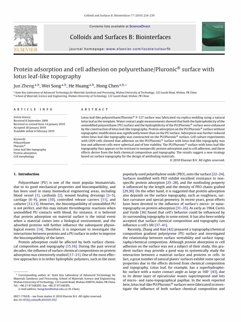
Colloids and Surfaces B: Biointerfaces 77 (2010) 234–239
Contents lists available at ScienceDirect
Colloids and Surfaces B: Biointerfaces
journa l homepage: www.e lsev ier .com/ locate /co lsur fb
rotein adsorption and cell adhesion on polyurethane/Pluronic® surface withotus leaf-like topography
un Zhenga,b, Wei Songa,b, He Huanga,b, Hong Chena,b,∗
State Key Laboratory of Advanced Technology for Materials Synthesis and Processing, Wuhan University of Technology, 122 Luoshi Road, Wuhan, PR ChinaSchool of Materials Science and Engineering, Wuhan University of Technology, 122 Luoshi Road, Wuhan, PR China
r t i c l e i n f o
rticle history:eceived 8 September 2009eceived in revised form 14 January 2010ccepted 28 January 2010vailable online 6 February 2010
a b s t r a c t
Lotus leaf-like polyurethane/Pluronic® F-127 surface was fabricated via replica molding using a naturallotus leaf as the template. Water contact angle measurements showed that both the hydrophobicity of theunmodified polyurethane (PU) surface and the hydrophilicity of the PU/Pluronic® surface were enhancedby the construction of lotus leaf-like topography. Protein adsorption on the PU/Pluronic® surface without
eywords:olyurethaneluronic®
otus leaf-like topography
topographic modification was significantly lower than on the PU surface. Adsorption was further reducedwhen lotus leaf-like topography was constructed on the PU/Pluronic® surface. Cell culture experimentswith L929 cells showed that adhesion on the PU/Pluronic® surface with lotus leaf-like topography waslow and adherent cells were spherical and of low viability. The PU/Pluronic® surface with lotus leaf-liketopography thus appears to be resistant to nonspecific protein adsorption and to cell adhesion, and these
oth chphy f
rotein adsorptionell morphology
effects derive from the bbased on surface topogra
. Introduction
Polyurethane (PU) is one of the most popular biomaterials,ue to its good mechanical properties and biocompatibility, andas been used in many biomedical engineering areas, includinglood vessel [1], cardioids [2], wound healing dressings [3–5],artilage [6–9], joint [10], controlled release carriers [11], andatheter [12,13]. However, the biocompatibility of unmodified PUs not perfect, and this may initiate thrombogenic reactions whennmodified PU contacts with blood, for instance, it is believedhat protein adsorption on material surface is the initial eventhen a material comes into a biological environment, and the
dsorbed proteins will further influence the subsequent physio-ogical events [14]. Therefore, it is important to investigate thenteractions between proteins and a PU surface in order to improvehe biocompatibility of the latter.
Protein adsorption could be affected by both surface chemi-
al composition and topography [15,16]. During the past severalecades, the influence of surface chemical compositions on proteindsorption was extensively studied [17–21]. One of the most effec-ive approaches is to tether hydrophilic polymers, such as the most∗ Corresponding author at: State Key Laboratory of Advanced Technology foraterials Synthesis and Processing, School of Materials Science and Engineering,uhan University of Technology, 122 Luoshi Road, Wuhan 430070, Hubei, PR China.
el.: +86 27 87168305; fax: +86 27 87168305.E-mail address: [email protected] (H. Chen).
927-7765/$ – see front matter © 2010 Elsevier B.V. All rights reserved.oi:10.1016/j.colsurfb.2010.01.032
emical composition and topography. The results suggest a new strategyor the design of antifouling materials.
© 2010 Elsevier B.V. All rights reserved.
popularly used polyethylene oxide (PEO), onto the surface [22–24].Surfaces modified with PEO exhibit excellent resistance to non-specific protein adsorption [25–28], and the nonfouling propertyis influenced by the length and the density of PEO chains grafted[29,30]. On the other hand, it is suggested that protein adsorptionalso depends on the surface topography, such as roughness, sur-face curvature and special geometry. In recent years, great effortshave been devoted to the influence of surface’s micro- or nano-topography on protein adsorption [31–35]. As early as 1964, Curtisand Varde [36] found that cell’s behavior could be influenced byits surrounding topography in some extent. It has also been widelyaccepted that surface chemical composition and topography caninfluence a cell’s life [37–41].
Recently, Zhang and Han [42] prepared a topography/chemicalcomposition gradient polystyrene (PS) surface and investigatedthe relationship between surface wettability and surface topog-raphy/chemical composition. Although protein absorption or celladhesion on the surface was not a subject of their study, this gra-dient surface may provide a good way to systemically study theinteraction between a material surface and proteins or cells. Infact, a great number of natural plants’ surfaces exhibit some specialproperties due to the effects derived from chemical compositionand topography. Lotus leaf, for example, has a superhydropho-
bic surface with a water contact angle as large as 160◦ [43], dueto its dense layer of epicuticular waxes superimposed and lotsof micro- and nano-topographical papillae. In the work reportedhere, lotus leaf-like PU/Pluronic® surfaces were fabricated to inves-tigate the influence of both surface chemical composition and
ces B:
tg
2
2
C1MwDfmwCw
uCoT
2t
cfll
t6Pcu36pstsatwDr
2
1
rp
2
wowpTra
J. Zheng et al. / Colloids and Surfa
opography on surface wettability, protein adsorption and cellrowth.
. Materials and methods
.1. Reagents and physical characterization methods
Poly(dimethyl siloxane) (PDMS) was purchased from Doworning (Sylgard 184, Midland, MI). Polyurethane (PU) pellets (TT-095A, Tecothane®) were obtained from Thermedics (Wilmington,A). Pluronic® F127 was bought from Sigma–Aldrich. Fibrinogenas obtained as a lyophilized powder from CalBioChem. N,N-imethylformamide (DMF) and other reagents were purchased
rom Sinopharm Chemical Reagent Corporation. Penicillin, strepto-ycin, trypsin, trypanblau and methyl thiazolyl tetrazolium (MTT)ere purchased from Amresco. L929 cell was purchased from Chinaenter for Type Culture Collection. Millipore water (18 M�/cm)as used in all experiments.
Advancing and receding water contact angle were measuredsing C201 automatic contact angle meter (Shanghai Solon tech.o. Ltd.) with a drop volume of 2 �L Milli-Q water. Three samplesf each surface (24 pieces in total) were used for the measurements.he data were expressed as the average ± standard deviation.
.2. Fabrication of PU/Pluronic® blend surface with lotus leaf-likeopography
A piece of fresh natural lotus leaf was placed on the bottom ofell culture plate, and then PDMS prepolymer was poured onto theresh lotus leaf. After cured at room temperature for 72 h, the PDMSayer, a film with complementary topography of the original lotuseaf, was peeled off. This PDMS film was called as negative template.
PU pellets were extracted for 3 days (Soxhlet) with methanolo remove impurities, and dried in a vacuum oven overnight at0 ◦C. The extracted PU pellets were dissolved in DMF and thenluronic® was added (PU/Pluronic® = 4:1, w/w). The solution wasast onto the PDMS negative template and degassed for 1 h at vac-um environment. The solvent was slowly evaporated at 60 ◦C fordays in a drying oven, followed by vacuum drying for 24 h at
0 ◦C. Finally, the film was peeled off and lotus leaf-like topogra-hy was formed on the surface of a PU/Pluronic®substrate. Smoothurface on a PU/Pluronic® substrate was also prepared by castinghe PU/Pluronic® solution on a clean glass Petri dish following theame procedure. (In this paper, smooth surfaces without Pluronic®
nd with 20% Pluronic® are designated as PU and PU/P, respec-ively; meanwhile, lotus leaf-like surfaces without Pluronic® andith 20% Pluronic® are designated as PUL and PUL/P, respectively.)iscs approximately 7.29 mm in diameter were cut from the film,
insed in methanol and vacuum dried for 24 h at 45 ◦C.
.3. Protein adsorption
Fibrinogen (Fg) and bovine serum albumin (BSA), labeled with25I (China Gaotong Isotope Co. Ltd.) using the ICl method [44],espectively. In all cases, radioactivity was converted to adsorbedrotein amount.
.3.1. Fibrinogen adsorption from bufferFor study of Fg adsorption from buffer, labeled Fg was mixed
ith unlabeled Fg (1:9, labeled:unlabeled) at a total concentrationf 1.0 mg/mL. Substrates with different surfaces were incubated
ith Fg in Tris buffered saline (TBS, pH 7.4) for 3 h at room tem-erature. Then they were rinsed three times (10 min each withBS), wicked onto filter paper, and transferred to clean tubes foradioactivity determination by gamma counting (Wallac 1480 Wiz-rd, Perkin Elmer).Biointerfaces 77 (2010) 234–239 235
2.3.2. Bovine serum albumin adsorption from cell culture mediumFor study of BSA adsorption from cell culture medium, labeled
BSA was mixed with unlabeled BSA in culture medium (1:10,labeled:unlabeled) at a total concentration of 2.73 mg/mL. Surfaceswere incubated with BSA in cell culture medium for 3 h at 37 ◦Cwith 98% humidity in air containing 5% CO2, the same condition ascell culture. Then they were rinsed three times (10 min each withPBS), wicked onto filter paper, and transferred to clean tubes forradioactivity determination by gamma counting.
2.4. Cell culture
L929 cells were cultured in RPMI medium 1640 (Gibco) with10% fetal bovine serum, 2 mg/mL NaHCO3, 100 U/mL penicillin and10 mg/mL streptomycin and incubated at 37 ◦C with 98% humidityin air containing 5% CO2. Cells were harvested by trypsinizationand trypanblau staining test showed that cell viability was above90% after trypsinization. L929 cells were seeded onto smooth andlotus leaf-like surfaces. When the cells were cultured for 1 day, theculture medium was changed to remove nonadherent cells. Aftercultured 3 days, the cells on these surfaces were tested with MTTand SEM assay.
2.5. MTT assay
Mitochondrial enzymes in living cells can decompose a tetra-zollium salt (MTT) to a colored formazan product. Samples werecarefully stuck on a 24-well tissue culture plate with lotus leaf-like topography facing outside. The cells were seeded with densityof 3 × 10⁴ cells/cm2. After the cells were cultured for 3 days, thediscs were carefully transferred into a 96-well tissue culture plateand 20 �L of 5 mg/mL solution of MTT in 0.1 M phosphate bufferedsaline (PBS, pH 7.4) was added to each well. After the cells werecultured at 37 ◦C for 4–5 h, the medium was carefully removed andthe purple products were dissolved in 0.22 mL of dimethyl sulfox-ide (DMSO). Then the purple solution was transferred into the newculture plate and the optical density (OD) of the dissolved solutewas measured by an ELISA reader (Thermo Labsystems 1500) at490 nm wavelength.
2.6. SEM assay
Samples were fixed with 2.5% glutaraldehyde at room tempera-ture for 30 min. Then cells were gradually dehydrated for 10 minat each concentration using ascending grades of ethanol (30%,50%, 70%, 90%, 95% and 100%). Then cells were further dehydratedwith hexamethyldisilane (HMDS) for 10 min and naturally driedover night at 4 ◦C. Finally, samples were mounted on aluminumspecimen stubs, coated with gold and examined using SEM (JSM-5610LV) at accelerating voltage of 20 kV. The negative template andlotus leaf-like surfaces were also examined using SEM under thesame condition.
3. Results and discussion
3.1. Lotus leaf-like surface topography of the polyurethane(PU)/Pluronic® blend
A PU/Pluronic® blend film with lotus leaf-like topography wasfabricated by replica molding, and typical SEM micrographs areshown in Fig. 1. There were many holes with diameters ranging
from 5 to 10 �m on the PDMS negative template (Fig. 1a and b). Amass of micro-papillae with diameters around 7–10 �m were dis-tributed randomly and evenly on the PU/Pluronic® surface, formingthe lotus leaf-like topography (Fig. 1c–f). These papillae were com-plementary with those holes on PDMS negative template. Some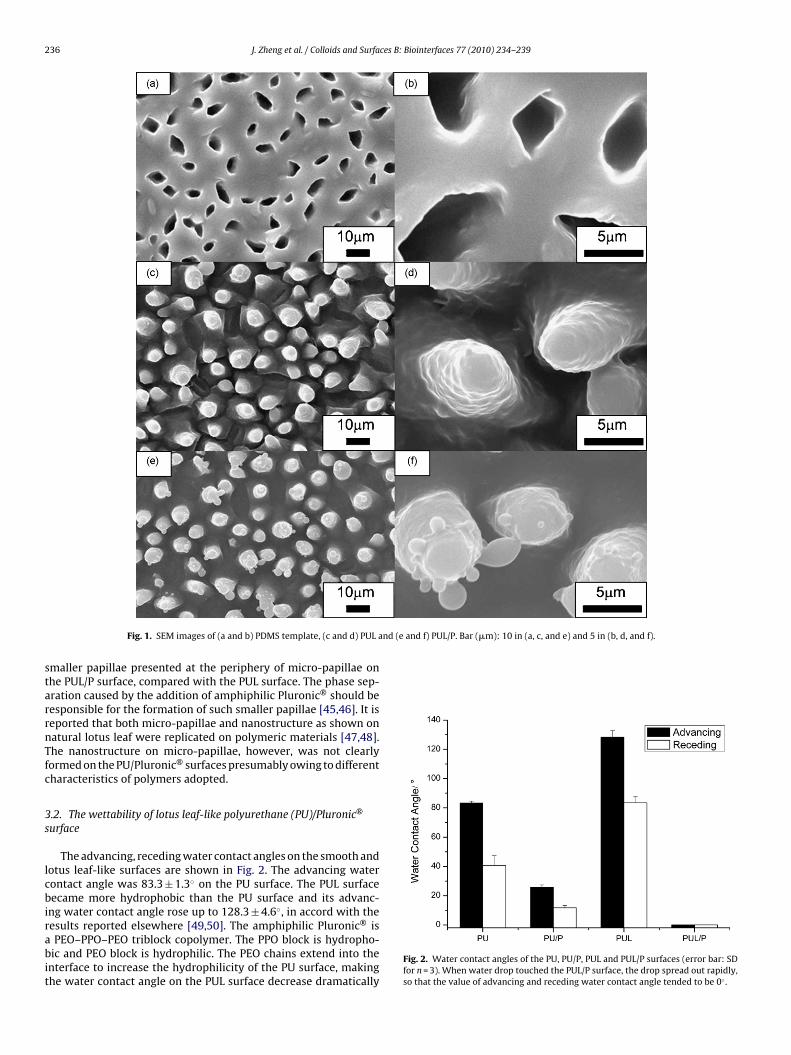
236 J. Zheng et al. / Colloids and Surfaces B: Biointerfaces 77 (2010) 234–239
nd (e and f) PUL/P. Bar (�m): 10 in (a, c, and e) and 5 in (b, d, and f).
starrnTfc
3s
lcbirabit
Fig. 1. SEM images of (a and b) PDMS template, (c and d) PUL a
maller papillae presented at the periphery of micro-papillae onhe PUL/P surface, compared with the PUL surface. The phase sep-ration caused by the addition of amphiphilic Pluronic® should beesponsible for the formation of such smaller papillae [45,46]. It iseported that both micro-papillae and nanostructure as shown onatural lotus leaf were replicated on polymeric materials [47,48].he nanostructure on micro-papillae, however, was not clearlyormed on the PU/Pluronic® surfaces presumably owing to differentharacteristics of polymers adopted.
.2. The wettability of lotus leaf-like polyurethane (PU)/Pluronic®
urface
The advancing, receding water contact angles on the smooth andotus leaf-like surfaces are shown in Fig. 2. The advancing waterontact angle was 83.3 ± 1.3◦ on the PU surface. The PUL surfaceecame more hydrophobic than the PU surface and its advanc-
ng water contact angle rose up to 128.3 ± 4.6◦, in accord with the
esults reported elsewhere [49,50]. The amphiphilic Pluronic® isPEO–PPO–PEO triblock copolymer. The PPO block is hydropho-ic and PEO block is hydrophilic. The PEO chains extend into thenterface to increase the hydrophilicity of the PU surface, makinghe water contact angle on the PUL surface decrease dramatically
Fig. 2. Water contact angles of the PU, PU/P, PUL and PUL/P surfaces (error bar: SDfor n = 3). When water drop touched the PUL/P surface, the drop spread out rapidly,so that the value of advancing and receding water contact angle tended to be 0◦ .

J. Zheng et al. / Colloids and Surfaces B: Biointerfaces 77 (2010) 234–239 237
F(
(idacthfcrwa
3
aiopFwP(fe[so
tomcattPoict
3
f
pressed by both the chemical composition and topography of asurface. The morphology of the cells was observed intuitively bythe SEM measurement (Fig. 6).
ig. 3. The amount of adsorbed fibrinogen on each disc of PU, PU/P, PUL and PUL/Pdiameter of disc is around 7.29 mm; error bar: SD for n = 3).
PUadv = 83.3 ± 1.3◦, PU/Padv = 25.8 ± 1.7◦). However, the advanc-ng water contact angle on the PUL/P surface was less than 10◦
ue to the lotus leaf-like pattern which increases the surfacerea of the PU/P surface, and leads to a higher content of PEOhains on the PUL/P surface than on the PU/P surface. Meanwhile,he surface roughness also plays an important role in enhancingydrophilicity of the PUL/P surface comparing with the PU/P sur-
ace (PUL/Padv < 10◦, PU/Padv = 25.8 ± 1.7◦) [51]. The receding waterontact angle can be influenced by many factors, such as surfaceoughness and surface chemistry. All the values of the recedingater contact angle were less than the advancing water contact
ngle, and the trend was the same (Fig. 2).
.3. Protein adsorption on smooth and lotus leaf-like surfaces
Fg and BSA were used here as model proteins to study proteindsorption on smooth and lotus leaf-like surfaces. Fg is a key proteinnvolved in blood clotting process, thus, the amount of adsorbed Fgn material surface is always quantified to evaluate the blood com-atibility of biomaterial [20]. It is seen that, in Fig. 3, the adsorbedg on the PU surface was 0.289 ± 0.046 �g and on the PUL surfaceas 0.544 ± 0.027 �g, resulting from the larger surface area on the
UL surface. Fg adsorption was much lower on the PU/P surface0.032 ± 0.007 �g) than on the PU surface, a reduction of 88.9% wasound. The decrease is attributed to the protein resistance of PEO,xtended into the interface to provide PEO-rich hydrophilic surface46]. The amount of adsorbed Fg was further reduced on the PUL/Purface (0.016 ± 0.012 �g) than on the PU surface, with a reductionf 94.5%.
It is crucial to investigate the protein adsorption in the cell cul-ure medium in order to correlate the effect of protein adsorptionn a cell’s behavior. There are many proteins in the cell cultureedium. BSA is a major and representative protein in the cell
ulture medium with the highest concentration. Therefore, BSAdsorption in the cell culture medium was carried out. The adsorp-ion trend of BSA from cell culture medium (Fig. 4) is similar tohat of Fg (Fig. 5), while the decline rates of BSA adsorption onU/P (19.7%) and PUL/P surface (67.6%) are not as much as thatf Fg adsorption, compared with the PU surface. The above resultsndicate that protein adsorption was affected by both the surfacehemical composition and topography, and adjusting the two fac-ors might reduce the amount of adsorption to a lower level.
.4. Cell growth on smooth and lotus leaf-like surfaces
Cell viability is presented in Fig. 5 after cells were culturedor 3 days. The OD value of PU, PU/P and PUL/P surfaces is
Fig. 4. The amount of adsorbed bovine serum albumin on each disc of PU, PU/P, PULand PUL/P from cell culture medium (diameter of disc is around 7.29 mm; error bar:SD for n = 5).
1.02 ± 0.19, 0.64 ± 0.11 and 0.47 ± 0.11, respectively. The viabil-ity of cells on a PU/P surface was much lower than on a PUsurface. It is well accepted that the interaction between a celland a surface is mediated by the extracellular matrix proteins[52]. Only when these proteins maintain their normal physiolog-ical function during the interaction, the cell could adhere on thesurface and grow well. In cell culture medium, the hydrophilicPEO blocks in Pluronic® extend into the interface to greatlyenhance surface hydrophilicity and protein resistance (accord-ing to BSA adsorption in Fig. 4), so that the proteins in the ECMand on the cell membrane cannot absorb well on the surface ormaintain their normal physiological function. Therefore the cellscould not grow normally, and the viability of the cells declinedrapidly (ODPU = 1.02 ± 0.19, ODPU/P = 0.64 ± 0.11). When topogra-phy was constructed on the hydrophilic surface, the hydrophilicityof the surface would increase further and strengthen the ability ofrepelling protein adsorption (according to BSA adsorption in Fig. 4).That is the reason for the lowest cell viability on the PUL/P surface(ODPUL/P = 0.47 ± 0.11).
The MTT result indicates that cell viability can be further sup-
Fig. 5. Cell growth on PU, PU/P and PUL/P surfaces for 3 days (error bar: SD for n = 5).

238 J. Zheng et al. / Colloids and Surfaces B: Biointerfaces 77 (2010) 234–239
/P and
wPs(atfcep
4
gafltstlst
A
do
R
[
[
[
[
[
[[
[
[[[
[[[[[[
[[[[[
[
[[[
[[
[[[[
Fig. 6. SEM morphology of cell growth on (a and b) PU, (c and d) PU
The adherent cells on PU are long fusiform and fully spread outith the largest density (Fig. 6a and b). Although the cells on the
U/P surface are fusiform, the quantity is much lower than on the PUurface, namely the number of cells has been strongly suppressedFig. 6c and d). The amount of cells on the PUL/P surface is the lowestnd the cells are more spherical, rather than fusiform. Obviously,he number of cells has been suppressed even larger (Fig. 6e and). This result is consistent with that of the MTT assay, also indi-ating that the growth of cells can be further suppressed by theffects derived from surface chemical composition and topogra-hy.
. Conclusions
A lotus leaf-like PU/Pluronic® surface was fabricated to investi-ate the effects of chemical composition and topography on proteindsorption and cell adhesion. Proteins adsorbed on the PU/P sur-ace were reduced 88.9% compared with the PU surface. When lotuseaf-like topography was constructed on the PU/P surface, however,he value reduced further to 97.1%. In addition, cells on the PUL/Purface were spherical and had the lowest viability. It is clear thathe PU/Pluronic® surface with lotus leaf-like topography has excel-ent properties of nonspecific protein resistance and cell adhesionuppression, lying on the effects of both chemical composition andopography of a surface.
cknowledgements
This work was supported by the National Natural Science Foun-ation of China (90606013, 20634030, 20920102035), the Ministryf Education of China (107080, NCET0606055).
eferences
[1] M.R. Williamson, R. Black, C. Kielty, Biomaterials 27 (2006) 3608.[2] C. Alperin, P.W. Zandstra, K.A. Woodhouse, Biomaterials 26 (2005) 7377.[3] S. Akita, K. Akino, T. Imaizumi, K. Tanaka, K. Anraku, H. Yano, A. Hirano, Burns
32 (2006) 447.[4] H.J. Yoo, H.D. Kim, J. Biomed. Mater. Res. B 85B (2008) 326.
[5] M.S. Khil, D.I. Cha, H.Y. Kim, I.S. Kim, N. Bhattarai, J. Biomed. Mater. Res. B 67B(2003) 675.[6] D. Eyrich, H. Wiese, G. Mailer, D. Skodacek, B. Appel, H. Sarhan, J. Tessmar,
R. Staudenmaier, M.M. Wenzel, A. Goepferich, T. Blunk, Tissue Eng. 13 (2007)2207.
[7] S.L. Chia, K. Gorna, S. Gogolewski, M. Alini, Tissue Eng. 12 (2006) 1945.
[[[[
[
(e and f) PUL/P. Bar (�m): 100 in (a, c, and e) and 10 in (b, d, and f).
[8] Y.C. Liu, K. Webb, K.R. Kirker, N.J. Bernshaw, P.A. Tresco, S.D. Gray, G.D. Prest-wich, Tissue Eng. 10 (2004) 1084.
[9] S. Grad, L. Kupcsik, K. Gorna, S. Gogolewski, M. Alini, Biomaterials 24 (2003)5163.
10] C.J. Spaans, V.W. Belgraver, O. Rienstra, J.H. de Groot, R.P.H. Veth, A.J. Pennings,Biomaterials 21 (2000) 2453.
11] A. Simmons, A.D. Padsalgikar, L.M. Ferris, L.A. Poole-Warren, Biomaterials 29(2008) 2987.
12] J. Poelaert, P. Depuydt, A. De Wolf, S.V. de Velde, I. Herck, S. Blot, J. Thorac.Cardiovasc. Surg. 135 (2008) 771.
13] Y.J. Du, J.L. Brash, G. McClung, L.R. Berry, P. Klement, A.K.C. Chan, J. Biomed.Mater. Res. A 80A (2007) 216.
14] E. Ostuni, R.G. Chapman, M.N. Liang, G. Meluleni, G. Pier, D.E. Ingber, G.M.Whitesides, Langmuir 17 (2001) 6336.
15] P.A. George, B.C. Donose, J.J. Cooper-White, Biomaterials 30 (2009) 2449.16] H. Chen, W. Song, F. Zhou, Z.K. Wu, H. Huang, J.H. Zhang, Q. Lin, B. Yang, Colloids
Surf. B 71 (2009) 275.17] E.S. Hatakeyama, H. Ju, C.J. Gabriel, J.L. Lohr, J.E. Bara, R.D. Noble, B.D. Freeman,
D.L. Gin, J. Membr. Sci. 330 (2009) 104.18] P. Roach, D. Farrar, C.C. Perry, J. Am. Chem. Soc. 127 (2005) 8168.19] Y.H. Kim, D.K. Han, K.D. Park, S.H. Kim, Biomaterials 24 (2003) 2213.20] M.C. Shen, M.S. Wagner, D.G. Castner, B.D. Ratner, T.A. Horbett, Langmuir 19
(2003) 1692.21] P. Kingshott, H.J. Griesser, Curr. Opin. Solid State Mater. Sci. 4 (1999) 403.22] Z.Q. Wu, H. Chen, X.L. Liu, Y.X. Zhang, D. Li, H. Huang, Langmuir 25 (2009) 2900.23] T. Goda, T. Konno, M. Takai, T. Moro, K. Ishihara, Biomaterials 27 (2006) 5151.24] H. Chen, M.A. Brook, H. Sheardown, Biomaterials 25 (2004) 2273.25] H. Chen, L. Yuan, W. Song, Z.K. Wu, D. Li, Prog. Polym. Sci. 33 (2008) 1059.26] H. Chen, Z. Zhang, Y. Chen, M.A. Brook, H. Sheardown, Biomaterials 26 (2005)
2391.27] J.G. Archambault, J.L. Brash, Colloids Surf. B 33 (2004) 111.28] J.H. Lee, H.B. Lee, J.D. Andrade, Prog. Polym. Sci. 20 (1995) 1043.29] L.D. Unsworth, H. Sheardown, J.L. Brash, Biomaterials 26 (2005) 5927.30] J.G. Archambault, J.L. Brash, Colloids Surf. B 39 (2004) 9.31] M.B. Hovgaard, K. Rechendorff, J. Chevallier, M. Foss, F. Besenbacher, J. Phys.
Chem. B 112 (2008) 8241.32] A. Dolatshahi-Pirouz, K. Rechendorff, M.B. Hovgaard, M. Foss, J. Chevallier, F.
Besenbacher, Colloids Surf. B 66 (2008) 53.33] P. Roach, D. Farrar, C.C. Perry, J. Am. Chem. Soc. 128 (2006) 3939.34] M. Lundqvist, I. Sethson, B.H. Jonsson, Langmuir 20 (2004) 10639.35] C. Galli, M.C. Coen, R. Hauert, V.L. Katanaev, P. Gröning, L. Schlapbach, Colloid
Surf. B 26 (2002) 255.36] A.S.G. Curtis, M. Varde, J. Natl. Cancer Inst. 33 (1964) 15.37] C.S. Chen, M. Mrksich, S. Huang, G.M. Whitesides, D.E. Ingber, Science 276
(1997) 1425.38] T.L. Sun, H. Tan, D. Han, Q. Fu, L. Jiang, Small 1 (2005) 959.39] K.Y. Cai, J. Bossert, K.D. Jandt, Colloids Surf. B 49 (2006) 136.40] Z.Z. Wu, Y.P. Zhao, W.S. Kisaalita, Colloids Surf. B 52 (2006) 14.41] B.G. Keselowsky, D.M. Collard, A.J. Garcia, Proc. Natl. Acad. Sci. U.S.A. 102 (2005)
5953.
42] J.L. Zhang, Y.C. Han, Langmuir 24 (2008) 796.43] T.L. Sun, L. Feng, X.F. Gao, L. Jiang, Acc. Chem. Res. 38 (2005) 644.44] M.S. Wagner, T.A. Horbett, D.G. Castner, Biomaterials 24 (2003) 1897.45] B. Wesslen, M. Kober, C. Freij-Larsson, Å. Ljungh, M. Paulsson, Biomaterials 15(1994) 278.46] J.H. Lee, Y.M. Ju, D.M. Kim, Biomaterials 21 (2000) 683.

ces B: Biointerfaces 77 (2010) 234–239 239
[
[[
J. Zheng et al. / Colloids and Surfa
47] M.H. Sun, C.X. Luo, L.P. Xu, H. Ji, O.Y. Qi, D.P. Yu, Y. Chen, Langmuir 21 (2005)8978.
48] Z.Q. Yuan, H. Chen, J. Zhang, Appl. Surf. Sci. 254 (2008) 1593.49] C.W. Guo, L. Feng, J. Zhai, G.J. Wang, Y.L. Song, L. Jiang, D.B. Zhu, ChemPhysChem
5 (2004) 750.
[
[[
50] Q.D. Xie, G.Q. Fan, N. Zhao, X.L. Guo, J. Xu, J.Y. Dong, L.Y. Zhang, Y.J. Zhang, C.C.Han, Adv. Mater. 16 (2004) 1830.
51] R.N. Wenzel, Ind. Eng. Chem. 59 (1936) 335.52] K. Anselme, Biomaterials 21 (2000) 667.