Embed Size (px)
Citation preview
INFECTION AND IMMUNITY,0019-9567/99/$04.0010
Feb. 1999, p. 675–680 Vol. 67, No. 2
Copyright © 1999, American Society for Microbiology. All Rights Reserved.
Protective Immunization with a Novel Membrane Proteinof Plasmodium yoelii-Infected ErythrocytesJAMES M. BURNS, JR.,* ERIC K. ADEEKU, AND PATRICIA D. DUNN
Department of Microbiology, Meharry Medical College,Nashville, Tennessee 37208
Received 24 August 1998/Returned for modification 5 October 1998/Accepted 11 November 1998
Immunization with a particulate fraction of blood-stage antigens was shown previously to protect miceagainst Plasmodium yoelii malaria. To identify antigens inducing the protective response, sera from immunizedmice were used to screen a P. yoelii cDNA expression library. Sequence analysis of one 2.6-kb cDNA clone in-dicated that the identified gene, pypag-1, encoded a novel plasmodial antigen. Two nonoverlapping regions ofpypag-1 were expressed in Escherichia coli. The first recombinant antigen, pAg-1N, contained the N-terminal337 residues, which included a putative transmembrane domain and a region relatively rich in tryptophanresidues. The second recombinant antigen, pAg-1C, contained the remaining C-terminal 211 residues, whichincluded 31 copies of a 5-amino-acid degenerative repeat. Immunoblot studies using rabbit antiserum raisedagainst recombinant pAg-1N showed that the native pypAg-1 protein migrated at approximately 98 kDa, con-siderably slower than its predicted molecular mass of 66 kDa. Immunofluorescence studies localized the ex-pression of the native pypAg-1 protein both to the cytoplasm and at the surface of P. yoelii-infected erythrocytes.Immunization with either pAg-1N or pAg-1C induced a four- to sevenfold reduction in P. yoelii blood-stage pa-rasitemia. As such, pypAg-1 appears to contain at least two distinct protective epitopes. To our knowledge, thisis the first characterization of a protective antigen of P. yoelii that is associated with the erythrocyte membrane.
Malaria is clearly a major public health problem with signif-icant economic and social consequences for many developingcountries, particularly those of sub-Saharan Africa (47). Thereis a great need to implement effective malaria control pro-grams, with the construction of a multivalent subunit vaccinebeing a major consideration (18). The vaccine effort has fo-cused primarily on Plasmodium falciparum, the protozoan par-asite responsible for the majority of severe disease and deathworldwide. Several pre-erythrocytic-stage, asexual blood-stage,and sexual-stage antigens have been identified as vaccine can-didates. Although limited, some encouraging results have beenreported from P. falciparum vaccine trials (12). Nevertheless,additional efforts in the selection of vaccine antigens, adju-vants, and delivery systems will be necessary to improve on thefirst generation of subunit malaria vaccines.
Studies utilizing various animal models of malaria have iden-tified protective antigens and protective immune responses. Inmurine models, the resolution of blood-stage infection resultsin sterilizing immunity which can be primarily cell mediated, aswith Plasmodium chabaudi, or primarily antibody mediated, aswith Plasmodium yoelii (31). Protection against P. yoelii malariacan also be induced by immunization with crude preparationsof blood-stage antigens in various adjuvants (4, 30, 32, 33, 44).Furthermore, studies with P. yoelii homologues of P. falcipa-rum pre-erythrocytic stage (10, 27, 40) and asexual blood-stage(5, 9, 19, 23, 24) antigens have been particularly useful inaugmenting vaccine development efforts.
Previously, we utilized the P. yoelii murine model to inves-tigate immunization-induced protective responses (4). Uponimmunization with a particulate fraction of a blood-stage an-tigen preparation, we induced protection against nonlethal andlethal P. yoelii infection. Both Th1- and Th2-type cytokines
were produced in protected mice, with a bias toward a Th2phenotype evident. In addition, the protection was shown to beB-cell dependent and associated with the production of para-site-specific immunoglobulin G1 (IgG1) and IgG2b antibodies.This particulate antigen fraction, designated pAg, represented25 to 30% of the total P. yoelii blood-stage antigen preparation.Of particular interest, protective immunization induced anti-bodies that recognized a limited subset of six to eight P. yoeliiantigens. We have employed this model to identify novel vac-cine candidate antigens that were selected for their ability toimmunize against blood-stage infection. In this paper, we re-port the identification and characterization of one of the P.yoelii blood-stage antigens that contributed to the pAg-inducedprotective response.
MATERIALS AND METHODS
Experimental infections. Male C57BL/6 or CByB6F1/J (BALB/cByJ 3C57BL/6J) mice, 5 to 6 weeks of age, were purchased from The Jackson Labo-ratories (Bar Harbor, Maine) and housed in the American Association forAccreditation of Laboratory Animal Care-approved Animal Care Facility ofMeharry Medical College, Nashville, Tenn. The lethal 17XL and nonlethal 17Xstrains of P. yoelii were originally obtained from William P. Weidanz (Universityof Wisconsin, Madison) and maintained as cryopreserved stabilates. Blood-stageinfections were initiated by intraperitoneal injection of parasitized erythrocytesobtained from donor mice. Resulting parasitemias were monitored by enumer-ating parasitized erythrocytes in thin tail-blood smears stained with Giemsa stain(17). Routine screenings were conducted throughout these studies to ensure thatmice were free of infection with common mouse pathogens. Sentinel animalshoused with immunized and/or infected mice remained seronegative for a panelof 19 viral and bacterial pathogens (Assessment Plus Profile; Charles RiverLaboratories, Wilmington, Mass.).
Sera. Sera from CByB6F1/J mice (n 5 5) were obtained 1 week followingsecondary immunization with pAg, but prior to infection. Nonimmune controlsera were similarly obtained from adjuvant control mice (n 5 5) immunized withQuil A alone. The generation and characterization of these sera have beenpreviously reported (4).
A high-titer rabbit antiserum against purified recombinant pAg-1N was com-mercially prepared (Lampire Biological Laboratories, Pipersville, Pa.). Briefly,rabbits received a total of five immunizations over an 8-week period. Each dosecontained 200 mg of purified antigen. The first immunization was with antigenemulsified in complete Freund’s adjuvant. For subsequent boosters, antigen wasadministered in incomplete Freund’s adjuvant. Preimmune serum was collected
* Corresponding author. Mailing address: Meharry Medical Col-lege, Department of Microbiology, 1005 D.B. Todd Blvd., Nashville,TN 37208. Phone: (615) 327-5726. Fax: (615) 327-6072. E-mail: [email protected].
675
on June 4, 2018 by guesthttp://iai.asm
.org/D
ownloaded from
prior to the first immunization. Immune serum was collected 2 weeks followingthe last booster.
cDNA cloning and sequence analysis. Parasitized blood was collected from 40C57BL/6 mice infected with P. yoelii 17XL when parasitemias averaged 35 to40%. This blood contained a mixture of ring, trophozoite, and schizont blood-stage parasites. Mouse leukocytes were removed by passage over columns ofmicrocrystalline cellulose (11). Erythrocytes were collected and lysed with 0.01%saponin. Total RNA was extracted from pelleted parasites, and 30 mg ofpoly(A)1 RNA representing 1.5% of total RNA was isolated. A cDNA librarywas constructed in the lambda Uni-ZAP XR expression vector, in a directionallyoriented manner according to the manufacturer’s protocols (Stratagene, LaJolla, Calif.). The library contained 3.25 3 106 clones, 94.8% of which wererecombinant. The majority of cDNA inserts were between 600 bp and 4 kbp inlength.
A pool of sera from mice immunized with pAg was preadsorbed to removeEscherichia coli reactivities (38), and used to screen the P. yoelii cDNA expres-sion library. Bound antibody was detected with 125I-labeled protein A (.30mCi/mg; ICN Biomedicals, Inc., Irvine, Calif.) followed by autoradiography. Ex-cision of pBSK(2) phagemid sequences from identified clones was carried outaccording to the manufacturer’s protocol. Nested deletions of clone pypag-1 weregenerated by exonuclease III digestion (15) and used to obtain the completesequences of the coding and noncoding strands. Single-stranded template wasprepared (6) and the sequence was obtained by Sanger dideoxynucleotide chaintermination with [35S]dATP (1,250 Ci/mmol; NEN Life Science Products, Bos-ton, Mass.) (39) or by fluorescence-based sequencing with an ABI Prism 377automated DNA sequencer (Molecular Biology Core Facility, Meharry MedicalCollege). Sequence analysis utilized a DNASIS for Windows software package(Hitachi Software, South San Francisco, Calif.). Homologies with data banksequences were determined by BLAST search (1) through the National Centerfor Biotechnology Information, National Library of Medicine.
Expression and purification of recombinant pypAg-1. Two pypag-1-encodedrecombinant proteins were produced in E. coli by using the pET plasmid vectorsand T7 RNA polymerase expression system (42). The N-terminal nonrepeatdomain, pAg-1N (amino acids 1 to 337), was PCR amplified from the pypag-1cDNA clone by using oligonucleotide primers 59-AAAAATCCATATGAGTGGGCAACTTAC-39 (primer A, based on nucleotides 593 to 619) and 59-AGCTCGAGCCTATACGGAAGTTGATTCAGATGCAG-39 (primer B, based onnucleotides 1591 to 1625) as 59 and 39 primers. The C-terminal repeat domain,pAg-1C (amino acids 339 to 549), was PCR amplified from the pypag-1 cDNAclone by using oligonucleotide primers 59-CTTCCGTACATATGAACGCTGACG-39 (primer C, based on nucleotides 1606 to 1629) and 59-GTAATCTCGAGTCACTATATAAGTATCG-39 (primer D, based on nucleotides 2239 to 2266)as 59 and 39 primers. To facilitate subcloning, NdeI and XhoI restriction siteswere incorporated into the 59 and 39 primers, respectively. The amplified frag-ments were gel purified, digested with NdeI and XhoI, and ligated into NdeI/XhoI-digested pET-15b (Novagen, Madison, Wis.). E. coli BL21(DE3)(pLysS)was used for expression. Each recombinant protein contained 20 plasmid-en-coded amino acids fused to its N terminus, including six histidine residues.
Pellets of induced bacterial cultures (approximately 1 g [wet weight]) wereresuspended in 15 ml of TNE (50 mM Tris-HCl [pH 8.0], 100 mM NaCl, 10 mMEDTA) and lysed by treatment with lysozyme and sonication. The 43-kDapAg-1N recombinant protein was purified from an insoluble inclusion bodyfraction. The inclusion body fraction was recovered by centrifugation for 10 minat 2,000 3 g and washed twice in TNE containing 0.1% deoxycholate and twicein TNE containing 2 M urea. The final pellet was solubilized in 10 ml of 100 mMTris-HCl (pH 8.5)–10 mM EDTA containing 6 M guanidine-HCl. Followingdialysis, pAg-1N was purified by nickel-chelate affinity chromatography in thepresence of 6 M guanidine-HCl. Eluted material remained soluble followingdialysis into 5 mM glycine-HCl (pH 3.0).
Following induction and lysis as described above, the 27-kDa pAg-1C proteinwas purified from a soluble fraction of lysed bacteria obtained following centrif-ugation for 30 min at 25,000 3 g. A 50 to 80% ammonium sulfate fraction of theinitial lysate was separated by preparative isoelectric focusing with a Rotofor IEFcell (Bio-Rad Laboratories, Hercules, Calif.) in the presence of 1% 3/10 am-pholytes and 8 M urea. Fractions containing recombinant pAg-1C were pooled.Final antigen purification was by nickel-chelate affinity chromatography in thepresence of 6 M urea. The eluted protein was dialyzed into 10 mM Tris-HCl (pH8.0). Protein concentrations were determined by using the Pierce bicinchoninicacid protein assay (Pierce, Rockford, Ill.), and purity was assessed by Coomassieblue staining following sodium dodecyl sulfate-polyacrylamide gel electrophore-sis (SDS-PAGE) (26). Endotoxin levels were monitored by using a chromogenicLimulus amebocyte lysate assay (BioWhittaker, Inc., Walkersville, Md.). Endo-toxin levels were less than 2.5 endotoxin units (EU) per mg of purified recom-binant antigen.
Immunoblot analysis. Total P. yoelii 17XL blood-stage antigens and the pAgfraction were prepared as previously described (4) and solubilized in SDS-PAGEsample buffer containing 2.5% SDS. Total blood-stage antigens (10 mg/lane), thepAg fraction (10 mg/lane), and purified recombinant pAg-1 antigens (1 mg/lane)were separated by SDS-PAGE on 10% polyacrylamide gels as described byLaemmli (26). Separated proteins were transferred electrophoretically to nitro-cellulose membranes (46). Membranes were blocked for 1 h with TBS (25 mMTris-HCl [pH 8], 150 mM NaCl) containing 5% nonfat dried milk. Blots were
incubated for 1 h with pAg immunization or control mouse sera (1:100) orpreimmune or immune rabbit serum (1:500) diluted in TBS–0.1% Tween 20containing 1% bovine serum albumin. Bound antibody was detected with 125I-labeled protein A (ICN Biomedicals) followed by autoradiography.
Indirect immunofluorescence assay. Parasitized blood was collected from miceinfected with P. yoelii 17XL when parasitemias averaged 15 to 20%. Erythrocyteswere pelleted by centrifugation for 10 min at 700 3 g and washed three times inphosphate-buffered saline (PBS). The final pellet was resuspended in an equalvolume of PBS containing 1% gelatin. Thin blood smears were prepared, airdried, and fixed in acetone-methanol (1:1) for 20 min at 220°C. Fixed slides wereair dried and stored at 4°C until use. Prior to use, slides were warmed to roomtemperature and equilibrated with PBS. Fixed cells were incubated for 30 min at37°C in a humidified chamber with preimmune or immune rabbit serum (1:200)diluted in PBS. Slides were washed three times in PBS and then incubated asabove with a fluorescein isothiocyanate-conjugated goat anti-rabbit IgG serum(Sigma Chemical Company, St. Louis, Mo.) diluted 1:160 in PBS. Stained cellswere then washed, mounted with Fluoromount-G (Southern Biotechnology As-sociates, Inc., Birmingham, Ala.), and visualized by fluorescence microscopy.
Immunizations. Recombinant pAg-1N and pAg-1C were each encapsulatedinto negatively charged liposomes containing L-a-phosphatidylcholine, dicetylphosphate, and cholesterol at a ratio of 7:2:1 (Sigma Chemical Company). Theencapsulation procedure was essentially as described previously (35), with minormodifications. Briefly, the liposome mixture in chloroform was evaporated todryness under a stream of nitrogen, leaving a thin coat on the walls of a 15-mlglass Corex tube. Recombinant antigens were added in aqueous solution at aconcentration of 1.25 mg/ml and vortexed for 10 min. The mixture was dilutedwith 4 volumes of PBS, and liposomes were pelleted by centrifugation for 15 minat 3,500 3 g. The supernatant was removed, and liposomes were resuspended inPBS. Encapsulation efficiency was monitored by SDS-PAGE and immunoblotanalysis using known quantities of purified recombinant pAg-1 antigens as stan-dards.
Groups of CByB6F1/J mice (n 5 5) were immunized subcutaneously with 10to 20 mg of liposome-associated recombinant antigen, utilizing 5 mmol of lipidper mouse per immunization. Quil A (Accurate Chemical and Scientific Corpo-ration, Westbury, N.Y.) was added to the liposome preparation prior to immu-nization, at a dose of 25 mg per mouse. Control animals were immunized withempty liposomes plus Quil A. Following the primary immunization, animals wereboosted twice at 3-week intervals with the same dose of antigen and adjuvant.Seven days later, mice were challenged with 106 P. yoelii 17X-parasitized eryth-rocytes, and blood parasitemias were monitored. The statistical significance ofdifferences in the mean peak parasitemia between groups was calculated withStudent’s t test.
Nucleotide sequence accession number. The sequence reported in this paperhas been deposited in the GenBank database under accession no. AF103869.
RESULTS
Cloning and sequence analysis of the pypag-1 gene of P. yoe-lii. A pool of sera from mice protected against P. yoelii malariaby pAg immunization was used to screen a P. yoelii cDNAexpression library. Screening of approximately 23,000 recom-binants yielded 26 seroreactive clones, with cDNA inserts rang-ing from 500 bp to 2.75 kb. Through cross-hybridization stud-ies, 23 of the P. yoelii cDNA clones were grouped and shown torepresent four distinct antigen genes (data not shown). Theworking designations assigned to these genes were pypag-1,pypag-2, pypag-3, and pypag-4. Sixteen clones contained pypag-1gene sequences.
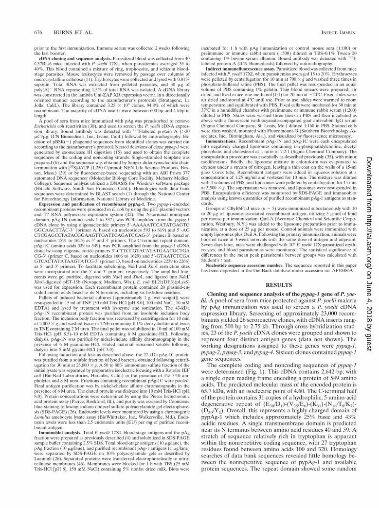
The complete coding and noncoding sequences of pypag-1were determined (Fig. 1). This cDNA contains 2,642 bp, witha single open reading frame encoding a protein of 549 aminoacids. The predicted molecular mass of the encoded protein is65.7 kDa, with an isoelectric point of 4.60. The C-terminal halfof the protein contains 31 copies of a hydrophilic, 5-amino-aciddegenerative repeat of (E30/D1)-(V25/E6)-(K31)-(N16/T8/K7)-(D30/Y1). Overall, this represents a highly charged domain ofpypAg-1 which includes approximately 25% basic and 43%acidic residues. A single transmembrane domain is predictednear its N terminus between amino acid residues 40 and 59. Astretch of sequence relatively rich in tryptophan is apparentwithin the nonrepetitive coding sequence, with 27 tryptophanresidues found between amino acids 100 and 320. Homologysearches of data bank sequences revealed little homology be-tween the nonrepetitive sequence of pypAg-1 and availableprotein sequences. The repeat domain showed some random
676 BURNS ET AL. INFECT. IMMUN.
on June 4, 2018 by guesthttp://iai.asm
.org/D
ownloaded from
similarity with degenerative repeats of otherwise unrelated ma-larial antigens.
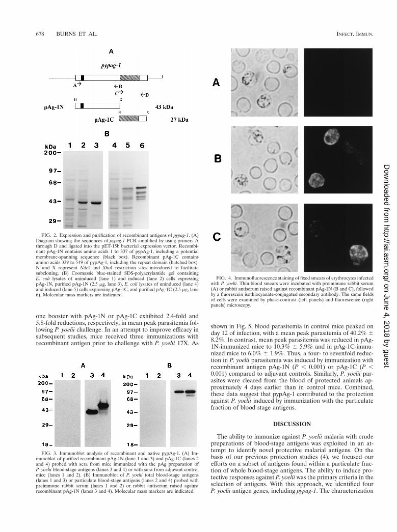
Expression and purification of recombinant pAg-1 proteins.Constructs were made to express two nonoverlapping regionsof pypag-1 in E. coli (Fig. 2A). Recombinant antigen pAg-1Ncontained residues 1 to 337 of the N terminus of pypAg-1.Recombinant antigen pAg-1C contained residues 339 to 549 ofthe C terminus of pypAg-1. pAg-1C contained the repeat do-main followed by 54 nonrepetitive amino acids. Both recom-binant proteins contained an N-terminal six-residue histidinetag which was utilized for purification by nickel-chelate affinitychromatography. A Coomassie blue-stained polyacrylamidegel of the purified recombinant proteins is shown in Fig. 2B.pAg-1N migrated close to its predicted molecular mass of 43kDa (Fig. 2B, lane 3). pAg-1C migrated much slower than itspredicted molecular mass of 27 kDa, as a cluster of four bandsranging from 45 to 55 kDa (Fig. 2B, lane 6). The aberrantmobility of pAg-1C was most likely related to the repetitivenature of this protein sequence.
Immunoblot analysis of pypAg-1 antigens. The reactivity ofpAg-1N and pAg-1C with sera from mice protected againstP. yoelii by pAg-plus-Quil A immunization was assessed byimmunoblot. As shown in Fig. 3A, both pAg-1N (lane 3) andpAg-1C (lane 4) were strongly recognized by pAg immuniza-tion sera. No reactivity was observed with control sera frommice immunized with Quil A alone (Fig. 3A, lanes 1 and 2). Assuch, protective immunization with a mixture of particulateblood-stage antigens of P. yoelii induced antibodies that rec-ognized both the repeat and nonrepeat domains of pypAg-1.
To identify native pypAg-1, a high-titer polyclonal rabbitserum was raised against recombinant antigen pAg-1N. Immu-noblot analysis using this antiserum identified a 98-kDa pro-tein present in a total antigen preparation (Fig. 3B, lane 3) aswell as the particulate antigen fraction (Fig. 3B, lane 4) ofP. yoelii 17XL blood-stage parasites. As with recombinant pAg-1C, native pypAg-1 migrated considerably slower than its pre-dicted molecular mass of 65 to 66 kDa. No reactivity wasobserved with preimmune rabbit serum (Fig. 3B, lanes 1 and2).
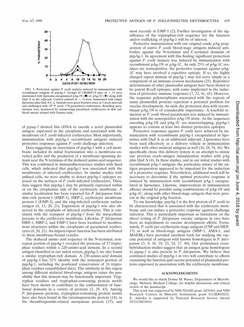
Localization of pypAg-1 expression. The localization ofpypAg-1 expression in parasitized erythrocytes was assessed byindirect immunofluorescence. The reactivity of the polyclonalrabbit serum raised against recombinant pAg-1N was assayedwith thin blood films of a mixture of P. yoelii 17XL blood-stageparasites that were air dried and fixed. As shown in Fig. 4B andC, a granular pattern of fluorescence was observed within thecytoplasms of infected erythrocytes. This fluorescence was notassociated with the trophozoite itself. Of significance, positivefluorescence was also noted at the erythrocyte membrane.However, pypAg-1-specific fluorescence was not observed onsurface of P. yoelii-infected erythrocytes when wet-mount prep-arations of unfixed cells were assayed (data not shown). Nofluorescence of P. yoelii-parasitized erythrocytes incubatedwith preimmune rabbit serum was observed (Fig. 4A).
Immunization with recombinant pAg-1. To assess the vac-cine potential of pypAg-1, mice were immunized with lipo-some-encapsulated pAg-1N or pAg-1C, along with Quil A asthe adjuvant. Initially, animals that were immunized and given
FIG. 1. Sequence analysis of pypag-1. The DNA and deduced amino acid sequences of the 2,642-bp insert of pypag-1 are shown. Tryptophan residues are in bold.Each 5-amino-acid repeat is underlined. A putative transmembrane segment (residues 40 to 59) is double underlined.
VOL. 67, 1999 PROTECTIVE ANTIGEN OF P. YOELII-INFECTED ERYTHROCYTES 677
on June 4, 2018 by guesthttp://iai.asm
.org/D
ownloaded from
one booster with pAg-1N or pAg-1C exhibited 2.4-fold and5.8-fold reductions, respectively, in mean peak parasitemia fol-lowing P. yoelii challenge. In an attempt to improve efficacy insubsequent studies, mice received three immunizations withrecombinant antigen prior to challenge with P. yoelii 17X. As
shown in Fig. 5, blood parasitemia in control mice peaked onday 12 of infection, with a mean peak parasitemia of 40.2% 68.2%. In contrast, mean peak parasitemia was reduced in pAg-1N-immunized mice to 10.3% 6 5.9% and in pAg-1C-immu-nized mice to 6.0% 6 1.9%. Thus, a four- to sevenfold reduc-tion in P. yoelii parasitemia was induced by immunization withrecombinant antigen pAg-1N (P , 0.001) or pAg-1C (P ,0.001) compared to adjuvant controls. Similarly, P. yoelii par-asites were cleared from the blood of protected animals ap-proximately 4 days earlier than in control mice. Combined,these data suggest that pypAg-1 contributed to the protectionagainst P. yoelii induced by immunization with the particulatefraction of blood-stage antigens.
DISCUSSION
The ability to immunize against P. yoelii malaria with crudepreparations of blood-stage antigens was exploited in an at-tempt to identify novel protective malarial antigens. On thebasis of our previous protection studies (4), we focused ourefforts on a subset of antigens found within a particulate frac-tion of whole blood-stage antigens. The ability to induce pro-tective responses against P. yoelii was the primary criteria in theselection of antigens. With this approach, we identified fourP. yoelii antigen genes, including pypag-1. The characterization
FIG. 2. Expression and purification of recombinant antigens of pypag-1. (A)Diagram showing the sequences of pypag-1 PCR amplified by using primers Athrough D and ligated into the pET-15b bacterial expression vector. Recombi-nant pAg-1N contains amino acids 1 to 337 of pypAg-1, including a potentialmembrane-spanning sequence (black box). Recombinant pAg-1C containsamino acids 339 to 549 of pypAg-1, including the repeat domain (hatched box).N and X represent NdeI and XhoI restriction sites introduced to facilitatesubcloning. (B) Coomassie blue-stained SDS-polyacrylamide gel containingE. coli lysates of uninduced (lane 1) and induced (lane 2) cells expressingpAg-1N, purified pAg-1N (2.5 mg, lane 3), E. coli lysates of uninduced (lane 4)and induced (lane 5) cells expressing pAg-1C, and purified pAg-1C (2.5 mg, lane6). Molecular mass markers are indicated.
FIG. 3. Immunoblot analysis of recombinant and native pypAg-1. (A) Im-munoblot of purified recombinant pAg-1N (lane 1 and 3) and pAg-1C (lanes 2and 4) probed with sera from mice immunized with the pAg preparation ofP. yoelii blood-stage antigens (lanes 3 and 4) or with sera from adjuvant controlmice (lanes 1 and 2). (B) Immunoblot of P. yoelii total blood-stage antigens(lanes 1 and 3) or particulate blood-stage antigens (lanes 2 and 4) probed withpreimmune rabbit serum (lanes 1 and 2) or rabbit antiserum raised againstrecombinant pAg-1N (lanes 3 and 4). Molecular mass markers are indicated.
FIG. 4. Immunofluorescence staining of fixed smears of erythrocytes infectedwith P. yoelii. Thin blood smears were incubated with preimmune rabbit serum(A) or rabbit antiserum raised against recombinant pAg-1N (B and C), followedby a fluorescein isothiocyanate-conjugated secondary antibody. The same fieldsof cells were examined by phase-contrast (left panels) and fluorescence (rightpanels) microscopy.
678 BURNS ET AL. INFECT. IMMUN.
on June 4, 2018 by guesthttp://iai.asm
.org/D
ownloaded from
of pypag-1 showed this cDNA to encode a novel plasmodialantigen, expressed in the cytoplasm and associated with themembrane of P. yoelii-infected erythrocytes. Most importantly,immunization with pypAg-1 recombinant antigens inducedprotective responses against P. yoelii challenge infection.
Data suggesting an association of pypAg-1 with a cell mem-brane included its initial fractionation into a membrane-en-riched pellet and the prediction of a membrane-spanning do-main near the N terminus of the deduced amino acid sequence.This was confirmed in immunofluorescence studies with fixedcells, which indicated that pypAg-1 was associated with themembranes of infected erythrocytes. In similar studies withunfixed cells, we were unable to detect pypAg-1 epitopes ex-posed on the surfaces of P. yoelii-infected erythrocytes. Thesedata suggest that pypAg-1 may be primarily expressed withinor on the cytoplasmic side of the erythrocyte membrane. Asimilar localization has been reported for P. falciparum histi-dine-rich protein-1 (HRP-1), HRP-2, erythrocyte membraneprotein-2 (EMP-2), and the ring-infected erythrocyte surfaceantigen (8, 13, 20, 21). Expression of pypAg-1 was also ob-served in the cytoplasm of infected erythrocytes. This is con-sistent with the transport of pypAg-1 from the intracellularparasite to the erythrocyte membrane. Likewise, P. falciparumHRP-1, HRP-2, and EMP-2 have been localized to membra-nous structures within the cytoplasms of parasitized erythro-cytes (8, 20, 21). An import/export function has been attributedto these membrane-bound vesicles.
The deduced amino acid sequence of the N-terminal, non-repeat portion of pypAg-1 revealed the presence of 27 trypto-phan residues within a 220-amino-acid domain. In a secondantigen identified in our initial screen, pypAg-3, we also founda similar tryptophan-rich domain. A 230-amino-acid domainof pypAg-3 has 32% identity with the nonrepeat portion ofpypAg-1, including the positional conservation of 24 trypto-phan residues (unpublished data). The similarity in this regionamong different malarial blood-stage antigens raises the pos-sibility that this domain may be functionally important. Tryp-tophan residues and tryptophan-containing protein motifshave been shown to contribute to the conformation of func-tional domains in a variety of proteins (3, 29, 43). AmongP. falciparum proteins, tryptophan-containing protein motifshave also been found in the circumsporozoite protein (14), inthe thrombospondin-related anonymous protein (37), and
most recently in EMP-1 (2). Further investigation of the sig-nificance of the tryptophan-rich sequence for the functionand/or trafficking of pypAg-1 will be of interest.
Protective immunization with our original crude pAg prep-aration of native P. yoelii blood-stage antigens induced anti-bodies against the N-terminal and C-terminal domains ofpypAg-1. In agreement with this finding, significant protectionagainst P. yoelii malaria was induced by immunization withrecombinant pAg-1N or pAg-1C. As only 25% of pAg-1C res-idues are nonrepetitive, the protective response against pAg-1C may have involved a repetitive epitope. If so, the highlycharged repeat domain of pypAg-1 may not serve simply as acomponent of an immune evasion mechanism (25). Repetitivedeterminants of other plasmodial antigens have been shown tobe potent B-cell epitopes, with some implicated in the induc-tion of protective immune responses (7, 22, 41, 45). However,the sequence polymorphisms associated with the repeats ofmany plasmodial proteins represent a potential problem forvaccine development. As such, the protection data with recom-binant pAg-1N is of considerable importance. A fourfold re-duction in P. yoelii blood parasitemia was induced by immuni-zation with the nonrepetitive pAg-1N alone. As the sequencesencoding pAg-1N and pAg-1C are nonoverlapping, pypAg-1appears to contain at least two distinct protective epitopes.
Protective responses against P. yoelii were achieved by im-munization with recombinant pypAg-1 encapsulated in lipo-somes with Quil A as an additional adjuvant. Liposomes havebeen used effectively as a delivery vehicle in immunizationstudies with other malarial antigens as well (16, 28, 34, 36). Wespecifically chose this delivery system in an attempt to mimicour previous crude-antigen immunization studies with pAgplus Quil A (4). In these studies, and in our initial studies withrecombinant pAg-1 antigens, the particulate nature of the an-tigen preparation appeared to be important for the inductionof a protective response. Nevertheless, additional work will benecessary to determine if the optimal protective response isinduced by immunization with recombinant pAg-1 encapsu-lated in liposomes. Likewise, improvement in immunizationefficacy should be possible using combinations of pAg-1N andpAg-1C, along with other blood-stage antigens that were pres-ent in the original pAg fraction.
To our knowledge, pypAg-1 is the first protein of P. yoelii tobe characterized that is associated with the erythrocyte mem-brane and can induce a protective response against challengeinfection. This is particularly important as limitations on thedirect testing of P. falciparum vaccine antigens in vivo havebeen an impediment to vaccine development efforts. Fortu-nately, P. yoelii pre-erythrocytic-stage antigens (CSP and HEP-17) as well as blood-stage antigens (MSP-1, AMA-1, andMAEBL) have provided excellent tools for studying the vac-cine potential of antigens with known homologues in P. falci-parum (5, 9, 10, 19, 23, 24, 27, 40). Our preliminary cross-hybridization studies suggest that an antigen gene homologousto pypag-1 is also present in P. falciparum. We believe thatcontinued studies of pypAg-1 in vivo will contribute to effortsexamining the function and vaccine potential of plasmodial pro-teins expressed in association with the erythrocyte membrane.
ACKNOWLEDGMENTS
We would like to thank Donna M. Russo, Department of Microbi-ology, Meharry Medical College, for helpful discussions and criticalreview of the manuscript.
This work was supported by NIH-NIAID grant AI35661 and NIHResearch Centers in Minority Institutions grant G12RR03032.E. Adeeku is supported by National Research Service Award5F31GM18916.
FIG. 5. Protection against P. yoelii malaria induced by immunization withrecombinant antigens of pypAg-1. Groups of CByB6F1/J mice (n 5 5) wereimmunized with liposome-encapsulated pAg-1N (F) or pAg-1C (}) along withQuil A as the adjuvant. Control animals (n 5 5) were immunized with emptyliposomes plus Quil A (h). Animals were given boosters twice at 3-week intervalsand challenged with 106 P. yoelii 17X-parasitized erythrocytes. Resulting para-sitemias were monitored by enumerating parasitized erythrocytes in thin tail-blood smears stained with Giemsa stain.
VOL. 67, 1999 PROTECTIVE ANTIGEN OF P. YOELII-INFECTED ERYTHROCYTES 679
on June 4, 2018 by guesthttp://iai.asm
.org/D
ownloaded from

REFERENCES
1. Altschul, S. F., T. L. Madden, A. A. Schaffer, J. Zhang, Z. Zhang, W. Miller,and D. J. Lipman. 1997. Gapped BLAST and PSI-BLAST: a new generationof protein database search programs. Nucleic Acids Res. 25:3389–3402.
2. Baruch, D. I., X. C. Ma, H. B. Singh, X. Bi, B. L. Pasloske, and R. J. Howard.1997. Identification of a region of PfEMP1 that mediates adherence ofPlasmodium falciparum infected erythrocytes to CD36: conserved functionwith variant sequence. Blood 90:3766–3775.
3. Bazan, J. F. 1990. Structural design and molecular evolution of a cytokinereceptor superfamily. Proc. Natl. Acad. Sci. USA 87:6934–6938.
4. Burns, Jr., J. M., P. D. Dunn, and D. M. Russo. 1997. Protective immunityagainst Plasmodium yoelii malaria induced by immunization with particulateblood-stage antigens. Infect. Immun. 65:3138–3145.
5. Burns, Jr., J. M., T. M. Daly, A. B. Vaidya, and C. A. Long. 1988. The 39portion of the gene for a Plasmodium yoelii merozoite surface antigen en-codes the epitope recognized by a protective monoclonal antibody. Proc.Natl. Acad. Sci. USA 85:602–606.
6. Burns, Jr., J. M., W. G. Shreffler, D. E. Rosman, P. R. Sleath, and S. G. Reed.1992. Identification and synthesis of a major conserved antigenic epitope ofTrypanosoma cruzi. Proc. Natl. Acad. Sci. USA 89:1239–1243.
7. Collins, W. E., R. F. Anders, M. Pappaioanou, G. H. Campbell, G. V. Brown,D. J. Kemp, R. L. Coppel, J. C. Skinner, P. M. Andrysiak, J. F. Favaloro,L. M. Corcoran, J. R. Broderson, G. F. Mitchell, and C. C. Campbell. 1986.Immunization of Aotus monkeys with recombinant proteins of an erythrocytesurface antigen of Plasmodium falciparum. Nature 323:259–262.
8. Coppel, R. L., J. G. Culvenor, A. E. Bianco, P. E. Crewther, H. Stahl, G. V.Brown, R. F. Anders, and D. J. Kemp. 1986. Variable antigen associated withthe surface of erythrocytes infected with the mature stages of Plasmodiumfalciparum. Mol. Biochem. Parasitol. 20:265–277.
9. Daly, T. M., and C. A. Long. 1995. Humoral response to a carboxyl-terminalregion of the merozoite surface protein-1 plays a predominant role in con-trolling blood-stage infection in rodent malaria. J. Immunol. 155:236–243.
10. Doolan, D. L., R. C. Hedstrom, W. O. Rogers, Y. Charoenvit, M. Rogers,P. de la Vega, and S. L. Hoffman. 1996. Identification and characterization ofthe protective hepatocyte erythrocyte protein 17 kDa gene of Plasmodiumyoelii, homolog of Plasmodium falciparum exported protein 1. J. Biol. Chem.271:17861–17868.
11. Eling, W. 1977. Ficoll fractionation for the separation of parasitized eryth-rocytes from malaria infected blood. Bull. W. H. O. 55:105–114.
12. Engers, H. D., and T. Godal. 1998. Malaria vaccine development: currentstatus. Parasitol. Today 14:56–64.
13. Foley, M., L. Tilley, W. H. Sawyer, and R. F. Anders. 1991. The ring-infectederythrocyte surface antigen of Plasmodium falciparum associates with spec-trin in the erythrocytes membrane. Mol. Biochem. Parasitol. 46:137–147.
14. Goundis, D., and K. B. M. Reid. 1988. Properdin, the terminal complementcomponents, thrombospondin and the circumsporozoite protein of malariaparasites contain similar sequence motifs. Nature 335:82–85.
15. Henikoff, S. 1987. Unidirectional digestion with exonuclease III in DNAsequence analysis. Methods Enzymol. 155:156–165.
16. Heppner, D. G., D. M. Gordon, M. Gross, B. Wellde, W. Leitner, U. Krzych,I. Schneider, R. A. Wirtz, R. L. Richards, A. Trofa, T. Hall, J. C. Sadoff,P. Boerger, C. R. Alving, D. R. Sylvester, T. G. Porter, and W. R. Ballou.1996. Safety, immunogenicity, and efficacy of Plasmodium falciparum repeat-less circumsporozoite protein vaccine encapsulated in liposomes. J. Infect.Dis. 174:361–366.
17. Hoffman, E. J., W. P. Weidanz, and C. A. Long. 1984. Susceptibility of CXBrecombinant inbred mice to murine plasmodia. Infect. Immun. 43:981–985.
18. Hoffman, S. L., and L. H. Miller. 1996. Perspectives on malaria vaccinedevelopment, p. 1–13. In S. L. Hoffman (ed.), Malaria vaccine development:a multi-immune response approach. ASM Press, Washington, D.C.
19. Holder, A. A., and R. R. Freeman. 1981. Immunization against blood-stagerodent malaria using purified parasite antigens. Nature 294:361–364.
20. Howard, R. J. 1988. Plasmodium falciparum proteins at the host erythrocytemembrane: their biological and immunological significance and novel para-site organelles which deliver them to the cell surface, p. 111–145. In P. T.Englund and A. Sher (ed.), The biology of parasitism. Alan R. Liss, Inc., NewYork, N.Y.
21. Howard, R. J., S. Uni, M. Aikawa, S. B. Aley, J. H. Leech, A. M. Lew,T. E. Wellems, J. Rener, and D. W. Taylor. 1986. Secretion of malariahistidine-rich protein (PFHRPII) from Plasmodium falciparum infectederythrocytes. J. Cell Biol. 103:1269–1277.
22. Inselburg, J., I. C. Bathurst, J. Kansopon, G. L. Barchfeld, P. J. Barr, andR. N. Rossan. 1993. Protective immunity induced in Aotus monkeys by arecombinant SERA protein of Plasmodium falciparum: adjuvant effects oninduction of protective immunity. Infect. Immun. 61:2041–2047.
23. Kappe, S. H., and J. H. Adams. 1996. Sequence analysis of the apicalmembrane antigen-1 genes (ama-1) of Plasmodium yoelii yoelii and Plasmo-
dium berghei. Mol. Biochem. Parasitol. 78:279–283.24. Kappe, S. H. I., A. R. Noe, T. S. Fraser, P. L. Blair, and J. H. Adams. 1998.
A family of chimeric erythrocyte binding proteins of malaria parasites. Proc.Natl. Acad. Sci. USA 95:1230–1235.
25. Kemp, D. J., R. L. Coppel, and R. F. Anders. 1987. Repetitive proteins andgenes of malaria. Annu. Rev. Microbiol. 41:181–208.
26. Laemmli, U. K. 1970. Cleavage of structural proteins during the assembly ofthe head of bacteriophage T4. Nature 227:680–685.
27. Lal, A. A., V. F. de la Cruz, J. A. Welsh, Y. Charoenvit, W. L. Maloy, and T. F.McCutchan. 1987. Structure of the gene encoding the circumsporozoiteprotein of Plasmodium yoelii. J. Biol. Chem. 262:2937–2940.
28. Ling, I. T., S. A. Ogun, P. Momin, R. L. Richards, N. Garcon, J. Cohen, W. R.Ballou, and A. A. Holder. 1997. Immunization against the murine malariaparasite Plasmodium yoelii using recombinant protein with adjuvants devel-oped for clinical use. Vaccine 15:1562–1567.
29. Macias, M. J., M. Hyvonen, E. Baraldi, J. Schultz, M. Sudol, M. Saraste, andH. Oschkinat. 1996. Structure of the WW domain of a kinase-associatedprotein complexed with a proline-rich peptide. Nature 382:646–649.
30. McColm, A. A., R. Bomford, and L. Dalton. 1982. A comparison of saponinfor the potentiation of protective immunity by killed Plasmodium yoeliivaccine in the mouse. Parasite Immunol. 4:337–347.
31. Melancon-Kaplan, J., J. M. Burns, Jr., A. B. Vaidya, H. K. Webster, andW. P. Weidanz. 1993. Malaria, p. 302–351. In K. Warren (ed.), Immunologyand molecular biology of parasitic infections, 3rd ed. Blackwell ScientificPublications, Inc., Cambridge, Mass.
32. Playfair, J. H. L., and J. B. De Souza. 1987. Recombinant gamma interferonis a potent adjuvant for a malaria vaccine in mice. Clin. Exp. Immunol. 67:5–10.
33. Playfair, J. H. L., J. B. De Souza, and B. J. Cottrell. 1977. Protection of miceagainst malaria by a killed vaccine: differences in effectiveness against Plas-modium yoelii and Plasmodium berghei. Immunology 33:507–515.
34. Pye, D., K. L. Vandenberg, S. L. Dyer, D. O. Irving, N. H. Goss, G. C.Woodrow, A. Saul, C. R. Alving, R. L. Richards, W. R. Ballou, M. Wu, K.Skoff, and R. F. Anders. 1997. Selection of an adjuvant for vaccination withthe malaria antigen, MSA-2. Vaccine 15:1017–1023.
35. Reed, S. G., M. Barral-Netto, and J. A. Inverso. 1984. Treatment of exper-imental visceral leishmaniasis with lymphokine encapsulated in liposomes.J. Immunol. 132:3116–3119.
36. Richards, R. L., M. Rao, N. M. Wassef, G. M. Glenn, S. W. Rothwell, andC. R. Alving. 1998. Liposomes containing lipid A serve as an adjuvant forinduction of antibody and cytotoxic T cell responses against RTS,S malariaantigen. Infect. Immun. 66:2859–2865.
37. Robson, K. J. H., J. R. S. Hall, M. W. Jennings, T. J. R. Harris, K. Marsh,C. I. Newbold, V. E. Tate, and D. J. Weatherall. 1988. A highly conservedamino-acid sequence in thrombospondin, properdin and in proteins fromsporozoites and blood stages of a human malaria parasite. Nature 335:79–82.
38. Sambrook, J., E. F. Fritsch, and T. Maniatis. 1989. Molecular cloning: alaboratory manual, 2nd ed. Cold Spring Harbor Laboratory Press, ColdSpring Harbor, N.Y.
39. Sanger, F., S. Nicklen, and A. R. Coulson. 1977. DNA sequencing withchain-terminating inhibitors. Proc. Natl. Acad. Sci. USA 74:5463–5467.
40. Sedegah, M., R. Hedstrom, P. Hobart, and S. L. Hoffman. 1994. Protectionagainst malaria by immunization with plasmid DNA encoding circumsporo-zoite protein. Proc. Natl. Acad. Sci. USA 91:9866–9870.
41. Stoute, J. A., M. Slaoui, D. G. Heppner, P. Momin, K. E. Kester, P. Desmons,B. T. Wellde, N. Garcon, U. Krzych, M. Marchand, W. R. Ballou, and J. D.Cohen. 1997. A preliminary evaluation of a recombinant circumsporozoite pro-tein vaccine against Plasmodium falciparum malaria. N. Engl. J. Med. 336:86–91.
42. Studier, F. W., A. H. Rosenberg, J. J. Dunn, and J. W. Dubendorff. 1990. Useof T7 RNA polymerase to direct expression of cloned genes. Methods En-zymol. 185:60–89.
43. Taylor, K. M., A. R. Trimby, and A. K. Campbell. 1997. Mutation of recom-binant complement component C9 reveals the significance of the N-terminalregion for polymerization. Immunology 91:20–27.
44. ten Hagen, T. M. L., A. J. Sulzer, M. R. Kidd, A. A. Lal, and R. L. Hunter.1993. Role of adjuvants in the modulation of antibody isotype, specificity,and induction of protection by whole blood-stage Plasmodium yoelii vaccines.J. Immunol. 151:7077–7085.
45. Theisen, M., S. Soe, C. Oeuvray, A. W. Thomas, J. Vuust, S. Danielsen, S.Jepsen, and P. Druilhe. 1998. The glutamate-rich protein (GLURP) ofPlasmodium falciparum is a target for antibody-dependent monocyte-medi-ated inhibition of parasite growth in vitro. Infect. Immun. 66:11–17.
46. Towbin, H., T. Staehelin, and J. Gordon. 1979. Electrophoretic transfer ofproteins from polyacrylamide gels to nitrocellulose sheets: procedure andsome applications. Proc. Natl. Acad. Sci. USA 76:4350–4354.
47. UNDP/World Bank/WHO Special Programme for Research and Training inTropical Diseases. 1997. Tropical Disease Research Thirteenth ProgrammeReport, p. 40–61. World Health Organization, Geneva, Switzerland.
Editor: J. M. Mansfield
680 BURNS ET AL. INFECT. IMMUN.
on June 4, 2018 by guesthttp://iai.asm
.org/D
ownloaded from