Embed Size (px)
Citation preview
Protective effect of ginger extract against alterations of rat thyroid structureinduced by cypermethrin administration
Doaa Mohammed Yousef, Samaa Salah Abd El-Fatah, Abdelmonem Awad Hegazy
Yousef DM, Abd El-Fatah SS, Hegazy AA. Protective effect of gingerextract against alterations of rat thyroid structure induced by cypermethrinadministration. J Exp Med Biol. 2019;1(1):19-25.
Cypermethrin (CYP) a type of insecticides is in common use. It has beenfound to be accumulated in the different body tissues leading to organdysfunction. This study aimed to investigate the toxicity of CYP onthyroid gland structure and the possible ameliorating effect of ginger. Fiftyadult male albino rats were used and equally divided into 5 groups (10rats/group). Group I received only balanced diet and tap water. Group IIrats were given corn oil (solvent of CYP). Group III (ginger extract group)received 750 mg/Kg body weight (BW) by gavage. Group IV (CYP-treated group) rats were given CYP (20 mg/kg BW) dissolved in corn oilby gavage. Group V (CYP and ginger extract- treated group) receivedCYP followed with ginger extract by the same manner mentioned above.After 14 days, venous blood samples were collected for assaying serumT3, T4 and TSH levels. Rats were anaesthetized then sacrificed. The
thyroid gland was harvest for light and electron microscope examinations.The CYP-treated group showed histopathological and ultrastructurechanges of thyroid follicles that appeared distended with flattened liningepithelium. Follicular cells showed vacuolation in their cytoplasm. Otherfollicles were lined by multiple layers of follicular cells (stratification).Many electron empty zones devoid of organelles and disrupted dilatedcisternae of rough endoplasmic reticulum were noticed. Absence ofmicrovilli congested dilated blood vessels and many collagen fibers werealso detected. Follicular cells had apoptotic nuclei. Such alterations wereassociated with a highly significant increase in TSH (p<0.001)concomitant with decrease of T3 and T4. On the other hand, co-administration of ginger extract was noticed to ameliorate the damagingeffects of CYP on thyroid tissues in animals of group V. It is suggestedthat co-administration of ginger could alleviate or minimize the possibletoxicity resulting from CYP exposure.Key Words: Thyroid; Microanatomy; Cypermethrin; Ginger; Amelioratingtoxicity
INTRODUCTION
Pyrethroid pesticides have been used as worldwide insecticides since thepast decade. They are a group of man-made products used in pest controlin households and agriculture. Such pyrethroids have been classified asendocrine-disrupting compounds (EDCs) as they possess hormonalactivities forming a potentially threat to human and wildlife [1]. EDCshave different mechanisms to decrease the concentration of naturalhormones. First one (agonist action), binds to and activates the varioushormone receptors and then mimic the natural hormone’s action. Secondone (antagonist action) binds to these receptors without activating themand blocks the receptors and inhibits their action. Third mechanism mayinterfere with the synthesis, transport, metabolism and elimination ofhormones [2]. Research on the endocrine disrupting effects of pyrethroidshould be therefore given more concern.
The thyroid gland is considered as the main goal of endocrine disruptingchemicals (EDCs). Disruption may have severe drawback as thyroidhormones play an important role in the maintenance of normal bodyphysiological status [3].
Cypermethrin (CYP), type II pyrethroid insecticides, is being extensivelyused for pest management and a chemotherapeutic agent againstectoparasite infestation in animals. It can be found in trace amounts or athigher concentrations in soil and air [4]. Usage of CYP has arrived at afrightening rate and causes a number of undesirable effects on non-targetorganisms including human beings by causing abnormal generation ofreactive oxygen species (ROS) and significant damage to cell structure,lipids, proteins, carbohydrates and nucleic acids. Consistent with itslipophilic nature, it has been found to accumulate in body fat, skin, liver,kidneys, adrenal glands, ovaries and brains leading to oxidative damage asa result of increase in ROS causing organ dysfunction [5]. Administrationof CYP to rats leads to disturbance of thyroid hormones, which areessential for some physiological processes as a controller of metabolic
activity, including bone remodeling, cardiovascular activities, andabnormal behaviour [6].
Nowadays, people tend to use complementary and alternative medicine,especially herbal drugs, because of fewer side effects and lower cost.Medicinal plants have attracted the attentions due to the inherentantioxidant potential of the phytochemicals that reduce the free-radicalinduced oxidative damage. Consumption of plants parts that have highamounts of total phenolic and antioxidant compounds is important inprotecting against oxidative damage [7]. Ginger is one among the plantswhich has been used for thousands of years in Asian and other Easterncultures as an anti-vomiting, diabetes mellitus, and cancer treatment.Ginger (Zingiber officinale), belongs to the family Zingiberaceae, andcontains flavonoids and polyphenolic constituents that have antioxidant,anti-inflammatory, antidiabetic and analgesic properties [8]. Therefore, thepresent work aimed to investigate the changes of thyroid structure inducedby administration of CYP; and to highlight the possible protecting role ofginger.
MATERIALS AND METHODS
ChemicalsCypermethrin: CYP 98% purity liquid produced by Jiangsu YangnongChemical Group Co L.T.D., China. 2-Corn oil: was brought fromcommercial sources.
Preparation of ethanolic ginger extractGinger (Z. officinale Roscoe) rhizomes were purchased from the Zagazigmarkets. One kilogram fresh ginger rhizome was cleaned, washed underrunning tap water, cut into small pieces, air dried and powdered. 100 g ofthis powder were macerated in 1000 ml of ethanol for 12 h at room
RESEARCH ARTICLE
Faculty of Medicine, Department of Anatomy and Embryology, Zagazig University, Zagazig, Egypt
Correspondence: Dr. Abdelmonem Awad Hegazy, Professor and Former Chairman, Faculty of Medicine, Department of Anatomy and Embryology,Zagazig University, Zagazig 44519, Egypt, Tel: +201110504321; E-mail: [email protected]
Received: January 27, 2019, Accepted: February 13, 2019, Published: February 20, 2019
This open-access article is distributed under the terms of the Creative Commons Attribution Non-Commercial License (CC BY-NC) (http://creativecommons.org/licenses/by-nc/4.0/), which permits reuse, distribution and reproduction of the article, provided that the original work isproperly cited and the reuse is restricted to noncommercial purposes. For commercial reuse, contact [email protected]
J Exp Med Biol. Vol.1 No.1 2019 19
temperature and then filtered. The extract concentration was 750 mg/ml.Each animal was orally given 1 ml of the final aqueous extract [9].
Experimental animalsThis study was carried out on fifty adult male albino rats each weighing150-250 gm. They were obtained from the animal house of Faculty ofMedicine, Zagazig University.
Experimental designAll animals were kept under hygienic conditions. Standard food and waterad-libitum were administrated. They were kept in animal house inhygienic cages. Temperature was maintained at 23°C ± 2°C. They wereaccommodated to the laboratory conditions for 15 days before beingexperimented. All rats were handled in accordance to the standard guidefor the care and use of laboratory animals. The animals were equallydivided into 5 groups (10 rats/each group) as the following:
Group I (Negative control group)
The animals received only regular diet and tap water to measure the basicparameters.
Group II (Positive control group)
Rats were given (1 ml/kg/day) of corn oil (solvent of CYP).
Group III (Ginger extract group)
Rats were gavaged orally with ginger extract (750 mg/kg body weight),once daily for 14 days [10].
Group IV (CYP treated group)
Rats were gavaged orally with CYP (20 mg/kg body weight) dissolved in1 ml of corn oil, once daily for 14 days [11].
Group V (CYP and ginger extract-treated group)
Each animal was given CYP followed with ginger extract by the samemanner mentioned above.
At the end of the experiment, venous blood samples were collected fromretro orbital plexuses for assaying serum thyroid stimulating hormone(TSH), triiodothyronine (T3), thyroxine (T4) levels. Then, the animalswere anaesthetized with sodium pentobarbital (50 mg/kg, intraperitonealinjection) then sacrificed. The skin of the neck was incised and the tracheawas exposed and dissected out. The thyroid gland was removed for lightand electron microscope examinations.
Biochemical investigationsBlood samples were left for serum separation, and then centrifugation wasdone at 400 x g (times gravity) for 5 min and serum was kept at -20°C forfurther analysis. The T3 and T4 levels were determined using aradioimmunoassay (RIA) kit while TSH levels by enzyme-linkedimmunosorbent assay (ELISA), performed at Clinical Pathology Lab,Faculty of Veterinary Medicine, Zagazig University.
Light microscope (LM) studyAfter thyroid removal, it was immediately cut up into small pieces formicroscope examination half of specimens for LM and the other for EM).The specimens for LM were immediately immersed in 10% bufferedformaldehyde for 48 hours at 4ºC. After that, the specimens wereprocessed for preparation of paraffin sections. These sections were stainedby Hematoxylin and Eosin (H&E) [12]. Stained slides were thenexamined by light microscope at the Anatomy Department, Faculty ofMedicine, Zagazig University.
Electron microscope (EM) studyThyroid gland specimens from the control and treated rats wereimmediately fixed in 3% gluatraldehyde buffer at pH 7.4. The tissues were
removed; and further fixed in 1.3% osmium tetroxide in phosphate buffer(pH 7.4) for 1 h. The samples were then processed and embedded inepoxy resin mixture. Semi-thin sections (1 ml) of thyroid gland tissuewere achieved using an LKB ultra-microtome, then stained with toluidineblue and examined by a light microscope. Ultrathin sections (70-90 nm)were prepared and stained with uranyl acetate and lead citrate [13].Stained grids were then examined by a JEOL electron microscope 1010 atthe Regional Centre of Mycology and Biotechnology, Al-AzherUniversity.
Statistical analysisData analysis was done using SPSS version 23 for windows. The meansand Standard Deviation (SD) values of variables were calculated as wellas Pearson correlation. One-way ANOVA test were used for comparisonbetween different groups. The p-value ≤ 0.05 was considered statisticallysignificant. P-values of serum TSH, T3 and T4 levels of adult male albinorats in each pairs of groups using independent t-test were calculated.Correlation was a bivariate analysis that measured the strength ofassociation between two variables and the direction of the relationship. Interms of the strength of relationship, the value of the correlationcoefficient varied between +1 and -1 [14].
RESULTS
LM resultsGroup I (negative control group)
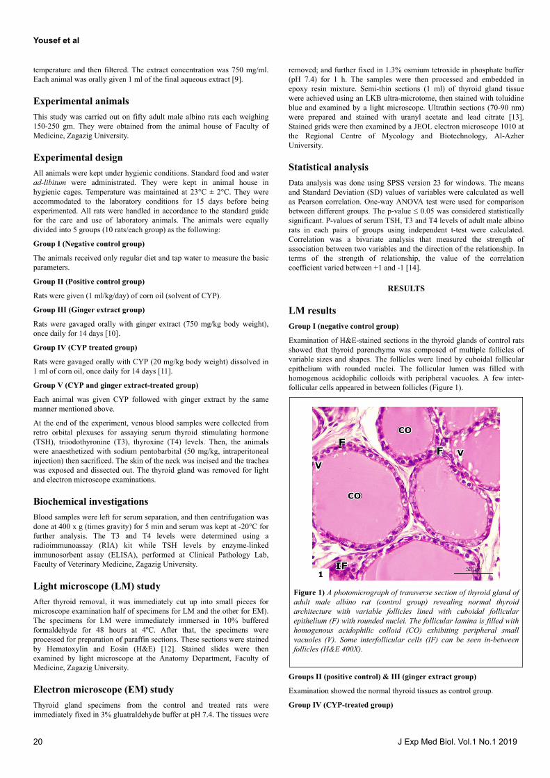
Examination of H&E-stained sections in the thyroid glands of control ratsshowed that thyroid parenchyma was composed of multiple follicles ofvariable sizes and shapes. The follicles were lined by cuboidal follicularepithelium with rounded nuclei. The follicular lumen was filled withhomogenous acidophilic colloids with peripheral vacuoles. A few inter-follicular cells appeared in between follicles (Figure 1).
Figure 1) A photomicrograph of transverse section of thyroid gland ofadult male albino rat (control group) revealing normal thyroidarchitecture with variable follicles lined with cuboidal follicularepithelium (F) with rounded nuclei. The follicular lamina is filled withhomogenous acidophilic colloid (CO) exhibiting peripheral smallvacuoles (V). Some interfollicular cells (IF) can be seen in-betweenfollicles (H&E 400X).
Groups II (positive control) & III (ginger extract group)
Examination showed the normal thyroid tissues as control group.
Group IV (CYP-treated group)
Yousef et al
20 J Exp Med Biol. Vol.1 No.1 2019
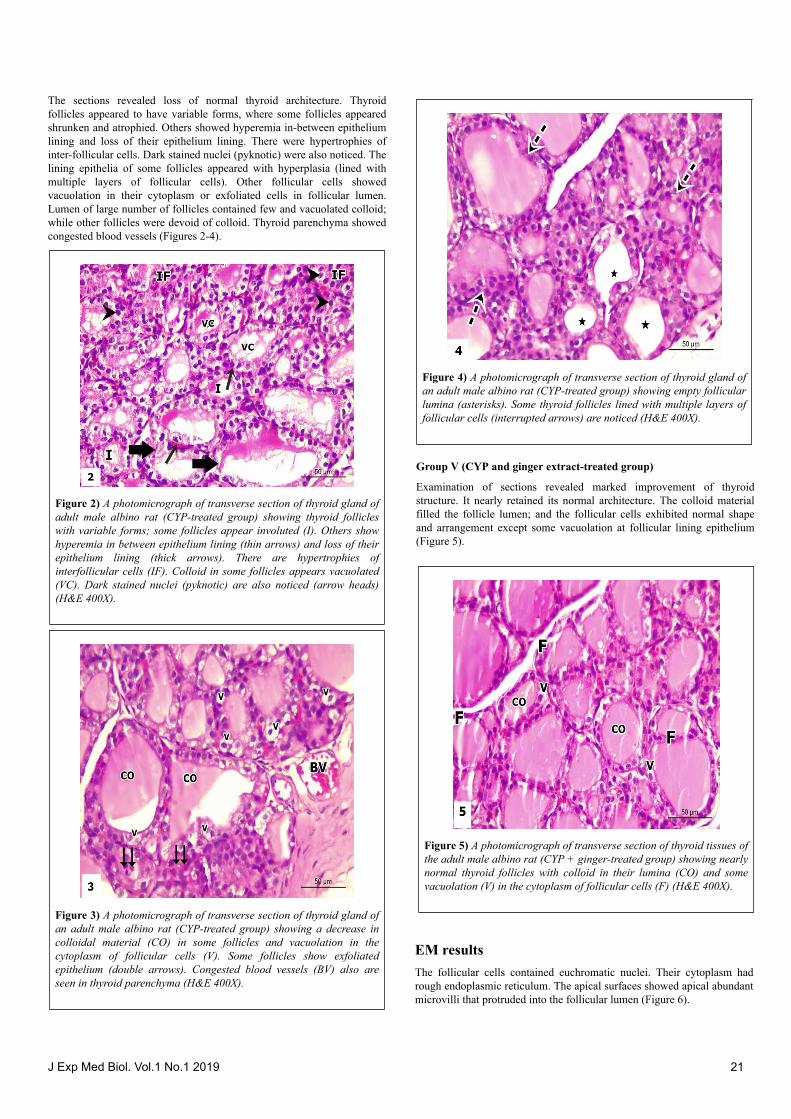
The sections revealed loss of normal thyroid architecture. Thyroidfollicles appeared to have variable forms, where some follicles appearedshrunken and atrophied. Others showed hyperemia in-between epitheliumlining and loss of their epithelium lining. There were hypertrophies ofinter-follicular cells. Dark stained nuclei (pyknotic) were also noticed. Thelining epithelia of some follicles appeared with hyperplasia (lined withmultiple layers of follicular cells). Other follicular cells showedvacuolation in their cytoplasm or exfoliated cells in follicular lumen.Lumen of large number of follicles contained few and vacuolated colloid;while other follicles were devoid of colloid. Thyroid parenchyma showedcongested blood vessels (Figures 2-4).
Figure 2) A photomicrograph of transverse section of thyroid gland ofadult male albino rat (CYP-treated group) showing thyroid follicleswith variable forms; some follicles appear involuted (I). Others showhyperemia in between epithelium lining (thin arrows) and loss of theirepithelium lining (thick arrows). There are hypertrophies ofinterfollicular cells (IF). Colloid in some follicles appears vacuolated(VC). Dark stained nuclei (pyknotic) are also noticed (arrow heads)(H&E 400X).
Figure 3) A photomicrograph of transverse section of thyroid gland ofan adult male albino rat (CYP-treated group) showing a decrease incolloidal material (CO) in some follicles and vacuolation in thecytoplasm of follicular cells (V). Some follicles show exfoliatedepithelium (double arrows). Congested blood vessels (BV) also areseen in thyroid parenchyma (H&E 400X).
Figure 4) A photomicrograph of transverse section of thyroid gland ofan adult male albino rat (CYP-treated group) showing empty follicularlumina (asterisks). Some thyroid follicles lined with multiple layers offollicular cells (interrupted arrows) are noticed (H&E 400X).
Group V (CYP and ginger extract-treated group)
Examination of sections revealed marked improvement of thyroidstructure. It nearly retained its normal architecture. The colloid materialfilled the follicle lumen; and the follicular cells exhibited normal shapeand arrangement except some vacuolation at follicular lining epithelium(Figure 5).
Figure 5) A photomicrograph of transverse section of thyroid tissues ofthe adult male albino rat (CYP + ginger-treated group) showing nearlynormal thyroid follicles with colloid in their lumina (CO) and somevacuolation (V) in the cytoplasm of follicular cells (F) (H&E 400X).
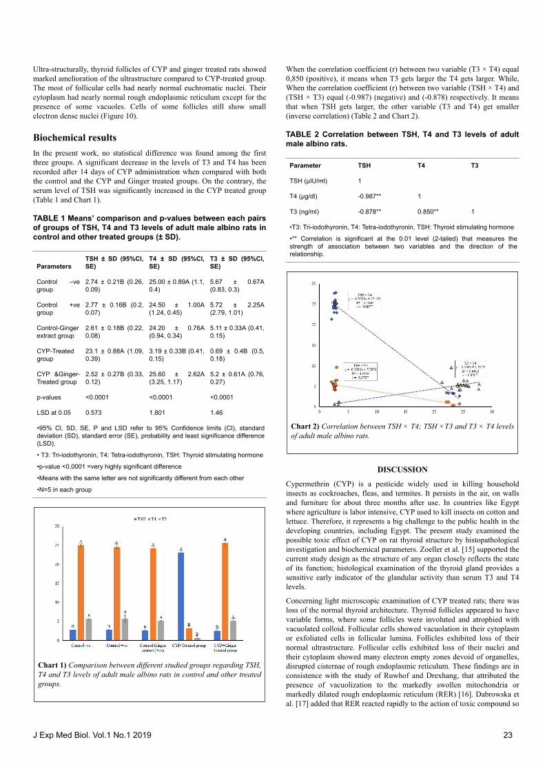
EM resultsThe follicular cells contained euchromatic nuclei. Their cytoplasm hadrough endoplasmic reticulum. The apical surfaces showed apical abundantmicrovilli that protruded into the follicular lumen (Figure 6).
J Exp Med Biol. Vol.1 No.1 2019 21
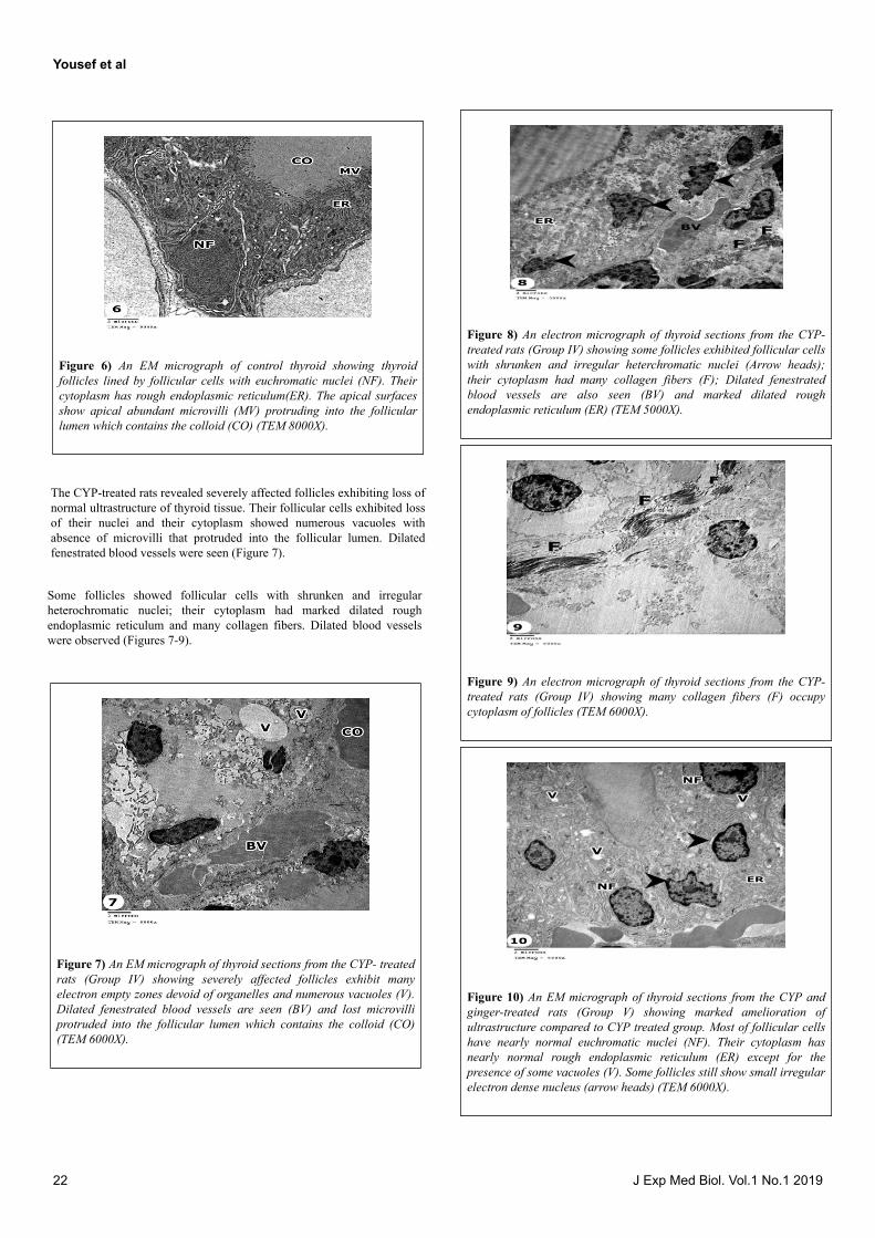
Figure 6) An EM micrograph of control thyroid showing thyroidfollicles lined by follicular cells with euchromatic nuclei (NF). Theircytoplasm has rough endoplasmic reticulum(ER). The apical surfacesshow apical abundant microvilli (MV) protruding into the follicularlumen which contains the colloid (CO) (TEM 8000X).
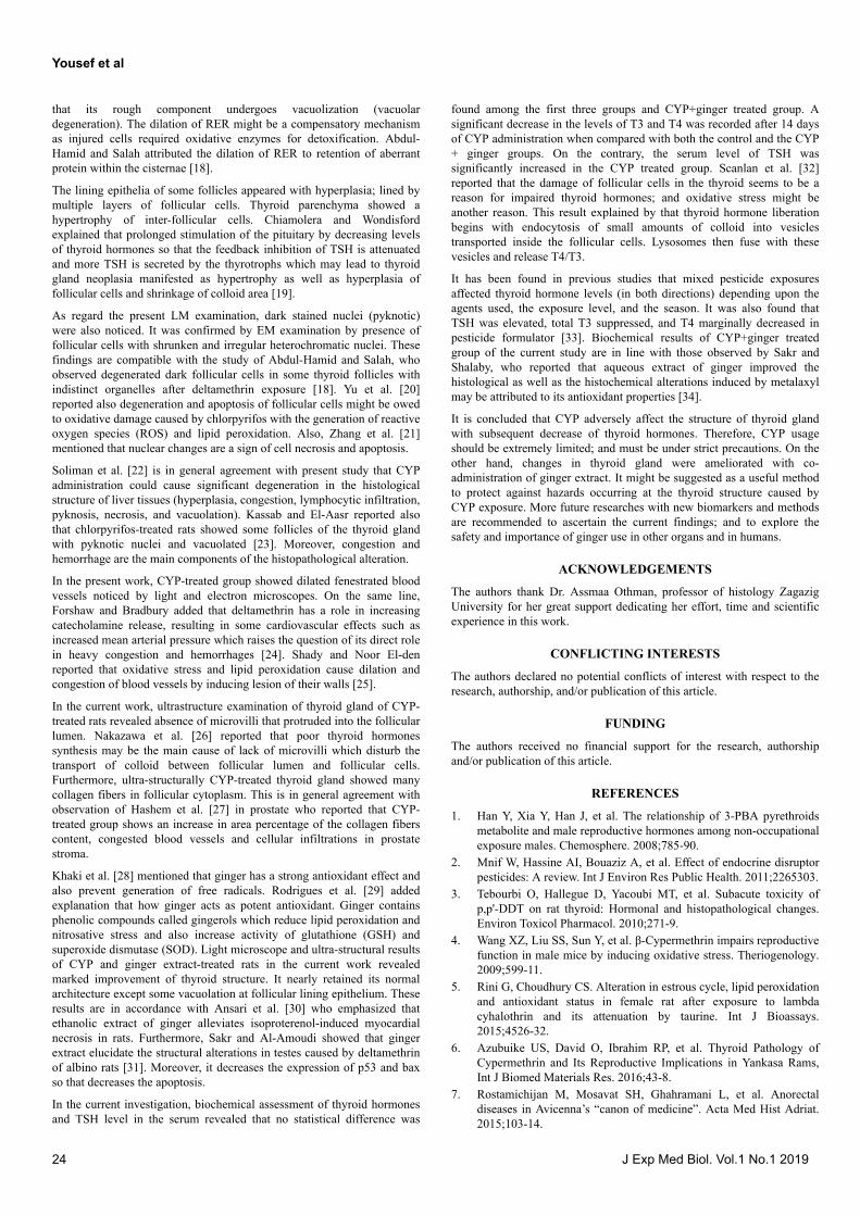
The CYP-treated rats revealed severely affected follicles exhibiting loss ofnormal ultrastructure of thyroid tissue. Their follicular cells exhibited lossof their nuclei and their cytoplasm showed numerous vacuoles withabsence of microvilli that protruded into the follicular lumen. Dilatedfenestrated blood vessels were seen (Figure 7).
Some follicles showed follicular cells with shrunken and irregularheterochromatic nuclei; their cytoplasm had marked dilated roughendoplasmic reticulum and many collagen fibers. Dilated blood vesselswere observed (Figures 7-9).
Figure 7) An EM micrograph of thyroid sections from the CYP- treatedrats (Group IV) showing severely affected follicles exhibit manyelectron empty zones devoid of organelles and numerous vacuoles (V).Dilated fenestrated blood vessels are seen (BV) and lost microvilliprotruded into the follicular lumen which contains the colloid (CO)(TEM 6000X).
Figure 8) An electron micrograph of thyroid sections from the CYP-treated rats (Group IV) showing some follicles exhibited follicular cellswith shrunken and irregular heterchromatic nuclei (Arrow heads);their cytoplasm had many collagen fibers (F); Dilated fenestratedblood vessels are also seen (BV) and marked dilated roughendoplasmic reticulum (ER) (TEM 5000X).
Figure 9) An electron micrograph of thyroid sections from the CYP-treated rats (Group IV) showing many collagen fibers (F) occupycytoplasm of follicles (TEM 6000X).
Figure 10) An EM micrograph of thyroid sections from the CYP andginger-treated rats (Group V) showing marked amelioration ofultrastructure compared to CYP treated group. Most of follicular cellshave nearly normal euchromatic nuclei (NF). Their cytoplasm hasnearly normal rough endoplasmic reticulum (ER) except for thepresence of some vacuoles (V). Some follicles still show small irregularelectron dense nucleus (arrow heads) (TEM 6000X).
Yousef et al
22 J Exp Med Biol. Vol.1 No.1 2019
Ultra-structurally, thyroid follicles of CYP and ginger treated rats showedmarked amelioration of the ultrastructure compared to CYP-treated group.The most of follicular cells had nearly normal euchromatic nuclei. Theircytoplasm had nearly normal rough endoplasmic reticulum except for thepresence of some vacuoles. Cells of some follicles still show smallelectron dense nuclei (Figure 10).
Biochemical resultsIn the present work, no statistical difference was found among the firstthree groups. A significant decrease in the levels of T3 and T4 has beenrecorded after 14 days of CYP administration when compared with boththe control and the CYP and Ginger treated groups. On the contrary, theserum level of TSH was significantly increased in the CYP treated group(Table 1 and Chart 1).
TABLE 1 Means’ comparison and p-values between each pairsof groups of TSH, T4 and T3 levels of adult male albino rats incontrol and other treated groups (± SD).
ParametersTSH ± SD (95%Cl,SE)
T4 ± SD (95%Cl,SE)
T3 ± SD (95%Cl,SE)
Control –vegroup
2.74 ± 0.21B (0.26,0.09)
25.00 ± 0.89A (1.1,0.4)
5.67 ± 0.67A(0.83, 0.3)
Control +vegroup
2.77 ± 0.16B (0.2,0.07)
24.50 ± 1.00A(1.24, 0.45)
5.72 ± 2.25A(2.79, 1.01)
Control-Gingerextract group
2.61 ± 0.18B (0.22,0.08)
24.20 ± 0.76A(0.94, 0.34)
5.11 ± 0.33A (0.41,0.15)
CYP-Treatedgroup
23.1 ± 0.88A (1.09,0.39)
3.19 ± 0.33B (0.41,0.15)
0.69 ± 0.4B (0.5,0.18)
CYP &Ginger-Treated group
2.52 ± 0.27B (0.33,0.12)
25.60 ± 2.62A(3.25, 1.17)
5.2 ± 0.61A (0.76,0.27)
p-values <0.0001 <0.0001 <0.0001
LSD at 0.05 0.573 1.801 1.46
•95% Cl, SD, SE, P and LSD refer to 95% Confidence limits (Cl), standarddeviation (SD), standard error (SE), probability and least significance difference(LSD).
• T3: Tri-iodothyronin, T4: Tetra-iodothyronin, TSH: Thyroid stimulating hormone
•p-value <0.0001 =very highly significant difference
•Means with the same letter are not significantly different from each other
•N=5 in each group
Chart 1) Comparison between different studied groups regarding TSH,T4 and T3 levels of adult male albino rats in control and other treatedgroups.
When the correlation coefficient (r) between two variable (T3 × T4) equal0,850 (positive), it means when T3 gets larger the T4 gets larger. While,When the correlation coefficient (r) between two variable (TSH × T4) and(TSH × T3) equal (-0.987) (negative) and (-0.878) respectively. It meansthat when TSH gets larger, the other variable (T3 and T4) get smaller(inverse correlation) (Table 2 and Chart 2).
TABLE 2 Correlation between TSH, T4 and T3 levels of adultmale albino rats.
Parameter TSH T4 T3
TSH (µIU/ml) 1
T4 (µg/dl) -0.987** 1
T3 (ng/ml) -0.878** 0.850** 1
•T3: Tri-iodothyronin, T4: Tetra-iodothyronin, TSH: Thyroid stimulating hormone
•** Correlation is significant at the 0.01 level (2-tailed) that measures thestrength of association between two variables and the direction of therelationship.
Chart 2) Correlation between TSH × T4; TSH ×T3 and T3 × T4 levelsof adult male albino rats.
DISCUSSION
Cypermethrin (CYP) is a pesticide widely used in killing householdinsects as cockroaches, fleas, and termites. It persists in the air, on wallsand furniture for about three months after use. In countries like Egyptwhere agriculture is labor intensive, CYP used to kill insects on cotton andlettuce. Therefore, it represents a big challenge to the public health in thedeveloping countries, including Egypt. The present study examined thepossible toxic effect of CYP on rat thyroid structure by histopathologicalinvestigation and biochemical parameters. Zoeller et al. [15] supported thecurrent study design as the structure of any organ closely reflects the stateof its function; histological examination of the thyroid gland provides asensitive early indicator of the glandular activity than serum T3 and T4levels.
Concerning light microscopic examination of CYP treated rats; there wasloss of the normal thyroid architecture. Thyroid follicles appeared to havevariable forms, where some follicles were involuted and atrophied withvacuolated colloid. Follicular cells showed vacuolation in their cytoplasmor exfoliated cells in follicular lumina. Follicles exhibited loss of theirnormal ultrastructure. Follicular cells exhibited loss of their nuclei andtheir cytoplasm showed many electron empty zones devoid of organelles,disrupted cisternae of rough endoplasmic reticulum. These findings are inconsistence with the study of Ruwhof and Drexhang, that attributed thepresence of vacuolization to the markedly swollen mitochondria ormarkedly dilated rough endoplasmic reticulum (RER) [16]. Dabrowska etal. [17] added that RER reacted rapidly to the action of toxic compound so
J Exp Med Biol. Vol.1 No.1 2019 23
that its rough component undergoes vacuolization (vacuolardegeneration). The dilation of RER might be a compensatory mechanismas injured cells required oxidative enzymes for detoxification. Abdul-Hamid and Salah attributed the dilation of RER to retention of aberrantprotein within the cisternae [18].
The lining epithelia of some follicles appeared with hyperplasia; lined bymultiple layers of follicular cells. Thyroid parenchyma showed ahypertrophy of inter-follicular cells. Chiamolera and Wondisfordexplained that prolonged stimulation of the pituitary by decreasing levelsof thyroid hormones so that the feedback inhibition of TSH is attenuatedand more TSH is secreted by the thyrotrophs which may lead to thyroidgland neoplasia manifested as hypertrophy as well as hyperplasia offollicular cells and shrinkage of colloid area [19].
As regard the present LM examination, dark stained nuclei (pyknotic)were also noticed. It was confirmed by EM examination by presence offollicular cells with shrunken and irregular heterochromatic nuclei. Thesefindings are compatible with the study of Abdul-Hamid and Salah, whoobserved degenerated dark follicular cells in some thyroid follicles withindistinct organelles after deltamethrin exposure [18]. Yu et al. [20]reported also degeneration and apoptosis of follicular cells might be owedto oxidative damage caused by chlorpyrifos with the generation of reactiveoxygen species (ROS) and lipid peroxidation. Also, Zhang et al. [21]mentioned that nuclear changes are a sign of cell necrosis and apoptosis.
Soliman et al. [22] is in general agreement with present study that CYPadministration could cause significant degeneration in the histologicalstructure of liver tissues (hyperplasia, congestion, lymphocytic infiltration,pyknosis, necrosis, and vacuolation). Kassab and El-Aasr reported alsothat chlorpyrifos-treated rats showed some follicles of the thyroid glandwith pyknotic nuclei and vacuolated [23]. Moreover, congestion andhemorrhage are the main components of the histopathological alteration.
In the present work, CYP-treated group showed dilated fenestrated bloodvessels noticed by light and electron microscopes. On the same line,Forshaw and Bradbury added that deltamethrin has a role in increasingcatecholamine release, resulting in some cardiovascular effects such asincreased mean arterial pressure which raises the question of its direct rolein heavy congestion and hemorrhages [24]. Shady and Noor El-denreported that oxidative stress and lipid peroxidation cause dilation andcongestion of blood vessels by inducing lesion of their walls [25].
In the current work, ultrastructure examination of thyroid gland of CYP-treated rats revealed absence of microvilli that protruded into the follicularlumen. Nakazawa et al. [26] reported that poor thyroid hormonessynthesis may be the main cause of lack of microvilli which disturb thetransport of colloid between follicular lumen and follicular cells.Furthermore, ultra-structurally CYP-treated thyroid gland showed manycollagen fibers in follicular cytoplasm. This is in general agreement withobservation of Hashem et al. [27] in prostate who reported that CYP-treated group shows an increase in area percentage of the collagen fiberscontent, congested blood vessels and cellular infiltrations in prostatestroma.
Khaki et al. [28] mentioned that ginger has a strong antioxidant effect andalso prevent generation of free radicals. Rodrigues et al. [29] addedexplanation that how ginger acts as potent antioxidant. Ginger containsphenolic compounds called gingerols which reduce lipid peroxidation andnitrosative stress and also increase activity of glutathione (GSH) andsuperoxide dismutase (SOD). Light microscope and ultra-structural resultsof CYP and ginger extract-treated rats in the current work revealedmarked improvement of thyroid structure. It nearly retained its normalarchitecture except some vacuolation at follicular lining epithelium. Theseresults are in accordance with Ansari et al. [30] who emphasized thatethanolic extract of ginger alleviates isoproterenol-induced myocardialnecrosis in rats. Furthermore, Sakr and Al-Amoudi showed that gingerextract elucidate the structural alterations in testes caused by deltamethrinof albino rats [31]. Moreover, it decreases the expression of p53 and baxso that decreases the apoptosis.
In the current investigation, biochemical assessment of thyroid hormonesand TSH level in the serum revealed that no statistical difference was
found among the first three groups and CYP+ginger treated group. Asignificant decrease in the levels of T3 and T4 was recorded after 14 daysof CYP administration when compared with both the control and the CYP+ ginger groups. On the contrary, the serum level of TSH wassignificantly increased in the CYP treated group. Scanlan et al. [32]reported that the damage of follicular cells in the thyroid seems to be areason for impaired thyroid hormones; and oxidative stress might beanother reason. This result explained by that thyroid hormone liberationbegins with endocytosis of small amounts of colloid into vesiclestransported inside the follicular cells. Lysosomes then fuse with thesevesicles and release T4/T3.
It has been found in previous studies that mixed pesticide exposuresaffected thyroid hormone levels (in both directions) depending upon theagents used, the exposure level, and the season. It was also found thatTSH was elevated, total T3 suppressed, and T4 marginally decreased inpesticide formulator [33]. Biochemical results of CYP+ginger treatedgroup of the current study are in line with those observed by Sakr andShalaby, who reported that aqueous extract of ginger improved thehistological as well as the histochemical alterations induced by metalaxylmay be attributed to its antioxidant properties [34].
It is concluded that CYP adversely affect the structure of thyroid glandwith subsequent decrease of thyroid hormones. Therefore, CYP usageshould be extremely limited; and must be under strict precautions. On theother hand, changes in thyroid gland were ameliorated with co-administration of ginger extract. It might be suggested as a useful methodto protect against hazards occurring at the thyroid structure caused byCYP exposure. More future researches with new biomarkers and methodsare recommended to ascertain the current findings; and to explore thesafety and importance of ginger use in other organs and in humans.
ACKNOWLEDGEMENTS
The authors thank Dr. Assmaa Othman, professor of histology ZagazigUniversity for her great support dedicating her effort, time and scientificexperience in this work.
CONFLICTING INTERESTS
The authors declared no potential conflicts of interest with respect to theresearch, authorship, and/or publication of this article.
FUNDING
The authors received no financial support for the research, authorshipand/or publication of this article.
REFERENCES
1. Han Y, Xia Y, Han J, et al. The relationship of 3-PBA pyrethroidsmetabolite and male reproductive hormones among non-occupationalexposure males. Chemosphere. 2008;785-90.
2. Mnif W, Hassine AI, Bouaziz A, et al. Effect of endocrine disruptorpesticides: A review. Int J Environ Res Public Health. 2011;2265303.
3. Tebourbi O, Hallegue D, Yacoubi MT, et al. Subacute toxicity ofp,p'-DDT on rat thyroid: Hormonal and histopathological changes.Environ Toxicol Pharmacol. 2010;271-9.
4. Wang XZ, Liu SS, Sun Y, et al. β-Cypermethrin impairs reproductivefunction in male mice by inducing oxidative stress. Theriogenology.2009;599-11.
5. Rini G, Choudhury CS. Alteration in estrous cycle, lipid peroxidationand antioxidant status in female rat after exposure to lambdacyhalothrin and its attenuation by taurine. Int J Bioassays.2015;4526-32.
6. Azubuike US, David O, Ibrahim RP, et al. Thyroid Pathology ofCypermethrin and Its Reproductive Implications in Yankasa Rams,Int J Biomed Materials Res. 2016;43-8.
7. Rostamichijan M, Mosavat SH, Ghahramani L, et al. Anorectaldiseases in Avicenna’s “canon of medicine”. Acta Med Hist Adriat.2015;103-14.
Yousef et al
24 J Exp Med Biol. Vol.1 No.1 2019
8. Daily JW, Yang M, Kim DS, et al. Efficacy of ginger for treatingType 2 diabetes: A systematic review and meta-analysis ofrandomized clinical trials. J Ethnic Foods. 2015;36-43.
9. Kamtchouing P1, Mbongue Fandio GY, Dimo T, et al. Evaluation ofandrogenic activity of Zingiber officinale and Pentadiplandrabrazzeana in male rats. Asian J Androl. 2002;299-01.
10. Aljahawey MHO, Soeharto S, Sujuti H. The effect of ginger extract(zingiber officinale roscoe) on male leydig cell and testosteroneLevel in carbofuran induced rat. Int J Pharm Tech Res. 2015;879-88.
11. Grewal KK, Sandhu GS, Kaur R, et al. Toxic impacts ofcypermethrin on behavior and histology of certain tissues of albinorats. Toxicol Int. 2010;94-8.
12. Hegazy R, Hegazy A. Hegazy’ Simplified Method of TissueProcessing (Consuming Less Time and Chemicals), Ann Int MedDen Res. 2015;57-61.
13. Gluert AM, Lewis PR. Biological specimen preparation fortransmission electron microscopy, Princeton University Press, 1998.
14. Dawson B, Trapp RG. Basic & Clinical Biostatistics, 4th Edition,McGraw-Hill, 2004.
15. Zoeller RT, Tan SW, Tyl RW. General background on thehypothalamic-pituitary-thyroid (HPT) axis. Crit Rev Toxicol.2007;11-53.
16. Ruwhof C, Drexhang HA. Iodine and thyroid autoimmune disease inanimal models. Thyroid, 2001;427-36.
17. Dabrowska A, Jacewicz D, Lapinska A, et al. Pivotal participation ofnitrogen dioxide in L-arginine induced acute necrotizing pancreatitis:protective role of superoxide scavenger 4-OH-TEMPO. BiochemBiophys Res Commun. 2005;313-20.
18. Abdul-Hamid M, Salah M. Lycopene reduces deltamethrin effectsinduced thyroid toxicity and DNA damage in albino rats. J BasicAppl Zool. 2013;155-63.
19. Chiamolera MI, Wondisford FE. Minireview: Thyrotropin-releasinghormone and the thyroid hormone feedback mechanism.Endocrinology. 2009;1091-96.
20. Yu F, Wang Z, Ju B, et al. Apoptotic effect of organophosphorusinsecticide chlorpyrifos on mouse retina in vivo via oxidative stressand protection of combination of vitamins C and E. Exp ToxicolPathol. 2008;415-23.
21. Zhang C, Niu W, Wang Z, et al. The effect of gonadotropin onglucose transport and apoptosis in rat ovary. PloS One. 2012;e42406.
22. Soliman MM, Attia HF, El-Ella GA. Genetic and histopathologicalalterations induced by cypermethrin in rat kidney and liver:Protection by sesame oil. Int J Immunopathol Pharmacol.2015;508-20.
23. Kassab A, El-Aasr M. Effect of Avocado pulp extract onchlorpyrifos-induced thyroid gland injury in rats: A histological andmorphometric study. Egypt J Hist. 2018;83-92.
24. Forshaw PJ, Bradburry JE. Pharmacological effects of prethroids onthe cardiovascular system of the rat. Eur. J. Pharmacol. 1983;207-13.
25. Shady AM, Noor El-deen F. Effect of chlorpyrifos on thyroid glandof adult male albino rats. Egypt J Histol. 2010;441-50.
26. Nakazawa T, Murata S, Kondo T, et al. Histopathology of the thyroidin amiodaroneinduced hypothyroidism. Pathol Int. 2008;55-8.
27. Hashem HE, Abd El-Haleem MR, Abass MA. Epithelial and stromalalterations in prostate after cypermethrin administration in adultalbino rats (histological and biochemical study). Tissue Cell.2015;366-72.
28. Khaki A, Fathiazad F, Nouri M, et al. The effects of ginger onspermatogenesis and sperm parameters of rat. Iran J Reprod Med.2009;7-12.
29. Rodrigues SM, Ximenes CF, de Batista PR, et al. Tributyltincontributes in reducing the vascular reactivity to phenylephrine inisolated aortic rings from female rats. Toxicol Lett. 2014;378-85.
30. Ansari MN, Bhandari U, Pillai KK. Ethanolic Zingiber officinale R.extract pretreatment alleviates isoproterenol-induced oxidativemyocardial necrosis in rats. Indian J Exp Biol. 2006;892-97.
31. Sakr SA, Al-Amoudi WM. Effect of ginger extract on deltamethrininduced histomorphological and immunohistochemical changes inTestes of Albino Rats. Life Science J. 2012;771-78.
32. Scanlan TS, Suchland KL, Hart ME, et al. 3-Iodothyronamine is anendogenous and rapid-acting derivative of thyroid hormone. NatMed. 2004;638-42.
33. Zaidi SS, Bhatnagar VK, Gandhi SJ, et al. Assessment of thyroidfunction in pesticide formulators. Hum Exp Toxicol. 2000;497-01.
34. Sakr SA, Shalaby YS. Ginger extract protects metalaxyl-inducedhistomorphological and histochemical alterations in testes of albinomice. J Applied Pharmaceut Sci. 2011;36-42.
J Exp Med Biol. Vol.1 No.1 2019 25