Embed Size (px)
Citation preview

Protective effect of flavanone - naringenin on highglucose-induced hepatotoxicity to Hep G2 cells
Petlevski, Roberta; Knežević, T.; Ris, M.; Filipašić, A.
Source / Izvornik: Farmaceutski glasnik, 2017, 73, 85 - 97
Journal article, Published versionRad u časopisu, Objavljena verzija rada (izdavačev PDF)
Permanent link / Trajna poveznica: https://urn.nsk.hr/urn:nbn:hr:163:659506
Rights / Prava: In copyright
Download date / Datum preuzimanja: 2022-03-12
Repository / Repozitorij:
Repository of Faculty of Pharmacy and Biochemistry University of Zagreb

ZNANSTVENI RADOVI hfd
Protective effect of flavanone - naringenin
on high glucose-induced hepatotoxicity to
Hep G2 cells
R. PETLEVSKI, T. KNEŽEVIĆ, M. RIS, A. FILIPAŠIĆ
University of Zagreb, Faculty of Pharmacy and Biochemistry, Department of Medica I
Biochemistry and Haematology, Domagojeva 2, Zagreb. Croatia
Protektivni učinak flavanona naringenina na glukozom izazvanu hepatotoksičnost u Hep G2 stanicama
S a ž e t a k - Nrzringenin je jlavanon, vrsta jlavonoida. Opisan je aktivni učinak.fiavonoida na ijuds!w zdravije. Djeluju kao antioksidansi i čistači slobodnih radikala, imaju protuupalni učinak, potiču metabolizam ug{jikohidrata te al,tivnost imunološkog sustava. Ciij je ovog rada hio utvrditi lwja od ispitivanih lwncentracija naringenina u msponu 1-1000 µM ima protektivni učinal, na Hep G2 stanice lcoje se nalaze u hiperglil,emijskim uvjetima. Hiperglikemija je u šećernoj bolesti primarni uzrok oksidacijskom stresu koji !,asnije dovodi do komplileacija. U Hep G2 stanicama koje se nalaze u hiperglikemijsleim uvjetima dolazi do značajnog porasta jetrenih funkcijskih marleera leao što su AS7,' AL7,' LDH i GGT; smai�jene vijabilnosti stanica (MTT test) i smanjene aktivnosti antio/,sidativnih enzima: glutation-peroksidaze (GPX) i superoksid-dizmutaze (SOD ). Naringenin pri lwncentracijskom rasponu od 1-100 µMIL ima hepatoprotektivni učinak na Hep G2 stanice u hiperglikemijslcim uvjetima, dol, više koncentracije naringenina (I 000 µMIL) ne pol,azuju više taj ulinale.
INTRODUCTION
111e !iver plays an important role in maintaining glucose homeostasis during fasting as well as the postprandial s tate (1). Several studies have shown that high levels of ALT and GGT are independent predictors of type 2 diabetes incidence (2,3). "I11e authors Lee et al. 2004., investigated hepatoprotective effects of naringenin in rats (4). Citrus fruits (including naringenin) and their potential benefits in the management
85

R. Petlevski, T. Knežević, M. Ris, A. Filipašić Protective effect of flavanone naringenin on high glucose-induced hepatotoxicity to Hep G2 cells, Farmaceutski glasnik 73, 2/2017
of diabetes is described in study of the authors Aruoma et al. 2012 (5). Furthermore, naringenin potentiates intracellular signaling responses to low insulin doses, suggesting that naringenin sensitizes hepatocytes to insulin (6). So far no study of the naringenin effect in the hyperglycemic condition in vitro has been performed.
Naringenin, the predominant flavanone in grapefruits, oranges and tomatoes (skin), is a type of flavonoid that is considered to have a bioactive effect on human health as antioxidant, free-radical scavenger, anti-inflammatory, carbohydrate metabolism promoter, immune system modulator (7) and has been shown to reduce oxidative damage to DNA in vivo (8).
The results of the authors Tsai et al. 2012 indicate that naringenin could attenuate diabetic nephropathy via its anti-inflammatory and antifibrotic activities (9). It has also been reported that high consumption of grapefruit juice improved blood lipid profile in hyperlipidemic humans (1 O, 11). Naringenin has an inhibitory effect on the human cytochmme P450 isoform (CYP1A2) (12,13).
In the hyperglycemia environment the high level of reactive oxygen species (ROS) exist (14). Oxidative stress is the principal mechanism in the progression of diabetes and actively leads to cellular injury that can precede the onset of many diabetic complications (15). TI1e development of insulin resistance is also closely related to the presence of cellular oxidative stress (16). Diabetes is associated with the generation of reactive oxygen species, which cause oxidative damage, particularly to heart, !iver, eyes, nerves and kidney (17). Hyperglycemia can degrade antioxidant enzyme defenses (18), thereby allowing ROS to damage other enzymes and also structural proteins. Antioxidants in foods, such as vitamin C, vitamin E, selenium and many phytochemicals can eliminate these free radicals (19). TI1e ethnomedical approach to plant drug discovery is practical, cost-effective, and logical. Phytochemicals with antioxidant activiry are: allyl sulfides (onions, leeks, garlic), carotenoids (fruit, carrots), flavonoids (fruit, vegetables), polyphenols (tea, grapes).
TI1e present study was planned to elucidate whether naringenin, when administered, can ameliorate toxic effects of glucose in Hep G2 cells.
Materials and Methods
Materials and chemicals Naringenin and D-glucose were from Sigma (St. Louis, MO, USA). Ali other
chemicals used in these measurements were of the highest puriry commercially available.
Cell culture Human Caucasian hepatocyte carcinoma (Hep G2) cells from European Collec
tion of Cell Cultures (ECACC) were maintained in an incubator at 37 °C with a
86

R. Petlevski, T. Knežević, M. Ris, A. Filipašić: Protective effect of flavanone - naringenin on high glucose-induced hepatotoxicity to Hep G2 cells, Farmaceutski glasnik 73, 2/2017
humidified atmosphere of 5 % C02 and cultured in Minimum Essential Media (MEM) (Gibco") supplemented with 10 % (vlv) feral bovine serum (FBS) and 20 IUlmL penicillin, 20 µglmL streptomycin. The medium was refreshed twice a week. For the experiments, cells were seeded into six-well plates (or in 96-well plates for mitochondrial dehydrogen activity assay and LDH activity). Medium was changed to FBS-free medium 24 h before the assay. The Hep G2 cells were treated another 24 h with 30 mM glucose and naringenin in following concentrations: 1000 µmol/L, 100 µmol/L, 1 O µmol/L, and 1 µmol/L. The control cells were grown in the medium which contained 5.56 mM D-glucose.
Treatment of the HeJJ G2 cells
Before treatment Hep G2 cells were culturcd in 6-wcll Nunc plates in complete MEM mcdium. On the day of treatment complete MEM medi um is removed, and Hep G2 cells were devided in six groups:
C - negative control Hcp G2 cells were incubated in FBS- frcc MEM medium, containing 5.56 mM glucose
D - positive control ln this group, Hcp G2 cclls werc culturcd in FBS- free MEM, supplemented with D-glucose (Sigma) in concentration of 30 mM
DIN1_1ooo ;mwl!L -Jour treated groups with naringenin Hep G2 cclls were cultured in FBS- free MEM, supplemented with D-glucosc (Sigma) in concentration of 30 mM + different concentrations of naringenin: DIN 1-1000 pmol/L
For statistical comparison is used D - positive control vs. DIN 1_1000 pmol/L (*) and C VS. D (**)
Cell viability assay
Cell viability was assessed by MTT test (20). The cytotoxicity was carried out by MTT [3-(4,5-dimethylthiazol-2-yl)-2,5-diphenyltetrazolium bromidc] (Sigma) assay for investigating changes in mitochondrial/11011-mitochondrial dehydrogenase activity. After overnight growth, cells wcre treated with diffcrent concentrations of naringenin dcscribed above for 24 h. At the end of the treatment the media were removed from wells (for LDH activity). Cells were washed twice with sterile phosphate buffered saline (PBS). MTT plus medium was added. After 4 h incubation at 37 °C, the supernatant was removed and formazan crystals were dissolved by D MSO (100 % ) . Finally, the absorbance at a wavelength of 595 nm was recorded using a plate reader (Victor). The absorbance at 595 nm was taken as an index of the activity of mitochondria.
87

R. Petlevski. T Knežević. M. Ris, A. Filipašić: Protective effect of flavanone naringenin on high glucose-induced hepatotoxicity to Hep G2 cells, Farmaceutski glasnik 73, 2/2017
Cell membrane damage Activity of lactate dehydrogenase (LDH) in the cell medium was measured as an
index of cell membrane damage. Hep G2 cells were cultured in 96-well plate at a density of 1x10⁵ cells / 200 µL. Cells were treated 24 h wirh naringenin. At the end of the trearment rhe media were collected. 111e LDH activity was measured with spectrophotometric method using lactate as subsrrate (21). For this, 200 µL aliquots of culture medium were added to the LDH assay system, and the absorbance at 340 nm was recorded.
Detennination of glutathione /Jeroxidase (GPx) activity 1he cells were seeded in six-well plates at concentration of lxI06 cells/mL and
24 h afrer plating, were treated with naringenin (in the concentration range I tolOOO �tmol/L) in the serum free media. Afrer additional 24 h, the medium was removed, the cells were washed twice with sterile PBS solution, and scraped. 1he harvested cells were suspended in I O mM phosphate buffer (pH 7.5) and then lysed on ice by ultrasonicating one for 15 sec. Then, Triton X-100 (1 %) was added to the lysates and incubated for 1 O min on ice. 1l1e lysaces were clarified by centrifugacion at 14 000 rpm for 20 min at + 4 °C to remove cellular debris.
We used Glutathione Peroxidase Assay Kit (Cayman Chemical Company) for GPx activity in the cell lysate (22, 23). Cayman's GPx Assay measures GPx activity indirectly by a coupled reaction with glutathione reductase (GR). Oxidized glutathione (GSSG), produced upon reduction of hydroperoxide by GPx was recycled to its reduced stare by GR and NADPH. 'Il1e oxidation ofNADPH to NADP+ was accompanied by a decrease in absorbance at 340 nm. Under conditions in which the GPx activity is rate limiting, the rate of decrease in the A340 nm is directly proportional to the GPx activity in the sample. 1l1e A,40 nm was recorded using VictorTM multilabel reader (Perkin Elmer). GPx activity was expressed as unirs/mg protein.
Detennination of suJJeroxide dismutase (SOD) activity We used Superoxide Dismutase Assay Kit (Cayman Chemical Company), (24,25)
for SOD activity in rhe cell lysate. Cayman's utilizes tetrazolium sale for detection of superoxide radicals generated by xantine oxidase and hypoxanthine. One unit of SOD is defined as the amount of enzyme needed to exhibit 50 % dismutation of rhe superoxide radical. 1l1e SOD assay measures ali rhree types of SOD (Cu/Zn, Mn and Fe SOD). 1l1e A450 nm was recorded using VictorTM mulrilabel reader (Perkin Elmer). Tora! SOD activity was expressed as unirs/mg protein.
The JJrotein content in HeJJ G2 cells by QuibitTM Quantitation System 1l1is is a fluorescence-based quantitation assay. 'The kit provides concentrated assay
reagent, dilution buffer, and pre-diluced BSA standards. 1be Quibi?M fluoromecer
88

R. Petlevski, T. Knežević, M. Ris, A. Filipašić: Protective effect of flavanone naringenin on high glucose-induced hepatotoxicity to Hep G2 cells, Farmaceutski glasnik 73, 2/2017
generates concentration data based on the relationship berween the three standards used in calibration and gives values for the proteins in cells in µg/mL.
Biochemical assays of liver enzymes
lhe activities of aspartate aminotransferase (AST) and alanine aminotransferase (Al'.T) were measured spectrophotometrically using the a-ketoglutarate reaction according to the standard procedures for automatic analyzer (Olympus, AU 1000). y-glutamyl transferase (GGT) activity was determined by the method of Rosalki et al.
1970 using y-glutamyl-p-nitroanilide as substrate (26). Ali enzymes were analyzed in Hep G2 cell lysate.
Statistical analysis
Ali analyses were performed using SigmaStat 2.0 Sofrware programme. Results were expressed as means ± SEM. For multiple comparisons, one-way analysis of variance was followed by the Holm-Sidak method. P-value< O.OS was considered to be statistically significant (27).
Results
Effects of naringenin on the cell viability
The exposure of Hep G2 cells to 30 mM glucose led to a loss of cell viability (p < O.OS) but the treatment with naringenin in concentration 1 µmol/L protected cells against damage induced by 30 mM glucose (p<O.OS) (Fig.1).
0.8
-� -� ti o.e e
i 0.4
I: ;;i; 0.2
o control WmM WmM 30mM 30mM 30mM
lJIUCO-!l& gtucooo „ gtucooo + gh.1CO&e „ grucooe +
1000 umolll. 100 umo6t. 10 UITIOW. 1 umoVL nng11111n m11ngen1n nartngeniln narlngemn
Fig. 1. EFFECT OF NARINGENIN AGAINST 30 Mm GLUCOSE-INDUCED CYTOTOXICITY IN HEP G2 CELLS. The data were expressed at the mean ± SEM of three independent experiments.
' p < 0.05 was considered to be statistically significant.
89
R. Petlevski, T. Knežević. M. Ris, A. Filipašić: Protective effect of flavanone - naringenin on high glucose-induced hepatotoxicity to Hep G2 cells, Farmaceutski glasnik 73, 2/2017
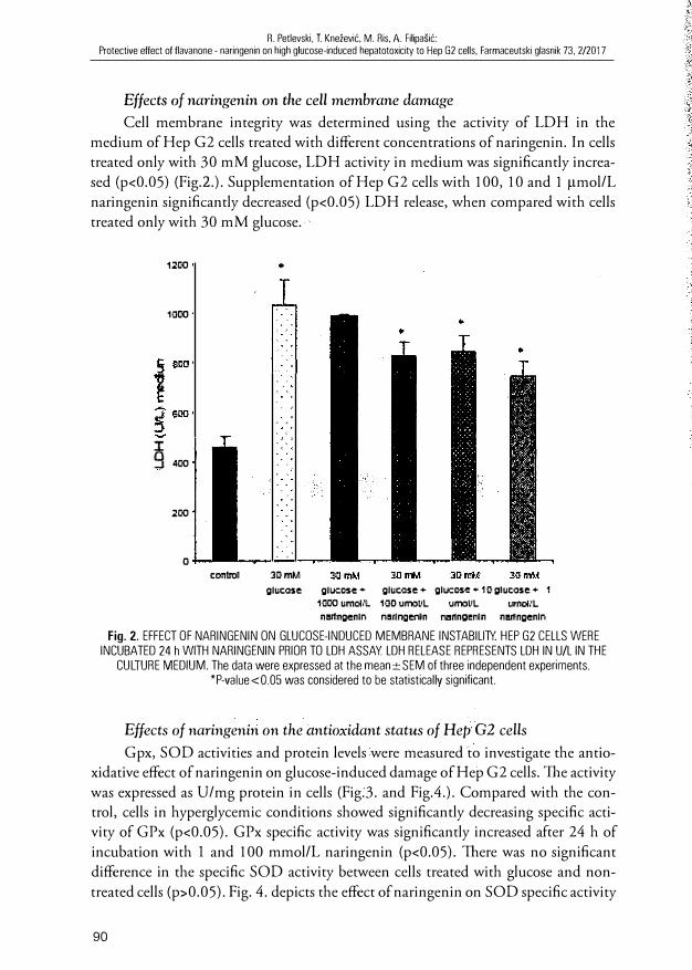
Effects of naringenin on the cell membrane damage
Cell membrane integrity was determined using the activity of LDH in the medium of Hep G2 cells treated with different concentrations of naringenin. In cells treated only with 30 mM glucose, LDH activity in medium was significantly increased (p<0.05) (Fig.2.). Supplementation ofHep G2 cells with 100, 10 and 1 µmol/L naringenin significantly decreased (p<0.05) LDH release, when compared with cells treated only with 30 mM glucose.
1200
1000
� 400
200
o
control
•
3DmM glucose glucos,e • gluc-ose + glucos,e + 10 glucose + 1
1000 umom. 100 urn:1t11. urn:1u1. Ull'IC<IL nl!lf,ngenln narlngl!n'ln Mrlnge1111n nl!llingenln
Fig. 2. EFFECT OF NARINGENIN ON GLUCOSE-INDUCED MEMBRANE INSTABILITY. HEP G2 CELLS WERE INCUBATED 24 h WITH NARINGENIN PRIOR TO LDH ASSAY LDH RELEASE REPRESENTS LDH IN U/L IN THE
CULTURE MEDIUM. The data were expressed at the mean±SEM of three independent experiments. *P-value<0.05 was considered to be statistically significant.
Effects of naringenin on the antioxidant status of Hep G2 cells
Gpx, SOD activities and protein levels were measured to investigate the antioxidative effect of naringenin on glucose-induced dam age of Hep G2 cells. "foe activity was expressed as U/mg protein in cells (Fig.3. and Fig.4.). Compared with the control, cells in hyperglycemic conditions showed significantly decreasing specific activity of GPx (p<0.05). GPx specific activity was significantly increased after 24 h of incubation with I and 100 mmol/L naringenin (p<0.05). There was no significant difference in the specific SOD activity between cells treated with glucose and nontreated cells (p>0.05). Fig. 4. depicts the effect of naringenin on SOD specific activity
90

R. Petlevski, T. Knežević, M. Ris. A. Filipašić: Protective ettect of flavanone - naringenin on high glucose-induced hepatotoxicity to Hep G2 cells, Farmaceutski glasnik 73, 2/2017
0,07
i 0,00 e � 0,05
i 0,04 j om,
� 0,02 &'. C) 0,01
o control 30 rrM 30mM
glucose glucose + 10DD
uroovL naringenin
* *
30 rrM 3DmM 30 rr,M
glueose + glucose + glucose + 100 uroolll 10 urooVL 1 umoNL narilgenin na,ringenin naringenin
Fig. 3. SPECIFIC ACTIVITY OF GLUTATHIONE PEROXIDASE (GPx) IN HEP G2 CELLS INCUBATED FOR 24 h WITH NARINGENIN AND GLUCOSE. The data were expressed at the mean ± SEM of three independent experiments.
*P-value<0.05 was considered to be statistically significant.
1.6
1.4
0 1.2
li
r 1
� 0.8 .� j D.6
8 0.4
0.2
o glucose glucose + glucose + glucose + 1 O glucose + 1
1000 urroVL 100 um:il'l. urmll.. urmVL naringenin naringerm naringenin naringenin
Fig. 4. SPECIFIC ACTIVITY OF SUPEROXIDE DISMUTASE (SOO) IN HEP G2 CELLS INCUBATED FOR 24 h WITH NARINGENIN AND GLUCOSE. The data were expressed at the mean ± SEM of three independent experiments.
*P-value<0.05 was considered to be statistically significant
upon the exposure of 30 mM glucose. There was a significant increase in SOD activity after the treatment with 100 and I O and 1 µmol/L naringenin (p<0.05).
HepatoJJrotective effect of naringenin on HeJJ G2 cells
Table 1. shows the hepatic functional markers of the conrrol and treated Hep G2 cells. The treatment of Hep G2 cells with 30 mM glucose significantly increased rhe levels of hepatic functional markers such as AST, ALT and GGT when compared with the control, untreated cells (p<0.05). The administration of naringenin in concentration from 1 to 100 µmol/L, significantly decreased the activities of hepatic biochemical markers when compared Hep G2 cells treated only with 30 mM glucose (P<0.05).
91

R. Petlevski, l Knežević, M. Ris. A. Filipašić: Protective effect of flavanone - naringenin on high glucose-induced hepatotoxicity to l-lep G2 cells, Farmaceutski glasnik 73, 2/2017
Table 1 . ACTIVITY OF AST. ALT AND GGT IN HEP G2 CELLS LYSATE AFTER NARINGENIN TREATMENT UNDER HYPERGLYCEMIC CONOITION (30 mM GLUCOSE)
Sample
Control 30 mM glucose 30 mM glucose+ 1 000 pmol/L naringenin 30 mM glucose+ 1 00 pmol/L naringenin 30 mM glucose+ 1 O pmol/L naringenin 30 mM 1 pmol/L
Data expressed as mean ± S.E.M. a control vs. 30 mM glucose, P<0,05 h naringenin vs. 30 mM glucose, P<0,05
Discussion
AST (U/L)
63,67±4,6 133,67±8,53
1 54,67 ±26,6 I 08,67 ±6,61, 1 22,33±9,3 1 02,00±5,i'
---·------ALT (U/L)
1 6,67± 1 ,5 38,33±3, 1 " 32,33±2, 1 b
24,67±0,61, 1 9,00±0, 1 h 1 3,33± 1 , 51,
GGT (U/L)
1 5,00±5,0 30,00±5,0" 38,00± 1 ,2 33,67± 1 ,4 45,00±7,i' 3 1 ,00±2,0
In the present study, we showed that naringenin in concentrations of 1 - 1 00 µmol/L had hepatoprotective effect on Hep G2 cells under hyperglycemic condition. Naringenin is a natura! flavonoid responsible for the bitter taste in several plam foods. l11e study of Oritz-Andrade et al. 2008. revealed that naringenin exerted its antidiabetic effect by lowering carbohydrate absorption from the intestine, which alleviated postprandial blood glucose level (28) . Kannapan and Anuradha, 20 1 0 reported that naringenin enhanced insulin sensitivity in fructose-fed animals (29) .
Diabetes mellitus has been shown to be a stare of increased free radical formation (30) . As the level of ROS was high in the hyperglycemia environment, oxidative stress may be increased.
Flavonoids have free radical scavenging effect with the formation of less reactive phenoxyl radicals by donation of electron or hydrogen, the abiliry to chelate a transition metal ! ike iron, thereby suppressing the hydrogen peroxide-driven Fenton reaction (3 1) .
Iron chelating properties and radical scavenging activity of flavonoid are closely related: iron is chelated by the flavonoid and the reactive oxygen species which are formed in its vicinity are subsequently scavenged by the flavonoids (32) .
LDH is a cytoplasmic enzyme present in all cells. Its release into the cell culture medi um is a sensitive indicator of cell membrane injury. G lucose in concentration of 30 mM caused LDH leakage from the cells imo the medium and naringenin in low concentrations decreased this leakage. l11e glucose-mediated increase in LDH leakage observed here (Fig.2.) indicates rhat cell membranes were damaged, possibly due to
oxidative injury. Naringenin in concentrations 1 00, 1 O and 1 µmol/L reduced LDH leakage, possibly by lowering oxidative stress.
92

R. Petlevski, T. Knežević, M. Ris, A. Filipašić Protective effect of flavanone - naiingenin on high glucose-induced hepatotoxicity to Hep G2 cells, Farmaceutski glasnik 73, 2/2017
To elucidate the protective effects on the cell against a high glucose level, we measured cell viability and demonstrated that the exposure of Hep G2 cells to high glucose resulted in the loss of cell viability. However, naringenin in concentration of 1 µM protected Hep G2 cells from high glucose-induced cytotoxicity (Fig. 1.).
The cellular antioxidative enzyme system plays a crucial role in the defense against !iver injury with high glucose level. The cytoprotective mechanism of naringenin against glucose-induced toxicity in Hep G2 cells was investigated by assessing the status of various antioxidant enzymes including GPx and SOD. ln the study of Jain et al., 2011, antioxidant potential of naringenin and silymarin in the dose of 50 mg/ kg each was evaluated in rats (33). They showed that naringenin and silymarin significantly protected SOD, CAT and GPx activities by directly scavenging ROS as well as by inhibiting lipid peroxidation, suggesting antioxidant properties of both flavonoids.
Glutathione peroxidase (GPx) catalyses the reduction of hydroperoxides, including hydrogen peroxide, by reducing glutathione and thus protects the cell from oxidative damage. (22). ln our study, specific GPx activity was significantly increased (p<0.05) after 24 h of incubation with 1-100 µM of naringenin, except of the naringenin in the highest concentration (Fig. 3).
In recent years, antioxidants and prooxidants have been extensively studied and it seems that most of the dietary antioxidants can behave as prooxidants; it ali depends on their concentration and rhe nature of neighbouring molecules (34). ln work of the authors Martin and Appel 2009 described that high concentrations of phenolic antioxidants, high pH, presence of iron phenolic antioxidants can initiate an auto-oxidation process and behave !ike pro-oxidants (35). Flavonoids are an andoxidant group of compounds composed of flavonols, flavanols, antocyanins, isoflavonoids, flavanones and flavones. l11e antioxidant properties are conferred on flavonoids by tbe phenolic hydroxyl groups, attached to ring structures and they can act as reducing agens, hydrogen donators, singlet oxygen quenchers, superoxide radical scavengers and even as metal chelators (36). ln work of tbe authors Yen et a!. 2003, the human lymphocytes were incubated with different concentrations of flavonoids included naringenin (up to a fina! concentrations of 25-200 µM) for 30 min at 37 °C. Conclusion of this study was rhat ali tbe flavonoids (included naringenin) above 100 µM were highly cytotoxic toward the lymphocyte cells (37). In our experiments, naringenin in a fina! concentration of 1000 µmol/L in ali experiments did not have a positive effect on hyperglycemia induced changes and this finding can be explained with a prooxidative effect of high concentration of naringenin on Hep G2 cells.
Superoxide dismutases (SODs) are metalloenzymes rhat caralyze the dismutation of the superoxide anion to molecular oxygen and hydrogen peroxide and thus form a crucial part of the cellular antioxidant defense mechanism. 1he reaction catalyzed
93

R. Petlevski, T. Knežević, M. Ris, A. Filipašić: Protective effect of flavanone · naringenin on high glucose-induced hepatotoxicity to Hep GZ cells, Farmaceutski glasnik 73. 2/2017
by SOD is extremely fast, having a turnover of 2xl09 M- 1 sec- 1 and the presence of sufficient amount of the enzyme in cells and tissues typically keeps the concentration of superoxide very low (38). Despite no differences in SOD activity between control (C) cells and glucose-treated cells (D), naringenin in lower concentrations (1-100 �tM) significantly increased the activity of SOD in Hep G2 cells showing the protective, amioxidative role (Fig. 4).
These observations supported the idea that naringenin did protect Hep G2 cells from glucose-induced cytotoxicity by its hepatoprotective property. In our study we also measured the activities of two aminotransferases: AST and ALT. 1hese enzymes are widely used in clinical practice as a sensitive, albeit 11011-specific index of acute damage to hepatocytes irrespective of its etiology. GGT is a microsomal enzyme that is widely discributed in tissues including liver and renal tubules, and it is a very sensitive index of !iver pathology. ln acute hepatic damage, changes in GGT activity parallel those of the aminotransferases. An increase in these enzymatic activities reflects active !iver damage.
TI1e !iver plays an important role in maimaining glucose homeostasis during fasting as well as the postprandial stare. 1be term 11011-alcoholic fatty !iver disease (NAFLD) refers to a spectrum of conditions that ranges from simple hepatic steatosis to more severe disorders, including 11011 alcoholic steatohepatitis (NASH), which can progress to fibrosis and cirrhosis. The NAFLD is associated with obesity, insulin resistance and Type 2 diabetes. The NAFLD is characterised by elevated levels of !iver enzymes, including ALT, AST and GGT (39) Several prospective studies have shown that high levels of ALT and GGT are independed predictors of incident Type 2 diabetes. It remains undefined whether subjects with 1-h post-load glucose level are at increased risk of having elevated of liver enzymes (40).
Glucose treatment of Hep G2 cells, in our study, has a significant role in the alteration of !iver functions (Table 1) because the activities of AS1: Al.T and GGT were significantly higher (p<0.05) than those of norma! value. Table 1 shows that naringenin decreased the activities of hepatic enzymes.
As a result, it may be concluded that naringenin in low concentrations (1-100 �tmol/L) possesses hepatoprotective activities in hypergliycemic conditions. The results of the study could serve as a step towards the development of a mechanism-based therapeutic approach for the management of diabetes.
However, further experimentation needs to be done to find a possible mecl1anism by which naringenin could have a positive effect in hyperglycemia.
Conflict of interest statement
The authors of the present manuscript declare that there are no conflicts of interest.
94

R. Petlevski, T. Knežević, M. Ris, A. Filipašić: Protective effect of flavanone - naringenin on high glucose-induced hepatotoxicity to Hep G2 cells. Farmaceutski glasnik 73, 2/2017
Acknowledgments
This work was supported by the Ministry of Sciences, Education and Sports of the Republic of Croatia project No. 006-006 1246-1251.
ABSTRACT
Naringenin is a flavanone, a type of flavonoid that is considered to have a bioactive effect on human health as antioxidant, free-radical scavenger, anti-inflammatory, carbohydrate metabolism promoter, and immune system modulator. This study was initiated to determine in which of the examined concentrations at the range 1-1000 µM of the naringenin has the protective effect on high glucose-induced toxicity in Hep G2 cells. The treatment of Hep G2 cells with 30 mM glucose significantly increased the levels of hepatic functional markers such as AST, ALT, LDH and GGT, decreased cell viability (MTT test), and decreased the activity ofhepatic antioxidative enzyme: glutathione peroxidase (GPx), when compared with norma! glucose level (p<0.05).
We showed, that naringenin at concentration 1 to 100 µmol/L had hepatoprotective effect on Hep G2 cells under hyperglycemic condition, but at a concentration of 1000 µmol/L this effect is lost and naringenin showed toxic effect.
Keywords: naringenin, hepatoprotective, Hep G2 cells, MTT test, !iver enzymes
1 . Yki-Jarvincn H. Liver fat in thc patogenesis of insulin rcsistance and typc2 diabctcs. Dig. Dis. 20 1 0; 28: 203-209
2. Vozarova B, 5tefan N, Lindsay R5, 5arcmi A, Pratlcy RE, Bogardus C. et. a l . , High alanine aminotransferase is associatcd with decreascd hepatic insulin sensitivicy and predicts the developmcnt of type 2 diabetes. Diabetcs. 2002; 5 1 : 1 889- 1 895
3. Hanley AJ, Williams K, Festa A, Wagcnkneicht LE, Agostino JrD, Hirai 5, Kim YI, Goto T, Kang M5, Yoshimura M , Obata A, YU R, Kawada 1� l nhibitory effect of nari ngenin chalcone on inflammatory changes in the i nteraction between adipocytes and macrophages. Life 5ci. 2007; 8 1 : 1 272-1 279
4. Lee MH, Yoon 5, Moon JO. The flavonoid naringen in inhibits dimethylnitrosamineinduced !iver damage in rats. Bio! . & Pharm. Bulletin. 2004; 27. 72-76
5. Aruoma O, Landes B, Ramful-Babooaill D, Bourdon E, Neergheen-Bhujun V, Wagner K-H, Bahourn T. Functional benefits of citrus fruits in the management of diabetes. Preventive Med. 20 1 2; 54: 5 1 2-5 1 6
6. Mulvihi l l EE, All ister EM, 5utherland BG, Telford DE, 5awyez CG,Edwards JY, Marklc JM, Hcgcle RA, Huff MW. Naringcnin prevcnts dyslipidemia, apolipoprotcin B ovcrproduction, and hypcrinsulincmia in LDL receptor-nuli micc with dict-induced insulin rcsistance. Diabetcs. 2009; 58: 2 1 98-22 1 O
7. Erlund I. Review of the flavonoids quercctin, hesperctin and naringenin. Dicrary sourccs bioacrivitics, bioavailabi lity and cpidcmiology. Nutr. Rcs. 2004; 4: 8 5 1 -874
95

R. Petlevski, T. Knežević. M. Ris, A. Filipašić: Protective effect of flavanone - naringenin on high glucose-induced hepatotoxicity to Hep G2 cells, Farmaceutski glasnik 73, 2/2017
96
8. Oršolić N, Gajski G, Garaj-Vrhovac V, Đikić D, Špancir-Prskalo Z, Sirovina D. DNAprotectivc cffccts of quercctin or naringeni n in alloxan-induced diabetic mice. Eur. J . Pharm. 20 1 1 ; 656 : 1 1 0-1 1 8
9. Tsai SJ, Huang CS, Mong MC, Kam WY, Huang HY, Yin MC. Anti-inflammatory and antifibrotic effects of naringenin in diabetic mice. J . Agric. Food Chem. 20 1 2; 60: 5 1 4-521
1 0. Gorinstein S , Caspi A, Libman I . ec. a l . Red grapefruit positively influences serum triglyceridc levcl in paticnrs suffering from coronary atherosclerosis: studics in vitro and in humans. J . Agric. Food Chcm. 2006; 54: 1 887-1 892
1 1 . Kurowska EM, Spencc JD, Jordan J. et. al. H DL-cholesrerol-raising effcct of orange juicc in subjects with hypercholesterolemia. Americ. J. Clin. Nutr. 2000; 72: 1 095-1 1 00
1 2. Eduards DJ and Bernier SM. Inhibitory effccts of grapefruit ju ice and its bitter principal , naringenin, on CYP I A2 depended metabol ism of caffcine in man. Lifc Sci. 1 996; 59 : ( 1 3) 1 025-1 030
1 3. Rodriguez RJ, Miranda CL, Stevens JF, Deinzer ML, Buhler DR. Influence of prenylared and non-prenylated flavonoids on ! iver microsomal lipid peroxidarion and oxidarive injury in rat hepatocytes. Food Chem. Toxicol. 200 1 ; 39: 437-445
1 4. Yang H, Jin X, Lam WKC., Yan SK. Oxidative srress and diabetes mellitus. Clin. Chem. Lab. Med. 201 1 ; 49:( 1 1 ) 1 773-1 782
1 5. Maisc K, Morhan SD, Chong ZZ. Oxidative stress biology and ccll i nj ury during type I and rype 2 diaberes melli tus. Curr. Neurovasc. Res. 2007; 4: 63-71
1 6. Niclo VI, Fernandez US, Alvaro C, Rondinone CM, Valverde AM, Lorenzo M. Proteintyrosine phospharase l B-deficienr myocytes shows increased insulin sensiriviry and protcction against tumor necrosis factor-alpha-induccd i nsul in resisrence. Diabctes. 2007; 56: 404-4 1 3
17 . Obrosova IG, Fathallah L, Liu E, Nourooz-Zadeh .J. Early oxidative stress i n the diabetic kidney: cffect of alpha-lipoic acid. Free Rad. Bio!. Med. 2003; 34: 1 86-1 95
1 8. Perlevski R, Hadžija M, Slijepčcvić M, Jurctić D, Petrik J. Glutarhione S-transfcrases and Malondialdchyde in the ! iver of NOD mice on short-term rrearmenr wirh plam mixturc cxtracr P-980 1 09 1 . Phytoth. Res. 2003; 1 7: 3 1 1 -3 1 4
1 9. Wcst I C. Radicals and oxidative strcss i n diabetes. Diab. Med. 2000; 1 7: 1 7 1 - 1 80 20. Mosmann T. Rapid colorimerric assay for ccllular growth and survival: application to
prol ifcration and cytotoxicity assays. J. lmmun. Methods. 1 983; 65 : ( J -2) 55-63 21 . Granado-Serrano A, Martin M, Bravo L, Goya L, Ramos S. Quercetin induccs apop
tosis via caspase activation, rcgulation of Bcl-2, and inhibition of Pl-3-Kinasc/Akt and ERK pathways in a human hematoma ccll ! ine (Hep G2). J. Nutr. 2006; 1 36: 27 1 5-272 1
22. Ursini F, Maiorino M, Grgol i n C. The selenoenzyme phospholipid hydroperoxide glutathione peroxidase. Biochim. Biophys. Acta. 1 985 ; 839: 62-70
23. Forstrom JW, Zakowski JJ, Tappcl AL. Idcntification of the catalytic site of rat I iver glutathione peroxidasc as sclenocysteinc. Biochemistry. 1 978; 1 7: 2639-2644
24. Maier CM. and Chan PH. Role ofaupcroxide dismutases in oxidative damage and neurodegenerative disorders. The Neuroscicntists. 2002; 8: (4) 323-334
25. Marklund S. Distribution ofCuZn superoxide dismutase and Mn supcroxidc dismutasc i n human tissues and extracellular fluids. Acta Physiol. Scand. 1 980; Suppl. 492: 1 9-23
26. Rosalki SB, Rav D, Lchman D, Prenticc M. Determination of serum gamma-glutamyl transpeptidasc activity and its cliniĆal applications. Ann. Clin. Biochem. 1 970; 7: 1 43-1 47

R. Petlevski, T. Knežević, M. Ris, A. Filipašić Protective effect of fbvanone naringenin on high glucose-induced hepatotoxicity to G2 cells, Farmaceutski 73, 2/2017
27. Marusteri M . and Bacarea V. Comparing groups for statistical differences: how to choose the right statistical test? Biochem. Med. 201 O; 20: 1 5-32
28. Oritz-Andrade RR, Sanchez-Salgado JC, Navarette-Vazquez G,Webster SI� Binnie M . , Garcia-Jimenez S , Leon-Rivcra I , Cigarroa-Vazquez P, Villalobos-Moll ina R EstradaSoto S. Antidiabetic and toxicological evaluations of naringenin in normoglycemic and NIDDM rat models and its impications on extra-pancrcatic glucosc regulation. Diab. Obcs. Metab. 2008; 1 0: 1 097- 1 1 04
29. Kannapan S. and Anuradha CV. Naringenin cnhances insulin-stimulated ryrosine phosphorylation and improves thc cellular actions of insulin in a dictary model of mctabol ic syndromc. Europ. J. Nutr. 20 1 O; 49: 1 O 1 - 1 09
30. Aruoma O, Nccrghcen V, Bahorun 'C Jen L-S. Free radicals, antioxidants and diabctcs: embryophaty, rctinophaty, ncuropathy, ncphropaty and cardiovascularcomplications. Ncuroembryol . Aging. 2007; 4: 1 1 7- 1 37
3 1 . Arora A, Nair MG, Srrasburg GM. Structure-activity relationship for antioxidant activities of a serics of flavonoids in a l iposomal systcm. Frcc Radic. Bio! . Med. 1 998; 24: 1 355-1 363
32. Kaiscrova H, Simunek T, Van dcr Vijgh WJ, Bast A, Kvasnickova F. Flavonoids as protcctors against doxorubicin cardiotoxicity: role of iron chelation, antioxidant activity and inhibition of carbonyl reductasc. Biochim. Biophys. Acta. 2007; 1 772: 1 065-1 074
33. Jain A, Yadav A, Bozhkov A!, Padalko VI, Flora SJS. Therapcutic efftcacy of silymarin and naringcnin in rcducing arsenic-induccd hcpatic damage in young rats. Ecotox. Environ. Safcty. 201 1 ; 74: 607-6 1 4
34. Vi l lanucva C, Kross RD. Antioxidant-induced srcss. !nt . J . Mol. Sci. 20 1 2; 1 3: 209 1 -2 1 09
35. Martin KR, Appel CL. Polyphenols as dietary supplemcnts: A double-cdged sword. Nutr.Diet. 20 1 0; Suppl. 1 - 1 2
36. Carocho M, Ferreira ICF. A rcvicw o n antioxidants, prooxidants and rclated conrroversy:Natural and synthctic compounds, screening and analysis methodologies and future perspectives. Food Chem. Toxicol. 20 1 3; 5 1 : 1 5-25
37. Yen GC, Duh PO, Tsai HL, Huang SL. Pro-oxidativc properties offlavonoids in human lymhocytcs. Biosci. Biotechnol. Biochcm. 2003; 67: (6) 1 2 1 5- 1222
38. Malstrom B, Andrcasson L, and Reinhammer B. 1 975, in The Enzymes. Boyer, P. editor XII B, pp. 533 - 537, Academic Prcss, New York
39. Kcmpf K. Elcvations in markcrs of ! iver injury and risk of type 2 diabctes: the insul in resistance atherosclerosis study. Diabetcs. 2004; 53: 2623-2632
40. Succuro E. Onc-hour post-load plasma glucose levels are associate with elevated ! iver enzymes. Nutrit. Metab. & Cardiovasc. Dis. 20 1 l ; 2 1 : 7 1 3-7 1 8
Accepted I st Decernher 2016
97