Embed Size (px)
Citation preview
UNIVERSIDADE DO PORTO
FACULDADE DE ENGENHARIA
INSTITUTO DE CIÊNCIAS BIOMÉDICAS ABEL SALAZAR
Protective effect against neosporosis induced in
mice by mucosal immunisation with
Neospora caninum antigens
Joana Dias
Mestrado Integrado em Bioengenharia – Ramo Biotecnologia Molecular
Supervisor: Manuel Vilanova (ICBAS)
Porto | 2012

2
III
Protective effect against neosporosis induced in mice by
mucosal immunisation with Neospora caninum antigens
Joana Dias
Mestrado Integrado em Bioengenharia – Ramo Biotecnologia Molecular
Supervisor: Manuel Vilanova
Developed in
Universidade do Porto
Instituto de Ciências Biomédicas Abel Salazar
Departamento de Imuno-Fisiologia e Farmacologia
Laboratório de Imunologia Mário Arala Chaves
Approved in public defence
Date
July 20, 2012
Jury
President: Alexandre Quintanilha (IBMC, Universidade do Porto)
External examiner: África González Fernández (CINBIO, Universidad de Vigo)
Supervisor: Manuel Vilanova (ICBAS, Universidade do Porto)

IV
V
This work was funded by FEDER funds through the Programa Operacional Factores de
Competitividade – COMPETE and by Portuguese funds through FCT – Fundação para a
Ciência e a Tecnologia in the framework of the projects PTDC/CVT/115126/2009 and PEst-
C/SAU/LA0002/2011.

VI
VII
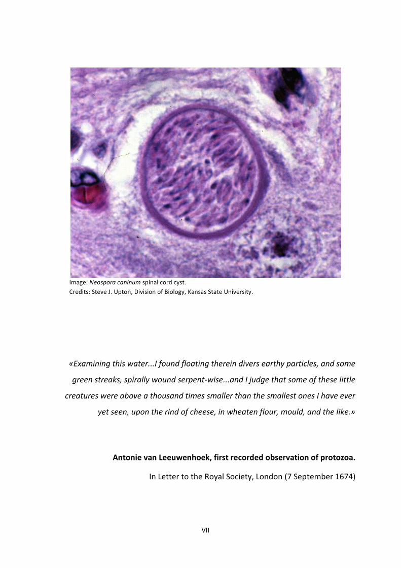
Image: Neospora caninum spinal cord cyst.
Credits: Steve J. Upton, Division of Biology, Kansas State University.
«Examining this water...I found floating therein divers earthy particles, and some
green streaks, spirally wound serpent-wise...and I judge that some of these little
creatures were above a thousand times smaller than the smallest ones I have ever
yet seen, upon the rind of cheese, in wheaten flour, mould, and the like.»
Antonie van Leeuwenhoek, first recorded observation of protozoa.
In Letter to the Royal Society, London (7 September 1674)

VIII
IX
Abstract
Neospora caninum is an obligate intracellular parasite of animals. Dogs, coyotes and
dingoes are definitive hosts, while cattle are intermediate hosts and the most commonly affected
animals in the field of neosporosis, the disease caused by N. caninum. This parasite is a major cause
of abortion in cattle around the world leading to heavy economic losses to dairy and beef industry.
The only vaccine commercially available against neosporosis has only a limited effect in preventing
abortion in cattle herds. The parasite can enter cattle hosts through the gastrointestinal tract and
this entry route is indispensable for sustaining infection within a herd. Thus, the present work
aimed at 1) characterizing the immune response induced in mice by mucosal (intra-nasal) CpG-
adjuvanted immunisation with membrane proteins from N. caninum tachyzoites (NcT) and 2)
evaluating the protective effect conferred by this strategy against N. caninum infection induced by
the intragastric route. Host protection is generally associated with a N. caninum-specific TH1
immune response whereas parasite invasion and infection are favoured by a TH2-biased immune
response.
The immunisation with NcT membrane proteins plus CpG was performed by two intra-nasal
administrations three weeks apart. It was shown here to be effective at promoting a N. caninum-
specific immune response in the intestinal mucosa, as indicated by the significantly higher anti-NcT
IgA ELISA titres detected in mice that received this treatment. The detection, by ELISA, of anti-NcT
IgG in the serum suggested that the immunisation strategy used also induced a systemic immune
response. The IgG1/IgG2a ratio, lower than 1.00, indicated that the N. caninum-specific immune
response was predominantly of the TH1-type, the prototypic protective response against N.
caninum. Protection against this parasite was confirmed by the absence of parasites in the liver and
brain of the immunised mice, seven days after infection with NcT, as evaluated by qPCR. At last,
serum IgG antibodies produced upon the immunisation were shown, by flow cytometry, to bind
the NcT surface in vitro. Western blot analysis indicated that IgG1 and IgG2a bound, respectively,
NcT membrane proteins of 35 and 39 kDa and antibodies of both the IgG subclasses also bound a
17 kDa protein.
Altogether, this study suggests that intra-nasal CpG-adjuvanted immunisation with NcT
membrane proteins is a promising strategy to prevent neosporosis. If successfully applied in cattle,
this strategy can aid combating the horizontal transmission route of N. caninum in this group of
animals and prevent, at least in part, the economic losses associated with neosporosis.
Keywords: Neospora caninum; neosporosis; gastrointestinal tract; mucosal immunity;
immunisation; adjuvant; CpG

X
XI
Resumo
Neospora caninum é um parasita intracelular obrigatório de animais. Os cães, coiotes e
dingos são os hospedeiros definitivos, enquanto que os bovinos são hospedeiros intermediários e o
grupo de animais mais afetado no que diz respeito à neosporose, doença causada por N. caninum.
Este parasita é a principal causa mundial de aborto em bovinos e conduz a prejuízos económicos
elevados na indústria de lacticínios e da carne. Uma vacina contra a neosporose atualmente
disponível no mercado demonstrou prevenir apenas parcialmente a ocorrência de abortos em
manadas. Em bovinos, o parasita pode entrar pelo trato gastrointestinal, sendo esta via de entrada
indispensável para manter a infeção numa manada. Deste modo, o presente estudo teve como
objetivo 1) caracterizar a resposta imunitária induzida em ratinhos por imunização na mucosa
(intranasal) com proteínas de membrana provenientes dos taquizoítos de N. caninum (NcT) e com
o adjuvante CpG, e 2) avaliar o efeito protetor conferido por esta estratégia na infeção por N.
caninum induzida pela via intragástrica. A proteção do hospedeiro está geralmente associada a
uma resposta imunitária específica para N. caninum do tipo TH1, enquanto que a invasão e infeção
pelo parasita são favorecidas por uma resposta enviesada para o tipo TH2.
A imunização com proteínas de membrana de NcT juntamente com CpG foi efetuada por
duas administrações intranasais, com um intervalo de três semanas entre elas. Esta mostrou ser
eficaz na indução de uma resposta específica para N. caninum na mucosa intestinal, como
demonstrado pelos níveis significativamente mais elevados de IgA específica para NcT, detetados
por ELISA, em ratinhos que receberam este tratamento. A deteção, também por ELISA, de IgG
específica para NcT no soro sugeriu que a imunização em estudo era também capaz de induzir uma
resposta imunitária sistémica. O facto de o rácio IgG1/IgG2a ser menor que 1.00 nestes ratinhos
indicou que a resposta imunitária específica para N. caninum induzida pela imunização era
predominantemente do tipo TH1, portanto presumivelmente protetora contra N. caninum. A
proteção contra este parasita foi confirmada, usando qPCR, pela ausência de parasita no fígado e
cérebro dos ratinhos imunizados, sete dias após a infeção com NcT. Por fim, demonstrou-se, por
citometria de fluxo, que IgGs do soro produzidas em resposta à imunização em estudo se ligavam à
superfície de NcT. A análise efetuada por Western blot demonstrou que a IgG1 e IgG2a se ligavam,
respetivamente, a proteína de membrana de NcT com 35 e 39 kDa e os anticorpos de ambas as
subclasses também se ligavam à proteína de 17 kDa.
Em resumo, este estudo sugere que a imunização intranasal com proteínas de membrana
de NcT e com o adjuvante CpG é promissora. Se aplicada com sucesso semelhante em bovinos, no
futuro, esta estratégia poderá ajudar a combater a via horizontal de transmissão de N. caninum
XII
neste grupo de animais e evitar, pelo menos em parte, os prejuízos económicos associados à
neosporose.
Palavras-chave: Neospora caninum; neosporose; trato gastrointestinal; imunidade na mucosa;
imunização; adjuvante; CpG.
XIII
Acknowledgments
Sendo este trabalho o último passo de um percurso de cinco anos tão fugaz mas ao mesmo tempo
tão intenso, não poderia deixar de incluir nele um agradecimento especial a todos os que, à sua
maneira, para ele contribuíram e me acompanharam ao longo desta caminhada. Deste modo,
agradeço:
Em primeiro lugar ao meu orientador, Professor Manuel Vilanova, por me ter acolhido no seu
laboratório e por me demonstrar, nas mais diversas situações, que são as dúvidas e o sentido de
descoberta os maiores estímulos para o exercício desta atividade tão fascinante que é a
Investigação Científica.
Ao Pedro Ferreirinha, por toda a dedicação e ajuda ao longo destes meses, pela paciência em
esclarecer as minhas dúvidas, por saber lidar tão bem com o meu stress diário, por me transmitir
tantos conhecimentos, pelos conselhos, pela amizade.
À Alexandra e Luzia por partilharem comigo os seus conhecimentos, pela força que sempre me
deram, pela amizade ao longo dos últimos anos. À Begoña e Adília pela ajuda e companheirismo ao
longo do tempo. À Joana Maia, Amanda, Rita, Sílvia e Filipe, assim como à Joana Alves, Elva, Liliana,
Pedro Madureira, Vanessa e Cristina pela boa disposição e bom ambiente de trabalho que sempre
proporcionaram. À Encarnação pela simpatia e disponibilidade constantemente demonstradas. À
Professora Paula Ferreira por ter sido sempre tão afável. Ao Professor Rui Appelberg pelas
apreciações críticas no decorrer do projeto.
Aos restantes Professores do MIB, quer da FEUP quer do ICBAS, por ao longo destes cinco anos
terem transmitido conhecimentos de áreas tão diferentes e demonstrado continuamente como
estão interligados. Em especial, estou grata ao Professor Nuno Azevedo por ter demonstrado de
forma tão clara e paciente durante os três últimos anos a ponte entre a componente «bio» e a
componente de «engenharia»; ao Professor Pedro Moradas Ferreira por, entre outros, ter tornado
possível o período de estágio ao abrigo do programa Erasmus, o levou a uma experiencia de vida
verdadeiramente única e cujas memórias ficarão para a vida. Ao Professor Alexandre Quintanilha
por ter sido uma fonte de inspiração ao longo destes cinco anos e por ter apoiado o meu fascínio
pela Imunologia, ao consentir que esta fosse a área de desenvolvimento da minha dissertação.
XIV
Aos colegas de Bioengenharia da turma de 2007 que ao longo destes anos me acompanharam
nesta caminhada, por todos os momentos passados, os bons e os menos bons, por todas as
«batalhas» ultrapassadas com sucesso e pelo companheirismo e solidariedade quando outras não
foram tão bem sucedidas. Pela boa disposição e pelos momentos de descontração diários. Em
especial, um muito obrigada à Andreia, Paulina e André Meireles pela entreajuda constante, pela
partilha de experiências, pela grande amizade que se construiu ao longo do tempo.
À Joana Oliveira e Marlene Fernandes, assim como à Teresa, Ana, Liliana e João Tiago pela amizade
de longa data, pelo apoio incondicional.
À Susana e ao César pela partilha de impressões, opiniões e emoções ao longo de tantos anos,
pelos conselhos sempre tão úteis, pela amizade tão sólida, por estarem «sempre lá».
Por último, aos grandes pilares da minha vida: à minha querida Irmã Sónia por toda a paciência que
sempre teve e tem comigo, pelos conselhos tão práticos, pela força que sempre me deu de uma
maneira tão única; aos meus Pais por «tudo», sendo esta palavra demasiado ingrata para resumir o
que me demonstram e proporcionam incondicionalmente todos os dias, todo o amor, todo o
apoio, toda a força, toda a paciência, toda a compreensão, toda a dedicação. Um eterno obrigada,
Irmã, Mãe e Pai, porque sem vós nunca teria conseguido!
XV
Contents
ABSTRACT IX
RESUMO XI
ACKNOWLEDGMENTS XIII
LIST OF FIGURES XVII
LIST OF TABLES XVII
LIST OF ABBREVIATIONS XVIII
1. INTRODUCTION 1
1.1 PROJECT BACKGROUND 1
1.2 PROJECT OBJECTIVES 1
1.3 THESIS ORGANISATION 1
2. STATE OF THE ART 3
2.1 NEOSPORA CANINUM 3
2.1.1 Life cycle 3
2.1.2 Modes of transmission in cattle 5
2.1.3 Surface and secretory proteins of NcT 6
2.2 NEOSPOROSIS IN CATTLE 7
2.2.1 Clinical signs 7
2.2.2 Diagnostic techniques 8
2.2.3 Economic impact 9
2.2.4 Prevention and control 9
2.3 MUCOSAL IMMUNITY 10
2.3.1 Innate immunity 10
2.3.2 Adaptive immunity 11
2.3.2.1 Inductive and effector sites 11
2.3.2.2 Cellular traffic in the mucosa and antigen sampling 13
2.3.2.3 Humoral immune response 14
2.3.2.4 Cellular immune response 15
2.4 MUCOSAL IMMUNISATION 16
2.4.1 Types of vaccines 16
2.4.2 Adjuvants 17
2.5 IMMUNE RESPONSE TO N. CANINUM 18

XVI
2.6 N. CANINUM AND VACCINATION 19
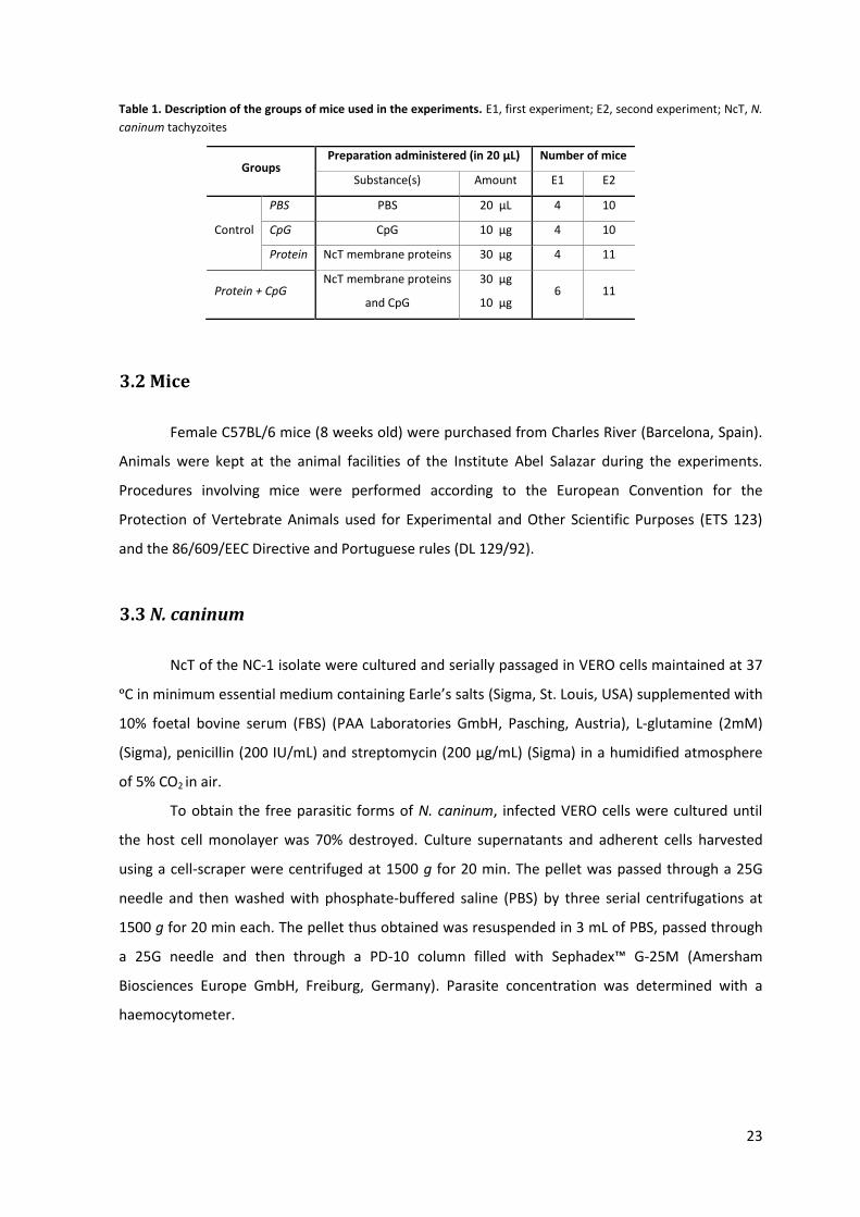
3. MATERIALS AND METHODS 22
3.1 EXPERIMENT SET-UP 22
3.2 MICE 23
3.3 N. CANINUM 23
3.4 IMMUNISATIONS 24
3.5 CHALLENGE INFECTIONS 24
3.6 EXTRACTION OF MEMBRANE PROTEINS OF NCT 24
3.7 COLLECTION OF BIOLOGICAL SAMPLES 25
3.8 IN VITRO CELL CULTURE 25
3.9 CYTOKINE INTRACELLULAR STAINING 26
3.10 ANTIBODY MEASUREMENT 26
3.11 DNA EXTRACTION 27
3.12 QPCR 27
3.13 SDS-PAGE AND WESTERN BLOT 28
3.14 ANTIBODY BINDING ASSAY TO NCT 28
3.15 FLOW CYTOMETRIC ANALYSIS 28
3.16 STATISTICAL ANALYSIS 29
4. RESULTS 30
4.1 EXTRACTION OF NCT MEMBRANE PROTEINS 30
4.2 ANTIBODY PRODUCTION IN MICE UPON CPG-ADJUVANTED IMMUNISATION WITH NCT MEMBRANE
PROTEINS AND FOLLOWING NCT INFECTION 31
4.2.1 NcT-specific IgA production 31
4.2.2 NcT-specific IgG1 and IgG2a production 33
4.3 CYTOKINE PRODUCTION BY MLN AND SPLEEN CELLS FOLLOWING CPG-ADJUVANTED PRIME-BOOST
IMMUNISATION WITH NCT MEMBRANE PROTEINS AND NCT INFECTION 36
4.4 PARASITE LOAD IN LIVER AND BRAIN OF MICE FOLLOWING PRIME-BOOST CPG-ADJUVANTED
IMMUNISATION WITH NCT MEMBRANE PROTEINS AND NCT INFECTION 37
4.5 SERUM IGG BINDING TO THE SURFACE OF NCT 38
5. DISCUSSION 40
6. CONCLUSIONS AND FUTURE WORK 48
REFERENCES 50
XVII
List of Figures
Figure 1. Life cycle of N. caninum. 5
Figure 2. Composition and cellular traffic in inductive and effector sites. 12
Figure 3. Timeline of tasks performed during the experiment. 22
Figure 4. SDS-PAGE gels of NcT membrane protein extracts obtained by the Triton X-114 extraction
procedure. 30
Figure 5. Anti-NcT IgA ELISA titres in mice vaginal lavage fluids after the prime immunisation. 31
Figure 6. Anti-NcT IgA ELISA titres in mice vaginal lavage fluids after the boost immunisation. 32
Figure 7. Anti-NcT IgA ELISA titres in mice intestinal lavage fluids after the immunisations and the
NcT challenge infection. 33
Figure 8. Anti-NcT IgG1 and IgG2a ELISA titres in mice serum after the boost immunisation. 34
Figure 9. Anti-NcT IgG1 and IgG2a ELISA titres in mice serum after the immunisations and the NcT
challenge infection. 35
Figure 10. Cytokine production by CD4+ and CD8+ cells from MLNs and spleen after the
immunisations and the NcT challenge infection. 36
Figure 11. N. caninum load in liver and brain of mice after the immunisations and the NcT challenge
infection. 37
Figure 12. In vitro binding of mice serum IgGs to the NcT surface. 38
Figure 13. Western blot analysis of the NcT membrane proteins to which IgG1 and IgG2a from the
immune serum were bound. 39
List of Tables
Table 1. Description of the groups of mice used in the experiments. 23
XVIII
List of Abbreviations
ADCC Antibody-dependent cell-mediated cytotoxicity
AP Alkaline phosphatase
APC Antigen-presenting cell
BCIP 5-bromo-4-chloro-3-indolyl-phosphate
BDL Below detection limit
BSA Bovine serum albumin
CALT Conjunctiva-associated lymphoid tissue
CCL CC-chemokine ligand
CCR CC-chemokine receptor
CMI Cell-mediated immunity
CMIS Common mucosal immune system
CT Cholera toxin
CTL Cytotoxic T lymphocyte
Cy Cychrome
DC Dendritic cell
DG Dense granule
DTH Delayed-type hypersensitivity
EDTA Ethylenediamine tetraacetic acid
ELISA Enzyme-linked immunosorbent assay
FACS Fluorescent-activated cell sorter
FAE Follicle-associated epithelium
FBS Foetal bovine serum
FCA Freund’s complete adjuvant
FcRn Neonatal Fc receptor
FIA Freund’s incomplete adjuvant
FITC Fluorescein isothiocyanate
GALT Gut-associated lymphoid tissue
GPI Glycophosphatidylinositol
HBSS Hank’s Buffered Salt Solution
HEL Hen egg lysozyme
HEV High endothelial venule
XIX
i.g. Intragastric
i.n. Intra-nasal
IEL Intraepithelial lymphocyte
IFAT Indirect fluorescent antibody test
IFN-γ Interferon-gamma
Ig Immunoglobulin
IL Interleukin
Iscom Immune stimulating complex
J-chain Joining chain
LN Lymph node
LP Lamina propria
MALT Mucosa-associated lymphoid tissue
MFI Mean fluorescence intensity
MLN Mesenteric LN
N. caninum Neospora caninum
NALT Nasopharynx-associated lymphoid tissue
NAT Neospora agglutination test
NBT Nitro blue tetrazolium
NcT N. caninum tachyzoites
NK Natural killer
NTPase Nucleotide triphosphate hydrolase
PAMP Pathogen-associated molecular pattern
PBS Phosphate-buffered saline
PCR Polymerase chain reaction
PDI Protein disulfide isomerase
PE Phycoerythrin
PerCP Peridin-chlorophyll protein
pIgR Polymeric Ig receptor
PLG Poly(D,L-lactide-co-glycolide)
PMA Phorbol myristate acetate
PMN Polymorphonuclear leukocyte
PP Peyer’s patch
PRR Pattern recognition receptor
qPCR Real-time quantitative PCR
XX
RNS Reactive nitrogen species
s.c. Subcutaneous
SAG Surface antigen
SDS Sodium dodecyl sulfate
SED Subepithelial dome
sIgA Secretory IgA
SRS SAG-related sequence
TC T cytotoxic
TCR T cell receptor
TH T helper
TLR Toll-like receptor
TNF-α Tumour necrosis factor-alpha
1
1. Introduction
1.1 Project background
Neospora caninum is an obligate intracellular parasite that causes disease, neosporosis, in a
wide range of veterinary species including dogs, sheep, goats and cattle. The latter are the most
commonly affected animals and the economically relevant hosts. Cattle can be infected
transplacentally, a very frequent and effective way of parasite transmission, or post-natally via the
gastrointestinal (GI) tract. Although less common, this infection route is indispensable to sustain
infection within a herd. Neosporosis is a major cause of abortion in cattle worldwide and, to date,
no completely effective control measure against the parasite has been developed. Moreover,
although a vaccine is commercially available, it has proven to be only partially effective in
preventing abortions within herds.
1.2 Project objectives
The present work aimed at characterizing the immune response elicited in mice by mucosal
(intra-nasal) (i.n.) CpG-adjuvanted immunisation with membrane proteins of N. caninum
tachyzoites (NcT) and at evaluating the protective effect conferred by this immunisation procedure
against neosporosis established by the GI tract. The ultimate purpose would be to establish an
effective immunisation protocol that could be applied in cattle to prevent post-natal N. caninum
transmission. Since horizontally transmitted neosporosis can also result in foetal infection and
possibly abortion, the development of a novel vaccine that could confer successful immunisation
would significantly contribute to improve the management and decrease the worldwide impact
caused by N. caninum infections.
1.3 Thesis organisation
This document is organised into six chapters: Introduction, State of the Art, Materials and
Methods, Results, Discussion and Conclusions and future work. The first four chapters are divided
into several subheadings so that the different subjects and ideas can be more easily accessed.
Chapter 2, State of the Art, entails a comprehensive literature review in the field and
provides the theoretical framework where the project is included.

2
Chapter 3, Materials and Methods, describes the reagents and data collection instruments
used, as well as the methods employed to carry out the research study. A section of this chapter
named Experiment set-up explains how the research was conducted by describing the organisation
in time of the several tasks that the project comprised.
Chapter 4, Results, includes a detailed presentation of the data obtained which are then
critically appreciated in the context of the stated objectives and other reported data in chapter 5,
Discussion.
Conclusions drawn from the study as well as recommendations for future studies are
documented in chapter 6, Conclusions and future work.
3
2. State of the Art
2.1 Neospora caninum
N. caninum is an obligate intracellular protozoan parasite that was first recognised in
Norway in 1984 as the causative agent of encephalomyelitis and myositis in dogs, which had no
antibodies to Toxoplasma gondii1. It was classified later on, in 1988, by Dubey et al.2, who
described the new genus Neospora and the type species Neospora caninum belonging to the
Phylum Apicomplexa and the family Sarcocystidae. In the same year, the parasite was isolated from
paralyzed dogs3 and that strain was then designated NC-14.
Unlike T. gondii, viable N. caninum is difficult to isolate5 and it has been obtained from only
a few hosts, namely cattle6, sheep7, water buffaloes8, white-tailed deers9 and bisons10, besides
dogs. Prevalence of N. caninum-specific antibodies has been demonstrated in cattle, sheep, goats,
dogs, pigs, coyotes and other domestic and wild animals5. N. caninum specific DNA in turn was
found in chickens, rats and rabbits, among others, but its finding should not be regarded as
equivalent to finding viable N. caninum5. Altogether, these reports stress out that a wide range of
domestic and wild animals have been exposed to N. caninum.
At present, the zoonotic potential of N. caninum is still questionable. Neither the parasite
itself nor its DNA were so far detected in human tissues11. Moreover, in one study using serum
samples from women with a history of idiopathic repeated abortions, no N. caninum-specific
antibodies were detected by any of the serological tests performed12. Nevertheless, N. caninum
infection was successfully established in macaque foetuses that were infected with the parasite,
either directly or transplacentally13, and N. caninum-specific immunoglobulin G (IgG) were detected
in HIV-infected patients and in patients with neurological disorders14.
2.1.1 Life cycle
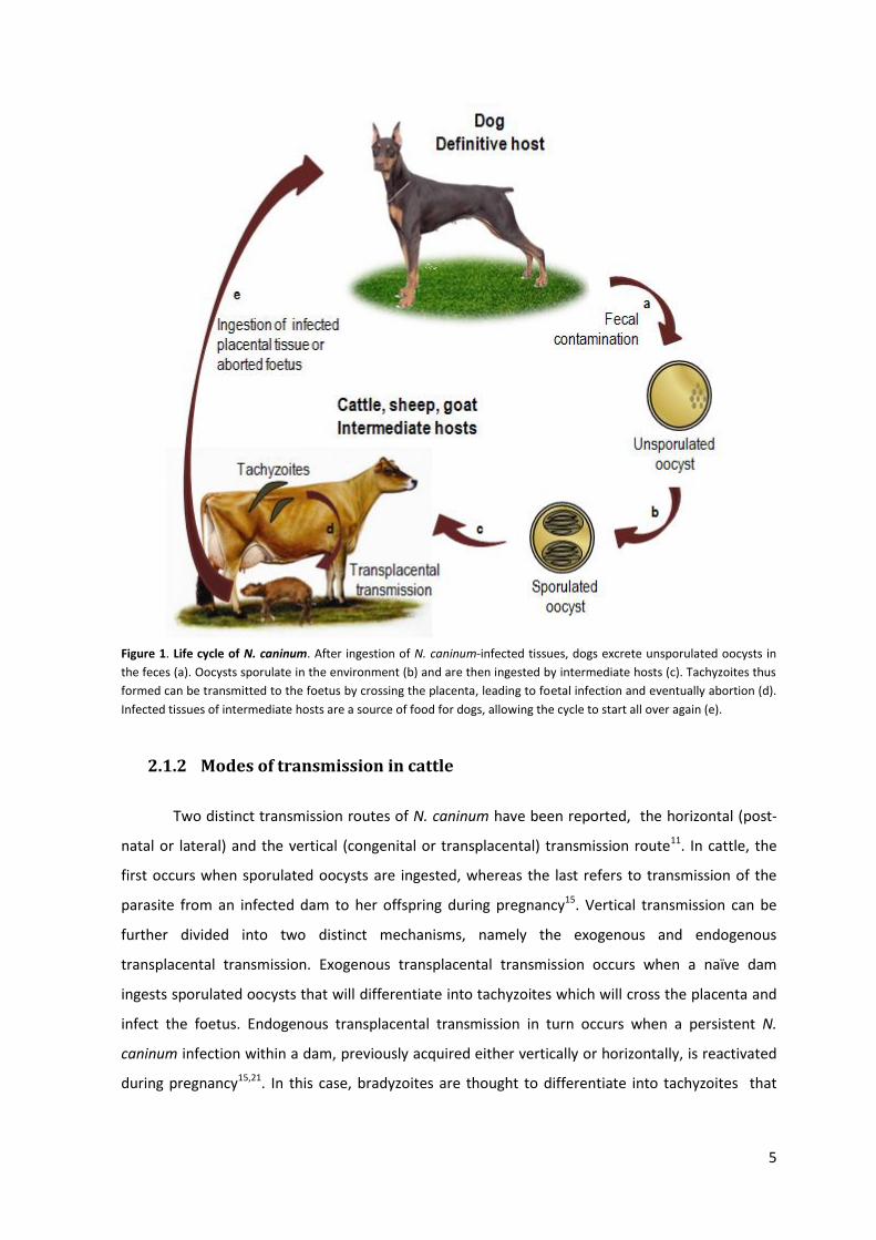
N. caninum is a single-celled coccidian parasite with a heteroxenous life cycle, which means
that more than one host is needed to complete it (Figure 1)15. To date, the only identified definitive
hosts of N. caninum, i.e., the animals in which sexual reproduction of the parasite takes place16, are
the domestic dogs (Canis lupus familiaris)17,18, Australian dingoes (Canis lupus dingo)16 and coyotes
(Canis latrans)19. Intermediate hosts, i.e., animals in which parasite multiplication occurs by asexual
reproduction16, include a wide range of warm-blooded animals like sheep, goats and felids20, being
however cattle the most common and economically important host animals21.
4
Three parasite infectious stages have been identified during N. caninum life cycle, namely
oocysts, tachyzoites (short-lived, rapidly dividing stage) and bradyzoites (longer-lived, slowly
dividing stage)20. Oocysts, environmentally resistant and measuring approximately 10 × 12 μm22,23,
constitute the sexual stage of the parasite and are excreted in an unsporulated phase in the feces
of acutely infected dogs, after ingestion of N. caninum-infected tissues24,25. Low numbers of oocysts
are usually excreted5. Shedding can start between 5 and 13 days after consumption of bradyzoite-
containing tissue cysts and last for up to 27 days26 or even continue for several months in some
dogs27. Oocysts sporulate in the environment within 24 to 72 hours after having been excreted,
giving rise to two sporocysts, each containing four sporozoites24,26 that measure 6.5 × 2 μm22. Once
cattle or other intermediate hosts ingest water or food contaminated with sporulated oocysts they
enable the sporozoites to be released in the intestinal tract, where they invade cells and
differentiate into tachyzoites24 which are lunate-shaped and measure approximately 6 × 2 μm15.
Tachyzoites divide within host cells by endodyogeny (process of asexual reproduction in which two
daughter cells develop within the parent cell), forming numerous organisms that are released in
the blood15,28. The resulting parasitaemia provides a means for dissemination of the parasite
throughout the body15. Okeoma et al.29 detected N. caninum DNA in the leukocyte fraction of blood
from naturally infected pregnant heifers. Tachyzoites were reported to infect many different cell
types, including myocytes, hepatocytes, neural cells, vascular endothelial cells, alveolar
macrophages, renal cells and placental trophoblasts, the last leading to transmission of the parasite
to the foetus15. At a cellular level, tachyzoites are located within a parasitophorous vacuole in the
host cell cytoplasm4. If the host is immunocompetent, its immune system will be activated in
response to the invasion and the multiplication of tachyzoites in order to contain the spread of the
parasite30. To be protected from the attack mounted by the host immune system, tachyzoites
differentiate into bradyzoites, which are slender, measuring approximately 6.5 × 1.5 μm31, and are
usually located in the central nervous system surrounded by a cyst wall, thus forming intracellular
tissue cysts24,32. However, they have also been identified in the retina of a naturally infected dog33
and in skeletal muscles of naturally infected dogs and cattle34. Tissue cysts are often round or oval
in shape and vary in size depending on the number of bradyzoites enclosed15,34. Their diameter and
cyst wall thickness are, respectively, up to 107 μm and up to 4 μm in dogs and approximately 50
μm and less than 2.5 μm in cattle4,35,36. Bradyzoites represent a quiescent stage of the parasite that
remain latent until the host’s immune response is suppressed, which allow them to recrudesce24,
and may act as a source of foetal infection in subsequent pregnancies28. Dogs can then ingest
tachyzoites in placental tissue or bradyzoites in tissue cysts, which allow them to implant in the GI
tract and undergo maturation. Oocysts are then excreted in the feces of these animals and the N.
caninum life cycle starts all over again24,26,37 (Figure 1).
5
Figure 1. Life cycle of N. caninum. After ingestion of N. caninum-infected tissues, dogs excrete unsporulated oocysts in
the feces (a). Oocysts sporulate in the environment (b) and are then ingested by intermediate hosts (c). Tachyzoites thus
formed can be transmitted to the foetus by crossing the placenta, leading to foetal infection and eventually abortion (d).
Infected tissues of intermediate hosts are a source of food for dogs, allowing the cycle to start all over again (e).
2.1.2 Modes of transmission in cattle
Two distinct transmission routes of N. caninum have been reported, the horizontal (post-
natal or lateral) and the vertical (congenital or transplacental) transmission route11. In cattle, the
first occurs when sporulated oocysts are ingested, whereas the last refers to transmission of the
parasite from an infected dam to her offspring during pregnancy15. Vertical transmission can be
further divided into two distinct mechanisms, namely the exogenous and endogenous
transplacental transmission. Exogenous transplacental transmission occurs when a naïve dam
ingests sporulated oocysts that will differentiate into tachyzoites which will cross the placenta and
infect the foetus. Endogenous transplacental transmission in turn occurs when a persistent N.
caninum infection within a dam, previously acquired either vertically or horizontally, is reactivated
during pregnancy15,21. In this case, bradyzoites are thought to differentiate into tachyzoites that
6
spread to distinct host tissues including the uteri, where they cross the placenta and infect the
foetus21.
In cattle, vertical transmission is a very efficient way of spreading N. caninum to a new
host, with transmission rates ranging from approximately 80% to 95%38,39, and can occur over
several and consecutive pregnancies40,41 or intermittently41. The endogenous route of vertical
transmission is thought to account for the majority of infections occurring in these animals42, being,
in contrast, less frequent the occurrence of horizontal transmission (between 1% and 2% per cow
per year)39,43. However, it was shown by theoretical models that even low levels of horizontal
transmission are needed to sustain N. caninum infection within a herd44.
Other natural post-natal modes of transmission were tested for the possibility of also being
responsible for N. caninum infection in cattle. Cow to cow transmission does not seem to occur45
and was not observed so far5 and venereal transmission is unlikely to exist. Dams naturally bred
with experimentally infected bulls did not seroconvert46 and even though N. caninum DNA has
been detected in the semen of seropositive bulls, only a few parasites were present there47.
Furthermore, although neonatal calves may become experimentally infected after ingestion of
tachyzoite-contaminated milk, lactogenic transmission does not appear to be an important natural
route of N. caninum transmission in cattle48,49. Therefore, the only demonstrated natural mode of
infection in cattle is through the ingestion of sporulated oocysts from the environment5.
2.1.3 Surface and secretory proteins of NcT
Two major immunodominant glycophosphatidylinositol (GPI)- anchored proteins populate
the NcT surface and contribute to the initial low-affinity host-parasite contact. These are
homologues of T. gondii surface antigen 1 (SAG1) and SAG-related sequence 2 (SRS2) and are
commonly designated NcSAG1 and NcSRS2 with 29 and 35 kDa, respectively32,50. Once the initial
contact has been established, NcT discharge secretory organelles, namely micronemes, dense
granules (DGs) and rhoptries. Microneme proteins, released at the onset of host-parasite adhesion
and therefore mediators of the physical interaction between the parasite and the host cell
surface51, include soluble molecules, such as NcMIC1, NcMIC2 and NcMIC4 as well as membrane-
bound molecules, such as NcMIC332. NcSUB1, a serine protease produced by the parasite, also
displays microneme localization52. As suggested by in vitro studies, rhoptry proteins, in particular
NcROP2, may be involved in host cell invasion, consistent with the fact that these organelles are
exocytosed upon invasion of the host cell53,54. Also, rhoptry components, as well as proteins from
DGs which are secreted during the invasion process, are important for the formation and
functioning of the parasitophorous vacuole in which the parasite develops53,54. Proteins from DGs
7
include NCDG1, NCDG2, nucleotide triphosphate hydrolase (NTPase), NcGRA2 and NcGRA753.
Another protein, existent on the surface of NcT and in the micronemes but absent in rhoptries and
DGs, is protein disulfide isomerase (PDI)55. NcPDI is involved in parasite-host cell interactions since
it catalyses the reduction, oxidation and isomerisation of disulfide bonds and many components of
both the adhesion and invasion machinery of N. caninum are cysteine-rich and depend on correct
folding through the formation of disulfide bonds55.
2.2 Neosporosis in cattle
Since its discovery, neosporosis, the disease caused by N. caninum, has emerged as a major
cause of disease in dogs26 and, in particular, abortions in cattle around the world11. Before
recognition of N. caninum as a new species and the characterization of the disease itself,
neosporosis was often misdiagnosed as toxoplasmosis due to close similarities between either the
causative parasites or the respective diseases both clinically and pathologically2,56. N. caninum and
T. gondii are, in fact, morphologically, genetically and antigenically closely related parasites4,50,57
and have similar life cycles11. However, they are biologically different as neosporosis is mainly a
disease of cattle and dogs and is not considered zoonotic, whereas toxoplasmosis affects primarily
humans, sheep and cats, the last being definitive hosts of that parasite5,20.
2.2.1 Clinical signs
In cattle, neosporosis manifests in the placenta and developing foetus but rarely in adult
animals30. Foetuses may either be aborted from the third month of gestation until the end, with
most abortions occurring between the fifth and sixth month, or born weak with neurological
symptoms or born clinically normal but persistently infected5,57. The outcome depends on the
timing in gestation at which the infection occurs30, with infections occurring early in pregnancy
being more harmful to the foetus than infections occurring later on58. Cows infected with N.
caninum are three to seven times more likely to abort than uninfected cows and the risk is highest
in the first pregnancy41,59,60. Other clinical signs of neosporosis have only been reported in cattle
with less than 2 months of age and include underweight and inability to rise. The hind limbs or
forelimbs may be flexed or hiperextended and neurological problems such as ataxia, decreased
patella reflexes and loss of conscious proprioception may develop. Calves may also have
exophthalmia or an asymmetrical appearance in the eyes and, occasionally, birth defects such as
hydrocephalus and narrowing of the spinal cord may occur5.

8
2.2.2 Diagnostic techniques
A variety of serological tests can be performed to diagnose N. caninum infection in cattle,
including the Indirect Fluorescent Antibody Test (IFAT), several Enzyme-Linked Immunosorbent
Assays (ELISAs), the Neospora Agglutination Test (NAT) and Western Blot61. Even though IFAT is
very specific for N. caninum, it is an expensive and time-consuming test, not being commonly
performed to screen cattle populations for that infection24,62. Avidity-ELISA is a useful test to
distinguish between acute and chronic infections, based, respectively, on the lower or higher
binding strengths (avidity) of IgG antibodies to a N. caninum antigen63. Another way of doing that is
by performing ELISAs using the recombinant proteins NcGRA7 and NcSAG4 (a bradyzoite stage-
specific protein) to detect antibodies of the acute phase and chronic phase, respectively64.
However, it is worth mentioning that antibody levels can fluctuate considerably and may even drop
below the cut-off value of the test used, giving rise to a negative result when N. caninum-specific
antibodies actually exist65,66. Therefore, negative serological results are not informative61.
Antibody fluctuations also occur in bovine foetuses as factors such as the stage of gestation
and the time between infection and abortion influence antibody production61. In addition,
serological results in the foetus only provide information about exposure to the parasite and
histologic examination should therefore be carried out for a definitive diagnosis of neosporosis
abortion61. However, few N. caninum parasites are generally present in autolysed tissues and these
are hardly visible in histological sections. Therefore, immunohistochemistry is usually performed.
The foetal brain is the tissue of choice either for histology or immunohistochemistry, as it is the
most consistently affected organ24,61. In severely autolysed foetuses, however,
immunohistochemistry is insensitive and may underestimate the existence of infection61. In such
cases, PCR can thus be used to detect N. caninum in the brain tissues of the aborted calves61.
Diagnosis of bovine abortion is often difficult since finding N. caninum-specific DNA, antibodies or
even the parasite itself in foetal tissues may not necessarily mean that N. caninum was the cause of
the abortion, as asymptomatic and congenitally infected cows, many of them being serologically
positive, are commonly found5,11. Thus, a comprehensive diagnostic approach is usually conducted
in which the aforementioned serologic, histologic and molecular methods are integrated to
investigate the existence of a cause-effect relationship between N. caninum and abortion15.
Although N. caninum is closely related to T. gondii and Sarcocystis species, cattle serum
cross-reactivity has not been a major issue61. N. caninum can also be distinguished from those
parasites by immunohistochemistry and, at the DNA level, by PCR61. In addition, infection of bovine
foetuses with T. gondii is rare and Sarcocystis cruzi is rarely (< 0.1%) found in aborted foetal
9
brains61. However, there is the possibility of serum cross-reaction with Neospora hughesi, a
parasite closely related to N. caninum about which little is known67.
2.2.3 Economic impact
N. caninum is a major cause of epidemic and endemic abortions in both dairy and beef
cattle around the world5,15 and economic losses due to Neospora-associated abortions have been
estimated to exceed hundreds of millions of dollars every year53. In addition to that direct cost,
indirect costs include veterinary assistance, diagnostic procedures, rebreeding, replacement costs
when aborted cows have to be culled and possible loss of milk production5. Post-natal losses
caused by N. caninum are difficult to assess since clinical disease has not been reported in calves
older than 2 months of age5.
2.2.4 Prevention and control
In cattle, control measures that may reduce neosporosis include embryo transfer, test-and-
cull strategies, chemotherapeutic treatment, vaccination and increased biosecurity in herds.
Transfer of embryos from N. caninum-infected dams to only uninfected and seronegative recipient
cows is a way of recovering uninfected calves and preventing endogenous transplacental
transmission of the parasite5,68. The test-and-cull approach includes testing and culling seropositive
dams or seropositive aborting dams or testing and either excluding the progeny of seropositive
dams from breeding or inseminating the progeny with beef bull semen only5 which has been
demonstrated to reduce the risk of abortion in Neospora-seropositive heifers and cows69,70. Despite
having been successfully implemented in the field71 and considered one of the most efficacious
solutions to control neosporosis, the test-and-cull approach is very expensive and likely
uneconomical, especially in herds with a large proportion of infected cattle53,72. Until now, no
chemotherapeutic treatment has proven to be safe and effective enough to treat neosporosis in
either dairy or beef cattle. Moreover, safety concerns arising from the possibility that drug
administration could produce undesirable residues in milk or meat, may render these products
unacceptable for consumption5. In addition, and similarly to the test-and-cull strategy,
chemotherapy does not seem to provide an economically viable control option, as suggested by
previous analysis72. In contrast, efficacious vaccination of all susceptible animals (both seropositive
and seronegative cattle in a herd) does seem to be economically viable72 (further discussion on this
topic in 2.6 N. caninum and vaccination). Farmers can also adopt biosecurity measures to prevent
horizontal transmission of N. caninum, such as the avoidance of contact between dogs and infected
10
bovine tissues to hinder oocysts shedding and the coverage of feed, silage and bedding materials to
minimize contamination with dog feces28. No control measures directed to public health are
recommended since, at present, neosporosis is not considered a zoonotic disease11.
2.3 Mucosal immunity
The intestine is the largest area of the organism in contact with the external environment73.
In there, permanent antigenic challenge occurs from birth to death, in the form of food antigens,
antigens of the microbiota and pathogens acquired by the oral route. This organ is thus considered
a particularly vulnerable source of infection and a constant threat of disease74,75. However, it has
the ability to maintain the number of resident bacteria and simultaneously protect against
pathogens by inducing mucosal innate and adaptive immune responses76.
2.3.1 Innate immunity
In the intestinal mucosa, the innate immune response is carried out by strongly
interconnected physical, chemical and cellular components of the epithelial barrier. A single
epithelial cell layer, with tight junctions between the cells, accounts for the physical barrier but yet
allows selective entry of essential nutrients. Chemical obstacles include the mucus overlying the
epithelium as well as the bile, pancreatic enzymes and antimicrobial peptides. The cellular barrier
in turn is composed of immune cells that trigger nonspecific defences against pathogens successful
in breaching the epithelial barrier76–78. Epithelial cells lining the intestinal mucosal surface are able
to recognize pathogen-associated molecular patterns (PAMPs) which are usually highly conserved
and essential for pathogens survival but absent in the host through pattern-recognition receptors
(PRRs) such as Toll-like receptors (TLRs)76,77. That recognition induces the production and secretion
of cytokines and chemokines that attract and activate polymorphonuclear leukocytes (PMNs),
macrophages and dendritic cells (DCs), ultimately resulting in a nonspecific immune response
aimed at clearing the pathogen76,77. Interleukin-8 (IL-8), produced by epithelial cells, attracts and
activates PMNs, which are usually the first cells to arrive at the site of infection79. Interferon-
gamma (IFN-γ) can be secreted by natural killer (NK) cells stimulated by cytokines secreted by
infected epithelial cells such as IL-15, and is known to activate DCs and macrophages whose
microbicidal activity is largely dependent on production of reactive nitrogen species (RNS)76,79. Both
macrophages and DCs are also a source of cytokines, such as IL-12, that are able to trigger adaptive
immune responses76,77.
11
2.3.2 Adaptive immunity
The production and presence of secretory IgA (sIgA) at luminal mucosal surfaces is
considered the hallmark of mucosal adaptive immunity80. However, more complex antibody and
cellular interactions can occur, depending on the degree of pathogen invasion and pathogenicity80.
Whatever the mechanism of adaptive immunity could be, it requires the existence of inductive and
effector sites, as well as acquisition of antigens by antigen-presenting cells (APCs).
2.3.2.1 Inductive and effector sites
In the inductive sites, antigens are taken up and B- and T-cell responses are induced with
the help of APCs, whereas in the effector sites, the previously stimulated effector cells accomplish
their function upon extravasation, retention and differentiation. An illustrative example may be
their contribution to the formation of sIgA81.
The inductive sites are composed of mucosa-associated lymphoid tissue (MALT) and
mucosa-draining lymph nodes (LNs) (Figure 2A-B)81. MALT is an organised lymphoid tissue in close
contact to mucosal surfaces and, according to the anatomical localization, it can be subdivided into
several components, such as the conjunctiva-associated lymphoid tissue (CALT), nasopharynx-
associated lymphoid tissue (NALT) and gut-associated lymphoid tissue (GALT)82, with highly variable
composition and distribution among different species81. All MALT subtypes are structurally
organised into several compartments, namely the lymphoid follicles, containing predominantly B
cells, the interfollicular areas, rich in T cells and high endothelial venules (HEVs), the follicle-
associated epithelium (FAE), containing M-cells, and the subepithelial dome (SED), which separates
the FAE from the follicle81,83. These MALT subtypes also contain a variety of APCs, such as DCs and
macrophages, and in contrast to LNs, lack afferent lymphatics, being the antigens directly sampled
from mucosal surfaces through M-cells81. These specialized epithelial cells, so called because they
display a thin membranous apical membrane with microfolds or microvilli84, have the ability to
endocytose and transport microorganisms and macro- and soluble molecules from the gut lumen
directly to the SED region, thereby allowing the lymphoid tissue to access luminal antigens81,83.
Due to their reduced mucus layer and microvilli, these cells are more susceptible to pathogen
invasion80. It should be mentioned that since mucosa-draining LNs do not sample antigens directly
from the lumen but have instead afferent lymphatic drainage, they are not considered MALT
structures, yet they constitute mucosal inductive sites (Figure 2A-B)81. For antigens derived from
the intestinal lumen, Peyer’s patches (PPs), isolated lymphoid follicles and the appendix, all of them
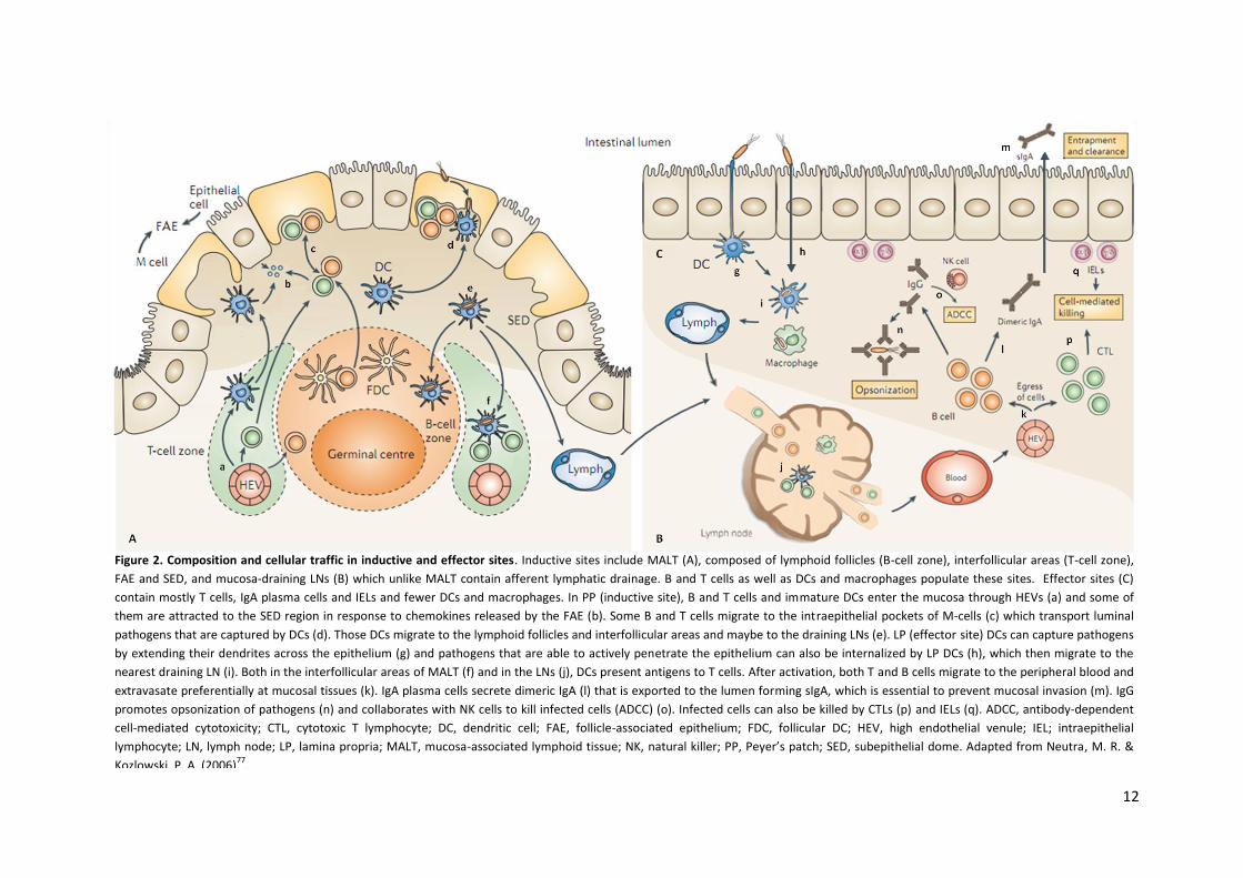
12
Figure 2. Composition and cellular traffic in inductive and effector sites. Inductive sites include MALT (A), composed of lymphoid follicles (B-cell zone), interfollicular areas (T-cell zone),
FAE and SED, and mucosa-draining LNs (B) which unlike MALT contain afferent lymphatic drainage. B and T cells as well as DCs and macrophages populate these sites. Effector sites (C)
contain mostly T cells, IgA plasma cells and IELs and fewer DCs and macrophages. In PP (inductive site), B and T cells and immature DCs enter the mucosa through HEVs (a) and some of
them are attracted to the SED region in response to chemokines released by the FAE (b). Some B and T cells migrate to the intraepithelial pockets of M-cells (c) which transport luminal
pathogens that are captured by DCs (d). Those DCs migrate to the lymphoid follicles and interfollicular areas and maybe to the draining LNs (e). LP (effector site) DCs can capture pathogens
by extending their dendrites across the epithelium (g) and pathogens that are able to actively penetrate the epithelium can also be internalized by LP DCs (h), which then migrate to the
nearest draining LN (i). Both in the interfollicular areas of MALT (f) and in the LNs (j), DCs present antigens to T cells. After activation, both T and B cells migrate to the peripheral blood and
extravasate preferentially at mucosal tissues (k). IgA plasma cells secrete dimeric IgA (l) that is exported to the lumen forming sIgA, which is essential to prevent mucosal invasion (m). IgG
promotes opsonization of pathogens (n) and collaborates with NK cells to kill infected cells (ADCC) (o). Infected cells can also be killed by CTLs (p) and IELs (q). ADCC, antibody-dependent
cell-mediated cytotoxicity; CTL, cytotoxic T lymphocyte; DC, dendritic cell; FAE, follicle-associated epithelium; FDC, follicular DC; HEV, high endothelial venule; IEL; intraepithelial
lymphocyte; LN, lymph node; LP, lamina propria; MALT, mucosa-associated lymphoid tissue; NK, natural killer; PP, Peyer’s patch; SED, subepithelial dome. Adapted from Neutra, M. R. &
Kozlowski, P. A. (2006)77
.
13
GALT structures, are considered inductive sites for mucosal B and T cells, together with the
mesenteric LNs (MLNs)85,86.
The effector sites include different compartments, such as the lamina propria (LP) of
various mucosal tissues and the stroma of exocrine glands81. At a cellular level, they are composed
of T-cells (mostly CD4+), IgA plasma cells, fewer IgM and IgG plasma cells and few B cells, DCs and
macrophages (Figure 2C)83. Another effector site is the intraepithelial lymphocyte (IEL)
compartment which generally refers to the epithelium of the small intestine where most IELs
occur81 (Figure 2C). IELs consist mostly of T cells belonging to the T cell receptor-γδ (TCRγδ)+ and
TCRαβ+ lineages87,88. They are in direct contact with epithelial cells and very close to antigens in the
gut lumen and thereby contribute to the physical barrier against invading pathogens87. Unlike
conventional T cells, they are antigen-experienced T cells that upon encountering antigens mediate
killing of the infected target cells or release cytokines right away87,88. IELs are therefore able to
mount and regulate both innate and adaptive immune responses87,88 (Figure 2C).
2.3.2.2 Cellular traffic in the mucosa and antigen sampling
At intestinal mucosal tissues with organised MALT, specifically at PPs, B and T lymphocytes
as well as immature DCs, essential APCs in initiating mucosal T cell responses, enter the mucosa
through HEVs77,89 and some of these cells can be then attracted to the SED region due to
chemokines released by FAE (Figure 2). CC-chemokine ligand 20 (CCL20) and CCL9, produced by
FAE of mouse small-intestinal PPs, are known to attract lymphocytes and DCs expressing CC-
chemokine receptor 6 (CCR6) or CCR1, respectively90–92. Some B and T lymphocytes migrate into
the intraepithelial pockets formed by M-cells, where they express maturation or memory markers,
while most of the immature DCs remain in the SED region77. DCs capture antigens and
microorganisms that are transported across the FAE by M-cells93 and then migrate from the SED
region to underlying B-cell follicles and interfollicular T-cell areas, where they upregulate the
expression of maturation markers and MHC molecules93,94. In the interfollicular areas, they process
the antigens and present them to naïve T cells77. It is postulated that some DCs can also carry the
antigens to draining LNs where they are presented to T cells95 (Figure 2).
Other strategies for antigen sampling have been reported, like the one that is exploited by
LP DCs (Figure 2). These cells use exclusive proteins to separate the epithelial junctions, which
allow them to extend their dendrites across the epithelium and directly sample the pathogens
within the gut lumen, yet preserving the monolayer integrity96,97. This strategy can also occur in
tissues with a stratified, pseudostratified or simple epithelium that lack organised lymphoid follicles
and M-cells, such as the female genital tract and, for that reason, it may also be more
14
immunologically important there77. After antigen capture, those DCs can migrate to the nearest
draining LN and present antigens to T cells77. Alternatively, pathogens able to damage or actively
penetrate the mucosal epithelium may also be captured by LP DCs that will be innately activated
through recognition of PAMPs by their PRRs and will then carry the internalised antigens to
draining LNs76,80 (Figure 2).
After being primed, naïve T and B cells become memory or effector cells and migrate from
MALT to LNs and from LNs to the peripheral blood, and then extravasate at mucosal effector sites98
(Figure 2). Therefore, mucosal infection at one site can provide protection at a distant mucosal site,
which created the concept of common mucosal immune system (CMIS)99. This term has been then
refined to compartmentalised mucosal immune system since not all the mucosal tissues seem to be
interconnected and cells may preferentially migrate to certain mucosal effector tissues than to all
mucosal tissues non-selectively100. Whatever the exact tissue is, the guiding of those cells to the
mucosa again is dictated by the upregulation of tissue-specific adhesion molecules and chemokine
receptors that bind complementary ligands in the endothelial cells of the mucosal vasculature101,102.
2.3.2.3 Humoral immune response
Production and secretion of sIgA is the most important defence factor against mucosa-
invading pathogens73. It starts with class switching from IgM to IgA by activated B cells in the
inductive sites followed by their migration through the bloodstream to various mucosal
tissues80,103. Within the LP (effector site), those B cells differentiate into plasma cells, which results
in secretion of dimeric IgA whose monomers are linked by a joining chain (J-chain)80. The
interaction of the IgA dimer with the polymeric Ig receptor (pIgR) on the basolateral surface of
epithelial cells induces their transcytosis to the apical surface104. There, pIgR is proteolytically
cleaved giving rise to a polypeptide named secretory component that remains attached to the IgA
dimer, thus forming sIgA80,104.
sIgA displays unique properties when compared with other classes of antibodies, namely, it
is resistant to protease degradation, for which its dimerization, high degree of glycosylation and
association with the secretory component account for, and can interact with mucus and effector
leukocytes77,80. However, sIgA is unable to activate complement or stimulate the release of
inflammatory mediators by innate immune cells80,103. Through a mechanism known as immune
exclusion, sIgA promotes the entrapment of pathogens in the mucus, thus preventing their direct
contact with mucosal surfaces77. Also, sIgA molecules of appropriate specificity might sterically
hinder pathogen surface molecules involved in epithelial attachment105 or might intercept, during
its pIgR-mediated transport, incoming pathogens retained in vesicles of epithelial cells106.
15
Moreover, it was shown that association of sIgA with pathogens might facilitate the antigen
sampling by DCs107. Dimeric IgA molecules present in the fluid underlying the epithelial barrier and
produced by local IgA plasma cells can also prevent mucosal infection by carrying pathogens that
have breached the epithelium back to the lumen through the pIgR-mediated transport (Figure
2)77,108.
IgG also contributes to adaptive immune responses at mucosal surfaces by promoting
antibody-dependent cell-mediated cytotoxicity (ADCC), a mechanism through which antibody-
coated infected cells are lysed by NK cells (Figure 2)77. In addition, and through the interaction with
an IgG-specific Fc receptor, the neonatal Fc Receptor (FcRn), IgG can be exported across the
intestinal epithelial barrier to the lumen, where it binds antigens, and then be transported back
into the LP, where the antigens are taken up by DCs for further processing and presentation to
CD4+ T lymphocytes109. IgG is the predominant antibody class in bovine secretions, specifically in
the colostrum and milk, in contrast to IgA which is present there at much reduced concentrations
and is the major Ig present in the human milk110.
2.3.2.4 Cellular immune response
When it comes to cell-mediated immunity (CMI), both cytotoxic T lymphocytes (CTLs, i.e.,
effector T cytotoxic (TC) CD8+ cells), and T helper (TH, CD4+) cells have been found to be important
for mucosal immune defence against pathogens (Figure 2)111. Upon antigen presentation by APCs,
naïve antigen-specific T cells are activated and become effector cells79. Induction of IgA production
against mucosal pathogens is dependent on effector TH cells111. Depending on the cytokine
environment in which TH cells differentiate, distinct cell subsets, such as TH1 or TH2, may arise. TH1
cells secrete IFN-γ and promote secretion of IL-12, a key cytokine for differentiation of TH1 cells, by
macrophages and DCs79. TH2 cells in turn secrete IL-4, which induces the differentiation of naive T
cells into TH2 cells, and also IL-10, which along with IL-4 suppresses the expansion of TH1
populations79. In the same way, IFN-γ inhibits the development and activity of the TH2 subset79. TH1
immune responses, usually set up against intracellular pathogens, are further characterized by
secretion of tumour necrosis factor-alpha (TNF-α) and opsonizing antibodies, such as those of the
IgG2a isotype, and strong CTL responses that mediate killing of target cells79,112. TH2 immune
responses are generally activated to control helminthic pathogens and allergic diseases, with
production of IgG1 and IgE antibodies but weak CTL responses79,112. Another type of TH cells exists,
namely the TH17 cells. They are activated by IL-23 and produce IL-17, a highly pro-inflammatory
cytokine responsible for tissue damage in delayed-type hypersensitivity (DTH) reactions, as it
increases chemokine production that leads to recruitment of monocytes and neutrophils to the site
16
of inflammation. DTH reactions are in turn considered a crucial component of the host immune
response against bacteria and intracellular parasites79,113. Moreover, IL-17, as well as IL-22, both
produced by TH17 cells, are important effectors of mucosal immunity, as they have been shown to
regulate the expression of antimicrobial peptides114.
2.4 Mucosal immunisation
As mucosal surfaces are the main sites of entry and colonization for many pathogens, there
is an increasing interest in effectively protecting them through activation of the mucosal immune
system115. This can be accomplished by mucosal immunisation, as the administration of vaccines
specifically onto mucosal surfaces results in the stimulation of mucosal and also systemic immune
responses. Systemic immunisation in turn can effectively elicit systemic responses but is generally a
poor inducer of mucosal immunity99,116.
Compared with systemic immunisations, mucosal immunisations are safer since direct
contact between potentially toxic components of the vaccine and systemic circulation can be
avoided and adverse effects can thus be minimized. Also, mucosal immunisation reduces the need
for trained personnel to administer the vaccine and it can also enhance its efficacy due to the
ability of eliciting both mucosal and systemic immune responses115.
An ideal mucosal vaccine would adhere to mucosal surfaces, effectively induce innate
responses and provide both humoral and cellular adaptive immunity at the relevant mucosal site of
administration and also throughout the body77. However, it comes out that vaccines administered
in the mucosa face the same bottlenecks as do pathogens, namely, they are attacked by proteases
and nucleases, diluted in mucosal secretions, captured in the mucus layer, excluded by epithelial
barriers and, in the case of vaccine antigens, their uptake generally occurs at low levels77,115. As a
result, multiple administrations of vaccine (prime-boost combinations) and in large doses are
usually required to induce a suitable protective immune response77,115.
Various routes of mucosal immunisation, namely the i.n., oral, rectal or vaginal, are being
continuously tested for their efficacy and the choice of the optimal route may depend on the
species to be immunised, the expected site of challenge and the type of vaccine used (live, killed or
subunit, among others)77.
2.4.1 Types of vaccines
Live vaccines consist of attenuated pathogens, i.e., pathogens that have lost the ability to
cause disease but are still capable of growing within an inoculated host79. This kind of vaccine is
17
more likely to stimulate appropriate CMI against intracellular pathogens as it more closely mimics
what happens during natural infection57,79. Its effectiveness as mucosal vaccine is in part due to the
adaptation mechanisms of the component pathogens to survive in the luminal environment and
effectively invade mucosal tissues77. However, these vaccines have a limited shelf-life and safety
concerns arise from the possibility of reversal to virulence30,79. Killed vaccines in turn comprise
inactivated pathogens that are not able to replicate in the host, yet they can raise an immune
response79. They are generally regarded as safe but are unable to induce CMI and can lack some of
the immune-relevant antigens due to the inactivation process or because they are only secreted by
the pathogens alive30,79. Moreover, killed vaccines can include immunosuppressive antigens or
antigens inducing undesired responses and subunit vaccines, which consist of specific and purified
pathogen-derived macromolecules, thereby constitute an advantageous alternative30,79. Several
systems can be used to deliver the antigen at mucosal surfaces, such as lipid particles (e.g.,
liposomes and immune stimulating complexes (iscoms), the last being cage-like structures formed
by phospholipids, cholesterol and saponins) and polymeric particles (made up of, e.g., poly(D,L-
lactide-co-glycolide) (PLG) or chitosan)115,117. DNA vaccines are in turn composed of plasmid DNA
that encodes antigenic proteins, allowing these molecules to be expressed in the host in their
natural conformation, and induce both humoral and CMI79. Finally, recombinant vector vaccines
make use of attenuated organisms, such as bacteria or viruses, to introduce antigen-coding genes
from infectious pathogens into the host and can therefore maximize CMI to those antigens79.
As with systemic immunisation, the administration at mucosal surfaces of antigens that
have no inherent immunostimulatory properties generates a weak or undetectable immune
response and co-delivery of substances known as adjuvants is therefore required80,118.
2.4.2 Adjuvants
Adjuvants are substances incorporated into vaccine formulations in order to enhance,
accelerate and prolong the immune response elicited towards vaccine antigens119. Most of them
are chemicals, microbial components or mammalian proteins and can be grouped according to
their mechanism of action117, which includes enhancement of antigen presentation, improvement
of antigen stability or immunomodulation (in this case, adjuvants alter the cytokine network,
increasing the concentration of some cytokines while decreasing the concentration of others, in
that way, shifting the type of immune response)118. A single adjuvant can also combine more than
one mechanism of action120.
By enhancing antigen immunogenicity, adjuvants allow the number of boost immunisations
and the amount of antigen needed to provide a successful immune response to be reduced,
18
making the vaccine more cost-effective117,119. If they display immunomodulatory properties, they
may also change the type of immune response to the most effective one against a given
pathogen117,119. However, as immune potentiators, adjuvants may also induce adverse reactions.
Systemic adverse effects observed in laboratory animals include fever, arthritis, uveitis and
malaise, and adjuvants that act locally can cause inflammation and more rarely granulomas or
sterile abcesses118. In most instances, these effects are mild and the benefits coming out of their
use outweigh the potential drawbacks117.
A wide variety of substances with adjuvant activity exists nowadays with different
mechanisms of action and host range for application. These include alum compounds, the first
adjuvants licensed to be used in humans, oil emulsions such as the Freund’s complete adjuvant
(FCA), which consists of a water-in-oil emulsion with mycobacteria and was abandoned due to high
toxicity, and the Freund’s incomplete adjuvant (FIA), that contains no mycobacteria and is still used
when the inflammation is not a major issue and when a strong adjuvant is needed77,117. Also,
cytokines, microparticles and nanoparticles have adjuvant properties as well as diverse bacterial
products including lipid A, cholera toxin (CT) and CpG, all of them being candidates for mucosal
adjuvants77,117.
CpG oligonucleotides are adjuvants that contain a central unmethylated CpG dinucleotide
preferentially flanked by two 5’ purines and two 3’ pirimidines121. They are underrepresented in
vertebrates and mimic a DNA motif from bacteria which, contrarily to vertebrates, lack a cytosine
methylase117,122. CpG oligonucleotides stimulate macrophages123, DCs124 and NK cells125 as well as B-
cell proliferation126 and one of their mechanisms of action in cells has been reported to be
dependent on TLR-9127. They are immunomodulators117, particularly effective in inducing a TH1
antigen-specific immune response121 and induce production of cytokines such as IL-6128, IL-12125,
IFN-γ125 and TNF-α123. When delivered systemically with diverse antigens, CpG oligonucleotides are
strong adjuvants115 and when used as mucosal adjuvants, both systemic and mucosal immune
responses were shown to be induced77,122.
2.5 Immune response to N. caninum
In vivo studies on the immunology of N. caninum infection are commonly performed in
mice. The availability of murine models with selective immunological defects as well as the fact
that vertical transmission of the parasite has been demonstrated in these animals is of great
advantage129,130. Exposure of mice to N. caninum has been associated with encephalitis, myositis,
acute primary pneumonia and pancreatitis131. However, particular mouse strains appear to be
19
more susceptible to brain infection than others. BD10.2 mice do not develop clinical neosporosis,
whereas BALB/c and C57BL/6 mice are highly susceptible to N. caninum-induced encephalitis132.
Several components of the innate immune system seem to be activated upon N. caninum
infection. It was shown that NK cells are able to produce IFN-γ and kill N. caninum-infected cells133
and that activation of macrophages with IFN-γ resulted in increased production of RNS and killing
activity against N. caninum134.
As N. caninum is an obligate intracellular parasite, CMI is likely to play a major role in
protective immunity. Evidences for the major importance of a TH1 immune response came from in
vitro studies, showing that IFN-γ and TNF-α significantly inhibited intracellular multiplication of the
parasite135,136, as well as from in vivo studies, where mice whose IFN-γ or IL-12 were depleted by
means of specific monoclonal antibodies137,138 and IFN-γ-knockout mice139 exhibited a significantly
increased vulnerability to N. caninum infection. The importance of CD4+ T cells in protective
immunity was demonstrated in vivo when most of the mice in which CD4+ T cells had been
depleted succumbed to fatal neosporosis within 30 days upon challenged with N. caninum,
contrarily to mice with no CD8+ T cells or those of the control group140. Also, CD4+ T cells from
infected cattle were shown to directly lyse parasite-infected autologous target cells in vitro141.
Humoral immunity was also shown to be important to control N. caninum infection, since B-cell
(and consequently antibody)-deficient μMT mice experimentally infected with NcT were found to
be more susceptible to infection than the wild-type mice142. It is also known that the humoral
component of the protective immunity against neosporosis is characterized by a major
predominance of serum IgG2a antibodies over the IgG1 subclass132,138, which further stresses out
the importance of TH1 immunity in protection against the parasite.
Antigen-specific IL-4 responses as well as a predominance of IgG1 antibodies are associated
with susceptibility to neosporosis, which shows that a TH2-biased immune response, in detriment
of the aforementioned TH1 protective immune response, favours invasion and infection by N.
caninum30,132,143.
2.6 N. caninum and vaccination
When attention has been paid at the economic impact of neosporosis in both beef and
dairy industry as well as at the lack of successful and cost-effective measures to control the
disease, a urgent need for the development of a safe and efficacious vaccine has come out in order
to reduce the overall losses caused by N. caninum. The assumption that vaccination may be a
feasible goal is supported by observations showing that N. caninum-infected cows may develop
protective immunity against abortion and transmission of the parasite11. Therefore, many attempts
20
have been made along the last years to develop an effective immunisation protocol aimed at
preventing N. caninum infection, transmission, disease or abortion129. The efficacy of those
attempts can be firstly evaluated in mouse models but it ultimately needs to be carried out in
cattle, for these are the target hosts53.
Several live vaccines have been tested for their efficacy against N. caninum. In particular,
temperature-sensitive strains have been reported to induce significant protection against N.
caninum in mice144. Also, natural N. caninum isolates attenuated in their ability to cause disease in
mice have emerged and include NC-Nowra and Nc-Spain-1H. Administration of NC-Nowra
tachyzoites in mice145 and cattle146 before pregnancy resulted, respectively, in a dramatic reduction
in transplacental transmission and total prevention of foetal death after challenging during
pregnancy. Nc-Spain-1H in turn has been reported to prevent abortion in a pregnant mouse
model147. Prevention of foetal death has therefore been experimentally demonstrated in
exogenously, but not in endogenously, N. caninum-infected cows5.
When it comes to killed vaccines, irradiated NcT protected mice from an otherwise lethal
challenge148 and a crude N. caninum extract prevented vertical transmission of the parasite in
mice149. On the other hand, cattle immunised with a NcT preparation together with the adjuvant
POLYGEN induced IFN-γ production but failed to prevent vertical transmission in pregnant cattle
after experimental challenge with NcT150. The only vaccine commercially available at present,
NeoGuard® marketed by Intervet Inc. (Merriam, KS, USA), is an Havlogen-adjuvanted vaccine
containing inactivated NcT30,53. However, it has proven to only partially protect herds in New
Zealand151 and to reduce only in some extent the incidence of abortions in Costa Rican dairy
cattle152, with a calculated efficacy of 46%11.
Among the large number of proteins existing in N. caninum153, the choice of candidate
molecules to subunit vaccines has been focused on those located on the parasite surface, as well as
on those involved in parasite survival (host cell invasion and intracellular development)53. In the
latter case, proteins present and excreted by secretory organelles constitute a common choice53.
Immunisation of mice with recombinant NcSAG1 and NcSRS2 together with the corresponding DNA
vaccine significantly protected mice from cerebral infection154. Other studies reported that BALB/c
mice subcutaneously (s.c.) immunised with iscoms containing recombinant NcSRS2 displayed lower
cerebral N. caninum burden when compared to the control group155 and that s.c. inoculation of
BALB/c mice with native NcSRS2 prevented transplacental transmission of the parasite156. In cattle,
Freund’s adjuvanted-injection of NcSRS2 coupled to palmitic acid elicited T-cell activation and IFN-γ
production, similarly to what happens upon a live parasite infection157. In turn, immunisation of
C57BL/6 mice with recombinant NcMIC1158 and NcMIC3159 significantly protected mice from
cerebral infection, and FIA or saponin-adjuvanted administration of recombinant NcROP2
21
protected C57BL/6 mice from clinical signs of neosporosis and also significantly reduced the
cerebral parasite burden in vaccinated mice54. Both NCDG1 and NCDG2 are known to be
immunogenic in cattle and they were used in immunological assays to demonstrate antibody
responses in these animals160. I.n. immunisation of recombinant NcPDI was recently reported to
provide highly protective immunity against N. caninum. It was also shown that immunisation of
BALB/c mice with plasmid DNA coding for NcGRA7 protected against vertical transmission of N.
caninum161 and the protective effect was increased when the plasmid DNA was administered
together with the CpG adjuvant162.
Intraperitoneal vaccination of BALB/c mice with a recombinant vaccinia virus carrying
either NcSAG1 or NcSRS2 gene protected mice against cerebral infection163 and vertical
transmission of the parasite164, being the best protection achieved with the vaccinia virus
expressing the NcSRS2 antigen. Brucella abortus strain RB51 was also used to deliver several N.
caninum antigens, namely NcMIC1, NcMIC3, NcGRA2, NcGRA6 and NcSRS2, and has shown to
protect C57BL/6 mice from lethal infection165 and vertical transmission of N. caninum166. However,
that approach might not be acceptable in cattle populations of countries that also want to prove to
be free of bovine brucellosis53.
The aforementioned studies of subunit, DNA and vector vaccines reflect a preferential use
of purified proteins or recombinant DNA as vaccine agents in most of the approaches aimed at
developing an effective immunisation protocol against N. caninum. However, native proteins also
show promise as vaccine agents, as suggested by the already mentioned study with native
NcSRS2156, and they should therefore also be considered in future studies53.
22
3. Materials and Methods
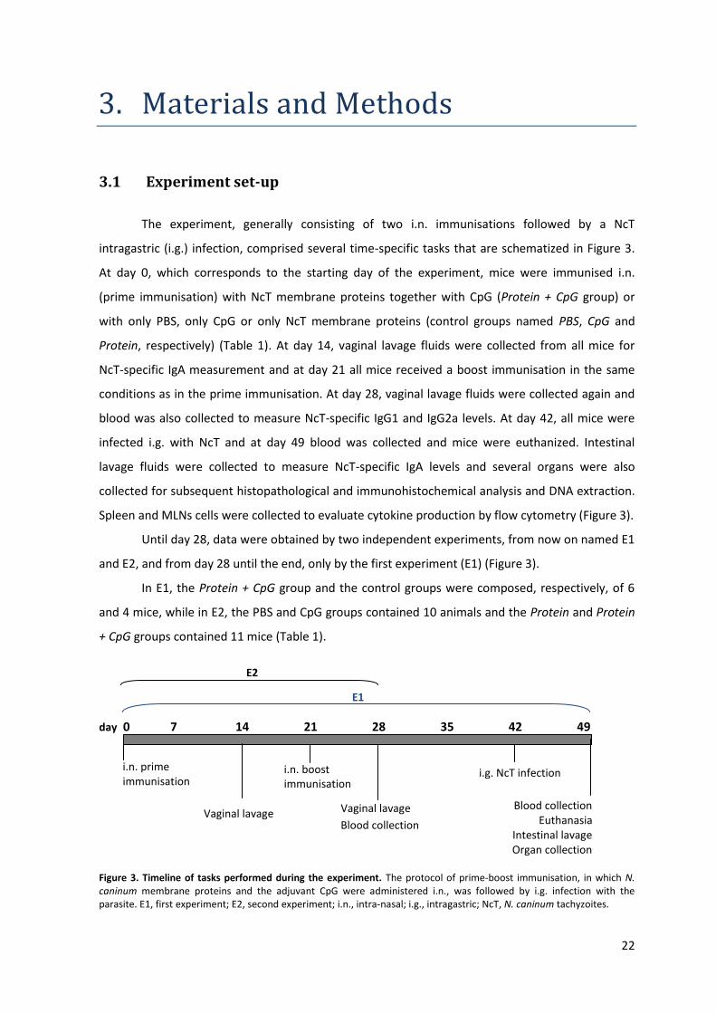
3.1 Experiment set-up
The experiment, generally consisting of two i.n. immunisations followed by a NcT
intragastric (i.g.) infection, comprised several time-specific tasks that are schematized in Figure 3.
At day 0, which corresponds to the starting day of the experiment, mice were immunised i.n.
(prime immunisation) with NcT membrane proteins together with CpG (Protein + CpG group) or
with only PBS, only CpG or only NcT membrane proteins (control groups named PBS, CpG and
Protein, respectively) (Table 1). At day 14, vaginal lavage fluids were collected from all mice for
NcT-specific IgA measurement and at day 21 all mice received a boost immunisation in the same
conditions as in the prime immunisation. At day 28, vaginal lavage fluids were collected again and
blood was also collected to measure NcT-specific IgG1 and IgG2a levels. At day 42, all mice were
infected i.g. with NcT and at day 49 blood was collected and mice were euthanized. Intestinal
lavage fluids were collected to measure NcT-specific IgA levels and several organs were also
collected for subsequent histopathological and immunohistochemical analysis and DNA extraction.
Spleen and MLNs cells were collected to evaluate cytokine production by flow cytometry (Figure 3).
Until day 28, data were obtained by two independent experiments, from now on named E1
and E2, and from day 28 until the end, only by the first experiment (E1) (Figure 3).
In E1, the Protein + CpG group and the control groups were composed, respectively, of 6
and 4 mice, while in E2, the PBS and CpG groups contained 10 animals and the Protein and Protein
+ CpG groups contained 11 mice (Table 1).
day 0 7 14 21 28 35 42 49
Figure 3. Timeline of tasks performed during the experiment. The protocol of prime-boost immunisation, in which N. caninum membrane proteins and the adjuvant CpG were administered i.n., was followed by i.g. infection with the parasite. E1, first experiment; E2, second experiment; i.n., intra-nasal; i.g., intragastric; NcT, N. caninum tachyzoites.
E2
E1
i.n. boost immunisation
i.n. prime immunisation
Vaginal lavage Vaginal lavage
Blood collection
i.g. NcT infection
Blood collection Euthanasia
Intestinal lavage Organ collection
23
Table 1. Description of the groups of mice used in the experiments. E1, first experiment; E2, second experiment; NcT, N.
caninum tachyzoites
3.2 Mice
Female C57BL/6 mice (8 weeks old) were purchased from Charles River (Barcelona, Spain).
Animals were kept at the animal facilities of the Institute Abel Salazar during the experiments.
Procedures involving mice were performed according to the European Convention for the
Protection of Vertebrate Animals used for Experimental and Other Scientific Purposes (ETS 123)
and the 86/609/EEC Directive and Portuguese rules (DL 129/92).
3.3 N. caninum
NcT of the NC-1 isolate were cultured and serially passaged in VERO cells maintained at 37
ᵒC in minimum essential medium containing Earle’s salts (Sigma, St. Louis, USA) supplemented with
10% foetal bovine serum (FBS) (PAA Laboratories GmbH, Pasching, Austria), L-glutamine (2mM)
(Sigma), penicillin (200 IU/mL) and streptomycin (200 μg/mL) (Sigma) in a humidified atmosphere
of 5% CO2 in air.
To obtain the free parasitic forms of N. caninum, infected VERO cells were cultured until
the host cell monolayer was 70% destroyed. Culture supernatants and adherent cells harvested
using a cell-scraper were centrifuged at 1500 g for 20 min. The pellet was passed through a 25G
needle and then washed with phosphate-buffered saline (PBS) by three serial centrifugations at
1500 g for 20 min each. The pellet thus obtained was resuspended in 3 mL of PBS, passed through
a 25G needle and then through a PD-10 column filled with Sephadex™ G-25M (Amersham
Biosciences Europe GmbH, Freiburg, Germany). Parasite concentration was determined with a
haemocytometer.
Groups Preparation administered (in 20 μL) Number of mice
Substance(s) Amount E1 E2
Control
PBS PBS 20 μL 4 10
CpG CpG 10 μg 4 10
Protein NcT membrane proteins 30 μg 4 11
Protein + CpG NcT membrane proteins
and CpG
30 μg
10 μg 6 11
24
3.4 Immunisations
Mice were immunised i.n., at day 0 and 21 (twice at 3-week intervals), with 20 μL of the
preparation containing 30 μg of NcT membrane proteins and 10 μg of CpG (Invitrogen, Carlsbad,
CA, USA) in PBS (Protein + CpG group). Control animals received 20 μL of PBS alone (PBS group), 20
μL of PBS with 10 μg of CpG (CpG group) or 20 μL of PBS with 30 μg of NcT membrane proteins
(Protein group) (Table 1). Mice were briefly anaesthetized by isoflurane (IsoFlo®, Veterinaria
Esteve, Bologna, Italy) inhalation before the immunisations.
3.5 Challenge infections
At day 42, N. caninum infection was performed in all mice by the i.g. route. Briefly, 4 hr
before infection, mice were deprived of food. At the time of infection, animals were anaesthetized
by intramuscular injection of 20 μL of a 4:5 mixture containing xylazine (Rompum®, Bayer Portugal,
S.A., Carnaxide) and ketamine (Imalgene® 1000, Merial, Rio de Mouro, Portugal). In order to
neutralize stomach acidity, 50 μL of a 10% sodium hydrogenocarbonate solution in water were
then directly administered into the stomach, through a gavage probe. Ten minutes later, the same
procedure was followed to inoculate 200 μL PBS containing 5 × 10⁷ NcT.
3.6 Extraction of membrane proteins of NcT
NcT membrane proteins were obtained by using a protocol previously described by Zintl et
al.167. Namely, parasites were resuspended in PBS containing 0.75% Triton-X114 (5 × 10⁷ – 5 × 10⁸
NcT per 3 mL of solution), gently vortexed and incubated on ice for 10 min. The preparation was
centrifuged at 10000 g for 30 min (4 ᵒC) to remove cytoskeletal proteins and the supernatant was
incubated at 30 ᵒC for 3 min, cooled on ice for 10 min and then centrifuged at 10000 g for 30 min (4
ᵒC) to remove possible aggregates. The resulting supernatant was incubated at 30 ᵒC for 3 min and
then centrifuged at 1000 g for 4 min (room temperature). The upper hydrophilic phase was
discarded and absolute ethanol was added to the lower detergent phase (volume of ethanol
equivalent to four times the volume of the detergent phase). This final fraction was gently vortexed
and then incubated on ice for 1 hr with vortex-mixing each 15 min. Following incubation, a
centrifugation step was performed at 12000 g for 20 min (4 ᵒC) and the pellet was resuspended in
PBS. Protein concentration was determined by the method of Lowry and using the
spectrophotometer 6300 (Jenway, Essex, UK).
25
3.7 Collection of biological samples
At day 14 and 28, vaginal lavages were performed with three times 50 μL PBS. Samples of
vaginal lavage fluids were then centrifuged at 3500 g for 8 min (4 ᵒC) and stored at -20 ᵒC for NcT-
specific IgA measurement.
At day 28, blood was collected from the submandibular vein and allowed to clot overnight.
Serum was obtained by centrifugation at 16000 g for 15 min (4 ᵒC) and was then stored at -20 ᵒC
for NcT-specific IgG1 and IgG2a measurement.
At day 49, mice were briefly anaesthetized by isoflurane inhalation and blood was collected
by retro-orbital bleeding for NcT-specific IgG1 and IgG2a measurement. The animals were then
euthanized by cervical dislocation and brain, liver, pancreas, stomach, heart, lungs and kidneys
were collected and sectioned longitudinally. Large intestine was also collected and the colon and
cecum were transversally divided in two parts each. Small intestine was used to collect intestinal
secretions for NcT-specific IgA measurement and it was then transversally cut in three parts. Half of
each organ/part was preserved in 10% buffered formalin for histopathological and
immunohistochemical analysis and the other half was stored at -80 ᵒC for DNA extraction. To
collect intestinal secretions, small intestines were washed with 2 mL of a solution of protease
inhibitor cocktail (Complete Mini Protease Inhibitor Cocktail Tablets, Roche Diagnostics GmbH,
Mannheim, Germany) in PBS. The intestinal lavage fluids were centrifuged, first at 4000 g for 15
min (4 ᵒC) and then at 8500 g for 60 min (4 ᵒC), and were then stored at -20 ᵒC. Spleen and MLNs
were also removed and placed in Hank’s Buffered Salt Solution (HBSS, Gibco, Invitrogen
Corporation, Carlsbad, CA, USA) supplemented with 2% FBS (PAA Laboratories GmbH) and
penicillin (100 IU/mL) and streptomycin (100 µg/mL) (Sigma) for in vitro cell culture.
3.8 In vitro cell culture
Spleen and MLN cells were obtained by gently teasing the organs in HBSS (Gibco)
supplemented with 2% FBS (PAA Laboratories GmbH) and penicillin (100 IU/mL) and streptomycin
(100 µg/mL) (Sigma). Cell suspensions were centrifuged at 300 g for 10 min (4 ᵒC) and the pellets of
MLN cells were resuspended in RPMI-1640 medium (Sigma) supplemented with 10% FBS (PAA
Laboratories GmbH), HEPES (10 mM) (Sigma), β-mercaptoethanol (7 mM) (Merck, Darmstadt,
Germany) and penicillin (100 IU/mL) and streptomycin (100 µg/mL) (Sigma). The pellets of spleen
cells were resuspended in ACK lysing solution, consisting of NH4Cl (150 mM), KHCO3 (10 mM) and
ethylenediamine tetraacetic acid (EDTA, 0.1 mM) in water, and incubated for 4 min at room
temperature. Spleen cell suspensions were then centrifuged at 300 g for 10 min and the pellets
26
were resuspended in the RPMI medium just described above. Cells were distributed in 96-well
plates (1 × 10⁶ cells/well) and cultured for 5 hr at 37 ᵒC in a humidified atmosphere of 5% CO2 in air
with 10 μg/mL of Brefeldin A (Epicentre Biotechnologies, Madison, WI, USA) and with the 1X
phorbol myristate acetate (PMA) (Sigma) and ionomycin (Merck) preparation, for cytokine
intracellular staining and flow cytometric analysis.
3.9 Cytokine intracellular staining
Spleen and MLN cells, incubated at 37 ᵒC in RPMI-1640 medium (Sigma) supplemented with
10% FBS (PAA Laboratories GmbH), HEPES (10 mM) (Sigma), penicillin (100 IU/mL) and
streptomycin (100 µg/mL) (Sigma) and 7 mM β-mercaptoethanol (Merck) with 10 μg/mL of
Brefeldin A (Epicentre Biotechnologies) and the 1X PMA (Sigma) and ionomycin (Merck)
preparation, were subsequently pre-incubated with anti-FcγR mAb and then surface stained with
phycoerythrin-cychrome 5 (PE-Cy5) anti-mouse CD4 (clone RM4-5) or peridinin-chlorophyll-
protein-Cy5.5 (PerCP-Cy5.5) anti-mouse CD8 (clone 53-6.7) (both from BD Biosciences Pharmingen,
San Diego, CA, USA). Cells were then fixed with 2% formaldehyde, washed, permeabilized with
0.5% saponin (Sigma) and pre-incubated with anti-FcγR mAb before intracellular staining with the
following antibodies: fluorescein isothiocyanate (FITC) anti-mouse IFN-γ (clone XMG1.2) (BD
Biosciences) or the FITC isotype-matched control (FITC Rat IgG1, κ, clone R3-34) (BD Biosciences),
PE anti-mouse IL-10 (clone JES5-16E3) or PE anti-mouse IL-4 (clone BVD4-1D11) (BD Biosciences).
3.10 Antibody measurement
Antibodies specific for NcT in vaginal and intestinal lavage fluids as well as in sera were
quantified by ELISA. Briefly, polystyrene microtitre plates (Nunc, Roskilde, Denmark) were coated
with 5 μg/mL of NcT membrane proteins and incubated overnight at 4 ᵒC. Wells were then
saturated with TST buffer (10 mM Tris [pH 8.0], 150 mM NaCl, 0.05% Tween 20) with 2% bovine
serum albumin (BSA), for 1 hr at room temperature. Serial dilutions of the samples in TST buffer
with 1% BSA were subsequently plated and incubated for 1 hr at room temperature. After washing,
bound antibodies were detected by addition of alkaline phosphatase (AP)-coupled monoclonal goat
anti-mouse-IgA, diluted 1:500 in TST buffer with 1% BSA, goat anti-mouse IgG1 or goat anti-mouse
IgG2a, both diluted 1:1000 in TST buffer with 1% BSA, for 1 hr at room temperature (all antibodies
from Southern Biotechnology Associates, Birmingham, AL, USA). Substrate solution containing p-
nitrophenyl phosphate (Sigma) diluted in AP buffer (1 mM MgCl2, 50 mM NaCO3 [pH 9.8]) was
added after washing and the reaction was stopped by addition of 0.1 M EDTA pH 8.0. Absorbance
27
values were measured at 405 and 570 nm using the plate reader Multiskan EX (Thermo Electron
Corporation, Vantaa, Finland) and the Ascent software version 2.6 (Thermo Electron Corporation).
To determine the antibody titres, the absorbance values at 570 nm were first subtracted to the
absorbance values measured at 405 nm in order to compensate for imperfections of the plate, as
described by the manufacturer. The ELISA antibody titres were expressed as the reciprocal of the
highest dilution that gives an absorbance equal to that of the control (no sample added).
3.11 DNA extraction
DNA from the brain and liver was extracted by digesting each organ overnight at 55 ᵒC in SE
buffer (75 mM NaCl, 25 mM EDTA, pH 8.0) with 1% sodium dodecyl sulfate (SDS) solution and 0.5
mg/mL proteinase K (Sigma). DNA was then purified by the phenol (Sigma)-chloroform (Merck)
method and ethanol precipitated. DNA concentration was determined using the NanoDrop 1000
Spectrophotometer (Thermo Scientific, Wilmington, DE, USA) and the NanoDrop ND-1000 software
version 3.6.0 (Thermo Scientific) and was adjusted to 200 ng/μL for real-time quantitative
polymerase chain reaction (qPCR) analysis.
3.12 qPCR
N. caninum DNA amplification was performed with 1 μL of template DNA in a final volume
of 10 μL containing 1X EXPRESS SYBR® GreenER™ qPCR Supermix (Invitrogen) and 0.5 μM of each
primer (forward primer Np21plus: 5’ – CCC AGT GCG TCC AAT CCT GTA AC, reverse primer Np6
plus: 5’ – CTC GCC AGT CAA CCT ACG TCT TCT) (TIB-Molbiol, Berlin, Germany). The PCR programme
was run as follows: (1) denaturation at 95 ᵒC, 10 min; (2) amplification in 45 cycles (including
denaturation [95 ᵒC, 30s], annealing [63 ᵒC, 15s] and extension [72 ᵒC, 45s]); (3) cooling at 4 ᵒC. A
melting temperature curve was made from 65 ᵒC to 95 ᵒC with increments of 0.02 ᵒC/s to confirm
the specificity of the qPCR product through the presence of a single melting-temperature peak.
Amplifications were made in a Rotor-Gene 6000 (Corbett Life Science, Sydney, Australia) and the
quantitative evaluation of fluorescent signals from the PCR products was performed using the
Rotor-Gene 6000 software version 1.7 (Corbett Life Science). Standards consisting of known
amounts of N. caninum DNA, from 10-3 ng to 10 ng, were included in each run and a standard curve
was plotted with the absorbance values thus obtained. Quantification of the fluorescent signals
from the samples was performed by interpolation of the standard curve. In each run, DNA of
uninfected organs and NcT DNA were used, respectively, as negative and positive controls.
28
3.13 SDS-PAGE and Western blot
NcT membrane proteins were denaturated by boiling for 5 min in loading buffer before
electrophoresis, which was then carried out at 45 mA constant current on a 4% polyacrylamide
stacking gel (pH 6.8) followed by a 10% polyacrylamide resolving gel (pH 8.8). Gels were either
stained by the silver nitrate staining protocol or electrophoretically transferred overnight at 60 mA
constant current to a nitrocellulose membrane (Sigma) for Western blot. After transferring, the
membrane was saturated with TST buffer with 1% BSA for 1 hr at room temperature and blotted
proteins were subsequently incubated with vaginal lavage fluids or sera, diluted 1:500 and 1:1000
in TST buffer with 0.1% BSA, respectively, for 2 hr at room temperature with shaking. After
washing, AP-coupled monoclonal goat anti-mouse-IgA, diluted 1:500 in TST buffer with 0.1 % BSA,
goat anti-mouse IgG1 or goat anti-mouse IgG2a, both diluted 1:1000 in TST buffer with 0.1 % BSA
(all antibodies from Southern Biotechnology Associates), were added and the membrane was
incubated for 1 hr at room temperature with shaking. Bound secondary antibodies were detected
by adding the substrate solution containing nitro blue tetrazolium / 5-bromo-4-chloro-3-indolyl-
phosphate (NBT/BCIP, Roche) diluted in AP buffer (5 mM MgCl2, 0.1 M NaCl, 0.1 M Tris [pH 9.5])
and the reaction was stopped by transferring the membrane to water.
3.14 Antibody binding assay to NcT
NcT were incubated at 37 ᵒC for 30 min with mice serum (containing the antibodies),
previously decomplemented at 56 ᵒC for 30 min and diluted 1:10, 1:100, 1:500 and 1:1000 in
fluorescent-activated cell sorter (FACS) buffer (PBS with 1% BSA and 10 mM NaN3) (5 × 10⁵ NcT per
antibody dilution). After a centrifugation step at 6000 g for 15 min (4 ᵒC), parasite suspensions
were incubated on ice for 30 min with FITC anti-mouse IgG (BD Biosciences), diluted 1:100 in FACS
buffer, in order to detect parasite-bound serum IgG. Unbound secondary antibody was then
removed by centrifuging the preparations at 6000 g for 15 min (4 ᵒC).
3.15 Flow cytometric analysis
All immunofluorescence cytometric analyses were performed on an EPICS XL Flow
Cytometer (Beckman Coulter, Miami, FL, USA) using the CellQuest Software (Becton Dickinson, San
Jose, CA, USA).
29
3.16 Statistical analysis
Statistical difference of the results between the pairs of groups to be compared, i.e., the
PBS with the CpG group and the Protein with Protein + CpG group, was determined by Student t-
test, using the GraphPad Software, Inc. version 5.00. p<0.05 was considered to be statistically
significant. In the following bar charts, the bars represent the mean plus 1 SEM of the data
obtained for each group. Groups contained a variable number of mice (see 3.1 Experiment set-up).
The absence of SEM in some bars in the following charts means that the parameter in study was
detected in only one mouse among all that constitute that group. When this happens in the Protein
group, no statistical analysis could be made in relation to the Protein + CpG group, yet the
difference in the parameter in study was considered significant.
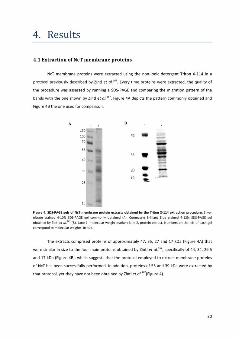
30
4. Results
4.1 Extraction of NcT membrane proteins
NcT membrane proteins were extracted using the non-ionic detergent Triton X-114 in a
protocol previously described by Zintl et al.167. Every time proteins were extracted, the quality of
the procedure was assessed by running a SDS-PAGE and comparing the migration pattern of the
bands with the one shown by Zintl et al.167. Figure 4A depicts the pattern commonly obtained and
Figure 4B the one used for comparison.
Figure 4. SDS-PAGE gels of NcT membrane protein extracts obtained by the Triton X-114 extraction procedure. Silver
nitrate stained 4-10% SDS-PAGE gel commonly obtained (A). Coomassie Brilliant Blue stained 4-12% SDS-PAGE gel
obtained by Zintl et al.167
(B). Lane 1, molecular weight marker; lane 2, protein extract. Numbers on the left of each gel
correspond to molecular weights, in kDa.
The extracts comprised proteins of approximately 47, 35, 27 and 17 kDa (Figure 4A) that
were similar in size to the four main proteins obtained by Zintl et al.167, specifically of 44, 34, 29.5
and 17 kDa (Figure 4B), which suggests that the protocol employed to extract membrane proteins
of NcT has been successfully performed. In addition, proteins of 55 and 39 kDa were extracted by
that protocol, yet they have not been obtained by Zintl et al.167(Figure 4).
130
100
70
55
40
35
25
15
1 2 1 2 A B
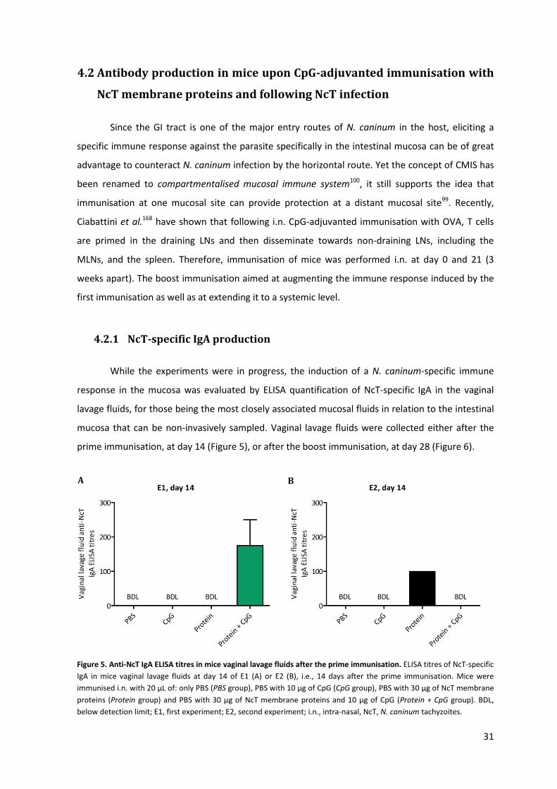
31
4.2 Antibody production in mice upon CpG-adjuvanted immunisation with
NcT membrane proteins and following NcT infection
Since the GI tract is one of the major entry routes of N. caninum in the host, eliciting a
specific immune response against the parasite specifically in the intestinal mucosa can be of great
advantage to counteract N. caninum infection by the horizontal route. Yet the concept of CMIS has
been renamed to compartmentalised mucosal immune system100, it still supports the idea that
immunisation at one mucosal site can provide protection at a distant mucosal site99. Recently,
Ciabattini et al.168 have shown that following i.n. CpG-adjuvanted immunisation with OVA, T cells
are primed in the draining LNs and then disseminate towards non-draining LNs, including the
MLNs, and the spleen. Therefore, immunisation of mice was performed i.n. at day 0 and 21 (3
weeks apart). The boost immunisation aimed at augmenting the immune response induced by the
first immunisation as well as at extending it to a systemic level.
4.2.1 NcT-specific IgA production
While the experiments were in progress, the induction of a N. caninum-specific immune
response in the mucosa was evaluated by ELISA quantification of NcT-specific IgA in the vaginal
lavage fluids, for those being the most closely associated mucosal fluids in relation to the intestinal
mucosa that can be non-invasively sampled. Vaginal lavage fluids were collected either after the
prime immunisation, at day 14 (Figure 5), or after the boost immunisation, at day 28 (Figure 6).
Figure 5. Anti-NcT IgA ELISA titres in mice vaginal lavage fluids after the prime immunisation. ELISA titres of NcT-specific
IgA in mice vaginal lavage fluids at day 14 of E1 (A) or E2 (B), i.e., 14 days after the prime immunisation. Mice were
immunised i.n. with 20 μL of: only PBS (PBS group), PBS with 10 μg of CpG (CpG group), PBS with 30 μg of NcT membrane
proteins (Protein group) and PBS with 30 μg of NcT membrane proteins and 10 μg of CpG (Protein + CpG group). BDL,
below detection limit; E1, first experiment; E2, second experiment; i.n., intra-nasal, NcT, N. caninum tachyzoites.
A B
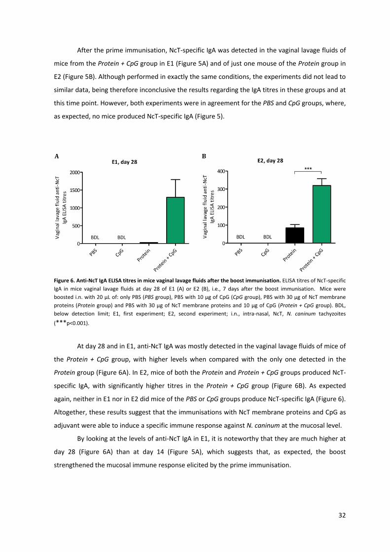
32
After the prime immunisation, NcT-specific IgA was detected in the vaginal lavage fluids of
mice from the Protein + CpG group in E1 (Figure 5A) and of just one mouse of the Protein group in
E2 (Figure 5B). Although performed in exactly the same conditions, the experiments did not lead to
similar data, being therefore inconclusive the results regarding the IgA titres in these groups and at
this time point. However, both experiments were in agreement for the PBS and CpG groups, where,
as expected, no mice produced NcT-specific IgA (Figure 5).
Figure 6. Anti-NcT IgA ELISA titres in mice vaginal lavage fluids after the boost immunisation. ELISA titres of NcT-specific
IgA in mice vaginal lavage fluids at day 28 of E1 (A) or E2 (B), i.e., 7 days after the boost immunisation. Mice were
boosted i.n. with 20 μL of: only PBS (PBS group), PBS with 10 μg of CpG (CpG group), PBS with 30 μg of NcT membrane
proteins (Protein group) and PBS with 30 μg of NcT membrane proteins and 10 μg of CpG (Protein + CpG group). BDL,
below detection limit; E1, first experiment; E2, second experiment; i.n., intra-nasal, NcT, N. caninum tachyzoites
(***p<0.001).
At day 28 and in E1, anti-NcT IgA was mostly detected in the vaginal lavage fluids of mice of
the Protein + CpG group, with higher levels when compared with the only one detected in the
Protein group (Figure 6A). In E2, mice of both the Protein and Protein + CpG groups produced NcT-
specific IgA, with significantly higher titres in the Protein + CpG group (Figure 6B). As expected
again, neither in E1 nor in E2 did mice of the PBS or CpG groups produce NcT-specific IgA (Figure 6).
Altogether, these results suggest that the immunisations with NcT membrane proteins and CpG as
adjuvant were able to induce a specific immune response against N. caninum at the mucosal level.
By looking at the levels of anti-NcT IgA in E1, it is noteworthy that they are much higher at
day 28 (Figure 6A) than at day 14 (Figure 5A), which suggests that, as expected, the boost
strengthened the mucosal immune response elicited by the prime immunisation.
B
N
A B
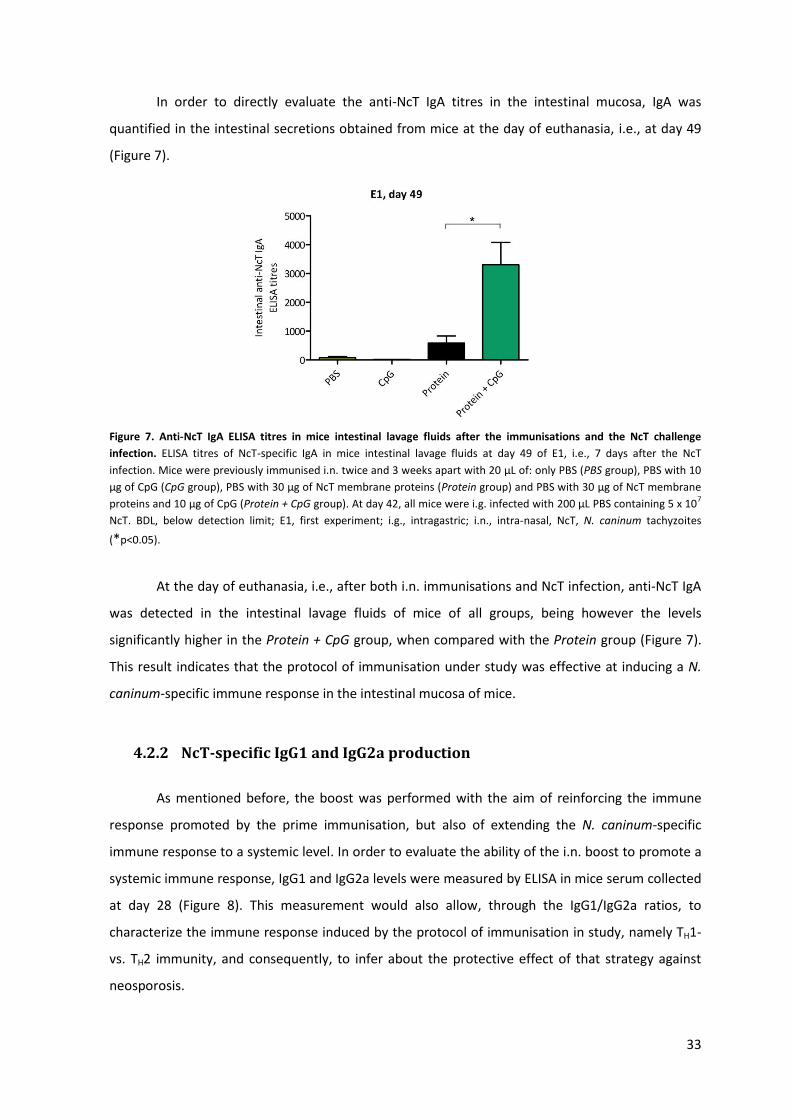
33
In order to directly evaluate the anti-NcT IgA titres in the intestinal mucosa, IgA was
quantified in the intestinal secretions obtained from mice at the day of euthanasia, i.e., at day 49
(Figure 7).
Figure 7. Anti-NcT IgA ELISA titres in mice intestinal lavage fluids after the immunisations and the NcT challenge
infection. ELISA titres of NcT-specific IgA in mice intestinal lavage fluids at day 49 of E1, i.e., 7 days after the NcT
infection. Mice were previously immunised i.n. twice and 3 weeks apart with 20 μL of: only PBS (PBS group), PBS with 10
μg of CpG (CpG group), PBS with 30 μg of NcT membrane proteins (Protein group) and PBS with 30 μg of NcT membrane
proteins and 10 μg of CpG (Protein + CpG group). At day 42, all mice were i.g. infected with 200 μL PBS containing 5 x 10⁷
NcT. BDL, below detection limit; E1, first experiment; i.g., intragastric; i.n., intra-nasal, NcT, N. caninum tachyzoites
(*p<0.05).
At the day of euthanasia, i.e., after both i.n. immunisations and NcT infection, anti-NcT IgA
was detected in the intestinal lavage fluids of mice of all groups, being however the levels
significantly higher in the Protein + CpG group, when compared with the Protein group (Figure 7).
This result indicates that the protocol of immunisation under study was effective at inducing a N.
caninum-specific immune response in the intestinal mucosa of mice.
4.2.2 NcT-specific IgG1 and IgG2a production
As mentioned before, the boost was performed with the aim of reinforcing the immune
response promoted by the prime immunisation, but also of extending the N. caninum-specific
immune response to a systemic level. In order to evaluate the ability of the i.n. boost to promote a
systemic immune response, IgG1 and IgG2a levels were measured by ELISA in mice serum collected
at day 28 (Figure 8). This measurement would also allow, through the IgG1/IgG2a ratios, to
characterize the immune response induced by the protocol of immunisation in study, namely TH1-
vs. TH2 immunity, and consequently, to infer about the protective effect of that strategy against
neosporosis.
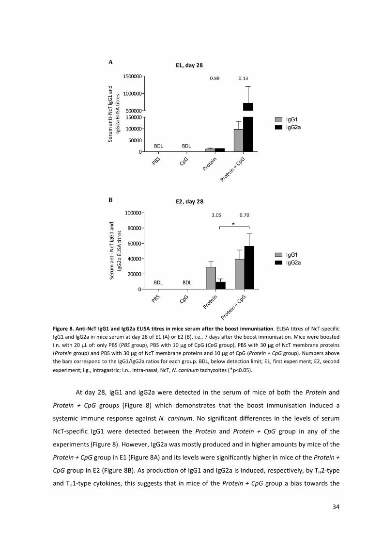
34
Figure 8. Anti-NcT IgG1 and IgG2a ELISA titres in mice serum after the boost immunisation. ELISA titres of NcT-specific
IgG1 and IgG2a in mice serum at day 28 of E1 (A) or E2 (B), i.e., 7 days after the boost immunisation. Mice were boosted
i.n. with 20 μL of: only PBS (PBS group), PBS with 10 μg of CpG (CpG group), PBS with 30 μg of NcT membrane proteins
(Protein group) and PBS with 30 μg of NcT membrane proteins and 10 μg of CpG (Protein + CpG group). Numbers above
the bars correspond to the IgG1/IgG2a ratios for each group. BDL, below detection limit; E1, first experiment; E2, second
experiment; i.g., intragastric; i.n., intra-nasal, NcT, N. caninum tachyzoites (*p<0.05).
At day 28, IgG1 and IgG2a were detected in the serum of mice of both the Protein and
Protein + CpG groups (Figure 8) which demonstrates that the boost immunisation induced a
systemic immune response against N. caninum. No significant differences in the levels of serum
NcT-specific IgG1 were detected between the Protein and Protein + CpG group in any of the
experiments (Figure 8). However, IgG2a was mostly produced and in higher amounts by mice of the
Protein + CpG group in E1 (Figure 8A) and its levels were significantly higher in mice of the Protein +
CpG group in E2 (Figure 8B). As production of IgG1 and IgG2a is induced, respectively, by TH2-type
and TH1-type cytokines, this suggests that in mice of the Protein + CpG group a bias towards the
A
B
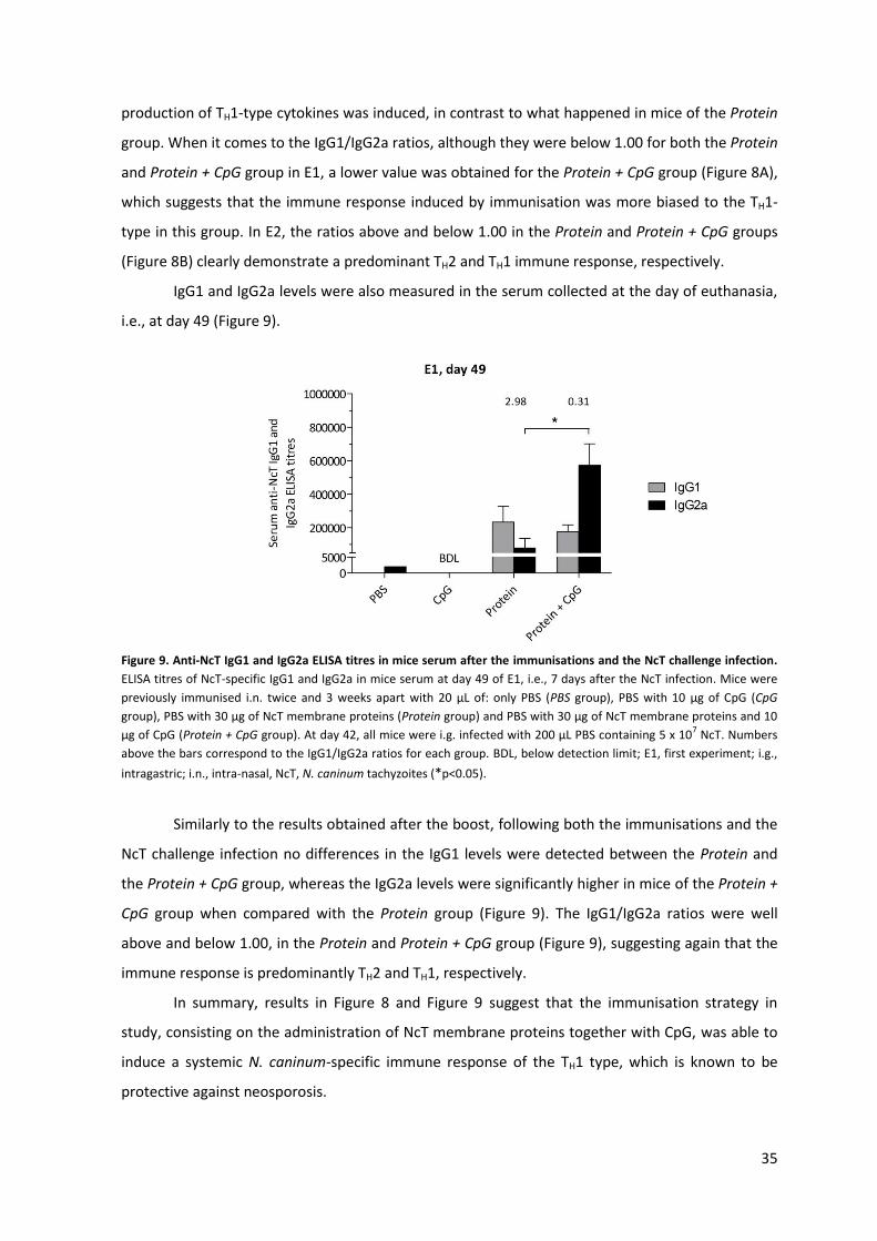
35
production of TH1-type cytokines was induced, in contrast to what happened in mice of the Protein
group. When it comes to the IgG1/IgG2a ratios, although they were below 1.00 for both the Protein
and Protein + CpG group in E1, a lower value was obtained for the Protein + CpG group (Figure 8A),
which suggests that the immune response induced by immunisation was more biased to the TH1-
type in this group. In E2, the ratios above and below 1.00 in the Protein and Protein + CpG groups
(Figure 8B) clearly demonstrate a predominant TH2 and TH1 immune response, respectively.
IgG1 and IgG2a levels were also measured in the serum collected at the day of euthanasia,
i.e., at day 49 (Figure 9).
Figure 9. Anti-NcT IgG1 and IgG2a ELISA titres in mice serum after the immunisations and the NcT challenge infection.
ELISA titres of NcT-specific IgG1 and IgG2a in mice serum at day 49 of E1, i.e., 7 days after the NcT infection. Mice were
previously immunised i.n. twice and 3 weeks apart with 20 μL of: only PBS (PBS group), PBS with 10 μg of CpG (CpG
group), PBS with 30 μg of NcT membrane proteins (Protein group) and PBS with 30 μg of NcT membrane proteins and 10
μg of CpG (Protein + CpG group). At day 42, all mice were i.g. infected with 200 μL PBS containing 5 x 10⁷ NcT. Numbers
above the bars correspond to the IgG1/IgG2a ratios for each group. BDL, below detection limit; E1, first experiment; i.g.,
intragastric; i.n., intra-nasal, NcT, N. caninum tachyzoites (*p<0.05).
Similarly to the results obtained after the boost, following both the immunisations and the
NcT challenge infection no differences in the IgG1 levels were detected between the Protein and
the Protein + CpG group, whereas the IgG2a levels were significantly higher in mice of the Protein +
CpG group when compared with the Protein group (Figure 9). The IgG1/IgG2a ratios were well
above and below 1.00, in the Protein and Protein + CpG group (Figure 9), suggesting again that the
immune response is predominantly TH2 and TH1, respectively.
In summary, results in Figure 8 and Figure 9 suggest that the immunisation strategy in
study, consisting on the administration of NcT membrane proteins together with CpG, was able to
induce a systemic N. caninum-specific immune response of the TH1 type, which is known to be
protective against neosporosis.
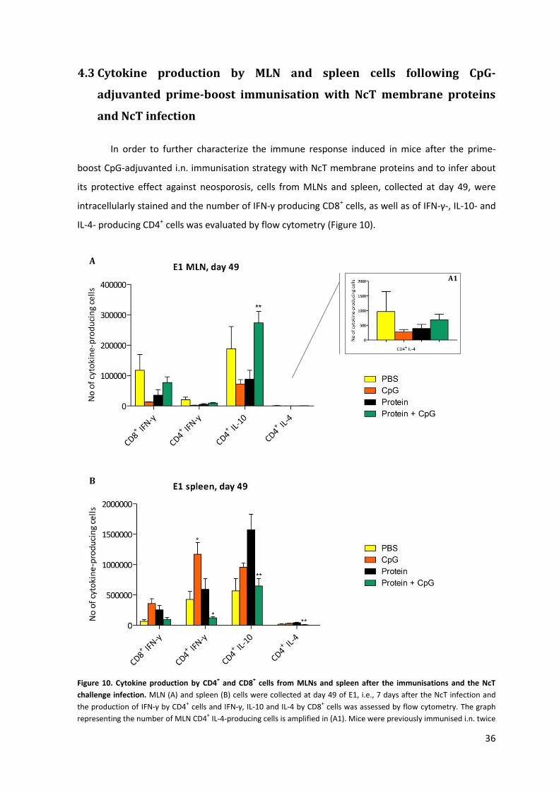
36
4.3 Cytokine production by MLN and spleen cells following CpG-
adjuvanted prime-boost immunisation with NcT membrane proteins
and NcT infection
In order to further characterize the immune response induced in mice after the prime-
boost CpG-adjuvanted i.n. immunisation strategy with NcT membrane proteins and to infer about
its protective effect against neosporosis, cells from MLNs and spleen, collected at day 49, were
intracellularly stained and the number of IFN-γ producing CD8+ cells, as well as of IFN-γ-, IL-10- and
IL-4- producing CD4+ cells was evaluated by flow cytometry (Figure 10).
Figure 10. Cytokine production by CD4+ and CD8
+ cells from MLNs and spleen after the immunisations and the NcT
challenge infection. MLN (A) and spleen (B) cells were collected at day 49 of E1, i.e., 7 days after the NcT infection and
the production of IFN-γ by CD4+ cells and IFN-γ, IL-10 and IL-4 by CD8
+ cells was assessed by flow cytometry. The graph
representing the number of MLN CD4+ IL-4-producing cells is amplified in (A1). Mice were previously immunised i.n. twice
A
B
A1
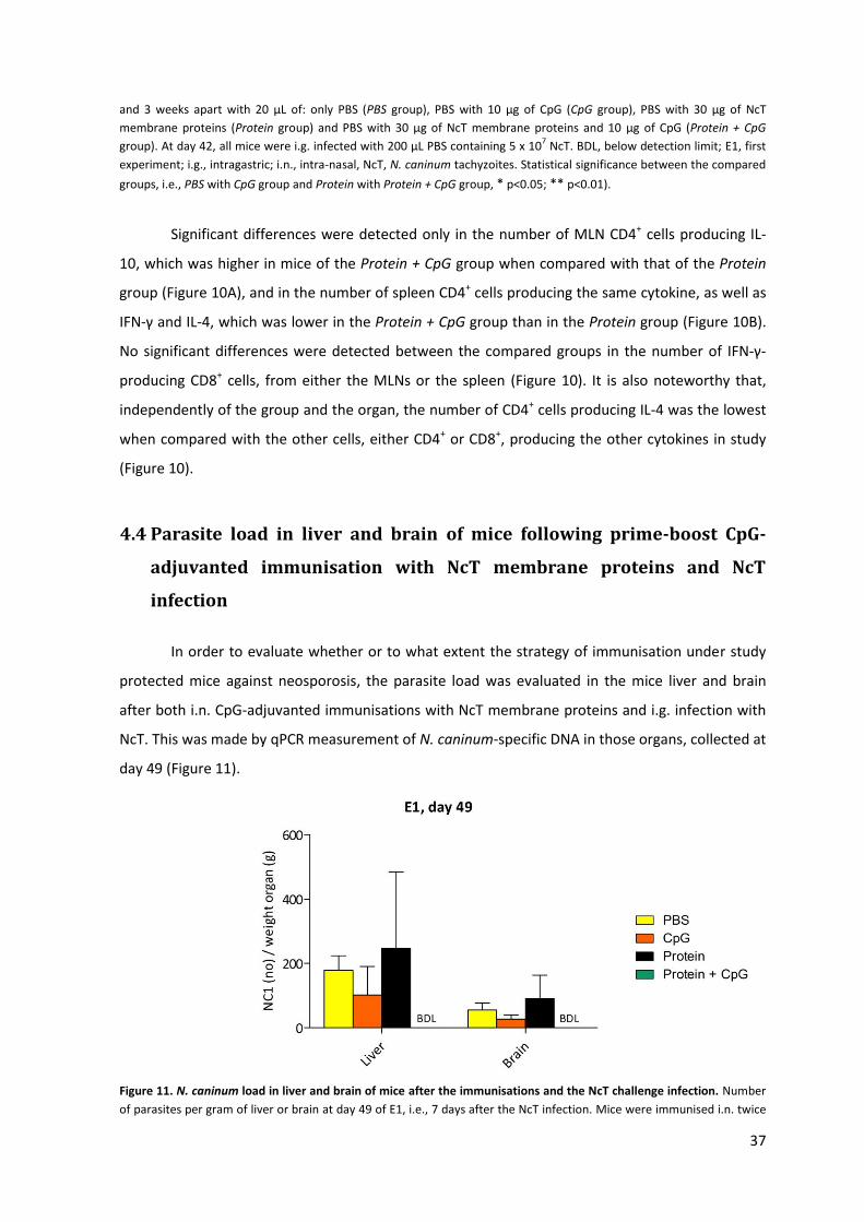
37
and 3 weeks apart with 20 μL of: only PBS (PBS group), PBS with 10 μg of CpG (CpG group), PBS with 30 μg of NcT
membrane proteins (Protein group) and PBS with 30 μg of NcT membrane proteins and 10 μg of CpG (Protein + CpG
group). At day 42, all mice were i.g. infected with 200 μL PBS containing 5 x 10⁷ NcT. BDL, below detection limit; E1, first
experiment; i.g., intragastric; i.n., intra-nasal, NcT, N. caninum tachyzoites. Statistical significance between the compared
groups, i.e., PBS with CpG group and Protein with Protein + CpG group, * p<0.05; ** p<0.01).
Significant differences were detected only in the number of MLN CD4+ cells producing IL-
10, which was higher in mice of the Protein + CpG group when compared with that of the Protein
group (Figure 10A), and in the number of spleen CD4+ cells producing the same cytokine, as well as
IFN-γ and IL-4, which was lower in the Protein + CpG group than in the Protein group (Figure 10B).
No significant differences were detected between the compared groups in the number of IFN-γ-
producing CD8+ cells, from either the MLNs or the spleen (Figure 10). It is also noteworthy that,
independently of the group and the organ, the number of CD4+ cells producing IL-4 was the lowest
when compared with the other cells, either CD4+ or CD8+, producing the other cytokines in study
(Figure 10).
4.4 Parasite load in liver and brain of mice following prime-boost CpG-
adjuvanted immunisation with NcT membrane proteins and NcT
infection
In order to evaluate whether or to what extent the strategy of immunisation under study
protected mice against neosporosis, the parasite load was evaluated in the mice liver and brain
after both i.n. CpG-adjuvanted immunisations with NcT membrane proteins and i.g. infection with
NcT. This was made by qPCR measurement of N. caninum-specific DNA in those organs, collected at
day 49 (Figure 11).
Figure 11. N. caninum load in liver and brain of mice after the immunisations and the NcT challenge infection. Number
of parasites per gram of liver or brain at day 49 of E1, i.e., 7 days after the NcT infection. Mice were immunised i.n. twice
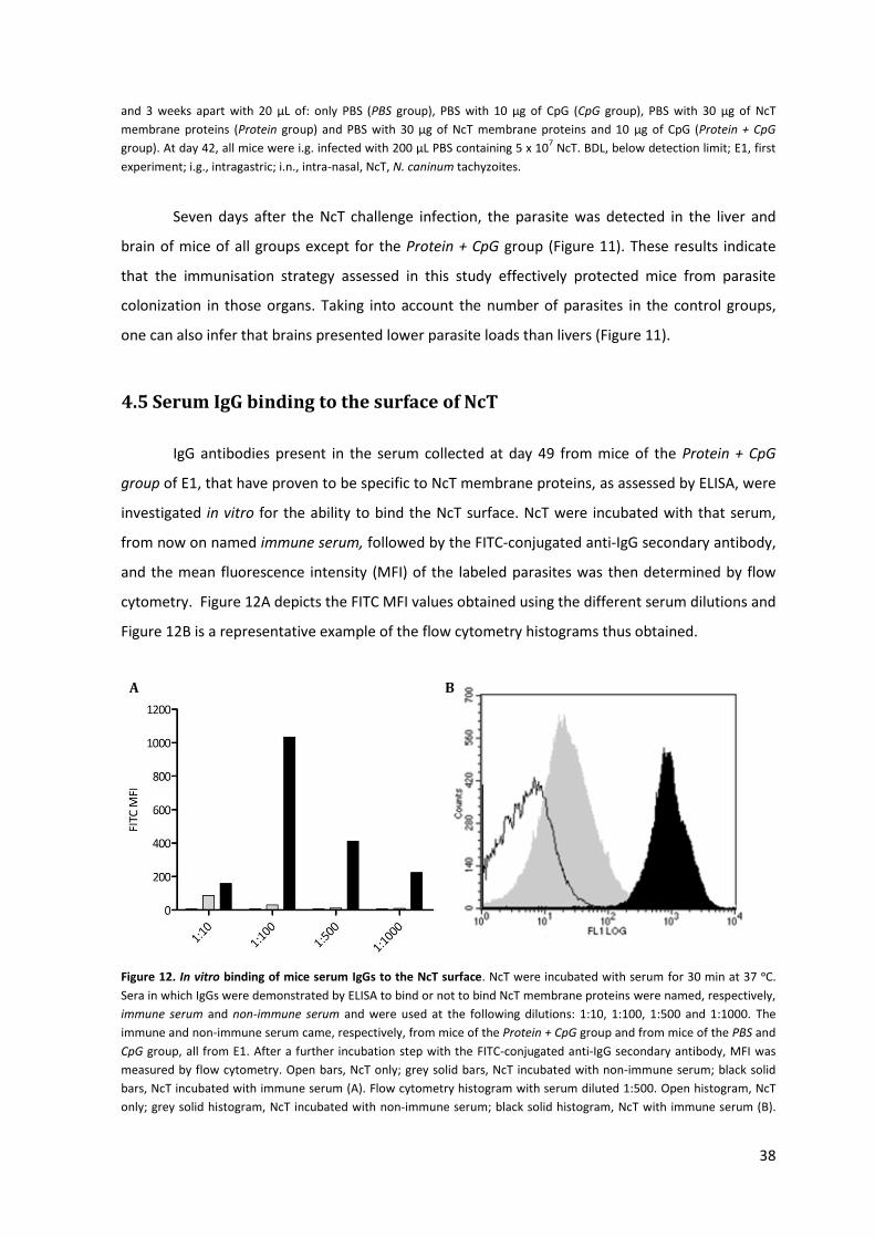
38
and 3 weeks apart with 20 μL of: only PBS (PBS group), PBS with 10 μg of CpG (CpG group), PBS with 30 μg of NcT
membrane proteins (Protein group) and PBS with 30 μg of NcT membrane proteins and 10 μg of CpG (Protein + CpG
group). At day 42, all mice were i.g. infected with 200 μL PBS containing 5 x 10⁷ NcT. BDL, below detection limit; E1, first
experiment; i.g., intragastric; i.n., intra-nasal, NcT, N. caninum tachyzoites.
Seven days after the NcT challenge infection, the parasite was detected in the liver and
brain of mice of all groups except for the Protein + CpG group (Figure 11). These results indicate
that the immunisation strategy assessed in this study effectively protected mice from parasite
colonization in those organs. Taking into account the number of parasites in the control groups,
one can also infer that brains presented lower parasite loads than livers (Figure 11).
4.5 Serum IgG binding to the surface of NcT
IgG antibodies present in the serum collected at day 49 from mice of the Protein + CpG
group of E1, that have proven to be specific to NcT membrane proteins, as assessed by ELISA, were
investigated in vitro for the ability to bind the NcT surface. NcT were incubated with that serum,
from now on named immune serum, followed by the FITC-conjugated anti-IgG secondary antibody,
and the mean fluorescence intensity (MFI) of the labeled parasites was then determined by flow
cytometry. Figure 12A depicts the FITC MFI values obtained using the different serum dilutions and
Figure 12B is a representative example of the flow cytometry histograms thus obtained.
Figure 12. In vitro binding of mice serum IgGs to the NcT surface. NcT were incubated with serum for 30 min at 37 ᵒC.
Sera in which IgGs were demonstrated by ELISA to bind or not to bind NcT membrane proteins were named, respectively,
immune serum and non-immune serum and were used at the following dilutions: 1:10, 1:100, 1:500 and 1:1000. The
immune and non-immune serum came, respectively, from mice of the Protein + CpG group and from mice of the PBS and
CpG group, all from E1. After a further incubation step with the FITC-conjugated anti-IgG secondary antibody, MFI was
measured by flow cytometry. Open bars, NcT only; grey solid bars, NcT incubated with non-immune serum; black solid
bars, NcT incubated with immune serum (A). Flow cytometry histogram with serum diluted 1:500. Open histogram, NcT
only; grey solid histogram, NcT incubated with non-immune serum; black solid histogram, NcT with immune serum (B).
A
A B
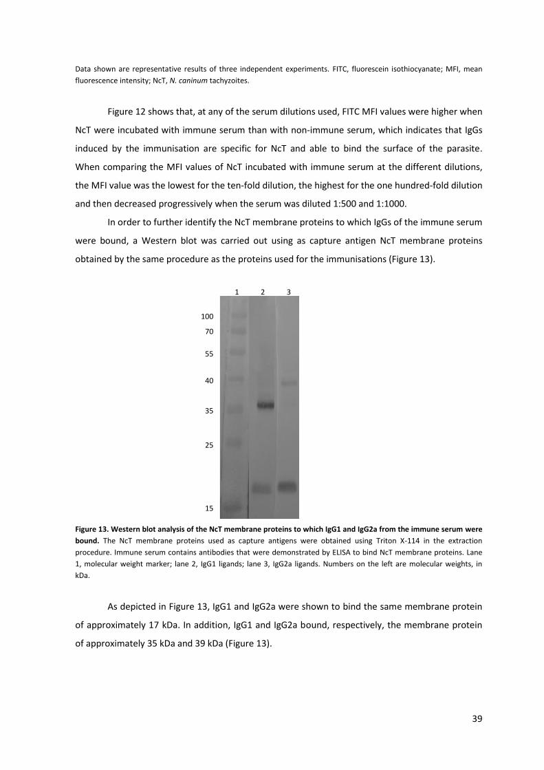
39
Data shown are representative results of three independent experiments. FITC, fluorescein isothiocyanate; MFI, mean
fluorescence intensity; NcT, N. caninum tachyzoites.
Figure 12 shows that, at any of the serum dilutions used, FITC MFI values were higher when
NcT were incubated with immune serum than with non-immune serum, which indicates that IgGs
induced by the immunisation are specific for NcT and able to bind the surface of the parasite.
When comparing the MFI values of NcT incubated with immune serum at the different dilutions,
the MFI value was the lowest for the ten-fold dilution, the highest for the one hundred-fold dilution
and then decreased progressively when the serum was diluted 1:500 and 1:1000.
In order to further identify the NcT membrane proteins to which IgGs of the immune serum
were bound, a Western blot was carried out using as capture antigen NcT membrane proteins
obtained by the same procedure as the proteins used for the immunisations (Figure 13).
Figure 13. Western blot analysis of the NcT membrane proteins to which IgG1 and IgG2a from the immune serum were
bound. The NcT membrane proteins used as capture antigens were obtained using Triton X-114 in the extraction
procedure. Immune serum contains antibodies that were demonstrated by ELISA to bind NcT membrane proteins. Lane
1, molecular weight marker; lane 2, IgG1 ligands; lane 3, IgG2a ligands. Numbers on the left are molecular weights, in
kDa.
As depicted in Figure 13, IgG1 and IgG2a were shown to bind the same membrane protein
of approximately 17 kDa. In addition, IgG1 and IgG2a bound, respectively, the membrane protein
of approximately 35 kDa and 39 kDa (Figure 13).
100
70
55
40
35
25
15
1 2 3

40
5. Discussion
In cattle, N. caninum infection can be acquired either post-natally or transplacentally,
being, respectively, the GI tract and the placenta the parasite entry routes into the host11. Since
most cattle are infected transplacentally, a successful vaccine targeting that mode of infection
could counteract neosporosis. However, it would be only partially effective since some form of
post-natal transmission is known to be required in order for the infection to be sustained in a
herd44. Therefore, the development of an immunisation strategy targeting this last route of
infection is essential to cope with the disease. Taking into account that N. caninum post-natal
infection originates in the GI tract, stimulation of an immune response directly in the intestinal
mucosa could be a way of counteracting infection and dissemination of the parasite soon after it
contacts with the host. The existence, at the very beginning, of antibodies specific to membrane
proteins essential for adhesion and/or invasion by the parasite could impede it of penetrating host
cells and thus cross the intestinal mucosal barriers, by antibody-mediated neutralization.
Therefore, this project aimed at characterizing the immune response promoted in mice by mucosal
immunisation with NcT membrane proteins as well as at evaluating the protective effect of that
strategy against i.g. established murine neosporosis. This infection route was chosen in order to
more closely simulate the horizontal route of N. caninum transmission that naturally happens in
herds.
The NcT membrane proteins used for immunisation were obtained by employing a protocol
that uses Triton-X114. Extraction protocols using this non-ionic detergent have been employed in
previous studies to isolate cell-surface membrane-associated proteins from N. caninum169 and
other parasites, such as T. gondii170 and Leishmania major171 . The specific protocol followed in this
project has been previously described and compared by Zintl et al.167 with others aimed at
obtaining N. caninum antigens, namely using sonication of parasites or solubilisation of NcT with
SDS or detergent and non-detergent sulfobetaines. Compared with those methodologies, the
Triton-X114 extraction is highly selective and has been considered a useful method to concentrate
a subset of N. caninum membrane proteins167. By employing that procedure, a protein extract
comprising six proteins with approximately 55, 47, 39, 35, 27 and 17 kDa, was obtained and
resulted in a SDS-PAGE migration pattern similar to the one obtained by Zintl et al167 (Figure 4).
Therefore, the extraction protocol was considered successful and used every time NcT membrane
proteins were needed, either for the immunisations or to set ELISA and Western blot tests (as
capture antigens). The observation that two proteins of 55 and 39 kDa were commonly obtained in
41
this work, but not by Zintl et al., could be explained by a slight different composition of the NcT
membrane. Although the strains of N. caninum were the same in both protocols (NC-1), the fact
that the isolates could have been passed different times and/or in different strains of mice could
have accounted for the existence of different antigens in the membrane and, consequently, slight
differences in the virulence between them (the strain used in this study has been previously passed
once in C57BL/6 IL-12KO mice). Regarding the components of the extract, the proteins of
approximately 35 and 27 kDa may most likely correspond to NcSRS2 and NcSAG1, respectively,
which are the two immunodominant surface proteins of NcT50. Nonetheless, the exact
identification of the proteins composing the extract would require the performance of peptide
mass fingerprint.
Since a protective immune response at one mucosal site can be induced by immunisation
at a distant mucosal site (concept of CMIS, then renamed to compartmentalised mucosal immune
system)99,100, and supported by a recently published study demonstrating that T cells primed
following i.n. CpG-adjuvanted immunisation with OVA migrate to the spleen and to non-draining
LNs, including the MLNs168, the i.n. route was chosen to immunise the mice. This choice was
further encouraged by the fact that, once its efficacy has been proven in mice, i.n. immunisation,
contrarily to other routes of mucosal immunisation such as the rectal or vaginal ones, could be
easily applied in cattle, the ultimate target species of this protocol. In addition to the first
immunisation, a boost was administered to all mice three weeks later in order to strengthen the
immune response promoted by the prime immunisation and enlarge the scope of the N. caninum-
specific immunity to a systemic level.
To investigate the efficacy of the immunisation strategy under study, the evaluation of a N.
caninum-specific immune response at the mucosa of the GI tract, a site of entry of the pathogen
into the host, seemed essential. For that purpose, IgA, the most characteristic mediator of mucosal
immunity80, would ideally be measured in the intestinal mucosal secretions. However, their
sampling requires invasive methods that are not feasible for routine diagnostics172. Therefore, the
IgA measurement was made, either after the prime or the boost immunisation, in the vaginal
lavage fluids, as those are the closest mucosal fluids in relation to the intestinal secretions that
could be collected by a non-invasive procedure. At the day of euthanasia, intestinal secretions were
nevertheless collected to quantify NcT-specific IgA directly at the intestinal mucosal site. The
protocol of immunisation studied here, namely the i.n. prime-boost immunisation with NcT
membrane proteins plus CpG, was demonstrated to induce an immune response against the
parasite at the mucosal level (Figure 6) and specifically at the intestinal mucosal site (Figure 7). This
was indicated by the significantly higher ELISA titres of anti-NcT IgA, respectively detected in the
vaginal lavage fluids after the boost (Figure 6) and in the intestinal lavage fluids at the day of
42
euthanasia (Figure 7), in the Protein + CpG group when compared with the Protein group. The
results concerning the anti-NcT IgA titres detected after the prime immunisation were inconclusive
since different data were obtained in the two independent experiments performed in the same
conditions (Figure 5). Further replicate experiments would therefore be necessary to better
ascertain the anti-NcT IgA titres in mice of the Protein and Protein + CpG groups, at this time point.
Nevertheless, they were useful, by suggesting that the boost induced an augmentation in the
magnitude of the immune response promoted by the prime immunisation, as assessed by the
increased anti-NcT IgA titres in the vaginal lavage fluids of the Protein + CpG group at day 28, when
compared with those at day 14 (Figure 5A and 6A). The fact that mice of the control PBS and CpG
groups did not produce IgA specific to N. caninum neither after the prime nor after the boost
immunisation (Figure 5 and 6) was already expected since until these time points those animals
were not administered with N. caninum antigens, contrarily to those of the Protein and Protein +
CpG groups. However, it is understandable that NcT-specific IgA had been detected in the intestinal
lavage fluids of those mice (Figure 7) since they had been infected with NcT seven days before
sampling these secretions. The lower IgA titres detected when compared with those of the Protein
and Protein + CpG groups (Figure 7) further stresses out the role of the encounter with the antigen
before the infection, and consequently the importance of the immunisation, to mount a significant
mucosal immune response against the pathogen once the infection has occurred.
Besides the aim of strengthening the immune response promoted by the prime
immunisation, the boost was also performed to extend it to a systemic level and thus to make the
immune response even more protective against the parasite. This second aim was achieved since
IgG1 and IgG2a specific for N. caninum were detected after the boost in the serum of mice of the
Protein and Protein + CpG groups (Figure 8) and also after the NcT challenge infection (Figure 9). As
already expected, and similarly to what has been reported in respect to the IgA titres, mice of the
PBS and CpG groups did not produce parasite-specific antibodies of any of the IgG isotypes after
the boost immunisation (Figure 8). One mouse of the PBS group did however produce IgG2a in
residual amounts after the NcT challenge infection (Figure 9), which is consistent with only having
encountered N. caninum antigens from the infection on.
It is well established that the production of IgG2a antibodies is induced by TH1 cytokines,
such as IL-12 and IFN-γ, whereas the production of IgG1 antibodies is favoured by TH2 cytokines,
such as IL-432. In this way, the levels of antibody subclasses may reflect the in vivo production of
cytokines, as well as the type of immune response mounted against the pathogen, namely when
attention is paid to the ratio IgG1/IgG2a32. In this case, the significantly higher levels of serum
IgG2a in the Protein +CpG group when compared with the Protein group, either after the boost
(Figure 8) or after both immunisations and the NcT infection (Figure 9), suggest that these mice
43
were producing more TH1-type cytokines than the ones of the Protein group. In contrast, the levels
of TH2-type cytokines did not appear to significantly differ between the compared groups, as no
statistically significant differences in the serum IgG1 titres were reported (Figure 8 and 9). By
looking at the IgG1/IgG2a ratios, it was clearly established that the immune responses promoted in
mice of the Protein and Protein + CpG group were, respectively, of the TH2-type (ratios above 1.00)
and TH1-type (ratios below 1.00), either after the boost immunisation (Figure 8B) or after both
immunisations and the NcT challenge infection (Figure 9). Only for the first experiment and after
the boost were the ratios for both groups below 1.00 (Figure 8A). Nevertheless, since the ratio for
the Protein + CpG group was much lower than the ratio for the Protein group (0.13 vs. 0.88,
respectively), one can also infer that the immune responses were, respectively, more biased to the
TH1- and TH2-type.
From the antibody analyses just presented, it can be inferred that the administration of
NcT membrane proteins together with CpG (Protein + CpG group) has been able to induce in mice,
both at the mucosal and systemic level, a protective immune status effective against N. caninum.
This status was characterized by significantly higher antigen-specific IgA and IgG2a titres, when
compared with mice of the Protein group, and biased to the TH1-type of immunity. The immune
protection conferred by the immunisation was supported by data on liver and brain parasite load,
measured seven days after the i.g. NcT challenge infection (Figure 11). In fact, no mouse of this
group was colonized by N. caninum, neither in the liver nor in the brain, contrarily to mice of the
Protein group. This observation is consistent with the previously inferred TH2 immune response for
these mice that is known to be associated with susceptibility to N. caninum infection. Also, and as
expected, mice of the PBS and CpG groups presented N. caninum colonization in both organs
(Figure 11), in agreement with the fact that none of these animals had been previously immunised
with N. caninum antigens in order to be protected at the time of infection. Although not
statistically significant, mice of the CpG group were less colonized than those of the PBS group and
this could be explained by CpG being an immunomodulator effective at inducing TH1-type immune
responses121. This type of immune response is known to be protective against neosporosis and may
have been responsible for the lower N. caninum colonization observed, when compared with that
in animals treated with PBS alone. Comparing the number of parasites in the livers and brains of
mice of the control groups, it is noteworthy that the first appeared to be more colonized than the
last (Figure 11). A possible explanation could be that at the time point at which the N. caninum load
was assessed (seven days after the NcT challenge infection), the parasite had still not evolved into
the bradyzoite stage, which is preferentially located in the central nervous system24,32, and was still
disseminating throughout the organism in the tachyzoite form.
44
After the prime-boost immunisation and the NcT challenge, the cytokine production by
MLN and spleen cells was evaluated with the aim of further characterizing the induced immune
response. A study conducted by Velge-Roussel et al.173 supported the analysis of the cytokine
production in cells coming from the MLNs to evaluate the mucosal immune response, as they
demonstrated that i.n. CT-adjuvanted immunisation with T. gondii SAG1 induced a specific cellular
response in the NALT, the cervical LN and MLNs. On the other hand, systemic immunity could be
evaluated using cells obtained from the spleen173. If the type of immune response in the Protein
and Protein + CpG groups, either TH1 or TH2, has been deducted from the serum IgG1/IgG2a ratios,
the same did not happen when the production of cytokines was analysed (Figure 10). In fact, the
only significant difference in the MLNs was noticed in the total number of CD4+ IL-10-producing
cells that was significantly higher in mice of the Protein + CpG groups than in animals of the Protein
group (Figure 10A), which contrasts with the TH1 immune response for the first group, as could be
deducted from the IgG1/IgG2a ratios. On the other hand, the number of spleen CD4+ cells
producing IL-4 and IL-10 was significantly lower in the Protein + CpG group than in the Protein
group, which could be in agreement with the TH1 immune response previously inferred if the
number of IFN-γ producing cells was not also significantly lower than in the Protein group, as it has
actually been demonstrated (Figure 10B). Low levels of IFN-γ in mice that contain higher IgG2a than
IgG1 titres could be explained by IgG2a production independent of IFN-γ as has been demonstrated
to occur upon parasitic infections174. In this study, Markine-Goriaynoff et al.174 have shown that
either IFN-γ receptor-knockout mice or wild-type mice treated with anti-IFN-γ did produce IgG2a
after infection with T. gondii and Trypanosoma cruzi, respectively, clearly indicating that at least
some of the in vivo production of IgG2a is independent of IFN-γ. One can also hypothesize that the
low levels of IFN-γ in mice of the Protein + CpG group were due to a decrease in the number of
parasites able to enter the host, due to NcT-specific IgA-mediated neutralization in the intestinal
mucosa. Alternatively, if they could have actually entered, it can be hypothesized that the NcT-
specific IgG1 and IgG2a generated upon immunisation have been successful in preventing its
dissemination. If one of those hypotheses did happen, the parasite could have not been able to
stimulate the immune system and therefore to induce the production of IFN-γ, typically associated
with protection against neosporosis. Also, it could be that, as mice were shown to be protected
against N. caninum, by assessing the parasite load in the liver and brain, the threshold level of IFN-γ
for protection had been achieved with the low amounts detected, which means that more IFN-γ
would not lead to more protection. At last, it should be noted that it is possible that the cytokine
analysis was made too late after the infection and the peak of IFN-γ production already subsided by
the time the analysis was made. Therefore, the cytokine analysis must be done in another
experiment where the animals would be analysed sooner than seven days after the NcT challenge.
45
When it comes to the significant differences between the Protein and the Protein + CpG
group besides the cytokine analysis, it is reasonable to postulate that they are generally assigned to
the effect caused by the administration of CpG. In fact, CpG has been reported to be a potent
mucosal adjuvant when delivered i.n. together with bacterial antigens122 and its administration
with NcT membrane proteins in the present study has likely been responsible for the significantly
higher IgA titres detected in the Protein + CpG group. In the same study, CpG has demonstrated to
induce not only a mucosal but also a systemic immune response when delivered i.n.122. In this case,
however, the systemic immune response promoted by the boost immunisation can not be entirely
attributed to CpG, since mice that only received NcT membrane proteins also produced detectable
amounts of antibodies of the IgG isotypes. One can infer that the NcT membrane proteins used in
the immunisation are slightly immunogenic by themselves and capable of stimulating an immune
response, both at the systemic level and at the mucosal level, which in this last case would also
explain the fact that mice of the Protein group produced some NcT-specific IgA. Also, it is
reasonable to hypothesize that the route of administration (i.n.) was itself responsible for
promoting not only a mucosal but also a systemic immune response in both groups, as discussed
below. Nonetheless, CpG is most likely responsible for the significantly higher IgG2a titres detected
in the Protein + CpG group when compared with the Protein group and for the TH1 immune
response inferred in these mice based on the IgG1/IgG2a ratios. A study carried out by Teixeira et
al.175 showed that intradermal immunisation of BALB/c mice with N. caninum sonicates in the alum
or Freund’s adjuvant or with irradiated NcT without adjuvant led to an increased susceptibility to
the parasite, with predominant production of IgG1 comparatively to IgG2a and a decrease in the
IFN-γ mRNA expression upon NcT challenge. Also, in a clear explanatory study, Chu et al.121
showed that co-administration of CpG with hen egg lysozyme (HEL), in either TH1- or TH2-biased
mice, induced a TH1 immune response characterized by high levels of IFN-γ production and low
levels of IL-5 secretion. Moreover, a TH1 immune response with high production of IgG2a was
switched on when FIA, that was shown to induce TH2 immune responses when co-administered as
the sole adjuvant with HEL, was administered together with CpG and HEL. The potent TH1-
immunomodulatory ability of CpG has also been demonstrated in an experimental study conducted
by Weeratna et al.112 who showed that the combination of CpG with FIA and alum compounds,
both of them strongly TH2-biased, induced even stronger TH1 immune responses than when CpG
was administered alone. In the same study, CpG has also shown to incur only minimal tissue
damage at the injection site upon intramuscular administration112. Also, and according to its TH1-
immunomodulatory property, CpG is known to be a potent inducer of TH1 cytokines, such as IL-12
and IFN-γ125, and Rosa et al176 highlighted the critical role of IFN-γ in the adjuvant activity of CpG
when co-administered i.n. with recombinant proteins. Namely, significantly lower IgG titres were
46
detected in IFN-γ-knockout mice , an effect that was specific for the CpG adjuvant176. In the same
study, the importance of CD4+ and CD8+ cells as IFN-γ sources in vivo following CpG-adjuvanted
immunisation has been demonstrated176. According to this, the fact that neither a significantly
higher number of CD4+ nor of CD8+ cells produced IFN-γ in the present study, further supports the
idea that the cytokine analysis should be repeated.
In order to further understand the functional properties of IgG antibodies that have
proven, by ELISA, to bind NcT membrane proteins, an assay was conducted in which these
antibodies were investigated for the ability to bind the surface of NcT in vitro. Independently of the
dilution at which the serum containing those IgGs (immune serum) was incubated with NcT, they
have been shown to bind the NcT surface (Figure 12), which is in agreement with the fact that they
were generated upon immunisation with NcT membrane proteins, the same to which they have
then proven to bind, by ELISA, as previously referred. The IgG binding was proven by the higher
MFI values of the opsonized parasites when compared with the ones obtained when the NcT were
incubated with the non-immune serum. Nonetheless, it is of note that the ten-fold dilution of the
serum was the one that led to the least binding, probably due to an excess of antibodies that
competed for the same ligand on the membrane of the parasite and thus sterically hindered the
binding of more IgG. The one hundred-fold dilution led, in turn, to the highest binding, among all
the four dilutions tested. The five hundred- and one thousand-fold dilution resulted in minimal
background, i.e., the differences in the MFI values between NcT incubated with non-immune
serum and with no serum at all were minimal, but the MFI values progressively decreased from the
one hundred- until the one thousand-fold dilution. One can hypothesize that this was due to a
progressive lack of IgGs to bind all the surface of the parasite. In summary, the one hundred-fold
dilution of the serum seems to lead to an ideal ratio antibody: parasite and is therefore
recommended for future in vitro assays of this type. The specificity of these antibodies for the NcT
membrane proteins was further investigated by Western blot where it was demonstrated that IgG1
and IgG2a bind, respectively, the membrane proteins of 35 and 39 kDa and both of them also bind
the one of 17 kDa.
Several studies have used the i.n. route to immunise animals against a variety of parasites,
including T. gondii. I.n. immunisation of CBA/J mice with T. gondii SAG1 protein and using non-toxic
mutant heat-labile enterotoxins as adjuvants induced both mucosal and systemic immune
response and protected mice against oral infection with T. gondii177. More recently, i.n. CT-
adjuvanted immunisation of BALB/c mice with the recombinant proteins TgROP2, TgGRA5 and
TgGRA7 has been shown to induce partial protection against tissue cyst formation after oral
challenge infection with tissue cysts of T. gondii178. Besides mice, i.n. immunisation has also been
applied in sheep using T. gondii tachyzoite antigens encapsulated into PLG microspheres, which
47
resulted in both systemic and mucosal, humoral and cell-mediated immune responses179. When it
comes to N. caninum, a pertinent study and the first on i.n. immunisation of mice against this
parasite, has been recently published by Debache et al.180, who demonstrated that the outcome of
protection depends not only on the nature of the antigen but also on the route of delivery180. Three
recombinant N. caninum proteins, namely NcPDI, NcROP2 and NcMAG1, were used for saponin-
adjuvanted i.p. immunisation or CT-adjuvanted i.n. immunisation of C57BL/6 mice which were then
challenged by i.p. infection with NcT. In the case of recombinant NcPDI, the i.n. route provided 90%
protection rate against neosporosis but only 20% when the protein was delivered i.p180.
The interest in developing vaccines of defined composition is progressively increasing.
Although there is one commercially available vaccine against N. caninum which consists of a crude
extract of inactivated NcT and the Havlogen adjuvant and has proven to be partially effective in
reducing the incidence of abortions in herds, the composition of this kind of vaccines made of
crude extracts may vary from one batch to another and thus can not be accurately controlled.
Moreover, these vaccines may include non-protective or immunosuppressive factors32. Therefore,
the possibility of combining purified N. caninum antigens (instead of the whole parasite) and the
strong TH1-immunomodulator CpG with the i.n. route of delivery, that has recurrently proven to
induce both mucosal and systemic immunity, seemed promising in order to develop an efficient
immunisation protocol against N. caninum. At least so far, this challenge seems to be on the good
way for success.
48
6. Conclusions and future work
The present study was carried out with the aim of characterizing the immune response
induced in mice by mucosal CpG-adjuvanted immunisation with NcT membrane proteins as well as
of evaluating the protective effect of that protocol of immunisation against neosporosis. The
strategy of immunisation under study was efficient at inducing an immune response against N.
caninum, both at the intestinal mucosa and the systemic level, as indicated by the ELISA NcT-
specific IgA and IgG titres, respectively. Taking into account that serum IgG1/IgG2a ratios were
lower than 1.00, the immune response thus induced was predominantly of the TH1 type, known to
be protective against neosporosis. In accordance with this, seven days after i.g. infection with NcT,
no parasites were detected in the liver and brain of mice subject to that protocol of immunisation,
as evaluated by qPCR. At last, serum IgG1 and IgG2a produced by the immunisation were shown by
flow cytometry to bind the NcT surface in vitro and were specific for three of the NcT membrane
proteins used to immunise the animals as determined by Western blot. IgG1 was specific to the
protein of 35 kDa, IgG2a to the protein of 39 kDa and antibodies of both IgG isotypes to the protein
of 17 kDa.
In the near future, peptide mass fingerprint could be performed in order to precisely
identify the proteins present in the extract that is used for immunisation. The knowledge of the
NcT proteins to which intestinal IgA binds would be useful to infer about whether IgA binding to
the parasite at the intestinal mucosa is important to prevent its adhesion to the intestinal cells
and/or invasion of the host. The ability of the intestinal IgA to prevent parasite entry into cells
could be experimentally investigated by incubating them with the parasite and then with the VERO
cells. The role of intestinal IgA could be further studied by an in vivo experiment of the course of N.
caninum infection as soon as it contacts with the host by the i.g. route. For this, immunised mice
could be challenged with NcT at different time points and sections of the intestine could be used
for histological examination and immunohistochemical analysis to investigate the damage caused
by the parasite in the intestine as well as its localization along time. If the parasite is shown to
invade the host after contacting with the intestinal mucosa, the consequences of serum IgG1 and
IgG2a binding to the NcT membrane, namely which parasite functions are neutralized upon binding
(adhesion, invasion), could be inferred if the proteins to which they bind have been identified by
peptide mass fingerprint. The effect of serum IgG1 and IgG2a binding in parasite entry into cells
could be also investigated by the aforementioned in vitro study. If IgG1 and IgG2a did not prove to
prevent parasite invasion of host cells, their ability to activate the complement system, that would
49
then destroy the N. caninum-infected host cells, could be evaluated as a possible reason to explain
why the parasite has not been detected in the liver and brain of the immunised mice.
With regard to the experiment set-up followed in this project, the possibility of decreasing
the time between the NcT challenge infection and the euthanasia of the animals should be
considered in order to probably get a better profile of the cytokine production by MLN and spleen
cells after the infection. Also, it would be interesting to carry out the same type of experiment but
with a longer time gap between the last immunisation and the NcT challenge infection. This would
be useful to investigate if the immunisation could provide long-term protection against the
parasite. Furthermore, the protective effect of the immunisation strategy in study, either the
present or the long-term one, could be evaluated by carrying out survival studies.
The possibility of combining another adjuvant with CpG could be considered in order to
possibly increase the efficiency of the immunisation. Weeratna et al.112 reported that the
combination of CpG with alum compounds induced the strongest immune responses to the
hepatitis B surface antigen when delivered intramuscularly and with minimal damage in the
injected muscle, among all the adjuvants alone and combinations of adjuvants tested.
The ultimate task of this project would be to apply in cattle the immunisation protocol that
has proven to be successful in mice and evaluate the efficacy of the strategy in that group of
animals. When proven to be successful, a way of preventing the post-natal route of N. caninum
transmission will have been established and the overall negative impact caused by N. caninum in
cattle will have, at least in part, been contradicted.
50
References
1. Bjerkås, I., Mohn, S. F. & Presthus, J. Unidentified cyst-forming sporozoon causing
encephalomyelitis and myositis in dogs. Z Parasitenkd 70, 271–274 (1984).
2. Dubey, J. P., Carpenter, J. L., Speer, C. A., Topper, M. J. & Uggla, A. Newly recognized fatal
protozoan disease of dogs. J. Am. Vet. Med. Assoc. 192, 1269–1285 (1988).
3. Dubey, J. P., Hattel, A. L., Lindsay, D. S. & Topper, M. J. Neonatal Neospora caninum infection
in dogs: isolation of the causative agent and experimental transmission. J. Am. Vet. Med. Assoc.
193, 1259–1263 (1988).
4. Dubey, J. P. et al. Redescription of Neospora caninum and its differentiation from related
coccidia. Int. J. Parasitol. 32, 929–946 (2002).
5. Dubey, J. P. & Schares, G. Neosporosis in animals--the last five years. Vet. Parasitol. 180, 90–
108 (2011).
6. Miller, C. M. D., Quinn, H. E., Windsor, P. A. & Ellis, J. T. Characterisation of the first
Australian isolate of Neospora caninum from cattle. Aust. Vet. J. 80, 620–625 (2002).
7. Koyama, T. et al. Isolation of Neospora caninum from the brain of a pregnant sheep. J.
Parasitol. 87, 1486–1488 (2001).
8. Rodrigues, A. A. R. et al. Shedding of Neospora caninum oocysts by dogs fed tissues from
naturally infected water buffaloes (Bubalus bubalis) from Brazil. Vet. Parasitol. 124, 139–150
(2004).
9. Vianna, M. C. B., Sreekumar, C., Miska, K. B., Hill, D. E. & Dubey, J. P. Isolation of Neospora
caninum from naturally infected white-tailed deer (Odocoileus virginianus). Vet. Parasitol. 129,
253–257 (2005).
10. Bień, J., Moskwa, B. & Cabaj, W. In vitro isolation and identification of the first Neospora
caninum isolate from European bison (Bison bonasus bonasus L.). Vet. Parasitol. 173, 200–205
(2010).
11. Dubey, J. P., Schares, G. & Ortega-Mora, L. M. Epidemiology and control of neosporosis and
Neospora caninum. Clin. Microbiol. Rev. 20, 323–367 (2007).
12. Petersen, E. et al. Neospora caninum infection and repeated abortions in humans. Emerging
Infect. Dis. 5, 278–280 (1999).
13. Barr, B. C., Conrad, P. A., Sverlow, K. W., Tarantal, A. F. & Hendrickx, A. G. Experimental fetal
and transplacental Neospora infection in the nonhuman primate. Lab. Invest. 71, 236–242 (1994).
51
14. Lobato, J. et al. Detection of immunoglobulin G antibodies to Neospora caninum in humans:
high seropositivity rates in patients who are infected by human immunodeficiency virus or have
neurological disorders. Clin. Vaccine Immunol. 13, 84–89 (2006).
15. Dubey, J. P., Buxton, D. & Wouda, W. Pathogenesis of bovine neosporosis. J. Comp. Pathol.
134, 267–289 (2006).
16. King, J. S. et al. Australian dingoes are definitive hosts of Neospora caninum. Int. J. Parasitol.
40, 945–950 (2010).
17. McAllister, M. M. et al. Dogs are definitive hosts of Neospora caninum. Int. J. Parasitol. 28,
1473–1478 (1998).
18. Lindsay, D. S., Dubey, J. P. & Duncan, R. B. Confirmation that the dog is a definitive host for
Neospora caninum. Vet. Parasitol. 82, 327–333 (1999).
19. Gondim, L. F. P., McAllister, M. M., Pitt, W. C. & Zemlicka, D. E. Coyotes (Canis latrans) are
definitive hosts of Neospora caninum. Int. J. Parasitol. 34, 159–161 (2004).
20. Lyon, C. Update on the diagnosis and management of Neospora caninum infections in dogs.
Top Companion Anim Med 25, 170–175 (2010).
21. Williams, D. J. L., Hartley, C. S., Björkman, C. & Trees, A. J. Endogenous and exogenous
transplacental transmission of Neospora caninum - how the route of transmission impacts on
epidemiology and control of disease. Parasitology 136, 1895–1900 (2009).
22. Lindsay, D. S., Upton, S. J. & Dubey, J. P. A structural study of the Neospora caninum oocyst.
Int. J. Parasitol. 29, 1521–1523 (1999).
23. Alves Neto, A. F. et al. Viability of sporulated oocysts of Neospora caninum after exposure to
different physical and chemical treatments. J. Parasitol. 97, 135–139 (2011).
24. Haddad, J. P. A., Dohoo, I. R. & VanLeewen, J. A. A review of Neospora caninum in dairy and
beef cattle--a Canadian perspective. Can. Vet. J. 46, 230–243 (2005).
25. Dijkstra, T. et al. Dogs shed Neospora caninum oocysts after ingestion of naturally infected
bovine placenta but not after ingestion of colostrum spiked with Neospora caninum tachyzoites.
Int. J. Parasitol. 31, 747–752 (2001).
26. Reichel, M. P., Ellis, J. T. & Dubey, J. P. Neosporosis and hammondiosis in dogs. J Small Anim
Pract 48, 308–312 (2007).
27. McGarry, J. W., Stockton, C. M., Williams, D. J. L. & Trees, A. J. Protracted shedding of oocysts
of Neospora caninum by a naturally infected foxhound. J. Parasitol. 89, 628–630 (2003).
28. Innes, E. A. The host-parasite relationship in pregnant cattle infected with Neospora
caninum. Parasitology 134, 1903–1910 (2007).
52
29. Okeoma, C. M., Williamson, N. B., Pomroy, W. E., Stowell, K. M. & Gillespie, L. The use of PCR
to detect Neospora caninum DNA in the blood of naturally infected cows. Vet. Parasitol. 122, 307–
315 (2004).
30. Innes, E. A., Andrianarivo, A. G., Björkman, C., Williams, D. J. L. & Conrad, P. A. Immune
responses to Neospora caninum and prospects for vaccination. Trends Parasitol. 18, 497–504
(2002).
31. Dubey, J. P. et al. Biologic, morphologic, and molecular characterisation of Neospora
caninum isolates from littermate dogs. Int. J. Parasitol. 34, 1157–1167 (2004).
32. Hemphill, A., Vonlaufen, N. & Naguleswaran, A. Cellular and immunological basis of the host-
parasite relationship during infection with Neospora caninum. Parasitology 133, 261–278 (2006).
33. Peters, M., Wagner, F. & Schares, G. Canine neosporosis: clinical and pathological findings
and first isolation of Neospora caninum in Germany. Parasitol. Res. 86, 1–7 (2000).
34. Peters, M., Lütkefels, E., Heckeroth, A. R. & Schares, G. Immunohistochemical and
ultrastructural evidence for Neospora caninum tissue cysts in skeletal muscles of naturally infected
dogs and cattle. Int. J. Parasitol. 31, 1144–1148 (2001).
35. Barr, B. C., Conrad, P. A., Dubey, J. P. & Anderson, M. L. Neospora-like encephalomyelitis in a
calf: pathology, ultrastructure, and immunoreactivity. J. Vet. Diagn. Invest. 3, 39–46 (1991).
36. Dubey, J. P., Leathers, C. W. & Lindsay, D. S. Neospora caninum-like protozoon associated
with fatal myelitis in newborn calves. J. Parasitol. 75, 146–148 (1989).
37. Gondim, L. F. P., Gao, L. & McAllister, M. M. Improved production of Neospora caninum
oocysts, cyclical oral transmission between dogs and cattle, and in vitro isolation from oocysts. J.
Parasitol. 88, 1159–1163 (2002).
38. Paré, J., Thurmond, M. C. & Hietala, S. K. Congenital Neospora caninum infection in dairy
cattle and associated calfhood mortality. Can. J. Vet. Res. 60, 133–139 (1996).
39. Davison, H. C., Otter, A. & Trees, A. J. Estimation of vertical and horizontal transmission
parameters of Neospora caninum infections in dairy cattle. Int. J. Parasitol. 29, 1683–1689 (1999).
40. Piergili Fioretti, D., Pasquali, P., Diaferia, M., Mangili, V. & Rosignoli, L. Neospora caninum
infection and congenital transmission: serological and parasitological study of cows up to the
fourth gestation. J. Vet. Med. B Infect. Dis. Vet. Public Health 50, 399–404 (2003).
41. Wouda, W., Moen, A. R. & Schukken, Y. H. Abortion risk in progeny of cows after a Neospora
caninum epidemic. Theriogenology 49, 1311–1316 (1998).
42. Trees, A. J. & Williams, D. J. L. Endogenous and exogenous transplacental infection in
Neospora caninum and Toxoplasma gondii. Trends Parasitol. 21, 558–561 (2005).
43. Hietala, S. K. & Thurmond, M. C. Postnatal Neospora caninum transmission and transient
serologic responses in two dairies. Int. J. Parasitol. 29, 1669–1676 (1999).
53
44. French, N. P., Clancy, D., Davison, H. C. & Trees, A. J. Mathematical models of Neospora
caninum infection in dairy cattle: transmission and options for control. Int. J. Parasitol. 29, 1691–
1704 (1999).
45. Anderson, M. L. et al. Evidence of vertical transmission of Neospora sp infection in dairy
cattle. J. Am. Vet. Med. Assoc. 210, 1169–1172 (1997).
46. Osoro, K., Ortega-Mora, L. M., Martínez, A., Serrano-Martínez, E. & Ferre, I. Natural breeding
with bulls experimentally infected with Neospora caninum failed to induce seroconversion in dams.
Theriogenology 71, 639–642 (2009).
47. Ferre, I. et al. Detection of Neospora caninum in the semen and blood of naturally infected
bulls. Theriogenology 63, 1504–1518 (2005).
48. Uggla, A. et al. Oral Neospora caninum inoculation of neonatal calves. Int. J. Parasitol. 28,
1467–1472 (1998).
49. Davison, H. C. et al. Experimental studies on the transmission of Neospora caninum between
cattle. Res. Vet. Sci. 70, 163–168 (2001).
50. Howe, D. K., Crawford, A. C., Lindsay, D. & Sibley, L. D. The p29 and p35 immunodominant
antigens of Neospora caninum tachyzoites are homologous to the family of surface antigens of
Toxoplasma gondii. Infect. Immun. 66, 5322–5328 (1998).
51. Naguleswaran, A. et al. Neospora caninum microneme protein NcMIC3: secretion,
subcellular localization, and functional involvement in host cell interaction. Infect. Immun. 69,
6483–6494 (2001).
52. Louie, K., Nordhausen, R., Robinson, T. W., Barr, B. C. & Conrad, P. A. Characterization of
Neospora caninum protease, NcSUB1 (NC-P65), with rabbit anti-N54. J. Parasitol. 88, 1113–1119
(2002).
53. Reichel, M. P. & Ellis, J. T. Neospora caninum--how close are we to development of an
efficacious vaccine that prevents abortion in cattle? Int. J. Parasitol. 39, 1173–1187 (2009).
54. Debache, K., Guionaud, C., Alaeddine, F., Mevissen, M. & Hemphill, A. Vaccination of mice
with recombinant NcROP2 antigen reduces mortality and cerebral infection in mice infected with
Neospora caninum tachyzoites. Int. J. Parasitol. 38, 1455–1463 (2008).
55. Naguleswaran, A. et al. Neospora caninum protein disulfide isomerase is involved in
tachyzoite-host cell interaction. Int. J. Parasitol. 35, 1459–1472 (2005).
56. Speer, C. A., Dubey, J. P., McAllister, M. M. & Blixt, J. A. Comparative ultrastructure of
tachyzoites, bradyzoites, and tissue cysts of Neospora caninum and Toxoplasma gondii. Int. J.
Parasitol. 29, 1509–1519 (1999).
57. Innes, E. A. & Vermeulen, A. N. Vaccination as a control strategy against the coccidial
parasites Eimeria, Toxoplasma and Neospora. Parasitology 133 Suppl, S145–168 (2006).

54
58. Williams, D. J. et al. Neospora caninum-associated abortion in cattle: the time of
experimentally-induced parasitaemia during gestation determines foetal survival. Parasitology 121
( Pt 4), 347–358 (2000).
59. Thurmond, M. C. & Hietala, S. K. Effect of congenitally acquired Neospora caninum infection
on risk of abortion and subsequent abortions in dairy cattle. Am. J. Vet. Res. 58, 1381–1385 (1997).
60. Moen, A. R., Wouda, W., Mul, M. F., Graat, E. A. & van Werven, T. Increased risk of abortion
following Neospora caninum abortion outbreaks: a retrospective and prospective cohort study in
four dairy herds. Theriogenology 49, 1301–1309 (1998).
61. Dubey, J. P. Review of Neospora caninum and neosporosis in animals. Korean J. Parasitol. 41,
1–16 (2003).
62. Dubey, J. P. et al. Serologic responses of cattle and other animals infected with Neospora
caninum. Am. J. Vet. Res. 57, 329–336 (1996).
63. Björkman, C. et al. An IgG avidity ELISA to discriminate between recent and chronic Neospora
caninum infection. J. Vet. Diagn. Invest. 11, 41–44 (1999).
64. Aguado-Martínez, A. et al. Usefulness of rNcGRA7- and rNcSAG4-based ELISA tests for
distinguishing primo-infection, recrudescence, and chronic bovine neosporosis. Vet. Parasitol. 157,
182–195 (2008).
65. Nogareda, C. et al. Dynamics of anti-Neospora caninum antibodies during gestation in
chronically infected dairy cows. Vet. Parasitol. 148, 193–199 (2007).
66. Jenkins, M. C. et al. Serological investigation of an outbreak of Neospora caninum-associated
abortion in a dairy herd in southeastern United States. Vet. Parasitol. 94, 17–26 (2000).
67. Gondim, L. F. P., Lindsay, D. S. & McAllister, M. M. Canine and bovine Neospora caninum
control sera examined for cross-reactivity using Neospora caninum and Neospora hughesi indirect
fluorescent antibody tests. J. Parasitol. 95, 86–88 (2009).
68. de Oliveira, V. S. F., Alvarez-Garcia, G., Ortega-Mora, L. M., Borges, L. M. F. & da Silva, A. C.
Abortions in bovines and Neospora caninum transmission in an embryo transfer center. Vet.
Parasitol. 173, 206–210 (2010).
69. Almería, S. et al. Effects of crossbreed pregnancies on the abortion risk of Neospora
caninum-infected dairy cows. Vet. Parasitol. 163, 323–329 (2009).
70. Yániz, J. L. et al. Some factors affecting the abortion rate in dairy herds with high incidence of
Neospora-associated abortions are different in cows and heifers. Reprod. Domest. Anim. 45, 699–
705 (2010).
71. Hall, C. A., Reichel, M. P. & Ellis, J. T. Neospora abortions in dairy cattle: diagnosis, mode of
transmission and control. Vet. Parasitol. 128, 231–241 (2005).
55
72. Reichel, M. P. & Ellis, J. T. If control of Neospora caninum infection is technically feasible
does it make economic sense? Vet. Parasitol. 142, 23–34 (2006).
73. Kaneko, M., Akiyama, Y., Takimoto, H. & Kumazawa, Y. Mechanism of up-regulation of
immunoglobulin A production in the intestine of mice unresponsive to lipopolysaccharide.
Immunology 116, 64–70 (2005).
74. MacDonald, T. T. The mucosal immune system. Parasite Immunol. 25, 235–246 (2003).
75. Moens, E. & Veldhoen, M. Epithelial barrier biology: good fences make good neighbours.
Immunology 135, 1–8 (2012).
76. Buzoni-Gatel, D., Schulthess, J., Menard, L. C. & Kasper, L. H. Mucosal defences against orally
acquired protozoan parasites, emphasis on Toxoplasma gondii infections. Cell. Microbiol. 8, 535–
544 (2006).
77. Neutra, M. R. & Kozlowski, P. A. Mucosal vaccines: the promise and the challenge. Nat. Rev.
Immunol. 6, 148–158 (2006).
78. Rescigno, M. The intestinal epithelial barrier in the control of homeostasis and immunity.
Trends Immunol. 32, 256–264 (2011).
79. Kindt, T. J., Goldsby, R. A. & Osborne, B. A. Kuby Immunology. (W. H. Freeman and Company:
New York, 2007).
80. Meeusen, E. N. Exploiting mucosal surfaces for the development of mucosal vaccines.
Vaccine 29, 8506–8511 (2011).
81. Brandtzaeg, P., Kiyono, H., Pabst, R. & Russell, M. W. Terminology: nomenclature of mucosa-
associated lymphoid tissue. Mucosal Immunol 1, 31–37 (2008).
82. Liebler-Tenorio, E. M. & Pabst, R. MALT structure and function in farm animals. Vet. Res. 37,
257–280 (2006).
83. Cesta, M. F. Normal structure, function, and histology of mucosa-associated lymphoid tissue.
Toxicol Pathol 34, 599–608 (2006).
84. Liu, H., Meagher, C. K., Moore, C. P. & Phillips, T. E. M cells in the follicle-associated
epithelium of the rabbit conjunctiva preferentially bind and translocate latex beads. Invest.
Ophthalmol. Vis. Sci. 46, 4217–4223 (2005).
85. Brandtzaeg, P. & Johansen, F.-E. Mucosal B cells: phenotypic characteristics, transcriptional
regulation, and homing properties. Immunol. Rev. 206, 32–63 (2005).
86. Brandtzaeg, P. & Pabst, R. Let’s go mucosal: communication on slippery ground. Trends
Immunol. 25, 570–577 (2004).
87. Cheroutre, H., Lambolez, F. & Mucida, D. The light and dark sides of intestinal intraepithelial
lymphocytes. Nat. Rev. Immunol. 11, 445–456 (2011).
56
88. Sheridan, B. S. & Lefrançois, L. Intraepithelial lymphocytes: to serve and protect. Curr
Gastroenterol Rep 12, 513–521 (2010).
89. Lefrançois, L. & Puddington, L. Intestinal and pulmonary mucosal T cells: local heroes fight to
maintain the status quo. Annu. Rev. Immunol. 24, 681–704 (2006).
90. Cook, D. N. et al. CCR6 mediates dendritic cell localization, lymphocyte homeostasis, and
immune responses in mucosal tissue. Immunity 12, 495–503 (2000).
91. Iwasaki, A. & Kelsall, B. L. Localization of distinct Peyer’s patch dendritic cell subsets and their
recruitment by chemokines macrophage inflammatory protein (MIP)-3alpha, MIP-3beta, and
secondary lymphoid organ chemokine. J. Exp. Med. 191, 1381–1394 (2000).
92. Zhao, X. et al. CCL9 is secreted by the follicle-associated epithelium and recruits dome region
Peyer’s patch CD11b+ dendritic cells. J. Immunol. 171, 2797–2803 (2003).
93. Shreedhar, V. K., Kelsall, B. L. & Neutra, M. R. Cholera toxin induces migration of dendritic
cells from the subepithelial dome region to T- and B-cell areas of Peyer’s patches. Infect. Immun.
71, 504–509 (2003).
94. Iwasaki, A. & Kelsall, B. L. Unique functions of CD11b+, CD8 alpha+, and double-negative
Peyer’s patch dendritic cells. J. Immunol. 166, 4884–4890 (2001).
95. MacPherson, G. G. & Liu, L. M. Dendritic cells and Langerhans cells in the uptake of mucosal
antigens. Curr. Top. Microbiol. Immunol. 236, 33–53 (1999).
96. Rescigno, M. et al. Dendritic cells express tight junction proteins and penetrate gut epithelial
monolayers to sample bacteria. Nat. Immunol. 2, 361–367 (2001).
97. Niess, J. H. et al. CX3CR1-mediated dendritic cell access to the intestinal lumen and bacterial
clearance. Science 307, 254–258 (2005).
98. Brandtzaeg, P. Mucosal immunity: induction, dissemination, and effector functions. Scand. J.
Immunol. 70, 505–515 (2009).
99. Mestecky, J. The common mucosal immune system and current strategies for induction of
immune responses in external secretions. J. Clin. Immunol. 7, 265–276 (1987).
100. Czerkinsky, C. & Holmgren, J. Topical immunization strategies. Mucosal Immunol 3, 545–555
(2010).
101. Kunkel, E. J. & Butcher, E. C. Plasma-cell homing. Nat. Rev. Immunol. 3, 822–829 (2003).
102. Mora, J. R. et al. Selective imprinting of gut-homing T cells by Peyer’s patch dendritic cells.
Nature 424, 88–93 (2003).
103. Cerutti, A. et al. Regulation of mucosal IgA responses: lessons from primary
immunodeficiencies. Ann. N. Y. Acad. Sci. 1238, 132–144 (2011).
104. Mostov, K. E. Transepithelial transport of immunoglobulins. Annu. Rev. Immunol. 12, 63–84
(1994).
57
105. Hutchings, A. B. et al. Secretory immunoglobulin A antibodies against the sigma1 outer
capsid protein of reovirus type 1 Lang prevent infection of mouse Peyer’s patches. J. Virol. 78, 947–
957 (2004).
106. Lamm, M. E. Interaction of antigens and antibodies at mucosal surfaces. Annu. Rev.
Microbiol. 51, 311–340 (1997).
107. Kadaoui, K. A. & Corthésy, B. Secretory IgA mediates bacterial translocation to dendritic cells
in mouse Peyer’s patches with restriction to mucosal compartment. J. Immunol. 179, 7751–7757
(2007).
108. Robinson, J. K., Blanchard, T. G., Levine, A. D., Emancipator, S. N. & Lamm, M. E. A mucosal
IgA-mediated excretory immune system in vivo. J. Immunol. 166, 3688–3692 (2001).
109. Yoshida, M. et al. Human neonatal Fc receptor mediates transport of IgG into luminal
secretions for delivery of antigens to mucosal dendritic cells. Immunity 20, 769–783 (2004).
110. Stelwagen, K., Carpenter, E., Haigh, B., Hodgkinson, A. & Wheeler, T. T. Immune components
of bovine colostrum and milk. J. Anim. Sci. 87, 3–9 (2009).
111. Holmgren, J. & Czerkinsky, C. Mucosal immunity and vaccines. Nat. Med. 11, S45–53 (2005).
112. Weeratna, R. D., McCluskie, M. J., Xu, Y. & Davis, H. L. CpG DNA induces stronger immune
responses with less toxicity than other adjuvants. Vaccine 18, 1755–1762 (2000).
113. Bettelli, E., Oukka, M. & Kuchroo, V. K. TH-17 cells in the circle of immunity and
autoimmunity. Nature Immunology 8, 345–350 (2007).
114. Kolls, J. K., McCray, P. B., Jr & Chan, Y. R. Cytokine-mediated regulation of antimicrobial
proteins. Nat. Rev. Immunol. 8, 829–835 (2008).
115. Chen, H. Recent advances in mucosal vaccine development. J Control Release 67, 117–128
(2000).
116. McGhee, J. R. et al. The mucosal immune system: from fundamental concepts to vaccine
development. Vaccine 10, 75–88 (1992).
117. Spickler, A. R. & Roth, J. A. Adjuvants in veterinary vaccines: modes of action and adverse
effects. J. Vet. Intern. Med. 17, 273–281 (2003).
118. Vogel, F. R. Improving vaccine performance with adjuvants. Clin. Infect. Dis. 30 Suppl 3,
S266–270 (2000).
119. Committee for Medicinal Products for Human Use Guideline on adjuvants in vaccines for
human use. (2005).
120. Cox, J. C. & Coulter, A. R. Adjuvants--a classification and review of their modes of action.
Vaccine 15, 248–256 (1997).
58
121. Chu, R. S., Targoni, O. S., Krieg, A. M., Lehmann, P. V. & Harding, C. V. CpG
oligodeoxynucleotides act as adjuvants that switch on T helper 1 (Th1) immunity. J. Exp. Med. 186,
1623–1631 (1997).
122. Jiang, W., Baker, H. J. & Smith, B. F. Mucosal immunization with helicobacter, CpG DNA, and
cholera toxin is protective. Infect. Immun. 71, 40–46 (2003).
123. Stacey, K. J., Sweet, M. J. & Hume, D. A. Macrophages ingest and are activated by bacterial
DNA. J. Immunol. 157, 2116–2122 (1996).
124. Jakob, T., Walker, P. S., Krieg, A. M., Udey, M. C. & Vogel, J. C. Activation of cutaneous
dendritic cells by CpG-containing oligodeoxynucleotides: a role for dendritic cells in the
augmentation of Th1 responses by immunostimulatory DNA. J. Immunol. 161, 3042–3049 (1998).
125. Chace, J. H., Hooker, N. A., Mildenstein, K. L., Krieg, A. M. & Cowdery, J. S. Bacterial DNA-
induced NK cell IFN-gamma production is dependent on macrophage secretion of IL-12. Clin.
Immunol. Immunopathol. 84, 185–193 (1997).
126. Krieg, A. M. et al. CpG motifs in bacterial DNA trigger direct B-cell activation. Nature 374,
546–549 (1995).
127. Bauer, S. et al. Human TLR9 confers responsiveness to bacterial DNA via species-specific CpG
motif recognition. Proc. Natl. Acad. Sci. U.S.A. 98, 9237–9242 (2001).
128. Yi, A. K., Klinman, D. M., Martin, T. L., Matson, S. & Krieg, A. M. Rapid immune activation by
CpG motifs in bacterial DNA. Systemic induction of IL-6 transcription through an antioxidant-
sensitive pathway. J. Immunol. 157, 5394–5402 (1996).
129. Nishikawa, Y., Mikami, T. & Nagasawa, H. Vaccine development against Neospora caninum
infection. J. Vet. Med. Sci. 64, 1–5 (2002).
130. Cole, R. A., Lindsay, D. S., Blagburn, B. L. & Dubey, J. P. Vertical transmission of Neospora
caninum in mice. J. Parasitol. 81, 730–732 (1995).
131. Lindsay, D. S. & Dubey, J. P. Neospora caninum (Protozoa: apicomplexa) infections in mice. J.
Parasitol. 75, 772–779 (1989).
132. Long, M. T., Baszler, T. V. & Mathison, B. A. Comparison of intracerebral parasite load, lesion
development, and systemic cytokines in mouse strains infected with Neospora caninum. J.
Parasitol. 84, 316–320 (1998).
133. Boysen, P., Klevar, S., Olsen, I. & Storset, A. K. The protozoan Neospora caninum directly
triggers bovine NK cells to produce gamma interferon and to kill infected fibroblasts. Infect. Immun.
74, 953–960 (2006).
134. Tanaka, T., Nagasawa, H., Fujisaki, K., Suzuki, N. & Mikami, T. Growth-inhibitory effects of
interferon-gamma on Neospora caninum in murine macrophages by a nitric oxide mechanism.
Parasitol. Res. 86, 768–771 (2000).
59
135. Innes, E. A. et al. Interferon gamma inhibits the intracellular multiplication of Neospora
caninum, as shown by incorporation of 3H uracil. J. Comp. Pathol. 113, 95–100 (1995).
136. Yamane, I. et al. The inhibitory effect of interferon gamma and tumor necrosis factor alpha
on intracellular multiplication of Neospora caninum in primary bovine brain cells. J. Vet. Med. Sci.
62, 347–351 (2000).
137. Khan, I. A., Schwartzman, J. D., Fonseka, S. & Kasper, L. H. Neospora caninum: role for
immune cytokines in host immunity. Exp. Parasitol. 85, 24–34 (1997).
138. Baszler, T. V., Long, M. T., McElwain, T. F. & Mathison, B. A. Interferon-gamma and
interleukin-12 mediate protection to acute Neospora caninum infection in BALB/c mice. Int. J.
Parasitol. 29, 1635–1646 (1999).
139. Dubey, J. P. et al. Canine neosporosis: clinical signs, diagnosis, treatment and isolation of
Neospora caninum in mice and cell culture. Int. J. Parasitol. 28, 1293–1304 (1998).
140. Tanaka, T. et al. The role of CD4(+) or CD8(+) T cells in the protective immune response of
BALB/c mice to Neospora caninum infection. Vet. Parasitol. 90, 183–191 (2000).
141. Staska, L. M., McGuire, T. C., Davies, C. J., Lewin, H. A. & Baszler, T. V. Neospora caninum-
infected cattle develop parasite-specific CD4+ cytotoxic T lymphocytes. Infect. Immun. 71, 3272–
3279 (2003).
142. Eperon, S., Brönnimann, K., Hemphill, A. & Gottstein, B. Susceptibility of B-cell deficient
C57BL/6 (microMT) mice to Neospora caninum infection. Parasite Immunol. 21, 225–236 (1999).
143. Long, M. T. & Baszler, T. V. Neutralization of maternal IL-4 modulates congenital protozoal
transmission: comparison of innate versus acquired immune responses. J. Immunol. 164, 4768–
4774 (2000).
144. Lindsay, D. S., Lenz, S. D., Blagburn, B. L. & Brake, D. A. Characterization of temperature-
sensitive strains of Neospora caninum in mice. J. Parasitol. 85, 64–67 (1999).
145. Miller, C., Quinn, H., Ryce, C., Reichel, M. P. & Ellis, J. T. Reduction in transplacental
transmission of Neospora caninum in outbred mice by vaccination. Int. J. Parasitol. 35, 821–828
(2005).
146. Williams, D. J. L. et al. Immunization of cattle with live tachyzoites of Neospora caninum
confers protection against fetal death. Infect. Immun. 75, 1343–1348 (2007).
147. Rojo-Montejo, S. et al. Isolation and characterization of a bovine isolate of Neospora
caninum with low virulence. Vet. Parasitol. 159, 7–16 (2009).
148. Ramamoorthy, S. et al. Vaccination with gamma-irradiated Neospora caninum tachyzoites
protects mice against acute challenge with N. caninum. J. Eukaryot. Microbiol. 53, 151–156 (2006).
149. Liddell, S., Jenkins, M. C., Collica, C. M. & Dubey, J. P. Prevention of vertical transfer of
Neospora caninum in BALB/c mice by vaccination. J. Parasitol. 85, 1072–1075 (1999).
60
150. Andrianarivo, A. G. et al. A POLYGEN-adjuvanted killed Neospora caninum tachyzoite
preparation failed to prevent foetal infection in pregnant cattle following i.v./i.m. experimental
tachyzoite challenge. Int. J. Parasitol. 30, 985–990 (2000).
151. Heuer, C., Nicholson, C., Russel, D. & Weston, J. Field study in dairy cattle from New Zealand.
Vet. Parasitol. 125, 137–146 (2004).
152. Romero, J. J., Pérez, E. & Frankena, K. Effect of a killed whole Neospora caninum tachyzoite
vaccine on the crude abortion rate of Costa Rican dairy cows under field conditions. Vet. Parasitol.
123, 149–159 (2004).
153. Lee, E.-G. et al. Two-dimensional gel electrophoresis and immunoblot analysis of Neospora
caninum tachyzoites. J. Vet. Sci. 5, 139–145 (2004).
154. Cannas, A. et al. Vaccination of mice against experimental Neospora caninum infection using
NcSAG1- and NcSRS2-based recombinant antigens and DNA vaccines. Parasitology 126, 303–312
(2003).
155. Pinitkiatisakul, S. et al. Immunogenicity and protective effect against murine cerebral
neosporosis of recombinant NcSRS2 in different iscom formulations. Vaccine 25, 3658–3668 (2007).
156. Haldorson, G. J. et al. Immunization with native surface protein NcSRS2 induces a Th2
immune response and reduces congenital Neospora caninum transmission in mice. Int. J. Parasitol.
35, 1407–1415 (2005).
157. Baszler, T. V. et al. Bovine immune response to inoculation with Neospora caninum surface
antigen SRS2 lipopeptides mimics immune response to infection with live parasites. Clin. Vaccine
Immunol. 15, 659–667 (2008).
158. Alaeddine, F., Keller, N., Leepin, A. & Hemphill, A. Reduced infection and protection from
clinical signs of cerebral neosporosis in C57BL/6 mice vaccinated with recombinant microneme
antigen NcMIC1. J. Parasitol. 91, 657–665 (2005).
159. Cannas, A., Naguleswaran, A., Müller, N., Gottstein, B. & Hemphill, A. Reduced cerebral
infection of Neospora caninum-infected mice after vaccination with recombinant microneme
protein NcMIC3 and ribi adjuvant. J. Parasitol. 89, 44–50 (2003).
160. Lally, N. C., Jenkins, M. C. & Dubey, J. P. Evaluation of two Neospora caninum recombinant
antigens for use in an enzyme-linked immunosorbent assay for the diagnosis of bovine
neosporosis. Clin. Diagn. Lab. Immunol. 3, 275–279 (1996).
161. Liddell, S., Parker, C., Vinyard, B., Jenkins, M. & Dubey, J. P. Immunization of mice with
plasmid DNA coding for NcGRA7 or NcsHSP33 confers partial protection against vertical
transmission of Neospora caninum. J. Parasitol. 89, 496–500 (2003).
61
162. Jenkins, M., Parker, C., Tuo, W., Vinyard, B. & Dubey, J. P. Inclusion of CpG adjuvant with
plasmid DNA coding for NcGRA7 improves protection against congenital neosporosis. Infect.
Immun. 72, 1817–1819 (2004).
163. Nishikawa, Y. et al. Protective efficacy of vaccination by recombinant vaccinia virus against
Neospora caninum infection. Vaccine 19, 1381–1390 (2001).
164. Nishikawa, Y. et al. Prevention of vertical transmission of Neospora caninum in BALB/c mice
by recombinant vaccinia virus carrying NcSRS2 gene. Vaccine 19, 1710–1716 (2001).
165. Ramamoorthy, S. et al. Prevention of lethal experimental infection of C57BL/6 mice by
vaccination with Brucella abortus strain RB51 expressing Neospora caninum antigens. Int. J.
Parasitol. 37, 1521–1529 (2007).
166. Ramamoorthy, S. et al. Prevention of vertical transmission of Neospora caninum in C57BL/6
mice vaccinated with Brucella abortus strain RB51 expressing N. caninum protective antigens. Int. J.
Parasitol. 37, 1531–1538 (2007).
167. Zintl, A., Pennington, S. R. & Mulcahy, G. Comparison of different methods for the
solubilisation of Neospora caninum (Phylum Apicomplexa) antigen. Vet. Parasitol. 135, 205–213
(2006).
168. Ciabattini, A. et al. Distribution of primed T cells and antigen-loaded antigen presenting cells
following intranasal immunization in mice. PLoS ONE 6, e19346 (2011).
169. Hemphill, A., Fuchs, N., Sonda, S., Gottstein, B. & Hentrich, B. Identification and partial
characterization of a 36 kDa surface protein on Neospora caninum tachyzoites. Parasitology 115 (
Pt 4), 371–380 (1997).
170. Spano, F. et al. The SAG5 locus of Toxoplasma gondii encodes three novel proteins belonging
to the SAG1 family of surface antigens. Int. J. Parasitol. 32, 121–131 (2002).
171. Bouvier, J., Etges, R. J. & Bordier, C. Identification and purification of membrane and soluble
forms of the major surface protein of Leishmania promastigotes. J. Biol. Chem. 260, 15504–15509
(1985).
172. Externest, D., Meckelein, B., Schmidt, M. A. & Frey, A. Correlations between antibody
immune responses at different mucosal effector sites are controlled by antigen type and dosage.
Infect. Immun. 68, 3830–3839 (2000).
173. Velge-Roussel, F., Marcelo, P., Lepage, A. C., Buzoni-Gatel, D. & Bout, D. T. Intranasal
immunization with Toxoplasma gondii SAG1 induces protective cells into both NALT and GALT
compartments. Infect. Immun. 68, 969–972 (2000).
174. Markine-Goriaynoff, D. et al. IFN-gamma-independent IgG2a production in mice infected
with viruses and parasites. Int. Immunol. 12, 223–230 (2000).
62
175. Teixeira, L. et al. Characterization of the B-cell immune response elicited in BALB/c mice
challenged with Neospora caninum tachyzoites. Immunology 116, 38–52 (2005).
176. Rosa, D. S. et al. Role of interferon-gamma during CpG oligodeoxynucleotide-adjuvanted
immunization with recombinant proteins. Vaccine 25, 6007–6017 (2007).
177. Bonenfant, C. et al. Intranasal immunization with SAG1 and nontoxic mutant heat-labile
enterotoxins protects mice against Toxoplasma gondii. Infect. Immun. 69, 1605–1612 (2001).
178. Igarashi, M. et al. Toxoplasma gondii: evaluation of an intranasal vaccine using recombinant
proteins against brain cyst formation in BALB/c mice. Exp. Parasitol. 118, 386–392 (2008).
179. Stanley, A. C., Buxton, D., Innes, E. A. & Huntley, J. F. Intranasal immunisation with
Toxoplasma gondii tachyzoite antigen encapsulated into PLG microspheres induces humoral and
cell-mediated immunity in sheep. Vaccine 22, 3929–3941 (2004).
180. Debache, K., Guionaud, C., Alaeddine, F. & Hemphill, A. Intraperitoneal and intra-nasal
vaccination of mice with three distinct recombinant Neospora caninum antigens results in
differential effects with regard to protection against experimental challenge with Neospora
caninum tachyzoites. Parasitology 137, 229–240 (2010).