Embed Size (px)
Citation preview

Living organisms carry from thousands through
scores of thousands of protein-coding genes and a far
greater number of proteins encoded by them. Diverse
studies have been devoted to analyzing protein synthesis,
but the reverse process of protein degradation has long
remained outside the scope of proper attention. R.
Schoenheimer was a pioneer in studying protein degrada-
tion. In 1942 he published the results of his studies using
the “labeling” of molecules with radioactive isotopes,
according to which proteins in animals are constantly
synthesized and degraded [1]. As known, proteins differ
greatly from each other in lifetime, and the lifetime of
protein molecules in an organism depend on their role.
So, some structural proteins can remain unchanged for
many years, whereas regulatory proteins are frequently
required only for a few minutes to trigger a certain process
and after completing their function they should be
destroyed. In the course of time, cells accumulate a large
amount of aberrantly folded and oxidized protein that
should be also eliminated somehow. Degradation of faulty
proteins and the proteins that “have done their part”
should be selective and accomplished in isolated com-
partments, so that structural components of the cell and
proteins required for it will remain undamaged.
In a eukaryotic cell, one of the compartments for
protein processing is the lysosome. However, proteolysis
in lysosomes is a nonspecific process. In higher eukary-
otes, only membrane-associated proteins and alien pro-
teins captured during endocytosis (viral, bacterial, etc.)
are destroyed in lysosomes. Degradation of the vast
majority (80-90%) of intracellular proteins is realized by
the 26S proteasome (26S PR) [2, 3]. In this case, the iso-
lated compartment is the internal proteolytic cavity of its
core portion (20S CP) (the 20S proteasome), which has
several peptidase centers. The selection of substrates for
proteolysis is assured by the fact that the gate to the 20S
proteasome is usually closed and only proteins having a
special “label” can get in. The polyubiquitin (polyUb)
chain plays the role of “label”: degraded are proteins con-
jugated with polyUb consisting of at least four ubiquitin
(Ub) monomers. Upon entering the proteasome channel,
the polypeptide chain of the protein unfolds and stretch-
es along the channel, being hydrolyzed to short peptides
(3-25 amino acid residues), which are released from the
opposite ending of the channel [4-6]. Ubiquitin per se
does not get into the proteasome, and after destruction of
the “labeled” molecule it is released and labels another
ISSN 0006-2979, Biochemistry (Moscow), 2009, Vol. 74, No. 13, pp. 1411-1442. © Pleiades Publishing, Ltd., 2009.
Original Russian Text © A. V. Sorokin, E. R. Kim, L. P. Ovchinnikov, 2009, published in Uspekhi Biologicheskoi Khimii, 2009, Vol. 49, pp. 3-76.
REVIEW
1411
Abbreviations: Ub, ubiquitin; 19S RP, 19S regulatory particle;
20S CP, 20S core particle, core proteasome; 26S PR, 26S pro-
teasome.
* To whom correspondence should be addressed.
Proteasome System of Protein Degradation and Processing
A. V. Sorokin*, E. R. Kim, and L. P. Ovchinnikov
Institute of Protein Research, Russian Academy of Sciences, 142290 Pushchino,
Moscow Region, Russia; E-mail: [email protected]; [email protected]
Received February 5, 2009
Abstract—In eukaryotic cells, degradation of most intracellular proteins is realized by proteasomes. The substrates for pro-
teolysis are selected by the fact that the gate to the proteolytic chamber of the proteasome is usually closed, and only pro-
teins carrying a special “label” can get into it. A polyubiquitin chain plays the role of the “label”: degradation affects pro-
teins conjugated with a ubiquitin (Ub) chain that consists at minimum of four molecules. Upon entering the proteasome
channel, the polypeptide chain of the protein unfolds and stretches along it, being hydrolyzed to short peptides. Ubiquitin
per se does not get into the proteasome, but, after destruction of the “labeled” molecule, it is released and labels another
molecule. This process has been named “Ub-dependent protein degradation”. In this review we systematize current data on
the Ub–proteasome system, describe in detail proteasome structure, the ubiquitination system, and the classical ATP/Ub-
dependent mechanism of protein degradation, as well as try to focus readers’ attention on the existence of alternative mech-
anisms of proteasomal degradation and processing of proteins. Data on damages of the proteasome system that lead to the
development of different diseases are given separately.
DOI: 10.1134/S000629790913001X
Key words: proteasome, Ub, ubiquitin, degradation, processing

1412 SOROKIN et al.
BIOCHEMISTRY (Moscow) Vol. 74 No. 13 2009
molecule. This process has been named the “ubiquitin-
dependent degradation of protein” (Fig. 1; see color
insert). The discoverers of this phenomenon — A.
Ciechanover, A. Hershko, and I. Rose — were awarded
the Nobel Prize in 2004.
This scheme of Ub-dependent protein degradation
by proteasomes was corroborated by various researchers.
At the same time, by the end of the 1990s there accumu-
lated sufficiently numerous data evidencing that protea-
somes can destroy proteins in another Ub-independent
way. More than that, it became evident that the protea-
some can regulate not only the amount of proteins but
also their functions: in some cases proteins are not
hydrolyzed to short peptides but undergo limited proteo-
lysis (processing), as a result of which the protein func-
tions can change significantly. In this review, we will focus
special attention on the two latter “non-canonical” func-
tions of the proteasome.
PROTEASOME STRUCTURE
26S Proteasome
The proteasome that accomplishes Ub-dependent
degradation of proteins consists of two basic subcomplex-
es: the core 20S proteasome (20S CP, about 700 kDa) and
the PA700 activator or the 19S regulatory particle (19S
RP, about 900 kDa). The 20S CP contains protease sub-
units, while the 19S RP includes subunits capable of
binding the polyUb chains and the substrate, as well as
isopeptidases cleaving Ub and ATPases that unfold the
substrate and deliver it to the core proteasome channel
[7]. The 19S RP can dock at the 20S CP either from one
or both ends, as a consequence of which the 26S and 30S
proteasomes are formed, respectively. However, the term
“30S proteasome” is practically not used, and the name
“26S proteasome” has been accepted to designate both
isoforms. In addition to the 19S RP, the structure of the
26S proteasome can include alternative regulatory parti-
cles: PA28α/β (or 11S REG), PA28γ (or REGγ), PA200,
PI31, etc. (Fig. 2; see color insert). There also occur
asymmetric isoforms of the 26S proteasome containing
different regulatory particles at the ends of the 20S CP.
Moreover, proteasome isoforms were revealed in which
regulatory particles are substituted by a multisubunit pro-
tein complex PC530 or signalosome COP9 [8]. The struc-
ture and functions of proteasome subcomplexes are ana-
lyzed in detail below.
Core 20S Proteasome
Molecular structure. Prokaryotic and eukaryotic 20S
proteasomes consist of 28 subunits (Table 1). A prokary-
otic proteasome contains 14 copies of identical α-sub-
units and 14 copies of identical β-subunits (Fig. 3; see
color insert). A eukaryotic proteasome carries two copies
of seven different α-subunits and two copies of seven dif-
ferent β-subunits. In addition to the constitutive 20S pro-
teasome, in mammals there is also an immunoprotea-
some, the assembly of which within the cell begins after
Table 1. Nomenclature of 20S proteasome subunits
Subunit
α1
α2
α3
α4
α5
α6
α7
β1
β2
β3
β4
β5
β6
β7
T. acido-philum
α
α
α
α
α
α
α
β
β
β
β
β
β
β
S. cerevisiae
Scl1/Prc2/Prs2/C7
Pre8/Prs4/Y7
Pre9/Prs5/Y13
Pre6
Pup2/Doa5
Pre5
Pre10/Prc1/Prs1/C1
Pre3
Pup1
Pup3
Pre1/C11
Pre2/Doa3/ Prg1
Pre7/Prs3/ Pts1/C5
Pre4
Constitutive 20S
PSMA6/Pros27/Iota
PSMA2/C3/Lmpc3
PSMA4/C9
PSMA7/C7/XAPC7
PSMA5/Zeta
PSMA1/C2/Pros30
PSMA3/C8
PSMB6/Y/delta/LMPY/LMP19
PSMB7/Z/Mmc14
PSMB3/C10
PSMB2/C7
PSMB5/X/MB1
PSMB1/C5
PSMB4/N3/beta/LMP3
Immunoproteasome
PSMA6/PROS27/Iota
PSMA2/C3/Lmpc3
PSMA4/C9
PSMA7/C7/XAPC7
PSMA5/Zeta
PSMA1/C2/Pros30
PSMA3/C8
PSMB9/b1i/LMP2/Ring12
PSMB10/b2i/LMP10/MECL1
PSMB3/C10
PSMB2/C7
PSMB8/b5i/Ring10/Y2/C13/LMP7
PSMB1/C5
PSMB4/N3/beta/LMP3
Mammals
Note: γ-Interferon-induced subunits of immunoproteasomes are given in bold type. Different names of the same subunit are separated by a slash.

PROTEASOME SYSTEM OF PROTEIN DEGRADATION AND PROCESSING 1413
BIOCHEMISTRY (Moscow) Vol. 74 No. 13 2009
its stimulation by γ-interferon. This cytokine triggers the
synthesis of three additional proteasomal subunits – β1i,
β2i, and β5i – which in the course of assembling are
incorporated instead of constitutively synthesized sub-
units β1, β2, and β5 [9-11]. It is accepted that in contrast
to the constitutive proteasome, the immunoproteasome
generates peptides that are then used during antigen pres-
entation [12-14].
The quaternary structure of 20S CP is the same in
bacteria, archaea, and eukaryotes including mammals: α-
and β-subunits each form two heptamer rings arranged in
a stack. The external rings contain only α-subunits, and
the internal two rings have only β-subunits [15]. The spa-
tial structure of all proteasomal subunits is the same, which
follows from high homology of the amino acid sequence of
α- and β-subunits. The three-dimensional packing of sub-
units represents two antiparallel five-stranded β-sheets
(S1-S10) that are located between two α-helices from one
side (H1 and H2) and three α-helices from the other side
(H3, H4, and H5). The main difference between α- and β-
subunits is an additional N-terminal α-helix (H0, 35
amino acid residues) in α-subunits (Fig. 3a).
There are three compartments within the protea-
some: two external cavities (“antechambers”) and one
internal proteolytic chamber. The volume of an
antechamber of the Thermoplasma acidophilum 20S pro-
teasome is about 59 nm3, and that of the proteolytic
chamber is about 84 nm3, which can readily accommo-
date a globular protein of ∼70 kDa [16]. The distance
from the outside of the 20S proteasome to the catalytic
centers is ∼70 Å, which correlates with the length of an
unfolded peptide of about 20 amino acid residues.
The proteasome is ascribed to the class of N-termi-
nal nucleophilic hydrolases (NTN hydrolases). The N-
terminal threonine of β-subunits is vital for catalysis, and
its substitution by serine leads to lower hydrolysis effi-
ciency [17]. In prokaryotes, all of the 14 β-subunits are
identical and consequently the proteasome contains 14
protease centers (Fig. 3b). In eukaryotes, three of the
seven β-subunits have threonine-protease catalytic cen-
ters of diverse substrate specificity, i.e. every proteasome
has six protease centers (Fig. 3c). It is known that subunit
β1 has caspase-like activity (hydrolyzes the peptide bond
after negatively charged amino acid residues), and sub-
unit β2 has trypsin-like activity (hydrolyzes the peptide
bond mostly after positively charged amino acid
residues), whereas subunit β5 has chymotrypsin-like
activity (hydrolyzes the peptide bond after large
hydrophobic amino acid residues) [18, 19].
All the proteinase centers are facing the internal pro-
teolytic chamber formed by β-subunits, and the substrate
can access to them via the gate formed by α-subunits
(Fig. 3) [20, 21]. Besides the three main types of pro-
teinase centers, the existence of two additional centers
have been reported: (i) one hydrolyzing the peptide bond
following branched chain amino acid residues (BrAAP,
branched chain amino acid peptidase) and (ii) the other
hydrolyzing the peptide bond after small neutral amino
acid residues (SNAAP, small neutral amino acid pepti-
dase) [22]. However, the results of subsequent studies did
not support this conclusion [23]. Based on structural
data, it has been proposed recently that there exists a
SNAAP catalytic center in subunit β7, but no experimen-
tal corroboration or rejection has followed yet [24].
The substrate is translocated into the proteolytic
chamber via the gate formed by α-subunits. In the T. aci-
dophilum proteasome, the gate diameter is ∼13 Å.
Unfortunately, in the available structure it is impossible to
see the 12 N-terminal residues of α-subunits facing the
channel (Fig. 4; see color insert) [25]. This region has no
fixed structure and does not close the channel to the pro-
teolytic chamber. The T. acidophilum 20S proteasome is
active in cleaving peptides, but in this case the ATP-
dependent activator PAN is required for cleaving proteins
[26, 27]. In eukaryotes substrate access into the proteo-
lytic chamber is limited: the gate opens only upon activa-
tion of the proteasome (Fig. 4). The gate size shows that
it can transmit only α-helices with small side chains or β-
hairpins, i.e. peptides or unfolded proteins [20, 28]. But
the experimental data suggest that the gate can widen up
to 20 Å, which allows concurrent passage of three unfold-
ed polypeptide chains [29].
Gating of the 20S CP channel. Activation of ATP-
dependent proteases is frequently connected with
allosteric regulation of their proteolytic centers. In the
case of the eukaryotic 20S proteasome, there is no struc-
tural evidence for the existence of such regulation.
Therefore, activation of the proteasome is accepted to be
connected with the opening of the gate in 20S CP, which
provides for substrate access to the catalytic centers. The
available model of the mechanism of gate opening is
based on data on conformational rearrangements in the
N-terminal regions of the α-subunits forming the gate
[30-33]. Such changes in the conformation can occur (i)
upon interaction with regulatory particles [34-36], (ii)
upon interaction with substrates [37, 38], and (iii) upon
treatment with low concentrations of SDS, polylysine,
etc. [15, 39].
The key role in the gate opening belongs to subunit
α3, more exactly to its N-terminal residues. The N-ter-
minus of subunit α3 is unique in its packing: when com-
pared to N-termini of other α-subunits it protrudes most
of all into the channel crossing the axis of the pseudosev-
en-beam symmetry and forms contacts with every α-sub-
unit [30] (Fig. 4b). Deletion of nine N-terminal residues
of subunit α3 of the Saccharomyces cerevisiae proteasome
(below this mutant proteasome will be called α3∆N)
results in destabilization of the packing of N-termini of
the other α-subunits and opening of the gate to a diame-
ter comparable with that of the gate in the T. acidophilum
proteasome [20] or the “theoretically open” proteasome
of S. cerevisiae (in the structural model, nine N-terminal

1414 SOROKIN et al.
BIOCHEMISTRY (Moscow) Vol. 74 No. 13 2009
residues in all α-subunits are deleted) (Fig. 4b). This is
sufficient for the proteasome to become active in cleaving
short peptides rather than proteins, unfolding of the latter
requiring a regulatory particle. The same N-terminal
deletion in the subunit α7 (α7∆N) does not cause
remarkable enhancement of the peptidase activity of the
proteasome, i.e. does not cause large rearrangements in
the packing of residues forming the gate and does not lead
to its opening [40]. It was demonstrated that the α3∆N
mutation does not affect the stability of the 26S protea-
some. Moreover, the peptidase activity of the α3∆N 26S
proteasome does not differ greatly from that of the wild-
type 26S proteasome [7]. These facts suggest that the
docking of 19S RP to 20S CP results in changes in the
gate structure that are similar to those for the α3∆N
mutation. This means that besides involvement in recog-
nition, unfolding, and translocation of the substrate, 19S
RP operates as an “opener” of the gate. The role of regu-
latory particles in the functioning of the proteasome will
be described in detail below.
Assembling of the 20S proteasome. Escherichia coli is
devoid of the proteasome and therefore it is an ideal sys-
tem for expression of proteasomal subunits and studying
proteasome assembly. Coexpression of the T. acidophilum
proteasome α- and β-subunits in E. coli results in the
assembly of an active proteasome in it. If α-subunits are
expressed without β-subunits, they can form single or
coupled heptamer rings. The N-terminal α-helix (H0, 35
amino acid residues) is responsible for assembly of α-sub-
units into rings. It was shown that α-subunits lacking this
helix cannot assemble into rings [41]. In contrast to α-
subunits, β-subunits cannot form rings by themselves,
rather remaining in a monomer state that is proteolytical-
ly inactive. Most likely, β-subunits assemble into rings on
the α-subunit rings, thus forming a “half-proteasome”.
Two such “half-proteasomes” assemble into a pre-20S
proteasome. A mature 20S proteasome is formed upon
cleavage of the N-terminal propeptide (eight residues)
from every β-subunit. However, a propeptide is not nec-
essarily required for assembling of the 20S proteasome: it
can be formed from a mixture of α-subunits and
processed β-subunits [42, 43] (Fig. 5; see color insert).
The process of assembly of a eukaryotic proteasome
is more intricate and less studied. As in the case with the
T. acidophilum proteasome, H0 α-helices of α-subunits
are required for assembly of a eukaryotic proteasome [44].
It is known that like the α-subunit of the T. acidophilum
proteasome, the subunit α7 of the human proteasome can
form double ring-like structures upon expression in E.
coli [45]. The neighboring subunits (α1 and α6) do not
form rings upon their individual expression in E. coli, but
are involved in ring-like structures when expressed
together with subunit α7. In this case, the mutual
arrangement of subunits in such rings is quite diverse.
This shows that not each α-subunit “possesses” informa-
tion about its position in the ring [46].
Analysis of assembly intermediates of a eukaryotic
proteasome is complicated because they are very unstable
and heterogeneous. The involvement in proteasome mat-
uration of several specific chaperones also adds complex-
ity [47]. The assembly of the S. cerevisiae proteasome
begins with the formation of a ring of α-subunits (an α-
ring) with involvement of heterodimer Pba1–Pba2 (in S.
cerevisiae) or PAC1–PAC2 (in mammals). These chaper-
ones interact with an intermediate that contains all α-
subunits except α3 and α4 [48-50]. Pba1–Pba2 or
PAC1–PAC2 dimers attach to the external side of the α-
ring (facing the regulatory particles) and prevent the fol-
lowing assembly of the proteasome from immature inter-
mediates. Chaperones remain attached to α-rings until
complete assembly of 20S CP, after which they very like-
ly degrade under the action of the proteasome or are
removed by 19S RP or another regulatory particles [48].
Correct assembly of an α-ring accomplished with the par-
ticipation of another pair of chaperones, Pba3–
Pba4/PAC3–PAC3 [51-53]. In the absence of these
chaperones the subunit α4 is embedded into the α-ring in
the place of the subunit α3, which significantly reduces
the efficiency of assembly of the entire proteasome [51].
Dimer Pba3–Pba4/PAC3–PAC3 remains bound to the
inner side of the α-ring (facing the β-ring) [53] prior to
attaching subunits β2, β3, and β4. This intermediate
including the α-ring, subunits β2, β3, and β4 and
Pba1–Pba2/PAC1–PAC2 is called the “13S complex”
[10, 54]. It cannot form dimers, which may be connected
with the fact that in this case only subunit β4 of the sub-
units β2, β3, and β4 can be involved in the formation of
contacts. A special feature of the assembly of the
immunoproteasome is that at the stage of the “13S com-
plex”, in addition to β2i, β3, and β4, subunit βli is also
attached [10].
With the assistance of the proteasome maturation
factor Ump1 (underpinning maturation of proteasome),
subunits β1, β5, and β6 are also attached to this complex.
The result is a “pre-half-proteasome” (or “half-protea-
some minus β7”) [50, 55, 56]. After attaching subunit β7,
a “15S half-proteasome” or a “half-proteasome” is
formed. Only after this dimerization of half-proteasomes,
i.e. assembly of a “preproteasome” (or immature 20S
proteasome), can take place [56]. It is just the subunit β7
that plays a key role in dimerization: its extended C-ter-
minus is embedded in the channel between subunits β1
and β2 of the other half-proteasome, which results in a
strong coupling of the two half-proteasomes. Removal of
the C-terminus of subunit β7 greatly reduces the efficien-
cy of preproteasome assembly. Besides, the C-terminus of
subunit β7 stabilizes the conformation of subunit β1,
which is required for processing this catalytic subunit [56,
57].
A mammalian preproteasome assembled in vitro is
described as a 650-kDa complex with sedimentation
coefficient of 16S. Chaperone Hsc73 was found within it.

PROTEASOME SYSTEM OF PROTEIN DEGRADATION AND PROCESSING 1415
BIOCHEMISTRY (Moscow) Vol. 74 No. 13 2009
It was demonstrated that on incubation of the preprotea-
some with ATP, Hsc73 dissociates and a higher-molecu-
lar-weight complex is formed. However, even so the pro-
cessing of propeptides of β-subunits is not observed. It is
probable that additional factors are necessary for final
maturation [54].
Both the assembly of the preproteasome and its mat-
uration (cleavage of β-subunit propeptides) require the
Ump1 factor. Five of the seven β-subunits (β1, β2, β5,
β6, and β7) have N-terminal propeptides. They are not
conservative in sequences and differ greatly in length.
Their deletion does not affect the correct positioning of
β-subunits in the proteasome [58, 59]. However, they are
required for the folding of β-subunits and their more effi-
cient embedding into the proteasome, i.e. they perform a
chaperone-like function. Only deletion of the propeptide
of subunit β5 is lethal [60]. This propeptide is not vital for
embedding of subunit β5 per se into the proteasome: the
proteasome is assembled even with the processed subunit
β5 [55]. However, only in the presence of non-processed
subunit β5 the Ump1 factor occupies the right position in
the half-proteasome, which is necessary for processing of
all β-subunits in the preproteasome [61]. The Ump1 fac-
tor itself degrades upon cleavage of the propeptides of β-
subunits: it is not found either in the mature 20S protea-
some or in the 26S proteasome. In the end, the final mat-
uration of 20S CP involves the regulatory particle
PA200/Blm10 [56, 62].
19S Regulatory Particle
The 19S regulatory particle (19S RP) is the key reg-
ulatory component of the 26S proteasome. It is responsi-
ble for the recognition of polyubiquitinated proteins and
hence provides for selectivity of the substrate degradation.
The 19S RP is involved in opening the gate of the 20S CP,
the substrate unfolding, and its advancing into the proteo-
lytic chamber. The 19S RP can attach to the 20S CP from
one or both ends forming RP1CP- and RP2CP-isoforms
of the 26S proteasome, respectively. In S. cerevisiae, the
proteasome is mainly in the complex with two 19S RP
(the RP2CP-isoform) [63]; in mammals the ratio of 19S
RP and 20S CP is lower and therefore the cells contain a
significant amount of free 20S CP as well as RP1CP-iso-
forms of the 26S proteasome [64].
Molecular structure of 19S RP. The 19S RP isolated
from various organisms have a similar subunit composi-
tion and consist at least of 17 core subunits. Six subunits
are ATPases of the AAA-superfamily, members of which
are found within many multisubunit complexes such as
translocators, transporters, and proteases [65, 66]. These
regulatory particle subunits are homologous and are des-
ignated as Rpt (Regulatory particle tripleA-ATPase). The
other subunits are designated as Rpn (Regulatory particle
non-ATPase) (Table 2). Six Rpt subunits and three Rpn
subunits (Rpn1, Rpn2, and Rpn10) form the base of the
19S RP; subunits Rpn3, Rpn5, Rpn6, Rpn7, Rpn8,
Rpn9, Rpn11, and Rpn12 form the lid of the 19S RP (Fig.
6; see color insert). In S. cerevisiae the base contains an
additional Rpn13 subunit [67] and in mammals the S5b
subunit [68]. In some isoforms of mammalian 26S PR the
hRpn13 subunit is found within the 19S RP [69]. Besides
eight basic subunits of the lid (Rpn3, 5, 6, 7, 8, 9, 11, and
12), sometimes loosely or temporarily associated subunits
p28/gankyrin, p27, and Sem1/Rpn15 are observed [70-
72], and in S. cerevisiae also an additional Son1/Rpn4
subunit [73].
X-Ray studies of the 19S RP are complicated due to
a very high motility of the complex and a heterogeneous
set of subunits, some of which are associated rather weak-
ly [74]. A low-resolution three-dimensional model of the
19S RP was obtained by electron microscopy (Fig. 6a)
[75, 76]. Biochemical methods established 20 intersub-
unit contacts in the Caenorhabditis elegans 26S RP [77],
40 such contacts in the S. cerevisiae 26S PR [30, 74, 78-
83], and 114 contacts in the human 26S PR [84]. The
spatial structure is known only for subunits Rpn13 and
S5a. According to computer modeling of the structure,
subunits Rpn1 and Rpn2 have the packing of an α-helical
toroid [85, 86]. Rpn1 and Rpn2 are stacked and enclosed
in a ring of six Rpt-subunits [87]. It is likely that accesso-
ry Ub-binding proteins are attached to the base.
Simplified topology of core subunits of the 19S RP is
shown in Fig. 6b.
Functions of the 19S RP. Binding with 20S CP and
gate opening. The main role in the interaction of the 19S
RP with the 20S CP and the gate opening is given to C-
terminal peptides of subunits Rpt2 and Rpt5 [20, 88-90].
It is believed that the attachment of the 19S RP to the 20S
CP is accompanied by conformational rearrangements in
N-terminal regions of α-subunits leading to changes in
the gate structure, i.e. switching to an open conforma-
tion. To bind the 19S RP to the 20S CP and open the gate,
ATP hydrolysis should be used [91]. Recent data show
that in addition to subunits Rpt2 and Rpt5, subunits
Rpn1 and Rpn2 are involved in the gate opening [92].
Recognition of ubiquitin. The principal subunit of the
19S RP that ensures recognition of the polyubiquitinated
substrate is Rpn10 [93]. Rpn10 binds to Ub because of the
presence of a Ub-binding site in the C-terminal
hydrophobic cluster containing motif LALAL [94]. The
isolated Rpn10 subunit binds with the same efficiency
various forms of polyUb in which individual Ub mole-
cules are linked via Lys6, Lys11, or Lys48, whereas the
entire 26S PR binds only the “correct” polymeric Ub
formed via Lys48 [95]. This indicates the existence of
mechanisms providing not only efficient but correct bind-
ing of the substrate. Probably the correct recognition of
the quaternary structure of the polyUb chain and the
specificity of the substrate binding is due to loosely asso-
ciated accessory proteins such as Rad23 (hHR23a,

1416 SOROKIN et al.
BIOCHEMISTRY (Moscow) Vol. 74 No. 13 2009
hHR23b) and Dsk2 (hPLIC) [96-98]. Experiments with
S. cerevisiae on deleting the Ub-binding site in Rpn10
demonstrated that phenotypically such mutants do not
differ from the wild type [96]. This shows that Rpn10 is
not the only subunit in the proteasome that can bind the
ubiquitin chain. The binding of the polyubiquitinated
substrate apparently involves also subunits Rpn1, Rpn2,
Rpn13, and Rpt5 [77, 79, 99, 100].
Binding of substrate. Subunits of the 19S RP base are
believed to play the critical role in substrate binding. The
existence of leucine-rich repeats (LRR) allows subunits
Rpn1 and Rpn2 to be efficiently involved in non-specific
protein–protein interactions [101]. By analogy with reg-
ulatory domains of simple ATP-dependent proteases,
Rpt-subunits can directly bind to substrates and so retain
the substrate on the proteasome [102, 103]. Apparently,
such retention of the substrate by the 19S RP base is
required for its unfolding and correct orientation for
translocation into the 20S PR channel. With aberrant ori-
entation, Ub conjugated with the substrate would not per-
mit it to enter the channel.
Unfolding and translocation. ATP is required not only
for the assembly of the 26S proteasome but also for sub-
strate degradation [104]. Hydrolysis of ATP is necessary
for conformational rearrangements of the proteasomal
subunits occurring upon substrate unfolding and probably
for its translocation into the 20S CP proteolytic chamber.
The unfolding of the substrate is especially necessary
since the dimension of the gate of the 20S CP is too small
to allow a protein with a developed tertiary structure to
pass through it.
It is proposed that the binding and hydrolysis of ATP
on Rpt-subunits triggers the cycle of high- and low-affin-
ity states of the proteasome relative to the substrate,
Subunit
Rpt1/S7
Rpt2/S4
Rpt3/S6b
Rpt4/S10b
Rpt5/S6a
Rpt6/S8
Rpn1/S2
Rpn2/S1
Rpn3/S3
Rpn4
Rpn5/p55
Rpn6/S9
Rpn7/S10
Rpn8/S12
Rpn9/S11
Rpn10/S5a
S5b
Rpn11/S13
Rpn12/S14
Rpn13
p28
p27
Rpn15/Sem1
S. cerevisiae
ATPases
Rpt1/Cim5/Yta3
Rpt2/Yhs4/Yta5
Rpt3/Ynt1/Yta2
Rpt4/Crl13/Pcs1/Sug2
Rpt5/Yta1
Rpt6/Cim3/Crl3/Sug1/TbpY/Tby1
non-ATPases
Rpn1/Hrd2/Nas1/Rpd1
Rpn2/Sen3
Rpn3/Sun2
Rpn4/Son1/Ufd5
Rpn5/Nas5
Rpn6/Nas4
Rpn7
Rpn8
Rpn9/Nas7
Rpn10/Sun1/Mcb1
−
Rpn11/Mpr1
Rpn12/Nin1
Rpn13/Daq1
Nas6
Nas2
Rpn15/Sem1/Dsh1
Mammals
PSMC2/Mss1
PSMC1
PSMC4/Mip224/Tbp7
PSMC6/Sug2/p42
PSMC3/Tbp1
PSMC5/p45/Sug1/Trip1
PSMD2/p97/Trap2
PSMD1/p112
PSMD3/p58
−
PSMD12/p55
PSMD11/p44.5
PSMD6/p42a
PSMD7/p40/Mov34
PSMD13/p40.5
PSMD4/ S5a/Mcb1
PSMD5/KIAA0072
PSMD14/Pad1/Poh1
PSMD8/p31
−
PSMD10/p28/Gankyrin
PSMD9/p27
SHFM1/DSS1/SHFDG1
Note: Different names of subunit are separated by slash.
Table 2. Nomenclature of proteasome 19S regulatory particle subunits

PROTEASOME SYSTEM OF PROTEIN DEGRADATION AND PROCESSING 1417
BIOCHEMISTRY (Moscow) Vol. 74 No. 13 2009
which is required for retaining the substrate in an unfold-
ed state [88]. The mechanism of translocation remains
unclear, but it is accepted that from the mechanical point
of view the substrate unfolding is closely connected with
the translocation of the unfolded part of the substrate via
the gate into the 20S CP proteolytic chamber.
Experiments with the archaeal proteasome and PAN
(proteasome-activating nucleotidase, which is an analog
of eukaryotic 19S RP) demonstrated that the transloca-
tion of substrates is ATP-dependent [31].
Deubiquitination. Upon degradation of the substrate
or just after it, the Ub attached to the substrate is released.
The hydrolysis of the isopeptide link between the sub-
strate and the Ub molecule is catalyzed by the Zn2+-
dependent metallopeptidase activity of subunit Rpn11 of
the 19S RP lid. This reaction is ATP-dependent [105].
Some enzymes associated with the proteasome—Ubp6
(ubiquitin-binding protein 6) and UCH37 (ubiquitin C-
terminal hydrolase 37)—can function in a similar or addi-
tional way, for example, deubiquitinate conjugates of cer-
tain proteins to prevent their degradation or depolymer-
ize the Ub cleaved from the substrate to replenish the pool
of free Ub [106-108]. Doa4 is another deubiquitinating
enzyme (in contrast to Ubp6 and UCH37, it is more
loosely associated with the 26S PR) that also plays an
important role in reutilization of Ub [109].
Assembly of 19S RP and 26S PR. In in vitro experi-
ments, the 26S proteasome can dissociate into the 19S
RP and 20S CP and reassociate in an ATP-dependent
manner [110, 111]. However, it is still unclear just how
the 19S regulatory particle is assembled. One of the rea-
sons for this is incomplete knowledge of the structure of
19S RP per se and its heterogeneity in the set of subunits.
There are two possible pathways of assembly: (i) the 19S
RP is assembled from individual subunits separate of the
20S CP and only then it is attached to the 20S CP, or (ii)
the 19S RP is assembled on the 20S CP. The assembly of
the 19S RP on the 20S CP is supported by the fact that the
central part of the 19S RP base consisting of Rpn1 and
Rpn2 can be attached to the surface of the α-ring of the
20S CP and thus plays the role of a “germ” of the 19S RP
assembly [92]. This “germ” serves as a site for gathering of
other base subunits, but such a complex (20S CP/19S RP
base) is very unstable [112]. At the next stage, the lid of
the 19S RP is attached and a mature 26S PR is generated
with a completely formed 19S RP. It is thought that the
19S RP lid is assembled independently of the base. This is
corroborated by the fact that within the cells two types of
stable intermediates of the lid assembly are found: (i) the
germ from Rpn5, Rpn8, Rpn9, and Rpn11 (lidrpn6-1), or
(ii) that from Rpn5, Rpn6, Rpn8, Rpn9, and Rpn11
(lidrpn7-3) [82, 113]. All experiments studying the 19S RP
assembly were performed on mutant lines of S. cerevisiae.
All attempts to find assembly intermediates in wild-type
cells have failed, perhaps because the 19S RP assembly in
the cell proceeds extremely rapidly [112].
As mentioned above, in the in vitro experiments the
26S proteasome can dissociate into 19S RP and 20S CP
and reassemble again. It appeared that in vivo the 26S
proteasome can be assembled both de novo and, by anal-
ogy with the in vitro experiments, from the 20S CP and
the already assembled 19S RP [114-116]. In accord with
the experimental data, in the cell there is dynamic equi-
librium between the 20S CP and the 26S PR that can be
shifted depending on conditions in the cell [40].
A great variety of additional proteins, among which
there are many chaperones, are found in the complex
with the 26S PR [117-119]. Evidently, as in the case with
the 20S CP, they can be involved in the assembly of the
26S PR. Such a variety of proteins reflect, first of all, the
dynamic character of the proteasome complex, peculiar-
ities of intracellular distribution, or substrate specificity.
The involvement of only some of these proteins in the
proteasome assembly was corroborated. Chaperone
Hsp90, which is one of such proteins, is required not only
for assembly of the 26S PR, but also for maintaining the
proteasome in a stable state. Thus, upon inactivation of
this protein in the cell, the 26S PR dissociates into the
19S RP and the 20S CP [120]. The nuclear protein Nod1
is involved in the assembly of the 26S PR in growing cells
and in the transportation of the 20S CP to the nucleus
[121].
Alternative ATP-Independent Regulatory Particles
In addition to the 19S RP (PA700), eukaryotic cells
contain a great number of proteins that can interact with
α-rings of the 20S CP forming alternative isoforms of the
proteasome (Fig. 2). Many of these proteins affect the
proteolytic activity of the proteasome, but the physiolog-
ical functions of such alternative complexes are not
understood yet [122]. In contrast to the 19S RP, these
alternative regulators are not ATPases and do not bind
polyUb chains, i.e. they regulate Ub-independent sub-
strate degradation by the proteasome.
PA28. The family of PA28 regulators discovered in
mammals and in some other eukaryotes, excluding yeasts,
consists of two cognate complexes: PA28α/β (or 11S
REG) and PA28γ (or REGγ). The first complex activates
the peptidase activity of the 20S CP increasing the effi-
ciency of hydrolysis of some peptides a hundred times,
but it does not affect the degradation of folded proteins
[91, 123, 124].
PA28α/β is a heteroheptamer consisting of α- and
β-subunits that form a ring attached to one or both ends
of the 20S CP. The content of α- and β-subunits and con-
sequently PA28α/β increases in cells as a result of stimu-
lation by γ-interferon [36, 125]. It was also found that
eukaryotes have a cognate complex PA28γ (or Ki antigen,
or REGγ), which is a homoheptamer. Molecular peculi-
arities of the interaction of regulator PA28 with 20S CP

1418 SOROKIN et al.
BIOCHEMISTRY (Moscow) Vol. 74 No. 13 2009
and the mechanism of activation of the proteolytic activ-
ity of the proteasome were studied on PA26 from
Trypanosoma brucei, which is a homolog of PA28. Upon
binding of homoheptamer PA26 with helices H0 and H1
of α-subunits from the 20S CP, N-terminal regions of α-
subunits closing the channel are rearranged: they are
drawn into the central chamber of PA26 and open the
gate of the 20S CP [126]. Hence, these structural
rearrangements in the α-ring gate increase the peptidase
activity of the proteasome relative to peptides due to facil-
itation of their penetration into the proteolytic chamber
of the 20S CP.
PA200 (or Blm10, or Blm3). Regulator PA200 con-
sists of one 200-kDa protein of an asymmetric bell-like
shape attached to one or both ends of the 20S CP [127].
It was shown in in vitro experiments that PA200 activates
hydrolysis of short peptides but not folded proteins [128,
129]. As mentioned above, PA200 is implicated in matu-
ration of the 20S CP [56, 62]. One of its possible func-
tions is the involvement of the proteasome in enzymatic
complexes participating in DNA reparation: PA200 is a
peculiar linker between the 26S proteasome and proteins
recognizing damaged DNA regions. Due to this, the pro-
teasome is attracted to the damaged DNA regions and
destroys chromatin proteins there, thus exposing the
damaged DNA for reparation enzymes [127].
Ecm29. Ecm29 is a 200-kDa protein which is the
prominent among proteins found in the complex with the
S. cerevisiae 26S PR. It was demonstrated that in vitro it
interacts both with 19S RP and 20S CP, acting as a stabi-
lizer of 26S PR. When this protein is eliminated, the S.
cerevisiae 26S PR can disintegrate into the 19S RP and
20S CP in an ATP-independent manner [117]. It is
impossible to find mammalian Ecm29 in complex with
20S CP. It is of interest that its content in organs varies
greatly: a high content in brain, testicles, and lungs; it is
practically absent in the liver, kidneys, heart, and pan-
creas [130]. It is believed that Ecm29 serves as an adapter
via which the 26S proteasome is localized to endosomes,
centrosomes, and endoplasmic reticulum (EPR), i.e.
intracellular compartments with a high level of Ub-inde-
pendent degradation of proteins [130].
PI31. PI31 is a proline-rich 30-kDa protein. It com-
petes with other regulatory proteins (PA28 and PA200)
for binding with 20S CP, i.e. conjugates with α-rings of
20S CP. In contrast to these proteins, it is unable to form
a stable complex with the proteasome and probably mod-
ulates proteasome functions only at definite moments
[131]. It was shown that in vitro PI31 inhibits chy-
motrypsin- and caspase-like activity and stimulates
trypsin-like activity of the proteasome. This protein
localizes mainly to the nuclear membrane and the EPR.
It is believed that PI31 is not involved in usual ATP/Ub-
dependent degradation of proteins, but is involved in bio-
genesis and functioning of the immunoproteasome [122,
132].
PC530. It is a 530-kDa multisubunit regulatory
complex discovered in starfish [133]. PC530 is involved in
gametogenesis and fertilization. The most important
physiological functions of the proteasome with the PC530
regulator are the removal of defective sperm in the epi-
didymis and the elimination of paternal mitochondria in
fertilized eggs [134].
Signalosome COP9 (or CSN). The regulatory com-
plex COP9 (430-kDa) consists of eight subunits (CSN1-
8) that are highly homologous to core subunits of the 19S
RP lid. Signalosome COP9 was initially identified as a
repressor of photomorphogenesis in Arabidopsis thaliana
[135]. Later it was discovered in numerous eukaryotes,
where it participates in many cell processes including
those at the stage of embryonic development [136]. It is
generally accepted that COP9 interacts with cullin-con-
taining E3 ubiquitin ligases and is required for their accu-
rate functioning. Such E3 ligases are activated by a Ub-
like NEDD8 modification: after conjugating to NEDD8,
the E3 ligase becomes more active in attracting E2
enzymes (ubiquitination of proteins and functions of
enzymes E1, E2, and E3 in this process will be described
in detail below) [137]. The reaction of conjugation to
NEDD8 is called “NEDDylation”. The function of
COP9 is antagonistic to NEDDylation: the CSN5 sub-
unit of signalosome COP9 possesses an isopeptidase
activity that removes NEDD8 from cullin-containing E3
ligases, thus regulating the assembly and activity of ligas-
es of this class [138]. In addition, COP9 can bind to pro-
tein kinases and deubiquitinating enzymes and regulate
their degradation [136]. Signalosome COP9 also has de-
ubiquitinating activity [139-141].
Other regulators. The human immunodeficiency
virus protein Tat and hepatitis B virus protein X interact
with the proteasome and suppress its peptidase activity
[142, 143]. As reckoned, these proteins not only compete
with other regulatory particles for the binding with the
20S CP, but being bound to it they keep the proteasome
gate closed. Thus, the proteins serve as immunosuppres-
sors preventing antigen presentation upon viral infection.
The multisubunit proteasome inhibitor CF-2 is
found in human blood. It is a 240-kDa homooctamer of
the 35-kDa protein ALAD (6-aminolevulinic acid dehy-
dratase) involved in heme biosynthesis [144].
Physiological functions of this protein are not sufficiently
studied. Most likely distortions in its functions are one of
the reasons of acute intermittent porphyria [145].
By interacting with the 26S PR, protein E7 of the
human papilloma virus (HPV) 16 enhances the efficiency
of degradation of intracellular protein of retinoblastoma
(Rb protein). A reduction of the Rb protein level causes
transformation to cancer cells. Apparently, just this cir-
cumstance explains the oncogenicity of the high-risk
HPV protein E7 [146].
Alanine/proline-rich peptide Pr39 is an uncompeti-
tive inhibitor of the 26S PR that does not influence the

PROTEASOME SYSTEM OF PROTEIN DEGRADATION AND PROCESSING 1419
BIOCHEMISTRY (Moscow) Vol. 74 No. 13 2009
binding of other regulators with the 20S CP. It interacts
with subunit α7 of the 20S CP and causes structural
changes that lead to substrate-specific inhibition of the
proteasome relative to proteins IκBα and HIF-1α [147].
UBIQUITIN-DEPENDENT PROTEOLYSIS
Ubiquitin-dependent protein degradation has two
main stages: (i) attaching the “label” (a polyubiquitin
chain) to the substrate protein, and (ii) cleaving the
polyubiquitinated substrate protein by the 26S protea-
some with release of free Ub. The latter is mediated by
deubiquitinating enzymes (DUBs). The conjugation of
Ub, an evolutionarily highly conserved 76-amino acid
protein, to the substrate protein occurs by means of a
three-stage cascade reaction.
Ubiquitination/Deubiquitination System
In eucaryotes, most proteasome substrates are poly-
ubiquitinated. Polyubiquitination is performed by cas-
cade reactions catalyzed by three enzymes: E1, E2, and
E3. Figure 7 (see color insert) shows the ubiquitination
process in a simplified way. In the first stage, Ub-activat-
ing enzyme E1 with the use of ATP activates Ub, during
which a high-energy thiol ester intermediate (E1-S∼Ub)
is formed. Then one of the Ub-carrier enzymes E2
(UBC) via the formation of one more intermediate (E2-
S∼Ub) carries the activated Ub to ligase E3, which is
bound specifically to the substrate. In the case of RING-
domain-containing E3 ligases, Ub is delivered directly by
the ligase to the substrate. In the case of HECT-domain-
containing E3 ligases, Ub is delivered to the substrate via
the formation of an additional intermediate (E3-S∼Ub).
After connecting the first Ub to the substrate, E3 ligase
attaches sequentially another score of Ub molecules to
the first Ub molecule on the Lys residue. In some cases,
polyubiquitination involves additional U-box-domain-
containing E3 ligases (also called E4 ligases) [148]. As a
rule, the C-terminus of Ub forms an isopeptide bond with
the ε-amino group of Lys in the substrate molecule, but in
some cases Ub can conjugate via the N-terminus of the
substrate or via a cysteine side chain [149-151]. The min-
imal signal of degradation for the proteasome is a chain of
four Ub molecules connected in series by an isopeptide
link between the C-terminus of one molecule and Lys48
of another molecule [95]. The system of ubiquitination in
mammals contains several hundreds of various enzymes
including one E1 enzyme, about 50 E2 enzymes, and
about 500 E3 ligases. E3 ligases are vital for Ub-depend-
ent proteasomal degradation of proteins since they pro-
vide for specificity of polyubiquitination of the substrate.
Before the substrate gets into the proteolytic cham-
ber of the proteasome, Ub should be eliminated from it.
The reaction of deubiquitination is performed by deubi-
quitinating enzymes (DUBs). All known DUBs are cys-
teine proteases that specifically hydrolyze the isopeptide
chain just after the C-terminal residue of Ub (Gly76). In
mammals there are nearly 100 deubiquitinating enzymes
and, as mentioned above (section “Functions of the 19S
RP”), at least four of them (Rpn11, Ubp6, UCH37,
Doa4) are components of the 26S PR or are frequently
found in a complex with it. Based on the data of their
molecular mass, homology of amino acid sequence, and
catalytically significant residues in the peptidase center,
they are separated into two large subfamilies: UCHs
(ubiquitin COOH-terminal hydrolases) and USPs (or
UBPs, ubiquitin-specific proteases). UCH enzymes are
in general small proteins (20-30 kDa) cleaving Ub from
short and non-structured polypeptide chains. Like all
cysteine proteases, in the peptidase center they contain a
catalytic triad of amino acid residues (Cys, His, and Asp)
and an additional conservative Glu residue. USP enzymes
are a more heterogeneous group of proteins (30-100 kDa)
that cleave the isopeptide link both between Ub and the
substrate and between adjacent Ub molecules. Proteins of
this family also have a catalytic triad of amino acid
residues (Cys, His, and Asp) in the peptidase center [152,
153]. Besides these two DUBs families, some minor ones
can also be separated: OUTs (otubain proteases), MJDs
(Machado–Joseph disease proteases), and JAMM/MPN
[154]. The recently discovered OUT family is not yet
numerous. It includes otubain 1 and 2, cezanne, and A20.
All of these contain a catalytic OUT-domain with three
residues (Cys, His, and Asp). Protein ataxin-3 causing the
Machado–Joseph neurodegenerative disease belongs to
the MJD family. This protease also has the Cys, His, and
Asp catalytic triad. Subunit Rpn11 of 19S RP belongs to
the family of JAMM/MPN-proteases, which are Zn2+-
dependent metallopeptidases.
Ubiquitin
Ubiquitin is a 76-amino-acid-residue protein with a
well-studied α/β packing (Fig. 8; see color insert). It is
highly conservative in eukaryotes, but is absent in bacte-
ria and archaea. In eucaryotes, several genes encode Ub.
It is frequent that Ub is synthesized as an inactive poly-
ubiquitin precursor in which the number of monoubiqui-
tin repeats can differ in different organisms. Some genes
encode one copy of Ub linked to ribosomal proteins L40
and S27a [155]. Processing by deubiquitinating enzymes
is required to activate Ub (expose the C-terminal Gly
residue).
Most often Ub is attached to substrates by forming
an isopeptide link between the C-terminal Gly of Ub and
the ε-amino group of Lys in the substrate molecule.
Ubiquitin forms various types of modifications. The most
simple is monoubiquitination, i.e. attachment of one Ub

1420 SOROKIN et al.
BIOCHEMISTRY (Moscow) Vol. 74 No. 13 2009
molecule to the protein [156]. Monoubiquitination is a
predominant regulatory modification. Attachment of Ub
to histones and transcription factors can regulate tran-
scription [157]. Monoubiquitination of proteins PCNA
and FANCD2 plays an important role in DNA reparation
[158-160]. Attachment of monoubiquitin to different sur-
face cell receptors is a signal for their endocytosis and
subsequent degradation in lysosomes [156].
Another modification—multiubiquitination or mul-
tiple monoubiquitination—is characterized by the ability
of several Lys residues on the substrate to form conjugates
with single Ub molecules. Such a modification causes
endocytosis of the substrate and its subsequent degrada-
tion in lysosomes [161].
Ubiquitin molecules can conjugate between each
other forming different variants of chains. The attach-
ment of such ubiquitin chains to the substrate is poly-
ubiquitination [162]. In the Ub molecule per se there are
seven Lys residues (Lys6, Lys11, Lys27, Lys29, Lys33,
Lys48, and Lys63). It is assumed that all of them can be
involved in chain formation. Polyubiquitin chains formed
via Lys48 and Lys63 occur more frequently and are well
studied [163, 164]. Lys48-polyubiquitin chains are most
often, though not always, a signal for proteasomal degra-
dation of the substrate [165]. Lys63-polyubiquitin chains
participate in regulation of endocytosis, DNA reparation,
and protein kinase activation [166]; in in vitro experi-
ments they can cause substrate degradation [167].
Polyubiquitin chains formed via Lys6, Lys11, Lys27,
Lys29, and Lys33 are quite rare and their functions are
not explicit [168, 169].
Ubiquitin-like proteins. Since the discovery of Ub in
the middle of the 1970s, several related proteins have been
found. They are separated into two classes: proteins with
a Ub-like domain (UDP, ubiquitin-domain proteins) and
Ub-like modifiers (Ubl, ubiquitin-like modifiers). The
UDP proteins are highly homologous to Ub in their
amino acid sequence and similar in three-dimensional
structure, but they do not form conjugates with proteins.
They serve as adapters attaching to Ub or Ubl proteins
[170]. The Ubl proteins are not only homologous to Ub in
the amino acid sequence and three-dimensional structure
(Fig. 8), but they also have residue Gly at their C-termi-
nus, which allows them to form conjugates with proteins.
Analogously to Ub, they (i) attach to the substrate via cas-
cade reactions and (ii) are synthesized as inactive precur-
sors that are processed by specific proteases [154]. Below
we describe some of the Ubl proteins.
SUMO (Small ubiquitin-like modifier). This modifier
is involved in the regulation of various cell processes such
as nuclear transport, transcription, DNA reparation and
replication, apoptosis, and protein stabilization [171-
173]. In S. cerevisiae, SUMO is encoded by one gene—
Smt3 [174]; in vertebrates, four homologous genes have
been found—SUMO-1, SUMO-2 (sentrin-3, SMT3A),
SUMO-3 (Sentrin-2, SMT3B), and SUMO-4 [175].
Like ubiquitination, the attachment of SUMO to the sub-
strate (sumoylation) proceeds via the formation of an
isopeptide link between the C-terminal residue Gly in the
SUMO molecule and the ε-amino acid group of a Lys
residue in the substrate molecule. However, the Lys
residue by which sumoylation occurs usually lies in the
ψΚxE consensus (where ψ is a large hydrophobic residue
and x is a random residue). The sumoylation proceeds as
cascade reactions with the help of Uba2-Aos1 enzymes in
S. cerevisiae or SAE1-SAE2 enzymes in vertebrates
(enzyme E1), Ubc9 (enzyme E2), and Siz1, Siz2, and
Mtm21 (enzyme E3).
NEDD8 (Neuronal-precursor cell-expressed develop-
mentally down-regulated protein 8). This modifier sup-
presses expression of a set of genes in neuronal precursors
during brain development. NEDD8 is a 9-kDa protein
the amino acid sequence of which is 60% identical to that
of Ub. It has its own enzymes E1 (APPBP1-Uba), E2
(Ubc12), and E3 (Dcn1) for attaching to substrates [176].
Known substrates include cullins, p53, and Mdm2 [177].
ISG15 (IFN-stimulated gene 15). The content of this
modifier in the cell increases greatly when induced by
interferon [178]. In contrast to Ub and other Ub-like
modifiers, it consists of two domains, each of which is
30% identical to Ub and has a similar three-dimensional
structure. This modifier is involved in immune response
regulation, cell growth, and differentiation [179, 180].
The attachment of ISG15 is also mediated by cascade
reactions involving protein UbeL1 (enzyme E1). Most
probably the subsequent stages of the reaction are per-
formed by enzymes E2 and E3 of the ubiquitination sys-
tem. Substrates of this modifier are proteins STATq1,
Serpina3G/Sp12A, JAK1, MAPK3/ERK1, PLCγ1,
EIF2AK2/PKR, MX1/MXA, and RIG-1.
Atg (autophagy). When studying mutant strains of S.
cerevisiae with distortions in autophagy, two Ub-like
modifiers—Atg8 and Atg12—were found. In mammals
homologous modifiers were discovered: Atg12 and
orthologs of Atg8—proteins LC3, GABARAP, and
GATE-16 [181]. In mammals such modifiers are involved
in regulation of autophagy in neurodegenerative, neuro-
muscular, and oncological diseases, as well as in bacterial
and viral infections. In the case of Atg8, the substrate of
such modification is the phospholipid PE (phos-
phatidylethanolamine), and the reaction of conjugation is
mediated by enzymes Atg7 (E1) and Atg3 (E2).
Conjugation of Atg8 with PE is absolutely necessary for
normal autophagy [182].
FAT10 (F-adjacent transcript-10). This 18-kDa pro-
tein is encoded by the gene of the major histocompatibil-
ity complex and is induced by TNFα and γ-interferon. It
consists of two Ub-like domains, one of which can direct-
ly bind to the 26S PR and thus mediate Ub-independent
degradation of proteins [183].
Ufm1 (Ubiquitin-fold modifier 1). This modifier is
only 16% identical to Ub, but it has a similar spatial struc-
PROTEASOME SYSTEM OF PROTEIN DEGRADATION AND PROCESSING 1421
BIOCHEMISTRY (Moscow) Vol. 74 No. 13 2009
ture [184, 185]. Biological functions of this modification
are not yet established. Conjugation of Ufm1 with the
substrate is mediated by enzymes Uba5 (E1) and Ufc1
(E2).
NON-CLASSICAL PROTEASOME PROTEOLYSIS
The well-studied classical Ub-dependent proteaso-
mal degradation of proteins requires polyubiquitination
of the substrate. It proceeds with expenditure of the ener-
gy of ATP, and its products are short peptides. During
recent years much experimental data have accumulated
on deviations from this classical degradation pathway.
Thus, proteins can be subjected to (i) Ub-independent
degradation [186], (ii) ATP-independent degradation
[187], (iii) degradation by the core latent 20S PR rather
than the 26S PR [188], and (iv) processing rather than
complete degradation to small peptides [189]. Upon
degradation of a particular substrate, most frequently
only one of the mentioned deviations is observed (usually
it is independence from Ub), occasionally two (for exam-
ple, independence from Ub and ATP), and quite rarely
when all the deviations can be observed during proteoly-
sis of one protein [37]. Let us analyze in detail the Ub-
independent degradation and processing.
Ubiquitin-Independent Degradation
The vast majority of examples of non-classical pro-
teasomal degradation are associated with the Ub-inde-
pendent degradation of proteasome substrates. Upon
degradation by the 26S proteasome, the only distinction
from the classical mechanism is that another protein plays
the role of Ub, or the signal for degradation is contained
in the sequence of the protein substrate per se. In the case
of the 20S proteasome, in addition to the proteasome
recognition of the substrate, the problems of substrate
unfolding and opening of the gate into the proteasome
cavity should be solved somehow. Ornithine decarboxy-
lase (ODC) [190], α-synuclein [188], p21Cip1 [191], tau
[192], DHFR [193], RPN4 [194], p53 and p73 [195],
HIF-1α [196], Rb [197], p105 subunit of NF-κB [198],
pertussis toxin [199], NFAT5 [200], kinase Aurora-A
[201], pp89 [202], KLF5 [203], hepatitis C virus (HCV)
F protein [204], c-Jun [205], calmodulin (CaM) [206],
troponin C [207], and oxidized proteins [208] are pro-
teins that can undergo Ub-independent degradation.
Below we consider some examples.
Non-structured/damaged proteins. Most proteins
degraded by the proteasome in a Ub-independent man-
ner are non-structured due to some reason. (1) In accord
with the genome analysis, about one third of all eukaryot-
ic proteins have long regions with disordered secondary
structure [209]. Due to their activity, regulatory proteins
are involved in interactions with different ligands (pro-
teins, DNAs, RNAs, membranes, etc.). It is frequent that
one regulator interacts with several specific ligands. Such
a variety of its intermolecular links is possible only with
high plasticity of the three-dimensional structure.
Therefore, it is not surprising that very many regulatory
proteins are natively disordered and short living.
Probably, their disorder that in the long run leads to rapid
Ub-independent degradation is an additional level of reg-
ulation of the content of such proteins within the cell
depending on environmental conditions [210]. (2)
Another reason for protein disorder may be its damage.
Proteins in the cell are constantly subjected to sponta-
neous nonenzymatic modifications (deamination of Asn
residues, isomerization of Asp residues, oxidation), which
not only inactivate them but also decrease their lifetime.
Such modifications often result in protein unfolding and,
as a consequence, in enlarging of hydrophobic surface,
which in turn leads to Ub-independent recognition and
degradation by the 20S proteasome [211]. It was shown in
experiments in vitro that the 26S proteasome is inactive in
degradation of oxidized and unfolded proteins even in the
presence of Ub and ATP [208]. Unfolded proteins do not
require Ub for binding with the proteasome. In this case,
the function of Ub is fulfilled by hydrophobic regions of
the unfolded chain. For their degradation, no ATP
hydrolysis is required for the task of unfolding. In many
cases they can be processed without the 19S RP because
of their ability to open the gate into the proteasome cavi-
ty.
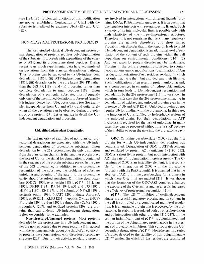
ODC. Ornithine decarboxylase (ODC) was the first
protein for which Ub-independent degradation was
demonstrated. Degradation of ODC is ATP-dependent
and regulated by protein AZ1 (antizyme 1) [190, 212].
ODC is a short living protein, but upon interaction with
AZ1 the rate of its degradation increases greatly. The C-
terminus of ODC is an instability element: it is responsi-
ble for the interaction of ODC with the proteasome
(probably with the Rpt5 subunit). It is assumed that in the
absence of AZ1 ornithine decarboxylase forms dimers in
which these C-termini are masked [213]. It was shown
that the formation of the ODC/AZ1 complex enhances
the exposure of the C-terminus and, as a result, increases
the efficiency of proteasomal recognition [214].
p21Cip1. The p21Cip1 inhibitor of cyclin-dependent
kinase is a crucial regulatory protein, and its content in
the cell is controlled by a complicated multilevel regula-
tion. It is an unstable protein that is processed by the pro-
teasome. Its stability is regulated both by phosphorylation
and by interaction with other proteins [215-217]. In the
cell, an insignificant part of p21Cip1 is ubiquitinated, and
the portion of the ubiquitinated protein grows in the pres-
ence of proteasome inhibitors. This corroborates the Ub-
dependent degradation of p21Cip1. Nonetheless, in a series
of studies devoted to the stability of non-ubiquitinatable
p21Cip1 analog (in which all Lys residues are substituted
1422 SOROKIN et al.
BIOCHEMISTRY (Moscow) Vol. 74 No. 13 2009
for Arg and the N-terminus is blocked with a tag prevent-
ing N-terminal ubiquitination) it has been reported that
such a modified protein remains unstable and undergoes
regulated degradation similar to the wild-type protein
[213]. These data demonstrate that ubiquitination of
p21Cip1 is not a necessary condition for its degradation,
i.e. its proteolysis can be also Ub-independent. PA28γ-
containing proteasomes contribute significantly to the
degradation of p21Cip1. So p21Cip1 is greatly stabilized in
cells depleted of PA28γ by RNA interference or in PA28γ-
knockout embryonic rat fibroblast cells [187, 218].
However, in some cell lines PA28γ is not required at all for
degradation of p21Cip1, and upon elimination of PA28γ by
RNA interference it is impossible to achieve complete
stabilization of p21Cip1 [187, 218]. This shows that other
proteasome isoforms participate in p21Cip1 degradation.
In particular, data of in vivo and in vitro experiments
demonstrate that the C-terminal part of p21Cip1 can inter-
act directly with subunit α7 of the 20S CP [219]. It is also
shown in the cited paper that wild-type p21Cip1, but not a
mutant isoform depleted of the C-terminal part, can be
degraded under the action of the 20S CP in vitro. A criti-
cal role in recognition and degradation of p21Cip1 caused
by PA28γ-containing proteasomes is also given to its C-
terminal part [187, 218]. As concerns the problem of pen-
etration into the proteolytic chamber, as in the case with
damaged proteins p21Cip1, being a non-structured protein
[220], can itself open the gate of the 20S proteasome [38].
It was shown that the degradation of p21Cip1 caused by the
26S proteasome in vitro is ATP-independent [38, 111].
The sole explanation of the ATP-independent degrada-
tion can be the ability of unfolded substrates to penetrate
into the proteolytic chamber of the 20S CP via an open
gate due to passive diffusion [111].
p53. Transcription factor p53 is an important coordi-
nator of the cell response to the DNA-damaging stress in
most if not in all cells. Depending on conditions and cell
type, the response to stress can be interruption of the cell
cycle, apoptosis, or aging [221]. Distortions in the func-
tions of p53 are found in more than 50% of cancer cases.
It is known that in non-transformed cells under normal
conditions p53 is labile and degrades quite rapidly. As a
result of stress, enhanced synthesis and stabilization of
p53 is observed in the cells. For p53, both Ub-independ-
ent and Ub-dependent degradation pathways have been
shown. The Ub-dependent mechanism will be briefly
described in section “Proteasomes and Medicine”.
Herein we will analyze the Ub-independent mechanisms.
At present two mechanisms of Ub-independent degrada-
tion of p53 are well studied.
E6-dependent Ub-independent degradation of p53.
High-risk human papilloma virus (types 16 and 18)
express protein E6 that binds to p53 and causes its degra-
dation [222]. Two binding sites of protein E6 and the p53
molecule are localized: one is in the DNA-binding
domain of p53 (amino acid residues 66-326) and the
other in the C-terminal part (amino acid residues 376-
384). The binding of E6 to the C-terminal region of p53
occurs without mediators, while the binding of E6 to the
DNA-binding domain of p53 is mediated by protein E6-
AP (E6-associated protein). After the formation of the
E6/p53 or E6/E6-AP/p53 complex, protein p53 under-
goes Ub-independent degradation by the proteasome
[223]. However, it is still unclear what proteasome com-
plex (20S or 26S) is involved in the degradation of p53
and in what way E6 presents p53 to the proteasome. It is
known that E6 can cause Ub-dependent degradation of
p53 as well. This mechanism also involves protein E6-AP.
This protein is a cell ligase E3 specific for p53, but under
normal conditions it does not ubiquitinate p53. The par-
ticipation of E6 and E6-AP in the Ub-dependent degra-
dation of p53 is described in more detail in the section
“Proteasomes and Medicine”.
NQO1-inhibited Ub-independent degradation of p53.
Protein NQO1 (NAD(P)H quinone oxidoreductase 1) is
an enzyme regulating the quinone content in the cell. A
considerable part of this protein is complexed with the
20S but not the 26S proteasome. NQO1 is able to inter-
act with p53, and in addition, NQO1, p53, and 20S are
found in the triple complex [195]. It was demonstrated
that the interaction of NQO1 with the 20S proteasome is
independent of NADH and is not destroyed by
dicumarol (a competitive inhibitor of NQO1). In con-
trast, the binding of NQO1 to p53 is intensified in the
presence of NADH and destroyed by an addition of
dicumarol or other inhibitors of NQO1. It was hypothe-
sized that the free protein NQO1 or that located on the
20S proteasome binds to p53 in the presence of NADH,
stabilizes it, and locally raises its concentration near the
20S proteasome. It is assumed that the catalytic activity
of NQO1 is not vital for stabilization of p53 [224]. In the
absence of NADH, p53 dissociates from NQO1 and is
destroyed by the 20S proteasome. However, the mecha-
nism of recognition and processing of p53 by the protea-
some remains unclear. It has been proposed that NQO1
plays the role of a peculiar gate-keeper that is able to
open the gate into the proteolytic chamber of the protea-
some for p53, p73α, ODC, and other nonstructural pro-
teins [225].
Rb. Like p107 and p130, oncosuppressive Rb pro-
teins are members of a multigene family. These proteins
play a crucial role in such cell processes as cell cycle con-
trol, response to DNA-damaging stress, DNA replication
and reparation, and cell differentiation and aging [226].
The content of these proteins in the cell is controlled due
to their proteasomal degradation. Both Ub-dependent
and Ub-independent degradation mechanisms have been
described.
pp71-induced Ub-independent degradation of Rb.
Several proteins encoded by DNA-containing viruses
that induce proteasomal degradation of Rb are known.
They are human papilloma virus protein E7 [227],
PROTEASOME SYSTEM OF PROTEIN DEGRADATION AND PROCESSING 1423
BIOCHEMISTRY (Moscow) Vol. 74 No. 13 2009
Epstein–Barr virus protein EBNA3C [228], hepatitis C
virus protein NS5B [229], and human cytomegalovirus
protein pp71 [230]. It was shown that proteins E7,
EBNA3C, and NS5B induce enhanced ubiquitination of
Rb and as a result its Ub-dependent degradation.
Protein pp71 causes Ub-independent degradation of Rb,
but the molecular mechanism of this is not sufficiently
studied.
Mdm2-induced Ub-independent degradation of Rb.
Oncoprotein Mdm2 is overexpressed in many types of
human cancer, and its C-terminal region is a RING-
domain-containing ligase E3. It was demonstrated that
Mdm2 binds specifically to the hypophosphorylated pro-
tein Rb and induces its proteasomal degradation [231,
232]. There are two contradicting points of view on the
mechanism of the Mdm2-induced degradation of Rb. (1)
Mdm2 enhances ubiquitination of Rb, which suggests the
Ub-dependent mechanism of Rb degradation in the cell
[232]. (2) Mdm2 does not induce ubiquitination of Rb in
the cell, and in such case the proteasomal degradation of
Rb proceeds by the Ub-independent pathway [231]. The
second mechanism is supported by the following data: Rb
is more sensitive to the 20S than to the 26S proteasome;
in experiments on gel filtration of cell extracts, Rb is
coeluted with the 20S and not the 26S proteasome.
Mdm2 and Rb bind to subunit α7 of the proteasome. The
RING-domain of Mdm2 and the C-pocket of Rb are
responsible for the binding to the subunit α7; both are
necessary for the Mdm2-induced degradation of Rb in
vivo. In the absence of the RING-domain, Mdm2 is
unable to promote/stabilize the interaction of Rb with
the proteasome subunit α7. All these data indicate a pos-
sibility of Ub-independent degradation of Rb in the cell.
However, it remains unclear why one group of
researchers observes ubiquitination of Rb while the other
does not.
Processing
As mentioned above, the classical mechanism of
protein degradation by the 26S proteasome includes
stages of polypeptide chain unfolding and translocation
into the proteolytic chamber. It is believed that the sub-
strate penetrates with one of its termini into the proteo-
lytic chamber of the 20S CP via the gate, and the degra-
dation occurs processively from the terminus (exoproteo-
lytically). However, it was found that the p50 subunit of
transcription factor NF-κB is formed from the p105 pre-
cursor cotranslationally by a proteasome-dependent
pathway. Based on the fact that the processed p50 subunit
contained the N-terminal region of the p105 precursor
and the C-terminal region was linked to the ribosome, a
hypothesis was offered on the ability of the proteasome to
perform endoproteolysis, i.e. to disrupt the polypeptide
chain of the substrate significantly far away from its ter-
mini [189]. During this, it was proposed that the growing
polypeptide chain folds like a hairpin, which slides into
the proteolytic chamber of the proteasome. Such an
opportunity was demonstrated in experiments in vitro on
degradation of barnase whose polypeptide chain was
crosslinked by disulfide links. It was shown in the cited
paper that the gate of the 20S CP can widen to 20 Å,
which allows three polypeptide chains to concurrently
pass through it [29]. Later the possibility of endoproteo-
lysis was demonstrated directly in in vitro experiments on
degradation of model substrates of the proteasome. The
substrates were proteins α-synuclein and p21Cip1, to the
C- and/or N-termini of which GFP protein had been
attached using a gene-engineering method. α-Synuclein
and p21Cip1 are unstructured proteins able to be cleaved by
the 20S and 26S PR in an Ub-independent pathway. In
contrast, GFP is a structured molecule resistant to pro-
teasomal degradation. It was shown in the above experi-
ments that both in GFP-α-synuclein and GFP-p21Cip1
(in which the N-terminus of unstructured proteins is pro-
tected from initiation of degradation) and in α-synucle-
in-GFP and p21Cip1-GFP (in which the C-terminus of
unstructured proteins is protected from initiation of
degradation), the 20S and 26S proteasomes can degrade
the unstructured part (α-synuclein or p21Cip1) leaving
GFP intact. This demonstrated that the substrate can be
degraded by the proteasome from either end of the
polypeptide chain. In spite of the protection of both their
termini from initiation of degradation, α-synuclein and
p21Cip1 in the substrates (GFP-α-synuclein-GFP and
GFP-p21Cip1-GFP) had also undergone proteasomal
degradation, whereas GFP remained intact. This points
to the endoproteolytic character of the degradation of
these model substrates and suggests limited degradation
or processing [38]. The publication of that experimental
paper let to the belief that the alternative Ub-independent
proteasomal degradation is not an artifact and initiated
consideration of the 20S proteasome as a valid enzyme
able to regulate the functions of proteins in the cell via
their degradation or processing.
NF-kB p105. NF-κB is a family of dimeric tran-
scription factors. By controlling the expression of a wide
range of genes, they are implicated in regulation of the
immune response, reparation reactions, and apoptosis.
The NF-κB family consists of five members: p50, p52,
p65/RelA, c-rel, and RelB [233]. p50 and p52 are formed
as a result of processing from precursors p105 and p100,
respectively. As mentioned above, the p50 subunit is the
N-terminal part of the p105-precursor. In accord with
experimental data, processing of p105 can be performed
both by the Ub-dependent pathway by the 26S protea-
some [234] and by the ATP-/Ub-independent pathway by
the 20S proteasome [198].
It was first assumed that the formation of p50 from
the p105-precursor occurs cotranslationally [189].
According to this hypothesis, p50 is formed not from a

1424 SOROKIN et al.
BIOCHEMISTRY (Moscow) Vol. 74 No. 13 2009
completely synthesized p105-precursor but cotransla-
tionally. Upon translation of the p105 mRNA, the ribo-
some makes a pause in the region of the triplet corre-
sponding to amino acid residue 530. At this moment, the
proteasome docks at the synthesized polypeptide and per-
forms limited endoproteolysis. As a result, the C-terminal
region is completely degraded and the N-terminal (amino
acid residues 1-433) remains intact. If the ribosome does
not make a pause, a full-size polypeptide p105 is synthe-
sized, which is no longer able to be processed into p50.
This protein functions as an inhibitor of the activity of a
variety of other proteins: it binds to proteins Tpl2, FLIP,
LYL1, and ZUD5 and subunits NF-κB [235-238]. The
action of definite stimuli activates IκB kinase β, which
phosphorylates p105 at residues Ser927 and Ser932, thus
triggering its proteasomal degradation [239].
Later there appeared data showing that in vitro p50
can be formed as a result of processing caused by the
action of the 20S proteasome from a completely synthe-
sized p105 precursor. It was demonstrated in experiments
in vivo that processing occurs independently of transla-
tion and does not require ubiquitination of p105 [198].
Experiments with a number of deletion mutations of p105
demonstrated that (i) the processing begins from endo-
proteolysis in the unstructured region of p105 correspon-
ding to amino acid residues 430-530, and (ii) the element
that prevents complete degradation of the protein is
region GRR (glycine-rich region, amino acid residues
365-430); elimination of this region leads to complete
degradation of p105. It remains unclear why GRR pre-
vents complete degradation.
YB-1. YB-1 is a DNA/RNA-binding nuclear-cyto-
plasmic protein of animal cells that is involved in practi-
cally all DNA- and mRNA-dependent processes. It
enhances the resistance of cells to ionizing radiation and
genotoxic xenobiotics, serves as a marker of both multiple
drug resistance and cancer, and can cause or suppress
oncogenic transformation of cells. Multiple functions of
YB-1 in the cell are dependent on its amount, activity,
and intracellular distribution [240].
In in vitro experiments, it was shown that YB-1 is
cleaved into two fragments by the 20S proteasome in the
ATP-/Ub-independent manner. The cleavage is per-
formed by the caspase-like activity of the 20S CP in the
peptide link after residue Glu219. The same cleavage of
YB-1 by the proteasome is observed in cancer cells after
their treatment by some therapeutic DNA-damaging
agents. However, in cells it is possible to find only a large
N-terminal fragment of YB-1 (most likely the C-terminal
region undergoes further degradation) [37]. A similar
cleavage was also revealed in endothelial cells treated with
thrombin, but the protease responsible for the processing
was not identified [241]. After the proteasome processing,
the N-terminal fragment of YB-1 (amino acid residues 1-
219) is translocated into the cell nucleus [37]. The
translocation into the nucleus of the truncated YB-1 can
be explained by the presence of a nuclear localization sig-
nal (amino acid residues 183-205) in it. The intact pro-
tein contains also another signal, i.e. the cytoplasmic
retention signal (amino acid residues 247-290) that is
localized in the cleaved C-terminal part of the molecule.
This signal predominates over the signal of nuclear local-
ization and masks it, which defines the cytoplasmic local-
ization of the intact protein [242]. It was elucidated that
accumulation of the processed YB-1 in nuclei correlates
with the appearance in the cells of resistance to DNA-
damaging reagents. At the present time, we do not know
whether processing of the YB-1 protein is required only
for regulating its nuclear–cytoplasmic distribution or for
modulating its functional activity as well. Molecular
mechanisms triggering the Ub-independent processing of
YB-1 have not yet been clarified.
Besides the ATP-/Ub-independent processing, YB-
1 can undergo a complete Ub-dependent degradation.
Initially it was found that cells treated with the protea-
some inhibitor accumulate ubiquitinated YB-1 [37], and
later E3 ligases involved in the process were also revealed.
One of them was the SCFFBX33 E3 ligase consisting of pro-
teins Skp1, Cul1, and the F-box of protein FBX33, which
is a substrate-recognizing component of this multisubunit
enzyme [243]. Another E3 ligase specific to YB-1 is the
RING-domain-containing protein RBBP6 (retinoblas-
toma binding protein 6) [244]. In all probability, the
amount of YB-1 in the cell is strictly controlled by the
Ub-dependent degradation, whereas the Ub-independ-
ent processing of YB-1 is triggered in particular condi-
tions and directed to regulate intracellular distribution
and/or modulation of YB-1 activities.
eIF4G and eIF3a. Translation initiation of cell and
viral mRNAs in eukaryotic cells occurs with the help of
special proteins—translation initiation factors. It has
been noted repeatedly that upon isolation of these factors
from rabbit reticulocyte lysates some of them are subject-
ed to proteolytic fragmentation, which remarkably affects
their activity. Most affected by such fragmentation are
subunits eIF4G of factor eIF4F and eIF3a of factor eIF3.
An addition of proteasome inhibitors to the cell lysate
upon fractionation of proteins prevented their fragmenta-
tion, based on which it was concluded that the protea-
some participates in their processing. This conclusion was
confirmed in experiments in vitro. It was shown that the
20S proteasome is able to cleave eIF4G and eIF3a in the
Ub-independent manner [245]. The cleavage of factors
eIF4F and eIF3 by the 20S proteasome has an inhibiting
effect on the translation of mRNAs that are dependent on
these factors. It is known that some cell and viral mRNAs
are “less strict” and can be effectively involved in transla-
tion even in the absence of some initiation factors [246,
247]. Based on these data, the authors of the cited paper
proposed that the proteasomal processing of proteins
eIF4G and eIF3a is one of the mechanisms of translation
regulation in the cell [245].
PROTEASOME SYSTEM OF PROTEIN DEGRADATION AND PROCESSING 1425
BIOCHEMISTRY (Moscow) Vol. 74 No. 13 2009
PROTEASOME REGULATION
The 26S proteasome is responsible for the regulated
proteolysis of many intracellular proteins, it per se always
being controlled. The relative content of the proteasome
and its localization in the cell change dynamically and
adjust in accord with the cell requirements and particular
stress conditions. The proteasome is constantly assem-
bled and disassembled, and its subunits are targets for a
great number of posttranslational modifications [248]. As
follows from this, the proteasome functioning is continu-
ously interacting with a variety of proteins, which stabi-
lize it, regulate its activity, assist in recognizing substrates
and in utilizing Ub, etc. Let us briefly characterize the
basic mechanisms of regulation of the Ub-dependent
degradation.
Heterogeneity of Proteasomes in the Cell
As known, the 26S PR consists of the 20S CP and
regulatory particles which can dock at one or both termi-
ni of the 20S CP. Heterogeneity of regulatory particles
and the number of combinations in which they can attach
to the 20S CP provide a whole set of proteasomes with
different functions. The content of regulatory particles in
the cell and correspondingly the functions of proteasomes
can change greatly (see section “Proteasome Structure”).
The immunoproteasome is another striking example of
regulation of proteasome functions by changing its sub-
unit composition. As shown above, the stimulation of
cells by γ-interferon activates the synthesis of three pro-
teasomal subunits (β1i, β2i, and β5i), which during pro-
teasome assembly are inserted instead of constitutively
synthesized subunits β1, β2, and β5. The peptides gener-
ated by the immunoproteasome are not subjected to fur-
ther degradation by cell peptidases and are used for anti-
gen presentation. Moreover, the content of individual
proteasomal subunits can depend on the gender of the
organism [249] or, as in the case of Rpn10, proteasomal
subunits may be synthesized as tissue-specific isoforms
appearing as a result of alternative splicing of their mRNA
precursors [250].
Synthesis of Proteasomal Subunits
Mechanisms of synthesis regulation of proteasomal
subunits are studied insufficiently, and at present only
several examples of such regulation are known.
Proteasomal subunits are synthesized in stoichiometric
amounts, which confirms the coordinated control of the
expression of proteasomal genes.
In S. cerevisiae the system of regulation at the tran-
scription level has been discovered. It was found that pro-
moters of genes of proteasomal subunits and a number of
other genes (their number exceeding 700) have regulato-
ry element PACE (proteasome activation control ele-
ment) that is recognized by the transcription factor Son1.
In earlier studies, this protein was found in complex with
proteasomal subunits; therefore it was named Rpn4.
Rpn4/Son1 binds to the PACE element in promoters of
proteasomal genes and activates their transcription. This
increases the proteasome content in the cell. Under nor-
mal conditions, Rpn4 is short living. Its half-life is about
2 min, and the degradation proceeds under the action of
newly synthesized proteasomes by the Ub-independent
mechanism [251]. Under stress, when a great number of
proteasomes in the cell is required or when their activity
is suppressed by mutations in subunits or by inhibitors,
the degradation of Rpn4 slows down and the half-life
increases several-fold [194]. The mechanism of regula-
tion of the synthesis of proteasomal subunits by Rpn4 is
corroborated by an elegant experiment in which the syn-
thesis of Rpn4 resistant to proteasomal degradation
resulted in a significant increase in the proteasome con-
tent [252].
It is known that the overall level of expression of pro-
teasomal subunits in higher eukaryotes depends on the
cell proliferation stage. So the synthesis of proteasomes in
proliferating leucosis cells is more active compared to
non-dividing cells at the final stage of differentiation
[253]. Enhanced synthesis of proteasomal subunits was
found in cells treated with proteasomal inhibitors [254].
The mechanisms of proteasomal synthesis regulation in
higher eukaryotes are not studied yet, but it is evident that
they differ from the above-described mechanism in
yeasts, since neither any ortholog of Rpn4 nor PACE ele-
ments in promoters of proteasomal genes have been
found in higher eukaryotic cells.
Localization of Proteasomes
Proteasomes in eukaryotic cells are localized both in
the cytoplasm and in the nucleus. For example, in hepa-
tocytes 17% of the overall number of proteasomes is in the
nucleus, 14% is linked to endoplasmic reticulum, and the
remaining portion is found in the cytoplasmic matrix; the
proteasomal content in the nuclei of lung epithelium cells
is 51% [255]. This indicates that the nuclear–cytoplasmic
distribution of proteasomes can be tissue-specific. The
distribution of proteasomes between the cytoplasm and
the nucleus changes remarkably during embryogenesis. In
spermatozoids and ovules, proteasomes are concentrated
in the cytoplasm, at early stages of subdivision they
translocate to the nucleus, and by the blastocyst stage the
intracellular localization of proteasomes is close to their
distribution in somatic cells [256, 257]. Besides, intracel-
lular distribution of proteasomes changes dynamically in
accord with the cell cycle phase [258]. The cell cycle is
regulated by two basic biochemical mechanisms. This is

1426 SOROKIN et al.
BIOCHEMISTRY (Moscow) Vol. 74 No. 13 2009
protein phosphorylation–dephosphorylation and proteo-
lysis of proteins implicated in the regulation of the cell
cycle (for example, cyclins and cyclin-dependent kinases)
[259]. Proteasomal degradation of cyclins in the nucleus
is a necessary condition for the normal course of the cell
cycle [260].
The mechanisms of regulation of intracellular distri-
bution of proteasomes are not sufficiently studied. Some
proteasomal subunits contain nuclear localization signals
(NLS) [261-263]. Many subunits are phosphorylated
[248]. At least six proteasomal subunits are phosphorylat-
ed on Tyr residues, and this phosphorylation might be
involved in regulation of proteasome redistribution
between the nucleus and the cytoplasm [262, 264, 265].
In some recent papers it is reported that proteasomes can
be localized not only in the cell, but also outside it.
Proteasomes have been found in blood serum of both nor-
mal subjects and patients with different malignant dis-
eases (leukemia, myeloma, carcinoma, etc.) [266-268].
Based on the fact that the proteasome level in the blood
serum of oncology patients was much higher, the authors
concluded that this might originate from their increased
secretion by tumor cells [266]. It was also demonstrated
that the level of proteasome synthesis in leucosis cells is
far greater than in normal blood cells [253]. An increased
proteasome level in the blood serum was also found in
patients with different autoimmune diseases, and at pres-
ent this is used as one of the clinical markers of disease
development (see section “Proteasomes and Medicine”).
The molecular mechanisms of proteasome secretion are
still little studied.
Modification of Proteasomal Subunits
In the cell, many proteins experience various struc-
tural changes as a result of cotranslational and posttrans-
lational modifications. More than 100 different modifica-
tions have been described; however, the role of most of
them is not clear. Some modifications are random and in
all probability have no functional significance, but there
are those that are vital for the cell because they are strict-
ly controlled by specific enzymes. It has been demon-
strated for subunits of 26S PR that they can be subjected
to phosphorylation, N-acetylation, N-terminal process-
ing, cleavage by caspases, N-myristoylation, O-glycosy-
lation, S-glutathionylation, alkylation, and oxidation of
sulfur-containing amino acid residues (see review [248]).
Some of these modifications can modulate proteasome
functions. Let us consider examples of such regulation.
Phosphorylation. Phosphorylation of the 20S PR by
protein kinase A in vitro (in subunits α1, α2, α3, β2, β3,
and β7) enhances the chymotrypsin- and caspase-like
activities [269]. Data are available according to which
phosphorylation of subunit α2 on Tyr120 is required for
nuclear localization of the proteasome [265].
Phosphorylation of subunits α3 and α7 of the 20S CP can
affect the attachment of regulatory particles to the ends of
the 20S CP [270, 271]. In particular, phosphorylation by
casein kinase II (CKII) in subunit α7 stabilizes the com-
plex of 20S CP with 19S RP, whereas dephosphorylation
enhances the binding of 20S CP to PA28α/β [272]. It has
been found recently that about half of all phosphorylated
residues of the proteasome are in consensuses for mito-
gen-activated and cyclin-dependent kinases, i.e. phos-
phorylation of the proteasome and consequently its func-
tions can change greatly depending on the cell cycle
phase [119].
N-Terminal processing. Five of the seven β-subunits
(β1, β2, β5, β6, and β7) of the 20S proteasome have N-
terminal propeptide sequences. They are required for
both the proteasome assembly and the protection of Thr1
residues in potential catalytic centers from acetylation
and inactivation [273]. Premature cleavage of propeptide
sequences from subunits β1, β2, and β5 leads to inactiva-
tion of catalytic centers and decreases the efficiency of
20S CP assembly.
N-Myristoylation. Using mass-spectral analysis, it
was discovered that subunit Rpt2 of the 19S RP undergoes
myristoylation in the N-terminal residue Gly. It is known
that such a modification influences protein–protein
interactions and the interaction of proteins with mem-
branes. It was assumed that myristoylation of subunit
Rpt2 also controls the proteasome interaction with pro-
teins and membranes [119]. But this has not been sup-
ported yet by experimental results.
S-Glutathionylation. It was shown that glutathiony-
lation of subunits of the 20S proteasome from S. cerevi-
siae on Cys residues inactivates the chymotrypsin- and
trypsin-like activities rather than the caspase-like activity
of the proteasome [274].
O-Glycosylation. Many Rpn, Rpt, and α- and β-
subunits of the proteasome are glycosylated. Since glyco-
sylation and phosphorylation is experienced by the same
residues of the polypeptide chain, Ser and Thr residues,
their influence on the functions being frequently antago-
nistic, the switching from one type of modification to
another type can rapidly modulate the proteasome activ-
ity. As known, the ATPase subunit Rpt2 of the 19S RP is
glycosylated in vivo and in vitro. Such a modification
results in inhibiting the ATPase activity and, as a conse-
quence, decreasing the proteolysis efficiency [275].
Cleavage by caspases. Upon apoptosis, some sub-
units of the 20S CP as well as subunits Rpn2, Rpn10, and
Rpt5 in human cells undergo cleavage by caspase 3.
Subunits Rpn2 and Rpn10 play an important role in
attaching the 19S RP base to the lid. Moreover, Rpn10 is
the main subunit implicated in the recognition of poly-
ubiquitinated substrates of the 26S PR. It is logical that
their cleavage leads to suppression of the protein degrada-
tion and accumulation of ubiquitinated proteins, includ-
ing proapoptotic ones [276].

PROTEASOME SYSTEM OF PROTEIN DEGRADATION AND PROCESSING 1427
BIOCHEMISTRY (Moscow) Vol. 74 No. 13 2009
Adaptor Proteins
The polyUb chain can be directly bound to protea-
somal subunits (described in detail in “19S Regulatory
Particle”). Nevertheless, the family of adaptor polyUb-
binding proteins UbL-UbA plays a great role in regula-
tion of the interaction of the polyubiquitinated substrate
with the proteasome. These proteins interact with the
proteasome by their N-terminal domain UbL (Ub-like
domain) and with the ubiquitinated substrate by the C-
terminal domain UbA (Ub-association domain). It was
shown that proteins UbL-UbA can have both diverse and
overlapping substrate specificity [97, 99, 277]. UbL-UbA
proteins include, for example, proteins Rad23, Dsk2, and
Ddi1 [67]. The specificity of recognition of substrates by
these adaptor proteins can provide for a local increase in
the proteasome concentration in regions where it is nec-
essary to process proteins promptly. Thus, e.g. under
DNA-damaging stress, protein Rad23 binds to protein
Rad4 implicated in DNA reparation, and thus attracts the
proteasome to damaged DNA sites involving it in protein
degradation in the chromatin sites required for effective
DNA reparation [278].
Some E3 ligases can play the role of an adaptor
between the proteasome and the substrate. They can
interact with the proteasome directly or via accessory
proteins. In this case, E3 ligase can provide for an effi-
cient substrate-specific degradation in two ways: directly,
by increasing locally the concentration of the specific
substrate, or mediated, by lengthening the polyUb chain
of the specific substrate and thus enhancing its affinity to
the proteasome [98].
For some substrates, the binding with the protea-
some is possible only in the presence of the complex
Cdc48/p97 attached to the proteasome [279]. Cdc48 is an
ATP-dependent chaperone consisting of six identical
subunits. It can interact with E3 ligases and deubiquiti-
nating enzymes and can bind substrates directly or via a
great family of adaptor proteins UbX-UbA. Besides rec-
ognizing specific substrates, the Cdc48 complex partici-
pates in their unfolding [98].
PROTEASOMES AND MEDICINE
Practically all intracellular processes including cell
cycle control, transcription, translation regulation, cell
response to stress, etc. are controlled by the Ub–protea-
some system. An impressive number of key regulatory
proteins in the cell are eliminated or processed by the
proteasome. They include cyclins, inhibitors of cyclin-
dependent kinases, phosphatases, kinases, and transcrip-
tion and translation factors. Numerous examples of the
involvement of proteasomes in the regulation of cell
processes are described in detail in the review of
Konstantinova et al. [248]. The important biological role
of the proteasome system implies that it should be
unavoidably involved in pathophysiological processes
causing the development of malignant, autoimmune, and
neurodegenerative diseases. Let us briefly analyze the
changes in the proteasome system that lead to the devel-
opment of various diseases.
Cancer
At present, several ways are known how the protea-
some system can be involved in the development of
malignant tumors. In general, the development of cancer
can be caused by stabilization of an oncoprotein or desta-
bilization of a tumor suppressor.
p53. The multifunctional protein p53 is one of the
most thoroughly studied tumor suppressors. It is impli-
cated in a wide range of cell processes including apopto-
sis induction, stimulation of DNA reparation, cell cycle
arrest, and regulation of basic metabolism [280]. Today
several mechanisms are known to regulate the activity of
p53 in the cell. Many are somehow related to the degra-
dation of this protein by the 26S or 20S proteasomes.
Several E3 ligases are known for p53: Mdm2, E6-AP/E6,
COP1, Pirh2, ARF-BP1, Topors, and CHIP [281-288].
Mutations in these enzymes, disturbance of their activity,
as well as duplication of their genes or disturbance of reg-
ulation of their expression can lead to malignant degener-
ation of cells as a result of inadequate activity of p53. This
can be readily demonstrated on Mdm2. In some types of
tumors, such disturbances as gene amplification [289,
290], enhanced protein synthesis [291-293], and incor-
rect splicing of pre-mRNA of Mdm2 [294, 295] were
found. Besides, in the promoter of the human Mdm2
gene (HDM2), single nucleotide polymorphism was dis-
covered: either thymine or guanine (SNP309T or
SNP309G) can be located in position 309 of the first
intron. The substitution of T by G leads to a tighter bind-
ing of transcription factor Sp1 to the intron DNA, as a
result of which the level of transcription from the Mdm2
gene increases [296]. According to the data of some
researchers, this polymorphism does not at all affect the
probability of cancer development [297-299]. But the
results obtained by other researchers show that the homo-
or heterozygote state of SNP309G correlates with higher
risk of malignant tumor development [300, 301], poor
prognosis for survival outcome [302], and earlier begin-
ning of the disease [296, 303, 304]. It should be noted that
Mdm2 can also promote the formation of tumors by a
mechanism independent of p53 [305].
When analyzing genes the expression of which is
enhanced in hepatocellular carcinoma, the protein
gankyrin was found, its content being higher in all tumor
samples analyzed. In an independent study, this protein
was identified as component p28/PSMD10 of the 19S
proteasomal regulatory particle and as a protein specifi-

1428 SOROKIN et al.
BIOCHEMISTRY (Moscow) Vol. 74 No. 13 2009
cally interacting with ATPase S6b/Tbp7/PSMC4 [306].
It was demonstrated that gankyrin can associate with pro-
tein Mdm2 and promote its interaction with p53.
Gankyrin enhances the ability of Mdm2 to mono- and
polyubiquitinate p53; as a consequence, the increase in
the content of this protein is accompanied by enhanced
Mdm2-dependent degradation of p53 [307]. Further-
more, it was proposed that gankyrin can deliver polyubi-
quitinated p53 in the complex with Mdm2 directly to the
proteasome due to the binding with S6b [308].
In addition to Mdm2, some tumors are characterized
also by a higher content of other E3 ligases specific to
p53. So, a higher amount of ARF-BP1 E3 ligase is con-
nected with the development of colorectal cancer [309],
the expression of COP1 E3 ligase is enhanced in many
ovary and breast carcinomas [284], and Pirh2 is enhanced
in lung tumors [310].
Enhanced degradation of p53 plays a vital role in the
development of cervix cancer caused by human high-
oncogenic-risk papilloma viruses (types 16 and 18). It was
found that the content of p53 in such tumors is signifi-
cantly lower, but at the same time in contrast to many
nonviral cervix carcinomas, they contain for the most
part a non-mutation gene of p53 [311]. Detailed investi-
gations demonstrated that protein E6 of the papilloma
virus can form a complex with cell proteins p53 and E6-
AP (E6-associated protein) [312-314]. E6-AP is an E3
ligase that as a rule does not interact with p53 [315]. E6
acts as a mediator that combines E6-AP and p53 into a
single complex; within this complex E6-AP can ubiquiti-
nate p53 [316]. One can say that in this case the E6-
AP/E6 complex plays the role of the E3 ligase.
p27Kip1. Protein p27Kip1 is an inhibitor of the activity
of Cdk2/CyclinA and Cdk2/CyclinE complexes and thus
prevents the passage of cells to S phase of the cell cycle
[317]. In response to the mitogenic stimuli, p27 is ubiqui-
tinated and subjected to proteasomal degradation. E3 lig-
ase specific to p27 is an SCFSkp2 complex consisting of
proteins Skp1, Cul1, and Skp2, the latter being a sub-
strate-recognizing component of the complex [318, 319].
The recognition and effective ubiquitination of p27 with
the SCFSkp2 complex also require the small accessory pro-
tein Cks1 [320, 321].
Being a negative regulator of cell division, p27 has
properties of a tumor suppressor. A decreased content of
p27 is a specific feature of cancer tumors of different ori-
gin [322-324]. As antagonists of p27, Skp2, and Cks1 are
oncoproteins, their enhanced synthesis correlates with a
low level of p27 and poor prognosis for the patient [322,
324-329]. Skp2 recognizes a number of tumor suppressors
in addition to p27, namely inhibitors of cyclin-dependent
kinases p21Cip1, p57, and p130 [330-332] and transcription
factor FOXO1 [333]. Therefore, an oncogenic function of
SKP2 independent of p27 can be postulated.
pVHL. Another example of how a mutation of one
component of the ubiquitinating complex can be con-
nected with the development of malignant tumors is von
Hippel–Lindau disease. This is a hereditary disease
caused by autosomal dominant mutation in one of the
alleles of gene VHL that encodes a subunit of the multi-
meric E3 ligase. The disease occurs as frequently as one
case per 35,000 subjects [334]. Such patients have a high-
er predisposition to the development of some types of
tumors such as pheochromocytomas, hemangioblastomas
of the central nervous system, clear-cell renal carcino-
mas, and retinal capillary angiomas. The development of
these tumors per se begins as a somatic inactivation of the
wild-type allele or after its mutation. Characteristic fea-
tures of tumors developing in von Hippel–Lindau disease
are a high degree of their vascularization and hyperex-
pression of proteins that are usually synthesized only at
hypoxia, such as VEGF (vascular endothelial growth fac-
tor) [335, 336].
Protein pVHL, a product of gene VHL, is a sub-
strate-recognizing component of a multimeric E3 ligase
[337-341]. Besides pVHL, this multisubunit complex
contains also proteins cullin 2A, Rbx/Hrt1, elongin B,
and elongin C [342-344]. The most studied and best-
known substrate of this enzyme is HIF1α that is a subunit
of heterodimeric transcription factor HIF-1.
The heterodimeric transcription factor HIF-1 plays a
key role in the cell response to hypoxia. It consists of two
subunits: one of the three versions of HIFα and HIFβ1
[345]. A sufficient amount of HIFβ1 is constantly present
in the cells, whereas HIFα is rapidly degraded in the pres-
ence of oxygen. Under hypoxia, HIFα is stabilized and,
together with HIFβ1, forms a functional transcription fac-
tor HIF-1, which by binding with the consensus sequence
HRE (hypoxia-responsive element) activates transcription
of some genes involved in angiogenesis, cell cycle control,
glucose metabolism, and apoptosis [345]. The mechanism
of such oxygen-dependent regulation of the HIF-1 level in
the cell has been disclosed lately. It was shown that in the
presence of oxygen, HIF1α is rapidly ubiquitinated and
cleaved by the proteasome [346]. This degradation was
dependent on the ODD (oxygen-dependent degradation)
domain that is recognized by pVHL. To facilitate the
recognition of HIF1α by protein pVHL, it is important
that residues Pro402 and especially Pro564 in HIF1α were
hydroxylated. Such hydroxylation is performed by special
prolylhydroxylases in the presence of oxygen [347-351].
Under conditions of hypoxia this reaction is virtually
absent; therefore, the level of HIF1α rises. The same
occurs on mutations of the VHL gene. The high extent of
tumor vascularization in von Hippel–Lindau disease may
be explained by active HIF-1-dependent transcription.
However, it is known that far from all cells with increased
level of HIF-1 undergo cancer transformation. This gives
grounds to believe that protein pVHL has additional func-
tions not related to HIF-1 [334].
BRCA1/BARD1. Protein BRCA1 (breast cancer
type 1 susceptibility protein) is a recognized suppressor of

PROTEASOME SYSTEM OF PROTEIN DEGRADATION AND PROCESSING 1429
BIOCHEMISTRY (Moscow) Vol. 74 No. 13 2009
breast and ovary tumors. Gene BRCA1 is frequently inac-
tive as a result of mutations in the case of both inheritable
and sporadic cancers [352, 353]. In addition, the BRCA1
dysfunction correlates with the basal phenotype of tumor
cells and poor prognosis for survival [354, 355].
BRCA1 contains a RING domain in the N-terminal
part of the molecule and forms a complex with the struc-
turally related protein BARD1 (BRCA1-associated
RING-domain protein 1), which also has a RING-
domain [356]. BARD1 mutations occur also, though less
frequently than BRCA1 mutations, in breast and ovary
cancers [357]. The BRCA1/BARD1 complex has E3 lig-
ase activity and is capable of autoubiquitination that
enhances its stability and activity [358-360]. There are
some indirect data showing that the functions of BRCA1
and BARD1 as tumor suppressors is realized via the activ-
ity of their heterodimeric complex and that during this of
importance is the integrity of domains involved in ubiqui-
tination [358, 361, 362]. Recently this has been directly
demonstrated on murine models, but it has remained
unclear whether the E3 ligase activity of the
BRCA1/BARD1 complex is essential for suppression
[363]. At present, several BRCA1/BARD1 substrates are
known including histones, γ-tubulin, phosphorylated
RNA-polymerase II, estrogen receptor, and others [360,
364-368]. Nonetheless, it is not clear yet the interaction
with what substrates mediate the antitumor effect of
BRCA1/BARD1.
Liddle Syndrome
This inheritable autosomal dominant disorder was
first described by Liddle in the 1960s. Its symptoms are
severe hypertension at an early age, hypokalemia, and
metabolic alkalosis caused by excess reabsorption of sodi-
um ions in kidneys with low secretion of aldosterone and
rennin [369, 370]. It was found that this disease is caused
by dysregulation of the epithelial Na+-channel (ENaC),
which consists of three homologous subunits α-, β-, and
γ-ENaC [371]. The activity of ENaC is mostly regulated
upon its inclusion into the cytoplasmic membrane. It was
demonstrated that nonsense and missense mutations in
C-terminal parts of β- and γ-ENaC involving the conser-
vative proline-rich motif cause an increase of the ENaC
amount in the membrane and development of Liddle syn-
drome [372-378]. It became clear that PY-motifs in the
C-terminal parts of ENaC subunits are binding sites of E3
ligases from the NEDD4 family that regulate negatively
the activity of ENaC [379-383]. Though ENaC can be
inhibited by several members of this family, the most sig-
nificant regulator in vivo is NEDD4-2 [384]. It is assumed
that ligase NEDD4-2 binds to ENaC on the cytoplasmic
membrane and ubiquitinates it. This results in the release
of ENaC subunits from the membrane and their subse-
quent degradation [385, 386]. The membrane always has
two pools of ENaC: intact channels and channels α- and
γ-subunits of which have undergone limited proteolysis in
the region of the extracellular domain. Cleaved channels
are more active, and the open-state probability in them is
higher. It is noteworthy that NEDD4-2 acts in such a way
that first the more active ENaC are removed from the
membrane [387].
Neurodegenerative Disorders
Neurodegenerative disorders include diseases the
main feature of which is the loss of functions and extinc-
tion of brain and spinal cord cells. A specific feature of
these diseases is the formation of large intracellular aggre-
gates consisting of irregularly folded protein molecules.
In many cases, aggregates are found to contain Ub and
proteasomes [388-391]. The role of the Ub–proteasome
system in pathogenesis of neurodegenerative diseases has
not been clarified yet. It is not clear whether inclusion
bodies are toxic for the cell. Initially it was proposed that
the reason for the formation of inclusion bodies in neu-
rodegenerative disorders is impairment of the Ub–pro-
teasome system functioning. Indeed, several teams of
researchers demonstrated that proteasomal inhibitors
cause the formation of inclusion bodies and apoptotic
death of neurons in model systems [392-394]. Moreover,
it appeared that the enzymatic activity of proteasomes is
reduced in neurons afflicted with Parkinson’s disease
[395]. On the other hand, it is known that aggregated pro-
teins per se can impair proteasome functioning [396-398].
It was established just to a certain extent of accuracy that
there is a correlation between the disease development
and dysfunction of the Ub–proteasome system for only
some types of inheritable Parkinson’s and Alzheimer’s
diseases. Let us consider one such case.
At the present time we know six mutations of genes
that can cause inheritable Parkinson’s disease. They are
α-synuclein, parkin, UCH-L1, DJ-1, PINK 1, and
LRRK2 [399-403]. The protein parkin is an E3 ligase
containing two RING-domains and an Ubl domain, due
to which it can bind to the Rpn10 subunit of the protea-
some [404-407]. Mutations reducing the ligase activity of
parkin lead to the disease development. Currently several
parkin substrates are known, including O-glycosylated α-
synuclein, CDCrel-1, and Pael receptor [404, 406, 408].
Though the available data suggest that parkin plays a quite
important role in the pathogenesis of inheritable
Parkinson’s disease, it is not clear whether the accumula-
tion of any of its substrates is toxic for neurons.
Viral Diseases
Viruses have elaborated various strategic pathways to
avoid recognition by the host immune system and multi-

1430 SOROKIN et al.
BIOCHEMISTRY (Moscow) Vol. 74 No. 13 2009
ply effectively. Some of these pathways are somehow con-
nected with the Ub–proteasome system.
HCMV (human cytomegalovirus). HCMV and its
related viruses prevent recognition of infected cells by
cytotoxic lymphocytes. These viruses induce reverse
transport of glycosylated heavy chains of MHC-I proteins
(major histocompatibility complex class I) from EPR
back to the cytosol, where they are deglycosylated, poly-
ubiquitinated, and undergo proteasomal degradation.
Responsible for these processes are transmembrane viral
glycoproteins US-2 and US-11 differing from each other
by the mechanism of transportation of heavy chains of
MHC-I [409-413]. To provide efficient US-11- and US-
2-dependent transportation of heavy chains of MHC-I,
the presence of the functional system of ubiquitination is
required; however, it is known that in the case of US-11-
dependent transportation heavy chains are not acceptors
of ubiquitin. The US-11-dependent degradation of heavy
chains of MHC-I requires proteins which when normal
are activated if EPR is overloaded with irregularly folded
proteins. US-11 triggers a signal pathway that leads to the
synthesis of these proteins [414, 415].
Furthermore, HCMV inhibits the process of antigen
presentation in one more way. It is known that peptide
ligands for MHC-I molecules are produced by immuno-
proteasomes. Specific subunits of immunoproteasomes
are synthesized in response to cell stimulation by γ-inter-
feron. Protein M27 of cytomegalovirus inhibits STAT2,
which is a component of the signal pathway beginning
from the interferon receptor and thus prevents the forma-
tion of immunoproteasomes [416].
Herpes viruses. Murine γ-herpes virus 68 and
Kaposi’s sarcoma-associated with herpes virus (herpes
virus 8) also impair the antigen presentation in infected
cells, but at a later stage than cytomegalovirus. Protein
MK3 of γ-herpes virus 68 and proteins K3 and K5 of her-
pes virus 8 are E3 ligases that ubiquitinate cytoplasmic
regions of MHC-I molecules and other molecules vital
for immune recognition, such as ICAM-1 and B7-2 [417-
420]. Ubiquitination of these molecules results in their
rapid release from the membrane by means of clathrin-
dependent endocytosis and cleavage in lysosomes.
EBV (Epstein–Barr virus). The Epstein–Barr virus
protein EBNA1 (Epstein–Barr virus (EBV)-encoded
nuclear antigen 1) is the only one of its nine proteins that
is always found in all EBV-associated tumors. Cytotoxic
lymphocytes do not recognize cells in which EBNA1 is
synthesized. This is connected with the presence of elon-
gated Gly-Ala-repeats in the N-terminal part of EBNA1.
These prevent the cleavage of the protein by the immuno-
proteasome [421, 422]. As a result, EBNA1 fragments are
not present on the membrane of the infected cell in com-
plex with MHC-I. It is assumed that Gly-Ala-repeats
impede the polypeptide chain translocation into the cat-
alytic cavity of the proteasome, therefore the proteolysis
of proteins with such repeats is incomplete [423, 424].
Paramyxoviruses. The JAK-STAT-signaling pathway
(Janus kinase–signal transducers and activators of tran-
scription) is the central one in protecting the host organ-
ism from viral infections; that is why the action of some
viruses is aimed at suppression of its activity. So, the
mumps virus protein V (as well as proteins V of its related
human parainfluenza virus type 2 and virus SV5) initiates
the assembly of a complex E3 ligase consisting of
protein V and a number of cell proteins. Such a complex
enzyme ubiquitinates STAT1 and STAT3 in the case of
the mumps virus, STAT2 in the case of the parainfluenza
virus, and STAT1 in the case of SV5 [425-427]. After
ubiquitination, these proteins are cleaved by the protea-
some.
Hepatitis C virus. There are examples of how viral
proteins can directly associate with the components of the
proteasome and change its proteolytic activity. Thus, pro-
tein NS3 of hepatitis C virus interacts with the LMT7
subunit of the immunoproteasome and reduces its
trypsin-like and caspase-like activities [428].
Inflammatory and Autoimmune Diseases
The Ub–proteasome system plays an essential role in
the mechanisms of immune protection of the organism.
First, it takes part in the antigen processing in antigen-
presenting cells. Second, it regulates the transmission of
signals from T-cell antigen receptors and the costimula-
tory CD28 molecule. And third, it is involved in activa-
tion of NF-κB, which is the key regulator of the activity
of genes of many inflammatory cytokines, adhesion mol-
ecules, and immune system receptors. Let us consider the
latter function of the Ub–proteasome system in more
detail.
The Ub–proteasome system activates hetero- and
homodimeric transcription factors of the NF-κB family
in two stages. At first, the proteasome performs Ub-
dependent processing of phosphorylated precursors p105
and p100 with the formation of active subunits of tran-
scription factors p50 (NF-κB1) and p52 (NF-κB2).
Active factors are retained by inhibitors of NF-κB (IκBs)
in the cytoplasm. After the signal-induced phosphoryla-
tion and ubiquitination, IκBs are cleaved by the protea-
some and the released factors are translocated to the
nucleus, where they activate the transcription of corre-
sponding genes [429]. It is believed that the pathologic
activation of NF-κB is a cause of many inflammatory dis-
eases.
The presence in blood of increased numbers of pro-
teasomes accompanies some autoimmune and oncologi-
cal diseases [268, 430, 431]. Furthermore, the presence of
antibodies to proteasome components and regulatory
particle PA28 is detected in blood of patients with sys-
temic lupus erythematosus, myositis, disseminated scle-
rosis, and Sjogren syndrome [432-435]. The functional

PROTEASOME SYSTEM OF PROTEIN DEGRADATION AND PROCESSING 1431
BIOCHEMISTRY (Moscow) Vol. 74 No. 13 2009
significance of these autoantibodies has not been estab-
lished yet, though it was demonstrated that they can block
activation of the proteasome by regulatory particle PA28.
This gives grounds to suggest that antibodies against the
proteasome are able to somehow regulate its activity
[436].
As known, pathological activation of T helpers 2
(Th2) leads to the development of asthma and allergy
symptoms. Activation of Th2 causes the JunB-dependent
transcription of genes of some interleukins (IL-4, IL-5,
IL-9, IL-10, IL-13) [437, 438]. The E3 ligase Itch plays a
vital role in the development of immunologic tolerance of
T cells. After phosphorylation by kinase JNK1, Itch is
activated and ubiquitinates JunB, which is then cleaved
by the proteasome [439]. It is known that mice lacking
gene Itch suffer various types of allergy [440].
Rheumatoid arthritis is an autoimmune disease
characterized by chronic inflammation of the synovial
tissue and bone and cartilage damage. It was demonstrat-
ed that activation of NF-κB is implicated in pathogenesis
of rheumatoid arthritis [441, 442].
Proteasomal inhibitors are considered as potential
remedies for autoimmune and inflammatory diseases,
acting chiefly by inhibiting NF-κB. The problem of using
inhibitors of the Ub–proteasome system in medicine is
discussed in more detail below.
Inhibitors of the Ubiquitin–Proteasome System
as Potential Remedies
As mentioned above, the disturbance of functioning
of any components of the Ub–proteasome system can
cause different disorders. Therefore, the search for specif-
ic inhibitors of this system seems to be an attractive direc-
tion. However, it is necessary to take into account that the
Ub–proteasome system is constantly involved in events
vital for the cell and that, as a result of its inhibition, the
normal sequence of a vast number of processes would be
violated.
Less specific inhibitors of the Ub–proteasome sys-
tem are inhibitors of the enzymatic activity of the 20S
proteasome and E1. The target of most of the used
inhibitors of the 20S proteasome is suppression of its chy-
motrypsin-like activity. This is connected, first, with the
fact that the blocking of just this activity leads to the
greatest reduction in the level of protein cleavage. And
second, inhibitors of such activity are usually quite
hydrophobic molecules and can therefore easily get into
the cell, in contrast to inhibitors of other catalytic centers
of the proteasome that contain charged regions [443]. In
accord with the chemical structure, the major part of
inhibitors is short peptides carrying chromophores that
interact with catalytic residues in the active center.
Peptide boronates are highly specific inhibitors of the 20S
proteasome [443].
At a rather prolonged action, inhibitors of the 20S
proteasome are toxic for cells and cause their death as a
result of apoptosis, the proliferating cells being typically
more sensitive to these substances [444-447]. Taking into
consideration this circumstance and the antiangiogenic
effect of inhibitors, it can be assumed that these sub-
stances should be effective in coping with oncologic dis-
eases [445, 448]. Bortezomib, which is one of the protea-
somal inhibitors (PS-341, pyrazinylcarbonyl-Phe-Leu-
boronate, Velcade), is now efficiently used to this end.
Bortezomib is a strong inhibitor of chymotrypsin-
like and partly of trypsin-like activity of the proteasome.
When the antitumor activity of bortezomib in vitro and in
vivo had been described, its clinical trials began [449,
450]. It was clarified that bortezomib is largely ineffective
as a monotherapy in treating solid tumors, but it gives
incredibly good results in multiple myeloma and other
hematologic disorders [451-456].
At present, a search is in progress for novel proteaso-
mal inhibitors that would have higher efficiency than
bortezomib and its derivatives. For example, the highly
specific inhibitor salinosporamide A (NPI-0052) was
recently isolated from marine bacteria. In contrast to
bortezomib, it can irreversibly bind to all catalytic centers
of the proteasome [457]. It was demonstrated in in vitro
studies that NPI-0052 is a better inhibitor of the protea-
some and NF-κB than bortezomib when used at equal
concentrations, and also a stronger stimulator of apopto-
sis of isolated lymphocytes from patients with chronic
lymphocytic leukemia [458, 459]. Moreover, NPI-0052
induces apoptosis of multiple myeloma cells resistant to
bortezomib and other drugs [460]. Clinical trials of this
substance have begun.
A more intricate approach than the use of inhibitors
of the 20S proteasome is the search and application of
inhibitors of E3 ligases and specific interactions of the
substrate and E3. At this point, several approaches can be
separated. First, peptides corresponding to the substrate
region to which E3 ligase is bound can be used as
inhibitors of E3. So, it was shown that the phosphopep-
tide appropriate to the N-terminal region of IκB protects
the entire protein from proteasomal degradation, and a
microinjection of this phosphopeptide into cells prevents
activation of NF-κB in them [461].
Second, it is possible to search for small molecules
that will specifically inhibit active centers of E3 or sites of
their binding to the substrate. The choice and efficient
modification of such molecules are much simpler if the
structures of substrate–E3 complexes are known. Let us
analyze the results of using such an approach on the
example of searching for inhibitors of the p53–Mdm2
interaction. As a consequence of screening of a large
library of small molecules, it became possible to identify
family HLI-98 whose members inhibited autoubiquitina-
tion of Mdm2 in vitro. Although these molecules inhibit-
ed other E3 ligases as well, and at high concentrations

1432 SOROKIN et al.
BIOCHEMISTRY (Moscow) Vol. 74 No. 13 2009
even E2, they were able to cause apoptosis of transformed
cells almost without any toxic effect on normal cells
[462]. This study demonstrated clearly that inhibition of
E3, in particular Mdm2, is a quite promising advance in
cancer treatment.
At the moment, several more specific low molecular
weight inhibitors of the p53–Mdm2 interaction are
known. First nutlins were found. They are derivatives of
cis-imidazoline, which are able to displace p53 from the
complex with Mdm2 [463]. Nutlins occupy the
hydrophobic pocket of Mdm2, where side chains of three
amino acids of p53 are usually located, and prevent the
latter from binding to Mdm2. Treatment with nutlins
leads to the accumulation of p53 and products of genes
activated by it (e.g. p21 and p27) in cells. Nutlin-3 caus-
es apoptosis of cancer cells from the wild-type p53, and in
normal cells it induces growth deceleration maintaining
their viability [463]. However, later it was found that in
addition to p53–Mdm2, nutlins inhibit other protein–
protein interactions as well. So, protein HIF1α binds to
the same region of Mdm2 as p53, and thus nutlin-3
impairs this interaction too [464]. Nutlins can also have a
therapeutic effect on cancer cells with mutant p53: in
such cells they cause enhanced sensitivity to many reme-
dies [465]. At present properties of some other inhibitors
of the p53–Mdm2 interaction (RITA, MI-63) are inves-
tigated, and their effects prove to be similar to that of nut-
lin [466-468].
Let us give some more examples of action of low
molecular weight inhibitors of E3-ligases. Peptide
aptamers binding human papilloma virus protein E6
cause apoptotic elimination of HPV16-positive cancer
cells without any effect on normal cells [469]. CpdA
(Compound A), a quite recently identified inhibitor of
complex ligase SCFSkp2, prevents incorporation of Skp2
into the composition of the enzyme. CpdA causes cell
cycle arrest and SCFSkp2- and p27-dependent cell death
affecting chiefly cancer cells [470].
On the whole, the therapeutic use of reagents modu-
lating the activity of the Ub–proteasome system seems to
be rather promising. Currently they are used first of all for
cancer treatment, but with account of the fact that distor-
tions in the well coordinated functioning of the Ub–pro-
teasome system occur in other diseases as well, it can be
suggested that in the near future the range of use of
inhibitors of this system in medicine will be widened.
CONCLUSION
The 20S proteasome was initially detected in 1968 on
electron micrographs of human erythrocyte lysate.
Because of the cylindrical shape, this structure was
named “cylindrin”, but at that time its functions were
unclear [471]. In the end of the 1970s and the beginning
of the 1980s, there appeared reports describing high
molecular weight proteases of a cylindrical shape with
molecular mass of 600-700 kDa. These proteases consist-
ed of several subunits with molecular mass from 24 to
28 kDa and hydrolyzed substrates following hydrophobic
and positively and negatively charged amino acids, i.e.
were multispecific [472-475]. These two properties served
to name the discovered enzyme “multicatalytic pro-
teinase”. At approximately the same time, but in another
pathway of investigations, subunits were found that were
called “prosomes” or the 19S RNP (19S ribonucleopro-
tein) [476]. In their size and subunit composition they
greatly resembled the multicatalytic proteinase, but it was
assumed that they were associated with mRNA and were
involved in the regulation of translation [477]. However,
later everything fell into place; it was shown that the “pro-
some” and “multicatalytic proteinase” are identical sub-
units [478]. At the same time, a new name for these par-
ticles appeared (it reflected their proteolytic essence and
compact structure)—the 20S proteasome. A quarter of a
century after the discovery of the proteasome it became
clear that the proteasome is the basis of a complex multi-
component cellular machine for utilizing exhausted pro-
teins in the cell, which was called the “ubiquitin–protea-
some system”. During that period remarkable progress in
studying the structure and functioning of the proteasome,
in particular of the Ub–proteasome system as a whole,
was achieved. The importance of the disclosure of the
Ub–proteasome system was corroborated by the fact that
in 2004 its authors were awarded the Nobel Prize in
Chemistry. However, even now there are many questions
on the functioning of the system.
Herein we have systemized the currently available
data on the Ub–proteasome system. We have described in
detail the proteasome structure, ubiquitination system,
classic ATP/Ub-dependent mechanism of protein degra-
dation, and have focused attention on the existence of
alternative mechanisms of proteasome-mediated degra-
dation and protein processing. Separately are given data
on disturbances of the proteasome system that cause the
development of various disorders. We hope that this
review will be of interest for a wide range of researchers.
This work was supported by the Russian Foundation
for Basic Research (No. 07-04-00403) and by the Presid-
ium of the Russian Academy of Sciences (Programs on
Molecular and Cell Biology and Basic Science to
Medicine).
REFERENCES
1. Schoenheimer, R. (1942) The Dynamic State of Body
Constituents, Harvard University Press, Cambridge, MA.
2. Rock, K. L., Gramm, C., Rothstein, L., Clark, K., Stein,
R., Dick, L., Hwang, D., and Goldberg, A. L. (1994) Cell,
78, 761-771.

PROTEASOME SYSTEM OF PROTEIN DEGRADATION AND PROCESSING 1433
BIOCHEMISTRY (Moscow) Vol. 74 No. 13 2009
3. Craiu, A., Gaczynska, M., Akopian, T., Gramm, C. F.,
Fenteany, G., Goldberg, A. L., and Rock, K. L. (1997) J.
Biol. Chem., 272, 13437-13445.
4. Kisselev, A. F., Akopian, T. N., and Goldberg, A. L. (1998)
J. Biol. Chem., 273, 1982-1989.
5. Kisselev, A. F., Akopian, T. N., Woo, K. M., and Goldberg,
A. L. (1999) J. Biol. Chem., 274, 3363-3371.
6. Glickman, M. H., and Ciechanover, A. (2002) Physiol.
Rev., 82, 373-428.
7. Groll, M., and Huber, R. (2003) Int. J. Biochem. Cell Biol.,
35, 606-616.
8. Dahlmann, B. (2005) Essays Biochem., 41, 31-48.
9. Frentzel, S., Pesold-Hurt, B., Seelig, A., and Kloetzel, P.
M. (1994) J. Mol. Biol., 236, 975-981.
10. Nandi, D., Woodward, E., Ginsburg, D. B., and Monaco,
J. J. (1997) EMBO J., 16, 5363-5375.
11. Griffin, T. A., Nandi, D., Cruz, M., Fehling, H. J., Kaer, L.
V., Monaco, J. J., and Colbert, R. A. (1998) J. Exp. Med.,
187, 97-104.
12. Kloetzel, P. M., Soza, A., and Stohwasser, R. (1999) Biol.
Chem., 380, 293-297.
13. Kloetzel, P. M. (2001) Nat. Rev. Mol. Cell. Biol., 2, 179-187.
14. Yewdell, J. W., Schubert, U., and Bennink, J. R. (2001) J.
Cell Sci., 114, 845-851.
15. Coux, O., Tanaka, K., and Goldberg, A. L. (1996) Annu.
Rev. Biochem., 65, 801-847.
16. Baumeister, W., Walz, J., Zuhl, F., and Seemuller, E. (1998)
Cell, 92, 367-380.
17. Kisselev, A. F., Songyang, Z., and Goldberg, A. L. (2000) J.
Biol. Chem., 275, 14831-14837.
18. Heinemeyer, W., Fischer, M., Krimmer, T., Stachon, U.,
and Wolf, D. H. (1997) J. Biol. Chem., 272, 25200-25209.
19. Arendt, C. S., and Hochstrasser, M. (1997) Proc. Natl.
Acad. Sci. USA, 94, 7156-7161.
20. Groll, M., Bajorek, M., Kohler, A., Moroder, L., Rubin,
D. M., Huber, R., Glickman, M. H., and Finley, D. (2000)
Nat. Struct. Biol., 7, 1062-1067.
21. Kohler, A., Bajorek, M., Groll, M., Moroder, L., Rubin,
D. M., Huber, R., Glickman, M. H., and Finley, D. (2001)
Biochimie, 83, 325-332.
22. Orlowski, M., Cardozo, C., and Michaud, C. (1993)
Biochemistry, 32, 1563-1572.
23. Dick, T. P., Nussbaum, A. K., Deeg, M., Heinemeyer, W.,
Groll, M., Schirle, M., Keilholz, W., Stevanovic, S., Wolf,
D. H., Huber, R., Rammensee, H. G., and Schild, H.
(1998) J. Biol. Chem., 273, 25637-25646.
24. Unno, M., Mizushima, T., Morimoto, Y., Tomisugi, Y.,
Tanaka, K., Yasuoka, N., and Tsukihara, T. (2002)
Structure, 10, 609-618.
25. Lowe, J., Stock, D., Jap, B., Zwickl, P., Baumeister, W.,
and Huber, R. (1995) Science, 268, 533-539.
26. Zwickl, P., Ng, D., Woo, K. M., Klenk, H. P., and
Goldberg, A. L. (1999) J. Biol. Chem., 274, 26008-26014.
27. Navon, A., and Goldberg, A. L. (2001) Mol. Cell, 8, 1339-1349.
28. Forster, A., and Hill, C. P. (2003) Trends Cell Biol., 13, 550-
553.
29. Lee, C., Prakash, S., and Matouschek, A. (2002) J. Biol.
Chem., 277, 34760-34765.
30. Groll, M., Ditzel, L., Lowe, J., Stock, D., Bochtler, M.,
Bartunik, H. D., and Huber, R. (1997) Nature, 386, 463-471.
31. Benaroudj, N., Zwickl, P., Seemuller, E., Baumeister, W.,
and Goldberg, A. L. (2003) Mol. Cell, 11, 69-78.
32. Smith, D. M., Kafri, G., Cheng, Y., Ng, D., Walz, T., and
Goldberg, A. L. (2005) Mol. Cell, 20, 687-698.
33. Smith, D. M., Chang, S. C., Park, S., Finley, D., Cheng,
Y., and Goldberg, A. L. (2007) Mol. Cell, 27, 731-744.
34. Glickman, M. H., Rubin, D. M., Coux, O., Wefes, I.,
Pfeifer, G., Cjeka, Z., Baumeister, W., Fried, V. A., and
Finley, D. (1998) Cell, 94, 615-623.
35. DeMartino, G. N., and Slaughter, C. A. (1999) J. Biol.
Chem., 274, 22123-22126.
36. Rechsteiner, M., Realini, C., and Ustrell, V. (2000)
Biochem. J., 345, Pt. 1, 1-15.
37. Sorokin, A. V., Selyutina, A. A., Skabkin, M. A., Guryanov,
S. G., Nazimov, I. V., Richard, C., Th’ng, J., Yau, J.,
Sorensen, P. H., Ovchinnikov, L. P., and Evdokimova, V.
(2005) EMBO J., 24, 3602-3612.
38. Liu, C. W., Corboy, M. J., DeMartino, G. N., and Thomas,
P. J. (2003) Science, 299, 408-411.
39. Tanaka, K., Yoshimura, T., and Ichihara, A. (1989) J.
Biochem., 106, 495-500.
40. Bajorek, M., Finley, D., and Glickman, M. H. (2003) Curr.
Biol., 13, 1140-1144.
41. Zwickl, P., Kleinz, J., and Baumeister, W. (1994) Nat.
Struct. Biol., 1, 765-770.
42. Seemuller, E., Lupas, A., and Baumeister, W. (1996)
Nature, 382, 468-471.
43. Grziwa, A., Maack, S., Puhler, G., Wiegand, G.,
Baumeister, W., and Jaenicke, R. (1994) Eur. J. Biochem.,
223, 1061-1067.
44. Seelig, A., Multhaup, G., Pesold-Hurt, B., Beyreuther, K.,
and Kloetzel, P. M. (1993) J. Biol. Chem., 268, 25561-
25567.
45. Gerards, W. L., Enzlin, J., Haner, M., Hendriks, I. L.,
Aebi, U., Bloemendal, H., and Boelens, W. (1997) J. Biol.
Chem., 272, 10080-10086.
46. Gerards, W. L., de Jong, W. W., Bloemendal, H., and
Boelens, W. (1998) J. Mol. Biol., 275, 113-121.
47. Rosenzweig, R., and Glickman, M. H. (2008) Biochem.
Soc. Trans., 36, 807-812.
48. Hirano, Y., Hendil, K. B., Yashiroda, H., Iemura, S.,
Nagane, R., Hioki, Y., Natsume, T., Tanaka, K., and
Murata, S. (2005) Nature, 437, 1381-1385.
49. Le Tallec, B., Barrault, M. B., Courbeyrette, R., Guerois,
R., Marsolier-Kergoat, M. C., and Peyroche, A. (2007)
Mol. Cell, 27, 660-674.
50. Li, X., Kusmierczyk, A. R., Wong, P., Emili, A., and
Hochstrasser, M. (2007) EMBO J., 26, 2339-2349.
51. Yashiroda, H., Mizushima, T., Okamoto, K., Kameyama,
T., Hayashi, H., Kishimoto, T., Niwa, S., Kasahara, M.,
Kurimoto, E., Sakata, E., Takagi, K., Suzuki, A., Hirano,
Y., Murata, S., Kato, K., Yamane, T., and Tanaka, K.
(2008) Nat. Struct. Mol. Biol., 15, 228-236.
52. Hirano, Y., Hayashi, H., Iemura, S., Hendil, K. B., Niwa,
S., Kishimoto, T., Kasahara, M., Natsume, T., Tanaka, K.,
and Murata, S. (2006) Mol. Cell, 24, 977-984.
53. Kusmierczyk, A. R., Kunjappu, M. J., Funakoshi, M., and
Hochstrasser, M. (2008) Nat. Struct. Mol. Biol., 15, 237-244.
54. Schmidtke, G., Schmidt, M., and Kloetzel, P. M. (1997) J.
Mol. Biol., 268, 95-106.
55. Ramos, P. C., Hockendorff, J., Johnson, E. S., Varshavsky,
A., and Dohmen, R. J. (1998) Cell, 92, 489-499.
56. Marques, A. J., Glanemann, C., Ramos, P. C., and
Dohmen, R. J. (2007) J. Biol. Chem., 282, 34869-34876.

1434 SOROKIN et al.
BIOCHEMISTRY (Moscow) Vol. 74 No. 13 2009
57. Ramos, P. C., Marques, A. J., London, M. K., and
Dohmen, R. J. (2004) J. Biol. Chem., 279, 14323-14330.
58. Schmidtke, G., Kraft, R., Kostka, S., Henklein, P.,
Frommel, C., Lowe, J., Huber, R., Kloetzel, P. M., and
Schmidt, M. (1996) EMBO J., 15, 6887-6898.
59. Schmidt, M., Zantopf, D., Kraft, R., Kostka, S., Preissner,
R., and Kloetzel, P. M. (1999) J. Mol. Biol., 288, 117-128.
60. Chen, P., and Hochstrasser, M. (1996) Cell, 86, 961-972.
61. Heinemeyer, W., Ramos, P. C., and Dohmen, R. J. (2004)
Cell. Mol. Life Sci., 61, 1562-1578.
62. Fehlker, M., Wendler, P., Lehmann, A., and Enenkel, C.
(2003) EMBO Rep., 4, 959-963.
63. Russell, S. J., Steger, K. A., and Johnston, S. A. (1999) J.
Biol. Chem., 274, 21943-21952.
64. Brooks, P., Fuertes, G., Murray, R. Z., Bose, S., Knecht,
E., Rechsteiner, M. C., Hendil, K. B., Tanaka, K., Dyson,
J., and Rivett, J. (2000) Biochem. J., 346, Pt. 1, 155-161.
65. Beyer, A. (1997) Protein Sci., 6, 2043-2058.
66. Ogura, T., and Wilkinson, A. J. (2001) Genes Cells, 6, 575-597.
67. Verma, R., Chen, S., Feldman, R., Schieltz, D., Yates, J.,
Dohmen, J., and Deshaies, R. J. (2000) Mol. Biol. Cell, 11,
3425-3439.
68. Deveraux, Q., Jensen, C., and Rechsteiner, M. (1995) J.
Biol. Chem., 270, 23726-23729.
69. Qiu, X. B., Ouyang, S. Y., Li, C. J., Miao, S., Wang, L., and
Goldberg, A. L. (2006) EMBO J., 25, 5742-5753.
70. Hori, T., Kato, S., Saeki, M., DeMartino, G. N.,
Slaughter, C. A., Takeuchi, J., Toh-e, A., and Tanaka, K.
(1998) Gene, 216, 113-122.
71. Watanabe, T. K., Saito, A., Suzuki, M., Fujiwara, T.,
Takahashi, E., Slaughter, C. A., DeMartino, G. N.,
Hendil, K. B., Chung, C. H., Tanahashi, N., and Tanaka,
K. (1998) Genomics, 50, 241-250.
72. Sone, T., Saeki, Y., Toh-e, A., and Yokosawa, H. (2004) J.
Biol. Chem., 279, 28807-28816.
73. Fujimuro, M., Tanaka, K., Yokosawa, H., and Toh-e, A.
(1998) FEBS Lett., 423, 149-154.
74. Fu, H., Reis, N., Lee, Y., Glickman, M. H., and Vierstra,
R. D. (2001) EMBO J., 20, 7096-7107.
75. Walz, J., Erdmann, A., Kania, M., Typke, D., Koster, A. J.,
and Baumeister, W. (1998) J. Struct. Biol., 121, 19-29.
76. Da Fonseca, P. C., and Morris, E. P. (2008) J. Biol. Chem.,
283, 23305-23314.
77. Davy, A., Bello, P., Thierry-Mieg, N., Vaglio, P., Hitti, J.,
Doucette-Stamm, L., Thierry-Mieg, D., Reboul, J.,
Boulton, S., Walhout, A. J., Coux, O., and Vidal, M. (2001)
EMBO Rep., 2, 821-828.
78. Uetz, P., Giot, L., Cagney, G., Mansfield, T. A., Judson, R.
S., Knight, J. R., Lockshon, D., Narayan, V., Srinivasan,
M., Pochart, P., Qureshi-Emili, A., Li, Y., Godwin, B.,
Conover, D., Kalbfleisch, T., Vijayadamodar, G., Yang, M.,
Johnston, M., Fields, S., and Rothberg, J. M. (2000)
Nature, 403, 623-627.
79. Cagney, G., Uetz, P., and Fields, S. (2001) Physiol. Genom.,
7, 27-34.
80. Ho, Y., Gruhler, A., Heilbut, A., Bader, G. D., Moore, L.,
Adams, S. L., Millar, A., Taylor, P., Bennett, K., Boutilier,
K., Yang, L., Wolting, C., Donaldson, I., Schandorff, S.,
Shewnarane, J., Vo, M., Taggart, J., Goudreault, M.,
Muskat, B., Alfarano, C., Dewar, D., Lin, Z.,
Michalickova, K., Willems, A. R., Sassi, H., Nielsen, P. A.,
Rasmussen, K. J., Andersen, J. R., Johansen, L. E.,
Hansen, L. H., Jespersen, H., Podtelejnikov, A., Nielsen,
E., Crawford, J., Poulsen, V., Sorensen, B. D.,
Matthiesen, J., Hendrickson, R. C., Gleeson, F., Pawson,
T., Moran, M. F., Durocher, D., Mann, M., Hogue, C. W.,
Figeys, D., and Tyers, M. (2002) Nature, 415, 180-183.
81. Santamaria, P. G., Finley, D., Ballesta, J. P., and
Remacha, M. (2003) J. Biol. Chem., 278, 6687-6695.
82. Isono, E., Saito, N., Kamata, N., Saeki, Y., and Toh-e, A.
(2005) J. Biol. Chem., 280, 6537-6547.
83. Sharon, M., Taverner, T., Ambroggio, X. I., Deshaies, R.
J., and Robinson, C. V. (2006) PLoS Biol., 4, e267.
84. Chen, C., Huang, C., Chen, S., Liang, J., Lin, W., Ke, G.,
Zhang, H., Wang, B., Huang, J., Han, Z., Ma, L., Huo,
K., Yang, X., Yang, P., He, F., and Tao, T. (2008)
Proteomics, 8, 508-520.
85. Kajava, A. V. (2002) J. Biol. Chem., 277, 49791-49798.
86. Kajava, A. V., Gorbea, C., Ortega, J., Rechsteiner, M., and
Steven, A. C. (2004) J. Struct. Biol., 146, 425-430.
87. Rosenzweig, R., and Glickman, M. H. (2008) Nat. Struct.
Mol. Biol., 15, 218-220.
88. Rubin, D. M., Glickman, M. H., Larsen, C. N.,
Dhruvakumar, S., and Finley, D. (1998) EMBO J., 17,
4909-4919.
89. Kohler, A., Cascio, P., Leggett, D. S., Woo, K. M.,
Goldberg, A. L., and Finley, D. (2001) Mol. Cell, 7, 1143-
1152.
90. Gillette, T. G., Kumar, B., Thompson, D., Slaughter, C.
A., and DeMartino, G. N. (2008) J. Biol. Chem., 283,
31813-31822.
91. Ma, C. P., Slaughter, C. A., and DeMartino, G. N. (1992)
J. Biol. Chem., 267, 10515-10523.
92. Rosenzweig, R., Osmulski, P. A., Gaczynska, M., and
Glickman, M. H. (2008) Nat. Struct. Mol. Biol., 15, 573-580.
93. Baboshina, O. V., and Haas, A. L. (1996) J. Biol. Chem.,
271, 2823-2831.
94. Fu, H., Sadis, S., Rubin, D. M., Glickman, M., van
Nocker, S., Finley, D., and Vierstra, R. D. (1998) J. Biol.
Chem., 273, 1970-1981.
95. Thrower, J. S., Hoffman, L., Rechsteiner, M., and Pickart,
C. M. (2000) EMBO J., 19, 94-102.
96. Van Nocker, S., Sadis, S., Rubin, D. M., Glickman, M.,
Fu, H., Coux, O., Wefes, I., Finley, D., and Vierstra, R. D.
(1996) Mol. Cell. Biol., 16, 6020-6028.
97. Elsasser, S., Chandler-Militello, D., Muller, B., Hanna,
J., and Finley, D. (2004) J. Biol. Chem., 279, 26817-26822.
98. Elsasser, S., and Finley, D. (2005) Nat. Cell Biol., 7, 742-
749.
99. Verma, R., Oania, R., Graumann, J., and Deshaies, R. J.
(2004) Cell, 118, 99-110.
100. Husnjak, K., Elsasser, S., Zhang, N., Chen, X., Randles,
L., Shi, Y., Hofmann, K., Walters, K. J., Finley, D., and
Dikic, I. (2008) Nature, 453, 481-488.
101. Lupas, A., Baumeister, W., and Hofmann, K. (1997)
Trends Biochem. Sci., 22, 195-196.
102. Braun, B. C., Glickman, M., Kraft, R., Dahlmann, B.,
Kloetzel, P. M., Finley, D., and Schmidt, M. (1999) Nat.
Cell Biol., 1, 221-226.
103. Strickland, E., Hakala, K., Thomas, P. J., and DeMartino,
G. N. (2000) J. Biol. Chem., 275, 5565-5572.
104. Hershko, A., Leshinsky, E., Ganoth, D., and Heller, H.
(1984) Proc. Natl. Acad. Sci. USA, 81, 1619-1623.
105. Yao, T., and Cohen, R. E. (2002) Nature, 419, 403-407.

PROTEASOME SYSTEM OF PROTEIN DEGRADATION AND PROCESSING 1435
BIOCHEMISTRY (Moscow) Vol. 74 No. 13 2009
106. Verma, R., Aravind, L., Oania, R., McDonald, W. H.,
Yates, J. R., 3rd, Koonin, E. V., and Deshaies, R. J. (2002)
Science, 298, 611-615.
107. Amerik, A. Y., and Hochstrasser, M. (2004) Biochim.
Biophys. Acta, 1695, 189-207.
108. Guterman, A., and Glickman, M. H. (2004) J. Biol.
Chem., 279, 1729-1738.
109. Papa, F. R., Amerik, A. Y., and Hochstrasser, M. (1999)
Mol. Biol. Cell, 10, 741-756.
110. Eytan, E., Ganoth, D., Armon, T., and Hershko, A. (1989)
Proc. Natl. Acad. Sci. USA, 86, 7751-7755.
111. Liu, C. W., Li, X., Thompson, D., Wooding, K., Chang, T.
L., Tang, Z., Yu, H., Thomas, P. J., and DeMartino, G. N.
(2006) Mol. Cell, 24, 39-50.
112. Isono, E., Nishihara, K., Saeki, Y., Yashiroda, H.,
Kamata, N., Ge, L., Ueda, T., Kikuchi, Y., Tanaka, K.,
Nakano, A., and Toh-e, A. (2007) Mol. Biol. Cell, 18, 569-
580.
113. Isono, E., Saeki, Y., Yokosawa, H., and Toh-e, A. (2004) J.
Biol. Chem., 279, 27168-27176.
114. Kurucz, E., Ando, I., Sumegi, M., Holzl, H., Kapelari, B.,
Baumeister, W., and Udvardy, A. (2002) Biochem. J., 365,
527-536.
115. Babbitt, S. E., Kiss, A., Deffenbaugh, A. E., Chang, Y. H.,
Bailly, E., Erdjument-Bromage, H., Tempst, P., Buranda,
T., Sklar, L. A., Baumler, J., Gogol, E., and Skowyra, D.
(2005) Cell, 121, 553-565.
116. Kiss, P., Szabo, A., Hunyadi-Gulyas, E., Medzihradszky,
K. F., Lipinszki, Z., Pal, M., and Udvardy, A. (2005)
Biochem. J., 391, 301-310.
117. Leggett, D. S., Hanna, J., Borodovsky, A., Crosas, B.,
Schmidt, M., Baker, R. T., Walz, T., Ploegh, H., and
Finley, D. (2002) Mol. Cell, 10, 495-507.
118. Schmidt, M., Hanna, J., Elsasser, S., and Finley, D.
(2005) Biol. Chem., 386, 725-737.
119. Wang, X., Chen, C. F., Baker, P. R., Chen, P. L., Kaiser, P.,
and Huang, L. (2007) Biochemistry, 46, 3553-3565.
120. Imai, J., Maruya, M., Yashiroda, H., Yahara, I., and
Tanaka, K. (2003) EMBO J., 22, 3557-3567.
121. Tone, Y., Tanahashi, N., Tanaka, K., Fujimuro, M.,
Yokosawa, H., and Toh-e, A. (2000) Gene, 243, 37-45.
122. Rechsteiner, M., and Hill, C. P. (2005) Trends Cell Biol.,
15, 27-33.
123. Dubiel, W., Pratt, G., Ferrell, K., and Rechsteiner, M.
(1992) J. Biol. Chem., 267, 22369-22377.
124. Stohwasser, R., Salzmann, U., Giesebrecht, J., Kloetzel,
P. M., and Holzhutter, H. G. (2000) Eur. J. Biochem., 267,
6221-6230.
125. Groettrup, M., Soza, A., Eggers, M., Kuehn, L., Dick, T.
P., Schild, H., Rammensee, H. G., Koszinowski, U. H.,
and Kloetzel, P. M. (1996) Nature, 381, 166-168.
126. Whitby, F. G., Masters, E. I., Kramer, L., Knowlton, J. R.,
Yao, Y., Wang, C. C., and Hill, C. P. (2000) Nature, 408,
115-120.
127. Ustrell, V., Hoffman, L., Pratt, G., and Rechsteiner, M.
(2002) EMBO J., 21, 3516-3525.
128. Schmidt, M., Haas, W., Crosas, B., Santamaria, P. G.,
Gygi, S. P., Walz, T., and Finley, D. (2005) Nat. Struct.
Mol. Biol., 12, 294-303.
129. Iwanczyk, J., Sadre-Bazzaz, K., Ferrell, K.,
Kondrashkina, E., Formosa, T., Hill, C. P., and Ortega, J.
(2006) J. Mol. Biol., 363, 648-659.
130. Gorbea, C., Goellner, G. M., Teter, K., Holmes, R. K., and
Rechsteiner, M. (2004) J. Biol. Chem., 279, 54849-54861.
131. Tanahashi, N., Kawahara, H., Murakami, Y., and Tanaka,
K. (1999) Mol. Biol. Rep., 26, 3-9.
132. Zaiss, D. M., Standera, S., Kloetzel, P. M., and Sijts, A. J.
(2002) Proc. Natl. Acad. Sci. USA, 99, 14344-14349.
133. Tanaka, E., Takagi Sawada, M., Morinaga, C., Yokosawa,
H., and Sawada, H. (2000) Arch. Biochem. Biophys., 374,
181-188.
134. Sakai, N., Sawada, M. T., and Sawada, H. (2004) Int. J.
Biochem. Cell Biol., 36, 776-784.
135. Wei, N., and Deng, X. W. (1992) Plant Cell, 4, 1507-1518.
136. Schwechheimer, C. (2004) Biochim. Biophys. Acta, 1695,
45-54.
137. Deshaies, R. J. (1999) Annu. Rev. Cell Dev. Biol., 15, 435-467.
138. Lyapina, S., Cope, G., Shevchenko, A., Serino, G., Tsuge,
T., Zhou, C., Wolf, D. A., Wei, N., and Deshaies, R. J.
(2001) Science, 292, 1382-1385.
139. Zhou, C., Wee, S., Rhee, E., Naumann, M., Dubiel, W.,
and Wolf, D. A. (2003) Mol. Cell, 11, 927-938.
140. Groisman, R., Polanowska, J., Kuraoka, I., Sawada, J.,
Saijo, M., Drapkin, R., Kisselev, A. F., Tanaka, K., and
Nakatani, Y. (2003) Cell, 113, 357-367.
141. Liu, C., Powell, K. A., Mundt, K., Wu, L., Carr, A. M.,
and Caspari, T. (2003) Genes Dev., 17, 1130-1140.
142. Seeger, M., Ferrell, K., Frank, R., and Dubiel, W. (1997)
J. Biol. Chem., 272, 8145-8148.
143. Hu, Z., Zhang, Z., Doo, E., Coux, O., Goldberg, A. L.,
and Liang, T. J. (1999) J. Virol., 73, 7231-7240.
144. Guo, G. G., Gu, M., and Etlinger, J. D. (1994) J. Biol.
Chem., 269, 12399-12402.
145. Grunberg-Etkovitz, N., Greenbaum, L., Grinblat, B., and
Malik, Z. (2006) Biochim. Biophys. Acta, 1762, 819-827.
146. Boyer, S. N., Wazer, D. E., and Band, V. (1996) Cancer
Res., 56, 4620-4624.
147. Gaczynska, M., Osmulski, P. A., Gao, Y., Post, M. J., and
Simons, M. (2003) Biochemistry, 42, 8663-8670.
148. Koegl, M., Hoppe, T., Schlenker, S., Ulrich, H. D.,
Mayer, T. U., and Jentsch, S. (1999) Cell, 96, 635-644.
149. Breitschopf, K., Bengal, E., Ziv, T., Admon, A., and
Ciechanover, A. (1998) EMBO J., 17, 5964-5973.
150. Ben-Saadon, R., Fajerman, I., Ziv, T., Hellman, U.,
Schwartz, A. L., and Ciechanover, A. (2004) J. Biol.
Chem., 279, 41414-41421.
151. Cadwell, K., and Coscoy, L. (2005) Science, 309, 127-130.
152. D’Andrea, A., and Pellman, D. (1998) Crit. Rev. Biochem.
Mol. Biol., 33, 337-352.
153. Chung, C. H., and Baek, S. H. (1999) Biochem. Biophys.
Res. Commun., 266, 633-640.
154. Ha, B. H., and Kim, E. E. (2008) BMB Rep., 41, 435-443.
155. Catic, A., Sun, Z. Y., Ratner, D. M., Misaghi, S., Spooner,
E., Samuelson, J., Wagner, G., and Ploegh, H. L. (2007)
EMBO J., 26, 3474-3483.
156. Hicke, L., and Dunn, R. (2003) Annu. Rev. Cell Dev. Biol.,
19, 141-172.
157. Muratani, M., and Tansey, W. P. (2003) Nat. Rev. Mol. Cell.
Biol., 4, 192-201.
158. Hoege, C., Pfander, B., Moldovan, G. L., Pyrowolakis,
G., and Jentsch, S. (2002) Nature, 419, 135-141.
159. Stelter, P., and Ulrich, H. D. (2003) Nature, 425, 188-191.
160. Gregory, R. C., Taniguchi, T., and D’Andrea, A. D. (2003)
Semin. Cancer Biol., 13, 77-82.

1436 SOROKIN et al.
BIOCHEMISTRY (Moscow) Vol. 74 No. 13 2009
161. Haglund, K., Di Fiore, P. P., and Dikic, I. (2003) Trends
Biochem. Sci., 28, 598-603.
162. Pickart, C. M., and Fushman, D. (2004) Curr. Opin. Chem.
Biol., 8, 610-616.
163. Varadan, R., Walker, O., Pickart, C., and Fushman, D.
(2002) J. Mol. Biol., 324, 637-647.
164. Varadan, R., Assfalg, M., Haririnia, A., Raasi, S., Pickart,
C., and Fushman, D. (2004) J. Biol. Chem., 279, 7055-
7063.
165. Hershko, A., and Ciechanover, A. (1998) Annu. Rev.
Biochem., 67, 425-479.
166. Wu, C. J., Conze, D. B., Li, T., Srinivasula, S. M., and
Ashwell, J. D. (2006) Nat. Cell Biol., 8, 398-406.
167. Hofmann, R. M., and Pickart, C. M. (2001) J. Biol.
Chem., 276, 27936-27943.
168. Peng, J., Schwartz, D., Elias, J. E., Thoreen, C. C.,
Cheng, D., Marsischky, G., Roelofs, J., Finley, D., and
Gygi, S. P. (2003) Nat. Biotechnol., 21, 921-926.
169. Kim, I., and Rao, H. (2006) Sci. STKE, 2006, pe18.
170. Jentsch, S., and Pyrowolakis, G. (2000) Trends Cell Biol.,
10, 335-342.
171. Girdwood, D., Bumpass, D., Vaughan, O. A., Thain, A.,
Anderson, L. A., Snowden, A. W., Garcia-Wilson, E.,
Perkins, N. D., and Hay, R. T. (2003) Mol. Cell, 11, 1043-
1054.
172. Seeler, J. S., and Dejean, A. (2003) Nat. Rev. Mol. Cell.
Biol., 4, 690-699.
173. Muller, S., Ledl, A., and Schmidt, D. (2004) Oncogene, 23,
1998-2008.
174. Meluh, P. B., and Koshland, D. (1995) Mol. Biol. Cell, 6,
793-807.
175. Bohren, K. M., Nadkarni, V., Song, J. H., Gabbay, K. H.,
and Owerbach, D. (2004) J. Biol. Chem., 279, 27233-
27238.
176. Hochstrasser, M. (1998) Genes Dev., 12, 901-907.
177. Xirodimas, D. P., Saville, M. K., Bourdon, J. C., Hay, R.
T., and Lane, D. P. (2004) Cell, 118, 83-97.
178. Korant, B. D., Blomstrom, D. C., Jonak, G. J., and
Knight, E., Jr. (1984) J. Biol. Chem., 259, 14835-14839.
179. Malakhova, O. A., Yan, M., Malakhov, M. P., Yuan, Y.,
Ritchie, K. J., Kim, K. I., Peterson, L. F., Shuai, K., and
Zhang, D. E. (2003) Genes Dev., 17, 455-460.
180. Zhao, C., Denison, C., Huibregtse, J. M., Gygi, S., and
Krug, R. M. (2005) Proc. Natl. Acad. Sci. USA, 102,
10200-10205.
181. Tanida, I., Ueno, T., and Kominami, E. (2004) Int. J.
Biochem. Cell Biol., 36, 2503-2518.
182. Ohsumi, Y. (2001) Nat. Rev. Mol. Cell. Biol., 2, 211-216.
183. Raasi, S., Schmidtke, G., and Groettrup, M. (2001) J.
Biol. Chem., 276, 35334-35343.
184. Komatsu, M., Chiba, T., Tatsumi, K., Iemura, S., Tanida,
I., Okazaki, N., Ueno, T., Kominami, E., Natsume, T.,
and Tanaka, K. (2004) EMBO J., 23, 1977-1986.
185. Sasakawa, H., Sakata, E., Yamaguchi, Y., Komatsu, M.,
Tatsumi, K., Kominami, E., Tanaka, K., and Kato, K.
(2006) Biochem. Biophys. Res. Commun., 343, 21-26.
186. Orlowski, M., and Wilk, S. (2003) Arch. Biochem. Biophys.,
415, 1-5.
187. Li, X., Amazit, L., Long, W., Lonard, D. M., Monaco, J.
J., and O’Malley, B. W. (2007) Mol. Cell, 26, 831-842.
188. Tofaris, G. K., Layfield, R., and Spillantini, M. G. (2001)
FEBS Lett., 509, 22-26.
189. Lin, L., DeMartino, G. N., and Greene, W. C. (1998) Cell,
92, 819-828.
190. Bercovich, Z., Rosenberg-Hasson, Y., Ciechanover, A.,
and Kahana, C. (1989) J. Biol. Chem., 264, 15949-15952.
191. Jin, Y., Lee, H., Zeng, S. X., Dai, M. S., and Lu, H. (2003)
EMBO J., 22, 6365-6377.
192. David, D. C., Layfield, R., Serpell, L., Narain, Y.,
Goedert, M., and Spillantini, M. G. (2002) J. Neurochem.,
83, 176-185.
193. Amici, M., Sagratini, D., Pettinari, A., Pucciarelli, S.,
Angeletti, M., and Eleuteri, A. M. (2004) Arch. Biochem.
Biophys., 422, 168-174.
194. Ju, D., and Xie, Y. (2004) J. Biol. Chem., 279, 23851-23854.
195. Asher, G., Tsvetkov, P., Kahana, C., and Shaul, Y. (2005)
Genes Dev., 19, 316-321.
196. Kong, X., Lin, Z., Liang, D., Fath, D., Sang, N., and
Caro, J. (2006) Mol. Cell. Biol., 26, 2019-2028.
197. Ying, H., and Xiao, Z. X. (2006) Cell Cycle, 5, 506-508.
198. Moorthy, A. K., Savinova, O. V., Ho, J. Q., Wang, V. Y.,
Vu, D., and Ghosh, G. (2006) EMBO J., 25, 1945-1956.
199. Pande, A. H., Moe, D., Jamnadas, M., Tatulian, S. A.,
and Teter, K. (2006) Biochemistry, 45, 13734-13740.
200. Ito, T., Fujio, Y., Takahashi, K., and Azuma, J. (2007) J.
Biol. Chem., 282, 1152-1160.
201. Lim, S. K., and Gopalan, G. (2007) Biochem. J., 403, 119-127.
202. Voigt, A., Salzmann, U., Seifert, U., Dathe, M., Soza, A.,
Kloetzel, P. M., and Kuckelkorn, U. (2007) Biochem.
Biophys. Res. Commun., 355, 549-554.
203. Chen, C., Zhou, Z., Guo, P., and Dong, J. T. (2007) FEBS
Lett., 581, 1124-1130.
204. Yuksek, K., Chen, W. L., Chien, D., and Ou, J. H. (2009)
J. Virol., 83, 612-621.
205. Jariel-Encontre, I., Pariat, M., Martin, F., Carillo, S.,
Salvat, C., and Piechaczyk, M. (1995) J. Biol. Chem., 270,
11623-11627.
206. Tarcsa, E., Szymanska, G., Lecker, S., O’Connor, C. M., and
Goldberg, A. L. (2000) J. Biol. Chem., 275, 20295-20301.
207. Benaroudj, N., Tarcsa, E., Cascio, P., and Goldberg, A. L.
(2001) Biochimie, 83, 311-318.
208. Grune, T., Reinheckel, T., and Davies, K. J. (1997) FASEB
J., 11, 526-534.
209. Fink, A. L. (2005) Curr. Opin. Struct. Biol., 15, 35-41.
210. Wright, P. E., and Dyson, H. J. (1999) J. Mol. Biol., 293,
321-331.
211. Davies, K. J. (2001) Biochimie, 83, 301-310.
212. Murakami, Y., Matsufuji, S., Kameji, T., Hayashi, S.,
Igarashi, K., Tamura, T., Tanaka, K., and Ichihara, A.
(1992) Nature, 360, 597-599.
213. Jariel-Encontre, I., Bossis, G., and Piechaczyk, M. (2008)
Biochim. Biophys. Acta, 1786, 153-177.
214. Zhang, M., Pickart, C. M., and Coffino, P. (2003) EMBO
J., 22, 1488-1496.
215. Blagosklonny, M. V., Wu, G. S., Omura, S., and el-Deiry,
W. S. (1996) Biochem. Biophys. Res. Commun., 227, 564-
569.
216. Kim, G. Y., Mercer, S. E., Ewton, D. Z., Yan, Z., Jin, K.,
and Friedman, E. (2002) J. Biol. Chem., 277, 29792-29802.
217. Jascur, T., Brickner, H., Salles-Passador, I., Barbier, V., El
Khissiin, A., Smith, B., Fotedar, R., and Fotedar, A.
(2005) Mol. Cell, 17, 237-249.
218. Chen, X., Barton, L. F., Chi, Y., Clurman, B. E., and
Roberts, J. M. (2007) Mol. Cell, 26, 843-852.

PROTEASOME SYSTEM OF PROTEIN DEGRADATION AND PROCESSING 1437
BIOCHEMISTRY (Moscow) Vol. 74 No. 13 2009
219. Touitou, R., Richardson, J., Bose, S., Nakanishi, M.,
Rivett, J., and Allday, M. J. (2001) EMBO J., 20, 2367-2375.
220. Kriwacki, R. W., Hengst, L., Tennant, L., Reed, S. I., and
Wright, P. E. (1996) Proc. Natl. Acad. Sci. USA, 93, 11504-
11509.
221. Vogelstein, B., Lane, D., and Levine, A. J. (2000) Nature,
408, 307-310.
222. Zur Hausen, H. (2000) J. Natl. Cancer Inst., 92, 690-698.
223. Camus, S., Menendez, S., Cheok, C. F., Stevenson, L. F.,
Lain, S., and Lane, D. P. (2007) Oncogene, 26, 4059-4070.
224. Anwar, A., Dehn, D., Siegel, D., Kepa, J. K., Tang, L. J.,
Pietenpol, J. A., and Ross, D. (2003) J. Biol. Chem., 278,
10368-10373.
225. Asher, G., Reuven, N., and Shaul, Y. (2006) Bioessays, 28,
844-849.
226. Classon, M., and Harlow, E. (2002) Nat. Rev. Cancer, 2,
910-917.
227. Scheffner, M., and Whitaker, N. J. (2003) Semin. Cancer
Biol., 13, 59-67.
228. Knight, J. S., Sharma, N., and Robertson, E. S. (2005)
Proc. Natl. Acad. Sci. USA, 102, 18562-18566.
229. Munakata, T., Nakamura, M., Liang, Y., Li, K., and
Lemon, S. M. (2005) Proc. Natl. Acad. Sci. USA, 102,
18159-18164.
230. Kalejta, R. F., Bechtel, J. T., and Shenk, T. (2003) Mol.
Cell. Biol., 23, 1885-1895.
231. Sdek, P., Ying, H., Chang, D. L., Qiu, W., Zheng, H.,
Touitou, R., Allday, M. J., and Xiao, Z. X. (2005) Mol.
Cell, 20, 699-708.
232. Uchida, C., Miwa, S., Kitagawa, K., Hattori, T., Isobe, T.,
Otani, S., Oda, T., Sugimura, H., Kamijo, T., Ookawa, K.,
Yasuda, H., and Kitagawa, M. (2005) EMBO J., 24, 160-
169.
233. Neumann, M., and Naumann, M. (2007) FASEB J., 21,
2642-2654.
234. Coux, O., and Goldberg, A. L. (1998) J. Biol. Chem., 273,
8820-8828.
235. Beinke, S., Deka, J., Lang, V., Belich, M. P., Walker, P. A.,
Howell, S., Smerdon, S. J., Gamblin, S. J., and Ley, S. C.
(2003) Mol. Cell. Biol., 23, 4739-4752.
236. Li, Z., Zhang, J., Chen, D., and Shu, H. B. (2003)
Biochem. Biophys. Res. Commun., 309, 980-985.
237. Zhang, J., Xu, L. G., Han, K. J., and Shu, H. B. (2004) J.
Biol. Chem., 279, 17819-17825.
238. Ferrier, R., Nougarede, R., Doucet, S., Kahn-Perles, B.,
Imbert, J., and Mathieu-Mahul, D. (1999) Oncogene, 18,
995-1005.
239. Lang, V., Janzen, J., Fischer, G. Z., Soneji, Y., Beinke, S.,
Salmeron, A., Allen, H., Hay, R. T., Ben-Neriah, Y., and
Ley, S. C. (2003) Mol. Cell. Biol., 23, 402-413.
240. Skabkin, M. A., Liabin, D. N., and Ovchinnikov, L. P.
(2006) Mol. Biol. (Moscow), 40, 620-633.
241. Stenina, O. I., Poptic, E. J., and DiCorleto, P. E. (2000) J.
Clin. Invest., 106, 579-587.
242. Jurchott, K., Bergmann, S., Stein, U., Walther, W., Janz,
M., Manni, I., Piaggio, G., Fietze, E., Dietel, M., and
Royer, H. D. (2003) J. Biol. Chem., 278, 27988-27996.
243. Lutz, M., Wempe, F., Bahr, I., Zopf, D., and von
Melchner, H. (2006) FEBS Lett., 580, 3921-3930.
244. Chibi, M., Meyer, M., Skepu, A. G., Rees, D. J.,
Moolman-Smook, J. C., and Pugh, D. J. (2008) J. Mol.
Biol., 384, 908-916.
245. Baugh, J. M., and Pilipenko, E. V. (2004) Mol. Cell, 16,
575-586.
246. Jackson, R. J. (2005) Biochem. Soc. Trans., 33, 1231-1241.
247. Qin, X., and Sarnow, P. (2004) J. Biol. Chem., 279, 13721-
13728.
248. Konstantinova, I. M., Tsimokha, A. S., and Mittenberg, A.
G. (2008) Int. Rev. Cell Mol. Biol., 267, 59-124.
249. Ma, J., Katz, E., and Belote, J. M. (2002) Insect Mol. Biol.,
11, 627-639.
250. Kawahara, H., Kasahara, M., Nishiyama, A., Ohsumi, K.,
Goto, T., Kishimoto, T., Saeki, Y., Yokosawa, H.,
Shimbara, N., Murata, S., Chiba, T., Suzuki, K., and
Tanaka, K. (2000) EMBO J., 19, 4144-4153.
251. Xie, Y., and Varshavsky, A. (2001) Proc. Natl. Acad. Sci.
USA, 98, 3056-3061.
252. Ju, D., Wang, L., Mao, X., and Xie, Y. (2004) Biochem.
Biophys. Res. Commun., 321, 51-57.
253. Kumatori, A., Tanaka, K., Inamura, N., Sone, S., Ogura,
T., Matsumoto, T., Tachikawa, T., Shin, S., and Ichihara,
A. (1990) Proc. Natl. Acad. Sci. USA, 87, 7071-7075.
254. Meiners, S., Heyken, D., Weller, A., Ludwig, A., Stangl,
K., Kloetzel, P. M., and Kruger, E. (2003) J. Biol. Chem.,
278, 21517-21525.
255. Rivett, A. J., Palmer, A., and Knecht, E. (1992) J.
Histochem. Cytochem., 40, 1165-1172.
256. Wojcik, C., Benchaib, M., Lornage, J., Czyba, J. C., and
Guerin, J. F. (2000) Mol. Hum. Reprod., 6, 331-336.
257. Wojcik, C., Benchaib, M., Lornage, J., Czyba, J. C., and
Guerin, J. F. (2000) Int. J. Androl., 23, 169-177.
258. Lafarga, M., Fernandez, R., Mayo, I., Berciano, M. T.,
and Castano, J. G. (2002) Glia, 38, 313-328.
259. Nurse, P. (2000) Cell, 100, 71-78.
260. Johnson, D. G., and Walker, C. L. (1999) Annu. Rev.
Pharmacol. Toxicol., 39, 295-312.
261. Nederlof, P. M., Wang, H. R., and Baumeister, W. (1995)
Proc. Natl. Acad. Sci. USA, 92, 12060-12064.
262. Wang, H. R., Kania, M., Baumeister, W., and Nederlof, P.
M. (1997) Eur. J. Cell Biol., 73, 105-113.
263. Sorokin, A. V., Kim, E. R., and Ovchinnikov, L. P. (2007)
Biochemistry (Moscow), 72, 1439-1457.
264. Tanaka, K., Yoshimura, T., Tamura, T., Fujiwara, T.,
Kumatori, A., and Ichihara, A. (1990) FEBS Lett., 271,
41-46.
265. Benedict, C. M., Ren, L., and Clawson, G. A. (1995)
Biochemistry, 34, 9587-9598.
266. Wada, M., Kosaka, M., Saito, S., Sano, T., Tanaka, K.,
and Ichihara, A. (1993) J. Lab. Clin. Med., 121, 215-223.
267. Feist, E., Brychcy, M., Hausdorf, G., Hoyer, B., Egerer,
K., Dorner, T., Kuckelkorn, U., and Burmester, G. R.
(2007) Ann. Rheum. Dis., 66, 5-11.
268. Jakob, C., Egerer, K., Liebisch, P., Turkmen, S., Zavrski,
I., Kuckelkorn, U., Heider, U., Kaiser, M., Fleissner, C.,
Sterz, J., Kleeberg, L., Feist, E., Burmester, G. R.,
Kloetzel, P. M., and Sezer, O. (2007) Blood, 109, 2100-
2105.
269. Zong, C., Gomes, A. V., Drews, O., Li, X., Young, G. W.,
Berhane, B., Qiao, X., French, S. W., Bardag-Gorce, F.,
and Ping, P. (2006) Circ. Res., 99, 372-380.
270. Bose, S., Mason, G. G., and Rivett, A. J. (1999) Mol. Biol.
Rep., 26, 11-14.
271. Bose, S., Brooks, P., Mason, G. G., and Rivett, A. J.
(2001) Biochem. J., 353, 291-297.

1438 SOROKIN et al.
BIOCHEMISTRY (Moscow) Vol. 74 No. 13 2009
272. Bose, S., Stratford, F. L., Broadfoot, K. I., Mason, G. G.,
and Rivett, A. J. (2004) Biochem. J., 378, 177-184.
273. Arendt, C. S., and Hochstrasser, M. (1999) EMBO J., 18,
3575-3585.
274. Demasi, M., Silva, G. M., and Netto, L. E. (2003) J. Biol.
Chem., 278, 679-685.
275. Zhang, F., Su, K., Yang, X., Bowe, D. B., Paterson, A. J.,
and Kudlow, J. E. (2003) Cell, 115, 715-725.
276. Sun, X. M., Butterworth, M., MacFarlane, M., Dubiel,
W., Ciechanover, A., and Cohen, G. M. (2004) Mol. Cell,
14, 81-93.
277. Medicherla, B., Kostova, Z., Schaefer, A., and Wolf, D. H.
(2004) EMBO Rep., 5, 692-697.
278. Schauber, C., Chen, L., Tongaonkar, P., Vega, I.,
Lambertson, D., Potts, W., and Madura, K. (1998) Nature,
391, 715-718.
279. Meyer, H. H., Wang, Y., and Warren, G. (2002) EMBO J.,
21, 5645-5652.
280. Chumakov, P. M. (2007) Biochemistry (Moscow), 72, 1399-
1421.
281. Haupt, Y., Maya, R., Kazaz, A., and Oren, M. (1997)
Nature, 387, 296-299.
282. Huibregtse, J. M., Scheffner, M., and Howley, P. M.
(1993) Mol. Cell. Biol., 13, 775-784.
283. Scheffner, M., Huibregtse, J. M., Vierstra, R. D., and
Howley, P. M. (1993) Cell, 75, 495-505.
284. Dornan, D., Bheddah, S., Newton, K., Ince, W., Frantz,
G. D., Dowd, P., Koeppen, H., Dixit, V. M., and French,
D. M. (2004) Cancer Res., 64, 7226-7230.
285. Leng, R. P., Lin, Y., Ma, W., Wu, H., Lemmers, B.,
Chung, S., Parant, J. M., Lozano, G., Hakem, R., and
Benchimol, S. (2003) Cell, 112, 779-791.
286. Chen, D., Kon, N., Li, M., Zhang, W., Qin, J., and Gu,
W. (2005) Cell, 121, 1071-1083.
287. Rajendra, R., Malegaonkar, D., Pungaliya, P., Marshall,
H., Rasheed, Z., Brownell, J., Liu, L. F., Lutzker, S.,
Saleem, A., and Rubin, E. H. (2004) J. Biol. Chem., 279,
36440-36444.
288. Esser, C., Scheffner, M., and Hohfeld, J. (2005) J. Biol.
Chem., 280, 27443-27448.
289. Oliner, J. D., Kinzler, K. W., Meltzer, P. S., George, D. L.,
and Vogelstein, B. (1992) Nature, 358, 80-83.
290. Forslund, A., Zeng, Z., Qin, L. X., Rosenberg, S.,
Ndubuisi, M., Pincas, H., Gerald, W., Notterman, D. A.,
Barany, F., and Paty, P. B. (2008) Mol. Cancer Res., 6, 205-
211.
291. Bueso-Ramos, C. E., Manshouri, T., Haidar, M. A., Yang,
Y., McCown, P., Ordonez, N., Glassman, A., Sneige, N.,
and Albitar, M. (1996) Breast Cancer Res. Treat., 37, 179-
188.
292. Lianes, P., Orlow, I., Zhang, Z. F., Oliva, M. R., Sarkis, A.
S., Reuter, V. E., and Cordon-Cardo, C. (1994) J. Natl.
Cancer Inst., 86, 1325-1330.
293. Lim, K. P., Sharifah, H., Lau, S. H., Teo, S. H., and
Cheong, S. C. (2005) Oncol. Rep., 14, 963-968.
294. Lukas, J., Gao, D. Q., Keshmeshian, M., Wen, W. H.,
Tsao-Wei, D., Rosenberg, S., and Press, M. F. (2001)
Cancer Res., 61, 3212-3219.
295. Bartel, F., Taubert, H., and Harris, L. C. (2002) Cancer
Cell, 2, 9-15.
296. Bond, G. L., Hu, W., Bond, E. E., Robins, H., Lutzker, S.
G., Arva, N. C., Bargonetti, J., Bartel, F., Taubert, H.,
Wuerl, P., Onel, K., Yip, L., Hwang, S. J., Strong, L. C.,
Lozano, G., and Levine, A. J. (2004) Cell, 119, 591-602.
297. Alhopuro, P., Ylisaukko-Oja, S. K., Koskinen, W. J.,
Bono, P., Arola, J., Jarvinen, H. J., Mecklin, J. P., Atula,
T., Kontio, R., Makitie, A. A., Suominen, S., Leivo, I.,
Vahteristo, P., Aaltonen, L. M., and Aaltonen, L. A. (2005)
J. Med. Genet., 42, 694-698.
298. Sotamaa, K., Liyanarachchi, S., Mecklin, J. P., Jarvinen,
H., Aaltonen, L. A., Peltomaki, P., and de la Chapelle, A.
(2005) Clin. Cancer Res., 11, 6840-6844.
299. Zainuddin, N., Berglund, M., Wanders, A., Ren, Z. P.,
Amini, R. M., Lindell, M., Kanduri, M., Roos, G.,
Rosenquist, R., and Enblad, G. (2009) Leuk. Res., 33, 60-
66.
300. Lind, H., Zienolddiny, S., Ekstrom, P. O., Skaug, V., and
Haugen, A. (2006) Int. J. Cancer, 119, 718-721.
301. Bond, G. L., Menin, C., Bertorelle, R., Alhopuro, P.,
Aaltonen, L. A., and Levine, A. J. (2006) J. Med. Genet.,
43, 950-952.
302. Han, J. Y., Lim, H. S., Yoo, Y. K., Shin, E. S., Park, Y. H.,
Lee, S. Y., Lee, J. E., Lee, D. H., Kim, H. T., and Lee, J.
S. (2007) Cancer, 110, 138-147.
303. Bougeard, G., Baert-Desurmont, S., Tournier, I., Vasseur,
S., Martin, C., Brugieres, L., Chompret, A., Bressac-de
Paillerets, B., Stoppa-Lyonnet, D., Bonaiti-Pellie, C., and
Frebourg, T. (2006) J. Med. Genet., 43, 531-533.
304. Menin, C., Scaini, M. C., de Salvo, G. L., Biscuola, M.,
Quaggio, M., Esposito, G., Belluco, C., Montagna, M.,
Agata, S., D’Andrea, E., Nitti, D., Amadori, A., and
Bertorelle, R. (2006) J. Natl. Cancer Inst., 98, 285-288.
305. Bouska, A., Lushnikova, T., Plaza, S., and Eischen, C. M.
(2008) Mol. Cell. Biol., 28, 4862-4874.
306. Dawson, S., Apcher, S., Mee, M., Higashitsuji, H., Baker,
R., Uhle, S., Dubiel, W., Fujita, J., and Mayer, R. J.
(2002) J. Biol. Chem., 277, 10893-10902.
307. Higashitsuji, H., Itoh, K., Sakurai, T., Nagao, T.,
Sumitomo, Y., Masuda, T., Dawson, S., Shimada, Y.,
Mayer, R. J., and Fujita, J. (2005) Cancer Cell, 8, 75-87.
308. Higashitsuji, H., Liu, Y., Mayer, R. J., and Fujita, J. (2005)
Cell Cycle, 4, 1335-1337.
309. Yoon, S. Y., Lee, Y., Kim, J. H., Chung, A. S., Joo, J. H.,
Kim, C. N., Kim, N. S., Choe, I. S., and Kim, J. W. (2005)
Biochem. Biophys. Res. Commun., 326, 7-17.
310. Duan, W., Gao, L., Druhan, L. J., Zhu, W. G., Morrison,
C., Otterson, G. A., and Villalona-Calero, M. A. (2004) J.
Natl. Cancer Inst., 96, 1718-1721.
311. Scheffner, M., Munger, K., Byrne, J. C., and Howley, P.
M. (1991) Proc. Natl. Acad. Sci. USA, 88, 5523-5527.
312. Scheffner, M., Werness, B. A., Huibregtse, J. M., Levine,
A. J., and Howley, P. M. (1990) Cell, 63, 1129-1136.
313. Huibregtse, J. M., Scheffner, M., and Howley, P. M.
(1991) EMBO J., 10, 4129-4135.
314. Thomas, M., Pim, D., and Banks, L. (1999) Oncogene, 18,
7690-7700.
315. Beer-Romero, P., Glass, S., and Rolfe, M. (1997)
Oncogene, 14, 595-602.
316. Huibregtse, J. M., Scheffner, M., and Howley, P. M.
(1993) Mol. Cell. Biol., 13, 4918-4927.
317. Sherr, C. J., and Roberts, J. M. (1999) Genes Dev., 13,
1501-1512.
318. Carrano, A. C., Eytan, E., Hershko, A., and Pagano, M.
(1999) Nat. Cell Biol., 1, 193-199.

PROTEASOME SYSTEM OF PROTEIN DEGRADATION AND PROCESSING 1439
BIOCHEMISTRY (Moscow) Vol. 74 No. 13 2009
319. Sutterluty, H., Chatelain, E., Marti, A., Wirbelauer, C.,
Senften, M., Muller, U., and Krek, W. (1999) Nat. Cell
Biol., 1, 207-214.
320. Ganoth, D., Bornstein, G., Ko, T. K., Larsen, B., Tyers,
M., Pagano, M., and Hershko, A. (2001) Nat. Cell Biol., 3,
321-324.
321. Spruck, C., Strohmaier, H., Watson, M., Smith, A. P.,
Ryan, A., Krek, T. W., and Reed, S. I. (2001) Mol. Cell, 7,
639-650.
322. Lahav-Baratz, S., Ben-Izhak, O., Sabo, E., Ben-Eliezer,
S., Lavie, O., Ishai, D., Ciechanover, A., and Dirnfeld, M.
(2004) Mol. Hum. Reprod., 10, 567-572.
323. Slotky, M., Shapira, M., Ben-Izhak, O., Linn, S.,
Futerman, B., Tsalic, M., and Hershko, D. D. (2005)
Breast Cancer Res., 7, R737-744.
324. Hershko, D. D., and Shapira, M. (2006) Cancer, 107, 668-
675.
325. Kudo, Y., Kitajima, S., Sato, S., Miyauchi, M., Ogawa, I.,
and Takata, T. (2001) Cancer Res., 61, 7044-7047.
326. Huang, H. Y., Kang, H. Y., Li, C. F., Eng, H. L., Chou, S.
C., Lin, C. N., and Hsiung, C. Y. (2006) Clin. Cancer Res.,
12, 487-498.
327. Inui, N., Kitagawa, K., Miwa, S., Hattori, T., Chida, K.,
Nakamura, H., and Kitagawa, M. (2003) Biochem.
Biophys. Res. Commun., 303, 978-984.
328. Kitajima, S., Kudo, Y., Ogawa, I., Bashir, T., Kitagawa,
M., Miyauchi, M., Pagano, M., and Takata, T. (2004) Am.
J. Pathol., 165, 2147-2155.
329. Yokoi, S., Yasui, K., Mori, M., Iizasa, T., Fujisawa, T., and
Inazawa, J. (2004) Am. J. Pathol., 165, 175-180.
330. Yu, Z. K., Gervais, J. L., and Zhang, H. (1998) Proc. Natl.
Acad. Sci. USA, 95, 11324-11329.
331. Kamura, T., Hara, T., Kotoshiba, S., Yada, M., Ishida, N.,
Imaki, H., Hatakeyama, S., Nakayama, K., and
Nakayama, K. I. (2003) Proc. Natl. Acad. Sci. USA, 100,
10231-10236.
332. Tedesco, D., Lukas, J., and Reed, S. I. (2002) Genes Dev.,
16, 2946-2957.
333. Huang, H., Regan, K. M., Wang, F., Wang, D., Smith, D.
I., van Deursen, J. M., and Tindall, D. J. (2005) Proc.
Natl. Acad. Sci. USA, 102, 1649-1654.
334. Kim, W. Y., and Kaelin, W. G. (2004) J. Clin. Oncol., 22,
4991-5004.
335. Igarashi, H., Esumi, M., Ishida, H., and Okada, K. (2002)
Cancer, 95, 47-53.
336. Stratmann, R., Krieg, M., Haas, R., and Plate, K. H.
(1997) J. Neuropathol. Exp. Neurol., 56, 1242-1252.
337. Lisztwan, J., Imbert, G., Wirbelauer, C., Gstaiger, M., and
Krek, W. (1999) Genes Dev., 13, 1822-1833.
338. Iwai, K., Yamanaka, K., Kamura, T., Minato, N., Conaway,
R. C., Conaway, J. W., Klausner, R. D., and Pause, A.
(1999) Proc. Natl. Acad. Sci. USA, 96, 12436-12441.
339. Maxwell, P. H., Wiesener, M. S., Chang, G. W., Clifford,
S. C., Vaux, E. C., Cockman, M. E., Wykoff, C. C., Pugh,
C. W., Maher, E. R., and Ratcliffe, P. J. (1999) Nature,
399, 271-275.
340. Tanimoto, K., Makino, Y., Pereira, T., and Poellinger, L.
(2000) EMBO J., 19, 4298-4309.
341. Cockman, M. E., Masson, N., Mole, D. R., Jaakkola, P.,
Chang, G. W., Clifford, S. C., Maher, E. R., Pugh, C. W.,
Ratcliffe, P. J., and Maxwell, P. H. (2000) J. Biol. Chem.,
275, 25733-25741.
342. Duan, D. R., Pause, A., Burgess, W. H., Aso, T., Chen, D.
Y., Garrett, K. P., Conaway, R. C., Conaway, J. W.,
Linehan, W. M., and Klausner, R. D. (1995) Science, 269,
1402-1406.
343. Pause, A., Lee, S., Worrell, R. A., Chen, D. Y., Burgess, W.
H., Linehan, W. M., and Klausner, R. D. (1997) Proc.
Natl. Acad. Sci. USA, 94, 2156-2161.
344. Kamura, T., Koepp, D. M., Conrad, M. N., Skowyra, D.,
Moreland, R. J., Iliopoulos, O., Lane, W. S., Kaelin, W.
G., Jr., Elledge, S. J., Conaway, R. C., Harper, J. W., and
Conaway, J. W. (1999) Science, 284, 657-661.
345. Patiar, S., and Harris, A. L. (2006) Endocr. Relat. Cancer,
13, Suppl. 1, S61-75.
346. Huang, L. E., Gu, J., Schau, M., and Bunn, H. F. (1998)
Proc. Natl. Acad. Sci. USA, 95, 7987-7992.
347. Bruick, R. K., and McKnight, S. L. (2001) Science, 294,
1337-1340.
348. Ivan, M., Kondo, K., Yang, H., Kim, W., Valiando, J.,
Ohh, M., Salic, A., Asara, J. M., Lane, W. S., and Kaelin,
W. G., Jr. (2001) Science, 292, 464-468.
349. Jaakkola, P., Mole, D. R., Tian, Y. M., Wilson, M. I.,
Gielbert, J., Gaskell, S. J., Kriegsheim, A., Hebestreit, H.
F., Mukherji, M., Schofield, C. J., Maxwell, P. H., Pugh,
C. W., and Ratcliffe, P. J. (2001) Science, 292, 468-472.
350. Masson, N., Willam, C., Maxwell, P. H., Pugh, C. W., and
Ratcliffe, P. J. (2001) EMBO J., 20, 5197-5206.
351. Yu, F., White, S. B., Zhao, Q., and Lee, F. S. (2001) Proc.
Natl. Acad. Sci. USA, 98, 9630-9635.
352. Gayther, S. A., Russell, P., Harrington, P., Antoniou, A.
C., Easton, D. F., and Ponder, B. A. (1999) Am. J. Hum.
Genet., 65, 1021-1029.
353. Staff, S., Isola, J., and Tanner, M. (2003) Cancer Res., 63,
4978-4983.
354. Sorlie, T., Tibshirani, R., Parker, J., Hastie, T., Marron, J.
S., Nobel, A., Deng, S., Johnsen, H., Pesich, R., Geisler,
S., Demeter, J., Perou, C. M., Lonning, P. E., Brown, P.
O., Borresen-Dale, A. L., and Botstein, D. (2003) Proc.
Natl. Acad. Sci. USA, 100, 8418-8423.
355. Turner, N. C., Reis-Filho, J. S., Russell, A. M., Springall,
R. J., Ryder, K., Steele, D., Savage, K., Gillett, C. E.,
Schmitt, F. C., Ashworth, A., and Tutt, A. N. (2007)
Oncogene, 26, 2126-2132.
356. Wu, L. C., Wang, Z. W., Tsan, J. T., Spillman, M. A.,
Phung, A., Xu, X. L., Yang, M. C., Hwang, L. Y.,
Bowcock, A. M., and Baer, R. (1996) Nat. Genet., 14, 430-
440.
357. Thai, T. H., Du, F., Tsan, J. T., Jin, Y., Phung, A.,
Spillman, M. A., Massa, H. F., Muller, C. Y., Ashfaq, R.,
Mathis, J. M., Miller, D. S., Trask, B. J., Baer, R., and
Bowcock, A. M. (1998) Hum. Mol. Genet., 7, 195-202.
358. Hashizume, R., Fukuda, M., Maeda, I., Nishikawa, H.,
Oyake, D., Yabuki, Y., Ogata, H., and Ohta, T. (2001) J.
Biol. Chem., 276, 14537-14540.
359. Chen, A., Kleiman, F. E., Manley, J. L., Ouchi, T., and
Pan, Z. Q. (2002) J. Biol. Chem., 277, 22085-22092.
360. Mallery, D. L., Vandenberg, C. J., and Hiom, K. (2002)
EMBO J., 21, 6755-6762.
361. Joukov, V., Chen, J., Fox, E. A., Green, J. B., and
Livingston, D. M. (2001) Proc. Natl. Acad. Sci. USA, 98,
12078-12083.
362. Baer, R., and Ludwig, T. (2002) Curr. Opin. Genet. Dev., 12,
86-91.
1440 SOROKIN et al.
BIOCHEMISTRY (Moscow) Vol. 74 No. 13 2009
363. Shakya, R., Szabolcs, M., McCarthy, E., Ospina, E.,
Basso, K., Nandula, S., Murty, V., Baer, R., and Ludwig,
T. (2008) Proc. Natl. Acad. Sci. USA, 105, 7040-7045.
364. Xia, Y., Pao, G. M., Chen, H. W., Verma, I. M., and
Hunter, T. (2003) J. Biol. Chem., 278, 5255-5263.
365. Starita, L. M., Machida, Y., Sankaran, S., Elias, J. E.,
Griffin, K., Schlegel, B. P., Gygi, S. P., and Parvin, J. D.
(2004) Mol. Cell. Biol., 24, 8457-8466.
366. Starita, L. M., Horwitz, A. A., Keogh, M. C., Ishioka, C.,
Parvin, J. D., and Chiba, N. (2005) J. Biol. Chem., 280,
24498-24505.
367. Eakin, C. M., Maccoss, M. J., Finney, G. L., and Klevit,
R. E. (2007) Proc. Natl. Acad. Sci. USA, 104, 5794-5799.
368. Starita, L. M., and Parvin, J. D. (2006) Cancer Biol. Ther.,
5, 137-141.
369. Reinstein, E., and Ciechanover, A. (2006) Ann. Intern.
Med., 145, 676-684.
370. Debigare, R., and Price, S. R. (2003) Am. J. Physiol. Renal
Physiol., 285, F1-8.
371. Snyder, P. M. (2005) Endocrinology, 146, 5079-5085.
372. Shimkets, R. A., Warnock, D. G., Bositis, C. M., Nelson-
Williams, C., Hansson, J. H., Schambelan, M., Gill, J. R.,
Jr., Ulick, S., Milora, R. V., Findling, J. W., et al. (1994)
Cell, 79, 407-414.
373. Firsov, D., Schild, L., Gautschi, I., Merillat, A. M.,
Schneeberger, E., and Rossier, B. C. (1996) Proc. Natl.
Acad. Sci. USA, 93, 15370-15375.
374. Schild, L., Lu, Y., Gautschi, I., Schneeberger, E., Lifton,
R. P., and Rossier, B. C. (1996) EMBO J., 15, 2381-2387.
375. Hansson, J. H., Schild, L., Lu, Y., Wilson, T. A., Gautschi,
I., Shimkets, R., Nelson-Williams, C., Rossier, B. C., and
Lifton, R. P. (1995) Proc. Natl. Acad. Sci. USA, 92, 11495-
11499.
376. Tamura, H., Schild, L., Enomoto, N., Matsui, N.,
Marumo, F., and Rossier, B. C. (1996) J. Clin. Invest., 97,
1780-1784.
377. Inoue, J., Iwaoka, T., Tokunaga, H., Takamune, K., Naomi,
S., Araki, M., Takahama, K., Yamaguchi, K., and Tomita,
K. (1998) J. Clin. Endocrinol. Metab., 83, 2210-2213.
378. Furuhashi, M., Kitamura, K., Adachi, M., Miyoshi, T.,
Wakida, N., Ura, N., Shikano, Y., Shinshi, Y., Sakamoto,
K., Hayashi, M., Satoh, N., Nishitani, T., Tomita, K., and
Shimamoto, K. (2005) J. Clin. Endocrinol. Metab., 90,
340-344.
379. Staub, O., Dho, S., Henry, P., Correa, J., Ishikawa, T.,
McGlade, J., and Rotin, D. (1996) EMBO J., 15, 2371-
2380.
380. Staub, O., Gautschi, I., Ishikawa, T., Breitschopf, K.,
Ciechanover, A., Schild, L., and Rotin, D. (1997) EMBO
J., 16, 6325-6336.
381. Goulet, C. C., Volk, K. A., Adams, C. M., Prince, L. S.,
Stokes, J. B., and Snyder, P. M. (1998) J. Biol. Chem., 273,
30012-30017.
382. Abriel, H., Loffing, J., Rebhun, J. F., Pratt, J. H., Schild,
L., Horisberger, J. D., Rotin, D., and Staub, O. (1999) J.
Clin. Invest., 103, 667-673.
383. Harvey, K. F., Dinudom, A., Cook, D. I., and Kumar, S.
(2001) J. Biol. Chem., 276, 8597-8601.
384. Snyder, P. M., Steines, J. C., and Olson, D. R. (2004) J.
Biol. Chem., 279, 5042-5046.
385. Zhou, R., Patel, S. V., and Snyder, P. M. (2007) J. Biol.
Chem., 282, 20207-20212.
386. Wiemuth, D., Ke, Y., Rohlfs, M., and McDonald, F. J.
(2007) Biochem. J., 405, 147-155.
387. Knight, K. K., Olson, D. R., Zhou, R., and Snyder, P. M.
(2006) Proc. Natl. Acad. Sci. USA, 103, 2805-2808.
388. Sieradzan, K. A., Mechan, A. O., Jones, L., Wanker, E. E.,
Nukina, N., and Mann, D. M. (1999) Exp. Neurol., 156,
92-99.
389. Ii, K., Ito, H., Tanaka, K., and Hirano, A. (1997) J.
Neuropathol. Exp. Neurol., 56, 125-131.
390. Kuzuhara, S., Mori, H., Izumiyama, N., Yoshimura, M.,
and Ihara, Y. (1988) Acta Neuropathol., 75, 345-353.
391. Ciechanover, A., and Brundin, P. (2003) Neuron, 40, 427-
446.
392. Sawada, H., Kohno, R., Kihara, T., Izumi, Y., Sakka, N.,
Ibi, M., Nakanishi, M., Nakamizo, T., Yamakawa, K.,
Shibasaki, H., Yamamoto, N., Akaike, A., Inden, M.,
Kitamura, Y., Taniguchi, T., and Shimohama, S. (2004) J.
Biol. Chem., 279, 10710-10719.
393. Rideout, H. J., Larsen, K. E., Sulzer, D., and Stefanis, L.
(2001) J. Neurochem., 78, 899-908.
394. Sun, F., Anantharam, V., Zhang, D., Latchoumycandane,
C., Kanthasamy, A., and Kanthasamy, A. G. (2006)
Neurotoxicology, 27, 807-815.
395. McNaught, K. S., and Jenner, P. (2001) Neurosci. Lett.,
297, 191-194.
396. Bence, N. F., Sampat, R. M., and Kopito, R. R. (2001)
Science, 292, 1552-1555.
397. Zhang, N. Y., Tang, Z., and Liu, C. W. (2008) J. Biol.
Chem., 283, 20288-20298.
398. Emmanouilidou, E., Stefanis, L., and Vekrellis, K. (2008)
Neurobiol. Aging, Epub ahead of print, DOI:
10.1016/j.neurobiolaging.2008.07.008.
399. Polymeropoulos, M. H., Lavedan, C., Leroy, E., Ide, S.
E., Dehejia, A., Dutra, A., Pike, B., Root, H., Rubenstein,
J., Boyer, R., Stenroos, E. S., Chandrasekharappa, S.,
Athanassiadou, A., Papapetropoulos, T., Johnson, W. G.,
Lazzarini, A. M., Duvoisin, R. C., Di Iorio, G., Golbe, L.
I., and Nussbaum, R. L. (1997) Science, 276, 2045-2047.
400. Kitada, T., Asakawa, S., Hattori, N., Matsumine, H.,
Yamamura, Y., Minoshima, S., Yokochi, M., Mizuno, Y.,
and Shimizu, N. (1998) Nature, 392, 605-608.
401. Liu, Y., Fallon, L., Lashuel, H. A., Liu, Z., and Lansbury,
P. T., Jr. (2002) Cell, 111, 209-218.
402. Valente, E. M., Abou-Sleiman, P. M., Caputo, V., Muqit,
M. M., Harvey, K., Gispert, S., Ali, Z., Del Turco, D.,
Bentivoglio, A. R., Healy, D. G., Albanese, A., Nussbaum,
R., Gonzalez-Maldonado, R., Deller, T., Salvi, S.,
Cortelli, P., Gilks, W. P., Latchman, D. S., Harvey, R. J.,
Dallapiccola, B., Auburger, G., and Wood, N. W. (2004)
Science, 304, 1158-1160.
403. Bonifati, V., Rizzu, P., van Baren, M. J., Schaap, O.,
Breedveld, G. J., Krieger, E., Dekker, M. C., Squitieri, F.,
Ibanez, P., Joosse, M., van Dongen, J. W., Vanacore, N.,
van Swieten, J. C., Brice, A., Meco, G., van Duijn, C. M.,
Oostra, B. A., and Heutink, P. (2003) Science, 299, 256-
259.
404. Zhang, Y., Gao, J., Chung, K. K., Huang, H., Dawson, V.
L., and Dawson, T. M. (2000) Proc. Natl. Acad. Sci. USA,
97, 13354-13359.
405. Chung, K. K., Zhang, Y., Lim, K. L., Tanaka, Y., Huang,
H., Gao, J., Ross, C. A., Dawson, V. L., and Dawson, T.
M. (2001) Nat. Med., 7, 1144-1150.

PROTEASOME SYSTEM OF PROTEIN DEGRADATION AND PROCESSING 1441
BIOCHEMISTRY (Moscow) Vol. 74 No. 13 2009
406. Shimura, H., Schlossmacher, M. G., Hattori, N., Frosch,
M. P., Trockenbacher, A., Schneider, R., Mizuno, Y., Kosik,
K. S., and Selkoe, D. J. (2001) Science, 293, 263-269.
407. Sakata, E., Yamaguchi, Y., Kurimoto, E., Kikuchi, J.,
Yokoyama, S., Yamada, S., Kawahara, H., Yokosawa, H.,
Hattori, N., Mizuno, Y., Tanaka, K., and Kato, K. (2003)
EMBO Rep., 4, 301-306.
408. Imai, Y., Soda, M., Inoue, H., Hattori, N., Mizuno, Y.,
and Takahashi, R. (2001) Cell, 105, 891-902.
409. Wiertz, E. J., Jones, T. R., Sun, L., Bogyo, M., Geuze, H.
J., and Ploegh, H. L. (1996) Cell, 84, 769-779.
410. Kikkert, M., Hassink, G., Barel, M., Hirsch, C., van der
Wal, F. J., and Wiertz, E. (2001) Biochem. J., 358, 369-377.
411. Shamu, C. E., Flierman, D., Ploegh, H. L., Rapoport, T.
A., and Chau, V. (2001) Mol. Biol. Cell, 12, 2546-2555.
412. Furman, M. H., Loureiro, J., Ploegh, H. L., and
Tortorella, D. (2003) J. Biol. Chem., 278, 34804-34811.
413. Hassink, G. C., Barel, M. T., van Voorden, S. B., Kikkert,
M., and Wiertz, E. J. (2006) J. Biol. Chem., 281, 30063-
30071.
414. Tirosh, B., Iwakoshi, N. N., Lilley, B. N., Lee, A. H.,
Glimcher, L. H., and Ploegh, H. L. (2005) J. Virol., 79,
2768-2779.
415. Barel, M. T., Hassink, G. C., van Voorden, S., and Wiertz,
E. J. (2006) Mol. Immunol., 43, 1258-1266.
416. Khan, S., Zimmermann, A., Basler, M., Groettrup, M.,
and Hengel, H. (2004) J. Virol., 78, 1831-1842.
417. Coscoy, L., and Ganem, D. (2000) Proc. Natl. Acad. Sci.
USA, 97, 8051-8056.
418. Coscoy, L., Sanchez, D. J., and Ganem, D. (2001) J. Cell
Biol., 155, 1265-1273.
419. Boname, J. M., and Stevenson, P. G. (2001) Immunity, 15,
627-636.
420. Coscoy, L., and Ganem, D. (2001) J. Clin. Invest., 107,
1599-1606.
421. Levitskaya, J., Coram, M., Levitsky, V., Imreh, S.,
Steigerwald-Mullen, P. M., Klein, G., Kurilla, M. G., and
Masucci, M. G. (1995) Nature, 375, 685-688.
422. Levitskaya, J., Sharipo, A., Leonchiks, A., Ciechanover,
A., and Masucci, M. G. (1997) Proc. Natl. Acad. Sci. USA,
94, 12616-12621.
423. Zhang, M., and Coffino, P. (2004) J. Biol. Chem., 279,
8635-8641.
424. Heessen, S., Leonchiks, A., Issaeva, N., Sharipo, A.,
Selivanova, G., Masucci, M. G., and Dantuma, N. P.
(2002) Proc. Natl. Acad. Sci. USA, 99, 1532-1537.
425. Andrejeva, J., Young, D. F., Goodbourn, S., and Randall,
R. E. (2002) J. Virol., 76, 2159-2167.
426. Ulane, C. M., Rodriguez, J. J., Parisien, J. P., and
Horvath, C. M. (2003) J. Virol., 77, 6385-6393.
427. Ulane, C. M., Kentsis, A., Cruz, C. D., Parisien, J. P.,
Schneider, K. L., and Horvath, C. M. (2005) J. Virol., 79,
10180-10189.
428. Khu, Y. L., Tan, Y. J., Lim, S. G., Hong, W., and Goh, P.
Y. (2004) Biochem. J., 384, 401-409.
429. Van Waes, C. (2007) Clin. Cancer Res., 13, 1076-1082.
430. Egerer, K., Kuckelkorn, U., Rudolph, P. E., Ruckert, J.
C., Dorner, T., Burmester, G. R., Kloetzel, P. M., and
Feist, E. (2002) J. Rheumatol., 29, 2045-2052.
431. Lavabre-Bertrand, T., Henry, L., Carillo, S., Guiraud, I.,
Ouali, A., Dutaud, D., Aubry, L., Rossi, J. F., and Bureau,
J. P. (2001) Cancer, 92, 2493-2500.
432. Feist, E., Dorner, T., Kuckelkorn, U., Schmidtke, G.,
Micheel, B., Hiepe, F., Burmester, G. R., and Kloetzel, P.
M. (1996) J. Exp. Med., 184, 1313-1318.
433. Feist, E., Kuckelkorn, U., Dorner, T., Donitz, H.,
Scheffler, S., Hiepe, F., Kloetzel, P. M., and Burmester, G.
R. (1999) Arthritis Rheum., 42, 697-702.
434. Mayo, I., Arribas, J., Villoslada, P., Alvarez DoForno, R.,
Rodriguez-Vilarino, S., Montalban, X., De Sagarra, M.
R., and Castano, J. G. (2002) Brain, 125, 2658-2667.
435. Matsushita, M., Takasaki, Y., Takeuchi, K., Yamada, H.,
Matsudaira, R., and Hashimoto, H. (2004) J. Rheumatol.,
31, 252-259.
436. Brychcy, M., Kuckelkorn, U., Hausdorf, G., Egerer, K.,
Kloetzel, P. M., Burmester, G. R., and Feist, E. (2006)
Arthritis Rheum., 54, 2175-2183.
437. Li, B., Tournier, C., Davis, R. J., and Flavell, R. A. (1999)
EMBO J., 18, 420-432.
438. Hartenstein, B., Teurich, S., Hess, J., Schenkel, J.,
Schorpp-Kistner, M., and Angel, P. (2002) EMBO J., 21,
6321-6329.
439. Gao, M., Labuda, T., Xia, Y., Gallagher, E., Fang, D., Liu,
Y. C., and Karin, M. (2004) Science, 306, 271-275.
440. Venuprasad, K., Elly, C., Gao, M., Salek-Ardakani, S.,
Harada, Y., Luo, J. L., Yang, C., Croft, M., Inoue, K.,
Karin, M., and Liu, Y. C. (2006) J. Clin. Invest., 116,
1117-1126.
441. Tsao, P. W., Suzuki, T., Totsuka, R., Murata, T., Takagi, T.,
Ohmachi, Y., Fujimura, H., and Takata, I. (1997) Clin.
Immunol. Immunopathol., 83, 173-178.
442. Firestein, G. S. (2004) Arthritis Rheum., 50, 2381-2386.
443. Kisselev, A. F., and Goldberg, A. L. (2001) Chem. Biol., 8,
739-758.
444. An, B., Goldfarb, R. H., Siman, R., and Dou, Q. P. (1998)
Cell Death Differ., 5, 1062-1075.
445. Drexler, H. C., Risau, W., and Konerding, M. A. (2000)
FASEB J., 14, 65-77.
446. Chen, L., Smith, L., Wang, Z., and Smith, J. B. (2003)
Mol. Pharmacol., 64, 334-345.
447. Fribley, A., and Wang, C. Y. (2006) Cancer Biol. Ther., 5,
745-748.
448. Roccaro, A. M., Hideshima, T., Raje, N., Kumar, S.,
Ishitsuka, K., Yasui, H., Shiraishi, N., Ribatti, D., Nico,
B., Vacca, A., Dammacco, F., Richardson, P. G., and
Anderson, K. C. (2006) Cancer Res., 66, 184-191.
449. Adams, J., Palombella, V. J., Sausville, E. A., Johnson, J.,
Destree, A., Lazarus, D. D., Maas, J., Pien, C. S.,
Prakash, S., and Elliott, P. J. (1999) Cancer Res., 59, 2615-
2622.
450. Teicher, B. A., Ara, G., Herbst, R., Palombella, V. J., and
Adams, J. (1999) Clin. Cancer Res., 5, 2638-2645.
451. Adams, J. (2002) Oncologist, 7, 9-16.
452. Maki, R. G., Kraft, A. S., Scheu, K., Yamada, J., Wadler,
S., Antonescu, C. R., Wright, J. J., and Schwartz, G. K.
(2005) Cancer, 103, 1431-1438.
453. Markovic, S. N., Geyer, S. M., Dawkins, F., Sharfman,
W., Albertini, M., Maples, W., Fracasso, P. M., Fitch, T.,
Lorusso, P., Adjei, A. A., and Erlichman, C. (2005)
Cancer, 103, 2584-2589.
454. Richardson, P. G., Barlogie, B., Berenson, J., Singhal, S.,
Jagannath, S., Irwin, D., Rajkumar, S. V., Srkalovic, G.,
Alsina, M., Alexanian, R., Siegel, D., Orlowski, R. Z.,
Kuter, D., Limentani, S. A., Lee, S., Hideshima, T.,

1442 SOROKIN et al.
BIOCHEMISTRY (Moscow) Vol. 74 No. 13 2009
Esseltine, D. L., Kauffman, M., Adams, J., Schenkein, D.
P., and Anderson, K. C. (2003) N. Engl. J. Med., 348,
2609-2617.
455. Ludwig, H., Khayat, D., Giaccone, G., and Facon, T.
(2005) Cancer, 104, 1794-1807.
456. Blade, J., Cibeira, M. T., and Rosinol, L. (2005) Acta
Oncol., 44, 440-448.
457. Feling, R. H., Buchanan, G. O., Mincer, T. J., Kauffman,
C. A., Jensen, P. R., and Fenical, W. (2003) Angew. Chem.
Int. Ed. Engl., 42, 355-357.
458. Ahn, K. S., Sethi, G., Chao, T. H., Neuteboom, S. T.,
Chaturvedi, M. M., Palladino, M. A., Younes, A., and
Aggarwal, B. B. (2007) Blood, 110, 2286-2295.
459. Ruiz, S., Krupnik, Y., Keating, M., Chandra, J.,
Palladino, M., and McConkey, D. (2006) Mol. Cancer
Ther., 5, 1836-1843.
460. Chauhan, D., Catley, L., Li, G., Podar, K., Hideshima,
T., Velankar, M., Mitsiades, C., Mitsiades, N., Yasui,
H., Letai, A., Ovaa, H., Berkers, C., Nicholson, B.,
Chao, T. H., Neuteboom, S. T., Richardson, P., Palladino,
M. A., and Anderson, K. C. (2005) Cancer Cell, 8, 407-
419.
461. Yaron, A., Gonen, H., Alkalay, I., Hatzubai, A., Jung, S.,
Beyth, S., Mercurio, F., Manning, A. M., Ciechanover, A.,
and Ben-Neriah, Y. (1997) EMBO J., 16, 6486-6494.
462. Yang, Y., Ludwig, R. L., Jensen, J. P., Pierre, S. A.,
Medaglia, M. V., Davydov, I. V., Safiran, Y. J., Oberoi, P.,
Kenten, J. H., Phillips, A. C., Weissman, A. M., and
Vousden, K. H. (2005) Cancer Cell, 7, 547-559.
463. Vassilev, L. T., Vu, B. T., Graves, B., Carvajal, D.,
Podlaski, F., Filipovic, Z., Kong, N., Kammlott, U.,
Lukacs, C., Klein, C., Fotouhi, N., and Liu, E. A. (2004)
Science, 303, 844-848.
464. LaRusch, G. A., Jackson, M. W., Dunbar, J. D., Warren,
R. S., Donner, D. B., and Mayo, L. D. (2007) Cancer Res.,
67, 450-454.
465. Ambrosini, G., Sambol, E. B., Carvajal, D., Vassilev, L. T.,
Singer, S., and Schwartz, G. K. (2007) Oncogene, 26,
3473-3481.
466. Issaeva, N., Bozko, P., Enge, M., Protopopova, M.,
Verhoef, L. G., Masucci, M., Pramanik, A., and
Selivanova, G. (2004) Nat. Med., 10, 1321-1328.
467. Ding, K., Lu, Y., Nikolovska-Coleska, Z., Wang, G., Qiu,
S., Shangary, S., Gao, W., Qin, D., Stuckey, J., Krajewski,
K., Roller, P. P., and Wang, S. (2006) J. Med. Chem., 49,
3432-3435.
468. Jones, R. J., Chen, Q., Voorhees, P. M., Young, K. H.,
Bruey-Sedano, N., Yang, D., and Orlowski, R. Z. (2008)
Clin. Cancer Res., 14, 5416-5425.
469. Butz, K., Denk, C., Ullmann, A., Scheffner, M., and
Hoppe-Seyler, F. (2000) Proc. Natl. Acad. Sci. USA, 97,
6693-6697.
470. Chen, Q., Xie, W., Kuhn, D. J., Voorhees, P. M., Lopez-
Girona, A., Mendy, D., Corral, L. G., Krenitsky, V. P., Xu,
W., Moutouh-de Parseval, L., Webb, D. R., Mercurio, F.,
Nakayama, K. I., Nakayama, K., and Orlowski, R. Z.
(2008) Blood, 111, 4690-4699.
471. Harris, J. R. (1968) Biochim. Biophys. Acta, 150, 534-537.
472. DeMartino, G. N., and Goldberg, A. L. (1979) J. Biol.
Chem., 254, 3712-3715.
473. Wilk, S., and Orlowski, M. (1980) J. Neurochem., 35,
1172-1182.
474. Hase, J., Kobashi, K., Nakai, N., Mitsui, K., Iwata, K., and
Takadera, T. (1980) Biochim. Biophys. Acta, 611, 205-213.
475. Kleinschmidt, J. A., Hugle, B., Grund, C., and Franke, W.
W. (1983) Eur. J. Cell Biol., 32, 143-156.
476. Schmid, H. P., Akhayat, O., Martins De Sa, C., Puvion, F.,
Koehler, K., and Scherrer, K. (1984) EMBO J., 3, 29-34.
477. Kloetzel, P. M., Falkenburg, P. E., Hossl, P., and Glatzer,
K. H. (1987) Exp. Cell Res., 170, 204-213.
478. Arrigo, A. P., Tanaka, K., Goldberg, A. L., and Welch, W.
J. (1988) Nature, 331, 192-194.

BIOCHEMISTRY (Moscow) Vol. 74 No. 13 2009
Fig. 1. (A. V. Sorokin et al.) Simplified model of ubiquitin-dependent degradation of proteins by proteasomes. A conventional model of ubiq-
uitin-dependent degradation of proteins by a proteasome includes the following stages. 1) Formation of the protein–substrate conjugate with
ubiquitin proceeds in several steps and is mediated by enzymes E1, E2, and E3. 2) Subunit Rpn10 of the proteasome regulatory particle rec-
ognizes the quaternary structure of the polyubiquitin chain. 3) Subunits Rpn1 and Rpn2 of the proteasome regulatory particle bind the sub-
strate. 4) Subunit ATPases of the regulatory particle unfold the substrate. The interaction of ATPases Rpt2 and Rpt5 with α-subunits of the
core proteasome causes the gating of the channel. 5) Subunit Rpn11 of the regulatory particle and deubiquitinating enzymes Uch37 and
Usp14/Ubp6 detach ubiquitin from the substrate. 6) Translocation of the substrate polypeptide chain into the proteolytic chamber of the pro-
teasome is facilitated by subunit ATPases of the regulatory particle, and hydrolysis of the peptide bonds is accomplished by protease subunits
(β1, β2, and β5; catalytic centers are designated by circles). The size of released peptides varies from three to 25 amino acid residues.
Designations: Ub, ubiquitin; (Ub)n-substrate, polyubiquitinated protein substrate; 26S PR, 26S isoform of proteasome; 20S, 20S core pro-
teasome (CP); 19S, 19S regulatory particle (RP).
Fig. 2. (A. V. Sorokin et al.) Schematic representation of the structure of 20S and 26S proteasomes and regulatory particles. a) The 20S protea-
some (20S CP) is a 700-kDa barrel consisting of four rings arranged in a stack. It includes 28 subunits, which are products of two groups of
homologous genes (α and β). The two external rings consist of only α-subunits, whereas the two internal rings consist of β-subunits. The func-
tions of α- and β-subunits are different: α-subunits form a physical barrier restricting the access of proteins to the internal proteolytic chamber,
and β-subunits are responsible for proteolytic activity of the proteasome (catalytic centers are designated by circles). The α-subunits also inter-
act with regulatory complexes, which affect the functions of the 26S proteasome. The gate formed by α-subunits opens only on activation of the
proteasome, and its diameter is 13 Å. (The 20S proteasome from Thermoplasma acidophilum has dimensions of 148 × 113 Å, and the dimen-
sions of the bovine 20S proteasome are 150 × 115 Å.) b) The 19S regulatory particle (19S RP) associates itself with one or both ends of the 20S
CP, as a result of which the 26S and 30S proteasomes are respectively formed. The 19S RP can be substituted by alternative regulatory particles
or other multisubunit protein complexes. c) Regulatory complexes. 19S RP is PA700 or the 19S regulatory particle. In mammals it consists of
18 core subunits. PA28 is PA28 regulatory particle. Two isoforms exist: PA28α/β and PA28γ. PA28α/β is regulator 11S REG which is a hetero-
hexamer consisting of α- and β-subunits. PA28γ is regulator REGγ which is a hexamer consisting of γ-subunits. PA200 is regulator PA200 con-
sisting of one 200-kDa protein having an asymmetric dome-like shape. PI31 is regulator PI31 consisting of one proline-rich 30-kDa protein.
(3) binding
unfolding, channel gating (4)
(2) recognition
translocation, proteolysis (6)
peptide release
substrate (Ub)n-substrate 26S PR
a c
b
(5)
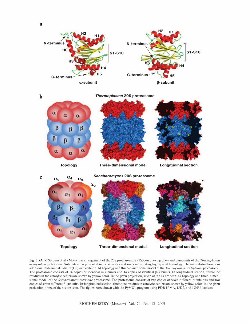
BIOCHEMISTRY (Moscow) Vol. 74 No. 13 2009
Fig. 3. (A. V. Sorokin et al.) Molecular arrangement of the 20S proteasome. a) Ribbon drawing of α- and β-subunits of the Thermoplasma
acidophilum proteasome. Subunits are represented in the same orientation demonstrating high spatial homology. The main distinction is an
additional N-terminal α-helix (H0) in α-subunit. b) Topology and three-dimensional model of the Thermoplasma acidophilum proteasome.
The proteasome consists of 14 copies of identical α-subunits and 14 copies of identical β-subunits. In longitudinal section, threonine
residues in the catalytic centers are shown by yellow color. In the given projection, seven of the 14 are seen. c) Topology and three-dimen-
sional model of the Saccharomyces cerevisiae proteasome. The proteasome consists of two copies of seven different α-subunits and two
copies of seven different β-subunits. In longitudinal section, threonine residues in catalytic centers are shown by yellow color. In the given
projection, three of the six are seen. The figures were drawn with the PyMOL program using PDB 1PMA, 1JD2, and 1G0U datasets.
a
c
b
H2H1
H0
N-terminus
C-terminus
H3
H5
H4
S1-S10
H2 H1
N-terminus
C-terminus
H3
H5
H4
S1-S10
a-subunit b-subunit
Thermoplasma 20S proteasome
Topology Three-dimensional model Longitudinal section
Saccharomyces 20S proteasome
Topology Three-dimensional model Longitudinal section
a5a4 a3
a2

BIOCHEMISTRY (Moscow) Vol. 74 No. 13 2009
a
b
Fig. 4. (A. V. Sorokin et al.) The 20S proteasome entrance. a) Three-dimensional model of a ring of α-subunits of the 20S proteasome (top
view). In the Thermoplasma acidophilum proteasome, 12 N-terminal residues of α-subunits are not ordered and are not given in the pre-
sented structure (they should be facing the channel). The three-dimensional structure of the “closed”, “open”, and “theoretically open”
conformations of the Saccharomyces cerevisiae proteasome. The “closed” conformation corresponds to the conformation of the wild-type
latent proteasome. The “open” conformation corresponds to the conformation of the α3∆N proteasome (the proteasome without nine N-
terminal residues in subunit α3). The “theoretically open” conformation was obtained from the three-dimensional structure of the “closed”
conformation by removing nine N-terminal residues in each α-subunit. b) Ribbon drawing of α-subunits of the 20S proteasome (top view).
The topology of α-subunits of the Saccharomyces cerevisiae 20S proteasome is shown in the “closed” conformation. The figures were drawn
with the PyMOL program using PDB 1PMA, 1JD2, and 1G0U datasets.
Fig. 5. (A. V. Sorokin et al.) Model of 20S proteasome assembly. a) Scheme of assembly of the proteasome from the archaeon Thermoplasma
acidophilum. b) Scheme of assembly of the eukaryotic proteasome. Intermediates that are not stable and cannot be found in vivo are given in
parentheses. See details in the text.
Thermoplasma 20S proteasome “closed” “open” “theoretically open”
Saccharomyces 20S proteasome
a-ring half-proteasome-b7 pre-proteasome
13S complex “half-proteasome” 20S proteasome
“half-proteasome”
pre-proteasome
20S proteasome
a
b

BIOCHEMISTRY (Moscow) Vol. 74 No. 13 2009
a b
lid
base
Fig. 6. (A. V. Sorokin et al.) Molecular arrangement of the 19S regulatory particle. a) A low-resolution three-dimensional model of the 19S RP
was obtained by electron microscopy. In 19S RP, subunits of the base, base arm, and lid are given in different colors. The figure is a modified
version from [76]. b) Topology of subunits of 19S RP. See details in text.
protein protein
Fig. 7. (A. V. Sorokin et al.) Stages of polyubiquitination of substrate protein. Ubiquitin (Ub) is activated by enzyme E1 and translocated to
enzyme E2. In the last stage, E3 ligase conjugates Ub to the substrate protein. The protein must be polyubiquitinated for Ub-dependent protein
degradation by the proteasome. See details in text.
a b
Fig. 8. (A. V. Sorokin et al.) Structure of Ub and Ubl proteins. a) Ribbon drawing of Ub, NEDD8, SUMO, and Ufm1. All of them have a com-
mon secondary structure ββααββαβ and β-grasp (ubiquitin-like) three-dimensional packing. b) Superposing ribbon drawings of Ub and Ubl
proteins. The figure demonstrates high spatial homology. The figure is modified from [154].
protein