Embed Size (px)
Citation preview
fpls-08-00107 February 1, 2017 Time: 17:25 # 1
ORIGINAL RESEARCHpublished: 03 February 2017
doi: 10.3389/fpls.2017.00107
Edited by:Scott C. Peck,
University of Missouri, USA
Reviewed by:Giridara Kumar Surabhi,
Regional Plant Resource Centre, IndiaAlex Jones,
University of Warwick, UK
*Correspondence:Renier A. L. van der Hoorn
Specialty section:This article was submitted to
Plant Proteomics,a section of the journal
Frontiers in Plant Science
Received: 09 July 2016Accepted: 18 January 2017
Published: 03 February 2017
Citation:Kovács J, Poór P, Kaschani F,
Chandrasekar B, Hong TN,Misas-Villamil JC, Xin BT, Kaiser M,
Overkleeft HS, Tari I andvan der Hoorn RAL (2017)
Proteasome Activity ProfilingUncovers Alteration of Catalytic β2
and β5 Subunits of theStress-Induced Proteasome during
Salinity Stress in Tomato Roots.Front. Plant Sci. 8:107.
doi: 10.3389/fpls.2017.00107
Proteasome Activity ProfilingUncovers Alteration of Catalytic β2and β5 Subunits of theStress-Induced Proteasome duringSalinity Stress in Tomato RootsJudit Kovács1, Péter Poór1, Farnusch Kaschani2, Balakumaran Chandrasekar3,4,Tram N. Hong3,4, Johana C. Misas-Villamil4,5, Bo T. Xin6, Markus Kaiser2,Herman S. Overkleeft6, Irma Tari1 and Renier A. L. van der Hoorn3,4*
1 Department of Plant Biology, University of Szeged, Szeged, Hungary, 2 Chemical Biology, Fakultät für Biologie, Zentrum fürMedizinische Biotechnologie, Universität Duisburg-Essen, Essen, Germany, 3 Plant Chemetics Laboratory, Department ofPlant Sciences, University of Oxford, Oxford, UK, 4 Plant Chemetics Laboratory, Max Planck Institute for Plant BreedingResearch, Cologne, Germany, 5 Botanical Institute and Cluster of Excellence on Plant Sciences, University of Cologne,Cologne, Germany, 6 Leiden Institute of Chemistry, Leiden University, Leiden, Netherlands
The stress proteasome in the animal kingdom facilitates faster conversion of oxidizedproteins during stress conditions by incorporating different catalytic β subunits. Plantsdeal with similar kind of stresses and also carry multiple paralogous genes encodingfor each of the three catalytic β subunits. Here, we investigated the existence of stressproteasomes upon abiotic stress (salt stress) in tomato roots. In contrast to Arabidopsisthaliana, tomato has a simplified proteasome gene set with single genes encoding eachβ subunit except for two genes encoding β2. Using proteasome activity profiling ontomato roots during salt stress, we discovered a transient modification of the catalyticsubunits of the proteasome coinciding with a loss of cell viability. This stress-inducedactive proteasome disappears at later time points and coincides with the need todegrade oxidized proteins during salt stress. Subunit-selective proteasome probes andMS analysis of fluorescent 2D gels demonstrated that the detected stress-inducedproteasome is not caused by an altered composition of subunits in active proteasomes,but involves an increased molecular weight of both labeled β2 and β5 subunits, and anadditional acidic pI shift for labeled β5, whilst labeled β1 remains mostly unchanged.Treatment with phosphatase or glycosidases did not affect the migration pattern. Thisstress-induced proteasome may play an important role in PCD during abiotic stress.
Keywords: 20S proteasome, immune proteasome, activity-based protein profiling, programmed cell death, saltstress, tomato root, catalytic subunit
INTRODUCTION
The proteasome plays a key role in protein degradation of cytonuclear proteins during bioticand biotic stress in plants. The 26S proteasome is a highly conserved protein complex whichhas a crucial role in selective protein degradation during cell death and development. The 26Sproteasome consists of a catalytic 20S core protease and a 19S regulatory particle (Coux, 1996;
Abbreviations: ABPP, activity-based protein profiling; IEF, isoelectric focusing; PCD, programmed cell death; PLCP, papain-like Cys protease; PTM, post-translational modification; ROS, reactive oxygen species
Frontiers in Plant Science | www.frontiersin.org 1 February 2017 | Volume 8 | Article 107
fpls-08-00107 February 1, 2017 Time: 17:25 # 2
Kovács et al. Stress-Induced Proteasome
Kurepa and Smalle, 2008). The core protease is composed offour heptameric rings; two outer α-rings and two inner β-rings,each of them composed of seven different α- and β subunits(Löwe et al., 1995). These four heptameric rings are arranged asa cylinder with three large internal chambers. The active sites arein the central chamber, residing in three catalytic subunits: β1, β2,and β5 which cleave after acidic, basic, and hydrophobic residuesand representing caspase-like, trypsin-like, and chymotrypsin-like activities, respectively (Löwe et al., 1995; Kurepa and Smalle,2008; Murata et al., 2009).
In addition to the standard β1, β2, and β5 subunits, animalshave additional “immuno”-subunits (β1i, β2i, and β5i) anda thymus-specific subunit (β5t; Klare et al., 2007; Murataet al., 2007). Replacement of standard catalytic subunits by thecatalytic “immuno”-subunits creates the immune proteasome(i-20S; Aki et al., 1994). These “immuno”-subunits can alsoco-exist with standard subunits within the same proteasomecomplex, creating an intermediate-type proteasome (Klare et al.,2007). Intermediate- and immune proteasomes play roles in thedegradation of damaged and misfolded proteins during cell deathand disease in animals (Zheng et al., 2012; Grigoreva et al., 2015).
Interestingly, there are indications of the existence ofan alternative proteasome in plants. A defense-induced β1subunit (β1din) in tobacco suggests the presence of a “defenseproteasome” in plants (Suty et al., 2003). Transcripts of β1dinaccumulate during the elicitin-induced response in tobacco(Lequeu et al., 2005) whereas salicylic acid signaling activatesthe proteasome post-translationally (Gu et al., 2010). But little isknown about modification and activation of specific proteasomesubunits in plants. An optimal 26S proteasome is essential formaintaining plant drought stress tolerance (Cho et al., 2008; Yeeand Goring, 2009). For instance, regulatory particle mutants haveincreased oxidative stress tolerance (Kurepa et al., 2008), andrpn1a mutants have increased salt hypersensitivity (Wang et al.,2009).
In this study, we investigated the proteasome during abioticstress. We focused our studies on salt stress-induced PCD intomato roots. The root is the primary organ recognizing salt stressand initiating signaling pathways. A moderate salt concentrationstops root growth but higher salt concentrations can inducePCD, characterized by an oxidative burst, cytochrome c release,and DNA fragmentation (Giannattasio et al., 2008; Shabala,2009; Andronis and Roubelakis-Angelakis, 2010), all triggered bysodium ions entering the cell (Shabala, 2000; Zhu, 2001). PCDin root tips upon salt stress is thought to be a defensive responseduring development to maintain the integrity of the root system(Hasegawa et al., 2000; Huh et al., 2002; Gémes et al., 2011).However, when lethal salt exposure is prolonged, it leads to deathnot only at the cellular level but also at tissue or organ level(Bagniewska-Zadworna and Arasimowicz-Jelonek, 2016).
Here, we tested the hypothesis that also plants have a stress-induced proteasome. We used ABPP (Cravatt et al., 2008)and proteomics on salt-stress in tomato roots to investigatethe molecular composition of this stress-induced proteasome.ABPP involves biotinylated or fluorescent chemical probes thatreact with the active site of enzymes in an activity-dependentmanner, creating an irreversible covalent bond that facilitates
detection and identification (Morimoto and Van der Hoorn,2016). ABPP displays changes in the activity level of theenzymes upon different treatments, for instance in the activityof PLCPs and serine hydrolases upon biotic stress, or vacuolarprocessing enzymes and proteasome during PCD (Shabab et al.,2008; Kaschani et al., 2009; Gu et al., 2012; van der Lindeet al., 2012; Misas-Villamil et al., 2013a,b; Sueldo et al., 2014).Our ABPP studies reveal previously unknown stress-associatedmodifications of the proteasome in tomato roots upon saltstress.
MATERIALS AND METHODS
BioinformaticsGenes encoding the β subunits of tomato were identified byBLASTp searches of the predicted proteome (ITAG release2.40) for homologs of the seven Arabidopsis β subunits at theSolGenomics website1. The β2a protein sequence was modeledonto polypeptide H of the structure of the yeast proteasome(2zcy, Groll et al., 2008) using Swiss Model2 (Biasini et al., 2014).This β2a model was used in PyMol to replace the β2 in thestructure of the yeast proteasome. Only the surface of one ringof β subunits was visualized and the various parts and residueswere colored using PyMol. Further annotations were added usingCorelDRAW. Transcript levels were extracted from publishedRNA sequencing experiments on different organs (The TomatoGenome Consortium, 2012).
Plant Materials and Growth ConditionsTomato (Solanum lycopersicum L. cv “Rio Fuego”) plants weregerminated at 26◦C for 3 days in the dark, and the seedlingswere subsequently transferred to perlite for 2 weeks. Plantswere grown hydroponically in a controlled environment in agreenhouse (300 µmol m−2 s−1 photon flux density with 12/12light/dark photoperiod, 25◦C, and 55–60% relative humidity) for3 weeks (Poór et al., 2011). Tomato plants were treated with0-, 100-, and 250 mM NaCl in the nutrient solution [2 mMCa(NO3)2, 1 mM MgSO4, 0.5 mM KCl, 0.5 mM, KH2PO4,0.5 mM Na2HPO4, 0.001 mM MnSO4, 0.005 mM ZnSO4,0.0001 mM (NH4)6Mo7O24, 0.01 mM H3BO4, 0.02 mM Fe(III)-EDTA]. Samples were made in at 9 a.m. and samples were takenin triplicate at 1, 6, and 24 h after salt exposure.
FDA StainingFluorescein diacetate (FDA; Sigma–Aldrich, St. Louis, MO, USA)was used to determine cell viability according to Gémes et al.(2011). Root tip segments were stained for 10 min at roomtemperature in the dark with 10 mM FDA dissolved in 3 ml10 mM 2-(N-morpholino)ethanesulfonic acid (MES) potassiumchloride (KCl) buffer (pH 6.15). After staining, the samples werewashed two times in 10 min with MES/KCl buffer (pH 6.15).Fluorescence intensity was detected with Zeiss Axiovert 200 Mtype fluorescent microscope (Carl Zeiss Inc., Jena, Germany)
1www.solgenomics.net2swissmodel.expasy.org
Frontiers in Plant Science | www.frontiersin.org 2 February 2017 | Volume 8 | Article 107

fpls-08-00107 February 1, 2017 Time: 17:25 # 3
Kovács et al. Stress-Induced Proteasome
equipped with an 5X objective. Digital photographs were takenfrom the samples with a high-resolution digital camera (AxiocamHR, HQ CCD camera; Carl Zeiss Inc., Jena, Germany) using afilter set 10 (excitation 450–495 nm, emission 515–565 nm) orfilter set 20HE (excitation: 535–585 nm, emission: 600–655 nm).The fluorescence emission (pixel intensity) was measured ondigital images with AXIOVISION REL. 4.8 software (Carl ZeissInc., Munich, Germany).
Small-Scale Labeling ReactionSample PreparationRoot tissue was homogenized in 50 mM Tris buffer at pH 7.5containing 5 mM DTT for labeling of the proteasome. The extractwas mixed and centrifuged at 10000 g for 10 min at 4◦C toremove cell debris and the supernatant was collected and usedfor labeling.
Labeling of Proteasome SubunitsHundred microgram/milliliterprotein extract was labeled with2 µM MV151 for 3 h or 0.2 µM MVB072 or co-labeled with0.8 µM LW124/MVB127 for 2 h at room temperature in the darkin 60 µl total volume. Equal volumes of DMSO were added forthe no-probe-control. For inhibition assays, extracts were pre-incubated with 50 or 100 µM epoxomicin or 50 µM N3β1 orN3β5 or DMSO and these extracts were labeled with the suitableprobe. The labeling reactions were stopped by adding gel loadingbuffer containing β-mercaptoethanol at 4X final concentrationand heating at 95◦C for 10 min. The reaction mixture wasseparated on 15% SDS gel at 200 V for 75 min. Labeled proteinswere visualized by in-gel fluorescence scanning using a Typhoon9400 Imager (GE Healthcare)3 using excitation and emissionwavelengths of 532/580 nm for MV151, MVB072, and MVB127and of 470/530 nm for LW124. 532/580 nm and 470/530 wereoverlaid and signals were quantified using ImageJ 1.48V.
IEF 2D SDS PAGELabeled and precipitated proteins were resuspended in UTCbuffer (8 M urea, 2 M thiourea, 4% (w/v) CHAPS, 1 g AG 501-X8Resin) containing 1% (v/v) ampholyte and 65 mM DTT. Sampleswere isoelectrically focused on 7 cm immobilized pH gradient(IPG) 3–10 pH strips (BioRad–ReadyStripTM IPG Strips) usingBioRad PROTEAN i12 IEF system with the following focusingconditions: 12 h passive rehydration; 250 V, 15 min, rapidramp; 4000 V, 1 h, slow ramp; 4000 V, 30000 Vhr, rapid ramp;500 V hold. After focusing, IPG strips were equilibrated in IEFEquilibration buffer [6 M urea, 5% SDS (w/v), 30% glycerol (v/v)]containing 1 % (w/v) DTT, then in IEF Equilibration buffercontaining 2.5% iodoacetamide (w/v). The second dimensionelectrophoresis was run on a 15% SDS gel. Gels were imagedusing a Typhoon 9400 Imager (GE Healthcare) using excitationand emission wavelengths of 532/580 nm. Images were quantifiedusing ImageJ 1.48V by multiplication of the fluorescenceintensity and the area of each of the spots (n= 3).
3http://www.gelifesciences.com
In-Gel Digestion and MSBands were excised by hand and treated with trypsin as describedelsewhere (Shevchenko et al., 2006). Tryptic digests were desaltedon home-made C18 StageTips as described (Rappsilber et al.,2007). After elution from the StageTips samples were driedusing a vacuum concentrator (Eppendorf) and the peptidestaken up in 10 µL 0.1 % formic acid solution. LC-MS/MSexperiments were performed on an Orbitrap Elite instrument(Thermo, Michalski et al., 2012) that was coupled to an EASY-nLC 1000 liquid chromatography (LC) system (Thermo). TheLC was operated in the two-column mode. The home-madefused silica column equipped with a glass fiber frit (Maiolicaet al., 2005) was packed with Reprosil-Pur 120 C18-AQ 3 µmresin (Dr. Maisch) and connected to the analytical column viaan UHPLC union (Upchurch; UH-432). The analytical columnwas a fused silica capillary (75 µm × 25 cm) with integratedPicoFrit emitter (New Objective) packed in-house with Reprosil-Pur 120 C18-AQ 3 µm resin (Dr. Maisch). The analytical columnwas attached to a nanospray flex ion source (Thermo). The LCwas equipped with two mobile phases: solvent A (0.1% formicacid, FA, in UPLC grade water) and solvent B (0.1% FA inacetonitrile, ACN). Peptides were delivered to the pre-columnvia the integrated autosampler at a flow rate of 2–3 µl/min in100% solvent A. Peptides were subsequently separated on theanalytical column by running a 70 min gradient of solvents Aand B (start with 7% B; gradient 7–35% B for 60 min; gradient35–100% B for 5 min and 100% B for 5 min) at a flow rate of300 nl/min. The mass spectrometer was operated using Xcalibursoftware (version 2.2 SP1.48) and was set in the positive ionmode. Precursor ion scanning was performed in the Orbitrapanalyzer (FTMS) in the scan range of m/z 300–1,500 and at aresolution of 120,000 with the internal lock mass option turnedon (lock mass was 445.120025 m/z, polysiloxane; Olsen et al.,2005). Product ion spectra were recorded in a data dependentfashion in the ion trap (ITMS) in a variable scan range and ata rapid scan rate. The ionization potential (spray voltage) wasset to 1.6–2.0 kV. Peptides were analyzed using a repeating cycleconsisting of a full precursor ion scan (1.0 × 10⁶ ions) followedby 15 product ion scans (1.0 × 10⁴ ions) where peptides areisolated based on their intensity in the full survey scan (thresholdof 500 counts) for tandem mass spectrum (MS2) generationthat permits peptide sequencing and identification. CID collisionenergy was set to 35% for the generation of MS2 spectra. DuringMS2 data acquisition dynamic ion exclusion was set to 120 s witha maximum list of excluded ions consisting of 500 members and arepeat count of one. Ion injection time prediction, preview modefor the FTMS, monoisotopic precursor selection and charge statescreening were enabled. Only charge states bigger than 1 wereconsidered for fragmentation.
Peptide and Protein IdentificationThe recorded RAW files were processed in ProteomeDiscoverer1.4 (PD14, Thermo). MS2 spectra were extracted using theSpectrum Selector node. Precursor selection was set to “use MS1precursor.” The mass range was set between 350 and 5,000 Dawith a minimum peak count of 1. Mass analyzer was set to “any”
Frontiers in Plant Science | www.frontiersin.org 3 February 2017 | Volume 8 | Article 107
fpls-08-00107 February 1, 2017 Time: 17:25 # 4
Kovács et al. Stress-Induced Proteasome
and MS order to “MS2.” Activation type was set to “is CID”and Scan type was defined as “full” with ionization source set to“is nanospray.” Selected spectra were submitted to the in houseMASCOT server [version 2.4.1 (Perkins et al., 1999)] using thePD14 MASCOT node.
Tandem mass spectrum spectra data were searched againstthe tomato_ITAG.fasta database4 (version 2.3; 34725 entries). Allsearches included a contaminants database (as implemented inMASCOT and MaxQuant, 263 sequences). The contaminantsdatabase contains known MS contaminants and was includedto estimate the level of contamination. Mascot and Andromedasearches allowed for oxidation of methionine residues (16 Da)and a static modification on cysteine (57 Da, alkylation withiodoacetamide). Enzyme specificity was set to Trypsin/P. Theinstrument type in MASCOT searches was set to ESI-TRAP andthe mass tolerance was set to ± 10 ppm for precursor mass and± 0.35 Da for product ion masses. MS2 spectra matches werethen evaluated using the peptide validation node of PD14 withthe standard settings [search against decoy database, target falsediscovery rate (FDR, strict): 0.01 and target FDR (released): 0.05].The reported results were further filtered. On peptide level onlypeptides with a minimum confidence ‘medium’ were reportedand on protein level only proteins with a minimum of at leasttwo peptide hits were reported.
Protein Phosphatase TreatmentSixty microliter of MVB072-labeled sample containing 1 M NaCl,25 mM MgCl2 10 mM DTT, and 12.5X protease inhibitor cocktail(Sigma) were treated with 1 or 5 µl of alkaline phosphatase(Sigma P0114) and incubated at 37◦C for 1 h. Samples wereanalyzed on 16% SDS-PAGE, whereas the remainder of thesample were separated on 10% SDS-PAGE and transferred topolyvinylidene difluoride (PVDF) membrane for detection ofphosphorylated MAPK using primary antibody, Phospho-p44/42MAPK (Erk1/2) (Thr202/Tyr204) antibody (CST, #9101) andsecondary Goat anti-Rabbit IgG (Thermo, #31466) and visualizedusing chemiluminescent substrates (SuperSignal West/PicoChemiluminescent substrates, Thermo scientific).
PNGaseF Treatment of Labeled ProteinsNine microliter of MVB072-labelled tomato root extract andBovine Fetuin (Promega) were treated with 1 µl of 10Xglycoprotein denaturing buffer (New England BioLabs) andheated at 95◦C for 5 min. The denatured proteins were chilled onice. Two microliter 10X GlycoBuffer (New England BioLabs), 2 µl10% NP40 (Promega), and 6 µl H2O was added to the reaction.The mixture was treated with 1 µl PNGase F (New EnglandBioLabs) or with 1 µl H2O and incubated at 37◦C for 1 h. Sampleswere analyzed on 16% SDS-PAGE.
Protein Deglycosylation of LabeledProteinsEighteen microliter of MVB072-labelled sample and BovineFetuin (Promega) were treated with 2 µl of 10X denaturing
4solgenomics.net
solution (Promega) and heated at 95◦C for 10 min. The denaturedproteins were chilled on ice for 5 min. To the denatured samples5 µl of 10X Deglycosylation Reaction Buffer (Promega), 5 µl of10% NP40 (Promega), and 15 µl of water were added. Sampleswere treated with 5 µl of Protein Deglycosylation Mix (Promega –PNGase F, O-Glycosidase, Neuraminidase, β1–4 Galactosidase,β-N-Acetylglucosaminidase) and incubated at 37◦C for 8 h.Samples were analyzed on 16% SDS-PAGE.
RESULTS
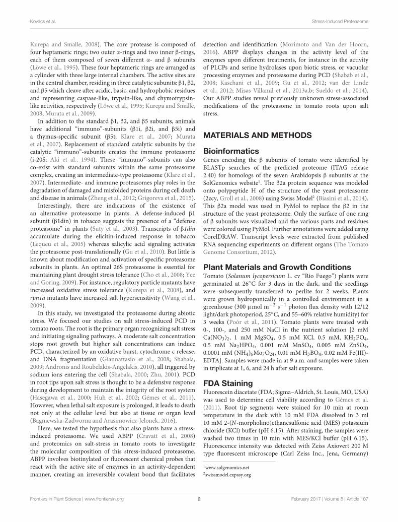
Tomato Has Eight Genes Encoding theSeven β SubunitsTo investigate the tomato proteasome, we performed BLASTsearches with the Arabidopsis β subunits on the predicted tomatoproteome5 and identified eight tomato genes encoding β subunits.Phylogenetic analysis of the tomato and Arabidopsis β subunitsrevealed that tomato genome has one gene for each of the six β
subunits (β1, β3, β4, β5, β6, and β7), and two genes encodingβ2 (Figure 1A). The two β2 proteins in tomato (β2a and β2b)are more closely related to each other when compared to thetwo β2 proteins of Arabidopsis (PBB1 and PBB2), consistent withthe fact that genome duplication occurred in each lineage, afterdivergence (The Tomato Genome Consortium, 2012). Tomato,however, must have lost the paralogous copies of each except oneproteasome β subunit. Sequence alignment with the Arabidopsisorthologs indicates that each of the eight tomato genes encodes aputative functional subunit, including an N-terminal pro-domainfor all subunits and a catalytic Thr for the β1, β2, and β5 subunits(Supplementary Figure S1).
The branch lengths indicate that the two β2 subunits aresubstantially different (Figure 1A). Indeed, we counted 18 aminoacid residues that differ between the mature β2a and β2bsubunits (Figure 1B). Most of these amino acid substitutions arebiochemically dissimilar. To estimate if these variant residues canaffect the proteolytic chamber, we generated a structural modelof the tomato β2 protein using the yeast proteasome (2zcy, Grollet al., 2008) as a template. Mapping the residues that vary betweenβ2a and β2b onto the structural model revealed that none ofthe variant residues are exposed to the proteolytic chamber(Figure 1C). Interestingly, nearly all the variant residues resideon the outer surface and are likely solvent-exposed (Figure 1C).
To determine which of the β subunit-encoding genes areexpressed in different tissues, we mined RNAseq datasets for thetranscript levels of each of these genes from RNAseq data (TheTomato Genome Consortium, 2012). As expected for subunitsthat assemble in stoichiometric complexes, transcript levels ofeach of the β subunit genes are very similar, with the exceptionof β2a transcripts, which accumulate 5- to 10-fold lower whencompared to β2b and the other β subunit-encoding transcripts(Figure 1D). Nevertheless, detection of β2a transcripts suggeststhat β2a is not a pseudogene, but the transcript levels arelow under normal conditions. The ratio between β2a and β2btranscript levels does not significantly change in different tissues
5www.solgenomics.org
Frontiers in Plant Science | www.frontiersin.org 4 February 2017 | Volume 8 | Article 107
fpls-08-00107 February 1, 2017 Time: 17:25 # 5
Kovács et al. Stress-Induced Proteasome
FIGURE 1 | Phylogeny and variation of beta proteasome subunits of tomato. (A) Phylogenetic tree of beta subunit genes of tomato and Arabidopsis.Neighbor-joining tree of protein sequences was build using ClustalW2. (B) Summary of the variant amino acid residues that differ between β2a and β2b. e, putativesolvent-exposed. Significant variation is printed in bold. (C) Location of variant residues in β2, modeled on the yeast proteasome. The tomato β2a protein wasmodeled using the β2 of yeast (2zcy) as a template. Residues that differ between β2a and β2b are highlighted in red in the topview (top) and sideview (bottom) of theβ-ring of the proteasome and summarized in the table. The proteolytic chamber is highlighted with a dashed orange line and catalytic sites are indicated with orangearrows. (D) Transcript levels of β subunit-encoding genes in various tomato organs. These data were extracted from The Tomato Genome Consortium (2012). Readsper kilobase of transcripts per million mapped reads (RPKM) values were extracted from the database for each gene.
(Figure 1D), suggesting that proteasome assembly might besimilar for β2a and β2b subunits in different tissues.
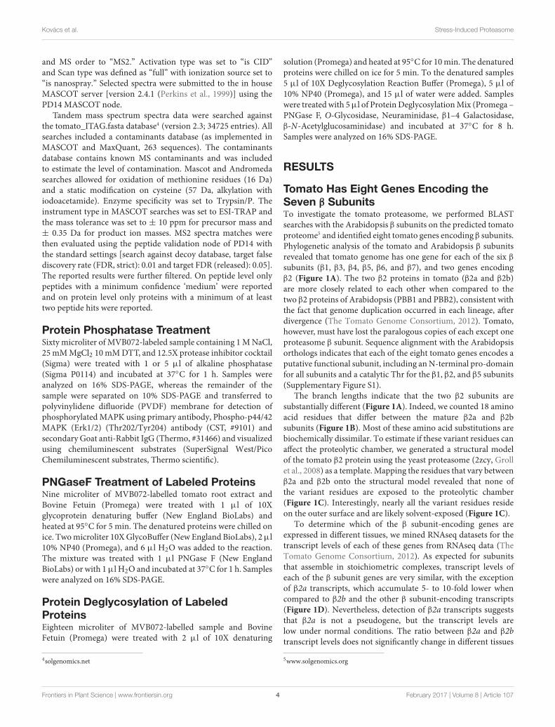
Salt Treatment Induces Loss of Viabilityin Tomato RootsSalt stress is common in plants and is associated with the releaseof ROS. Higher salt concentration also triggers PCD (Hasegawaet al., 2000; Poór et al., 2014). To investigate salt stress in tomatoroots, plants were treated with sublethal- (100 mM) and lethal(250 mM) concentrations of NaCl (Figure 2A). We studied theearly stages of abiotic stress by collecting samples at 1, 6, and 24 hafter salt exposure. Root tips were stained with FDA to detect andquantify viable cells. Low FDA staining after 6 h upon treatmentwith 250 mM NaCl indicates a massive and quick PCD thatcompletes within 24 h (Figure 2B). By contrast, treatment with100 mM NaCl caused a slower loss of viability, where decreasedviability was detected only at 24 h.
MV151 Labeling Uncovers DifferentialProteasome ActivityPapain-like cysteine Proteases and the proteasome have beenimplicated in stress and PCD. To examine the activity of
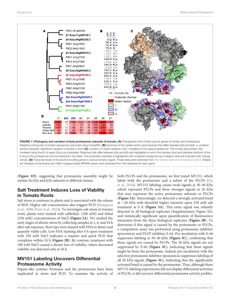
both PLCPs and the proteasome, we first tested MV151, whichlabels both the proteasome and a subset of the PLCPs (Guet al., 2010). MV151 labeling causes weak signals at 30–40 kDawhich represent PLCPs and three stronger signals at 26 kDathat may represent the active proteasome subunits or PLCPs(Figure 3A). Interestingly, we detected a strongly activated bandat ∼26 kDa with threefold higher intensity upon 250 mM salttreatment at 6 h (Figure 3A). This extra signal was robustlydetected in all biological replicates (Supplementary Figure S2),and statistically significant upon quantification of fluorescenceintensities from the three biological replicates (Figure 3B). Todetermine if this signal is caused by the proteasome or PLCPs,a competition assay was performed using proteasome inhibitorepoxomicin and PLCP inhibitor E-64. Pre-incubation with E-64suppresses labeling at 30–40 kDa (Figure 3C), confirming thatthese signals are caused by PLCPs. The 26 kDa signals are notsuppressed by E-64 (Figure 3C), indicating that these signalsmight be from the proteasome. Indeed, pre-incubation with theselective proteasome inhibitor epoxomicin suppresses labeling ofall 26 kDa signals (Figure 3C), indicating that the significantlyactivated band is caused by the proteasome. Thus, although theseMV151 labeling experiments did not display differential activitiesof PLCPs, it did uncover differential proteasome activity profiles.
Frontiers in Plant Science | www.frontiersin.org 5 February 2017 | Volume 8 | Article 107
fpls-08-00107 February 1, 2017 Time: 17:25 # 6
Kovács et al. Stress-Induced Proteasome
FIGURE 2 | Salt treatment induces loss of viability in tomato roots.(A) Experimental assay. Tomato (Solanum lycopersicum) plants were grown ina hydroponic system and 5-weeks old plants were treated with 0-, 100- and250 mM NaCl in the nutrient solution. Root tips were collected at 1, 6, and24 h. (B) Loss of viability upon salt stress. Root tips were stained withfluorescein diacetate (FDA) to detect the viable cells. Top: representativeimages are shown. Scale bar, 0.5 mm. Bottom: fluorescent intensities of FDAfluorescence levels, when compared to the control. Error bars represent SEMof n = 3 biological replicates.
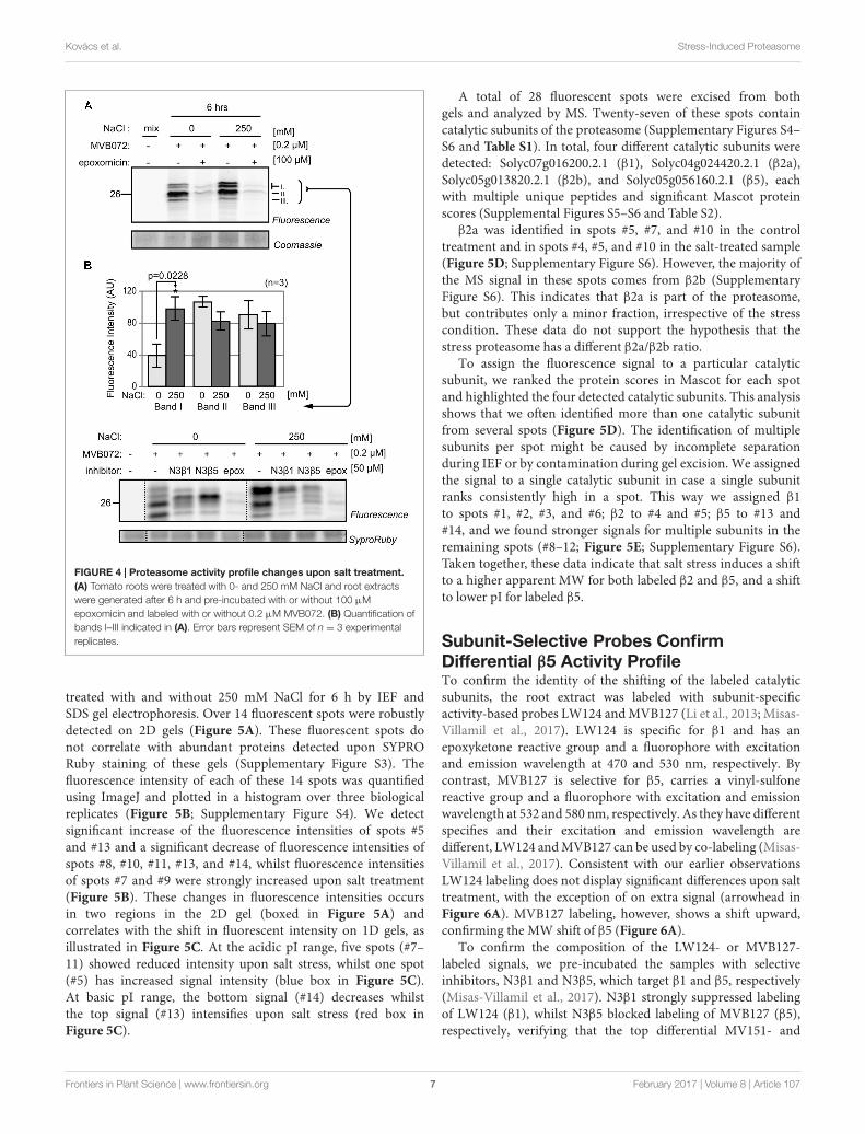
Salt Stress Alters the Activity Profile ofProteasome Catalytic SubunitsTo confirm differential proteasome activity, we used a newlyre-synthesized epoxomicin-based MVB072, which carries botha bodipy tag for fluorescent detection and a biotin tag foraffinity purification (Kolodziejek et al., 2011). When comparedto MV151, MVB072 is a much more selective proteasome probewithout any known off targets, ideal to confirm differentialproteasome activity in our samples (Kolodziejek et al., 2011).Importantly, MVB072 labeling displays the same altered activityprofile upon salt treatment as MV151 labeling (Figure 4A).Quantification of fluorescence intensities of the various signalsdemonstrate a highly reproducible increased intensity of theupper signal at 6 h upon 250 mM NaCl treatment over
FIGURE 3 | MV151 activity profile changes upon salt stress in roots.(A) Differential activity profiles with MV151. Tomato roots were treated with 0-,100-, 250 mM NaCl. Root extracts were generated after 1-, 6- and 24 h andlabeled with 2 µM MV151 at pH 6.0. A mix of all nine samples waspre-incubated with or without 50 µM E-64 and labeled with 2 µM MV151.Shown is a representative gel at long and short fluorescence exposure andupon coomassie staining. The other two experimental replicates are shown asSupplementary Figure S2. (B) Quantification of the upper differential MV151signal (arrowhead) taken from three experimental replicates (A)(Supplementary Figure S2). Error bars represent SEM of n = 3 experimentalreplicates. (C) Differential signal is suppressed by proteasome inhibitor. The6 h 0- and 250 mM NaCl treated samples were labeled with 2 µM MV151.A mix of the two samples was pre-incubated with or without 50 µM E-64 orepoxomicin and labeled with or without 2 µM MV151.
multiple biological replicates, whilst other signals seem to reduce(Figure 4B).
β2- and β5 Catalytic Subunits Migrate atDifferent Molecular Weight (MW) uponLethal Salt StressTo identify the differentially active catalytic subunits of theproteasome, we separated MVB072-labeled proteomes of roots
Frontiers in Plant Science | www.frontiersin.org 6 February 2017 | Volume 8 | Article 107
fpls-08-00107 February 1, 2017 Time: 17:25 # 7
Kovács et al. Stress-Induced Proteasome
FIGURE 4 | Proteasome activity profile changes upon salt treatment.(A) Tomato roots were treated with 0- and 250 mM NaCl and root extractswere generated after 6 h and pre-incubated with or without 100 µMepoxomicin and labeled with or without 0.2 µM MVB072. (B) Quantification ofbands I–III indicated in (A). Error bars represent SEM of n = 3 experimentalreplicates.
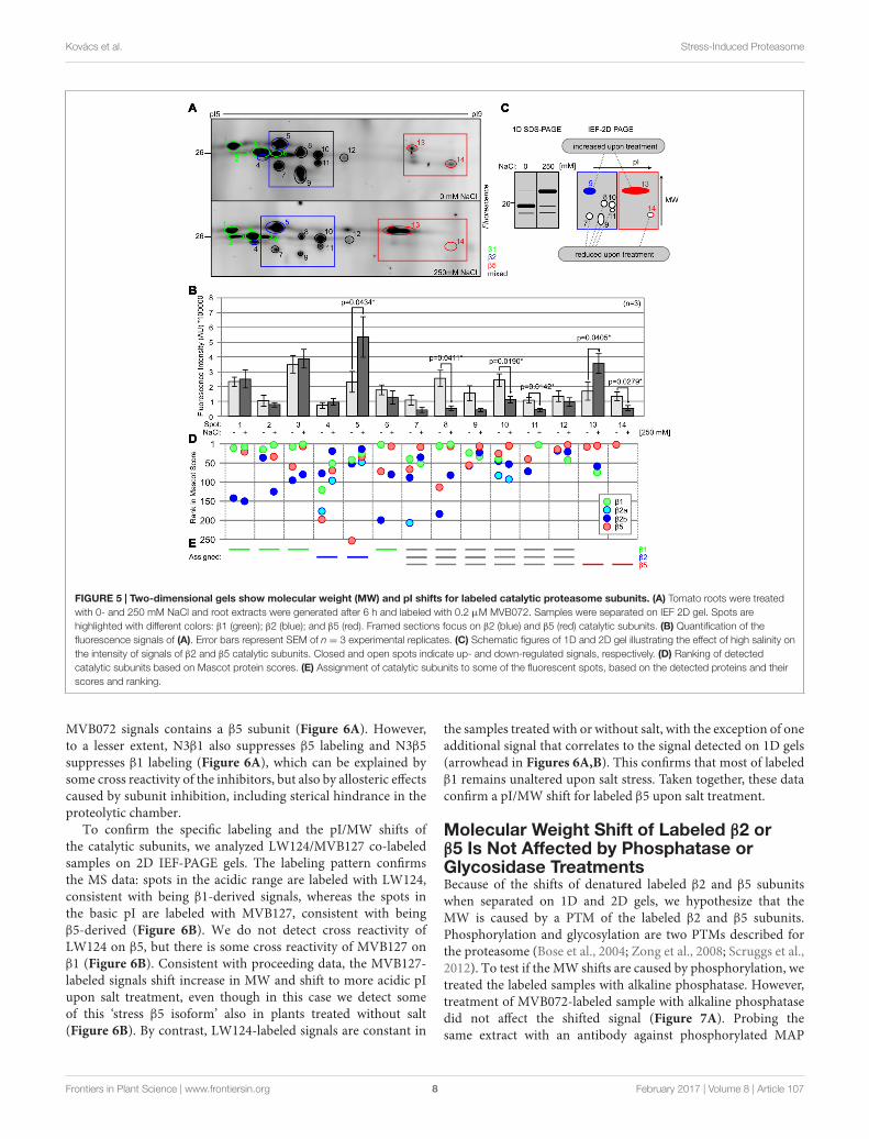
treated with and without 250 mM NaCl for 6 h by IEF andSDS gel electrophoresis. Over 14 fluorescent spots were robustlydetected on 2D gels (Figure 5A). These fluorescent spots donot correlate with abundant proteins detected upon SYPRORuby staining of these gels (Supplementary Figure S3). Thefluorescence intensity of each of these 14 spots was quantifiedusing ImageJ and plotted in a histogram over three biologicalreplicates (Figure 5B; Supplementary Figure S4). We detectsignificant increase of the fluorescence intensities of spots #5and #13 and a significant decrease of fluorescence intensities ofspots #8, #10, #11, #13, and #14, whilst fluorescence intensitiesof spots #7 and #9 were strongly increased upon salt treatment(Figure 5B). These changes in fluorescence intensities occursin two regions in the 2D gel (boxed in Figure 5A) andcorrelates with the shift in fluorescent intensity on 1D gels, asillustrated in Figure 5C. At the acidic pI range, five spots (#7–11) showed reduced intensity upon salt stress, whilst one spot(#5) has increased signal intensity (blue box in Figure 5C).At basic pI range, the bottom signal (#14) decreases whilstthe top signal (#13) intensifies upon salt stress (red box inFigure 5C).
A total of 28 fluorescent spots were excised from bothgels and analyzed by MS. Twenty-seven of these spots containcatalytic subunits of the proteasome (Supplementary Figures S4–S6 and Table S1). In total, four different catalytic subunits weredetected: Solyc07g016200.2.1 (β1), Solyc04g024420.2.1 (β2a),Solyc05g013820.2.1 (β2b), and Solyc05g056160.2.1 (β5), eachwith multiple unique peptides and significant Mascot proteinscores (Supplemental Figures S5–S6 and Table S2).
β2a was identified in spots #5, #7, and #10 in the controltreatment and in spots #4, #5, and #10 in the salt-treated sample(Figure 5D; Supplementary Figure S6). However, the majority ofthe MS signal in these spots comes from β2b (SupplementaryFigure S6). This indicates that β2a is part of the proteasome,but contributes only a minor fraction, irrespective of the stresscondition. These data do not support the hypothesis that thestress proteasome has a different β2a/β2b ratio.
To assign the fluorescence signal to a particular catalyticsubunit, we ranked the protein scores in Mascot for each spotand highlighted the four detected catalytic subunits. This analysisshows that we often identified more than one catalytic subunitfrom several spots (Figure 5D). The identification of multiplesubunits per spot might be caused by incomplete separationduring IEF or by contamination during gel excision. We assignedthe signal to a single catalytic subunit in case a single subunitranks consistently high in a spot. This way we assigned β1to spots #1, #2, #3, and #6; β2 to #4 and #5; β5 to #13 and#14, and we found stronger signals for multiple subunits in theremaining spots (#8–12; Figure 5E; Supplementary Figure S6).Taken together, these data indicate that salt stress induces a shiftto a higher apparent MW for both labeled β2 and β5, and a shiftto lower pI for labeled β5.
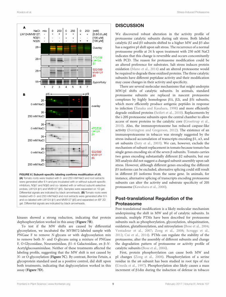
Subunit-Selective Probes ConfirmDifferential β5 Activity ProfileTo confirm the identity of the shifting of the labeled catalyticsubunits, the root extract was labeled with subunit-specificactivity-based probes LW124 and MVB127 (Li et al., 2013; Misas-Villamil et al., 2017). LW124 is specific for β1 and has anepoxyketone reactive group and a fluorophore with excitationand emission wavelength at 470 and 530 nm, respectively. Bycontrast, MVB127 is selective for β5, carries a vinyl-sulfonereactive group and a fluorophore with excitation and emissionwavelength at 532 and 580 nm, respectively. As they have differentspecifies and their excitation and emission wavelength aredifferent, LW124 and MVB127 can be used by co-labeling (Misas-Villamil et al., 2017). Consistent with our earlier observationsLW124 labeling does not display significant differences upon salttreatment, with the exception of on extra signal (arrowhead inFigure 6A). MVB127 labeling, however, shows a shift upward,confirming the MW shift of β5 (Figure 6A).
To confirm the composition of the LW124- or MVB127-labeled signals, we pre-incubated the samples with selectiveinhibitors, N3β1 and N3β5, which target β1 and β5, respectively(Misas-Villamil et al., 2017). N3β1 strongly suppressed labelingof LW124 (β1), whilst N3β5 blocked labeling of MVB127 (β5),respectively, verifying that the top differential MV151- and
Frontiers in Plant Science | www.frontiersin.org 7 February 2017 | Volume 8 | Article 107
fpls-08-00107 February 1, 2017 Time: 17:25 # 8
Kovács et al. Stress-Induced Proteasome
FIGURE 5 | Two-dimensional gels show molecular weight (MW) and pI shifts for labeled catalytic proteasome subunits. (A) Tomato roots were treatedwith 0- and 250 mM NaCl and root extracts were generated after 6 h and labeled with 0.2 µM MVB072. Samples were separated on IEF 2D gel. Spots arehighlighted with different colors: β1 (green); β2 (blue); and β5 (red). Framed sections focus on β2 (blue) and β5 (red) catalytic subunits. (B) Quantification of thefluorescence signals of (A). Error bars represent SEM of n = 3 experimental replicates. (C) Schematic figures of 1D and 2D gel illustrating the effect of high salinity onthe intensity of signals of β2 and β5 catalytic subunits. Closed and open spots indicate up- and down-regulated signals, respectively. (D) Ranking of detectedcatalytic subunits based on Mascot protein scores. (E) Assignment of catalytic subunits to some of the fluorescent spots, based on the detected proteins and theirscores and ranking.
MVB072 signals contains a β5 subunit (Figure 6A). However,to a lesser extent, N3β1 also suppresses β5 labeling and N3β5suppresses β1 labeling (Figure 6A), which can be explained bysome cross reactivity of the inhibitors, but also by allosteric effectscaused by subunit inhibition, including sterical hindrance in theproteolytic chamber.
To confirm the specific labeling and the pI/MW shifts ofthe catalytic subunits, we analyzed LW124/MVB127 co-labeledsamples on 2D IEF-PAGE gels. The labeling pattern confirmsthe MS data: spots in the acidic range are labeled with LW124,consistent with being β1-derived signals, whereas the spots inthe basic pI are labeled with MVB127, consistent with beingβ5-derived (Figure 6B). We do not detect cross reactivity ofLW124 on β5, but there is some cross reactivity of MVB127 onβ1 (Figure 6B). Consistent with proceeding data, the MVB127-labeled signals shift increase in MW and shift to more acidic pIupon salt treatment, even though in this case we detect someof this ‘stress β5 isoform’ also in plants treated without salt(Figure 6B). By contrast, LW124-labeled signals are constant in
the samples treated with or without salt, with the exception of oneadditional signal that correlates to the signal detected on 1D gels(arrowhead in Figures 6A,B). This confirms that most of labeledβ1 remains unaltered upon salt stress. Taken together, these dataconfirm a pI/MW shift for labeled β5 upon salt treatment.
Molecular Weight Shift of Labeled β2 orβ5 Is Not Affected by Phosphatase orGlycosidase TreatmentsBecause of the shifts of denatured labeled β2 and β5 subunitswhen separated on 1D and 2D gels, we hypothesize that theMW is caused by a PTM of the labeled β2 and β5 subunits.Phosphorylation and glycosylation are two PTMs described forthe proteasome (Bose et al., 2004; Zong et al., 2008; Scruggs et al.,2012). To test if the MW shifts are caused by phosphorylation, wetreated the labeled samples with alkaline phosphatase. However,treatment of MVB072-labeled sample with alkaline phosphatasedid not affect the shifted signal (Figure 7A). Probing thesame extract with an antibody against phosphorylated MAP
Frontiers in Plant Science | www.frontiersin.org 8 February 2017 | Volume 8 | Article 107
fpls-08-00107 February 1, 2017 Time: 17:25 # 9
Kovács et al. Stress-Induced Proteasome
FIGURE 6 | Subunit-specific labeling confirms modification of β5.(A) Tomato roots were treated with 0- and 250 mM NaCl and root extractswere generated after 6 h and pre-incubated with or without subunit-specificinhibitors, N3β1 and N3β5 and co- labeled with or without subunit-selectiveprobes, LW124 (β1) and MVB127 (β5). Samples were separated on 1D gel.Differential signals are indicated by black arrowheads. (B) Tomato roots weretreated with 0- and 250 mM NaCl and root extracts were generated after 6 hand co-labeled with LW124 (β1) and MVB127 (β5) and separated on IEF 2Dgel. Differential signals are indicated by black arrowheads.
kinases showed a strong reduction, indicating that proteindephosphorylation worked in this assay (Figure 7B).
To test if the MW shifts are caused by differentialglycosylation, we incubated the MVB072-labeled sample withPNGase F to remove N-glycans or with deglycosylation mixto remove both N- and O-glycans using a mixture of PNGaseF, O-Glycosidase, Neuraminidase, β1–4 Galactosidase, en β-N-Acetylglucosaminidase. Neither of these treatments affected thelabeling profile, suggesting that the MW shift is not caused byN- or O-glycosylation (Figure 7C). By contrast, Bovine Fetuin, aglycoprotein standard used as a positive control, did shift uponboth treatments, indicating that deglycosylation worked in thisassay (Figure 7D).
DISCUSSION
We discovered robust alteration in the activity profile ofproteasome catalytic subunits during salt stress. Both labeledcatalytic β2 and β5 subunits shifted to a higher MW and β5 alsohas a negative pI shift upon salt stress. The recurrence of a normalproteasome profile at 24 h upon treatment with 250 mM NaClindicates that this change is reversible and occurs concomitantlywith PCD. The reason for proteasome modification could bean altered preference for substrates. Salt stress induces proteinoxidation (Mano et al., 2014) and an altered proteasome wouldbe required to degrade these oxidized proteins. The three catalyticsubunits have different peptidase activity and their modificationmay cause changes in their activity and specificity.
There are several molecular mechanisms that might underpinMW/pI shifts of catalytic subunits. In animals, standardproteasome subunits are replaced in nascent proteasomecomplexes by highly homologous β1i, β2i, and β5i subunits,which more efficiently produce antigenic peptides in responseto infection (Tanaka and Kasahara, 1998) and more efficientlydegrade oxidized proteins (Seifert et al., 2010). Replacements bythe i-20S proteasome subunits open the central chamber to allowaccess of more proteins to the catalytic core (Groettrup et al.,2010). Also, the immunoproteasome has reduced caspase-likeactivity (Ferrington and Gregerson, 2012). The existence of animmunoproteasome in tobacco was strongly suggested by thestress-induced accumulation of transcripts encoding β1, α3, andα6 subunits (Suty et al., 2003). We can, however, exclude themechanism of subunit replacement in tomato because tomato hassingle genes encoding six of the seven β subunits. Tomato carriestwo genes encoding substantially different β2 subunits, but ourMS analysis did not suggest a changed subunit assembly upon saltstress. However, although different genes encoding the differentβ5 isoforms can be excluded, alternative splicing could still resultin different β5 isoforms from the same gene. In animals, forinstance, alternative splicing of transcripts encoding proteasomesubunits can alter the activity and substrate specificity of 20Sproteasome (Kawahara et al., 2000).
Post-translational Regulation of theProteasomePost-translational modification is a likely molecular mechanismunderpinning the shift in MW and pI of catalytic subunits. Inanimals, multiple PTMs have been described for proteasomesubunits such as phosphorylation, glycosylation, ubiquitination,oxidation, glutathionylation, and nitrosylation (Bose et al., 2004;Ventadour et al., 2007; Zong et al., 2008; Scruggs et al.,2012; Cui et al., 2014). PTMs can regulate the stability of theproteasome, alter the assembly of different subunits and changethe degradation pattern of proteasome or activity profile ofcatalytic subunits (Bose et al., 2004).
First, protein phosphorylation can cause both MW andpI changes (Zong et al., 2008). Phosphorylation of a serineresidue in the α6 subunit has been studied in root tips of rice(Umeda et al., 1997). Phosphorylation also likely causes a massincrement of β1din during the induction of defense in tobacco
Frontiers in Plant Science | www.frontiersin.org 9 February 2017 | Volume 8 | Article 107

fpls-08-00107 February 1, 2017 Time: 17:25 # 10
Kovács et al. Stress-Induced Proteasome
FIGURE 7 | Phosphatase and glycosidase treatments do not affect altered proteasome activity profile. (A) Root extracts of 250 mM treated samples,labeled by MVB072 were treated with alkaline phosphatase at different conditions. (B) Dephosphorylation of MAP kinase, used as a positive control, detected by ananti-phosphoMAPK antibody. (C) Root extracts of 250 mM treated-samples, labeled by MVB072 were treated with and without PNGase F or deglycosylation mix.(D) Enzymatic deglycosylation of Bovine Fetuin was used as a positive control.
(Suty et al., 2003). There are several predicted phosphorylationsites in catalytic subunits. The β2 subunit contains eight Ser,five Thr, four Tyr residues, whereas the β5 subunit contains10 Ser, one Thr, and three Tyr residues (SupplementaryFigure S3). However, searches or our MS data allowing phosphatemodifications, did not reveal any phosphorylated peptides fromthe β2 or β5 subunits. In addition, phosphatase treatment did notaffect the MVB072 activity profile in tomato roots upon 250 mMNaCl treatment (Figure 7A) suggesting that phosphorylationis not the underlying mechanism of the altered proteasomeupon salt stress. However, phosphorylation cannot be ruled outbecause some phosphorylations can endure alkaline phosphatasetreatment.
Second, proteasome subunits can be regulated byglycosylation. For instance O-Glycosylation reversibly inhibitsproteasome function via modification of Rpt2 ATPase in the19S regulatory particle of the proteasome in animals (Zhanget al., 2003). Likewise, N-Glycosylation is a key PTM of specificproteins during osmotic stress adaptation in plants (Koiwa et al.,2003). However, deglycosylation by PNGase or deglycosidasemix had no effect on the MW shift of β5 (Figure 7C) indicatingthat also glycosylation is not the underlying mechanism.
Third, ubiquitination is common on proteasome subunits.Both β2 and β5 catalytic subunits are ubiquitinated at Lys residues(Ventadour et al., 2007; Kim et al., 2013). However, the predictedMW shift of mono- or polyubiquitinated β2 and β5 is too large(>8.5 kDa) to explain the observed ∼2 kDa MW shift in thealtered proteasome. The same argument excludes sumoylation asa PTM underlying the observed shifts in MW.
In addition, ROS production leads to accumulation ofoxidatively modified proteins (e.g., carbonyl compounds) thatalter the function of enzymes (Basset et al., 2002). There isevidence of carbonylation of the 20S proteasome in responseto carbon starvation in maize root tips (Basset et al., 2002).Furthermore, glutathionylation has been detected on each of
the β5 subunits of the plant proteasome (Dixon et al., 2005).β2 has four and β5 has two Cys residues which may explain a2 kDa upon S-glutathionylation. Finally, Cys residues can alsobe modified by reactive nitrogen species, which are releasedduring stress (Hess et al., 2005). However, the use of reducingagents during our sample preparation would probably removeoxidation, glutathionylation, and nitrosylation from Cys residues,so these PTMs are not likely to explain the MW/pI shifts that wedetected. However, many additional PTMs are known and theycan also be combined in several ways. Also a combination ofPTMs might result in the observed MW/pI shifts in the activityprofile of β2 and β5 catalytic subunits.
CONCLUSION
We discovered an altered proteasome activity profile at the earlystage of salt stress-induced PCD in tomato roots. Modificationof proteasome profile is probably caused by yet unidentifiedcovalent modification of β2 and β5 proteasome catalytic subunits,which is not caused by differential subunit assembly, and may notbe caused by phosphorylation, glycosylation or ubiquitination ofcatalytic subunits. This modification of the proteasome catalyticsubunits is reversible and correlates with the need to degradeoxidized proteins during biotic and abiotic stress. Further workcan now focus at the structural and functional elucidation ofthe stress-induced proteasome to determine its role in stressresponses in plants.
DATA AVAILABILITY
The mass spectrometry proteomics data have been deposited tothe ProteomeXchange Consortium via the PRIDE (Vizcaíno et al.,2016) partner repository (https://www.ebi.ac.uk/pride/archive/)
Frontiers in Plant Science | www.frontiersin.org 10 February 2017 | Volume 8 | Article 107
fpls-08-00107 February 1, 2017 Time: 17:25 # 11
Kovács et al. Stress-Induced Proteasome
with the dataset identifier PXD005266. The samples have beenrenamed as summarized in Supplementary Table S3.
AUTHOR CONTRIBUTIONS
JK, BC, TH, and RvdH designed experiments. JK performed mostexperiments. PP performed microscopy. FK performed massspectrometry. JK, FK, JM-V, and RvdH analyzed the data. BX,MK, and HO contributed materials. JK and RvdH wrote themanuscript with suggestions from all coauthors. IT and RvdHsupervised the project.
FUNDING
This work was financially supported by grants from theHungarian National Scientific Research Foundation (OTKAK101243), the European Union and the State of Hungary,co-financed by the European Social Fund in the frameworkof TÁMOP 4.2.4. A/2-11-1-2012-0001 ‘National Excellence
Program.’ ERC Consolidator Grant 616449 ‘GreenProteases,’Max Planck Society for Plant Breeding Research in Cologne andthe University of Oxford.
ACKNOWLEDGMENTS
We would like to thank Hölger Kramer for his help with 2Dprotein gel electrophoresis, and Jiorgos Kourelis for help withbioinformatic analysis. We would like to thank Zsuzsa Koncz forcritically reading the manuscript.
SUPPLEMENTARY MATERIAL
The Supplementary Material for this article can be found onlineat: http://journal.frontiersin.org/article/10.3389/fpls.2017.00107/full#supplementary-material
TABLE S1 | Peptides and proteins of tomato detected by MS in 2D gels.
REFERENCESAki, M., Shimbara, N., Takashina, M., Akiyama, K., Kagawa, S., Tamura, T.,
et al. (1994). Interferon-gamma induces different subunit organizations andfunctional diversity of proteasomes. J. Biochem. 115, 257–269.
Andronis, E. A., and Roubelakis-Angelakis, K. A. (2010). Short-term salinity stressin tobacco plants leads to the onset of animal-like PCD hallmarks in plantain contrast to long-term stress. Planta 231, 437–448. doi: 10.1007/s00425-009-1060-x
Bagniewska-Zadworna, A., and Arasimowicz-Jelonek, M. (2016). The mystery ofunderground death: cell death in roots during ontogeny and in response toenvironmental factors. Plant Biol. 18, 171–184. doi: 10.1111/plb.12391
Basset, G., Raymond, P., Malek, L., and Brouquisse, R. (2002). Changes in theexpression and the enzymatic properties of the 20S proteasome in sugar-starvedmaize roots. Evidence for an in vivo oxidation of the proteasome. Plant Physiol.128, 1149–1162. doi: 10.1104/pp.010612
Biasini, M., Bienert, S., Waterhouse, A., Arnold, K., Studer, G., Schmidt, T., et al.(2014). SWISS-MODEL: modelling protein tertiary and quaternary structureusing evolutionary information. Nucleic Acids Res. 42, W252–W258. doi: 10.1093/nar/gku340
Bose, S., Stratford, F. L. L., Broadfoot, K. I., Mason, G. G. F., and Rivett, A. J.(2004). Phosphorylation of 20S proteasome alpha subunit C8 (α7) stabilizes the26S proteasome and plays a role in the regulation of proteasome complexes byγ –interferon. Biochem. J. 378, 177–184. doi: 10.1042/bj20031122
Cho, S. K., Ryu, M. Y., Song, C., Kwak, J. M., and Kim, W. T. (2008). ArabidopsisPUB22 and PUB23 are homologous U-Box E3 ubiquitin ligases that playcombinatory roles in response to drought stress. Plant Cell 20, 1899–1914.doi: 10.1105/tpc.108.060699
Coux, O. (1996). Structure and functions of the 20S and 26S proteasomes. Annu.Rev. Biochem. 65, 801–847. doi: 10.1146/annurev.biochem.65.1.801
Cravatt, B. F., Wright, A. T., and Kozarich, J. W. (2008). Activity-based proteinprofiling: from enzyme chemistry to proteomic chemistry. Annu. Rev. Biochem.77, 383–414. doi: 10.1146/annurev.biochem.75.101304.124125
Cui, Z., Scruggs, S. B., Gilda, J. E., Ping, P., and Gomes, A. V. (2014). Regulation ofcardiac proteasomes by ubiquitination, SUMOylation, and beyond. J. Mol. Cell.Cardiol. 71, 32–42. doi: 10.1016/j.yjmcc.2013.10.008
Dixon, D. P., Skipsey, M., Grundy, N. M., and Edwards, R. (2005). Stress-inducedproteins S-glutathionylation in Arabidopsis. Plant Physiol. 138, 2233–2244. doi:10.1104/pp.104.058917
Ferrington, D. A., and Gregerson, D. S. (2012). Immunoproteasomes: structure,function, and antigen presentation. Prog. Mol. Biol. Transl. Sci. 109, 75–112.doi: 10.1016/B978-0-12-397863-9.00003-1
Gémes, K., Poór, P., Horváth, E., Kolbert, Z., Szopkó, D., Szepesi, A., et al. (2011).Cross-talk between salicylic acid and NaCl-generated reactive oxygen speciesand nitric oxide in tomato during acclimation to high salinity. Physiol. Plant.142, 179–192. doi: 10.1111/j.1399-3054.2011.01461.x
Giannattasio, S., Atlante, A., Antonacci, L., Guaragnella, N., Lattanzio, P.,Passarella, S., et al. (2008). Cytochrome c is released from coupled mitochondriaof yeast en route to acetic acid-induced programmed cell death and can workas an electron donor and a ROS scavenger. FEBS Lett. 582, 1519–1525. doi:10.1016/j.febslet.2008.03.048
Grigoreva, T. A., Tribulovich, V. G., Garabadzhiu, A. V., Melino, G., and Barlev,N. A. (2015). The 26S proteasome is a multifaceted target for anti-cancertherapies. Oncotarget 6, 24733–24749. doi: 10.18632/oncotarget.4619
Groettrup, M., Kirk, C. J., and Basler, M. (2010). Proteasomes in immune cells:more than peptide producers? Nat. Rev. Immunol. 10, 73–78. doi: 10.1038/nri2687
Groll, M., Schellenberg, B., Bachmann, A. S., Archer, C. R., Huber, R., Powell,T. K., et al. (2008). A plant pathogen virulence factor inhibits the eukaryoticproteasome by a novel mechanism. Nature 452, 755–758. doi: 10.1038/nature06782
Gu, C., Kolodziejek, I., Misas-Villamil, J., Shindo, T., Colby, T., Verdoes, M.,et al. (2010). Proteasome activity profiling: a simple, robust and versatilemethod revealing subunit-selective inhibitors and cytoplasmic, defense inducedproteasome activities. Plant J. 62, 160–170. doi: 10.1111/j.1365-313X.2009.04122.x
Gu, C., Shabab, M., Strasser, R., Wolters, P. J., Shindo, T., Niemer, M., et al.(2012). Post-translational regulation and trafficking of the granulin-containingprotease RD21 of Arabidopsis thaliana. PLoS ONE 7:e32422. doi: 10.1371/journal.pone.0032422
Hasegawa, P. M., Bressan, R. A., Zhu, J. K., and Bohnert, H. J. (2000). Plant cellularand molecular responses to high salinity. Annu. Rev. Plant Phys. 51, 463–499.doi: 10.1146/annurev.arplant.51.1.463
Hess, D. T., Matsumoto, A., Kim, S. O., Marshall, H. E., and Stamler, J. S. (2005).Protein S-nitrosylation: purview and parameters. Nat. Rev. Mol. Cell Biol. 6,150–166. doi: 10.1038/nrm1569
Huh, G. H., Damsz, B., Matsumoto, T. K., Reddy, M. P., Rus, A. M., Ibeas,J. I., et al. (2002). Salt causes ion disequilibrium-induced programmed celldeath in yeast and plants. Plant J. 29, 649–659. doi: 10.1046/j.0960-7412.2001.01247.x
Kaschani, F., Gu, C., Niessen, S., Hoover, H., Cravatt, B. F., and Van der Hoorn,R. A. L. (2009). Diversity of serine hydrolase activities of unchallenged andbotrytis-infected Arabidopsis thaliana. Mol. Cell. Proteom. 8, 1082–1093. doi:10.1074/mcp.M800494-MCP200
Frontiers in Plant Science | www.frontiersin.org 11 February 2017 | Volume 8 | Article 107
fpls-08-00107 February 1, 2017 Time: 17:25 # 12
Kovács et al. Stress-Induced Proteasome
Kawahara, H., Kasahara, M., Nishiyama, A., Ohsumi, K., Goto, T., Kishimoto, T.,et al. (2000). Developmentally regulated, alternative splicing of the Rpn10 genegenerates multiple forms of 26S proteasomes. EMBO J. 19, 4144–4153. doi:10.1093/emboj/19.15.4144
Kim, D. Y., Scalf, M., Smith, L. M., and Vierstra, R. D. (2013). Advanced proteomicanalyses yield a deep catalog of ubiquitylation targets in Arabidopsis. Plant Cell25, 1523–1540. doi: 10.1105/tpc.112.108613
Klare, N., Seeger, M., Janek, K., Jungblut, P. R., and Dahlmann, B. (2007).Intermediate-type 20S proteasomes in HeLa Cells: “Asymmetric” subunitcomposition, diversity and adaptation. J. Mol. Biol. 373, 1–10. doi: 10.1016/j.jmb.2007.07.038
Koiwa, H., Li, F., McCully, M. G., Mendoza, I., Koizumi, N., Manabe, Y., et al.(2003). The STT3a subunit isoform of the Arabidopsis oligosaccharyltransferasecontrols adaptive responses to salt/osmotic stress. Plant Cell 15, 2273–2284.doi: 10.1105/tpc.013862
Kolodziejek, I., Misas-Villamil, J. C., Kaschani, F., Clerc, J., Gu, C., Krahn, D.,et al. (2011). Proteasome activity imaging and profiling characterizes bacterialeffector syringolin A. Plant Physiol. 155, 477–489. doi: 10.1104/pp.110.163733
Kurepa, J., and Smalle, J. A. (2008). Structure, function and regulation of plantproteasomes. Biochemie 90, 324–335. doi: 10.1016/j.biochi.2007.07.019
Kurepa, J., Toh-E, A., and Smalle, J. A. (2008). 26S proteasome regulatory particlemutants have increased oxidative stress tolerance. Plant J. 53, 102–114. doi:10.1111/j.1365-313X.2007.03322.x
Lequeu, J., Simon-Plas, F., Fromentin, J., Etienne, P., Petitot, A. S., Blein, J. P.,et al. (2005). Proteasome comprising a β1 inducible subunit acts as a negativeregulator of NADPH oxidase during elicitation of plant defense reactions. FEBSLett. 579, 4879–4886. doi: 10.1016/j.febslet.2005.07.073
Li, N., Kuo, C. L., Paniagua, G., Van den Elst, H., Verdoes, M., Willems, L. I.,et al. (2013). Relative quantification of proteasome activity by activity-basedprotein profiling and LC-MS/MS.Nat. Protoc. 8, 1155–1168. doi: 10.1038/nprot.2013.065
Löwe, J., Stock, D., Jap, B., Zwickl, P., Baumeister, W., and Hubert, R. (1995).Crystal structure of the 20S proteasome from the archaeon T. acidophilum at3.4 A resolution. Science 268, 533–539. doi: 10.1126/science.7725097
Maiolica, A., Borsotti, D., and Rappsilber, J. (2005). Self-made frits for nanoscalecolumns in proteomics. Proteomics 5, 3847–3850. doi: 10.1002/pmic.200402010
Mano, J., Nagata, M., Okamura, S., Shiraya, T., and Mitsui, T. (2014). Identificationof oxidatively modified proteins in salt-stressed Arabidopsis: a carbonyl-targeted proteomics approach. Plant Cell Physiol. 55, 1233–1244. doi: 10.1093/pcp/pcu072
Michalski, A., Damoc, E., Lange, O., Denisov, E., Nolting, D., Müller, M., et al.(2012). Ultra high resolution linear ion trap orbitrap mass spectrometer(orbitrap elite) facilitates top down LC MS/MS and versatile peptidefragmentation modes. Mol. Cell. Proteomics 11, O111.013698. doi: 10.1074/mcp.O111.013698
Misas-Villamil, J. C., Kolodziejek, I., Crabill, E., Kaschani, F., Niessen, S.,Shindo, T., et al. (2013a). Pseudomonas syringae pv. syringae uses proteasomeinhibitor syringolin A to colonize from wound infection sites. PLoS Pathog.9:e1003281. doi: 10.1371/journal.ppat.1003281
Misas-Villamil, J. C., Toenges, G., Kolodziejek, I., Sadaghiani, A. M., Kaschani, F.,Colby, T., et al. (2013b). Activity profiling of vacuolar processing enzymesreveals a role for VPE during oomycete infection. Plant J. 73, 689–700. doi:10.1111/tpj.12062
Misas-Villamil, J. C., van der Burgh, A. M., Grosse-Holz, F., Bach-Pages, M.,Kovács, J., Kaschani, F., et al. (2017). Subunit-selective proteasome activityprofiling uncovers uncoupled proteasome subunit activities during bacterialinfections. Plant J. (in press).
Morimoto, K., and Van der Hoorn, R. A. L. (2016). The increasing impact ofactivity-based protein profiling in plant science. Plant Cell Physiol. 57, 446–461.doi: 10.1093/pcp/pcw003
Murata, S., Sasaki, K., Kishimoto, T., Niwa, S., Hayashi, H., Takahama, Y.,et al. (2007). Regulation of CD8+ T cell development by thymus-specificproteasomes. Science 316, 1349–1353. doi: 10.1126/science.1141915
Murata, S., Yashiroda, H., and Tanaka, K. (2009). Molecular mechanisms ofproteasome assembly. Mol. Cell. Biol. 10, 104–115.
Olsen, J. V., de Godoy, L. M., Li, G., Macek, B., Mortensen, P., Pesch, R., et al.(2005). Parts per million mass accuracy on an Orbitrap mass spectrometer
via lock mass injection into a C-trap. Mol. Cell. Proteomics 4, 2010–2021.doi: 10.1074/mcp.T500030-MCP200
Perkins, D. N., Pappin, D. J., Creasy, D. M., and Cottrell, J. S. (1999). Probability-based protein identification by searching sequence databases using massspectrometry data. Electrophoresis 20, 3551–3567. doi: 10.1002/(SICI)1522-2683(19991201)20:18<3551::AID-ELPS3551<3.0.CO;2-2
Poór, P., Borbély, P., Kovács, J., Papp, A., Szepesi, Á, Takács, Z., et al. (2014).Opposite extremes in ethylene/nitric oxide ratio induce cell death in suspensionculture and root apices of tomato exposed to salt stress. Acta Biol. Hung. 65,428–438. doi: 10.1556/ABiol.65.2014.4.7
Poór, P., Gémes, K., Horváth, F., Szepesi, Á., Simon, M. L., and Tari, I. (2011).Salicylic acid treatment via the rooting medium interferes with stomatalresponse, CO2 fixation rate and carbohydrate metabolism in tomato, anddecreases harmful effects of subsequent salt stress. Plant Biol. 13, 105–114.doi: 10.1111/j.1438-8677.2010.00344.x
Rappsilber, J., Mann, M., and Ishihama, Y. (2007). Protocol for micro-purification, enrichment, pre-fractionation and storage of peptides forproteomics using StageTips.Nat. Protoc. 2, 1896–1906. doi: 10.1038/nprot.2007.261
Scruggs, S. B., Zong, N. C., Wang, D., Stefani, E., and Ping, P. (2012). Post-translational modification of cardiac proteasomes: functional delineationenabled by proteomics. Am. J. Physiol. 303, H9–H18.
Seifert, U., Bialy, L. P., Ebstein, F., Bech-Otschir, D., Voigt, A., Schröter, F.,et al. (2010). Immunoproteasomes preserve protein homeostasis uponinterferon-induced oxidative stress. Cell 142, 613–624. doi: 10.1016/j.cell.2010.07.036
Shabab, M., Shindo, T., Gu, C., Kaschani, F., Pansuriya, T., Chintha, R., et al.(2008). Fungal effector protein AVR2 targets diversifying defense-relatedCys proteases of tomato. Plant Cell 20, 1169–1183. doi: 10.1105/tpc.107.056325
Shabala, S. (2000). Ionic and osmotic components of salt stress specificallymodulate net ion fluxes from bean leaf mesophyll. Plant Cell Environ. 23,825–837. doi: 10.1046/j.1365-3040.2000.00606.x
Shabala, S. (2009). Salinity and programmed cell death: unravelling mechanismsfor ion specific signaling. J. Exp. Bot. 60, 709–712. doi: 10.1093/jxb/erp013
Shevchenko, A., Tomas, H., Havlis, J., Olsen, J. V., and Mann, M. (2006). In-geldigestion for mass spectrometric characterization of proteins and proteomes.Nat. Protoc. 1, 2856–2860. doi: 10.1038/nprot.2006.468
Sueldo, D., Ahmed, A., Misas-Villamil, J. C., Colby, T., Tameling, W., Joosten,M. H. A. J., et al. (2014). Dynamic hydrolase activities precede hypersensitivetissue collapse in tomato seedlings. New Phytol. 203, 913–925. doi: 10.1111/nph.12870
Suty, L., Lequeu, J., Lancon, A., Etienne, P., Petitot, A. S., and Blein, J. P. (2003).Preferential induction of 20S proteasome subunits during elicitation of plantdefense reactions: towards the characterization of “plant defense proteasomes.”Int. J. Biochem. Cell Biol. 35, 637–650. doi: 10.1016/S1357-2725(02)00386-2
Tanaka, K., and Kasahara, M. (1998). The MHC class I ligand-generating system:roles of immunoproteasomes and the interferon-Y-inducible proteasomeactivator PA28. Immunol. Rev. 163, 161–176. doi: 10.1111/j.1600-065X.1998.tb01195.x
The Tomato Genome Consortium (2012). The tomato genome sequence providesinsights into fleshy fruit evolution. Nature 485, 635–641. doi: 10.1038/nature11119
Umeda, M., Manabe, Y., and Uchimiya, H. (1997). Phosphorylation of the C2subunit of the proteasome in rice (Oryza sativa L.). FEBS Lett. 403, 313–317.doi: 10.1016/S0014-5793(97)00073-2
van der Linde, K., Mueller, A. N., Hemetsberger, C., Kashani, F., Van der Hoorn,R. A. L., and Doehlemann, G. (2012). The maize cystatin CC9 interacts withapoplastic cysteine proteases. Plant Sign. Behav. 7, 1397–1401. doi: 10.4161/psb.21902
Ventadour, S., Jarzaguet, M., Wing, S. S., Chambon, C., Combaret, L., Béchet, D.,et al. (2007). A new method of purification of proteasome substrates revealspolyubiquitination of 20 S proteasome subunits. J. Biol. Chem. 282, 5302–5309.doi: 10.1074/jbc.M610005200
Vizcaíno, J. A., Csordas, A., del-Toro, N., Dianes, J. A., Griss, J., Lavidas, I., et al.(2016). 2016 update of the PRIDE database and related tools. Nucleic Acids Res.44, 447–456. doi: 10.1093/nar/gkv1145
Frontiers in Plant Science | www.frontiersin.org 12 February 2017 | Volume 8 | Article 107

fpls-08-00107 February 1, 2017 Time: 17:25 # 13
Kovács et al. Stress-Induced Proteasome
Wang, S., Kurepa, J., and Smalle, J. A. (2009). The Arabidopsis 26Sproteasome subunit RPN1a is required for optimal plant growth andstress responses. Plant Cell Physiol. 50, 1721–1725. doi: 10.1093/pcp/pcp105
Yee, D., and Goring, D. R. (2009). The diversity of plant U-box E3 ubiquitinligases: from upstream activators to downstream target substrates. J. Exp. Bot.60, 1109–1121. doi: 10.1093/jxb/ern369
Zhang, F., Su, K., Yang, X., Bowe, D. B., Paterson, A. J., and Kudlow, J. E. (2003).O-GlcNAc modification is an endogenous inhibitor of the proteasome. Cell 115,715–725. doi: 10.1016/S0092-8674(03)00974-7
Zheng, J., Dasgupta, A., and Bizzozero, O. A. (2012). Changes in 20S subunitcomposition are largely responsible for altered proteasomal activities inexperimental autoimmune encephalomyelitis. J. Neurochem. 121, 486–494. doi:10.1111/j.1471-4159.2012.07699.x
Zhu, J. K. (2001). Plant salt tolerance. Trends Plant Sci. 6, 66–71. doi: 10.1016/S1360-1385(00)01838-0
Zong, C., Young, G. W., Wang, Y., Lu, H., Deng, N., Drews, O., et al. (2008).Two-dimensional electrophoresis-based characterization of post-translationalmodifications of mammalian 20S proteasome complexes. Proteomics 8, 5025–5037. doi: 10.1002/pmic.200800387
Conflict of Interest Statement: The authors declare that the research wasconducted in the absence of any commercial or financial relationships that couldbe construed as a potential conflict of interest.
Copyright © 2017 Kovács, Poór, Kaschani, Chandrasekar, Hong, Misas-Villamil, Xin,Kaiser, Overkleeft, Tari and van der Hoorn. This is an open-access article distributedunder the terms of the Creative Commons Attribution License (CC BY). The use,distribution or reproduction in other forums is permitted, provided the originalauthor(s) or licensor are credited and that the original publication in this journalis cited, in accordance with accepted academic practice. No use, distribution orreproduction is permitted which does not comply with these terms.
Frontiers in Plant Science | www.frontiersin.org 13 February 2017 | Volume 8 | Article 107


![Loss of 26S Proteasome Function Leads to …Loss of 26S Proteasome Function Leads to Increased Cell Size and Decreased Cell Number in Arabidopsis Shoot Organs1[C][W][OA] Jasmina Kurepa,](https://img.pdfslide.us/doc/110x75/5e564ca73cb3c5319d626adb/loss-of-26s-proteasome-function-leads-to-loss-of-26s-proteasome-function-leads-to.jpg)