Embed Size (px)
Citation preview
Protease Production by haloarchaea Natrinema sp.BTSH10 isolated from salt pan of South India
Thesis submitted to the
Cochin University of Science and TechnologyUnder the faculty of Science
in partial fulfillment of the requirementsfor the degree of
DOCTOR OF PHILOSOPHYIN
BIOTECHNOLOGY
ByR. MANJULA
(Reg. No. 3373)
Microbial Technology LaboratoryDepartment of Biotechnology
Cochin University of Science and TechnologyCochin - 22, Kerala, India
May 2014


Prof. (Dr.) M. Chandrasekaran 19-05-2014
CERTIFICATE
This is to certify that the research work presented in this thesis entitled “Protease
Production by haloarchaea Natrinema sp. BTSH10 isolated from salt pan of South
India”, is based on the original research work carried out by Mrs. R. Manjula under my
guidance and supervision at the Department of Biotechnology, Cochin University of
Science and Technology, in partial fulfillment of the requirements for the degree of
Doctor of Philosophy and that no part thereof has been presented for the award of any
degree, diploma, associateship or other similar titles or recognition.
M. CHANDRASEKARAN
DEPARTMENT OF BIOTECHNOLOGYCOCHIN UNIVERSITY OF SCIENCE AND TECHNOLOGY
COCHIN-682 022, KERALA, INDIAPh: 0484- 2576267 Fax: 0484- 2577595
Email: [email protected]


ACKNOWLEDGEMENTS
¨ÉÉiÉ®Æú Ê{ÉiÉ®Æú xÉi´ÉÉ<Ç·É®Æú ¾þnÂùMÉiÉÆ ¨ÉÖnù¨ÉÂ*ªÉiEÞòiÉÆ ºÉEò±ÉÆ i´ÉtMÉÖ û{ÉÉnùÉƤÉÖVÉä%{ÉǪÉä**
I bow my head before the Master of the Universe, the Omnipresent, Omniscient
and Omnipotent driving force of the Universe. Without His Will, this dream would
not have realized. He has blessed me with parents whose thoughtfulness, guidance
and sacrifice have helped me sail smoothly through life. I pay my respects to them.
I wish to thank all my teachers who have moulded me into what I am today. I
am blessed to be a student of Dr. M. Chandrasekaran, Professor, Department of
Biotechnology, Cochin University of Science and Technology. His words, emanating
a positive energy, were a constant source of moral support and encouragement, which
served to boost my morale and strengthened my resolve to complete the thesis in time.
His vast ocean of knowledge, logical way of thinking and deep vision, paved the way
for the realization of this dream. I am deeply indebted to him for having chosen me as
his student and for leading me to the accomplishment of this goal.
Words fail to express my deep sense of gratitude to Dr. Sarita G. Bhat, who was
a pillar of strength as my Co-Guide and Doctoral Committee Member. She goaded
me to complete the work in time, keeping an eye on the progress of the work. Her
suggestions and discussions were invaluable, which enabled me to take right decisions
at the right time.
I would like to place on record my gratitude to Dr. Padma Nambisan and Dr.
Ammini Joseph, Doctoral Committee Members for their support and co-operation.
I express my deep sense of gratitude to Dr. C.S. Paulose, Emeritus Professor, for his
support and co-operation.
I wish to thank the University Grants Commission, Govt. of India for
providing Fellowship under the FIP scheme.

I would like to express my gratitude to Dr. B.S. Krishnan, Chairman, Sree
Sankara College Association, Kalady for his kind support. He was a constant source
of inspiration and guidance. I wish to thank the Management, Principal and Staff,
Sree Sankara College, Kalady for their support. My thanks are due to Dr. Valsa A.K,
Dr. Sumi Mary Gerorge and Dr. S. Mohan, Department of Microbiology, Sree
Sankara College, Kalady. I wish to thank Dr.V.V. Anil Kumar and Dr. C.K. Sujesh,
Mrs. Moby, Sree Sankara College for their support and co-operation. Words fail to
express my gratitude to Dr. Sunanda C., Academic Consultant, College of Veterinary
and Animal Sciences, Mannuthy, for helping me with the statistical analysis of the
work.
My colleagues in the Microbial Technology Laboratory Dr. Roslin Alex, Mr.
Karthikeyan, Mr. Cikesh, Ms. Bindiya, Ms. Tina, Mr. Sajan, Mr. Doles, Dr. Beena,
Dr. Jissa, Mr. Ajith and Ms. Nasiya were always ready to lend a helping hand
whenever I had any problem. I place on record my gratitude to them.
The support and help rendered by the Research Scholars in the Microbial
Genetics Laboratory Mrs. Smitha, Mrs. Helvin, Ms. Mridula, Mrs. Harisree, Mr. Siju,
Dr. Raghul, Mrs. Vijaya, Mrs. Linda, Ms. Lakshmi, Mr. Noble Kurian, Ms. Anu and
Dr. Jeena, is duly acknowledged.
Dr. Manzur, Dr. Sapna, Ms. Rekha and Mr. Ramesh Kumar of Immuno
Technology Laboratory were always ready to lend a helping hand. Dr. Jikku, Dr.
Jasmine, Mrs. Sudha, Ms. Kiran, Mrs. Soumya, Ms. Anala Mrs. Arrinnia, Research
Scholars, Plant Biotechnology Laboratory were supportive and co-operative. I thank
them for their constant encouragement and support.
I also wish to place on record my gratitude to the Research Scholars of
Neuroscience Department, specially Dr. Anju, Dr. Jayanarayanan, Dr. Korah, Dr.
Smijin, Mr. Nigil, Dr. Shilpa, Ms. Roshini, Mr. Ajayan and Dr. Anitha. I would like
to convey my gratitude to the M.Sc. students of the Department of Biotechnology,
Cochin University of Science and Technology, for their help and support.
I also extend my gratitude to Dr. Jayachandran K., Associate Professor,
School of Biosciences, Mahatma Gandhi University, Kottayam, for his valuable

suggestions and ideas. I wish to thank Dr. I.S. Bright Singh, Coordinator, National
Centre for Aquatic Animal Health, Cochin University of Science and Technology, for
permitting the use of lyophilizer. My sincere thanks to Ms. Sareen Sarah John, Dr.
Soorej and Dr. Sreeja for their suggestions and innovative ideas.
I am grateful to the Section Officer, Staff and Librarian, Department of
Biotechnology, Cochin University of Science and Technology for all the help
rendered.
I wish to thank my husband, Dr. K. Madhusudhanan, for shouldering the
tough tasks, enduring hardships and his sacrifices which paved way for the
preparation of the thesis. Madhav, my son was patient enough to bear with my
temper, short-comings and time restraints to take care of his needs during this period.
I also wish to place on record my gratitude to my brothers Dr. Manoj and Mr. Pradeep
and their respective families for the encouragement and prayers.
I also remember with gratitude my friends and well wishers.

Abbreviations
% - Percentage
°C - Degree Celsius
µ - Micron
µg - microgram
µL - microlitre
APS - Ammonium persulfate
BLAST - Basic Local Alignment Search Tool
BSA - Bovine Serum Albumin
cm - centimeter
DMSO - Dimethyl sulphoxide
DNA - Deoxyribonucleic acid
DW - Distilled Water
EC - Enzyme Commission
EDTA - Ethylenediaminetetraacetic acid
Fig. - Figure
g - grams
g/L - grams per litre
h - hour
HPLC - High Performance Liquid Chromatography
kDa - Kilo Dalton
Km - substrate concentration at which the reaction
velocity is half Maximum
M - Molar
mg - milligram
min. - minutes
mL - millilitre
mm - millimetre
mM - milli Molar
MW - Molecular Weight
NCBI - National Centre for Biotechnology
Information
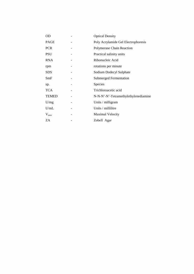
OD - Optical Density
PAGE - Poly Acrylamide Gel Electrophoresis
PCR - Polymerase Chain Reaction
PSU - Practical salinity units
RNA - Ribonucleic Acid
rpm - rotations per minute
SDS - Sodium Dodecyl Sulphate
SmF - Submerged Fermentation
sp. - Species
TCA - Trichloroacetic acid
TEMED - N-N-N’-N’-Tetramethylethylenediamine
U/mg - Units / milligram
U/mL - Units / millilitre
Vmax - Maximal Velocity
ZA - Zobell Agar
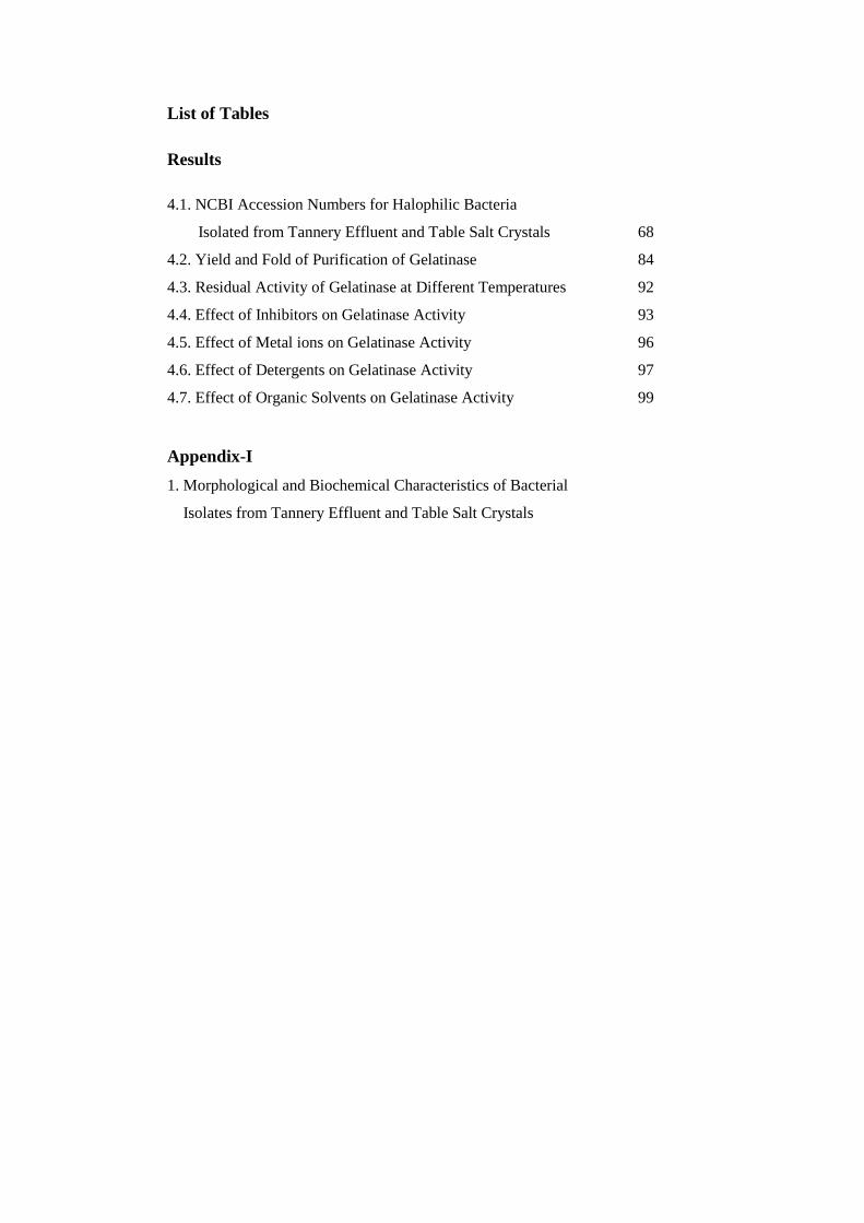
List of Tables
Results
4.1. NCBI Accession Numbers for Halophilic Bacteria
Isolated from Tannery Effluent and Table Salt Crystals 68
4.2. Yield and Fold of Purification of Gelatinase 84
4.3. Residual Activity of Gelatinase at Different Temperatures 92
4.4. Effect of Inhibitors on Gelatinase Activity 93
4.5. Effect of Metal ions on Gelatinase Activity 96
4.6. Effect of Detergents on Gelatinase Activity 97
4.7. Effect of Organic Solvents on Gelatinase Activity 99
Appendix-I
1. Morphological and Biochemical Characteristics of Bacterial
Isolates from Tannery Effluent and Table Salt Crystals


LIST OF FIGURES
1. INTRODUCTION
1.1. Global Industrial Enzyme Market, 2008-2015 1
2. REVIEW OF LITERATURE
2.1. Phylogenetic Tree of the three Domains of Life 14
3. RESULTS
4.1. Phylogenetic Tree of the Halophiles Isolated from Tannery
Effluent and Commercially Available Salt Crystals. 70
4.2. Gelatinase Production by BTSH10, BTSH03 and Bacteria
Isolated from Tannery Effluent and Food Grade Salt Crystals 71
4.3. Gelatin Plate with Bacteria Showing Gelatinase Production 71
4.4. Typical Growth of Natrinema sp. BTSH10 on Marine
Zobell’s Agar Supplemented with 15% NaCl 72
4.5. Selection of Media for Gelatinase Production 73
4.6. Growth Curve of Natrinema sp. BTSH10 73
4.7. Optimization of NaCl Concentration for Gelatinase
Production by Natrinema sp. BTSH10 74
4.8. Optimization of Initial pH of Medium for Gelatinase
Production by Natrinema sp. BTSH10 75
4.9. Optimization of Incubation Temperature for Gelatinase
Production by Natrinema sp. BTSH10 76
4.10. Optimization of Inoculum Concentration for Gelatinase
Production by Natrinema sp. BTSH10 77
4.11. Optimisation of Inoculum Age for Gelatinase Production by
Natrinema sp. BTSH10. 78
4.12. Effect of Agitation on Gelatinase Production by
Natrinema sp. BTSH10 78
4.13. Effect of Additional Carbon Sources on Gelatinase Production
by Natrinema sp. BTSH10 79
4.14.(a) Effect of Additional Inorganic Nitrogen Sources on Gelatinase
Production by Natrinema sp. BTSH10 80
4.14.(b) Effect of Additional Organic Nitrogen Source on Gelatinase
Production by Natrinema sp. BTSH10 81
4.15. Effect of Detergents on Gelatinase Production by
Natrinema sp. BTSH10 82
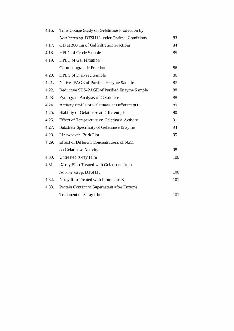
4.16. Time Course Study on Gelatinase Production by
Natrinema sp. BTSH10 under Optimal Conditions 83
4.17. OD at 280 nm of Gel Filtration Fractions 84
4.18. HPLC of Crude Sample 85
4.19. HPLC of Gel Filtration
Chromatographic Fraction 86
4.20. HPLC of Dialysed Sample 86
4.21. Native -PAGE of Purified Enzyme Sample 87
4.22. Reductive SDS-PAGE of Purified Enzyme Sample 88
4.23. Zymogram Analysis of Gelatinase 88
4.24. Activity Profile of Gelatinase at Different pH 89
4.25. Stability of Gelatinase at Different pH 90
4.26. Effect of Temperature on Gelatinase Activity 91
4.27. Substrate Specificity of Gelatinase Enzyme 94
4.28. Lineweaver- Burk Plot 95
4.29. Effect of Different Concentrations of NaCl
on Gelatinase Activity 98
4.30. Untreated X-ray Film 100
4.31. X-ray Film Treated with Gelatinase from
Natrinema sp. BTSH10 100
4.32. X-ray film Treated with Proteinase K 101
4.33. Protein Content of Supernatant after Enzyme
Treatment of X-ray film. 101
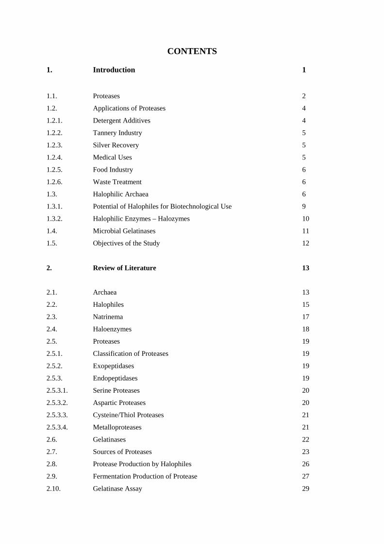
CONTENTS
1. Introduction 1
1.1. Proteases 2
1.2. Applications of Proteases 4
1.2.1. Detergent Additives 4
1.2.2. Tannery Industry 5
1.2.3. Silver Recovery 5
1.2.4. Medical Uses 5
1.2.5. Food Industry 6
1.2.6. Waste Treatment 6
1.3. Halophilic Archaea 6
1.3.1. Potential of Halophiles for Biotechnological Use 9
1.3.2. Halophilic Enzymes – Halozymes 10
1.4. Microbial Gelatinases 11
1.5. Objectives of the Study 12
2. Review of Literature 13
2.1. Archaea 13
2.2. Halophiles 15
2.3. Natrinema 17
2.4. Haloenzymes 18
2.5. Proteases 19
2.5.1. Classification of Proteases 19
2.5.2. Exopeptidases 19
2.5.3. Endopeptidases 19
2.5.3.1. Serine Proteases 20
2.5.3.2. Aspartic Proteases 20
2.5.3.3. Cysteine/Thiol Proteases 21
2.5.3.4. Metalloproteases 21
2.6. Gelatinases 22
2.7. Sources of Proteases 23
2.8. Protease Production by Halophiles 26
2.9. Fermentation Production of Protease 27
2.10. Gelatinase Assay 29

2.11. Purification of Protease 30
2.12. Characterization of Protease 32
2.13. Molecular Characterization 34
2.14. Recombinant Technology 36
2.15. Applications of Proteases 37
2.15.1. Detergent Industry 37
2.15.2. Leather Industry 37
2.15.3. Textile Industry 38
2.15.4. Pharmaceutical Industry 38
2.15.5. Food and Feed Industry 39
2.15.6. Peptide Synthesis 39
2.15.7. Silver Recovery 40
2.15.8. Other Applications 40
3. Materials and Methods 41
3.1. Isolation of Halophiles 41
3.1.1. Samples 41
3.1.2. Medium 41
3.1.3. Plating Procedures 41
3.1.4. Identification of Bacteria 42
3.1.4.1. Determination of Different Characteristics of Isolates 42
3.1.4.2. Molecular Classification of Isolates 42
3.2. Screening of Bacteria for Gelatinase Production 43
3.2.1. Media for Screening 43
3.2.2. Screening of Isolates Using Gelatin Media 44
3.2.3. Preparation of Crude Enzyme from Halobacterial and Archaeal Isolates
for Gelatinase Assay 45
3.2.4. Gelatinase Assay 45
3.3. Selection of Potential Haloarchaebacterium for Gelatinase Production 46
3.4. Selection of Media for Gelatinase Production 46
3.4.1. Culture Conditions in Liquid Media and Inoculum Preparation 47
3.4.2. Inoculation and Incubation 47
3.4.3. Recovery of Enzyme 48
3.5. Analytical Methods 48
3.5.1. Gelatinase Assay 48
3.5.2. Protein Estimation 48
3.5.3. Specific Activity 49
3.6. Growth Curve 49
3.7. Production of Protease by BTSH10 49
3.7.1. Inoculum Preparation and Incubation 49
3.7.2. Inoculation and Incubation 49
3.7.3. Recovery of Enzyme 49
3.7.4. Optimisation of Bioprocess Variables for Gelatinase Production
by BTSH10 49
3.7.4.1. NaCl Concentration 50
3.7.4.2. Initial pH of Medium 50
3.7.4.3. Incubation Temperature 50
3.7.4.4. Inoculum Concentration 51
3.7.4.5. Inoculum Age 51
3.7.4.6. Agitation 51
3.7.4.7. Additional Carbon Sources 52
3.7.4.8. Additional Nitrogen Sources 52
3.7.4.8.1. Inorganic Nitrogen Sources 52
3.7.4.8.2. Organic Nitrogen Sources 52
3.7.4.9. Detergents 53
3.7.4.10. Time Course Study Under Optimal Conditions 53
3.8. Purification of Enzyme. 54
3.8.1. Filtration 54
3.8.2. Gel Filtration Chromatography 54
3.8.2.1. Preparation of Column 54
3.8.2.2. Sample Preparation and Application on the Column 55
3.8.3. Dialysis 55
3.8.4. High Performance Liquid Chromatography 55
3.8.5. Polyacrylamide Gel Electrophoresis (PAGE) 56
3.8.5. 1. Reagents for Polyacrylamide Gel Electrophoresis 56
3.8.5. 2. Native – Polyacrylamide Gel Electrophoresis (Native -PAGE) 58
3.8.5. 2.1. Gel Preparation 58
3.8.5.2.2. Sample Preparation 59
3.8.5.2. 3. Procedure 59
3.8.5. 3. Reductive SDS-PAGE 60
3.8.5. 3.1. Gel Preparation 60
3.8.5. 3.2. Sample Preparation 60
3.8.5. 3.3. Procedure 61
3.8.5.4. Zymogram 61
3.8.6. Analytical Methods 61
3.8.7. Calculation of Yield of Protein, Yield of Enzyme Activity and
Fold of Purification 61
3.9. Characterization of Purified Enzyme 62
3.9.1. Optimal pH for Gelatinase Activity 62
3.9.2. Stability of Gelatinase at Different pH 62
3.9.3. Optimal Temperature for Gelatinase Activity 63
3.9.4. Stability of Gelatinase at Different Temperatures 63
3.9.5. Effect of Inhibitors on Gelatinase Activity 63
3.9.6. Substrate Specificity 63
3.9.7. Kinetic Studies 63
3.9.8. Effect of Various Metal ions on Gelatinase Activity 64
3.9.9. Effect of Various Detergents on Gelatinase Activity 64
3.9.10. Effect of Various Concentrations of NaCl on Gelatinase Activity 64
3.9.11. Effect of Organic Solvents on Gelatinase Activity 64
3.9.12. Analytical Methods 65
3.9.12.1. Residual Activity 65
3.9.12.2. Relative Activity 65
3.9.13. Application Studies 65
3.9.13.1. Decomposition of Gelatin Layer of X-ray film 65
3.10. Statistical analysis 66
4. Results 67
4.1. Isolation and Identification of Halophiles 67
4.1.2. Molecular classification of Isolates. 67
4.1.3. Phylogenetic Tree of Halophiles Isolated from Tannery Effluent and
Commercially Available Salt Crystals 69
4.2. Screening and Selection of Potential Halobacteria for
Gelatinase Production 70
4.3. Selection of Media for Gelatinase Production 72
4.4. Growth Curve of Natrinema sp. BTSH10 72
4.5. Optimization of Bio-Process Conditions for Gelatinase Production
by BTSH10 Natrinema sp. Under SmF 74
4.5.1. Optimisation of NaCl Concentration 74
4.5.2. Optimisation of Initial pH of Medium 75

4.5.3. Optimisation of Incubation Temperature 75
4.5.4. Optimisation of Inoculum Concentration 76
4.5.5. Optimisation of Inoculum Age 77
4.5.6. Effect of Agitation 77
4.5.7. Effect of Additional Carbon Sources 79
4.5.8. Effect of Additional Nitrogen Sources 79
4.5.8.1. Effect of Additional Inorganic Nitrogen Sources 79
4.5.8.2. Effect of Additional Organic Nitrogen Sources 80
4.5.9. Effect of Detergents 81
4.5.10. Time Course Experiment 82
4.6. Purification of Gelatinase 83
4.6.1. Filtration 83
4.6.2. Gel Filtration Chromatography 84
4.6.3. Dialysis 85
4.6.4. HPLC 85
4.6.5. Polyacrylamide Gel Electrophoresis 87
4.6.5.1. Native PAGE 87
4.6.5.2. Reductive SDS- PAGE 87
4.6.5.3. Zymogram 88
4.7. Characterisation of the Purified Enzyme 89
4.7.1. Optimal pH for Gelatinase Activity. 89
4.7.2. Stability of Gelatinase at Different pH 89
4.7.3. Optimal Temperature for Gelatinase Activity 90
4.7.4. Stability of Gelatinase at Different Temperatures 91
4.7.5. Effect of inhibitors on Gelatinase Activity 92
4.7.6. Substrate Specificity 93
4.7.7. Kinetic Studies 94
4.7.8. Effect of Various Metal ions on Gelatinase Activity 95
4.7.9. Effect of Various Detergents on Gelatinase Activity 97
4.7.10. Effect of Various Concentrations of NaCl on Gelatinase Activity 97
4.7.11. Effect of Organic Solvents on Gelatinase Activity 98
4.8. Application Studies 99
4.8.1. Decomposition of Gelatin Layer of X-ray Film 99

5. Discussion 103
5.1. Gelatinase production 105
5.2. Enzyme characteristics 110
6. Summary and Conclusions 117
References 121
List of Publications 157
Appendix-I

Introduction
1
Chapter 1
INTRODUCTION
Extracellular hydrolytic enzymes such as amylases, proteases, lipases,
DNases, pullulanases and xylanases have quite diverse potential usages in different
areas such as food industry, feed additives, biomedical sciences and chemical
industries.Industrial enzymes could be divided into four major categories, based on
application - detergent enzymes, technical enzymes, food enzymes and feed enzymes.
The technical enzymes could further be divided into textile enzymes, leather enzymes,
pulp and paper enzymes, fine chemicals enzymes, fuel ethanol enzymes and others
(van Beilen and Li, 2002).
According to the market research report on world enzymes published in 2007,
the world market for enzymes is expected to grow at the rate of 7.6% per year to $6
billion in 2011 (David et al., 2009). Survey on world sales of enzymes ascribes 31%
for food enzymes, 6% for feed enzymes and the remaining for technical enzymes
(Berka and Cherry, 2006; Agrahari, 2011).
Fig. 1.1. Global Industrial Enzyme Market 2008-2015 (Sarrouh et al., 2012)
A report by BCC Research (2011)stated that the global market for industrial
enzymes was estimated to reach a value of $3.3 billion in 2010 and is expected to

Introduction
2
reach $4.4 billion by 2015, a compound annual growth rate (CAGR) of 6% over the
5year forecast period. Singhal et al.(2012),reported that the world enzyme market was
at $5.1 billion and was expected to rise by 6.3% annually by 2013. According to
Sarrouh et al. (2012) technical enzymes, valued at over $1 billion in 2010, were
expected to increase at a 6.6% compound annual growth rate (CAGR) to reach $1.5
billion in 2015. The highest sales of technical enzymes occurred in the leather market,
followed by the bioethanol market (Fig. 1.1). The food and beverage enzymes
segment was speculated to reach about $1.3 billion by 2015, from a value of $975
million in 2010, rising at a compound annual growth rate (CAGR) of 5.1%. Within the
food and beverage enzymes segment, the milk and dairy market had the highest sales,
with $401.8 million in 2009 (Sarrouh et al., 2012).
Major enzyme producers are based in USA, Europe and Japan. Denmark
dominates the world enzyme production, with major players like Novozymes and
Danisco contributing 45% and 17% respectively; Genencor (USA), DSM (The
Netherlands) and BASF (Germany) making up the rest of world market (Binod et al.,
2008; BCC-Business Communications Company, Inc., 2009; Agrahari, 2011).
Microorganisms represents the most common source of enzymes as they are
relatively more stable and active than the enzymes derived from plant or animal
sources. They are preferred due to their broad biochemical diversity, feasibility, mass
culture and ease of genetic manipulations. Further microbes serve as a preferred
source of these enzymes owing to their rapid growth, requirement of limited space for
their cultivation and the ease with which they can be genetically manipulated to
generate new enzymes with altered properties that are desirable for their various
applications.
1.1.Proteases
Global Strategic Business Report (2012-
http://www.strategyr.com/Industrial_Enzymes_Market_Report.asp.), on enzymes
market highlighted the fact that proteases constitute the largest product segment in the
global industrial enzymes market. Among the various industrial enzymes
extracellular proteases are considered as important for the hydrolysis of external
proteins. Among the enzymes, microbial proteases account for approximately 60% of

Introduction
3
the total enzyme sales in the world (Singh et al., 2001, Banik and Prakash, 2004).
Proteases enable the cell to absorb and utilize hydrolytic products. Further proteases
hydrolyze peptide bonds in aqueous medium and also facilitate synthesis of peptides
in low water or non-aqueous medium. In fact, proteases that can be purified easily
have been commercially exploited to assist protein degradation in various industrial
processes. Thus, proteases are the largest selling industrial enzymes and their sale is
projected to increase further in the coming years with anticipated applications in
protein processing, peptide synthesis and detergent formulations. Microbial proteases
are probably one of the most extensively studied enzymes with wide ranging
applications in industries like detergent, pharmaceutical, food, waste management,
leather, diagnostics etc. (Tari et al., 2006; Bhaskar et al., 2007; Dodia et al., 2008;
Setati, 2010).
Industrial processes are carried out under specific physical and chemical
conditions, which cannot always be adjusted to the optimal values required for the
activity and stability of the available enzymes. Industrial applications of proteases
often require the enzymes to be stable in the presence of organic solvents. These most
often act to inactivate the enzymes and retard the rate of reaction. Several physical and
chemical methods such as chemical modification, immobilization, entrapment and
protein engineering have been employed for the stabilization of enzymes towards
organic solvents. However, if enzymes are naturally stable and exhibit high activities
in the presence of organic solvents, such stabilization is not necessary. Therefore, it
would be of great importance to have available enzymes showing optimal activities at
different values of pH, salt concentration, organic solvents and temperatures. In this
context, extensive research efforts are being directed to screen new sources of
proteases with novel properties.
Despite the facts that many different proteases have been identified and some
of them have been used in biotechnological and industrial applications, the present
proteases are not sufficient to meet most of the industrial demands. In view of these
restrictions, attention to isolation and characterization of proteases from extremophiles
is very important. In this context, halophiles are the most likely sources of such
enzymes, because not only are their enzymes salt tolerant but many are
thermotolerant.
Introduction
4
1.2.Applications of Proteases
Proteases are robust enzymes with considerable industrial potential in
detergents, leather processing, silver recovery, medical purposes, food processing,
meat tenderization, cheese making, dehairing, feeds, and chemical industries as well
as waste treatment. These enzymes contribute to the development of high value-added
applications or products by using enzyme aided (partial) digestion. Probably the
largest application of proteases is in laundry detergents, where they help removing
protein based stains from clothing (Banerjee et al., 1999). For an enzyme to be used
as an detergent additive it should be stable and active in the presence of typical
detergent ingredients, such as surfactants, builders, bleaching agents, bleach
activators, fillers, fabric softners and various other formulation aids. In textile
industry, proteases may also be used to remove the stiff and dull gum layers of
sericine from the raw silk fiber to achieve improved luster and softness. Protease
treatments can modify the surface of wool and silk fibers to provide new and unique
finish. Currently proteases are used in various industries including that of detergents,
food, pharmaceuticals, leather industry, basic research and for extraction of silver
from used X-ray films.
1.2.1.Detergent Additives
Enzymes used in detergents include proteases, amylases and lipases, of which
alkaline proteases hold a lion’s share and constitute 60-65% of the global industrial
enzyme market (Amoozegara et al., 2007). Microbial alkaline proteases dominate
commercial applications with a significant share of market owned by subtilisins and
/or alkaline proteases from Bacillus species for laundry detergent applications.
Alkaline proteases added to laundry detergents enable the release of proteinaceous
material from stains. The increased usage of these proteases as detergent additives is
mainly due to the cleaning capabilities of these enzymes in environmentally
acceptable, non phosphate detergents. In addition to improved washing efficiency, the
use of these enzymes allows lower wash temperatures and shorter period of agitations,
often after preliminary period of soaking. Ideally, proteases and other enzymes used
in detergent formulations should have high activity and stability over a broad range of
pH and temperature. The enzymes used should be effective at low levels and should
also be compatible with various detergent components along with oxidizing and

Introduction
5
sequestering agents. Very few published reports are available on the compatibility of
the alkaline proteases with detergents. Some cleaning applications are less demanding
than others. For instance, presoak formulations and contact lens cleaning solutions do
not require the same enzyme thermal stability as an all temperature laundry detergent.
1.2.2.Tannery Industry
Alkaline proteases possessing elastolytic and keratinolytic activity offer an
effective biotreatment of leather, especially the dehairing and bating of skins and
hides. The alkaline conditions enable the swelling of root hairs and subsequent attack
of proteases on the hair follicle protein allow for easy removal of the hair. The bating
followed by dehairing process involves the degradation of elastin and keratin, removal
of hair residues and the deswelling of collagen, which produce a good, soft leather
mainly used for making leather clothes and goods.
1.2.3.Silver Recovery
Alkaline proteases find potential applications in the bioprocessing of used X-
ray films for silver recovery. Used X-ray film contains approximately 1.5 to 2.0% (by
weight) silver in its gelatin layers. The conventional practice of silver recovery by
burning film causes a major environmental pollution problem. Thus, the enzymatic
hydrolysis of the gelatin layers on the X-ray films enables not only the silver, but also
the polyester film base to be recycled (Ishikawa et al., 1993).
1.2.4.Medical Uses
Collagenases with alkaline protease activity are increasingly used for the
therapeutic applications in the preparation of slow release dosage forms. A new semi
alkaline protease with high collagenolytic activity was obtained from Aspergillus
niger LCF9. The enzyme hydrolyzed various collagen types without aminoacid
release and liberated low molecular weight peptides of potential therapeutic use.
Similarly Elastoterase, a preparation with high elastolytic activity from Bacillus
subtilis 316M, was immobilized on a bandage for the therapeutic applications in the
treatments of burns and purulent wounds, furuncles and deep abscesses. Furthermore,
Bacillus species have been recognized as being safe to humans and an alkaline
protease having fibrinolytic activity has been used as a thrombolytic agent. Proteases
are also useful and important components in biopharmaceutical products such as
Introduction
6
contact-lens enzyme cleaners and enzymic debridement (Anwar and Saleemuddin,
2000). The proteolytic enzymes also offer a gentle and selective debridement
supporting the natural healing process in the successful local management of skin
ulcerations by efficient removal of the necrotic material (Sjodahl et al., 2002).
1.2.5.Food Industry
Alkaline proteases can hydrolyze proteins from plants, fishes or animals to
produce hydrolysates of well defined peptide profile. The commercial alkaline
protease-Alcalase has a broad specificity with some preference for terminal
hydrophobic aminoacids. Neutral proteases have been reported to be used to produce
soy sauce and soy products which are less bitter; they are used in brewing industry as
they are not sensitive to natural plant proteinase inhibitors (Rao et al., 1998).
1.2.6.Waste Treatment
Alkaline proteases from Bacillus subtilis was reported to be used for treatment
of waste feathers (Dalev, 1994).
1.3.HalophilicArchaea
Halophiles are microorganisms that adapt to moderate and high salt
concentrations. Halophiles include a great diversity of organisms, like moderately
halophilic aerobic bacteria, cyanobacteria, sulphur oxidizing bacteria, heterotrophic
bacteria, anaerobic bacteria, archaea, protozoa, fungi, algae and multicellular
eukaryotes. They are found in all three domains of life: Archaea, Bacteria and
Eukarya. Halophilic archaea is a member of the Halobacteriaceae family, the only
family in the Halobacteriales order. Halophilic bacteria grow over an extended range
of salt concentrations (3-15% NaCl, w/v and above). The term ‘halobacteria’ refers to
the red-pigmented extremely halophilic archaea, members of the Halobacteriaceae
family. Halobacteria is phylogenetically distinct from bacteria and eukaryotes, and
are classified as archaea. They exhibit features characteristic of the archaea, including
eukaryotic-like transcription and translation machinery, ether-linked lipids and like
some bacteria, a cell wall S-layer composed of glycoproteins (DasSarma and Arora,
2001). Microorganisms that are able to grow in the absence as well as in the presence
of salt are designated as halotolerant and those that are able to grow above
approximately 15% (w/v) NaCl (2.5 M) are considered extremely halotolerant.

Introduction
7
Extreme halophilic archaea are chemo-organotrophic organisms that satisfy
some of their energy requirements with light. These archaea are classified in one
order, Halobacteriales, and one family, Halobacteriaceae (Grant and Larsen, 1989).
Thereafter, 16S rDNA sequencing, DNA-DNA hybridization, polar lipid analysis and
other studies have recognized 40 genera (Parte, 2013). Some important genera are:
Halobacterium, Haloarchaea, Haloferax, Natronobacterium, Natronococcus (Tindall
et al., 1984; Torreblanca et al., 1986; Tindall, 1992; Grant and Larsen, 1989;
Halorubrum (McGenity and Grant, 1995), Halobaculum (Oren et al., 1995),
Natrialba (Kamekura and Dyall Smith, 1995), Natronomonas (Kamekura et al.,
1997), Halogeometricum (Montalvo-Rodriguez et al., 1998), Natrinema (McGenity et
al., 1998), Haloterrigena (Ventosa et al., 1999), Natronorubrum (Xu et al., 1999) and
Halorhabdus (Waino et al., 2000).
Members of the family Halobacteriaceae are characterized by red coloured
cells, the colour mainly being due to the presence of C50- carotenoids
(bacterioruberins) as the major carotenoids (Ronnekleiv and Liaaen-Jensen, 1995).
Some members of the genera Halobacterium and Haloarcula have been reported to
partially produce C40- carotenoids and Ketocarotenoids such as β-carotene, lycopene,
3-hydroxy echinenone and trans- astaxanthin and the minor carotenoids (Caloet al.,
1995).
Halophiles are microorganisms which grow over an extended range of salt
concentration (3-30% NaCl, w/v) and include the halotolerant bacteria and the
obligate halophilic archaea. They are found in salt marshes, marine ecosystems, salted
meats, hypersaline seas, salt evaporation pools and salt mines. Hypersaline
environment originates by the evaporation of sea water and are also called
thalassohaline environments. As water evaporates, sodium chloride precipitates and
salinity increases above 300 PSU. Despite the prevailing extreme environment, a
great diversity of extremophiles especially Haloarchaea have been reported in these
environments.
Halophiles are categorized as slight, moderate or extreme, by the extent of
their halotolerance. Slight halophiles prefer 0.3 to 0.8 M (1.8 to 4.7% - seawater is
0.6 M or 3.5%), moderate halophiles 0.8 to 3.4 M (4.7 to 20%), and extreme

Introduction
8
halophiles 3.4 to 5.1 M (20 to 30%) NaCl (Ventosa et al., 1998; Anton et al., 1999).
Halophiles require NaCl for growth in contrast to halotolerant organisms, which do
not require NaCl but can grow under saline conditions.
Halotolerant bacteria form a versatile group, adapted to life at the lower range
of salinities, with the possibility of rapid adjustment to the changes in the external salt
concentrations for survival. This property of halotolerant bacteria makes them better
candidates for bio-prospecting than their halophilic counterparts.
Halophilic archaea are considered as a potentially valuable resource in the
development of novel biotechnological processes and industrial applications in terms
of new pharmaceuticals, cosmetics, nutritional supplements, molecular probes,
enzymes and fine chemicals. Many of them are known to produce compounds of
industrial interests such as enzymes, polymers and osmoprotectants and some also
possess useful physiological properties which can facilitate their exploitation for
commercial purposes. Recently the biotechnological potential of these members of the
archaea has been recognized by researchers because of their unique features, which
facilitates many industrial products/ procedures.
Halophilic archaea have also been evaluated for bioremediation in harsh
environments for the degradation of organic pollutants (Margesin and Schinner, 2001)
and degradation of hydrocarbon by archaeal microbes under anoxic condition (Lovely,
2001). Biosurfactant producing halophilic archaea play a significant role in the
accelerated remediation of oil polluted saline environments. Certain strains of
halophilic archaea contain membrane bound retinal pigments, bacteriorhodopsin and
halorhodopsin, which enable microorganisms to use light energy to derive
bioenergetic processes (Oren, 1994; Lanyi, 1995). Furthermore, bacteriorhodopsin can
be exploited for the renewal of biochemical energy such as the back conversion of
ADP to ATP. A device based on bacteriorhodopsin and ATP synthesis has been
developed and patented (Saito et al., 1992). Thus, a wide variety of biotechnological
products such as bacteriorhodopsins, halorhodopsins, biopolymers, biosurfactants,
exopolysaccharides, polyhydroxyalkonates, flavoring agents, antitumor drugs and
enzymes are produced by halophilic archaea.

Introduction
9
1.3.1.Potential of Halophiles for Biotechnological Use
Extremozymes have great economic potential in many industrial processes
(e.g. agriculture, food, feed, drinks, detergents, textile, leather, pulp and paper).
Although there are controversial opinions about the potential of extremophiles, some
companies (e.g. Diversa, Genecor International Inc, Novozymes) and several research
groups are investing money and time, searching for these microbes and novel
applications of extremozymes.
The industrial and environmental applications of halophilic microorganisms
have been reviewed by Oren (2010). The review highlights the salient features of
halophiles, including their highly successful applications like β-carotene production
by Dunaliella and ectoine synthesis using Halomonas and other moderately halophilic
bacteria. BenAmotz and Avron(1989), have reported the use of Dunaliella for
production of β-carotene which is used as a food colourant, precursor of Vitamin A,
additive in cosmetics and preparation of multivitamins and health food preparations.
Bacteriorhodopsin the retinal protein proton pump of Halobacterium finds
applications in holography, artificial retina, neural network and optical computing.
Other possible use of halophilic microorganisms includes in the treatment of saline
and hyper saline waste waters and production of exopolysaccharides, poly β-
hydroxyalkanoate bioplastics and biofuels. Margesin and Schinner (2001) also
reported that H. cutrubrum was used for liposome production used in medicine and
cosmetics to transport compounds to specific target sites. Ectoine and hydroxyetoine
produced by Halomonas elongata KS3, are used in moisturizers and ectoine is also a
stabilizer in PCR (Motitschke et al., 2000). These microorganisms can be used as a
source of metabolites, compatible solutes and other compounds of industrial value.
Novel halophilic biomolecules may also be used for specialized applications
e.g. bacteriorhodopsin for biocomputing, pigments for food colouring and compatible
solutes as stress protectants (DasSarma et al., 2001). Exopolymer poly (γ-D-glutamic
acid) produced by Natrialba is used as a biodegradable thickener and drug carrier in
food or pharmaceutical industry (Kunioka,1997; Hezayen et al., 2000).
Biodegradation of organic pollutants by halophilic bacteria and archaea has been
recently reviewed (Le Borgne et al., 2008). These microorganisms are good
candidates for the bioremediation of hypersaline environments and the treatment of

Introduction
10
saline effluents. Halobacteriumis used for bioremediation of oil spills in saline
environments and degradation of n-alkanes with C10 – C30 (Kulichevskaya et al.,
1992).Yongsawatdigul et al. (2007), has reported the use of species of Halococcus,
Bacillus and Vibriobacillus for production of Thai fish sauce. Ryu et al.(1994)
reported the isolation of a serine protease from Halobacterium halobium which could
be used as a catalyst for the production of glycine containing peptides in presence of
organic solvents.
Halophilic bacteria are a potential source of extracellular hydrolases like
proteases with a wide array of industrial applications. These enzymes exhibit stability
over a range of saline conditions (Shivanand and Jayaraman, 2009). The importance
of proteases is highlighted by the fact that they have many practical applications in
biotechnology and industry (Rao et al., 1998).Halophilic bacteria constitute excellent
models for the molecular study of osmoregulatory mechanisms (Ventosa et al., 1998).
1.3.2.HalophilicEnzymes - Halozymes
Moderately halophilic bacteria that grow optimally in a media containing 3-
15% NaCl are considered as a likely source of such enzymes. Hence, halophilic
microorganisms are perceived to be a valuable source of enzymes with unique
structural features and properties. In order to survive in saline environments, these
organisms accumulate high concentration of salts (most often NaCl or KCl) or
osmolytes (e.g. betaine, glycerol) in the cytoplasm (Le Borgne et al., 2008). As a
consequence, their enzymes are generally salt stable. In terms of water availability,
saline environments are similar to non-aqueous systems. Therefore, halophilic
enzymes should logically be stable in organic solvents. Although large numbers of
salt-stable enzymes have been reported from halophilic sources, stability towards
organic solvents has been noticed in only few cases. Further, enzymes from the
halophilic archaea tend to be more thermostable than expected from the organism’s
growth temperature.
Halophiles from the archaeal domain provide the main source of extremely
halophilic enzymes. The potentials of halophiles and haloenzymes have been
highlighted in literature (Eichler, 2001; Oren, 2002). Extracellular halophilic enzymes
such as xylanases, amylases, proteases and lipases has been reported in many

Introduction
11
halophiles belonging to the genera Haloferax, Actinobacter, Halobacterium,
Marinococcus, Natronoccus, Halobacillus, Halorhabdus and Halothermothrix
(Adams et al., 1995; Sellek and Chaudhari, 1999; Madern et al., 2000; Mevarech et
al., 2000; Eichler, 2001). Halophilic microorganisms produce stable enzymes
(including many hydrolytic enzymes such as DNases, lipases, amylases, gelatinase
and protease) capable of functioning under high concentration of salt which leads to
precipitation or denaturation of most proteins. Most halophilic enzymes are
inactivated and denatured at concentration of NaCl below 1M. Examples of
halophilic enzymes are serine proteases from the extreme halophilic Halobacterium
halobium (Izotova et al., 1983), DNA topoisomerases from Methanopyus kandleri
(Kozyavkin et al.,1994), extremely halophilic β- galactosidase from Haloferax
alicantei (Holmes et al.,1997), D-hydantoinase from halophilic Pseudomonas species
(Sudge et al.,1998) and halophilic α- amylase from Nesterenkonia sp. (Shafiei et al.,
2012).
1.4.Microbial Gelatinases
Gelatinase is one type of diverse group of protease, an extracellular metallo-
endopeptidase or metalloproteinase which is able to hydrolyze gelatin and other
compounds such as pheromone, collagen, casein and fibrinogen (Makinen and
Makinen, 1994; Makinen et al., 1989). Gelatinase and collagenase are important
metalloproteases and these are widely used not only in chemical and medical
industries but also in food and basic biological sciences (Hisano et al., 1989).
Bacterial metalloproteases are associated with virulence and matrix
metalloproteases of eukaryotes play a role in processing of precursors which play
modulation roles in tumor formation (Lennarz et al., 1991; Makinen and Makenin,
1994). Mazotto et al.(2010) have reported the isolation of Bacillus subtilis AMR from
poultry wastes which could hydrolyze human hair producing serine peptidases with
keratinase and gelatinase activity. They suggested that the peptides obtained from
enzymatic hydrolysis of hair may be useful for the production of pharmaceutical and
cosmetic formulations. Thus, gelatinases could be used for recovery of silver from
used photographic films, treatment of waste (poultry and animal waste) and they are
medically important as targets for drug development and for design of inhibitors for
disease treatment. Mazollo et al. (2011) have isolated Bacillus sp. capable of acting

Introduction
12
on gelatin, keratin and casein from agroindustrial residues in a poultry farm. They
observed degradation of feather along with production of enzyme using feather as a
cheap eco-friendly substrate.
Gelatinase enzyme produced by microorganism hydrolyze gelatin into its sub
compounds (polypeptides, peptides and amino acids) that can cross the cell membrane
and be used by the organism. Forms of gelatinases are expressed in several bacteria
including Pseudomonas aeruginosa, Staphylococcus aureus, Clostridium perfringens,
Serratia marcescens and Bacillus (Shanmugasundaram et al., 2012). The potential
uses of gelatinase and their high demand, the need exists for the discovery of new
strains of bacteria that produce enzymes with novel properties.
1.5. Objectives of the Study
Among the proteases ‘gelatinases’ are those enzymes which cleave gelatin,
casein, fibrinogen, etc.to result in polypeptides, peptides and amino acids. They are
metalloendopeptidases which have applications in leather industry, production of fish
sauce, fish processing, peptide synthesis etc. Whereas literature available on
halophilic proteases, particularly gelatinases is rather scanty, while there is more
scope in exploring halophiles, as a source of proteases. Considering the potentials of
gelatinase for industrial applications and the lack of information available in literature
on haloarchaeal gelatinase it was desired to explore the haloarchaea towards isolating
potential gelatinase producing halophilic archaea towards their prospective utilization
in industry. Hence this study was planned with the following objectives.
Specific objectives of the present study include:
1) Isolation of halophiles and screening of gelatinase.
2) Optimization of bioprocess conditions for gelatinase production under SmF
by Natrinema sp. BTSH10.
3) Purification of the enzyme.
4) Characterization of the enzyme.
5) Application studies.

Review of literature
13
Chapter 2
REVIEW OF LITERATURE
2.1.Archaea
Archaea, which are highly adapted to survive in extreme environments,
comprise of hyperthermophiles, halophiles and methanogens and are more closely
related to the Eukarya than to the Eubacteria (Fig. 2.1) (Bullock, 2000). Based on 16S
rDNA analysis, archaea are classified into four major Kingdoms – Crenarchaeota,
Euryarchaeota, Korarchaeota (Grant and Larsen,1989) and Nanoarchaeota (Huber et
al., 2002).
The Kingdom Crenarchaeota comprises organisms that thrive in very hot and
very cold environments. Majority of cultured Crenarchaeotes are hyperthermophiles,
isolated from geothermally heated soils or wastes containing elemental sulphur and
sulphides. Psychrophilic Crenarchaeotes have been identified from community
sampling of 16S rRNA genes from many non thermal environments. Marine
planktonic Crenarchaeotes have been isolated in large numbers from Antarctic region
(Madigan et al., 2009).
The Kingdom Euryarchaeota includes thermophilic methanogens,
methanogens, halophiles and hyperthermophiles. Methanogens are obligate anaerobes
abundantly seen in intestinal tracts of animals, sewage treatment facilities, marine and
fresh-water sediments, bogs and deep soils. Extremely halophilic archaea are a
diverse group of prokaryotes that inhabit hyper saline environments such as solar salt
evaporation ponds, the surfaces of heavily salted foods like certain fish and meats, and
natural salt lakes. Extreme halophiles are obligate aerobes with a requirement of high
salt concentrations for growth (Madigan et al., 2009).
The Kingdom Korarchaeota includes hyper thermophiles growing optimally at
85°C and were originally discovered from iron and sulphur-rich Yellow Stone hot
spring, Obsidian Pool (Madigan et al., 2009).

Review of literature
14
Kingdom Nanoarchaeota has only one representative, Nanoarchaeum
equitans which is an obligatory symbiont on the archaeon Ignicoccus (Huber et al.,
2002). Nanoarchaeum equitans has the smallest archaeal genome (around 500 kb)
and the initial studies of single stranded ribosomal RNA indicated a vast difference
between this group and the Kingdoms Crearchaeota and Euryarchaeota. Brochier et
al.(2005) suggested that the initial sample of ribosomal RNA was biased and
Nanoarchaeum actually belongs to Euryarchaeota.
Fig. 2.1. Phylogenetic tree of the three domains of life (Allers & Mevarech,
2005).
The term extremophile collectively applies to a number of bacteria and
archaea that grow optimally under ‘extreme’ conditions such as acidic or alkaline pH,
extremes of temperature, extremes of atmospheric pressure and extremes of salt or
organic ion concentrations. Extremophiles are best characterised according to their

Review of literature
15
growth profiles, using marginal data, under certain culture or environmental
conditions, such as NaCl ranges (NaClopt, NaClmin, NaClmax) or temperature profile
(Topt, T min, T max) (Mesbah and Wiegel, 2008). Examples of extremophiles include,
thermophiles (high temperature), psychrophiles (low temperature), acidophiles (low
pH), alkaliphiles (high pH), piezophiles (high pressure, formerly known as
barophiles), halophiles (high salt concentration), osmophiles (high concentration of
organic solutes), oligotrophs (low concentration of solutes and or nutrients) and
xerophiles (very dry environment) (Mesbah and Wiegel, 2008). Extreme
environments are proving to be a valuable source of microorganisms that secrete
interesting new molecules and these properties seem to offer numerous applications in
various fields of industry (Margesin and Schinner, 2001).
2.2.Halophiles
Extremely hypersaline habitats are seen in hot, dry areas of the world. They
are of two types: thalassohaline and athalassohaline environments. Hypersaline
environments which originate by evaporation of sea water are called thalasssohaline
environments (eg. Great Salt Lake). Their salt composition is similar to that of sea
water, with the dominating ions being sodium and chloride ions. The pH is near
neutral or slightly alkaline. Thalassohaline brines (saltern crystallizer ponds) display
bright colouration due to the large numbers of pigmented microorganisms they
harbour. In athalassohaline hypersaline environments, like the Dead Sea, the
concentration of divalent cations exceeds that of monovalent cations and the pH is
around 6. Oren (1988) reported presence of microorganisms in the Dead Sea. Cayol
et al. (1994), have reported microorganisms capable of tolerating high salt
concentrations (200g/L) and high temperatures of around 68°C. Jie Lu et al. (2001),
isolated an extremely halotolerant Oceanobacillus ilheyensis from Ilheya Ridge at a
depth of 1050 m below sea level. Oren (2002) reported presence of microbial life in
alkaline soda lakes with high pH values of 11 and higher and high salt concentrations
about 300g/L. Rohban et al.(2009) reported hydrolytic enzyme producing
Oceanobacillus sp. isolated from Howz Soltan Lake in Iran.
Halophilic archaea have requirement for high concentrations of NaCl (3.5-4.5
M). Some strains may grow at low salt concentrations of 1.5 M NaCl while others
grow well in saturated NaCl (5.2 M). Haloarchaea accumulate KCl up to 5 M

Review of literature
16
(Matheson et al., 1976). It was reported that intracellular enzymes of halophilic
archaea have requirement for high levels of KCl (Kushner, 1985). Halobacillus
halophilus a moderate halophile was shown to use a hybrid strategy for
osmoadaptation by accumulating both molar concentrations of chloride and
compatible solutes (Hänelt and Müller, 2013). This distinctive feature enables H.
halophilus to grow over a broad range of salinities (up to 3 M) and to adapt
sufficiently to rapidly changing environments. The salinity and growth-phase
dependent adaptation of the accumulated solutes is incredible and probably
demonstrates a long lasting evolution being optimally prepared for its changing
environment. A dominant compatible solute, such as carbon and nitrogen is used to
guarantee energy optimization.
According to Grant et al. (1998), who reviewed the diversity of halophilic
bacteria and archaea, halophilic bacteria include Chlorobium limnicola, Thiocapsa
halophila, species of Acinetobacter, Alteromonas, Deleya, Flavobacterium,
Marinomonas, Pseudomonas and Vibrio. Species belonging to genera Marinococcus,
Bacillus, Sporosarcina and Salinococcus have been isolated from saline soils and
salterns. Hypersaline waters harbour archaeal genera including Haloarcula,
Halococcus, Halobaculum, Halobacterium, Halorubrum, Haloferax and
Haloterrigena. Halobacterium salinarum has been isolated from salted food. Hyper
saline lakes also harbour halophilic methanogens like Methanohalophilus mahii, M.
halophilus and M. evestigatum. According to Antón et al.(2000 and 2002),
Eubacteria belonging to genus Halorhodospira (γ-Proteobacteria), the actinomycete
Actinopolyspora halophila and Candidatus salinibacter resemble haloarchaebacteria
in their salt requirement.
The classification of halophilic archaea is as follows- Domain: Archaea,
Class: Halobacteria, Order: Halobacteriales and Family: Halobacteriaceae. Family
Halobacteriaceae comprises of 40 genera encompassing 137 species (Parte, 2013;
Minegishi, 2013). Some of the genera include Halalkalicoccus, Halobaculum,
Halobiforma, Halomicrobium, Halobacterium, Haloarcula, Haloferax, Halococcus,
Halorhabdus, Halorubrum, Halosimplex, Halostagnicola, Halovivax, Natrialba,
Halogeometricum, Haloterrigena, Natrinema, Natronolimnobius, Natronomonas,
Natronococcus and Natronorubrum (Grant et al.,2001; Oren et al.,2002; Itoh et

Review of literature
17
al.,2005; Castillo et al.,2006a and 2006b; Gutierrez et al.,2007). In fact the
composition of membrane polar lipids have been used as one of the key
chemotaxonomic criteria for the differentiation of haloarchaeal genera (Kamekura and
Kates, 1999).
2.3. Natrinema
Natrinema sp. J7, previously named as Halobacterium salinarum J7, was
isolated from a salt mine in Hubei province, China. It was found that this strain
harbors a high copy number plasmid pHH205 and possesses extracellular proteolytic
activity(Ye et al., 2003). A gene encoding an extracellular protease, SptA, was cloned
from the halophilic archaeon Natrinema sp. J7. The SptA gene was expressed in
Haloferax volcanii WFD11, and the recombinant enzyme could be secreted into the
medium in an active mature form. The N-terminal amino acid sequencing and
MALDI-TOF mass spectrometry analysis of the purified SptA protease indicated that
the 152-amino acid prepropeptide was cleaved and the C-terminal extension was not
processed after secretion. The SptA protease was optimally active at 50°C in 2.5 M
NaCl at pH 8.0. When the twin-arginine motif in the signal peptide of SptA protease
was replaced with a twin-lysine motif, the enzyme was not exported from Hfx.
volcanii WFD11 (Shi et al., 2006). A halophilic extracellular serine protease
produced by Natrinema sp. R6-5 with molecular size 62 kDa was purified using
bacitracin-Sepharose 4B chromatography. The protease exhibited optimum activity at
NaCl concentration of 3 mol/L. At the optimum NaCl concentration of 3 mol/L, the
optimum temperature and the optimum pH were 45°C and 8.0 (Shi et al., 2007).
Natrinema sp., isolated from a hypersaline lake in Iran produced 6 different types of
enzymes including protease, lipase, pullulanase, cellulase, chitinase and inulinase
(Makhdoumi Kakhki et al., 2011). Natrinema sp., isolated from Lonar lake in
Maharashtra was found to produce amylase, caseinase, cellulase and xylanase (Patil
and Bajekal, 2013).
Feng et al. (2012) sequenced the complete genome of Natrinema sp. J7-2, an
extreme haloarchaeon capable of growing on synthetic media without amino acid
supplements. The complete genome sequence of Natrinema sp. J7-2 was found to be
composed of a 3,697,626-bp chromosome and a 95,989-bp plasmid pJ7-I. This was
the first report of complete genome sequence of a member of the genus Natrinema.

Review of literature
18
They reconstructed the biosynthetic pathways for all 20 amino acids and discussed a
possible evolutionary relationship between the haloarchaeal arginine synthetic
pathway and the bacterial lysine synthetic pathway. The genome harboured the genes
for assimilation of ammonium and nitrite, but not nitrate, and had a denitrification
pathway to reduce nitrite to N2O. Natrinema sp. BTSH10 isolated from saltpan of
Kanyakumari, Tamilnadu, India was identified, and medium for enhanced production
of halocin SH10 was optimized (Karthikeyan et al., 2013).
2.4. Haloenzymes
Halozymes are enzymes produced by the halophilic archaea. Several
enzymes isolated from archaea such as xylanases and cellulases could play important
roles in the chemical, pharamaceutical, paper pulp or waste treatment industries.
Research on hydrolytic enzymes from halophilic organisms was pioneered by Norberg
and Hofsten in1969 (Norberg and Hofsten, 1969). Since then, a considerable amount
of effort has been directed towards the evaluation of extracellular salt-tolerant
enzymes of the moderately halophilic bacteria and the use of such enzymes in
biotechnological processes (Ventosa et al., 1998; Eichler, 2001; Oren, 2002; Zhang
and Kim, 2010). Halophilic enzymes have been suggested for use in biotechnological
applications due to their halotolerance, thermostability for long incubation periods and
ability to retain activity in presence of high levels of organic solvents (Eichler, 2001;
Madern et al., 2000). Halophilic bacteria producing alkaline proteases displaying
thermostability, activity at high pH, organic solvent stability and detergent
compatibility have been reported (Makhija et al., 2006). Hydrolases and isomerases
from extremely Haloarchaea have potential application in several biotransformations
in the production of supplements and are exploited in the production of fermented
food (Margesin and Schinner, 2001). Extracellular production of halophilic enzymes
such as xylanases, amylases, proteases and lipases has been reported from many
halophiles belonging to the genera Haloferax, Halobacterium, Halorhabdus,
Marinococcus, Micrococcus, Natronococcus, Halobacillus and Halothermothrix
(Eichler, 2001; Zhang and Kim, 2010). Production of a fructose-1,6-biphosphate
aldolase from Haloarcula vallismortis (Krishnan and Altekar, 1991), lipase by
Natronococcus sp. (Bhatnagar et al., 2005) and β-xylanase by Halorhabdus utahensis
(Waino and Ingvorsen, 2003) was also reported. In addition, use of halophilic
organisms and their enzymes for biodiesel production (Begemann et al.,2011) and for

Review of literature
19
degrading cellulosic biomass with reduced requirement for high temperature and pH
neutralization of pretreated biomass before fermentation, have been also reported.
2.5. Proteases
2.5.1. Classification of Proteases
Barrett (2001) has classified proteases based on three criteria:
(i)Type of reaction catalyzed,
(ii) Evolutionary relationship with reference to structure and
(iii) Chemical nature of catalytic site.
Based on their site of action proteases are broadly classified as exopeptidases
and endopeptidases. Based on their catalytic mechanism, proteases are classified into
four types, (a) serine proteases,(b) aspartic proteases,(c) cysteine proteases and (d)
metalloproteases. Depending on the pH of optimal activity, proteases are classified
into three types and they are acidic, neutral and alkaline proteases.
2.5.2.Exopeptidases
Exopeptidases cleave peptide bonds near the ends of the polypeptide chain
and are further classified as aminopeptidases (acting at amino terminus) and
carboxypeptidases (acting at carboxy terminus). Aminopeptidases act at N- terminus
of polypeptide chain liberating a single amino acid or a dipeptide or a tripeptide.
Carboxypeptidases act at C- terminus of the polypeptide chain liberating a single
amino acid or a dipeptide. Based on the nature of the amino acid residues at the active
site of the enzymes carboxypeptidases are mainly of three types (Rao et al., 1998)
and they are: (a) serine carboxypeptidases, (b) metallocarboxypeptidases and(c)
cysteine carboxypeptidases,
2.5.3. Endopeptidases
Endopeptidases cleave peptide bond within the polypeptide chain, the
presence of free amino or carboxyl group bears a negative influence on enzyme
activity. Based on their catalytic mechanism these enzymes are grouped into four
types: (a) Serine proteases, (b) Cysteine/thiol proteases, (c) Aspartic proteases and
(d) Metalloproteases.

Review of literature
20
2.5.3.1.Serine Proteases(Cera,2009)
The hall mark of serine proteases is the presence of a serine in their active site
(E.C.3.4.21).These enzymes are reported to have esterolytic and amidase activity also.
Barett (1994) classified serine proteases into 40 families, which were further
subdivided into about 13 clans. He has indicated four separate evolutionary origins
for serine proteases because the primary structure of four clans are unrelated
(Chymotrypsin (SA), subtilisin (SB), carboxypeptidase C (SC) and Escherichia D-
Ala-D-Ala peptidase A(SE)). Serine proteases are generally active at neutral and
alkaline pH, and the isoelectric points of serine proteases fall within the pH range 4-6.
Alkaline serine proteases embody the largest subgroup of serine proteases. They
cleave a peptide bond having a tyrosine, leucine or phenylalanine at the carboxyl side
of the splitting bond. The optimum pH for activity of alkaline proteases is about 10,
with the isoelectric point around pH 9. Serine proteases are irreversibly inhibited by
inhibitors like phenylmethylsulfonyl fluoride (PMSF) 3,4-dichloroisocoumarin (3,4-
DCI), tosyl-L-lysine chloromethyl ketone (TLCK), L-3-carboxy trans-2,3-
epoxypropyl-leucylamido (4-guanidine) butane (E.64) and di-
isopropylfluorophosphate (DFP). Some of the serine proteases, having a cysteine
residue near the active site are inhibited by thiol reagents such as p-
chloromercuricbenzoate (PCMB).
2.5.3.2. Aspartic Proteases
Aspartic proteasesalso called acidic proteases, have aspartic acid residues in
their catalytic site (E.C.3.4.23). Acidic proteases are placed in clan AA. Barett,
(1995), recognized three families - pepsin (A1), retropepsin (A2), and enzymes from
pararetroviruses (A3). Most aspartic proteases are active at low pH (pH 3 to 4) and
have isoelectric points in the range of pH 3 to 4.5. Inhibitors of aspartic proteases
include pepstatin (Fitzgerald et al., 1990), diazoketone compounds such as di-
azoacetyl-DL-norleucine methyl ester (DAN) and 1,2-epoxy-3-(p-nitrophenoxy)
propane (EPNP) in the presence of copper ions. Microbial aspartic proteases include
(i) pepsin-like enzymes produced by Penicillium, Aspergillus, Neurospora and
Rhizopus, and (ii) rennin-like enzymes produced by Mucor and Endothia (Rao et al.,
1998).

Review of literature
21
2.5.3.3.Cysteine/Thiol Proteases (Grzonka et al., 2001)
As many as 21 different families of cysteine proteases (E.C.3.4.22) have been
described, occuring in both prokaryotes and eukaryotes (Grzonka et al., 2001). The
catalytic activity of cysteine proteases depends on a dyad consisting of cysteine and
histidine residues. The order of Cys and His (Cys-His or His-Cys) residues differ
among the families (Barett, 1994). Cysteine proteases are active only in the presence
of reducing agents such as cysteine or HCN.
They are divided into four groups on the basis of their side chain specificity,
(i) papain-like, (ii) trypsin- like with preference for cleavage at the arginine residue,
(iii) specific to glutamic acid, and (iv) others. The optimum pH of cysteine proteases
is 7, but, lysosomal proteases are optimally active at acidic pH. They are susceptible
to sulfhydryl agents such as PCMB but are unaffected by DFP and metal-chelating
agents. Mechanism of action of cysteine proteases resembles that of aspartic
proteases. Cysteine proteases cause the hydrolysis of carboxylic acid derivatives via a
double-displacement pathway involving general acid-base formation and hydrolysis of
an acyl-thiol intermediate.
2.5.3.4.Metalloproteases
Metalloproteases (E.C.3.4.24) are a large, diverse group of proteases (Barett,
1995), which require a divalent metal ion for their activity and this accounts for their
inactivation by the addition of chelating agents or by dialysis. The ion coordinates to
the protein via three ligands (histidine, glutamate, aspartate, lysine or arginine) and a
labile water molecule. Rao et al. (1998) reported that neutral metalloproteases show
specificity for hydrophobic amino acids, while the alkaline metalloproteases have a
very broad specificity.
About 61 families of metalloproteases have been recognized (Jisha et al.,
2013), of which 17 contain only endopeptidases, 12 contain only exopeptidases, and 1
(M3) contains both endo- and exopeptidases (Rao et al., 1998). Families of
metalloproteases have been grouped into 16 clans based on the nature of the amino
acid that completes the metal-binding site; e.g., clan MA has the sequence HEXXH-E
and clan MB corresponds to the motif HEXXH-H (Rao et al.,1998). The activity of
metalloprotease requires the binding of a divalent metal ion to a His-Glu-Xaa-Xaa-His
Review of literature
22
motif. It is believed that the Glu143 helps the nucleophilic attack of a water molecule
on the carbonyl carbon of the scissile peptide bond polarized by the Zn2+ ion (Holm
and Mathews, 1981).
The HExxH motif forming a α-helix is well conserved active site in many
monozinc enzymes as in which the two histidine residues coordinate with the zinc ion.
Zinc-binding motifs, of some monozinc proteases are, HxxE(D)-aan-H in the
carboxypeptidase family and HxD-aa12-H-aa12-H in the matrix metalloprotease
family. Dipeptidyl peptidase (DPP) III has a unique zinc binding HELLGH motif as
active site which coordinated with a zinc ion. The motif HELLGH could not be found
in any other metalloproteases, it exists in three kinds of monooxygenases (tyrosine,
phenylalanine, and tryptophan hydroxylases) as an iron-binding site, as revealed by a
search of the NBRF-PIR protein sequence database (Fukasawa et al., 2011).
2.6.Gelatinases
Gelatinases are proteases and found in humans as matrix metalloproteinases
(MMP 2 & 9) which break down extracellular matrix, playing a role in embryonic
development, morphogenesis, reproduction and tissue remodelling as well as in
diseases like arthritis, cardiovascular and neurological diseases and also cancer and
metastasis hence, they are medically important as targets for drug development
(Pacheco et al., 1998; Stetler-Stevenson et al., 1993; Deryugina and Quigley, 2006;
Zitka et al., 2010). It has been proved that bacterial metalloproteases are associated
with virulence and matrix metalloproteases of eukaryotes play a significant role in
processing of precursors which play modulation roles in tumour formation (Lennarz et
al.,1991; Makinen et al.,1994). Metalloproteases have thus attracted considerable
attention for development of inhibitors for disease treatment (Tamaki et al.,1995).
Gelatinases have role in connective tissue degradation associated with tumour
metastasis, hence, they are medically important as targets for drug development
(Pacheco et al., 1998; Stetler-Stevenson et al., 1993). Matrix metalloproteinases
(MMPs) degrade components of extracellular matrix (Murphy and Docherty, 1992).
Gelatinases A and Gelatinase B cleave gelatins and also types IV and V collagens
(Collier et al., 1988) and elastin (Senior et al., 1991). Production and
characterization of a collagenolytic serine proteinase by Penicillium
aurantiogriseum URM 4622 was reported by Lima et al.(2011).
Review of literature
23
Tetracycline antibiotics inhibit mammalian tissue destructive proteinases like
collagenases and gelatinases during disease processes like tumour invasion,
metastasis, angiogenesis, periodontitis etc. and chemically modified non-antibacterial
analogues of tetracycline was reported to inhibit these enzymes without producing the
typical side effects induced of antibiotics (Golub et al.,1996). Inactivation of gene
encoding zinc metalloprotease gelatinase was found to prevent biofilm formation and
this enzyme was suggested as a unique target for therapeutic intervention in
enterococcal endocarditis (Hancock and Perego, 2004).
Gupta and Ramnani (2006) reported that feather hydrolysates obtained after
feather degradation could be used as additives for feedstuffs, fertilizers, glues and
films or used for the production of the rare amino acids -serine, cysteine, and proline.
Mazotto et al.(2010) isolated Bacillus subtilis AMR from poultry waste which could
hydrolyse human hair and reported that serine peptidases with keratinase and
gelatinase activity hydrolysed human hair indicating that the peptides obtained may
find applications in pharmaceutical and cosmetic formulations. Mazollo et al.(2011)
reported the isolation of Bacillus sp. from agroindustrial residues in a poultry farm,
which could degrade gelatine, keratin, casein etc. They demonstrated that feather
waste could be used as a cheap and ecofriendly substrate for the enzyme.
2.7. Sources of Proteases
Microbes are the preferred source of proteases owing to their great
biochemical diversity and susceptibility to genetic manipulation. They can be
cultured in vast quantities within a short span of time to ensure an abundant supply of
proteases. Extracellular nature of microbial proteases simplify the downstream
processing of enzyme and have a long shelf life, with less stringent storage
requirements. Only non-toxic and non-pathogenic microbes are used for commercial
production and referred to as “genetically regarded as safe”(GRAS).GRAS is an
acronym forthe phrase Generally Recognized As Safe
(http://www.gmo.hr/eng/Additional-content/Glossary/generally-recognized-as-safe-
GRAS). Bacterial neutral proteases have low thermotolerance and have an affinity for
hydrophobic amino acids. Some of them are metalloproteases with a requirement for
divalent metal ions for activity, others are serine proteases. Bacterial alkaline

Review of literature
24
proteases are characterized by optimal activity at high pH, broad substrate specificity
with optimal temperatures around 60°C which make them ideal for use in detergent
industry (http://novozymesbiotech.com). Bacillus sp. is a potent producer of neutral
and alkaline proteases among bacteria and prominent among them are Bacillus
licheniformis, Bacillus subtilis, Bacillus amyloliquefaciens etc. (Kumar et al., 1999a;
Rao et al., 1998; Schallmey et al., 2004; Fujinami and Fujisawa, 2010). Bacillus
subtilis (MTCC9102) was reported to produce keratinase by solid state fermentation
using horn meal as a substrate (Kumar et al., 2010). Pseudomonas sp. was
recognized as potent protease producers (Bayoudh et al., 2000). A metalloprotease
secreted by Pseudomonas aeruginosa MTCC 7926 which was found to be useful for
dehairing of animal skin, processing of X-ray film and exhibited antistaphylococcal
activity was reported by Patil and Chaudhari (2009). A Ca2+dependent
metalloprotease, capable of degrading insect tissues, was purified from
entomopathogenic bacterium Photorhabdus sp. strain EK1 (PhPrtPI) (Soroor et al.,
2009). Halophilic proteases have been isolated and characterized from several
bacterial species including Bacillus species (Shivanand and Jayaraman, 2009). A
marine Exiguobacterium sp. was reported for concomitant production of protease and
antioxidant materials from shrimp biowaste (Anil Kumar and Suresh, 2014). A
fibrinolytic enzyme producing Pseudoalteromonas sp. IND11 was isolated from the
fish scales using cow dung as a substrate by solid-state fermentation (Vijayaraghavan
and Vincent, 2014).
Aspergillus sp., (Chakrabarti et al., 2000), Rhizopus sp. (Banerjee and
Bhattacharyya, 1992), Conidiobolus sp. (Bhosale et al., 1995), Penicillium sp. are the
different fungi which elaborate proteases. Solid state fermentation for protease
production is usually done using fungi. Fungal acid proteases have optimum activity
at acidic pH and fungal neutral proteases are usually metalloproteases active at neutral
pH. Candida sp. (Poza et al., 2001) and Streptomyces (Petinate et al., 1999) are
known producers of proteases. Alkaline proteases from yeasts like Cryptococcus
aureus, Aureobasidium pullulans, Issatchenkia orientalis and Yarrowia lipolytica (Li
et al., 2009) and mushrooms have also been reported (Zhang et al., 2010; Zheng et
al., 2011).

Review of literature
25
Viruses are known to elaborate serine peptidases, aspartic peptidases and
cystine peptidases, all of which are endopeptidases (Rawlings and Barrett, 1993).
Methanococcus jannaschii, a thermophilic methanogen isolated from deep-sea
was shown to produce a hyperthermophilic and barophilic protease. The enzyme
showed specificity for leucine at the P1 site of polypeptide substrates and the activity
and thermal stability of the enzyme increased with increase in pressure (Michels and
Clark, 1997). Pyrococcus furiosus a hyperthermophilic archeaon was shown to
produce intracellular proteases (Halio et al., 1997). A moderately thermophilic and
halotolerant alkaline protease secreted by Salinivibrio sp. strain AF-2004 exhibiting
broad pH ranges (5.0–10.0) was described by Amoozegara et al. (2007).
A halophilic isolate Salimicrobium halophilum strain LY20 producing
extracellular amylase and protease was isolated by Li and Ying-Yu (2012). Both
enzymes were highly stable over broad temperature range (30 to 80 °C), pH (6.0
to12.0) and NaCl concentration (2.5 to 20%) ranges, showing excellent thermostable,
alkalistable, and halotolerant nature with remarkable stability in the presence of water
soluble organic solvents. A metalloprotease from Streptomyces olivochromogenes
was reported to be useful in organic solvent-based enzymatic synthesis and detergent
formulation by Simkhada et al. (2010). Protease production by solid state
fermentation using anchovy waste meal by moderate halophile Serratia
proteamaculans AP-CMST isolated from fish intestine was reported by Esakkiraj
et al.(2011). An extracellular organic solvent-tolerant protease, with outstanding
activity in organic solvents and imidazolium-based ionic liquids and having potential
application in low water synthetic section of industrial biotechnology was purified
from a novel moderately halophilic bacterium Salinivibrio sp. strain MS-7
(Karbalaei-Heidari et al., 2013).
Protease production is constitutive but it may also be inducible. Proteases are
mainly secreted during the stationary phase and are related to sporulation in many
bacilli (Hanlon and Hodges, 1981). Bierbaum et al.(1991), reported that protease
production and sporulation are two independent events and extracellullar protease
production is a manifestation of nutrient limitation seen in the beginning of stationary
phase.

Review of literature
26
2.8. Protease Production by Halophiles
Many halophilic archaea possess proteolytic activity and some of the
extracellular proteases isolated from the halophilic archaea are serine proteases which
enable the degradation of proteins and peptides in the natural environment (DeCastro
et al., 2006; Oren, 2002). Enzymes which exhibit optimal activities at various ranges
of salt concentration, pH and temperature are of tremendous importance for industrial
processes (Rohban et al., 2009).
Despite the growing interest in the use of halozymes for biotechnological
applications, only few reports are available regarding the production and
characterization of halophilic enzymes (Bhatnagar et al., 2005). These include
proteases of 40-66 kDa isolated from neutrophilic haloarchaea including the strains of
Halobacterium salinarum (H. halobium) (Ryu et al., 1994; Kim and Dordick, 1997),
Nab. asiatica 172 P1 (Kamekura et al., 1992) and Haloferax mediterranei 1538
(Stepanov et al., 1992) and R4 (Kamekura and Seno, 1993). A few extracellular
proteases from haloarchaea have been characterized and their activity has been found
to depend on high salt concentration with high pH (Giménez et al., 2000). Alkaline
protease from Halobacterium halobium S9 with potential application in debittering of
protein hydrolysates was reported by Capiralla et al. (2002). Extracellular alkaline
proteases from halophilic bacteria with high pH and thermostability, organic solvent
stability and compatibility with detergents also have been studied (Makhija et al.,
2006).
Moderate halophiles producing hydrolases having optimal activities at
different salt concentrations could be useful in several industrial processes (Gomez
and Steiner, 2004; Sanchez Porro et al., 2003a). Many species producing enzymes
have been described including Psuedoaltermonas species (Sanchez-Porroetal.,
2003b), Salinivobrio species (Amoozegara et al., 2007), Salicola species (Moreno et
al., 2009), Halobacillus species (Namwong et al., 2006; Karbalaei-Heidari et
al.,2009), Filobacillus species (Hiraga et al.,2005), Chromohalobacter species
(Vidyasagar et al., 2009), Nesterenkonia species (Bakhtiar et al., 2005) and
Virgibacillus species (Sinsuwan et al., 2010).
Review of literature
27
Production of proteases by halophilicarchaea in high salt medium has been
reported by several researchers; Natrinema sp. R6-5 (Shi et al., 2007), Halobacterium
sp.SP1(1) (Akolkar et al., 2008), Halobacterium salinarum (Kim and Dordick, 1997),
Natronomona spharonis (Stan-Lotter et al., 1999), Natrialba magadii (Giménez et al.,
2000), Natronococcus occultus (Studdert et al., 1997), Halogeometricum borinquense
(Vidyasagar et al., 2006), Halobacterium mediterranei (Stepanov et al., 1992),
Natrialba asiatica (Kamekura and Seno, 1990) and Haloferax lucentensis VKMM007
(Manikandan et al., 2009).
2.9. Fermentation Production of Protease
The total cost of enzyme production and downstream processing play a major
role in the successful application of technology in enzyme production. Optimization
of process parameters assesses the effect of media components on growth of
microorganism and enzyme production. Beg et al. (2003b), stated that the traditional
one-at-a-time approach leads to increase in production, but it does not take into
consideration the interaction of different physico-chemical parameters. Other
strategies for increasing protease production include screening for strains which
produce high amount of proteases, cloning and over-expression, optimization of
fermentation media etc. (Gupta et al., 2002).
Several statistical and non statistical methods have been used to study the
effect of medium components on protease production (Montgomery, 2002; Akolkar,
2009). In order to achieve maximum enzyme production, the composition of media is
different for different microorganisms, hence the media constituents and
concentrations have to be optimized. Optimum production of enzymes involves
efficient and economical process development (Oskouie et al., 2008). Stastical
optimization of growth medium for protease production by Haloferax lucentensis
VKMM 007 by response surface methodology was reported by Manikandan et al.
(2011).
Proteases are usually produced during the stationary phase and their
production is regulated by carbon and nitrogen stress. Submerged fermentation (SmF)
is used for protease production. Here, the microbes are cultivated in substrates
submerged or dissolved in aqueous phase. In this case, the different parameters can

Review of literature
28
be monitored and controlled and hence, the process of scale up from laboratory scale
to industry is much easier (Paul, 2005). In solid state fermentation (SSF), microbes
are grown on moist solid substrates without free flowing water. Use of cheap
substrates, less requirement for water and production of metabolites in concentrated
form resulting in downstream processing which is less expensive and time consuming,
make SSF a very economic alternative for enzyme production. Alkaline protease
production using Aspergillus versicolor PF/F/107 by solid state fermentation was
described by Choudhary and Jain (2012).
Production of alkaline proteases have also been carried out using different
agro-industrial wastes like green gram husk, chick pea husk, wheat bran, rice husk,
lentil husk, cotton stalk, crushed maize, millet cereal, tannery wastes, shrimp wastes,
date wastes, grass and potato peel, etc., in order to cut cost of production (Prakasham
et al., 2006; Khosravi-Darani et al., 2008; Mukherjee et al., 2008; Nadeem et al.,
2008; Akcan and Uyar, 2011; Haddar et al., 2011; Ravindran et al., 2011).
Various factors affect protease production such as the presence of amino acids
in the medium, presence of easily metabolizable sugar like glucose (Beg et al., 2002),
and metal ions (Varela et al., 1996). Factors like pH, temperature, aeration, inoculum
density etc also have a profound effect on protease production (Puri et al., 2002).
Protease production by microorganisms is influenced by various nutritional factors
such as metal ions, carbon and nitrogen source, easily utilizable sugars like glucose
and rapidly metabolizable amino acids. Metal ions such as Fe3+, Ca2+, Mg2+ and Zn2+
affect protease production in microorganisms (Singh et al., 2001; Thangman and
Rajkumar, 2002; Adinarayana et al., 2003).
Members of genus Halobacterium utilize amino acids, peptides and proteins
as carbon and energy source; carbohydrates are not utilized by these microorganisms
(Grant and Larsen, 1989). An alkaline salt tolerant protease produced by Halomonas
marisflava KCCM 10457 showed highest activity towards the substrate gelatin (In et
al., 2005). In the case of Natrialba magadii (D’Alessandro et al., 2007) and Bacillus
sp. (Patel et al., 2005), yeast extract and gelatin increased protease production
respectively. Skimmed milk and yeast extract have been reported as good carbon and
nitrogen source for haloarchaeal protease production and Ca2+ ions are known to

Review of literature
29
increase the production of haloarchaeal protease (Vidyasagar et al., 2006). It was
reported that the protease production by Halobacterium sp. SP1(1) was highest in
presence of protein rich soybean flour (Akolkar, 2009).
Halophilic archaea (extreme halophiles) generally requires 15-30% NaCl for
optimum growth, depending on the species, (Oren, 2002). It was reported that 12%
NaCl was optimum for protease production by Salinivibrio (Lama et al., 2005). In the
case of Halogeometricum sp. TSS101 Ca2+ at 200 mM concentration enhanced the
protease production and Fe2+ was found to be inhibitory (Vidyasagar et al., 2006).
Production of protease by Halobacterium sp. SP1(1) increased on supplementing the
medium with FeCl3 and Ca2+ at 200 mM concentration inhibited the growth of
Halobacterium sp. SP1(1) (Akolkar, 2009). Incorporation of Ca2+, Mn2+, Mg2+
together (200mM) in growth medium yielded maximum enzyme production in the
case of Halobacterium sp. (VijayAnand et al., 2010)
Surfactants are known to affect cell membrane permeability (Kragh-Hansen,
1998; Helander and Mattila-Sandholm, 2000) leading to increased secretion of
extracellular enzymes. Presence of CTAB caused an appreciable increase in activity
of serine protease from Halogeometricum borinquense strain TSS101(Vidyasagar et
al., 2006). CTAB had an inhibitory effect on growth and protease production by
Halobacteriumsp. SP1(1) and the enzymatic activity was reduced in presence of
CTAB and SDS (Akolkar, 2009). Protease production by Halobacterium sp. SP1(1)
was increased in presence of dicotylsulfosuccinate, whereas, Triton X-100, Tween 80
and Tween 20 did not affect the production (Akolkar, 2009).
2.10. Gelatinase Assay
Procedure for gelatinase assay was described by Dworschack et al.(1952).
This method was followed by Pisano et al.(1964) while studying the gelatinase
production by marine fungi. Purified calfskin gelatin was the substrate for the
reaction carried out at 40°C (pH 7.5). The degree of liquefaction was determined
viscometrically and amino nitrogen liberated measured by Sorensen’s titration as
modified by Dworschack et al. (1952). A spectrophotmetric assay for gelatinase
using succinylated gelatin was described by Baragi et al.(2000). This is based on the
measurement of primary amines by hydrolysis of substrate by gelatinases, with
Review of literature
30
sensitivity comparable to that of gelatin zymography. Tran and Nagano (2002)
described a gelatinase assay, wherein gelatinase was incubated with gelatin in TrisHCl
(pH 7.5) with 12 mM CaCl2. The reaction was stopped using HCl and the released
free amino group was measured by ninhydrin method. Gelatinase activity was
expressed as µmol of leucine equivalent /min/mL of the culture filtrate (Hamza et al.,
2006). The caseinolytic activity of halophilic proteases decreased beyond 2M NaCl
concentration because casein loses its original conformation at higher NaCl
concentrations limiting the availability of substrate to the enzyme (Capiralla et al.,
2002).
2.11.Purification of Protease
Purification of proteases is imperative for the understanding of their properties
and action. Most proteases are extracellular in nature. The culture of microorganism
is separated from the fermented broth by centrifugation or filtration and the culture
supernatant is subjected to different methods of purification. In case the protease is
intracellular, appropriate methods for cell disruption are to be adopted (Walsh, 2001).
Various methods have been used for downstream processing to purify extracellular
proteases from the culture broth. Commonly used methods include ammonium
sulphate precipitation followed by affinity chromatography and gel filtration (Kumar
et al., 1999a).
Ammonium sulphate is used for precipitating proteases from culture
supernatant. Ammonium sulphate is inexpensive, highly soluble and does not
denature the proteases, hence this is preferred by many researchers (Adinarayana et
al., 2003; Anita and Rabeeth, 2010). Acetone has been used for extraction (Kumar et
al., 1999a). Ethanol has also been used for extraction (El-Shanshoury et al., 1995).
Ultrafiltration is another method used for concentration with no loss of activity (Kang
et al.,1999). Diafiltration is another method for salt removal or changing salt
concentration (Peek et al., 1992). Other methods for concentration include
lyophilization (Manonmani and Joseph, 1993) and use of activated charcoal (Aikat et
al., 2001).
Different chromatographic techniques used for purification of protease
include:

Review of literature
31
1. Affinity chromatography using adsorbents like dydroxyapatite (Kobayashi et al.,
1996), aprotinin-agarose (Petinate et al., 1999), casein-agarose (Hutadilok-Towatana
et al., 1999) and bacitracin-sepharose (Manikandan et al., 2009) have been described.
The labile nature of affinity ligands and high cost of enzyme supports are the major
limitations of this method.
2. Ion Exchange chromatography, the matrices for which contain functional groups
like Diethylaminoethyl and carboxymethyl which get associated with the charged
protein molecules adsorbing them to matrices. The adsorbed protein is then eluted out
using gradient change in ionic strength or pH of eluting buffer. Sana et al.(2006) has
described the use of DEAE cellulose for purification of alkaline protease.
3. Hydrophobic interaction chromatography is based on the binding of hydrophobic
amino acid residues on proteins to hydrophobic surfaces. These interactions are
strengthened by high temperatures and high salt concentrations and are weakened
considerably in presence of miscible organic solvents or detergents. Karan and Khare
(2010) have reported the use of phenyl sepharose 6 for hydrophobic interaction
chromatography.
4. Gel filtration using Sephacryl (Kumar et al., 1999b), Sepharose (Singh et al., 2001)
and Sephadex (Adinarayana et al., 2003) is used for separation based on size. The
desired protein gets diluted and this method has lower capacity for loading proteins.
Affinity precipitation, in which, ligand polymer is added to enzyme solution under
conditions favouring binding of the desired protein. The ligand polymer is precipitated
and the supernatant is removed. The protein is eluted from the polymer using suitable
conditions. This method is used to purify alkaline protease from B. licheniformis (Pecs
et al., 1991).
Other techniques are also used for purification of proteases. Aqueous two
phase system of PEG/Citrate was used to purify proteases produced by Clostridum
perfringens from culture broth (Porto et al., 2008). Extraction of alkaline protease
from the culture broth of Nocardiopsis sp. was done using reverse micelles of sodium
di(2-ethyl hexyl) sulfosuccinate (AOT) in isooctane with equal phase volume ratio
(Monteiro et al., 2005).
Halophilic archaea produce proteases that are active and stable at high salt
concentrations (4 to 5 M) and the absence of salt cause irreversible inactivation of

Review of literature
32
these proteases (Lanyi, 1974). Hence, the conventional methods used for purification
of non-halophilic proteases fail to purify haloarchaeal proteases. The presence of high
salt in the medium inhibits the precipitation of haloarchaeal proteases by saturating
with ammonium sulphate. Ethanol precipitation method has been reported for
haloarchaeal proteases by several researchers (Vidyasagar et al., 2006). Capiralla et
al.(2002) have reported the use of ultrafiltration and the use of Bacitracin-Sepharose
column for purification of protease by Halobacterium halobium S9.
Halozymes are unstable at low concentration of neutral salts, this factor
imposes considerable restrictions on the choice of purification techniques (Madern et
al., 2000). Haloarchaeal proteases were purified by ethanol precipitation followed by
affinity chromatography and gel filtration (Gimenez et al., 2000; Vidyasagar et al.,
2006). The extracellular protease from Halobacterium sp. SP1(1) was purified by gel
permeation using sephacryl S-200 column (Akolkar, 2009).
2.12. Characterization of Protease
Some of the enzymes may display polyextremophilicity. For instance, the
enzymes may be haloalkaliphilic (Gupta et al., 2005) or halothermophilic
(Vidyasagar et al., 2009). Consequently, halophilic and halotolerant bacteria harbour
a pool of proteases that will be more suitable for application in food production
processes involved in the production of various protein rich foods including
processing of fish and meat-based products and the production of soy sauce (Setyorini
et al., 2006). Moreover, the enzymes derived from halophiles make excellent
additives for laundry detergent as most of them are either alkali tolerant or
alkaliphilic. Some proteases such as those from Nesterenkonia species have been
reported to display unique substrate specificities which might open up new application
opportunities (Bakhtiar et al., 2005).
The extracellular proteases isolated from halophilic archaea are serine
proteases and are closely related to the subtilisin-like serine protease subfamily S8A
and have been denoted as halolysins (DeCastro et al., 2006). Protein sequences of
haloarchaeal extracellular proteases have retained the conserved “Ser-His-Asp”
catalytic triad (DeCastro et al., 2006). A serine protease isolated from Halobacterium
salinarum P-535 had maximum activity at 37°C between pH 8-9 (Izotova et al.,

Review of literature
33
1983). A serine protease of Halobacterium mediterranei also shows optimum activity
at pH 8-8.5 (Stepanov et al., 1992).Most of the enzymes from halophilic archaea are
stable at high salt concentrations, representing a model for biocatalysis in low water
activity media (Sellek and Chaudhary, 1999) and an attractive example of adaptation
(Madern et al., 2000).
Capiralla et al. (2002) reported optimum activity of an extracellular serine
protease from Halobacterium halobium S9 at 40°C and pH 8.7. Lama et al.(2005)
noted that the optimum pH for a protease isolated from Salinivibrio was 8. These
enzymes displayed optimal activity in the presence of NaCl and maintained stability
over a wide pH range (pH 5-10). Further, the enzymes were active at temperatures of
40-75ºC, while some of the enzymes displayed an absolute requirement of NaCl
(Vidyasagar et al., 2009). The trypsin–like serine protease described by Manikandan
et al. (2009) showed optimum activity at pH 8 and its optimum temperature was 60°C
at 4.3 M NaCl. The optimum temperature for halophilic proteases are varied; 75°C for
a protease from Chromohalobacter sp. TVSP101 (Vidyasagar et al., 2009), 40°C for a
protease produced by Halobacterium sp. PB407 (Werasit et al., 2004), 60°C for
extracellular protease from Natronococcus occultus (Studdert et al., 2001), Haloferax
lucentensis VKMM007 (Manikandan et al., 2009) and Salinivibrio (Lama et al.,2005).
Thermostability of enzymes is enhanced by stabilizers like PEG, starch,
polyhydric alcohols or by manipulating the structure of enzymes by protein
engineering. Ca2+ ions increase the activity and stability of alkaline proteases at high
temperatures (Kumar, 2002) and ions like Mn2+, Ba2+, Mg2+ and Zn2+ are used for
stabilizing proteases (Johnvesly and Naik, 2001). These metal ions prevent thermal
denaturation and maintain active conformation of enzymes at high temperatures.
According to DeCastro et al. (2006) molecular mass of haloarchael proteases
are within the size range 41-66 kDa. Thus, proteases with different molecular masses
have been reported. Molecular mass of 49 kDa for protease isolated from
Natronobacterium sp. (Yu, 1991); a 41 kDa serine protease from Halobacterium
mediterranei (Stepanov et al., 1992); 66 kDa protease from Halobacterium halobium
(Kim and Dordick,1997), and Chromohalobacter sp. TVSP101 (Vidyasagar et
Review of literature
34
al.,2009) were reported. Protease of Natronococcus occultus with a size of 130 kDa
was the largest among the group (Studdert et al., 2001).
Kinetic parameters like Km, Vmax and Kcat are important for designing enzyme
reactors and quantifying applications of enzyme under different conditions. Banerjee
and Bhattacharyya (1993) reported that the Km and Vmax for an alkaline protease from
Rhizopus oryzae increased when the temperature increased from 37°C to 70°C. Beg et
al. (2002), reported that an alkaline protease from B. mojavensis the Km for casein
decreased with increase in Vmax when the reaction temperature was increased from
45°C to 60°C.
Characterization of the biochemical properties in combination with the gene
information would be helpful to improve the understanding of halophilic proteases.
The mechanism of secretion and activation of extracellular haloarchaeal proteins
remains to be elucidated (De Castro et al., 2006).
2.13. Molecular Characteristics of Halophilic Proteases
Nucleic acid and amino acid sequences of proteases serve to derive primary
structure and identify functional regions of proteases. They serve as a prelude to
phylogenetic analysis of proteins and help in predicting secondary structures of
proteins and DNA and also elucidate the structure-functional relationship of proteases
(Rao et al., 1998). Haloarchaea represent suitable model systems to study protein
breakdown in archaea. During the last decades several research advances have been
made to further our understanding of haloarchaeal proteases. Glutamate residues have
high water binding capacity and are found in excess on the surface of halophilic
proteases (Dym et al., 1995; Britton et al., 2006). Acidic amino acid residues
constitute up to 20-23% of halophilic protein (Ishibashi et al., 2001; De Castro et al.,
2008). Negatively charged amino acid residues in halophilic proteins bind hydrated
cations and maintain a surface hydration layer, reducing their surface hydrophobicity
and contribute to mutual electrostatic repulsion (Kastritis et al., 2007; Tadeo et al.,
2009). These properties prevent the aggregation of proteins at high salt concentration
(Elcock and McCammon, 1998). Halophilic proteins lack lysine residues on the
protein surface (Kennedy et al., 2001; Fukuchi et al., 2003; Paul et al., 2008). In
halophilic proteins, lysine residues are replaced by arginine residues, the greater
Review of literature
35
hydrophilicity of the guanidinyl side chain plays a role in maintaining active protein
structure (Pire et al., 2004; Britton et al., 2006; Esclapez et al., 2007; Kastritis et al.,
2007; Tadeo et al., 2009). The number of larger hydrophobic amino acid residues like
phenylalanine, isoleucine and leucine was less compared to small amino acid residues
like glycine and alanine and borderline hydrophobic amino acid residues like serine
and threonine (Fukuchi et al., 2003; Bolhuis et al., 2008; Paul et al., 2008). Another
interesting feature of halophilic proteins is that oppositely charged neighbouring
residues interact to form salt bridges which play a role in protein folding, structure and
oligomerization. An increase in salt concentration decreased electrostatic repulsion by
ion binding, stabilising oligomerization necessary for catalytic activity
(Bandyopadhyay and Sonawat, 2000). High salt concentration enhances the native
conformation and function in halophilic proteins (Jolley et al., 1997; Rao et al., 2009).
The salt concentration affects the folding, conformation, subunit structure and kinetics
of halophilic proteins, withdrawal of salt results in loss of structure and unfolding of
halophilic proteins (Müller-Santos et al., 2009).
Karan et al. (2011), cloned and characterized the gene encoding a solvent-
tolerant protease from the haloalkaliphilic bacterium Geomicrobium sp. EMB2.
Primers were designed based on the N-terminal amino acid sequence of the purified
EMB2 protease and were used to amplify a 1,505-bp open reading frame that had a
coding potential of a 42.7 kDa polypeptide. The EMB2 protein contained a 35.4 kDa
mature protein of 311 residues, with a high proportion of acidic amino acid residues.
Phylogenetic analysis placed the EMB2 gene close to a known serine protease from
Bacillus clausii KSM-K16. Primary sequence analysis indicated a hydrophobic
inclination of the protein. The 3D structure modelling elucidated a relatively higher
percentage of small (glycine, alanine, and valine) and borderline (serine and
threonine) hydrophobic residues on its surface. The structure analysis also highlighted
the fact that enrichment of acidic residues contributed to salt stability of the enzyme.
The solvent stabilities in Geomicrobium sp. protease may be accorded to the presence
of a number of small hydrophobic amino acid residues on the surface.
Giant proteolytic ‘nanocompartments’ have been isolated from haloarchaea,
including 20S proteosomes (Kaczowka and Maupin-Furlow, 2003) and tetrahedral
aminopeptides (TET) (Franzetti et al., 2002). The genome sequences of
Review of literature
36
Halobacterium sp. strain NRC-1 (Ng et al., 2000), and Haloarculamaris mortui
(Baliga et al., 2004) have been completed and many other haloarchaeal genome
sequencing projects are undertaken such as Haloferax volcanii DS2 (Hartman et al.,
2010) and Halobaculum gomorrense, Natrialba asiatica (Goo et al., 2004). General
secretory (Sec) and twin-arginine-transport (Tat) pathways for protein secretion are
conserved in haloarachaea (Ring and Eichler, 2004; Pohlschroder et al., 2005). Rose
et al.(2002) suggests that Tat pathway is the major protein secretion pathway of
Halobacterium sp. NRC-1, enabling the proteins to fold in the cytoplasmic milieu of
high KCl prior to secretion into high NaCl environment. They suggest that halolysins
are secreted by Tat pathway due to the presence of conserved twin-arginine motif
within the first 35 amino acid residues.
A serralysin-type metalloproteases from marine bacterium strain YS-80-122
with 463 amino acid residues and ANGTSSAFTQ as the N-terminal amino acid
sequence has been reported by Wang et al. (2010). An alkaline protease BPP-A from
Bacillus pumilus strain MS-1consisting of 275 amino acids (mature protein), was
reported by Miyaji et al. (2006). Amino acid sequence of halotolerant alkaline
proteases from Bacillus subtilis FP-133 was studied and it was found that the mature
protein consisted of 275 amino acid residues with a catalytic triad centre containing
Asp, His and Ser residues. Three dimensional modelling suggested that the acidic and
polar amino acid residues located on the surface stabilize protein structure in the
presence of relatively high NaCl concentrations (Takenaka et al., 2011).
Multiple sequence alignment of alkaline protease protein sequence of
different Aspergillus species revealed a stretch of conserved region for amino acid
residues from 69 to 110 and 130 to 204. A signature motif with an amino acid
sequence of 50 amino acids was uniformly observed in proteases protein sequences
indicating its involvement with the structure and enzymatic function (Morya et al.,
2012).
2.14. Recombinant Technology
Major biotechnological advances are expected in the area of protein
engineering. Identification of structural properties essential for thermal activity and
stability will enable development of proteins with the required catalytic and thermal

Review of literature
37
properties. The halolysin Nep is secreted by the alkaliphilic haloarchaeon Natrialba
magadii, and the recombinant active enzyme has been synthesized in Haloferax
volcanii. The study revealed the secretion and maturation of a Tat-dependent
halophilic subtilase (Ruiz et al., 2012).
2.15.Applications of Proteases
2.15.1.Detergent Industry
Proteases find application in laundry detergent and dish washing detergents
and cleaning detergents (Godfrey and West, 1996; Showell, 1999). Burnus the first
enzymatic detergent produced in 1913, consisted of crude pancreatic extract and
sodium carbonate. BIO-40, the first detergent containing bacterial enzyme was
produced in 1956. Novo Industry A/S introduced Alcalase® (produced by Bacillus
licheniformis) under the trade name BIOTEX. Gist-Brocades introduced a detergent
Maxatase. Genencor International and Novo Nordisk are the major suppliers of
detergent enzymes in the world market. Kannase, detergent protease active at low
temperatures (10-20°C) was introduced by Novo Nordisk Bioindustry, Japan. Several
oxidatively stable serine proteases (OSPs) suitable for use in detergents have been
isolated from alkaliphilic Bacillus strains (Saeki et al., 2000). Surgical instruments
can be cleaned using Pronod 153L (Gupta et al., 2002). Enzymes used for cleaning
membrane systems include- P-3 pardigm (Henkel-Ecolab, Dusseldorf, Germany),
Terga-zyme (Alconox, New York, U.S.A.) and Alkazym, (Novodan, Copenhagen,
Denmark).
2.15.2.Leather Industry
Alkaline proteases possessing keratinolytic and elastolytic activities find use
in leather industries. Treatment with proteases removes pigments, albumins and
globulins; and clean hide is produced. Chemical processing of leather used chemicals
like sodium sulfide which causes pollution and problems of effluent disposal.
Treatment of leather using enzymes is ecofriendly and is generally preferred
(Andersen, 1998). Alkaline proteases are used in combination with sodium chloride
and hydrated lime for dehairing. Alkaline conditions cause swelling of hair roots
followed by action of protease on hair follicle protein results in dehairing with
considerable reduction in waste water generated. Pyrase, NovoLime, NUE,
Aquaderm and Novocor S are different proteases manufactured by Novo Nordisk for

Review of literature
38
leather processing. A novel protease showing keratinolytic activity from B. subtilis
which was used for dehairing process of leather industry was described by
Arunachalam and Sarita (2009). A protease from Thermoactinomyces sp. RM4 was
used for dehairing goat hides by Verma et al. (2011).
2.15.3.Textile Industry
Proteases find application in processing of wool and degumming of silk.
Sericin which constitutes about 25% of weight of raw silk fibres confers a rough
texture to raw silk fibres. Conventional methods used to remove sericin from inner
core of fibroin is by conducting shrink-proofing and twist-setting for the silk yarns
using starch (Kanehisa, 2000). This is an expensive process, and the alternative
method is the use of proteases for degumming of silk prior to dyeing (Freddi et al.,
2003). Proteases are used to wash printing screens to remove proteinaceous gums
used for thickening of printing pastes. Bio-polishing and bio-stoning are the current
trends in the area of enzyme processing (Ramachandran and Karthik, 2004).
2.15.4.Pharmaceutical Industry
The immobilized alkaline protease from Bacillus subtilis possessing
therapeutic properties has been used for development of ointment compositions, soft
gel-based medicinal formulas, gauze, non-woven tissues and new bandage materials
(Davidenko, 1999). Rao et al. (1998) have reported the oral administration of
proteases from Aspergillus oryzae to correct certain lytic enzyme deficiency
syndromes.
Asparagine, seen in the bloodstream in cases of lymphocytic leukemia, is
removed using asparaginase produced by E. coli. Kim et al.(1996) has described an
alkaline protease with fibrinolytic activity which could be used as a thrombolytic
agent. Elastoesterase, produced from Bacillus subtilis 316M was used for treatment
of carbuncles, furuncles, deep abscesses, burns and purulent wounds (Kudrya and
Simonenko, 1994). Barthomeuf et al.(1992), described a collagenase produced by
Aspergillus niger LCF9 which could hydrolyse collagen to release low molecular
weight peptides with potential therapeutic use. Attempts were made to treat some
types of lytic enzyme deficiency syndromes by oral use of proteases (Luizym and
Nortase) from Aspergillus oryzae (Rao et al., 1998). Broad spectrum antibiotics in

Review of literature
39
combination with clostridial collagenase or subtilisin, is used to treat burns and
wounds. Aqua-Biotechnology (http://aquabiotechnology.com) has launched a skin
care product Zonase XTM, which removes the dead cells in the outer layers of the
human skin and accelerates the renewal and healing process of the skin.
2.15.5.Food and Feed Industry
Halophilic proteases are used for preparation of fish sauce and soy sauce as
they are prepared in high salt (20-30%) containing brines (Yongsawatdigul et al.,
2007). Proteases are used for the manufacture of protein rich therapeutic diets,
hypoallergenic infant food formulations and also fortification of fruit juices and soft
drinks. Alkaline protease from Bacillus licheniformis is used for the production of
protein hydrolysate with angiotensin I converting enzyme inhibitor activity from
sardine muscles; used in blood pressure regulation (Matsui et al., 1993). The brewing
industry uses Neutrase (neutral protease) insensitive to natural plant proteinase
inhibitors (Rao et al., 1998).
Alkaline elastase (Takagi et al., 1992) and alkaline protease (Wilson et al.,
1992) are meat tenderizing enzymes possessing the ability to hydrolyze muscle fibre
proteins as well as connective tissue proteins. Alkaline protease (B72) from Bacillus
subtilis and B. licheniformis PWD-1 were used for production of proteinaceous fodder
from feather keratin (Cheng et al., 1995; Dalev, 1990, 1994)
2.15.6.Peptide Synthesis
Peptide synthesis in organic medium using halophilic proteases is an
interesting application. The use of biocatalysis in organic media offers distinct
advantages of higher solubility of hydrophobic species, reduced microbial
contamination, and reduced water activity which alters the hydrolytic equilibrium
(Sellek and Chaudhuri, 1999). Kim and Dordick (1997), have reported the efficient
use of extracellular protease from Halobacterium halobium for peptide synthesis in
water/N -N –dimethylformamide. Okazaki et al.(2000) used surfactant-protease
complex as a biocatalyst for peptide synthesis in hydrophilic organic solvents. Chen
et al.(1991) reported the use of Alcalase as a catalyst for the resolution of N-protected
amino acid esters.

Review of literature
40
2.15.7.Silver Recovery
Silver is used in photographic industry in vast quantities. A photographic film
is made up of a support layer (glass, plastic sheet, or paper) coated with an emulsion
layer consisting of silver halide crystals in gelatin (Moore et al., 1996). Proteases can
be used for recovery of silver from used X-ray films which contain about 1.5 -2%
(w/w) silver in its gelatin layers (Nakiboglu et al., 2001). Silver can be recovered by
using chemical solutions to strip gelatin silver layer (Syed et al., 2002) and also by
oxidation of silver following electrolysis (Ajiwe and Anyadiegwn, 2000). Stripping
of gelatin using chemicals is hazardous, not economical and time consuming (Sankar
et al., 2010). An increase in temperature was reported to cause increase in gelatin
hydrolysis by Sankar et al. (2010). Nakiboglu et al. (2001) reported that 50ºC was
optimum for stripping gelatin at the optimum pH 8 by enzyme of Bacillus subtilis
ATCC 6633. Sankar et al.(2010) found that pH 10 is effective. Recovery of silver
from used X- ray films by burning causes environmental pollution which can be
overcome by the use of microbial enzymes, which also enables the polyester film base
to be recycled (Kumar and Takagi, 1999).
2.15.8.Other Applications
In molecular biology, proteases are used in the process of extraction of
nucleic acids (Kyon et al., 1994). They are also used in selective delignification of
hemp (Dorado et al., 2001) and also in pest control (Kim et al., 1999). Anita and
Rabeeth (2010) reported the use of proteases for biodiesel production. Proteases are
used for sequencing of proteins and also elucidation of structure- function
relationship. Another interesting application of halophilic proteases is in antifouling
coating preparations used to prevent biofouling of submarine equipments
(http//www.ttc.ust.hk/patents3/index5.htm). The ease in which halophiles are grown
and the absence of a necessity for aseptic conditions makes them very attractive for
commercial applications including production of biodegradable plastics (Lillo and
Rodriguez-Valera, 1990) and cosmetics (Galinski and Tindall, 1992).
Materials and Methods
41
Chapter 3
MATERIALS AND METHODS
3.1. Isolation of Halophiles
3.1.1. Sample
Tannery effluent (Sivabalan and Jayanthi, 2009) and food grade table salt
crystals were used for isolation of halophiles. The effluent was procured from a
tannery located in Erode district, Tamil Nadu, India. Food grade table salt crystals
available in local market were purchased and used as samples.
3.1.2. Medium
Zobell’s Agar (ZA) medium (HI Media, Mumbai, India) was used for the
isolation and cultivation of halophilic bacteria. The ZA medium was supplemented
with additional sodium chloride (NaCl) such that the final NaCl concentration in the
medium was 15% and suitable for the isolation of halophilic bacteria (Karthikeyan et
al., 2013)
3.1.3. Plating Procedures
An aliquot of 100 µL of the tannery effluent sample was spread plated on
Zobell’s Agar medium. In the case of table salt crystals, a solution of 1g of salt
crystals in 1 mL of sterile distilled water was prepared and 100 µL of the same was
spread plated on the prepared Zobell’s Agar medium. The inoculated plates were
incubated at 42°C for 7 days and observed for appearance of bacterial colonies. Those
bacterial colonies which appeared on the surface of agar medium were then isolated
based on their colony morphology and subcultured in fresh sterile Zobell’s Agar
medium. All the isolates obtained were repeatedly purified by streak plate method.
Single cell colonies obtained after purification were used for preparation of stock
cultures which were maintained as agar slope cultures under sterile liquid paraffin.
Glycerol stocks were also prepared and stored at ˗70°C for future studies.
Materials and Methods
42
3.1.4. Identification of Bacteria
All the bacterial strains isolated from the tannery effluent and table salt
crystals were identified based on polyphasic taxonomy which included morphological,
biochemical and physiological characteristics (Gillis et al., 2001). Molecular
identification of isolates was done by amplification and sequencing of 16S rDNA
(Shivaji et al., 2000).
3.1.4.1. Determination of Different Characteristics of Isolates
Gram staining of the isolates was performed according to the method
described by Dussault (1955). Biochemical characterization of the isolates was done
according to Cheesebrough (2006). All the media used for biochemical and
physiological characterization were supplemented with 15% NaCl. The tests included
IMViC, production of hydrogen sulphide (H2S), urease, oxidase, catalase, coagulase,
fermentation of glucose, sucrose, mannitol, lactose, glycerol, sorbitol, starch
hydrolysis (amylase), casein hydrolysis (protease) and tributyrin utilization (lipase).
Amylase production was checked on Zobell’s Agar supplemented with starch.
Addition of I2-KI solution (0.1% I2 - 0.2% KI) after growth of bacteria produced a
colourless halo around colonies that produced amylase (Gonzalez et al., 1978).
Lipase (Mourey and Kilbertus, 1976) production was indicated by a zone
around the colonies (grown in tributyrin agar medium (HI Media, Mumbai, India)
supplemented with 15% NaCl) which produced lipase. Protease production was
indicated by zones around colonies grown in casein agar plates supplemented with 15
% NaCl (Cojoc et al., 2009). Cultures which showed enzyme production were
subcultured, purified and glycerol stocks of isolated colonies were prepared.
3.1.4.2. Molecular Classification of Isolates
Eubacterial universal 16S rDNA primer (Vidyasagar et al., 2009) was used.
The primer sequence (Sigma Aldrich-India) was as follows. 16F27 (5¹ CCA GAG
TTT GAT CMT GGC TCA G 3¹) and 16R1 525XP (5¹ TTC TGC AGT CTA GAA
GGA GGT GWT CCA GCC 3¹).

Materials and Methods
43
The partial gene sequences of 16S rDNA of the bacterial isolates were
amplified by colony PCR (Kwon et al., 2004) on a thermocycler MJ Mini (Biorad,
USA) using eubacterial primers (Vidyasagar et al., 2009).
PCR process involved the initial denaturation at 95°C for 5 min., denaturation
at 95°C for 1 min., primer annealing at 65°C for 1 min., and extension at 72°C for 1
min. After 35 cycles additional extension was done at 72°C for 10 min.
Sequencing of the amplified 16S rDNA was done using Applied Biosystems
3730XL DNA Analyzer, according to Sanger’s dideoxy method. The obtained 16S
rDNA sequences were subjected to BLAST analysis for confirming DNA homology
and identity of the isolates. Sequences were submitted to NCBI
(http://www.ncbi.nlm.nih.gov/) (JX975066, JN228200, JN228197, JN228201,
JN228199, JN228198, KC019171, KC019170, JX975064, JX975065 and JX975062).
A Phylogenetic tree was constructed using MEGA4 (Tamura et al., 2007) software
and the evolutionary history was inferred using Maximum Parsimony method (Eck et
al., 1966).
3.2. Screening of Bacteria for Gelatinase Production
All the bacterial strains isolated from tannery effluent and table salts were
screened for gelatinase. In addition, halophilic archaeal strains Natrinema sp. BTSH10
(NCBI Accession no. JN228202) and Halorubrum sp. BTSH03 (NCBI Accession no.
JF830242) available in the culture collection of Microbial Technology Laboratory of
the Dept. of Biotechnology, Cochin University of Science and Technology Cochin,
Kerala, which were recognized to produce halocin as part of another study were also
screened for gelatinase production (Kanemitsu et al., 2001).
3.2.1. Media for Screening
Zobell’s medium (M385-500G, Hi Media, Mumbai, India) supplemented with
15% NaCl was used as broth for growth of isolates and gelatinase production.

Materials and Methods
44
Composition of the medium is as given below:
Ingredients g/L
Peptic digest of animal tissue 5.0
Yeast extract 1.0
Ferric Citrate 0.10
Sodium Chloride 19.45
Magnesium Chloride 8.80
Sodium Sulphate 3.24
Calcium Chloride 1.8
Potassium Chloride 0.55
Sodium Bicarbonate 0.16
Potassium Bromide 0.08
Strontium Chloride 0.034
Boric acid 0.022
Sodium Silicate 0.004
Sodium fluorate 0.0024
Ammonium nitrate 0.0016
Disodium phosphate 0.008
Final pH (at 25°C 7.6 ± 0.2).
Weighed 40.25 g of Zobell’s medium (M385-500G, Hi Media) and dissolved
in 1L of distilled water and added an additional 109.75 g of NaCl to make final
concentration to 15%. Agar (4%) was added to get solid media. The medium was
then autoclaved at 121°C at 15 lbs pressure for 15 min. The above said medium was
used as broth for preparation of inoculum and mass culture of halobacterial and
haloarchaeal isolates towards obtaining crude enzyme for gelatinase test.
3.2.2. Screening of Isolates using Gelatin Media
Gelatinase detection was performed on agar plates containing a medium
composed of 0.5% gelatin, 1.5% agar and 15% NaCl. The gelatin was dissolved in
little water under boiling conditions and added to the medium. The prepared medium
Materials and Methods
45
was autoclaved at 121°C at 15 lbs pressure for 15 min., cooled, and poured into sterile
plates aseptically.
3.2.3. Preparation of Crude Enzyme from Halobacterial and Archaeal
Isolates for Gelatinase Assay
All the isolates were grown in 100 mL of the Zobell’s broth (mentioned in
previous section 3.2.1) taken in conical flasks. The prepared broth was inoculated
using freshly subcultured agar slope culture as inoculum and incubated at 37°C in a
rotary shaker maintained at 125 rpm, for 5 days. After incubation the culture broth
was centrifuged at 10,000 rpm at 4°C for 15 min. and the cell free culture supernatant
was harvested. This supernatant was used as crude enzyme extract for gelatinase
assay.
3.2.4. Gelatinase Assay
Gelatinase was assayed according to a modified method described by
Kanemitsu et al. (2001). Proteinase K, known to act on gelatin was used as the
standard enzyme to estimate haloarchaeal gelatinase activity.
Wells were cut into gelatin agar plates prepared as mentioned in previous
section 3.2.2 and Proteinase K was added to each well in different concentrations.
The plate was incubated at 37°C for 24 hrs. After incubation 10% TCA was added to
precipitate gelatin, which resulted in the formation of zones around the wells
containing Proteinase K, as the enzyme could hydrolyze gelatin. The zone diameter
was measured and the correlation between the concentration of Proteinase K and zone
diameter was determined. The concentration of Proteinase K vs the zone diameter
was plotted, and a standard curve with good correlation was obtained (R2 =0.978).
Each mg of the standard Proteinase K contained 30 Units. This standard curve was
used for computing gelatinase activity of the haloarchaea.
The crude enzyme extract obtained from haloarchaeal isolates was added to
the wells cut out in gelatin plates and the zones formed on addition of TCA, after 24 h
incubation at 37°C, was measured. A halo zone size of 1.5 cm diameter was found to
be produced by 1 unit of gelatinase which was equivalent to proteinase K activity of

Materials and Methods
46
30 x 10-3 μg/mL. Gelatinase activity was expressed as U/mL. Using the standard
graph, the gelatinase activity of the haloarchaea could be converted to Proteinase K
activity and expressed as U/mL.
3.3. Selection of Potential Haloarchaea for Gelatinase Production
Among all the isolates of halobacteria obtained from tannery effluent and
table salts besides the 2 stock cultures, the strain belonging to the genus Natrinema sp.
BTSH10 was found to have the potential for gelatinase production. Hence, the same
was selected and used for further studies. The strain was maintained as glycerol stock
culture at ˗70°C, at the Microbial Technology Laboratory, Department of
Biotechnology, Cochin University of Science and Technology. The haloarchaeal
strain was periodically subcultured at 37°C for 4 days and stored at 4°C.
3.4. Selection of Media for Gelatinase Production
Five different media were evaluated to select the optimal medium that
supports maximum gelatinase production. They included the following:
Medium 1 - Medium suggested by Akolkar (2009) was used with
modifications as described in 3.4.2.
Medium 2 - Medium suggested by Vidyasagar et al. (2006) was modified
and used (200 g/L NaCl, 10 g/L MgCl2, 5 g/L KCl, 3 g/L Trisodium citrate and 10 g/L
gelatin; pH 7.2).
Medium 3 - Eimhjellen medium (Catherine et al., 2001) with yeast extract
5%, MgSO4.7H2O 2%, CaCl2.2 H2O 0.5% and NaCl 25%.
Medium 4 - Zobell’s medium (M385-500G, Hi Media, Mumbai, India) was
prepared as described in section 3.2.1.
Medium 5 - liquid Brown’s medium (Birbir et al., 2004) containing 5 g/L
yeast extract, 3 g/L Trisodium citrate, 20 g/L MgSO4.7 H2O, 2 g/L KCl and 250 g/L
NaCl.
Each of the media was prepared by dispensing 100 mL of the media in 250
mL Erlenmeyer flask and autoclaved at 121°C at 15 lbs pressure for 15 min.

Materials and Methods
47
Among the media evaluated, Medium 1 gave maximum gelatinase production
and hence, it was used for optimization of bioprocess conditions towards maximum
enzyme production.
3.4.1. Culture Conditions in Liquid Media and Inoculum Preparation
Single cell colony from freshly subcultured Natrinema sp. BTSH10 on
Zobell’s Agar medium was used as inoculum for preparation of a preculture in 10 mL
of Zobell’s broth. After 2 days of incubation 5 mL of the culture broth was transferred
to 100 mL of Zobell’s broth and incubated at 37°C in a rotary shaker maintained at
125 rpm for 3 days and the culture broth was used as inoculum. The prepared
inoculum was adjusted to 0.1 OD at 600 nm using sterile media.
3.4.2. Inoculation and Incubation
Production of gelatinase by Natrinema sp. BTSH10 was carried out using the
modified Medium 1, in which gelatin was used as substrate instead of soyabean.
The composition of the medium used is as given below:
Ingredients g/L
NaCl 250
KCl 2
MgSO4 20
Sodium Citrate 3
Gelatin 10
Gelatin was first dissolved in small quantity of distilled water taken in a
conical flask under boiling conditions in a water bath and then added to the conical
flask in which all the other ingredients were dissolved. Then the pH was adjusted to 7
and autoclaved at 15 lbs pressure for 15 min. 100 mL of the media prepared and taken
in a 250 mL Erlenmeyer flask was inoculated with 2% of the prepared inoculum and
incubated at 37°C in a rotary shaker maintained at 125 rpm, for 5 days. Samples were
withdrawn at regular intervals to check for production of gelatinase.

Materials and Methods
48
3.4.3. Recovery of Enzyme
After incubation, the culture broth was centrifuged at 10,000 rpm for 10 min.
at 4°C. After centrifugation the supernatant was collected and used as crude enzyme
for further studies.
3.5. Analytical Methods
3.5.1. Gelatinase Assay
Gelatinase assay was done according to a modified method described earlier
in section 3.2.4.
3.5.2. Protein Estimation
Protein content was estimated according to the method described by Lowry et
al. (1951) using Bovine Serum Albumin (BSA) as standard. Optical density was
measured at 750 nm and concentration of protein was expressed as mg/mL.
Reagents
A. Sodium carbonate in 0.1 N Sodium hydroxide 2% (w/v)
B. Cupric sulphate in water 0.5% (w/v)
C. Sodium potassium tartarate in distilled water 1% (w/v)
D. Working reagent -100 mL of reagent (A) was mixed with 1mL of reagent (B)
and reagent (C).
E. 1:1 Folin and Ciocalteu’s phenol reagent diluted with distilled water.
(D) and (E) were freshly prepared before use.
Estimation
An aliquot of 100 µL of the sample was made up to 1 mL with distilled water
and added to 2.5 mL of freshly prepared reagent (D), mixed and allowed to stand for
10 min. Blank was set up using 1 mL distilled water without sample. 250 µL of
reagent (E) was added and incubated for 30 min. The absorbance was measured at 750
nm in a UV-Visible spectrophotometer (Shimadzu, Japan).
Materials and Methods
49
3.5.3. Specific Activity
Specific activity was calculated by dividing the enzyme units with protein
content and expressed as U/mg protein.
Specific activity =
3.6. Growth Curve
Growth curve of Natrinema sp. BTSH10 was estimated using Zobell’s broth
supplemented with 15% NaCl. 100 mL of Zobell’s broth was inoculated with 2 mL of
inoculum prepared as described under section 3.4.1 and incubated at 37°C in a rotary
shaker at 125 rpm. At regular intervals samples were taken and growth was measured
in terms of turbidity at 600 nm in a UV-Visible spectrophotometer (Schimadzu,
Japan). Uninoculated medium served as control. The optical density values were
plotted against time and a growth curve was constructed. Generation time and growth
rate constant were calculated.
3.7. Production of Gelatinase by Natrinema sp. BTSH10
Media used for production was the same as described in section 3.4.2.
3.7.1. Inoculum Preparation and Incubation
Inoculum preparation and incubation were done as described in section 3.4.1.
3.7.2. Inoculation and Incubation
Inoculation and incubation were done as described in section 3.4.2.
3.7.3. Recovery of Enzyme
Recovery of enzyme was done as described in section 3.4.3.
3.7.4. Optimization of Bioprocess Variables for Gelatinase Production by
BTSH10.
Physico-chemical and bioprocess variables that influence gelatinase
production were optimized for maximal production under submerged fermentation.
Enzyme Activity (U/mL)
Protein (mg/mL)

Materials and Methods
50
The effects of different parameters were evaluated using ‘one-variable at a time’
approach. The medium 1 (Akolkar, 2009) as described in section 3.4.2 was used. The
different parameters optimized included incubation temperature, initial pH of medium,
agitation, effect of different carbon and nitrogen sources, sodium chloride
concentration, inoculum age, % of inoculum and incubation time. Preparation of
medium, preparation of inoculum, inoculation and incubation and recovery of enzyme
were done as described in sections 3.4.1, 3.4.2, 3.4.3 respectively. In each case,
enzyme assay and protein estimation was done as detailed in sections 3.2.4 and 3.5.2.
3.7.4.1. NaCl Concentration
Halophiles have requirement for high salt concentration and the concentration
of NaCl plays a prominent role in the growth and production of enzymes. Hence
enzyme production at different salt concentration (10%, 15%, 20%, 25% 30% and 35
%) were evaluated by adjusting the final salt concentration of the medium and
assaying gelatinase. The enzyme production after 120 h of incubation was determined.
Preparation of medium, preparation of inoculum, inoculation and incubation and
recovery of enzyme were done as described in sections 3.4.1, 3.4.2, 3.4.3 respectively.
In each case, enzyme assay and protein estimation was done as detailed in sections
3.2.4 and 3.5.2.
3.7.4.2. Initial pH of Medium
Initial pH of medium that could support maximal protease production was
determined by adjusting the pH of the medium from 2 - 12 using 1N NaOH or 1N HCl
and assaying the gelatinase produced at the respective pH of the medium. The
protease production after 120 h of incubation was determined. NaCl concentration of
media adjusted to 25%. Preparation of medium, preparation of inoculum, inoculation
and incubation and recovery of enzyme were done as described in sections 3.4.1,
3.4.2, 3.4.3 respectively. In each case, enzyme assay and protein estimation was done
as detailed in sections 3.2.4 and 3.5.2.
3.7.4.3. Incubation Temperature
The optimal incubation temperature that supported maximal gelatinase
production was determined by incubating the inoculated media at different
Materials and Methods
51
temperatures viz ; 22°C, 27°C, 32°C, 37°C, 42°C, 47°C and 52°C. The initial pH of
media was adjusted to 7 and the salt concentration was 25%. The protease production
after 120 h of incubation was determined. Preparation of medium, preparation of
inoculum, inoculation and incubation and recovery of enzyme were done as described
in sections 3.4.1, 3.4.2, 3.4.3 respectively. In each case, enzyme assay and protein
estimation was done as detailed in sections 3.2.4 and 3.5.2.
3.7.4.4. Inoculum Concentration
The optimal concentration of inoculum which supported the maximum
enzyme production was evaluated using different concentrations of inocula ranging
from 1% - 10%. The initial pH of media was adjusted to 7 and the salt concentration
was 25% and the incubation temperature was adjusted to 42°C. The protease
production after 120 h of incubation was determined. Preparation of medium,
preparation of inoculum, inoculation and incubation and recovery of enzyme were
done as described in sections 3.4.1, 3.4.2, 3.4.3 respectively. In each case, enzyme
assay and protein estimation was done as detailed in sections 3.2.4 and 3.5.2.
3.7.4.5. Inoculum Age
The age of inoculum affects the enzyme production and hence inoculum with
age of 24 h, 36 h, 48 h, 60 h, 72 h and 90 h were used. The enzyme production after
120 h of incubation was determined. The initial pH of media was adjusted to 7 and the
concentration of inoculum was 2%. The salt concentration was 25% and the
incubation temperature was adjusted to 42°C. Preparation of medium, preparation of
inoculum, inoculation and incubation and recovery of enzyme were done as described
in sections 3.4.1, 3.4.2, 3.4.3 respectively. In each case, enzyme assay and protein
estimation was done as detailed in sections 3.2.4 and 3.5.2.
3.7.4.6. Agitation
The effect of agitation on enzyme production was studied by incubating the
inoculated conical flasks at different agitation rates 0 rpm, 50 rpm, 100 rpm, 150 rpm,
200 rpm and 250 rpm. The enzyme production after 120 h of incubation was
determined. The initial pH of media was adjusted to 7 and the concentration of
inoculum was 2%. The age of inoculum was adjusted to 36 h. The salt concentration
Materials and Methods
52
was 25% and the incubation temperature was adjusted to 42°C. Preparation of
medium, preparation of inoculum, inoculation and incubation and recovery of enzyme
were done as described in sections 3.4.1, 3.4.2, 3.4.3 respectively. In each case,
enzyme assay and protein estimation was done as detailed in sections 3.2.4 and 3.5.2.
3.7.4.7. Additional Carbon Sources
Additional carbon sources (at a concentration of 0.1M) viz., dextrose, sucrose,
cellobiose, maltose, fructose, galactose, mannitol, mannose, lactose and sorbitol were
used to evaluate the suitable carbon source that supported the maximum enzyme
production. The enzyme production after 120 h of incubation was determined. The
initial pH of media was adjusted to 7 and the salt concentration was 25%. The
concentration of inoculum was 2% and the inoculum age was adjusted to 36 h. The
flasks were incubated at 42°C on a rotary shaker at 150 rpm. Preparation of medium,
preparation of inoculum, inoculation and incubation and recovery of enzyme were
done as described in sections 3.4.1, 3.4.2, 3.4.3 respectively. In each case, enzyme
assay and protein estimation was done as detailed in sections 3.2.4 and 3.5.2.
3.7.4.8. Additional Nitrogen Sources
Both organic and inorganic nitrogen sources were evaluated for maximum
enzyme production.
3.7.4.8.1. Inorganic Nitrogen Sources
Inorganic nitrogen sources like ammonium ferrous sulphate, ammonium
dihydrogen phosphate, ammonium hydrogen carbonate, ammonium acetate,
ammonium chloride, ammonium oxalate, ammonium nitrate, ammonium sulphate, Di
ammonium hydrogen phosphate and sodium nitrate at a concentration of 0.1M were
evaluated by incorporating into the medium as additional nitrogen source.
3.7.4.8.2. Organic Nitrogen Sources
Organic nitrogen sources like soyabean casein digest, beef extract, soyabean
meal, yeast extract, peptone and skimmed milk powder at a concentration of 0.5%
were evaluated by incorporating into the medium as additional nitrogen source.

Materials and Methods
53
The protease production after 120 h of incubation was determined. The initial
pH of media was adjusted to 7 and the salt concentration was 25%. The concentration
of inoculum was 2% and the inoculum age was 36 h. Media contained 0.1M sorbitol
was used. The flasks were incubated at 42°C on a rotary shaker at 150 rpm.
Preparation of medium, preparation of inoculum, inoculation and incubation and
recovery of enzyme were done as described in sections 3.4.1, 3.4.2 and 3.4.3
respectively. In each case, enzyme assay and protein estimation was done as detailed
in sections 3.2.4 and 3.5.2.
3.7.4.9. Detergents
Impact of different detergents (0.2g/L) including CTAB
(cetyltrimethylammonium bromide), Sodium taurocholate, TritonX100, Tween 20,
Tween 80 and sodium lauryl sulphate on gelatinase production was evaluated. The
enzyme production after 120 h of incubation was determined. The initial pH of media
was adjusted to 7 and the salt concentration was 25%. The concentration of inoculum
was 2% and the inoculum age was 36 h. Media containing 0.1M sorbitol, 0.5%
skimmed milk and 0.1 M ammonium sulphate was used. The flasks were incubated at
42°C on a rotary shaker at 150 rpm. Preparation of medium, preparation of inoculum,
inoculation and incubation and recovery of enzyme were done as described in sections
3.4.1, 3.4.2, 3.4.3 respectively. In each case, enzyme assay and protein estimation
was done as detailed in sections 3.2.4 and 3.5.2.
3.7.4.10. Time Course Study under Optimal Conditions
Time course Study was done with optimized conditions determined after
optimization of different variables. The conditions included
1. 2% inoculum concentration
2. Agitation at 150 rpm
3. 25% NaCl concentration
4. pH 7
5. 0.1M sorbitol, 0.1 M ammonium sulphate and 0.5% skim milk powder.
6. Incubation temperature 42°C
7. 36 h old inoculum

Materials and Methods
54
Preparation of medium, preparation of inoculum, inoculation and incubation and
recovery of enzyme were done as described in sections 3.4.1, 3.4.2, 3.4.3 respectively.
In each case, enzyme assay and protein estimation was done as detailed in sections
3.2.4 and 3.5.2.
3.8. Purification of Enzyme
The culture supernatant obtained after centrifugation at 10,000 rpm for 10
min. at 4°C was purified as detailed below:
3.8.1. Filtration
The crude enzyme was first subjected to filtration using Amicon filters
(Millipore) with a cut off size 10 kDa. The fraction containing proteins below 10 kDa
size did not show enzyme activity and hence the fraction was discarded. The fraction
containing proteins with size greater than 10 kDa showed enzyme activity and hence
further studies were done using this fraction. This fraction obtained was again
subjected to filtration using Amicon filters (Millipore) with a cut off size 30 kDa. The
fraction containing protein with size ranging from 10 kDa to 30 kDa showed enzyme
activity.
3.8.2. Gel Filtration Chromatography
Sephadex G-50 (Sigma-Aldrich) which can be used to separate proteins
ranging in size from 50 kDa to 3 kDa was used for gel filtration chromatograhy.
Sephadex G-50 column packed in Bio-Rad column of 55 cm height and 1 cm
diameter. The buffer used was Tris-HCl buffer 0.05 M (pH 8) with 2M NaCl.
3.8.2.1. Preparation of Column
a. 5 g of sephadex G-50 (Sigma-Aldrich) was suspended in 0.05 M Tris-HCl pH
8.0 (with 2M NaCl) and hydrated for 15 min. at 80°C in a water bath and the
fine particles were removed.
b. To the hydrated sephadex suspension some more buffer was added.
c. The glass column was filled with the column material without air bubble
formation maintaining a slow flow rate so that the column settled under
gravity.

Materials and Methods
55
d. The column was equilibrated by allowing 450 mL of 0.05 M Tris-HCl pH 8.0
(with 2M NaCl) buffer to pass through the column.
3.8.2.2. Sample Preparation and Application on the Column
A total volume of 1.6 mL of concentrated 10- 30 kDa molecular weight cut
off sample was loaded on to a packed column (55 cm x 1 cm). The sample was
completely dissolved and applied on column. Initial 10 mL of the buffer was collected
as a single fraction and then fractions of 1 mL/4 min. were collected and OD of the
fractions were checked at 280 nm using UV-Visible spectrophotometer (Shimadzu,
Japan). Gelatinase activities in the fractions were checked and all the fractions having
the activities were pooled together.
3.8.3. Dialysis
The lyophilized protein fraction with size ranging from 10-30 kDa was
dialysed against Tris- HCl buffer 0.05 M (pH 8). Dialysis tube (Sigma –Aldrich) was
first subjected to pre-treatment to remove glycerin and sulphur compounds associated
with it and to clear the pores of the tube. The process involved washing the tube for
3-4 h in running water, dipping in 0.3% (w/v) sodium sulfide for 1min. at 80°C,
washing with hot water (60°C) for 2 min. This was followed by rinsing in 0.2% (v/v)
sulphuric acid and rinsed with distilled water. The protein was dialysed using
pretreated dialysis tube at 4°C for about 48 h with several changes of buffer.
Gelatinase activity, protein content, specific activity and yield were calculated. The
treated tube retained proteins of molecular weight 12 kDa and more.
The dialysis was repeated using Tris HCl buffer 0.05 M (pH 8) with 2M
NaCl. All the other procedures were done as described earlier.
3.8.4. High Performance Liquid Chromatography
Crude enzyme, gel filtered fraction which showed activity and the dialysed
10-30 kDa fractions were subjected to HPLC (Schimadzu LC - 6 AD). Semi-
preparative column C 18 was used. The column was prior run with methanol followed
methanol (with 0.1% TFA) and the samples were injected. The solvent system used
Materials and Methods
56
for separation was methanol (with 0.1% TFA) and water. The procedure was
completed in about 70 min. with a flow rate of 2 mL/min.
3.8.5. Polyacrylamide Gel Electrophoresis
3.8.5.1. Reagents for Polyacrylamide Gel Electrophoresis
1. Stock acrylamide solution (30:0.8)
Acrylamide (30%) - 60.0 g
Bis-acrylamide (0.8%) - 1.6 g
Distilled water (DW) - 200.0 mL
The reagent prepared was stored at 4°C in amber coloured bottle.
2. Stacking gel buffer stock (0.5 M Tris-HCl, pH 6.8)
Tris buffer - 6 g in 40 mL DW
Titrated to pH 6.8 with 1M HCl and made up to 100 mL with DW.
The reagent was filtered using Whatman No: 1 filter paper and stored at 4°C.
3. Resolving gel buffer stock (3 M Tris-HCl, pH 8.8)
Tris buffer - 36.3 g
Titrated to pH 8.8 using 1M HCl and made up to 100 mL with DW.
The reagent was filtered using Whatman No:1 filter paper and stored at 4°C.
4. Reservoir buffer for Native- PAGE (pH 8.3)
Tris buffer - 3.0 g
Glycine - 14.4 g
Dissolved and made up to 1 L with DW.
The reagent is prepared in 10X concentration and stored at 4°C.
5. Reservoir buffer for SDS- PAGE (pH 8.3)
Tris buffer - 3.0 g
Glycine - 14.4 g
Sodium dodecyl sulfate (SDS) - 1.0 g
Dissolved and made up to 1L with DW. The reagent was prepared in 10X
concentration and stored at 4°C.
Materials and Methods
57
6. Sample buffer for Native -PAGE
Tris-HCl (pH 6.8) - 0.0625 M
Glycerol (optional) - 10% (v/v)
Bromophenol blue - 0.01%
The reagent is prepared in 2X concentration and stored at 4°C.
7. Sample buffer for Reductive SDS -PAGE
Tris- HCl (pH 6.8) - 0.0625 M
Glycerol (optional) - 10% (v/v)
SDS - 2%
Dithiothreitol - 0.1 M
Bromophenol blue - 0.01%
Prepared in 2X concentration and stored at 4°C
8. SDS (10%)
Sodium dodecyl sulfate (SDS) - 1 g in 10 mL DW
9. Sucrose (50%)
Sucrose - 5 g in 10 mL DW, autoclaved at 121°C for 15 min. and stored at 4 °C.
10. Silver Staining
a. Methanol : acetic acid: water (50:10:40, v/v). - 100 mL
b. 5% methanol - 100 mL
c. 0.02% sodium thiosulphate - 100 mL
d. 0.2% silver nitrate - 100 mL
e. Developing solution :- 2 mL of 0.02% sodium thiosulphate and 50 µL of
formaldehyde in 100 mL 3% sodium carbonate - 100 mL
f. 1.4% EDTA - 100 mL
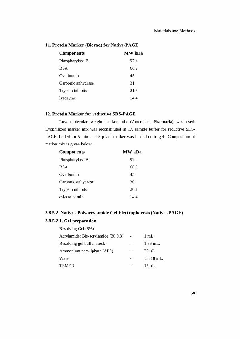
Materials and Methods
58
11. Protein Marker (Biorad) for Native-PAGE
Components MW kDa
Phosphorylase B 97.4
BSA 66.2
Ovalbumin 45
Carbonic anhydrase 31
Trypsin inhibitor 21.5
lysozyme 14.4
12. Protein Marker for reductive SDS-PAGE
Low molecular weight marker mix (Amersham Pharmacia) was used.
Lyophilized marker mix was reconstituted in 1X sample buffer for reductive SDS-
PAGE; boiled for 5 min. and 5 µL of marker was loaded on to gel. Composition of
marker mix is given below.
Components MW kDa
Phosphorylase B 97.0
BSA 66.0
Ovalbumin 45
Carbonic anhydrase 30
Trypsin inhibitor 20.1
α-lactalbumin 14.4
3.8.5.2. Native - Polyacrylamide Gel Electrophoresis (Native -PAGE)
3.8.5.2.1. Gel preparation
Resolving Gel (8%)
Acrylamide: Bis-acrylamide (30:0.8) - 1 mL.
Resolving gel buffer stock - 1.56 mL.
Ammonium persulphate (APS) - 75 µL
Water - 3.318 mL.
TEMED - 15 µL.

Materials and Methods
59
Stacking Gel (4%)
Acrylamide:Bis-acrylamide (30:.08) - 0.67 mL.
Stacking gel buffer stock - 1.25 mL
Ammonium persulphate (APS) - 40 µL.
Water - 3.075 mL.
TEMED - 12 µL.
3.8.5.2.2. Sample Preparation
The lyophilized sample was mixed with 50 mM Tris-HCl buffer (pH 8) and
15 µL of sample with 3 µL sample buffer for Native- PAGE was loaded on to the gel.
1 µL marker with 3 µL sample buffer for Native -PAGE was made up to 18 µL with
50 mM Tris-HCl buffer (pH 8) and loaded on to the gel.
3.8.5.2.3. Procedure
a. The gel plates were cleaned and assembled.
b. Resolving gel - All the components of the resolving gel were mixed
gently and the mixture was poured into the cast and a layer of butanol
was poured over the gel and allowed to solidify for at least 1 h.
c. Stacking gel - The butanol layer was poured off. The components
were mixed gently and poured into the cast above the resolving gel.
The comb was inserted between the glass plates and allowed to
solidify for 30 min.
d. The gel was placed in the electrophoresis unit and the reservoirs,
upper and lower were filled with reservoir buffer for Native-PAGE.
e. The gel was loaded with sample
f. The gel was run at 80 V until the sample entered the resolving gel.
g. The current was increased to 100 V.
h. The current was switched off when the dye front reached about 1 cm
above the lower end of glass plate.
i. The gel was removed and stained using silver staining method.
Modified method of Sambrook and Russel (2001) was employed to visualize
the protein bands on gel. The gel was immersed in fixing solution comprising of

Materials and Methods
60
methanol: acetic acid: water (50:10:40, v/v) for 30 min. Discarded the fixing solution,
and the gel was immersed in 5% methanol for 15 min. The gel was washed with
water thrice for 5 min. each. The gel was immersed in 0.02% sodium thiosulphate for
1 min. The gel was washed with water thrice for 30 sec. each and stained using 0.2%
silver nitrate for 25 min. Washed again with water thrice for 1 min. each. Developing
solution (2 mL of 0.02% sodium thiosulphate and 50 µL of formaldehyde in 100 mL
3% sodium carbonate) was added. After 10 min, the reaction was stopped by adding
1.4% EDTA.
3.8.5.3. Reductive SDS-PAGE.
3.8.5.3.1. Gel Preparation
Resolving Gel (8%)
Acrylamide: Bis-acrylamide (30:0.8) - 1 mL.
Resolving gel buffer stock - 1.56 mL.
10% SDS - 0.6 mL
Ammonium persulphate (APS) - 75 µL
Water - 3.318 mL.
TEMED - 15 µL.
Stacking Gel (4%)
Acrylamide: Bis-acrylamide (30:0.8) - 0.67 mL.
Stacking gel buffer stock - 1.25 mL
Ammonium persulphate (APS) - 40 µL.
10% SDS - 50 µL.
Water - 3.075 mL.
TEMED - 12 µL.
Sample Buffer (1X)
SDS-PAGE sample buffer (2X) - 1 mL
50% sucrose - 0.4 mL
DW - 0.6 mL
3.8.5.3.2. Sample Preparation
The gelatinase enzyme was mixed with the sample buffer and boiled for 5
min. in a water bath, cooled to room temperature and 20 µL was loaded to gel. 5 µL
of protein marker was loaded to the gel.

Materials and Methods
61
3.8.5.3.3. Procedure
Procedures for electrophoresis and staining were the same as described in
section 3.8.5.2.3, and the reservoir buffer used was that of SDS-PAGE.
3.8.5.4. Zymogram
Gelatinase activity of the purified enzyme protein band was confirmed by
performing zymogram. The method described by Raut et al. (2012) was modified by
addition of more NaCl and ZnSO4. Gelatin was incorporated into gel (500 mL of
resolving gel contained 500 µL of gelatin solution (0.1 g/10 mL)) and Native-PAGE
was performed. After running the protein, it was washed with water and immersed in
a developing solution which contained 50 mM Tris (pH 8) with 1 mM ZnSO4 and 15
% NaCl, and incubated at 37°C overnight. The gel was then dipped in Coomassie
brilliant blue stain for 1 h and destained using protein destaining solution.
Protein staining solution
Coomassie brilliant blue (0.1%) - 100 mg
Methanol (40%) - 40 mL
Glacial Acetic acid - 10 mL
DW - 50 mL
Protein destaining solution
Methanol (40%) - 40 mL
Glacial Acetic acid - 10 mL
DW - 50 mL
3.8.6. Analytical Methods
Gelatinase activity, protein content and specific activity were determined as
described in 3.2.4 and 3.5.2 and 3.5.3 and expressed as U/mL, mg/mL and U/mg
protein respectively.
3.8.7. Calculation of Yield of Protein, Yield of Enzyme Activity and Fold
of Purification
Yield of protein of each fraction during purification is the percentage obtained
Materials and Methods
62
by dividing the total protein content of the fraction with the total protein content of the
crude extract. Yield of enzyme activity of each fraction is the percentage activity
obtained by dividing the activity of the fraction by the activity of the crude extract.
Fold of purification was calculated by dividing the specific activity of the
fraction with that of the crude extract.
Yield of Protein = Total protein content of fraction x 100
Total protein content of the crude extract
Yield of Activity = Total activity of fraction x 100
Total activity of the crude extract
Fold of Purification = Specific activity of fraction x 100
Specific activity of the crude extract
3.9. Characterization of Purified Enzyme
3.9.1. Optimal pH for Gelatinase Activity.
Optimal pH for gelatinase activity was evaluated by assaying gelatinase in
plates with media in the pH range 2 to 13 as described in 3.2.4. The buffer systems
used included HCl-KCl buffer (pH 2), Citrate buffer (pH 3 to 5), Phosphate buffer
(pH 6 and 7), Tris-HCl buffer (pH 8), Glycine-NaOH buffer (pH 9), Bicarbonate
buffer (pH 10), disodium hydrogen phosphate-sodium hydroxide buffer (pH 11 and
12) and KCl-NaOH buffer (pH 13). Enzyme activity was calculated as described in
section 3.2.4.
3.9.2. Stability of Gelatinase at Different pH
Stability of gelatinase at different pH conditions was evaluated by incubating
the enzyme in different buffer systems of varying pH (pH 2 to 13) for 24 h at 4°C and
conducting gelatinase assay. The buffer systems used included HCl-KCl buffer (pH
2), Citrate buffer (pH 3 to 5), Phosphate buffer (pH 6 and 7), Tris-HCl buffer (pH 8),
Glycine-NaOH buffer (pH 9), Bicarbonate buffer (pH 10), disodium hydrogen
phosphate-sodium hydroxide buffer (pH 11 and 12) and KCl-NaOH buffer (pH 13).
After incubation, assay was done for gelatinase activity as described in section 3.2.4.
Materials and Methods
63
Residual activity was determined as described in section 3.9.12.1 and enzyme activity
at pH 8 was taken as control.
3.9.3. Optimal Temperature for Gelatinase Activity
Optimal temperature for gelatinase activity was evaluated by assaying
gelatinase in plates with media at different temperatures from 5°C to 47°C. Enzyme
activity was determined as described in section 3.2.4.
3.9.4. Stability of Gelatinase at Different Temperatures
Stability of gelatinase at different temperatures was determined by incubating
the enzyme at different temperatures ranging from 20°C to 80°C. The assay was
conducted at 30 min., 1 h, 2 h, 4 h, 6 h, 8 h, 10 h, 12 h and 24 h., as described in
section 3.2.4.
Enzyme activity of the sample kept at 4°C was taken as control. Residual
activity of the enzyme was calculated as described in section 3.9.12.1.
3.9.5. Effect of Inhibitors on Gelatinase Activity
Effect of inhibitors on gelatinase was evaluated using 0.2 mM to 1 mM
Aprotinin; 10 mM to 50 mM EDTA and 10 mM to 50 mM Iodoacetamide. The
inhibitors were added to the purified enzyme and incubated at room temperature for
30 min. After incubation enzyme assay was done as described in section 3.2.4.
Residual activity of the enzyme was calculated as described in section 3.9.12.1 and
expressed in percentage. Enzyme in the absence of inhibitors was used as control.
3.9.6. Substrate Specificity
Substrate specificity was determined by conducting the assay using plates of
gelatin, skim milk, Bovine serum albumin and haemoglobin (0.5% w/v). The enzyme
activity was done as described in section 3.2.4. and expressed in U/mL.
3.9.7. Kinetic Studies
Gelatinase was subjected to kinetic studies to determine the Km and Vmax. Km,
the substrate concentration at which reaction velocity is half maximum and Vmax the
maximum velocity of the enzyme reaction, were determined by conducting enzyme
Materials and Methods
64
assay using plates containing different concentrations of gelatin (0.2 -20 mg/mL) as
described in section 3.2.4.
3.9.8. Effect of Various Metal ions on Gelatinase Activity
The effect of various metal ions on gelatinase activity was studied by
incubating the enzyme along with different concentrations of metal ions for 30 min.,
followed by measuring the residual activity (section 3.9.12.1.). The metals studied
included 1 mM, 5 mM, 10 mM, 20 mM and 30 mM final concentrations of CaCl2,
MgSO4, ZnSO4, CuSO4, FeSO4, MnCl2, NiSO4, CoCl2, BaCl2, CdCl2 and Al2(SO4)3,
which contributed the metal ions, Ca2+, Mg2+ Zn2+, Cu2+, Fe2+, Mn2+, Ni2+,Co2+,Ba2+
Cd2+ Al3+ respectively. Enzyme in the absence of metal ions was used as control.
3.9.9. Effect of Various Detergents on Gelatinase Activity
The effect of various detergents on enzyme activity was evaluated by
incubating the enzyme in different concentrations of each detergent viz., 0.2 %, 0.4%,
0.6%, 0.8%, 1% and 5% for 30 min. and the residual enzyme activity was determined
as described in section 3.9.12.1. The detergents tested included CTAB
(cetyltrimethylammonium bromide), Sodium Lauryl Sulphate, Triton X 100, Tween
20 and Tween 80. Enzyme in the absence of detergents was used as control.
3.9.10. Effect of Various Concentrations of NaCl on Gelatinase Activity
Gelatin plates containing 0%, 5%, 10%, 15%, 20% and 25% NaCl were
prepared and assay was done as described in section 3.2.4. Gelatin plates with
concentration of NaCl above 25% could not be prepared as NaCl crystalizes on
solidification.
3.9.11. Effect of Organic Solvents on Gelatinase Activity
Effect of organic solvents on gelatinase activity was evaluated by incubating
the enzyme with the solvent for 30 min and the residual activity was assayed as
described in section 3.9.12.1. The organic solvents used included acetone, ethanol,
methanol, dimethyl sulphoxide, diethyl ether, acetonitrile amd isopropanol in the
concentrations 1%, 2%, 5%, 10% and 20% (v/v). Enzyme in the absence of organic
solvents was used as control
Materials and Methods
65
3.9.12. Analytical Methods
Gelatinase activity, protein content and specific activity were determined as
described in 3.2.4, 3.5.2 and 3.5.3 and expressed as U/mL, mg/mL and U/mg protein
respectively.
3.9.12.1. Residual Activity
Residual activity is the percent enzyme activity of the sample with respect to
the enzyme activity of the control sample.
Residual Activity = Activity of sample (U/mL) x 100
Activity of Control (U/mL)
3.9.12.2. Relative Activity
Relative activity is the percent enzyme activity of sample with respect to the
sample for which maximum activity was obtained.
Relative Activity = Activity of sample (U/mL) x 100
Activity of maximal enzyme activity obtained sample (U/mL)
3.9.13. Application Studies
3.9.13.1. Decomposition of Gelatin layer on X-ray film
Ability of gelatinase to hydrolyse gelatin layer of the X-ray film for recovery
of silver was studied by incubating the enzyme (12 U) with 2 g of X-ray film (Kumar
and Takagi, 1999).
The following sets were prepared and studied.
1. 19 mL Tris-HCl buffer (pH 8) with 15% NaCl + 2 g X-ray film + 1 mL
gelatinase enzyme (12 U).
2. 19 mL Tris-HCl buffer (pH 8) with 15% NaCl + 2 g X-ray film + 1 mL
Proteinase K enzyme (12 U).
3. 20 mL Tris-Hcl buffer (pH 8) with 15% NaCl + 2 g X-ray film.

Materials and Methods
66
The flasks were kept in a rotary shaker at 120 rpm at 37°C. After 3 h, the X-
ray film was removed and rinsed with tap water and examined visually. Protein
released by the action of enzyme into the supernatant was estimated by the method as
described in section 3.5.2 (Lowry et al., 1951).
3.10. Statistical Analysis
Statistical analysis was done using Microsoft Excel and average of five values
were taken.
Results
67
Chapter 4
RESULTS
4.1.1. Isolation and Identification of Halophiles
Halophilic bacteria associated with tannery effluent and commercially
available food grade table salt crystals were isolated using Zobell’s medium
employing standard plating procedures. Several colonies which were very similar and
identical in their morphological characteristics were noted on the plates. Hence,
representative colonies that showed identical morphological characteristics were
picked randomly and sub cultured. Thus, six representative colonies designated as
BTMT01, BTMT02, BTMT03, BTMT04, BTMT08 & BTMT10 were isolated from
tannery effluent. Similarly, five representative colonies designated as BTMT05,
BTMT06, BTMT11, BTMT12 and BTMT13 could be isolated from food grade table
salt crystals.
All the isolates obtained were studied for their cell characteristics,
biochemical and physiological characteristics, and 16S rDNA sequencing was done
for identification, as described under the materials and methods section. Interestingly,
all the isolates obtained from tannery effluent were found to be Gram positive while
the isolates obtained from food grade table salt crystals were Gram negative bacilli.
Among the isolates tested for production of different hydrolytic enzymes,
isolates BTMT01 and BTMT03 obtained from tannery effluent were observed to
produce large quantities of lipase and protease. However, the isolates BTMT08 and
BTMT10 did not show lipase production. All the isolates obtained from food grade
table salt crystals were observed to produce lipase, amylase and protease, although the
levels of the enzyme production were very less (refer Appendix-I).
4.1.2. Molecular classification of Isolates.
The partial gene sequences of 16S rDNA of the bacterial isolates were
amplified by colony PCR using universal eubacterial primers and the ~ 1kb amplicons
were sequenced. The identity of the nucleotide sequence was confirmed by BLAST
analysis and DNA homology was ascertained. Based on the information obtained, the

Results
68
identity of the isolates were inferred. The partial gene sequences of 16S rDNA were
submitted to NCBI and the details of the identity and NCBI Accession Numbers for
the halophilic bacterial strains isolated from tannery effluent and table salt crystals are
shown in Table 4.1.
Table 4.1.NCBI Accession Numbers for halophilic bacteria isolated fromtannery effluent and table salt crystals
SourceStrain
No.Bacteria
NCBI
Accession
Number
Organism showing
closest match
NCBI
Accession
Number
%
similarity
Tannery
effluent
BTMT01Oceanobacillus sp. JX975066
Oceanobacillus sp.
H-82KF021765 99
BTMT02
Staphylococcus
arlettaeJN228200
Staphylococcus
arlettae strain
VIT-RJ1
KJ716448 99
BTMT03Oceanobacillus sp. JN228197
Oceanobacillus sp.
803(2012)JN039425 99
BTMT04
Staphylococcus
arlettaeJN228201
Staphylococcus
arlettae strain NS2KP279979 98
BTMT08Salimicrobium sp. JN228199
Salimicrobium album
strain NBRC 102360NR114064 99
BTMT10Salimicrobium sp. JN228198
Salimicrobium album
strain NBRC 102360NR114064 99
Salt
crystals
BTMT05Halomonas elongata KC019171
Halomonas elongatastrain BK-AG18
KJ185382 99
BTMT06Halomonas elongata KC019170
Halomonas elongatastrain BK-AG18
KJ185382 99
BTMT11
Chromohalobactersalexigens
JX975064Chromohalobactersalexigens strain
DSM3043NR074225 98
BTMT12Halomonas elongata JX975065
Halomonas elongatastrain BK-AB8
KJ185379 99
BTMT13Halomonas elongata JX975062
Halomonas elongatastrain BK-AG18
KJ185382 99
From the results presented in Table 4.1., it was inferred that only species of
Oceanobacillus, Staphylococcus and Salimicrobium were associated with tannery
effluent while food grade table salt crystals harbored species of Halomonas and
Results
69
Chromohalobacter salexigens. Further, it was observed that Halomonas elongata was
dominant among the bacteria associated with the food grade table salt crystals. It was
also observed that both the samples of tannery effluent and food grade table salt
crystals harbored very different species of halophilic bacterial flora indicating that
source of sample influenced species diversity.
4.1.3. Phylogenetic Tree of the Halophiles Isolated from Tannery Effluent
and Commercially Available Salt Crystals
A phylogenetic tree was constructed using MEGA4 software and the
evolutionary history was inferred using Maximum Parsimony (MP) method (Eck and
Dayhoff,1966). Tree #12 out of 15 most parsimonious trees (length = 375) is shown.
The consistency index (0.955556), the retention index (0.986051), and the composite
index 0.943979 (0.942226) for all sites and parsimony-informative sites (presented in
parentheses) were computed. The percentages of replicate trees in which the
associated taxa clustered together in the bootstrap test (1000 replicates) are shown
above the branches (Felsenstein, 1985). The MP tree was obtained using the Close-
Neighbor-Interchange algorithm (Nei and Kumar, 2000) with search level 3
(Felsenstein, 1985; Nei and Kumar, 2000) in which the initial trees were obtained with
the random addition of sequences (10 replicates). The tree is drawn to scale; with
branch lengths calculated using the average pathway method (Nei and Kumar, 2000)
and are in the units of the number of changes over the whole sequence. All positions
containing gaps and missing data were eliminated from the dataset (Complete
Deletion Option). There were a total of 525 positions in the final dataset, out of which
311 were parsimony informative.
Although all the isolated bacteria were halotolerant, the phylogenetic tree
(Fig. 4.1) showed the bacteria isolated from tannery effluent and those isolated from
food grade table salt crystals in different clusters. Similar organisms are paired on the
same branch. Halomonas elongata and Chromohalobacter salexigens isolated from
salt crystals are seen in two branches, while species of Halomonas elongata are
clustered in a single branch, reflecting their similarity.

Results
70
Fig. 4.1.Phylogenetic tree of the halophiles isolated from tannery effluentand commercially available salt crystals. (Evolutionary relationships of 11taxa.Phylogenetic analyses were conducted with MEGA4 software)
4.2. Screening and Selection of Potential Haloarchaebacteria forGelatinase Production
All the bacterial strains isolated from tannery effluent and table salts were
screened for gelatinase production along with haloarchaea Natrinema sp.BTSH10 and
Halorubrum sp.BTSH03 using Zobell’s medium supplemented with 0.5% gelatin and
15% NaCl. Gelatinase activity was detected on gelatin supplemented agar medium in
terms of clearing zone produced around the inoculated colonies on addition of 10%
TCA after growth followed by computation of gelatinase enzyme units as described
under the materials and methods section 3.2.4.
From the results presented in Fig. 4.2, it was evident that Natrinema sp.
BTSH10 produced maximal quantities (11U/mL) of gelatinase, followed by
Halorubrum sp.BTSH03 (9 U/mL), Oceanobacillus sp. BTMT01 (5.9 U/mL) and
Oceanobacillus sp. BTMT03 (5.8 U/mL) while all other isolates produced only lesser
amounts of enzyme. Gelatinase activities demonstrated by three strains which showed
relatively maximal enzyme activities are shown in Fig. 4.3.

Results
71
Fig. 4.2.Gelatinase production by BTSH10, BTSH03 and bacteria isolatedfrom tannery effluent and food grade salt crystals
1. Halorubrumsp.BTSH032. Natrinema sp.BTSH103. Oceanobacillus sp. BTMT01
Fig. 4.3. Gelatin plate with bacteria showing gelatinase production
Based on the results obtained, Natrinema sp.BTSH10 was selected for further
optimization of bioprocess conditions for gelatinase production. Typical growth of
Natrinema sp.BTSH10 on Zobell’s agar medium supplemented with 15% NaCl is
shown in Fig.4.4.
3
1
2
0
2
4
6
8
10
12G
elat
inas
e ac
tivi
ty U
/mL
Bacterial and archaeal strains
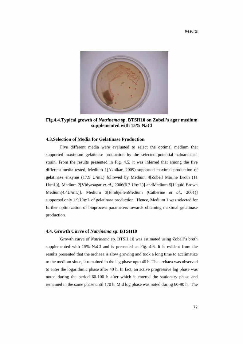
Results
72
Fig.4.4.Typical growth of Natrinema sp. BTSH10 on Zobell’s agar mediumsupplemented with 15% NaCl
4.3.Selection of Media for Gelatinase Production
Five different media were evaluated to select the optimal medium that
supported maximum gelatinase production by the selected potential haloarchaeal
strain. From the results presented in Fig. 4.5, it was inferred that among the five
different media tested, Medium 1(Akolkar, 2009) supported maximal production of
gelatinase enzyme (17.9 U/mL) followed by Medium 4[Zobell Marine Broth (11
U/mL)], Medium 2[Vidyasagar et al., 2006(6.7 U/mL)] andMedium 5[Liquid Brown
Medium(4.4U/mL)]. Medium 3[EimhjellenMedium (Catherine et al., 2001)]
supported only 1.9 U/mL of gelatinase production. Hence, Medium 1 was selected for
further optimization of bioprocess parameters towards obtaining maximal gelatinase
production.
4.4. Growth Curve of Natrinema sp. BTSH10
Growth curve of Natrinema sp. BTSH 10 was estimated using Zobell’s broth
supplemented with 15% NaCl and is presented as Fig. 4.6. It is evident from the
results presented that the archaea is slow growing and took a long time to acclimatize
to the medium since, it remained in the lag phase upto 40 h. The archaea was observed
to enter the logarithmic phase after 40 h. In fact, an active progressive log phase was
noted during the period 60-100 h after which it entered the stationary phase and
remained in the same phase until 170 h. Mid log phase was noted during 60-90 h. The
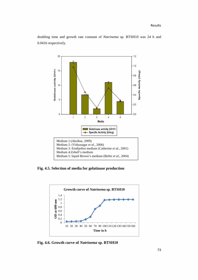
Results
73
doubling time and growth rate constant of Natrinema sp. BTSH10 was 24 h and
0.0416 respectively.
Fig. 4.5. Selection of media for gelatinase production
Fig. 4.6. Growth curve of Natrinema sp. BTSH10
00.20.40.60.8
11.21.4
10 20 30 40 50 60 70 90 100110120130140150160
OD
at
600
nm
Time in h
Growth curve of Natrinema sp. BTSH10
Medium 1:(Akolkar, 2009)Medium 2: (Vidyasagar et al., 2006)Medium 3: Eimhjellen medium (Catherine et al., 2001)Medium 4:Zobell’s mediumMedium 5: liquid Brown’s medium (Birbir et al., 2004)
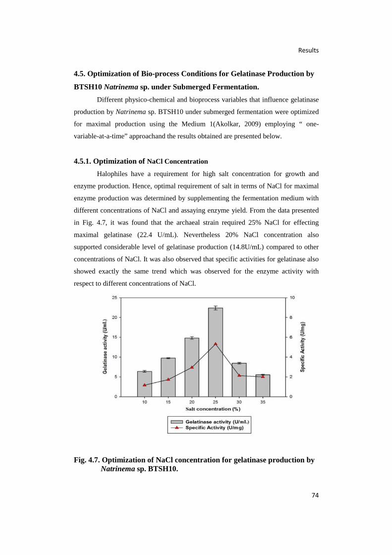
Results
74
4.5. Optimization of Bio-process Conditions for Gelatinase Production by
BTSH10 Natrinema sp. under Submerged Fermentation.
Different physico-chemical and bioprocess variables that influence gelatinase
production by Natrinema sp. BTSH10 under submerged fermentation were optimized
for maximal production using the Medium 1(Akolkar, 2009) employing “ one-
variable-at-a-time” approachand the results obtained are presented below.
4.5.1. Optimization of NaCl Concentration
Halophiles have a requirement for high salt concentration for growth and
enzyme production. Hence, optimal requirement of salt in terms of NaCl for maximal
enzyme production was determined by supplementing the fermentation medium with
different concentrations of NaCl and assaying enzyme yield. From the data presented
in Fig. 4.7, it was found that the archaeal strain required 25% NaCl for effecting
maximal gelatinase (22.4 U/mL). Nevertheless 20% NaCl concentration also
supported considerable level of gelatinase production (14.8U/mL) compared to other
concentrations of NaCl. It was also observed that specific activities for gelatinase also
showed exactly the same trend which was observed for the enzyme activity with
respect to different concentrations of NaCl.
Fig. 4.7. Optimization of NaCl concentration for gelatinase production byNatrinema sp. BTSH10.

Results
75
4.5.2. Optimization of Initial pH of Medium
Initial pH of the medium often influences the course of fermentation and rate
of enzyme production. Hence, optimal pH required for maximal enzyme production
was determined by adjusting the initial pH of the fermentation medium to varying pH
levels and assaying enzyme yield after fermentation. From the results documented in
Fig. 4.8.it was inferred that pH 7.0 is the optimal requirement for the archaea to record
maximum gelatinase (30.6 U/mL) production. It was observed that very little
gelatinase activities were supported by initial pH 2-5 while neutral pH supported
considerable levels of gelatinase production compared to alkaline pH conditions.
Further increase in pH above 9 did not support gelatinase production by the archaeal
strain. It was also observed that specific activities for gelatinase also showed exactly
the same trend which was observed for the enzyme activity with respect to different
levels of initial pH in the medium.
Fig.4.8. Optimization of initial pH of medium for gelatinase productionby Natrinema sp. BTSH10
4.5.3. Optimization of Incubation Temperature
Optimal incubation temperature requirement of the haloarchaea for maximal
gelatinase production was determined by incubating the inoculated medium at
different temperatures and assaying gelatinase yield. From the data presented in

Results
76
Fig.4.9., it was found that this archaeal strain preferred 42°C for recording maximal
gelatinase production (37 U/mL), compared to 37°C (15 U/mL) which was commonly
used for cultivation of the archaeal strain. Further, it was also observed that increase
in incubation temperatures above 42°C and incubation at temperatures below 37°C
resulted in reduced levels of gelatinase. It was observed that profile of specific
activities for gelatinase also demonstrate similar pattern which was observed for the
enzyme activity with respect to different incubation temperatures.
Fig. 4.9.Optimization of incubation temperature for gelatinaseproduction by Natrinema sp. BTSH10
4.5.4. Optimization of Inoculum Concentration
Optimal inoculum concentration required for maximal gelatinase production
by Natrinema sp. BTSH10 was determined by inoculating the medium with different
concentration of inocula and assaying enzyme yield. From the results presented in Fig.
4.10., it was evident that 2% inoculum concentration was optimum since maximal
enzyme (45 U/mL) yield was obtained when compared to other concentrations tested.
Concentrations above 2% recorded a decline in the gelatinase production. 1%
inoculum did not support enhanced level of enzyme production. In a similar manner,
the specific activities for gelatinase also followed exactly the same trend which was
observed for the enzyme activity with respect to different inoculum concentrations.

Results
77
Fig. 4.10.Optimization of inoculum concentration for gelatinaseproduction by Natrinema sp.BTSH10
4.5.5. Optimization of Inoculum Age
Impact of age of inocula used for inoculation on gelatinase production was
evaluated and the results are presented in Fig. 4.11. It was found that inoculation of
enzyme production media with inoculum age of 36 h supported maximal gelatinase
yield (60 U/mL). When inocula of 24 h age were used, only 11.2 U/mL of gelatinase
could be obtained. However, a yield of 22.7 U/mL could be obtained when 48 h
inoculum was used. Similarly inoculum of age above 36 h did not support enhanced
levels of gelatinase production since increase in the inoculum age was found to result
in a decline in the levels of gelatinase production. It was observed that pattern of
specific activities for gelatinase also showed exactly the same trend which was
observed for the enzyme activity with respect to different inoculum ages.
4.5.6. Effect of Agitation
Impact of agitation and optimal rate of agitation required for maximal
gelatinase production by Natrinema sp. BTSH10 was determined by incubating the
inoculated media in a rotary shaker at different rates of agitation and assaying enzyme
yield. From the data presented in Fig. 4.12, it was inferred that rate of agitation

Results
78
influenced the rate of enzyme production during incubation. Thus, under static
conditions, the enzyme production was only 2 U/mL which increased to only 3 U/mL
when it was subjected to 50 rpm. When the rate of agitation was enhanced to 100
rpm, the gelatinase production increased to 55 U/mL, which increased to a maximum
of 75 U/mL when the agitation rate was increased to 150 rpm. However, further
increase in agitation rates, did not enhance level of enzyme production but instead led
to a decline in the rate of gelatinase production. It was observed that specific activities
for gelatinase also showed exactly the same trend which was observed for the enzyme
activity with respect to different rates of agitation.
Fig.4.11. Optimization of inoculum age for gelatinase production byNatrinema sp. BTSH10.
Fig .4.12. Effect of agitation on gelatinase production by Natrinema sp.BTSH10
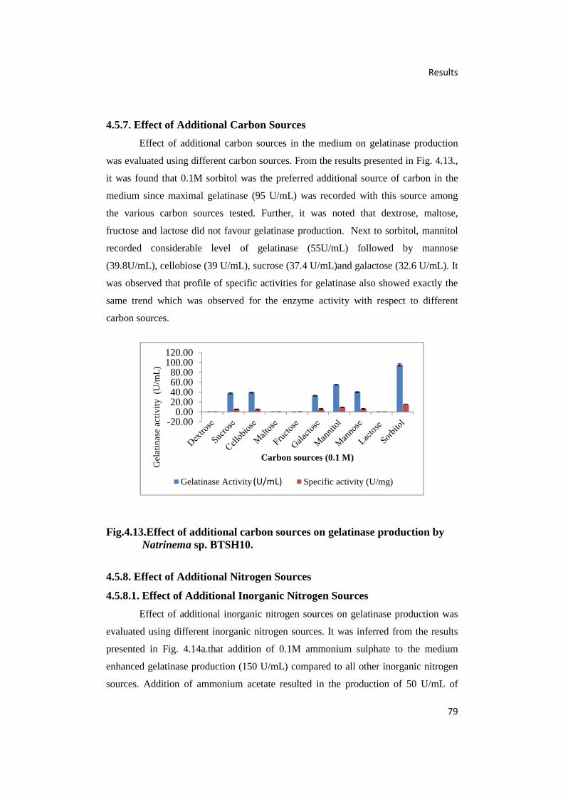
Results
79
4.5.7. Effect of Additional Carbon Sources
Effect of additional carbon sources in the medium on gelatinase production
was evaluated using different carbon sources. From the results presented in Fig. 4.13.,
it was found that 0.1M sorbitol was the preferred additional source of carbon in the
medium since maximal gelatinase (95 U/mL) was recorded with this source among
the various carbon sources tested. Further, it was noted that dextrose, maltose,
fructose and lactose did not favour gelatinase production. Next to sorbitol, mannitol
recorded considerable level of gelatinase (55U/mL) followed by mannose
(39.8U/mL), cellobiose (39 U/mL), sucrose (37.4 U/mL)and galactose (32.6 U/mL). It
was observed that profile of specific activities for gelatinase also showed exactly the
same trend which was observed for the enzyme activity with respect to different
carbon sources.
Fig.4.13.Effect of additional carbon sources on gelatinase production byNatrinema sp. BTSH10.
4.5.8. Effect of Additional Nitrogen Sources
4.5.8.1. Effect of Additional Inorganic Nitrogen Sources
Effect of additional inorganic nitrogen sources on gelatinase production was
evaluated using different inorganic nitrogen sources. It was inferred from the results
presented in Fig. 4.14a.that addition of 0.1M ammonium sulphate to the medium
enhanced gelatinase production (150 U/mL) compared to all other inorganic nitrogen
sources. Addition of ammonium acetate resulted in the production of 50 U/mL of
-20.000.00
20.0040.0060.0080.00
100.00120.00
Gel
atin
ase
activ
ity (
U/m
L)
Carbon sources (0.1 M)
Gelatinase Activity (U/Ml) Specific activity (U/mg)(U/mL)
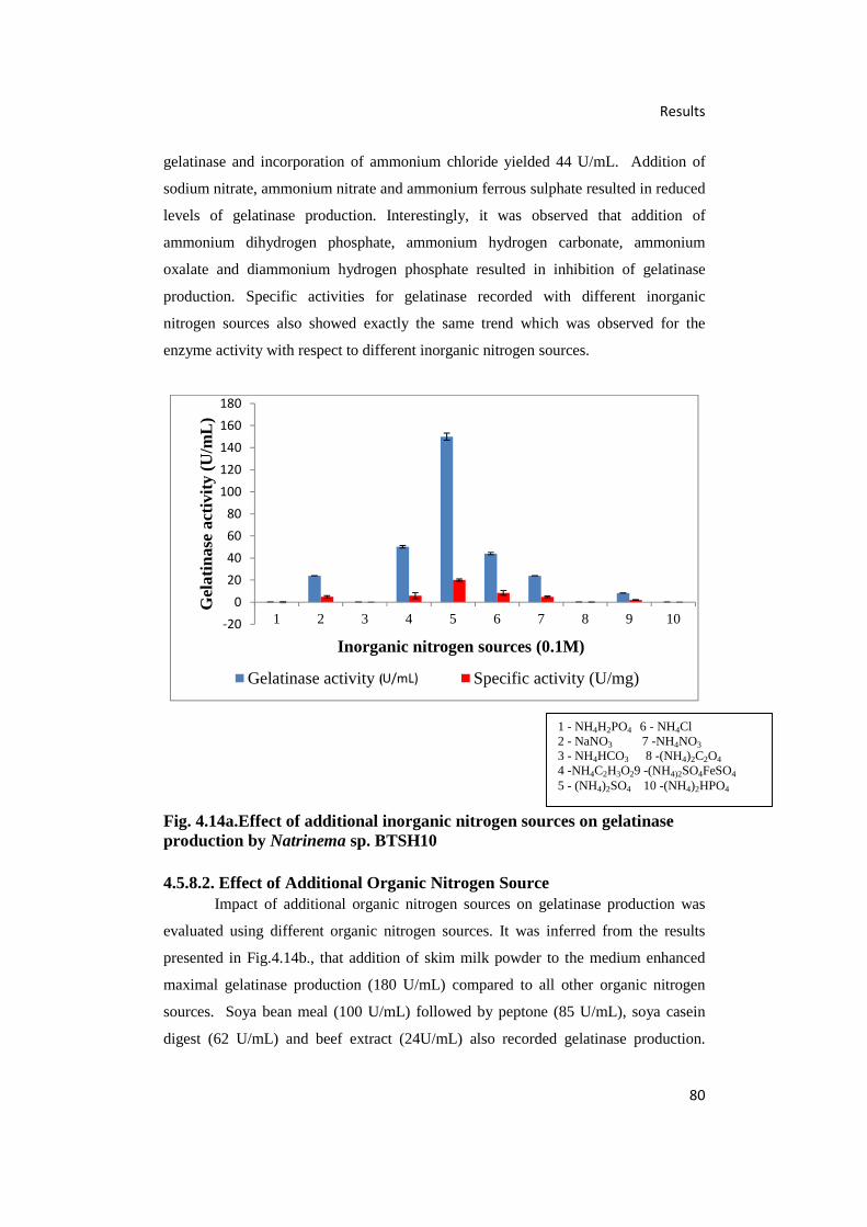
Results
80
gelatinase and incorporation of ammonium chloride yielded 44 U/mL. Addition of
sodium nitrate, ammonium nitrate and ammonium ferrous sulphate resulted in reduced
levels of gelatinase production. Interestingly, it was observed that addition of
ammonium dihydrogen phosphate, ammonium hydrogen carbonate, ammonium
oxalate and diammonium hydrogen phosphate resulted in inhibition of gelatinase
production. Specific activities for gelatinase recorded with different inorganic
nitrogen sources also showed exactly the same trend which was observed for the
enzyme activity with respect to different inorganic nitrogen sources.
Fig. 4.14a.Effect of additional inorganic nitrogen sources on gelatinaseproduction by Natrinema sp. BTSH10
4.5.8.2. Effect of Additional Organic Nitrogen SourceImpact of additional organic nitrogen sources on gelatinase production was
evaluated using different organic nitrogen sources. It was inferred from the results
presented in Fig.4.14b., that addition of skim milk powder to the medium enhanced
maximal gelatinase production (180 U/mL) compared to all other organic nitrogen
sources. Soya bean meal (100 U/mL) followed by peptone (85 U/mL), soya casein
digest (62 U/mL) and beef extract (24U/mL) also recorded gelatinase production.
-20020406080100120140160180
1 2 3 4 5 6 7 8 9 10
Gel
atin
ase
acti
vity
(U
/mL
)
Inorganic nitrogen sources (0.1M)
Gelatinase activity (U/mL) Specific activity (U/mg)
1 - NH4H2PO4 6 - NH4Cl2 - NaNO3 7 -NH4NO3
3 - NH4HCO3 8 -(NH4)2C2O4
4 -NH4C2H3O29 -(NH4)2SO4FeSO4
5 - (NH4)2SO4 10 -(NH4)2HPO4
U/mL)
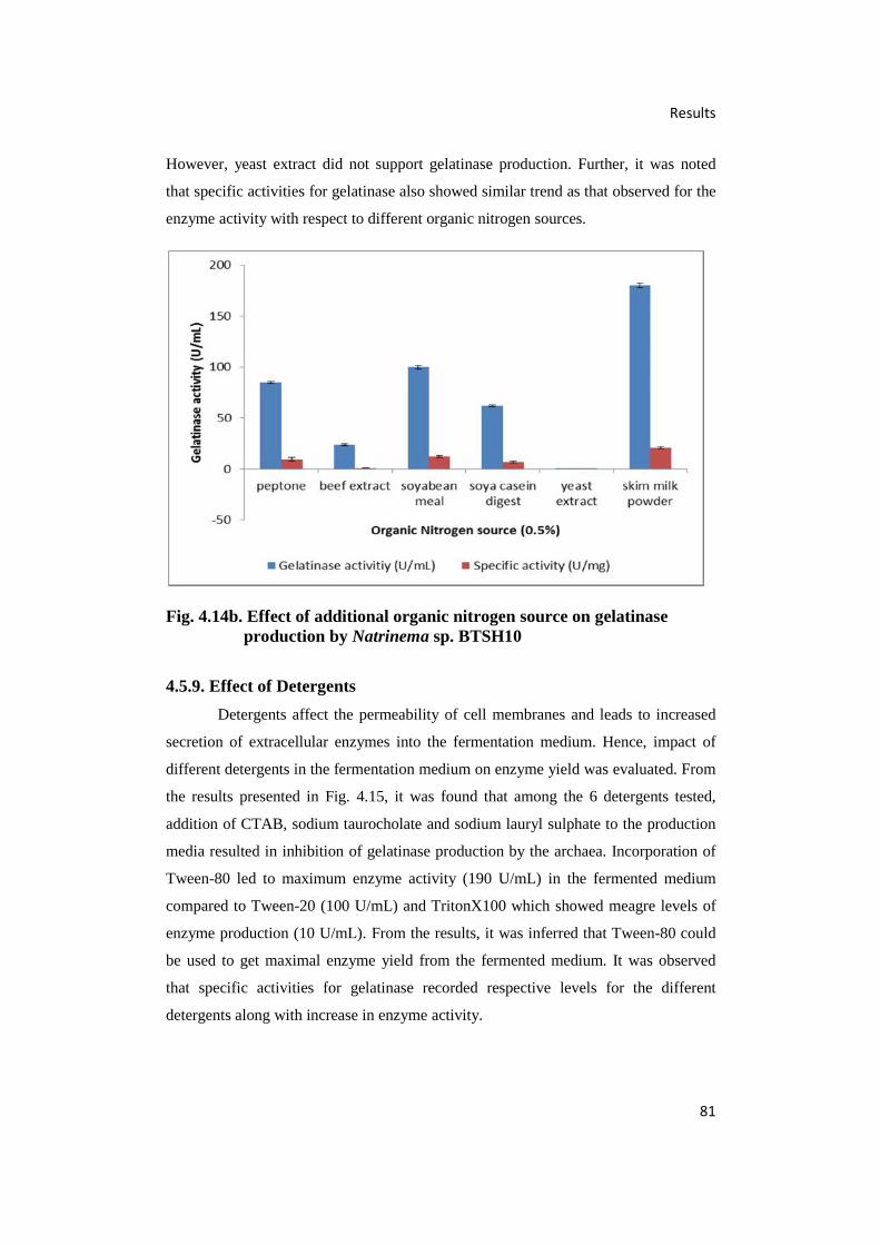
Results
81
However, yeast extract did not support gelatinase production. Further, it was noted
that specific activities for gelatinase also showed similar trend as that observed for the
enzyme activity with respect to different organic nitrogen sources.
Fig. 4.14b. Effect of additional organic nitrogen source on gelatinaseproduction by Natrinema sp. BTSH10
4.5.9. Effect of Detergents
Detergents affect the permeability of cell membranes and leads to increased
secretion of extracellular enzymes into the fermentation medium. Hence, impact of
different detergents in the fermentation medium on enzyme yield was evaluated. From
the results presented in Fig. 4.15, it was found that among the 6 detergents tested,
addition of CTAB, sodium taurocholate and sodium lauryl sulphate to the production
media resulted in inhibition of gelatinase production by the archaea. Incorporation of
Tween-80 led to maximum enzyme activity (190 U/mL) in the fermented medium
compared to Tween-20 (100 U/mL) and TritonX100 which showed meagre levels of
enzyme production (10 U/mL). From the results, it was inferred that Tween-80 could
be used to get maximal enzyme yield from the fermented medium. It was observed
that specific activities for gelatinase recorded respective levels for the different
detergents along with increase in enzyme activity.

Results
82
Fig. 4.15. Effect of detergents on gelatinase production by Natrinema sp.BTSH10
4.5.10. Time Course Experiment
A time course experiment was conducted after optimizing all the bioprocess
variables for obtaining maximal gelatinase production by Natrinema sp.BTSH10
during submerged fermentation over a period of 168 h under optimized conditions.
The results obtained are documented in Fig.4.16. From the results, it was evident that
gelatinase production by the archaeal strain commenced after 48 h growth and reached
a maximum after 120 h (229.4 U/mL). It was noted that there were very low levels of
gelatinase production at 72 h and 96 h which rapidly increased during the period
between 96h and 120 h. It is assumed that maximal gelatinase production has taken
place during late logarithmic phase of the growth curve compared to mid log phase.
Further, it was also noted that during incubation beyond 120 h the enzyme yield
decreased progressively along with increase in incubation time.It was observed that
specific activities for gelatinase also showed exactly the same trend which was
observed for the enzyme activity.

Results
83
Fig. 4.16. Time course study on gelatinase production by Natrinema sp.BTSH10 under optimal conditions
4.6. Purification of Gelatinase
The culture supernatant obtained after centrifugation at 10,000 rpm for 10 min
at 4°C, followed by removal of archaeal cells was considered as crude gelatinase
enzyme which was subjected to purification employing Membrane Filtration, Gel
Filtration Chromatography and Dialysis followed by HPLC. The results obtained are
presented in Table4.2.
4.6.1. Filtration
The crude enzyme prepared was initially subjected to filtration at 4°C using
Amicon filters with a cut–off size 10 kDa and all the fractions were analyzed for
presence of enzymes. From the results obtained, it was observed that only those
fractions comprising proteins with a size greater than 10 kDa showed enzyme activity.
Hence, these fractions were further subjected to filtration using Amicon filters with a
cut off size 30 kDa. It was found that the fractions consisting of proteins between 10 -
30 kDa showed gelatinase activities and these fractions were used for further
purification. Results presented in Table 4.2 show the efficiency of purification by
filtration.

Results
84
Table 4.2. Yield and fold of purification of gelatinase
Sample Volume(mL)
Protein(mg)
Activity(U)
Specificactivity(U/mg)
Yieldof
protein(%)
Yieldof
activity(%)
Fold ofpurification
Crude 1000 4500 11203.66 2.48 100 100 1
Filtration10 kDacut off(>10 kDafraction)
550 2447.5 9385.85 3.83 54.38 83.77 1.54
Filtration30 k Dacut off(10-30kDafraction)
330 1212.75 8577.80 7.07 26.95 76.56 2.84
Gelfiltration
2 3.1 79 25.48 0.06 0.70 10.23
HPLC 1 2.1 60.3 28.71 0.04 0.53 11.53
4.6.2.Gel Filtration Chromatography
Gel filtration chromatograhy was performed using Sephadex G-50 (Sigma-
Aldrich) to separate proteins ranging in size from 50 kDa -3 kDa. From the data
presented in Fig. 4.17., it was inferred that the fractions numbered 18 to 24 contained
gelatinase enzyme. These fractions were pooled, lyophilised and used for dialysis of
the enzyme fractions towards further purification.
Fig. 4.17. OD at 280 nm of gel filtration fractions
00.050.10.150.20.250.30.35
1 3 5 7 9 11 13 15 17 19 21 23 25 27 29 31 33 35
OD
at
280n
m
Gel filtration fractions
Gel filtration chromatography
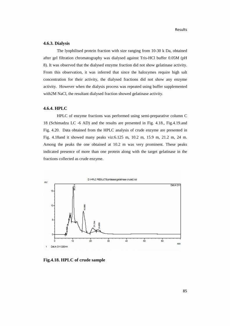
Results
85
4.6.3. Dialysis
The lyophilised protein fraction with size ranging from 10-30 k Da, obtained
after gel filtration chromatography was dialysed against Tris-HCl buffer 0.05M (pH
8). It was observed that the dialysed enzyme fraction did not show gelatinase activity.
From this observation, it was inferred that since the halozymes require high salt
concentration for their activity, the dialysed fractions did not show any enzyme
activity. However when the dialysis process was repeated using buffer supplemented
with2M NaCl, the resultant dialysed fraction showed gelatinase activity.
4.6.4. HPLC
HPLC of enzyme fractions was performed using semi-preparative column C
18 (Schimadzu LC -6 AD) and the results are presented in Fig. 4.18., Fig.4.19.and
Fig. 4.20. Data obtained from the HPLC analysis of crude enzyme are presented in
Fig. 4.18and it showed many peaks viz:6.125 m, 10.2 m, 15.9 m, 21.2 m, 24 m.
Among the peaks the one obtained at 10.2 m was very prominent. These peaks
indicated presence of more than one protein along with the target gelatinase in the
fractions collected as crude enzyme.
Fig.4.18. HPLC of crude sample

Results
86
The enzyme fractions obtained after gel filtration chromatography, when
subjected to HPLC, showed a single peak (6.828 m) indicating the effectiveness of
purification by gel filtration chromatography and the purity of enzyme. It was also
noted that this single peak was observed in the fraction collected during 6.5-7.5
m(Fig. 4.19.), when the concentration of methanol was between 9.3 % and 10.7 %
methanol.
Fig.4.19. HPLC of gel filtration chromatographic fraction
From the data depicted in Fig. 4.20. it was inferred that HPLC purification of
the dialysed sample (using buffer containing 2M NaCl) resulted in a single peak
which was observed at 6.132 m. The efficiency of the purification in terms of yield
and fold are presented in table 4.2.
Fig.4.20. HPLC of dialysed sample
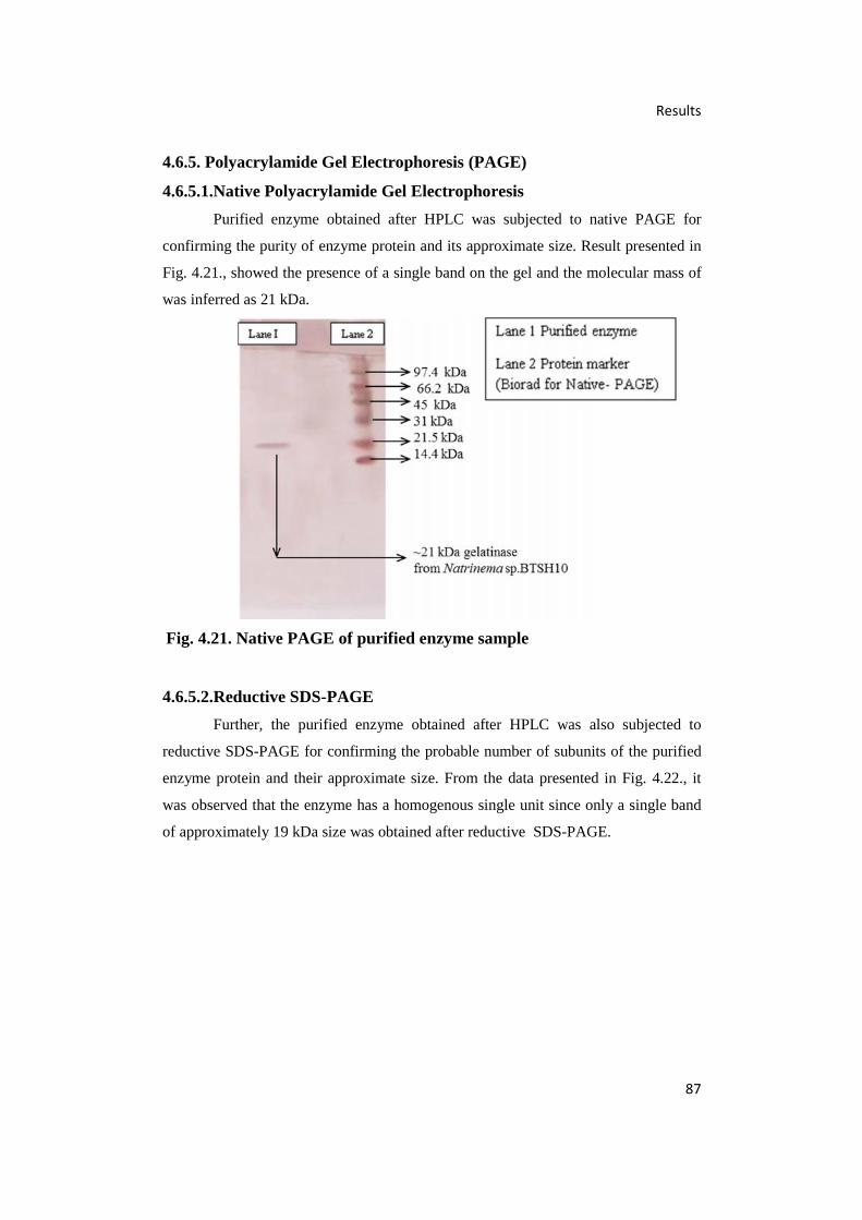
Results
87
4.6.5. Polyacrylamide Gel Electrophoresis (PAGE)
4.6.5.1.Native Polyacrylamide Gel Electrophoresis
Purified enzyme obtained after HPLC was subjected to native PAGE for
confirming the purity of enzyme protein and its approximate size. Result presented in
Fig. 4.21., showed the presence of a single band on the gel and the molecular mass of
was inferred as 21 kDa.
Fig. 4.21. Native PAGE of purified enzyme sample
4.6.5.2.Reductive SDS-PAGE
Further, the purified enzyme obtained after HPLC was also subjected to
reductive SDS-PAGE for confirming the probable number of subunits of the purified
enzyme protein and their approximate size. From the data presented in Fig. 4.22., it
was observed that the enzyme has a homogenous single unit since only a single band
of approximately 19 kDa size was obtained after reductive SDS-PAGE.

Results
88
Fig. 4.22. Reductive SDS-PAGE of purified enzyme sample
4.6.5.3.Zymogram
Gelatinase activity of the purified enzyme protein band was confirmed by
performing zymogram analysis. The results presented in Fig.4.23., indicate that the
purified enzyme protein showed good gelatinase activity since a clear zone in blue
coloured gel was observed around the site of protein band. The results confirmed that
the purified enzyme protein was gelatinase.
Clearing seen in lane 2
Fig. 4.23.Zymogram analysis of gelatinase
1 2 3 4
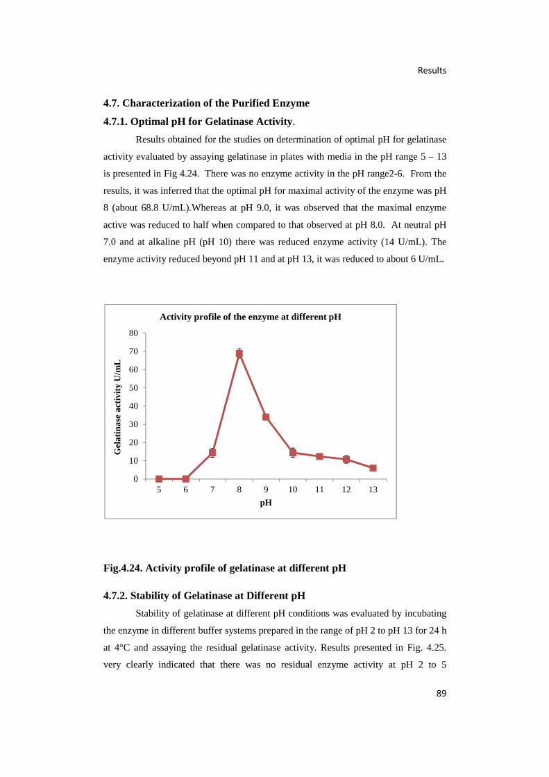
Results
89
4.7. Characterization of the Purified Enzyme
4.7.1. Optimal pH for Gelatinase Activity.
Results obtained for the studies on determination of optimal pH for gelatinase
activity evaluated by assaying gelatinase in plates with media in the pH range 5 – 13
is presented in Fig 4.24. There was no enzyme activity in the pH range2-6. From the
results, it was inferred that the optimal pH for maximal activity of the enzyme was pH
8 (about 68.8 U/mL).Whereas at pH 9.0, it was observed that the maximal enzyme
active was reduced to half when compared to that observed at pH 8.0. At neutral pH
7.0 and at alkaline pH (pH 10) there was reduced enzyme activity (14 U/mL). The
enzyme activity reduced beyond pH 11 and at pH 13, it was reduced to about 6 U/mL.
Fig.4.24. Activity profile of gelatinase at different pH
4.7.2. Stability of Gelatinase at Different pH
Stability of gelatinase at different pH conditions was evaluated by incubating
the enzyme in different buffer systems prepared in the range of pH 2 to pH 13 for 24 h
at 4°C and assaying the residual gelatinase activity. Results presented in Fig. 4.25.
very clearly indicated that there was no residual enzyme activity at pH 2 to 5
0
10
20
30
40
50
60
70
80
5 6 7 8 9 10 11 12 13
Gel
atin
ase
acti
vity
U/m
L
pH
Activity profile of the enzyme at different pH

Results
90
indicating that the enzyme was not stable under acidic conditions when compared to
alkaline pH conditions (pH 9 to pH 13). The residual activities were above 56% for
pH 12 and 49% for pH 13, about 86% for pH 9 to 11 range. At neutral pH 7, 65%
residual activity was recorded. Results indicated that probably the enzyme is
alkalophilic in nature.
Fig.4.25. Stability of gelatinase at different pH
4.7.3. Optimal Temperature for Gelatinase Activity
Optimal temperature required for gelatinase activity was evaluated by
assaying gelatinase by incubation at different temperatures varying from 5°C to 47°C.
Data presented in Fig. 4.26.indicated that the optimum temperature required for
maximal enzyme activity was 37°C. It was also observed that temperatures above
42°C and below 27°C led to a steep decline in activity. However, moderate levels of
enzyme activities, almost less than 50% of the level recorded at 37°C could be
observed at 32°C and 42°C.
-20
0
20
40
60
80
100
120
2 3 4 5 6 7 8 9 10 11 12 13
Res
idua
l enz
yme
activ
ity %
pH
Stability profile of the enzyme in different pH

Results
91
Fig.4.26. Effect of temperature on gelatinase activity
4.7.4. Stability of Gelatinase at Different Temperatures
Stability of gelatinase at different temperatures was determined by incubating
the enzyme at different temperatures ranging from 20°C to 80°C and assaying residual
enzyme activity after 30 m, 1 h, 2 h, 4 h, 6 h, 8 h, 10 h, 12 h and 24 h. From the
results presented in table 4.3, it was inferred that the enzyme was stable without losing
activity (about 99% residual activity) at 40°Cupto 4 h and upto 30 min. at 20°C, 30°C,
50°C and 60°C. The enzyme showed a residual activity of 63% upto 24 h at
temperatures of 20°C and 30°C; up to 12h at 40°C, upto 6h at 50°C and upto 4h at
60°C. Extended incubation at higher temperatures resulted in loss of activity since the
residual activity declined to about 43%. At higher temperatures of 70°C and 80°C,
there was no residual activity indicating loss of enzyme activity.
Temperature (C)
0 10 20 30 40 50
Gel
atin
ase
activ
ity (U
/ml)
0
20
40
60
80
100

Results
92
Table. 4.3.Residual activity of gelatinase at different temperatures
TimeResidual activity (%) at different temperatures (°C)
20 30 40 50 60 70 80
30min
99.0±
4.30
99.66 ±
2.48
99.66±
2.48
99.0±
4.3
98.66±
4.96
12.0±
4.3
1.1±
1.13
1h66.66 ±
8.95
66.66 ±
6.57
99.66±
2.48
67.0±
8.6
65.66±
2.48
0.96±
0.24
0.96±
0.24
2h65.33 ±
2.48
65.66 ±
2.48
99.0±
4.30
65.66±
2.48
64.66±
2.48
0.93±
0.24
0.96±
0.24
4h65.0±
4.30
65.33±
2.48
98.66±
4.96
64.33±
4.96
64.33±
2.48
0.96±
0.24
0.96±
0.24
6h64.66 ±
2.48
65.33 ±
2.48
66.0±
4.30
64.0±
4.3
43.66±
2.48
0.96±
0.24
0.96±
0.24
8h64.33±
2.48
64.66 ±
2.48
65.66±
2.48
42.0±
7.45
43.66±
2.48
0.96±
0.24
0.96±
0.24
10h64.0±
4.30
64.33±
2.48
65.66±
2.48
42.0±
6.57
42.0±
4.3
0.96±
0.24
0.96±
0.24
12h64.33 ±
2.48
63.66 ±
4.96
64.33±
2.48
28.33 ±
2.48
19.66 ±
6.57
0.93±
0.24
0.96±
0.24
24h64.0 ±
4.30
63.66 ±
4.96
29.0±
4.3
27.0±
4.3
18.66±
2.48
0.93±
0.24
0.96±
0.24
95% confidence interval for residual activity
4.7.5. Effect of Inhibitors on Gelatinase Activity
Effect of inhibitors on gelatinaseactivity was evaluated using Aprotinin,
EDTA and Iodoacetamide, and the results obtained are presented in table 4.4. From
the residual enzyme activities recorded for the various inhibitors it was found that the
enzyme showed marked decline in enzyme activities along with increase in the
concentration of the enzyme inhibitors in all the three cases. Among the three

Results
93
inhibitors tested, maximal inhibition was effected by EDTA which is a metallo-
protease inhibitor. Even at the lowest concentration 10 mM, marked inhibition (20%)
of the gelatinase was observed which declined rapidly to 3% in presence of 50 mM
EDTA. Whereas in the presence of aprotinin, the gelatinase showed a residual activity
of 75% at 0.2 mM which decreased to 49% when the concentration was increased to
0.6 mM. Further increase in inhibitor concentration to a maximum of 1 mM led only
to a marginal decline from 49% to 43%. In the case of Iodoacetamide 43% residual
activity was noted for the lesser concentrationsof 10mM and 20 mM indicating more
than 50% enzyme inhibition, while 27% residual activity was observed in the presence
of 50mM concentrations.
Table. 4.4. Effect of inhibitors on gelatinase activity.
95% confidence interval for residual activity
4.7.6. Substrate Specificity
Substrate specificity was determined by conducting the enzyme assay using
gelatin, skim milk, BSA and haemoglobin as substrates in agar plates incorporated
with respective substrates. From the results presented in Fig. 4.27., it was found that
Name of inhibitor
(class) used
Concentration(mM)
Residual Activity(%)
Aprotinin(Serine protease)
0.20.40.60.81.0
75.17 (± 4.50 )65.88 (± 4.38)49.19 (± 4.54)43.03 (± 4.31)43.70 (± 2.62)
EDTA(Metallo-protease)
1020304050
20.12 (± 5.13)18.52 (± 2.16)8.73 (± 2.78)5.41 (± 2.23)3.31 (± 1.18)
Iodoacetamide(Cysteine protease)
1020304050
43.03 (± 4.31)43.70 (± 2.23)29.09 (± 3.68)27.76 ± 2.9227.43 ± 3.22

Results
94
the ideal substrate that supported maximal activity was skim milk followed by gelatin.
Use of BSA and haemoglobin as substrates did not yield appreciable activity.
Fig.4.27. Substrate specificity of gelatinase enzyme
4.7.7. Kinetic Studies
Enzyme kinetic studies were conducted to determine the Km, the substrate
concentration at which reaction velocity is half maximum and Vmax the maximum
velocity of the enzyme reaction for gelatinase. Km, the substrate concentration at
which reaction velocity is half maximum and Vmax the maximum velocity of the
enzyme reaction, were determined by conducting enzyme assay using different
concentrations of gelatin. From the results obtained (Fig. 4.28) it was found that Km of
gelatinase was 21.38 mg/mL and Vmax was recorded as 55.55 U/mL.
0
5
10
15
20
25
30
35
40
Gelatin Skim milk BSA Haemoglobin
Enz
yme
acti
vity
U/m
l
substrates(0.5% w/v)

Results
95
Fig.4.28.Lineweaver- Burk Plot
4.7.8. Effect of Various Metal ions on Gelatinase Activity
The effect of CaCl2, MgSO4, ZnSO4, CuSO4, FeSO4, MnCl2, NiSO4, CoCl2, BaCl2,
CdCl2 and Al2 (SO4)3, which contributed the metal ions, Ca2+, Mg2+ Zn2+, Cu2+, Fe2+,
Mn2+, Ni2+,Co2+,Ba2+ Cd2+andAl3+ respectively on gelatinase activity was studied by
measuring the residual activities after incubating the enzyme along with different
concentrations of metal ions for 30 m.Results obtained are presented in table 4.5. In
general ZnSO4 (149%) CuSO4 (145%), FeSO4 (115%) andMnCl2 (130%) wereobserved
to enhance residual enzyme activities to levels above their original activities (100%)
at 1 mM. Whereas at other concentrations of these metals except ZnSO4 (149% at 5
mM) recorded reduced levels of residual activities indicating inhibition at
concentration of 5 mM and above. All other metals showed marked inhibition of
enzyme activities indicated by reduced levels of residual activities at all the
concentrations of the metals tested.CaCl2, MgSO4,BaCl2, CdCl2 and Al2(SO4)3 showed
marked inhibition of enzyme activities when compared to other metals at all
concentrations except Al2(SO4)3 which showed 66% residual activity at 1mM.
Interestingly, MnCl2 was observed to have relatively reduced effect on inhibition of
gelatinase at all concentrations tested since up to 72% residual activity was recorded
even at the maximum concentration of 30mM. It must be also noted that the reduction
in residual enzyme activity levels gradually declined from 97% at 5mM to 85% at
10mM and 20 mM. Further it was also noted that NiSO4, did not inhibit enzyme
y = 0.385x + 0.018R² = 0.948
-0.4-0.3-0.2-0.1
00.10.20.30.40.50.60.70.8
-1.5 -1 -0.5 0 0.5 1 1.5 2
1/V
1/[S]
Lineweaver -Burk Plot
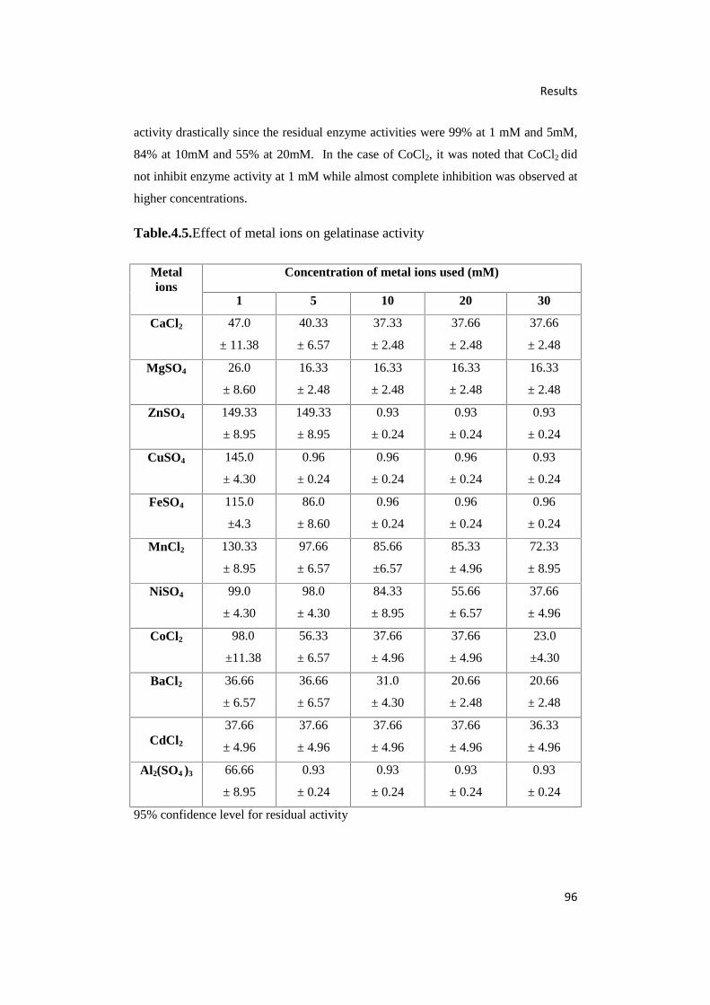
Results
96
activity drastically since the residual enzyme activities were 99% at 1 mM and 5mM,
84% at 10mM and 55% at 20mM. In the case of CoCl2, it was noted that CoCl2 did
not inhibit enzyme activity at 1 mM while almost complete inhibition was observed at
higher concentrations.
Table.4.5.Effect of metal ions on gelatinase activity
95% confidence level for residual activity
Metalions
Concentration of metal ions used (mM)
1 5 10 20 30
CaCl2 47.0
± 11.38
40.33
± 6.57
37.33
± 2.48
37.66
± 2.48
37.66
± 2.48
MgSO4 26.0
± 8.60
16.33
± 2.48
16.33
± 2.48
16.33
± 2.48
16.33
± 2.48
ZnSO4 149.33
± 8.95
149.33
± 8.95
0.93
± 0.24
0.93
± 0.24
0.93
± 0.24
CuSO4 145.0
± 4.30
0.96
± 0.24
0.96
± 0.24
0.96
± 0.24
0.93
± 0.24
FeSO4 115.0
±4.3
86.0
± 8.60
0.96
± 0.24
0.96
± 0.24
0.96
± 0.24
MnCl2 130.33
± 8.95
97.66
± 6.57
85.66
±6.57
85.33
± 4.96
72.33
± 8.95
NiSO4 99.0
± 4.30
98.0
± 4.30
84.33
± 8.95
55.66
± 6.57
37.66
± 4.96
CoCl2 98.0
±11.38
56.33
± 6.57
37.66
± 4.96
37.66
± 4.96
23.0
±4.30
BaCl2 36.66
± 6.57
36.66
± 6.57
31.0
± 4.30
20.66
± 2.48
20.66
± 2.48
CdCl2
37.66
± 4.96
37.66
± 4.96
37.66
± 4.96
37.66
± 4.96
36.33
± 4.96
Al2(SO4 )3 66.66
± 8.95
0.93
± 0.24
0.93
± 0.24
0.93
± 0.24
0.93
± 0.24
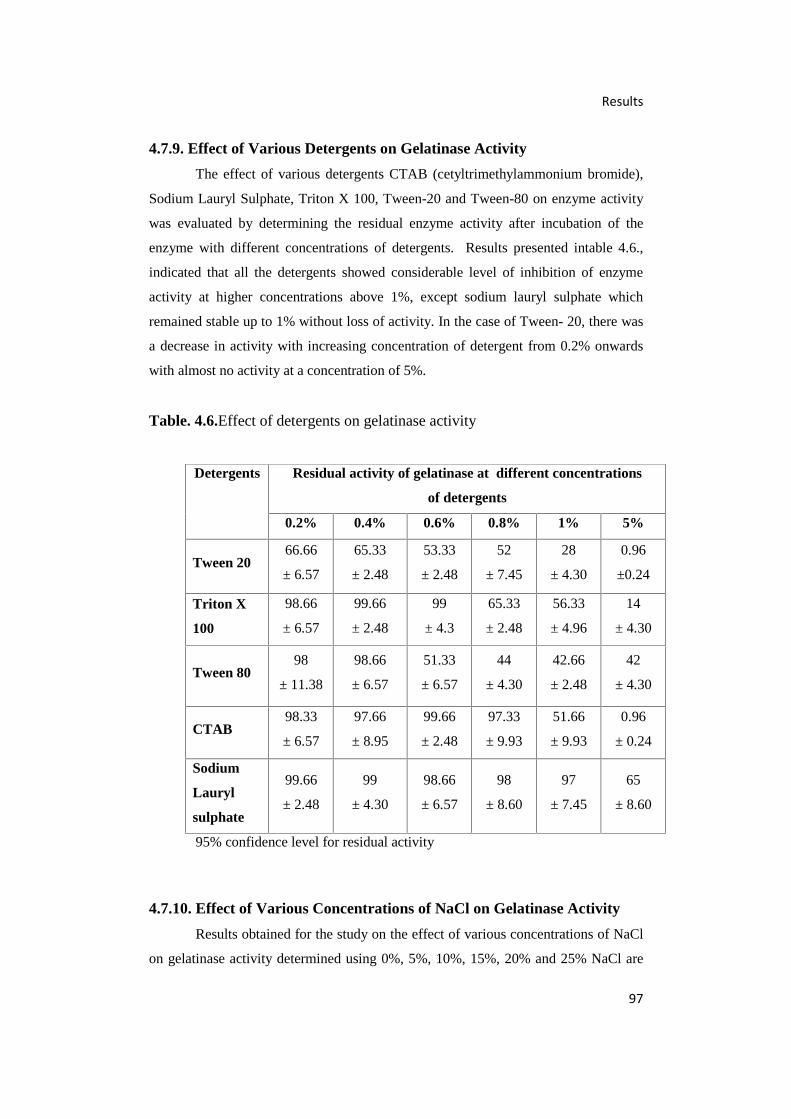
Results
97
4.7.9. Effect of Various Detergents on Gelatinase Activity
The effect of various detergents CTAB (cetyltrimethylammonium bromide),
Sodium Lauryl Sulphate, Triton X 100, Tween-20 and Tween-80 on enzyme activity
was evaluated by determining the residual enzyme activity after incubation of the
enzyme with different concentrations of detergents. Results presented intable 4.6.,
indicated that all the detergents showed considerable level of inhibition of enzyme
activity at higher concentrations above 1%, except sodium lauryl sulphate which
remained stable up to 1% without loss of activity. In the case of Tween- 20, there was
a decrease in activity with increasing concentration of detergent from 0.2% onwards
with almost no activity at a concentration of 5%.
Table. 4.6.Effect of detergents on gelatinase activity
95% confidence level for residual activity
4.7.10. Effect of Various Concentrations of NaCl on Gelatinase Activity
Results obtained for the study on the effect of various concentrations of NaCl
on gelatinase activity determined using 0%, 5%, 10%, 15%, 20% and 25% NaCl are
Detergents Residual activity of gelatinase at different concentrations
of detergents
0.2% 0.4% 0.6% 0.8% 1% 5%
Tween 2066.66
± 6.57
65.33
± 2.48
53.33
± 2.48
52
± 7.45
28
± 4.30
0.96
±0.24
Triton X
100
98.66
± 6.57
99.66
± 2.48
99
± 4.3
65.33
± 2.48
56.33
± 4.96
14
± 4.30
Tween 8098
± 11.38
98.66
± 6.57
51.33
± 6.57
44
± 4.30
42.66
± 2.48
42
± 4.30
CTAB98.33
± 6.57
97.66
± 8.95
99.66
± 2.48
97.33
± 9.93
51.66
± 9.93
0.96
± 0.24
Sodium
Lauryl
sulphate
99.66
± 2.48
99
± 4.30
98.66
± 6.57
98
± 8.60
97
± 7.45
65
± 8.60

Results
98
presented in Fig. 4.29. Gelatin plates with concentration of NaCl above 25% could not
be prepared as NaCl crystalizes on solidification. From the data, it was found that
NaCl concentration had a profound effect on enzyme activity. At 15% NaCl, the
enzyme showed maximum activity which declined on increase of salt concentration.
At concentrations below 10%, there was no enzyme activity.
Fig.4.29. Effect of different concentrations of NaCl on gelatinase activity
4.7.11. Effect of Organic Solvents on Gelatinase Activity
Effect of organic solvents on gelatinase activity was evaluated by incubating
the enzyme with the organic solvents such as acetone, ethanol, methanol, dimethyl
sulphoxide, diethyl ether, acetonitrile and isopropanol at concentrations of 1%, 2%,
5%, 10% and 20% (v/v) for 30 min and the residual activities were assayed. In
general, it was observed that the gelatinase was inhibited by all the organic solvents
although the percent level of inhibition varied for the solvents. It may be noted from
the results presented in table 4.7.that among the different solvents only ethanol
registered relatively higher levelof residual activities (65%) at a concentration of 1%-
2% indicated lesser levels of inhibition. Further increase in concentration of ethanol
resulted in more than 50% enzyme inhibition since there was a decline in residual
activities (43%) and a total inhibition was recorded at 20%. All the other organic
solvents showed more than 50% enzyme inhibition since they recorded residual

Results
99
activities varying from 20% (acetonitrile and isopropylalcohol) to 43% (diethyl ether)
44% (acetone) 45% (methanol) even at 1% of organic solvent in the reaction mixture.
Isopropylalcohol (20-12%) and DMSO (29-17%) recorded lesser levels of residual
activities indicating maximal enzyme inhibition for the various concentration tested
when compared to other solvents (table 4.7.). It was also observed that at a
concentration of 20%, only 1% residual activity of the enzyme was recorded in the
case of methanol, ethanol and diethyl ether indicating almost complete enzyme
inhibition.
Table. 4.7.Effect of organic solvents on gelatinase activity
Solvents
Concentration of organic solvents used
1% 2% 5% 10% 20%
Acetone44.66
± 6.57
43.33
± 2.48
43.0
± 4.30
42. 0
± 4.30
19.66
± 6.57
Ethanol66.33
± 6.57
65.33
± 2.48
43.33
±2.48
42.0
± 4.30
1.03
± 0.65
Methanol45.0
± 8.60
19.66
± 6.57
17.66
± 6.57
11.0
± 4.30
0.96
± 0.24
DMSO29.0
± 4.30
26.66
± 6.57
19.66
± 6.57
18.33
±2.48
17.66
± 2.48
Diethyl ether43.66
±13.14
28.66
± 4.96
28.33
± 2.48
16.66
±6.57
1.03
± 1.08
Acetonitrile20.66
±10.82
18.0
± 4.30
13.33
± 6.57
13.0
± 4.30
11.66
± 2.48
Isopropylalcohol20.66
± 6.57
20.33
± 4.96
11.33
±2.48
11.66
±2.48
12.0
±4.30
95% of confidence interval for residual activity
4.8. Application Studies
4.8.1. Decomposition of Gelatin Layer on X-ray film
Potential of gelatinase enzyme purified from Natrinema sp.BTSH10 to
degrade the protein layer of the X-ray film for recovery of silver was studied by
incubating the enzyme (12U) with 2g of X-ray film. Results presented in Fig. 4.33

Results
100
indicated, when compared to the control without enzyme where the protein content of
the supernatant was only 0.425 mg/mL (Fig. 4.30.), there was considerable level of
degradation of protein on the protein layer of X-ray film since the supernatant
estimated after incubation with gelatinase enzyme recorded 8.9 mg of protein per mL
(Fig. 4.31).Similarly incubation with Proteinase K yielded 11 mg/mL protein in the
supernatant (Fig. 4.32). Further visual examination of the X-ray film also indicated
that the enzyme could effectively decompose the gelatin layer.
Fig. 4.30. Untreated X-ray film
Fig. 4.31. X-ray film treated with gelatinase from Natrinema sp. BTSH10

Results
101
Fig. 4.32. X-ray film treated with Proteinase K
Fig. 4.33. Protein content of supernatant after enzyme treatment on X-rayfilm.
0
2
4
6
8
10
12
1.gelatinase treated 2.proteinase Ktreated
3.control
Pro
tein
mg/
mL
Protein content of supernatant estimated after enzymetreatment of X-ray film

Results
102

Discussion
103
Chapter 5
DISCUSSION
Halophilic bacteria associated with tannery effluent and commercially
available food grade table salt crystals were isolated using Zobell’s medium
employing standard plating procedures. Interestingly, all the isolates obtained
from tannery effluent were found to be Gram positive while the isolates obtained
from food grade table salt crystals were Gram negative bacilli. From the results of
the present study, it was inferred that only species of Oceanobacillus,
Staphylococcus and Salimicrobium were associated with tannery effluent while
food grade table salt crystals harbored species of Halomonas and
Chromohalobacter salexigens which are Gram negative. Further, it was observed
that Halomonas elongata was dominant among the bacteria associated with the
food grade table salt crystals. It was also observed that both the samples of
tannery effluent and food grade table salt crystals harbored very different species
of halophilic bacterial flora indicating that the source of sample influenced species
diversity. This is evidently seen in the phylogenetic tree which clearly reflects the
fact that the bacteria isolated from tannery effluent and those isolated from food
grade table salt crystals are seen in different clusters. Similar organisms are paired
on the same branch. Halomonas elongata and Chromohalobacter salexigens
isolated from salt crystals are seen in two branches.
In the present study, Halomonas sp. produced amylase, lipase and
protease and Chromohalobacter sp. produced lipase. Staphylococcus arlettae
isolated from tannery effluent could elaborate coagulase, an enzyme which is
usually seen in pathogenic species of Staphylococci. Salimicrobium sp. isolated
from tannery effluent could not produce lipase. Oceanobacillus sp. isolated from
tannery effluent produced large quantities of lipase and protease. All the bacteria
isolated from food grade table salts were also observed to produce lipase, amylase
and protease, albeit, in small amounts. It was reported that almost 10% of
moderately halophilic bacteria isolated from solar salterns and saline soils of Isla
Cristina (Spain) secrete proteases; while other isolates secrete enzymes like lipase
amylase etc. (Sanchez –Porro et al., 2003a). Birbir et al. (2004) isolated
extremely halophilic archaebacteria from Tuzkoy Salt Mine, Turkey which
included species belonging to Haloarcula, Halobacterium, Natrinema and

Discussion
104
Halorubrum. Most of them produced enzymes capable of degrading
macromolecules including casein, gelatin, starch, cellulose etc. They observed
that since the salt is used for preservation of hides and food, the presence of
halophilic archaeal community capable of elaborating hydrolytic enzymes in the
salt, could lead to the deterioration of the salted products.
In the present study, during screening and selection of potential
haloarchaebacteria for gelatinase production it was found that Natrinema sp.
BTSH10 produced gelatinase in maximal quantities compared to Halorubrum
sp.BTSH03 and all other isolates obtained from tannery effluent and table salt
crystals. Hence, this Natrinema sp. BTSH10 was selected for further studies.
Further, it was also noted that this strain was observed to grow well in Zobell’s
agar medium supplemented with 15% NaCl. However, it was observed to be slow
growing and took a long time to acclimatize to the medium since it remained in
the lag phase upto 40 h. Nevertheless the archaea showed an active progressive
log phase during the period 60-100 h after which it entered the stationary phase
and remained in the same phase until 170 h. Karthikeyan et al. (2013) during the
time course study on production of halocin SH10 by Natrinema sp. BTSH10
observed that it remained in lag phase up to 40h and recorded logarithmic phase
only during 60-100 h during which maximal halocin was secreted.
In the present study, the enzyme showed optimum activity at 15% NaCl
and no activity at 10% NaCl; indicating that at higher concentrations of NaCl,
probably, the configuration of casein was affected leading to its non availability to
the enzyme. Even if small amounts of tyrosine were produced on hydrolysis of
casein, it was probably masked due to the presence of high concentration of salt.
Hence, liquid assay using casein employing spectrophotometric method could not
be standardized. This led to the choice of the specific protease – gelatinase which
could be assayed as described by Kanemitsu et al.(2001). After adding gelatinase
to the gelatin plates and incubation, 10% TCA was added. The zone was visible
even when gelatin plates containing 15% NaCl was used. In this context, it may
also be noted that the caseinolytic activity of halophilic proteases decreased
beyond 2 M NaCl concentration because casein loses its original conformation at
higher NaCl concentrations limiting the availability of substrate to the enzyme
(Capiralla et al., 2002).
Discussion
105
5.1.Gelatinase Production
Five different media were evaluated to select the optimal medium that
supported maximum gelatinase production by the selected potential haloarchaeal
strain Natrinema sp. BTSH10, as described under materials and methods section.
Among the different media tested, modified Medium 1 (Akolkar, 2009) was found
to support maximal production of gelatinase enzyme compared to other media.
Probably the suitability of modified Medium 1 (Akolkar, 2009) could be attributed
to the composition of the medium with respect to 25% of NaCl and presence of
sodium citrate compared to the presence of trisoidum citrate and yeast extract in
other media. Of course, no conclusive reasons could be ascribed since optimal
requirement of various media ingredients by Natrinema sp. BTSH 10 could not be
studied in detail towards a comparative evaluation of various media. Further, it
was out of scope of the present study.
Various bioprocess variables that influence maximal production of
gelatinase by Natrinema sp. BTSH 10 were studied and very interesting results
were obtained. In fact, halophiles have a requirement for high salt concentration
for growth and also enzyme production. From the data obtained in the present
study, it was found that this archaea required 25% for effecting maximal
gelatinase production compared to the levels of NaCl tested. Nevertheless 20%
NaCl concentration also supported considerable level of gelatinase production
compared to other concentrations of NaCl. Further, it was observed that specific
activities of gelatinase also showed exactly the same trend which was observed for
the enzyme activities with respect to different concentrations of NaCl. Even
during the course of optimization of medium for gelatinase production, it was
observed that medium containing 25% NaCl alone supported maximal gelatinase.
The results evidently confirmed the halophilic nature of Natrinema sp BTSH 10
and hence, there is requirement for high concentration of NaCl. In a similar study
reported by Lama et al. (2005) it was reported that 12% NaCl was optimum for
protease production by Salinivibrio.
Initial pH of the medium often influences the course of fermentation and
rate of enzyme production, since pH of the culture media drastically affects the
conformation of the plasma membrane, and consequently affects the membrane
bound ribosomes involved in protein synthesis (Razak et al., 1997). Hence,
optimal pH requirement for maximal enzyme production by microorganism is
usually determined before finalization of enzyme production medium. Thus, in the

Discussion
106
present study optimal pH requirement for gelatinase production by Natrinema
sp.BTSH 10 was studied. From the results, it was inferred that pH 7.0 is the
optimal requirement for Natrinema sp. BTSH10 to record maximum gelatinase
production. Further, it was observed that very meagre gelatinase activities were
supported by initial pH 2-5 and pH levels above pH 9 did not support gelatinase
production. In fact, neutral pH supported considerable levels of gelatinase
production by Natrinema sp. BTSH 10 compared to alkaline pH conditions. It was
also noted that specific activities of gelatinase also showed exactly the same trend
which was observed for the enzyme activity with respect to different levels of
initial pH in the medium. The results of the present study are in agreement with
protease production reported for Halobacterium sp .Js1(VijayAnand et al., 2010).
Whereas, the same Natrinema sp. BTSH 10 was reported to record enhanced
growth and maximal production of halocin, at pH 8.0 compared to other pH levels
(Karthikeyan et al.,2013).
Incubation temperature significantly affects the properties of the cell
membrane and thereby influences the secretion of extracellular enzymes since
temperature probably controls enzyme synthesis at transcriptional and
translational levels (Votruba et al., 1991). Further, the incubation temperature also
profoundly affects the duration of enzyme synthesis phase and the enzyme yield
(Ramesh and Lonsane, 1987; Ray et al., 1992).Hence, the incubation temperature
for maximal gelatinase production by Natrinema sp. BTSH 10 was evaluated.
From the data obtained, it was found that this archaea preferred 42°C for recording
maximal gelatinase compared to that obtained with 37°C which was commonly
used for cultivation. Further, it was observed that increase in incubation
temperatures above 42°C and incubation at temperatures below 37°C resulted in
reduced levels of gelatinase production. Profile of specific activities of gelatinase
also demonstrated similar pattern which was observed for the enzyme activities
with respect to different incubation temperatures. Earlier, an optimum
temperature of 40°C was reported for protease production by Chromohalobacter
sp. TVSP101 (Vidyasagar et al., 2007) while 37°C was noted for protease
production by Halobacterium sp. SP1 (1) (Akolkar, 2009). Optimal requirement
for incubation temperature may be species specific and hence may vary for
different organisms.The same organism was reported to record enhanced growth
and maximal production of halocin, at 42°C compared to other temperatures
(Karthikeyan et al., 2013).

Discussion
107
Increase in inoculum size may adversely affect production of enzyme as it
causes nutrient depletion and lack of oxygen in the culture media (Rahman et al.,
2005). Hence, in the present study optimal requirements for inoculum
concentration and age of inocula were determined. From the results obtained, it
was noted that 2% inoculum concentration was optimum for Natrinema sp. BTSH
10 since maximal enzyme yield was obtained compared to other concentrations
tested. It was noted that concentrations above 2% led to a decline in the gelatinase
production while 1% inoculum did not support enhanced level of enzyme
production. Further, it was found that inoculation of enzyme production media
with inoculum age of 36 h supported maximal gelatinase yield compared to
decreased levels of gelatinase recorded with young age of inocula. Also, it was
noted that inoculum with age above 36 h did not support enhanced levels of
gelatinase production. Sehar and Hameed (2011) reported that maximum
extracellular alkaline protease was produced by Bacillus sp. with 24 h incubation
and 10% inoculum concentration. In the present study, a reduced level of
inoculum concentration of 2% gave best results.
At optimal agitation, increase in aeration of media caused increased
nutrient uptake which probably led to increased protease production (Beg et al.,
2003b). At low rpm rates, lesser availability of dissolved oxygen and low mixing
rates cause decrease in protease production (Potumarthi et al., 2007). Higher
agitation rates of 200 rpm or more could lead to denaturation of enzymes and
hence cause fall in production (Burkert et al., 2005; Shafee et al., 2005). It was
reported that 150 rpm was optimum for protease production by Bacillus sp.
isolated from Khewra salt range, Pakistan (Sehar and Hameed, 2011). Hence, in
the present study optimal agitation rate for maximal gelatinase production was
studied. From the data obtained, it was inferred that rate of agitation influenced the
rate of enzyme production by Natrinema sp. BTSH 10 during incubation. Thus,
under static conditions, the enzyme production was only 2 U/mL which increased
to a maximum of 75 U/mL when the agitation rate was increased to 150 rpm.
However, further increase in the rate of agitation rates, did not enhance level of
enzyme production and instead led to a decline in the rate of gelatinase
production. It was also observed that specific activities of gelatinase also showed
exactly the same trend which was observed for the enzyme activity with respect to
different rates of agitation. The same organism was however reported to record
enhanced growth and maximal production of halocin, at 200 and 250 rpm
compared to lesser agitation rates (Karthikeyan et al., 2013).

Discussion
108
Often additional carbon sources other than the substrate are required for
maximal production of any metabolite or enzyme. Hence, requirement for
additional carbon sources by Natrinema sp. BTSH 10 for gelatinase production
was studied using several carbon sources. From the results obtained it was
observed that 0.1M sorbitol was the preferred additional source of carbon in the
medium for maximal gelatinase production compared to other carbon sources
tested. Further, it was noted that dextrose, maltose, fructose and lactose did not
favour gelatinase production. Next to sorbitol, mannitol recorded considerable
level of gelatinase production, followed by mannose, cellobiose, sucrose and
galactose. It was also observed that the profile of specific activities of gelatinase
also showed exactly the same trend with respect to different carbon sources.
Preference of carbon sources by any microorganism is species specific and may
vary for the organisms as well as the product of interest. Thus, Natrinema sp.
BTSH10 was reported to prefer galactose followed by sorbitol maltose, glycerol
and glucose for halocin SH10 production (Karthikeyan et al., 2013).
Nitrogen sources, inorganic or organic, in the medium significantly
influence the cellular biosynthesis and secretion of metabolites and enzymes into
the medium. Hence, optimal requirements of additional nitrogen sources in the
enzyme production medium were evaluated using different inorganic nitrogen and
organic nitrogen sources. In the present study, from the results obtained it was
inferred that addition of 0.1M ammonium sulphate to the medium enhanced
maximal gelatinase production compared to all other inorganic nitrogen sources
tested. Whereas sodium nitrate, ammonium nitrate and ammonium ferrous
sulphate resulted in reduced levels of gelatinase production. Interestingly, it was
observed that addition of ammonium dihydrogen phosphate, ammonium hydrogen
carbonate, ammonium oxalate and diammonium hydrogen phosphate resulted in
inhibition of gelatinase production. Whereas addition of ammonium to the
medium was earlier reported to decrease enzyme activity, in the case of
Pseudoalteromonas sp. strain CP76 (Sánchez-Porro et al., 2003b) and Aeromonas
hydrophila (O’Reilly and Day, 1983). Further, it was also reported that inorganic
nitrogen sources like urea, NaNO3, NH4Cl, and (NH4)2SO4 did not support enzyme
production in Halobacterium sp. (VijayAnand et al., 2010). This observation with
respect to Natrinema sp. BTSH10 may be attributed to the preferential
requirement of inorganic nitrogen sources by microorganism which could be
Discussion
109
species specific and may vary for the organisms as well as with the product of
interest similar to that of carbon sources.
Protease production is usually constitutive and in some cases, it may be
inducible (Kalisz, 1988). Proteases are maximally produced during the stationary
phase and production is regulated by nitrogen and carbon stress (Hölker et al.,
2004). Incorporation of gelatin in medium increased protease production by
Salinivibrio (Lama et al., 2005). In the case of Natrialba magadii (D’Alessandro
et al., 2007) and Bacillus sp. (Patel et al., 2005) yeast extract and gelatin increased
protease production, respectively. Addition of casein was also reported to promote
protease production by Bacillus clausii under solid state fermentation using rice
bran (Sumantha et al., 2006). Protease production by Halobacterium sp. SP1 (1)
was highest in presence of protein rich soybean flour (Akolkar, 2009). Maximum
proteolytic activity by Halobacterium sp. Js1was obtained when medium
containing combination of skim milk powder (1%) and peptone (1%) were used.
While poor protease secretion was noted when medium contained gelatin, corn
steep, liquor, beef extract and yeast extract (VijayAnand et al., 2010). Whereas, in
the present study, skim milk powder followed by soy bean meal were noted as
good organic nitrogen sources for gelatinase production. It was inferred from the
results that addition of skim milk powder to the medium enhanced maximal
gelatinase production compared to all other organic nitrogen sources. Soya bean
meal followed by peptone, soya casein digest and beef extract also recorded
gelatinase production. However, yeast extract did not support gelatinase
production. Further, it was noted that specific activities of gelatinase also showed
similar trend as that observed for the enzyme activity with respect to different
organic nitrogen sources.
Surfactants are known to affect cell membrane permeability leading to
increased secretion of extracellular enzymes (Helander and Mattila-Sandholm,
2000). Protease production by Halobacterium sp. SP1 (1) was increased in
presence of dicotylsulfosuccinate, whereas, Triton X-100, Tween-80 and Tween-
20 did not affect the production (Akolkar, 2009). These results contradict earlier
reports on extracellular protease production by Bacillus cereus and Rhizopus
oryzae which was enhanced in the presence of Triton X-100 and Tween-80
(Esakkiraj et al., 2009). In the present study, it was inferred that Tween- 80 could
be used to get maximal enzyme yield from the fermented medium compared to
Tween-20 and TritonX-100 which showed meagre levels of enzyme production by

Discussion
110
Natrinema sp. BTSH10. CTAB had an inhibitory effect on growth and protease
production by Halobacterium sp. SP1 (1) and the enzymatic activity of the
protease was reduced in presence of CTAB and SDS (Akolkar, 2009). Presence of
CTAB caused an appreciable increase in activity of serine protease from
Halogeometricum borinquense strain TSS101 (Vidyasagar et al., 2006). Whereas,
in the present study, it was found that addition of CTAB, sodium taurocholate and
sodium lauryl sulphate to the production media resulted in inhibition of gelatinase
production by the archaea. It was also observed that for the different detergents
used, specific activities of gelatinase recorded levels corresponding to the increase
in enzyme activity.
A Time course experiment was conducted after optimizing all the
bioprocess variables for obtaining maximal gelatinase production by Natrinema
sp.BTSH10 during submerged fermentation over a period of 168 h under
optimized conditions. From the results, it was evident that gelatinase production
commenced after 48 h growth and reached a maximum after 120 h (229.4 U/mL).
It is assumed that maximal gelatinase production has taken place during late
logarithmic phase of the growth curve compared to mid log phase. Further, it was
also noted that during incubation beyond 120 h the enzyme yield decreased
progressively along with increase in incubation time.
5.2.Enzyme Characteristics
Strong acidic and alkaline conditions change the surface structure of
enzymes, which has an impact on the interactions between active site and
substrate. Thus, enzyme activity may be lost due to enzyme denaturation.
Gelatinase produced by Natrinema sp. BTSH10 was active at pH 8 with sharp
decline in activity at pH levels both above and below pH 8.0. Further, the
gelatinase was observed to be stable in the pH range 7-11 although further
increase in pH above pH 11 led to loss of activity. It was also observed that in
acidic pH conditions up to pH 6, there was no activity. The optimum pH for a
protease isolated from Salinivibrio was also reported as pH 8 (Lama et al., 2005).
The trypsin–like serine protease isolated from from Haloferax lucentensis
VKMM007 also showed optimum activity at pH 8 (Manikandan et al., 2009). The
optimum activity of an extracellular serine protease from Halobacterium halobium
S9 was observed at pH 8.7 (Capiralla et al., 2002). Results very clearly indicated
that there was no residual enzyme activity at pH 2 to 5, indicating that the
Discussion
111
gelatinase enzyme was not stable under acidic conditions when compared to
alkaline pH conditions (pH 9 to 13).
In the present study, results obtained indicated that the optimum
temperature required for maximal enzyme activity by gelatinase was 37°C and
temperatures above 42°C and below 27°C led to a steep decline in activity.
However, moderate levels of enzyme activities, almost less than 50% of the level
recorded at 37°C could be observed at 32°C and 42°C. Similar observations were
reported earlier in literature. Optimum activity of an extracellular serine protease
from Halobacterium halobium S9 was observed at 40°C (Capiralla et al., 2002).
Proteases from Halobacterium halobium ((Izotova et al., 1983) and Bacillus sp.
(Patel et al., 2006) were reported to have an optimum temperature of 37°C.
Moreover from the present study, it was inferred that the gelatinase enzyme was
stable without losing activity (about 99% residual activity) at 40°C upto 4 h and
upto 30 min at 20°C, 30°C, 50°C and 60°C. Extended incubation at higher
temperatures resulted in loss of activity and at 70°C and 80°C there was no
residual activity indicating loss of enzyme activity.
Impact of EDTA on enzyme is often studied since it is generally
considered as an enzyme inhibitor and EDTA inhibition of enzyme activity is
assumed to be the characteristic feature of metalloprotease. From the results
obtained for the experiments conducted with various inhibitors, it was found that
the enzyme showed marked decline in enzyme activities along with increase in the
concentration of the enzyme inhibitors and maximal inhibition was effected by
EDTA. Even at the lowest concentration of 10 mM EDTA, marked inhibition of
the gelatinase was observed. It was reported earlier that serine proteases can be
inhibited by metal chelating agents like EDTA (Izotova et al., 1983) and protease
from Salinivibrio sp., which had an optimum temperature of 65ºC and optimum
pH of 8.5, was reported to be inhibited by EDTA(Karbalaei-Heidari et al., 2007).
Similarly protease isolated from Halobacillus karajensis was also inhibited to
80% by 1mM EDTA (Karbalaei-Heidari et al., 2009).
It was observed, in the present study, that the best substrate for gelatinase
activity was skim milk and the second best substrate was gelatin. Use of BSA and
haemoglobin as substrates did not yield appreciable activity. In fact, according to
Grebeshova et al. (1999) protease potency is indicated by ability to hydrolyse
several substrates and alkaline proteases were reported to show highest activity

Discussion
112
towards casein, compared to other proteins (Kumar et al., 1999b). It may be noted
that skim milk powder contains casein and thus it has served as an ideal substrate
for gelatinase produced by Natrinema sp. BTSH10 and the present results are in
agreement with earlier reports.
In the present study, the presence of ZnSO4, CuSO4, FeSO4 and MnCl2
were observed to enhance residual enzyme activities of gelatinase to levels above
their original activities (control values) at 1 mM while at other concentrations,
except ZnSO4 at 5 mM, reduced levels of residual activities were recorded
indicating inhibition at concentration of 5 mM and above. CaCl2, MgSO4, BaCl2,
CdCl2,and Al2(SO4)3were observed to show marked inhibition of enzyme activities
when compared to other metals at all concentrations except Al2(SO4)3 which
showed 66% residual activity at 1mM. Present results contradict many of the
earlier reports on protease. Presence of 5mM concentrations of Ca2+ and Mn2+
was reported to cause an increase in activity of serine protease of Bacillus subtilis
PE 11 (Adinarayana et al., 2003). Fe 2+ and Mn2+ were shown to increase the
protease activity in Bacillus mojavensis (Beg et al., 2003a). Zn2+ ions at high
concentrations were reported to inhibit metalloproteases (Teo et al., 2003) by
forming zinc monohydroxide which bridges the catalytical zinc ion to the side
chain of the active site of the enzyme (Larsen and Auld, 1991). Serine protease
was reported to show increased activity in presence of Ba2+ and Ca2+, whereas,
Zn2+, Hg2+, Co2+ inactivated the enzyme (Sana et al., 2006). At 1 and 5 mM
concentrations, Ni2+ could inhibit the enzyme isolated from Salinivibrio sp.
(Karbalaei-Heidari et al., 2007). Presence of metal ions Ca2+, K+, Mg2+was
observed to enhance protease enzyme activity while Zn2+, Mn2+ and Cu2+ inhibited
activity (Manikandan et al., 2009). Protease production was increased in presence
of Ca2+ and Mg2+ ions and decreased in presence of Cu2+ and Zn2+ ions (Sehar and
Hameed, 2011).
According to Wyman and Gill (1990), metals bind to the active site of the
enzyme, thereby imparting stability to the protein. James et al. (1991) reported
that binding of Ca2+ ions prevent unfolding of protease at higher temperatures.
Ca2+ strengthens the interactions inside the molecule by binding to the autolytic
sites and inner surface of protease (Ghorbel et al., 2003). Cu2+ ions lead to
denaturing of protease enzyme (Demina and Lysenko, 1995). The differential
observation made with Natrinema sp. BTSH10 with respect to effect of different
metal ions on gelatinase may be species specific, particularly for haloarchaea. No

Discussion
113
conclusive inference could be made on probable reason on this varied response
and further specific experiments on the effect of metals on gelatinase may throw
more light on this aspect.
In the present study, the enzyme showed maximum activity at 15% NaCl
which decreased on increasing the concentration. At concentrations lower than
15%, there was a decline in enzyme activity. Gelatin plates with concentration of
NaCl above 25% could not be prepared as NaCl crystalises on solidification. From
the data obtained, it was found that NaCl concentration had a profound effect on
enzyme activity. The protease secreted by Haloferax mediterranei was completely
inactivated at low salt concentrations (Nolasco et al., 2002) and the denaturation
was irreversible as was seen in the case of other halophilic proteins (Kamekura
and Seno, 1990). In fact, extremely halophilic bacteria which have optimal salt
concentration of 20% have been reported to have enzymes which are inhibited by
salt concentrations greater than 3% (Louis and Fitt, 1971). Collagenolytic
activities of halophiles isolated from cured hides were reported to be inhibited at
7% NaCl, probably due to inactivation of enzyme by salt or due to inability to
produce the enzyme at that salt concentration (Thomson et al., 1972). A
halothermophilic serine protease purified from Chromohalobacter sp. isolated
from solar saltern samples was reported to retain 100% stability in the absence of
NaCl (Vidyasagar et al., 2009). In the present study, Natrinema sp. BTSH10
isolated from similar saltern ponds however required NaCl above 15%, to have
good growth and enzyme activity. These observations strongly indicate the true
halophilic nature of Natrinema sp. BTSH10 and suggest gelatinase as a halozyme.
Enzyme inhibition at a range of concentrations of detergents may be
attributed to changes in the tertiary structure due to reduction in hydrophobic
interactions and also direct interactions with the protein molecule (Creighton,
1989). According to Bressollier et al. (1999), increased substrate accessibility
may contribute to increased enzyme activity in the presence of detergents. In the
present study the results obtained for the experiments on the effect of detergents
on gelatinase activity indicated that all the detergents showed considerable level of
inhibition of enzyme activity at higher concentrations above 1%, except sodium
lauryl sulphate which remained stable up to 1% without loss of activity. In the
case of Tween -20, there was a decrease in activity with increasing concentration
of detergent from 0.2% onwards with almost no activity at a concentration of 5%.

Discussion
114
In the case of CTAB, there was a decrease in activity at lower concentrations
which was regained at 0.6% concentration, followed by decrease in activity.
Organic solvents have a stabilizing effect on protease due to the
replacement of water molecules in enzyme with organic molecules stabilizing the
enzyme structure (Frikha et al., 2005).Kim and Dordick (1997) reported an
organic solvent tolerant halophilic protease from Halobacterium halobium. Kumar
and Bhalla (2004) reported a thermostable protease from Bacillus sp. which
showed stability in the presence of methanol, petroleum and ethanol. A solvent
stable endopeptidase from a marine crab which showed increased activity in
presence of 2-propanol was reported by Saborowski et al. (2004). In the present
study it was observed that the gelatinase was inhibited by all the organic solvents
tested although the percent level of inhibition varied for the solvents. Among the
different solvents only ethanol registered relatively higher level of residual
activities at a concentration of 1% to 2% indicating lesser levels of inhibition.
Further increase in concentration of ethanol resulted in more than 50% enzyme
inhibition. Based on the results it was inferred that gelatinase from Natrinema sp.
BTSH10 is not tolerant to organic solvents, except to ethanol.
Akolkar (2009) studied the protease produced by Halobacterium sp.
SP1(1) and showed that the Km and Vmax values as determined by Lineweaver–
Burk plot for hydrolysis of casein in presence of 2M NaCl at 37°C and pH 7.2
were 0.262 mg/mL and 40.984 U/mL, respectively. In the presence of Na-
glutamate, the values of Km and Vmax for casein hydrolysis at 37°C increased to
1.266 mg/mL and 106.4 U/mL, respectively. Whereas, in the present study,Km for
gelatinase from Natrinema sp. BTSH 10 was found to be 21.38 mg/mL and Vmax
was recorded as 55.55 U/mL. The variations in values of Km and Vmax observed
with Natrinema sp. BTSH 10 could be species specific and enzyme specific and
hence no comparisons could be made.
DeCastro et al. (2006) have suggested that haloarchaeal proteases are
within the size range 41-66 kDa. The proteases produced by Chromohalobacter
sp. TVSP101 (Vidyasagar et al., 2009) and Halobacterium halobium (Kim and
Dordick, 1997) were reported to have a molecular mass of 66 kDa. Molecular
mass of protease of Natronococcus occultus was reported to be 130 kDa and was
the largest among the group (Studdert et al., 2001). Whereas in the present study

Discussion
115
the gelatinase of Natrinema sp. BTSH10 was found to have a molecular mass of,
about 19 kDa in size and is comprised of single polypeptide chain.
X-ray or photographic waste films contain 1.5 to 2.0% silver by weight in
their gelatin layer, which can be used as a good source of silver for a variety of
purposes, if appropriately recovered. Conventionally, silver is recovered by
burning the used X-ray films, which causes undesirable environmental pollution.
Enzymatic hydrolysis of gelatin not only helps in extracting silver, but also
enables recycling and reuse of the polyester film base. Hence, in the present study
hydrolytic activity of the gelatinase of Natrinema sp. BTSH 10 on the gelatin layer
of the X-ray film was evaluated by incubating the X-ray film in enzyme solution.
Results presented indicated, there was considerable level of degradation of protein
on the protein layer of X-ray film compared to the X-ray film in the control
(without enzyme) and was almost similar to that of Proteinase K indicating that
the gelatinase of Natrinema sp. BTSH10 could effectively decompose the gelatin
layer. Further, visual observation of the film conclusively testified the ability of
the enzyme to decompose the gelatin layer of the X-ray film. These observations
corroborate well with the reports made earlier for the alkaline proteases from
Bacillus sp. B21-2 (Ishikawa et al., 1993), Bacillus sp. B18’ (Fujiwara et al.,
1991), B. subtilis (Fujiwara et al., 1989), and B. coagulans PB-77 (Gajju et al.,
1996) and fungus Engyodontium album (Chellappan, 2005) which were observed
to be efficient in decomposing the gelatinous coating on used X-ray films from
which the silver could be recovered. It is inferred that the gelatinase of Natrinema
sp. BTSH10 has potential for application towards the recovery of silver and
recycling and reuse of the films.

Discussion
116

Summary and Conclusions
117
Chapter 6
SUMMARY AND CONCLUSIONS
Halophiles are potential source of extracellular hydrolases like proteases with
a wide array of industrial applications. These enzymes exhibit stability over a range of
saline conditions. ‘Gelatinases’ are those protease enzymes which cleave gelatin,
casein, fibrinogen, etc. to result in polypeptides, peptides and amino acids. They are
metalloendopeptidases which have applications in leather industry, production of fish
sauce, fish processing, peptide synthesis etc. Literature available on halophilic
proteases, particularly gelatinases is rather scanty, hence there is more scope in
exploring halophiles as source for proteases. Hence, this study was planned to isolate
gelatinase from haloarchaeal strain, purify and characterize the same.
Halophilic bacteria were isolated from samples of tannery effluent and food
grade table salt crystals. Six isolates were obtained from tannery effluent and five
isolates were obtained from food grade table salt crystals. The isolates associated with
tannery effluent were identified as species of Oceanobacillus, Staphylococcus and
Salimicrobium while species of Halomonas and Chromohalobacter salexigens were
found to be present in food grade table salt crystals. It was also observed that
Halomonas elongata was dominant among the bacteria associated with the food grade
table salt crystals. All the eleven isolates obtained were evaluated for gelatinase
production along with Natrinema sp. BTSH 10 and Halorubrum sp. BTSH 03 which
were isolated from saltern ponds in an earlier study and available as stock cultures in
the Department of Biotechnology, Cochin University of Science and Technology,
Cochin. Among the strains tested, Natrinema sp. BTSH 10 was found to produce
gelatinase in maximal quantities, and hence it was selected for further studies.
Among the various media evaluated, the medium suggested by Akolkar
(2009) was observed to support maximum gelatinase production by Natrinema sp.
BTSH10 when gelatin was used as substrate instead of soyabean. The optimal
conditions of different bioprocess variables that supported maximal gelatinase
production under submerged fermentation was determined employing ‘one factor at a

Summary and Conclusions
118
time approach’. The optimized variable was incorporated in the subsequent
experiments.
Maximal gelatinase production was recorded with 25% NaCl (22.4 U/mL),
pH 7 (30.6 U/mL), incubation at 42°C (37 U/mL), 2% inoculum concentration (45
U/mL), 36 h of inoculum age (60 U/mL), agitation rate of 150 rpm (75 U/mL),
sorbitol as additional carbon source (95 U/mL), addition of ammonium sulphate as
additional inorganic nitrogen source (150 U/mL), and skim milk powder as additional
organic nitrogen source (180 U/mL). Time course study conducted employing
optimized conditions of different variables resulted in an enhanced level of gelatinase
(229.4 U/mL).
The crude enzyme obtained as the culture free supernatant after submerged
fermentation was purified by filtration, gel filtration, dialysis and HPLC. The purified
enzyme fraction consisting of proteins between 10 - 30 kDa molecular mass showed
gelatinase activity. During purification, it was noted that dialysis against buffer
without salt resulted in complete loss of activity.
The purified gelatinase enzyme fraction, when subjected to reductive SDS
PAGE, showed only a single band indicating that the enzyme had a single unit with a
molecular mass of 19 kDa. Zymography also revealed a single band of clearance in
the gel, which appeared as a clear zone in blue coloured gel.
The purified enzyme was characterized for its optimal activities in response to
various physico-chemical variables. The optimal pH of the purified enzyme was found
to be pH 8 although it showed considerable level of activity in the pH range 7 to 10.
It was also found to be stable at pH levels in the pH range 6 to13. The optimum
temperature for the enzyme activity was noted as 37°C.
EDTA which is a metallo-protease inhibitor was observed to effect marked
inhibition of the gelatinase. Thus, only 20% residual activity was noted in the
presence of 10 mM which declined to 3% in the presence of 50 mM EDTA.
Kinetic studies on gelatinase conducted using gelatin as the substrate
indicated that the Km for the gelatinase was 21.38 mg/mL, and Vmax was 55.55 U/mL.
Summary and Conclusions
119
A marked increase in residual activity of gelatinase at lower concentrations of
ZnSO4 (1 mM and 5 mM) was observed. Similarly in the presence of CuSO4 (1 mM )
and FeSO4 (1 mM) also there was an increase in residual activity which decreased
along with the increase in the concentration of the metal ions.
The enzyme was found to show a maximum activity at 15% NaCl, which
declined on further increase of NaCl concentration. Skim milk powder was observed
as the ideal substrate for gelatinase activity, followed by gelatin.
In the presence of higher concentration of detergents, the gelatinase recorded
a decline in residual activity. With Tween-20 (0.2%), the residual activity was
recorded as 66%. Whereas in the presence of increasing concentration of solvents,
there was a decline in residual activity with ethanol recording 65% residual activity at
2% concentration.
Application studies conducted with X-ray film indicated that the purified
gelatinase enzyme could degrade the protein layer of the X-ray film, on incubation of
the X-ray film in enzyme solution. Results indicated scope for application of this
enzyme in removal of gelatin from X- ray film and recovery of silver.
Conclusions
The present study led to the recognition of Natrinema sp. BTSH 10 isolated
from saltern ponds, as an ideal candidate species for production of gelatinase, which
was noted as a halozyme capable of showing enzyme activity in the presence of 15%
NaCl. Results obtained during the course of the present study indicated potential for
application of this enzyme in industrial catalysis that are performed in the presence of
high concentrations of salt. The enzyme characteristics noted with this gelatinase also
indicate the scope for probable applications in leather industry, meat tenderization,
production of fish sauce and soy sauce. Since halophilic proteases are tolerant to
organic solvents, they could be used in antifouling coating preparations used to
prevent biofouling of submarine equipments. The gelatinase from haloarchaea could
be considered as a probable candidate for peptide synthesis. However, further studies
are warranted on this haloarcheal gelatinase particularly on structure elucidation and

Summary and Conclusions
120
enzyme engineering to suit a wide range of applications. There is immense scope for
developing this halozyme as an industrial enzyme once thorough biochemistry of this
gelatinase is studied and a pilot scale study is conducted towards industrial production
of this enzyme under fermentation is facilitated. Based on the present study it is
concluded that haloarchaea Natrinema sp. that inhabit solar saltern ponds are ideal
source for deriving industrially important halozymes and molecular studies on
enzymes are prerequisite for their probable industrial applications. This is the first
time this species of archaea is recognized as a source of gelatinase enzyme that has
potential for industrial applications.
References
121
REFERENCES
Adams, M.W.W., Perler, F.B. and Kelly, R.M.(1995). Extremozymes: Expanding the
limits of biocatalysts. Biotechnology13: 662-668.
Adinarayana, K., Ellaiah, P. and Siva Prasad, D.(2003). Purification and partial
characterization of thermostable serine alkaline protease from a newly isolated
Bacillus subtilis PE-11. AAPS Pharm. Sci. Tech. 4(4): 440–448.
Agrahari, S. (2011). Ph.D. thesis (Biotechnology). Production of enzymes and degradation
of feathers by soil microbes. Jaypee Institute of Information Technology, Noida,
U.P. 222 p.
Aikat, K., Maiti, T.K.and Bhattacharyya, B.C.(2001). Decolourization and purification of
crude protease from Rhizopus oryzae by activated charcoal and its electrophoretic
analysis. Biotechnol. Lett.23: 295-301.
Ajiwe, V.I.E. and Anyadiegwu, I.E.(2000).Recovery of silver from industrial wastes,
cassava solution effects. Separation and Purif. Tech. 18: 89-92.
Akcan, N. and Uyar, F. (2011). Production of extracellular alkaline protease from Bacillus
subtilis RSKK96 with solid state fermentation. Eur. Asian J. Biosci.5(8): 64-72.
Akolkar, A.V.(2009). Ph.D. Thesis (Microbiology), Isolation and characterization of
halophilic Archaea: production, characterization and application of extracellular
protease from Halobacterium sp. SP1(1).The Maharaja Sayajirao University,
Baroda, India. 195p
Akolkar,A.V., Deshpande,G.M., Raval,K.N., Durai,D., Nerurkar, A.S. and Desai,
A.J.(2008). Organic solvent tolerance of Halobacterium sp. SP1(1) and its
extracellular protease. J. of Basic Microbiol.48: 1-5.
Allers, T.and Mevarech, M.(2005). Archaeal genetics - the third way.Nat. Rev.Genetics
6: 58-73.
Amoozegara, M.A., Fatemia, A.Z., Karbalaei-Heidarib, H.R. and Razavic, M.R. (2007).
Production of an extracellular alkaline metalloprotease from a newly isolated,
moderately halophilic Salinivibrio sp. strain AF-2004. Microbiol. Res. 162: 369-
377.

References
122
Andersen, L.P.(1998). Method for dehairing of hides or skins by means of enzymes. US
Patent 5834299.
Anil Kumar,P.K. and Suresh, P.V.(2014). Biodegradation of shrimp biowaste by
marine Exiguobacterium sp. CFR26M and concomitant production of
extracellular protease and antioxidant materials: Production and process
optimization by response surface methodology. Marine Biotech.16(2): 202-
218.
Anita, A. and Rabeeth, M.(2010). Isolation, purification, characterization of protease
enzyme from Bacillus sp., possible use in biodiesel production. Asian J.
Microbiol. Biotech. Env. Sci.12: 71-77.
Antón, J., Llobet-Brossa, E., Rodríguez-Valera, F. and Amann, R.(1999).
Fluorescence insitu hybridization analysis of the prokaryotic community
inhabiting crystallizer ponds. Envir. Microbiol.1: 517-523.
Antón, J., Rosselló - Mora, R., Rodríguez-Valera, F. and Amann, R. (2000). Extremely
halophilic bacteria in crystallizer ponds from solar salterns. Appl. Environ.
Microbiol. 66: 3052-3057.
Antón, J., Oren, A., Benlloch, S., Rodríguez -Valera, F., Amann, R. and Rosselló –Mora,
R. (2002). Salinibacter ruber gen. nov., sp. nov., an new species of extremely
halophilic bacteria from saltern crystallizer ponds. Int. J. Syst. Evol. Microbiol. 52:
485-491.
Anwar, A. and Saleemuddin, M.(2000). Alkaline protease from Spilosoma obliqua:
Potential applications in bioformulations. Biotechnol. and Applied Biochem.31(2):
85-89.
Arunachalam, C. and Sarita, K.(2009). Protease enzyme: an eco-friendly alternative for
leather industry. Indian J. Sci. Technol. 2(12):29-32
Bakhtiar, S., Estiveira, R. J. and Hatti-Kaul, R.(2005). Substrate specificity of alkaline
protease from alkaliphilic feather-degrading Nesterenkonia sp. AL20. Enzyme and
Microbial Technol. 37(5): 534-540.

References
123
Baliga, N.S., Bonneau, R., Facciotti, M.T., Pan, M., Glusman, G., Deutsch, E.W.,
Shannon, P., Chiu, Y., Weng, R.S., Gan, R.R., Hung, P., Date, S.V., Marcottee,
E., Hood, L. and Ng, W.V. (2004). Genome sequence of Haloarcula marismortui:
A halophilic archaeon from the Dead Sea. Genome Res.14: 2221-223.
Bandyopadhyay, A.K. andSonawat, H.M.(2000). Salt dependent stability and unfolding of
[Fe2-S2] ferredoxin of Halobacterium salinarum: Spectroscopic investigations.
Biophys. J.79:501-510.
Banerjee, R. and Bhattacharyya, B.C.(1992). Extracellular alkaline protease of newly
isolate Rhizopus oryzae.Biotech. Lett. 14: 301-304.
Banerjee, R. and Bhattacharyya, B.C. (1993). Kinetic properties of extracellular alkaline
proteases of Rhizopus oryzae. J. Ferment. Bioeng. 75: 380-382.
Banerjee, U.C., Sani, R.K., Azmi, W. and Soni, R. (1999). Thermostable alkaline protease
from Bacillus brevis and its characterization as a laundry detergent additive.
Process Biochem.35(1): 213-219.
Banik, R.M. and Prakash, M.(2004). Laundry detergent compatibility of the alkaline
protease from Bacillus cereus. Microbiol. Res.159:135-140.
Baragi,V.M.,Shaw, B.J., Renkiewicz, R.R., Kuipers, P.J.,Welgus, H.G., Mathrubutham,
M., Cohen, J.R. and Rao, S.K. (2000). A versatile assay for gelatinases using
succinylated gelatin. Matrix Biology19: 267-273.
Barett, A. J. (1994). Proteolytic enzymes: serine and cysteine peptidases. Methods in
Enzymol.244: 1-15.
Barett, A. J. (1995). Proteolytic enzymes: aspartic and metallopeptidases. Methods in
Enzymol.248: 183.
Barrett, A.J.(2001). Proteolytic enzymes: Nomenclature and classification. In:Proteolytic
enzymes a practical approach. Beynon, R. and Bond, J.S.(eds), Oxford
University Press, New York, 1-21.
Barthomeuf, C., Pourrat, H. and Pourrat, A.(1992).Collagenolytic activity of a new
semialkaline protease from Aspergillus niger. J. Ferment. Bioeng.73: 233-236.

References
124
Bayoudh, A., Gharsallah, N., Chamkha, M., Dhouib, A., Ammar, S. and Nasri, M. (2000).
Purification and characterization of an alkaline protease from Pseudomonas
aeruginosa MN1. J. Ind. Microbiol. Biotechnol.24: 291-295.
BCC-Business Communications Company, Inc. (2009). In: Report FOD020C-World
markets for fermentation ingredients, Wellesley, MA 02481.
BCC Research. (2011). In: Report BIO030F - Enzymes in Industrial Applications: Global
Markets; BBC Research, Wellesley, MA, USA.
Beg, Q.K. and Gupta, R. (2003a). Purification and characterization of an oxidation -stable,
thiol-dependent serine alkaline protease from Bacillus mojavensis. Enzyme
Microb. Technol.32: 294-304.
Beg, Q.K., Sahai, V. and Gupta, R. (2003b). Statistical media optimization and alkaline
protease production from Bacillus mojavensis in a bioreactor. Process Biochem.
39: 203-209.
Beg, Q.K., Saxena, R.K. and Gupta, R. (2002). De-repression and subsequent induction of
protease synthesis by Bacillus mojavensis under fed-batch operations. Process
Biochem. 37: 1103-1109.
Begemann, M.B., Mormile, M.R., Paul, V.G. and Vidt, D.J. (2011). Potential enhancement
of biofuel production through enzymatic biomass degradation activity and
biodiesel production by halophilic microorganisms. In: Halophiles and
hypersaline environments. Springer, Berlin Heidelberg. pp 341-357.
Ben-Amotz and Avron (1989). The biotechnology of mass culturing Dunaliella for
products of commercial interest in algal and cyanobacterial biotechnology. Cress-
well, R. C., Ress, T.A.V. and Shah, N. (Eds.) Longman, Harlow, U.K./John Wiley
& Sons, New York, pp 90-114.
Berka, R.M. and Cherry, J.R. (2006). Enzyme biotechnology. In: Basic Biotechnology
(3rdedn), Ratledge, C. And Kristiansen, B. (Eds.) Cambridge University Press,
Cambridge, UK, pp 477-498.
Bhaskar, N., Sudeepa, E.S., Rashmi, H.N. and Selvi, A.T. (2007). Partial purification and
characterization of protease of Bacillus proteolyticus CFR 3001 isolated from fish
processing waste and its antibacterial activities. Biores. Technol. 98: 2758-2764.

References
125
Bhatnagar, T., Boutaiba, S., Hacene, H., Cayol, J.L., Fardeau, M.L., Ollivier, B. and
Baratti, J.C. (2005). Lipolytic activity from Halobacteria. Screening and
hydrolase production. FEMS Microbiol. Lett.248: 133-140.
Bhosale, S.H., Rao, M.B., Deshpande, V.V. and Srinivasan, M.C. (1995). Thermostability
of high-activity alkaline protease from Conidiobolus coronatus (NCL
86.8.20).Enzyme Microb. Technol.17:136-139.
Bierbaum, G., Giesecke, U.E. and Wandrey, C. (1991). Analysis of nucleotide pools
during protease production with Bacillus licheniformis. Appl. Microbiol.
Biotechnol. 35: 725-730.
Binod, P., Singhania, R.R., Soccol, C.R. and Pandey, A. (2008). Industrial enzymes, In:
Advances in fermentation technology. Pandey, A., Larroche, C., Soccol, C.R., and
Dussap, C.G.(Eds.) Asiatech Publishers, New Delhi, India, pp 291-320.
Birbir, M., Ogan, A., Calli, B. and Mertoglu, B.(2004). Enzyme characteristics of
extremely halophilic archaeal community in Tuzkoy salt mine, Turkey. World J.
of Microbiol.and Biotech.20:613-621.
Bolhuis, A., Kwan, D. and Thomas, J.R. (2008). Halophilic adaptations of proteins. In:
Protein adaptation in extremophiles. Siddiqui, K.S. and Thomas, T. (eds). Nova
Science Publishers Inc USA, New York,71-104.
Bressollier, P., Letourneau, F., Urdaci, M. and Verneuil, B. (1999). Purification and
characterization of a keratolytic serine protease from Streptomyces abidoflavus.
Appl. Env. Microbiol. 65: 2570-2576.
Britton, K.L., Baker, P.J., Fisher, M., Ruzheinikov, S., Gilmour, D.J., Bonete, M.J., Ferrer,
J., Pire, C., Esclapez, J. and Rice, D.W. (2006). Analysis of protein solvent
interactions in glucose dehydrogenase from the extreme halophile Haloferax
mediterranei. Proc. Natl. Acad. Sci. USA.103:4846-4851
Brochier, C.,Gribaldo, S., Zivanovic, Y., Confalonieri, F. and Forterre, P. (2005).
Nanoarchaea: representatives of a novel archaeal phylum or a fast evolving
euryarchaeal lineage related to Thermococcales. Genome Biol. 6(5): R42.
Bullock, C. (2000). The Archaea- a biochemical perspective. Biochem. Mol. Biol. Edu.28:
186-191.

References
126
Burkert, J.F., Maldonado, R.R., Filho, F.M and Roderigues, M.I. (2005). Comparison of
lipase production by Geotrichium candidum in stirring and airlift fermenters. J.
Chem. Technol.Biotechnol.80: 61-67.
Calo, P., de Miguel, T., Sieiro, C., Velazquez, J.B. and Villa, T.G. (1995).
Ketocarotenoids in halobacteria: 3-hydroxy echinenone and trans-astaxanthin. J.
Appl. Bacteriol. 79: 282-285.
Capiralla, H., Hiroi, T., Hirokawa, T. and Maeda, S. (2002). Purification and
characterization of a hydrophobic amino acid-specific endopeptidase from
Halobacterium halobium S9 with potential application in debittering of protein
hydrolysates.Process Biochem. 38: 571-579.
Castillo, A.M., Gutierrez, M.C., Kamekura, M., Ma, Y., Cowan, D.A., Jones, B. E., Grant,
W.D. and Ventosa, A. (2006a). Halovivax asiaticus gen. nov., sp. nov., a novel
extremely halophilic archaeon isolated from Inner Mongolia, China. Int. J. Syst.
Evol. Microbiol.56: 765–770.
Castillo, A.M., Gutierrez, M.C., Kamekura, M., Ma, Y., Cowan, D.A., Jones, B.E., Grant,
W. D. and Ventosa, A. (2006b). Halostagnicola larseniigen. gen. nov., sp. nov.,
an extremely halophilic archaeon from a saline lake in inner Mongolia, China.
Int.J. Syst. Evol. Microbiol. 56: 1515-1517.
Catherine, L., Monteoliva-Sanchez, M., Prado, B., Ramos- Cormwnzana, A., Weckesser,
J. and Campos,V.(2001). Taxonomic study of extreme halophilic Archaea isolated
from ‘Salar De Atacama’, Chile. System Appl. Microbiol. 24: 464-474.
Cayol, J.L., Ollivier, B., Patel, B.K.C., Prensier, G., Guezennec, J. and Gracia, J.L. (1994).
Isolation and characterization of Halothermothrix orenii gen. nov., sp. nov., a
halophilic, thermophilic, fermentative, strictly anaerobic bacterium. Int. J. Syst.
Bacteriol. 44: 534-540.
Cera, E.D. (2009). Serine proteases. Encyclopedia of Life Sciences 5: 510-515.
Chakrabarti, S.K., Matsumura, N. and Ranu R.S. (2000). Purification and characterization
of an extracellular alkaline serine protease from Aspergillus terreus (IJIRA
6.2).Curr. Microbiol.40: 239-244.
References
127
Cheesebrough, M. (2006). Bioochemical tests to identify bacteria. In: District laboratory
practice in tropical countries- Part II, 2nd(edn.). Cambridge University Press, New
York, 62-70.
Chellappan, S. (2005).Ph.D. thesis (Biotechnology), Alkaline protease production by
marine fungus Engyodontium album BTMFS10. Cochin University of Science and
Technology, Cochin, Kerala, India.
Chen, S.T., Chen, S.Y., Hsiao, S.C. and Wang, K.T. (1991). Kinetic resolution of N
protected amino acid esters in organic solvents catalysed by a stable industrial
alkaline protease. Biotechnol. Lett. 13: 773-78.
Cheng, S.W., Hu, M.N., Shen, S.W., Takagi, H., Asano, M. and Tsai, Y.C.(1995).
Production and characterization of keratinase of a feather-degrading Bacillus
licheniformis PWD-1. Biosci. Biotech. Biochem. 59: 2239-2243.
Choudhary, V. and Jain, P.C.(2012).Optimization of process parameters for alkaline
protease production by Aspergillus versicolor PF/F/107. J. Acad. Indus. Res. 1(1):
8-14.
Cojoc, R., Merciu, S., Popescu, G., Dumitru, L., Kamekura, M. and Enache, M. (2009).
Extracellular hydrolytic enzymes of halophilic bacteria isolated from a
subterranean rock salt crystal. Romanian Biotech. Lett.14(5): 4658-4664.
Collier, I.E., Wilhelm, S.M., Eisen, A.Z., Marmer, B.L., Grant, G.A., Seltzer, J.L.,
Kornberger, A., He, C.S., Bauer, E.A. and Goldberg, G.I. (1988). HRAS
oncogene- transformed human bronchial epithelial cells (TBE-1) secrete a single
metalloproteinase capable of degrading basement membrane collagen. J Biol.
Chem.263(14): 6579-6587.
Creighton, T.E.(1989). Protein structure: a practical approach. IRL Press, Oxford, 406 p
D’Alessandro, C.P., De Castro, R.E., Giménez, M.I. and Paggi, R.A. (2007). Effect of
nutritional conditions on extracellular protease production by haloalkaliphilic
archaeon Natrialba magadii. Lett. Appl. Microbiol. 44: 637-642.
Dalev, P. (1990). An enzyme-alkaline hydrolysis of feather keratin for obtaining a protein
concentrate for fodder. Biotech. Lett.12:71-72.

References
128
Dalev, P.G.(1994). Utilisation of waste feathers from poultry slaughter for production of a
protein concentrate. Biores. Technol. 48: 265-267.
DasSarma, S. and Arora, P.(2001). Halophiles. In: Encyclopedia of life sciences, Nature
publishing group, London, 1- 9.
David, L., Vierros, M., Hamon, G., Arico, S. and Monagle, C.(2009). Marine genetic
resources: a review of scientific and commercial interest. Mar. Policy 33: 183-
194.
Davidenko, T. (1999). Immobilization of alkaline protease on polysaccharides of microbial
origin. Pharm. Chem. J.33(9):487-489.
DeCastro, R.E., Maupin-Furlow, J.A., Giménez, M.I., Seitz, M.K.H. and Sánchez, J.J.
(2006). Haloarchaeal proteases and proteolytic systems. FEMS Microbiol.Rev. 30:
17-35.
DeCastro, R.E., Ruiz, D.M., Giménez, M.I., Silveyra, M.X., Paggi, R.A. and Maupin-
Furlow, J.A. (2008). Gene cloning and heterologous synthesis of a haloalkaliphilic
extracellular protease of Natrialba magadii (Nep). Extremophiles 5:677-687.
Demina, N.S. and Lysenko, S.V. (1995). Exoproteases of Streptomyces lavendulae.
Microbiol.64: 385-387.
Deryugina, E.I. and Quigley, J.P.(2006). Matrix metalloproteinases and tumor metastasis.
Cancer and Metastasis Rev.25(1): 9-34.
Dodia, M.S., Bhimani, H.G., Rawal, C.M., Joshi, R.H. and Singh, S.P. (2008). Salt
dependent resistance against chemical denaturation of alkaline protease from a
newly isolated halophilic Bacillus sp. Biores. Technol. 99: 6233-6227.
Dorado, J., Field, J.A., Almendros, G. and Alvarez, R.S. (2001). Nitrogen removal with
protease as a method to improve the selective delignification of hemp stem wood
by white-rot fungus Bjerkandera sp. strain BOS55. Appl. Microbiol. Biotechnol.
57:205-211.
Dussault, H.P. (1955).An improved technique for staining red halophilic bacteria. J.
Bacteriol.70: 484-485.
References
129
Dworschack, R.G., Koepsell, H.J. and Lagoda, A.A.(1952). Proteolytic enzymes of
microorganisms. Evaluation of proteinases produced by molds of the Apergillus
flavus - oryzae group in submerged culture. Arch. Biochem. Biophy.41:48-60.
Dym, O., Mevarech, M. and Sussman, J.L. (1995).Structural features that stabilize
halophilic malate dehydrogenase from an archaebacterium.Science 267:1344-
1346.
Eck, R.V. and Dayhoff, M.O. (1966). Atlas of Protein Sequence and Structure. Dayhoff,
M.O. (ed.). National Biomedical Research Foundation, Silver Springs,
Maryland.161-169.
Eichler, J. (2001). Biotechnological uses of archaeal extremozymes. Biotechnol.
Adv.19(4): 261-278.
Elcock, A.H. and McCammon, J.A. (1998). Electrostatic contributions to the stability of
halophilic proteins. J. Mol. Biol.4:731-748.
El-Shanshoury, A.R., El-Sayeed, M.A., Sammour, R.H. and El-Shouny, W.A.(1995).
Purification and partial characterization of two extracellular alkaline proteases
from Streptomyces corchorusii ST36. Can.J. Microbiol. 41: 99-104.
Esakkiraj, P., Immanuel, G., Sowmya, S.M., Iyapparaj, P. and Palavesam, A. (2009).
Evaluation of proteases-producing ability of fish gut isolate Bacillus cereus for
aqua feed. J.Food Bioprocess Technol.2: 383-390.
Esakkiraj, P., Sankaralingam, S., Usha, R., Palavesam, A. and Immanuel, G.(2011). Solid-
state protease production using anchovy waste meal by moderate halophile
Serratia proteamaculans AP-CMST isolated from fish intestine. Annals of
Microbiol.61(4):749-755.
Esclapez, J., Pire, C., Bautista, V., Martínez-Espinosa, R.M., Ferrer, J. and Bonete,
M.J.(2007). Analysis of acidic surface of Haloferax mediterranei glucose
dehydrogenase by site-directed mutagenesis. FEBS Lett. 581:837-842.
Felsenstein, J. (1985).Confidence limits on phylogenies: An approach using the bootstrap.
Evolution 39:783-791.
References
130
Feng, J., Liu, B., Zhang, Z., Ren, Y., Li, Y., Gan, F., Huang, Y., Chen, X., Shen, P., Wang,
L., Tang, B. and Tang, X-F. (2012).The complete genome sequence of Natrinema
sp. J7-2, a haloarchaeon capable of growth on synthetic media without amino acid
supplements.PLoSONE7(7): e41621.
Fitzgerald, P.M.D., McKeever, B.M., Van Middlesworth, J.F., Springer, J.P., Heimbach,
J.C., Leu, C.T., Herber, W.K., Dixon, R.A.F. and Darke, P.L. (1990).
Crystallographic analysis of a complex between human immunodeficiency virus
type 1 protease and acetyl pepstatin at 2.0A° resolution. J. Biol. Chem. 265:
14209-14219.
Franzetti, B., Schoehn, G., Hernandez, J.F., Jaquinod, M., Ruigrok, R.W., and Zaccai, G.
(2002).Tetrahedral aminopeptidase: a novel large protease complex from Archaea.
EMBO J. 21: 2132-2138.
Freddi, G., Mossotti, R. and Innocenti, R. (2003). Degumming of silk fabric with several
proteases. J. Biotechnol. 106:101-112.
Frikha, B., Kamoun, A., Fakhfakh, N., Haddar, A., Manni, L. and Nasri, M.(2005).
Production and purification of a calcium dependent protease from Bacillus cereus
BG1. J. Ind. Microbiol. Biotechnol.32:186-194.
Fujinami, S. and Fujisawa,M. (2010). Industrial applications of alkaliphiles and their
enzymes- past, present and future. Environ. Technol. 31: 845-856.
Fujiwara, N., Tsumiya, T., Katada, T., Hosobuchi, T. and Yamamoto, K.
(1989).Continuous recovery of silver from used X-ray films using a proteolytic
enzyme. Process Biochem.24: 155-156.
Fujiwara, N., Yamamoto, K. and Masui, A. (1991). Utilization of a thermostable alkaline
protease from an alkaliphilic thermophile for the recovery of silver from used X-
ray film. J. Ferment. Bioeng.72: 306-308.
Fukasawa, K.M., Hata, T., Ono, Y. and Hirose, J.(2011). Metal preferences of Zinc-
binding motif on metalloproteases. J. Amino Acids. Article ID 574816, 1-7.
Fukuchi, S., Yoshimune, K., Wakayama, M., Moriguchi, M. and Nishikawa, K.(2003).
Unique amino acid composition of proteins in halophilic bacteria. J. Mol.
Biol.327:347-357.

References
131
Gajju, H., Bhalla, T.C. and Agarwal, H.O. (1996). Utilization of thermostable alkaline
protease from Bacillus coagulans PB-77for silver recovery from used X-ray films.
In: Proceedings of the 37th Annual Conference of Association of Microbiologists of
India, Chennai, India (Abstract no. IM-4), 79-79.
Galinski, E.A. and Tindall, B.J. (1992). Biotechnological prospects for halophiles and
halotolerant microorganisms. In: Molecular Biology and Biotechnology of
Extremophiles. R.H. Hebert and R. Sharp. (Eds). Blackie and Sen, Glasgow. 76-
114.
Ghorbel,B., Kamoun, A.S., and Nasri, M. (2003).Stability studies of protease from
Bacillus cereus BG1. Enzyme Microb.Technol. 32: 513-518.
Gillis, M., Vandame, P., De Vos, P., Swings, J. and Kersters, K. (2001).Polyphasic
taxonomy. In: Bergey’s Manual of Systematic Bacteriology Vol 1: The archaea
and the deeply branching and phototrophic bacteria. Boone, D.R., Garrity, G. and
Castenholz, R.W. (Eds). Springer Verlag. 43-48.
Giménez, M.I., Studdert, C.A., Sánchez, J.J. and DeCastro, R.E. (2000). Extracellular
protease of Natrialba magadii: purification and biochemical characterization.
Extremophiles 4: 181-188.
Global Strategic Business Report (2012). Industrial Enzymes. Global Industry Analysts,
Inc., USA. p 428.
Godfrey, T. and West, S. (1996). Introduction to industrial enzymology. In: Industrial
enzymology. Godfrey, T. and West, S. (Eds.). The Macmillan Press Ltd, London.
pp1-8.
Golub, L.M., Schuler, E.K. and Gallagher, B.M.(1996). New therapeutic uses for an old
family of drugs: travels of a dental researcher from the lab to the university’s
office of technology transfer and beyond. Technol. and Health Care 4(3): 311-
316.
Gomez, J. and Steiner, W. (2004). The biocatalytic potential of extremophiles and
extremozymes. Food Technol. Biotechnol. 2: 223-235.
Gonzalez, C., Gutierrez, C. and Ramirez, C. (1978).Halobacterium vallismortis sp. nov. an
amylolytic and carbohydrate - metabolizing extremely halophilic bacterium. Can.
J.Microbiol. 24: 710-715.

References
132
Goo, Y.A., Roach, J., Glusman, G., Baliga, N.S., Deutsch, K., Pan, M., Kennedy, S.,
DasSarma, S., Ng, W.V. and Hood, L. (2004). Low-pass sequencing for microbial
comparative genomics. BMC Genomics 5(1): 3.
Grant, W.D. and Larsen, H. (1989). Extremely halophilic archaebacteria. In: Bergey’s
Manual of Systematic Bacteriology, 1stEdn., Vol. 3. Staley, Bryant, Pfennig and
Holt (Eds.). The Williams & Wilkins Co., Baltimore, pp. 2216-2219.
Grant, W.D., Gemmel, R.T. and McGenity,T.J. (1998). Halophiles, In: Extremophiles:
Microbial life in extreme environments. Horikoshi, K. and Grant, W.D. (Eds).
Wiley-Liss Inc., New York, pp 93-132.
Grant, W.D., Kamekura, M., McGenity, T.J. and Ventosa, A. (2001). Order I.
Halobacteriales Grant and Larsen 1989b, 495p(Effective publication: Grant and
Larsen 1989a, 2216). In: Bergey’s Manual of Systematic Bacteriology, 2nd edn.,
vol. 1..Boone, D.R., Castenhol, R.W. and Garrity, G.M. (Eds.). Springer, New
York,. pp. 294-299.
Grebeshova, R.N., Saldeco-Torres, L.E. and Hidalgo, M.A. (1999). Serine protease of
Bacillus subtilis. R. Appl. Biochem. Microbiol. 35:131-134.
Grzonka, Z., Jankowska, E., Kasprzykowski, F., Kasprzykowski, R., Lankiewicz, L.,
Wiczk, W., Wieczerzak, E., Ciarkowski, J., Drabik, P., Janowski, R., Kozak, M.,
Jaskolski, M. and Grubb, A. (2001). Structural studies of cysteine proteases and
their inhibitors. Acta Biochimica Polonica 48(1): 1-20.
Gupta, R. and Ramnani, P. (2006). Microbial keratinases and their prospective
applications: an overview. Appl. Microbiol. Biotechnol. 70(1):21-33.
Gupta, R., Beg, Q.K. and Lorenz, P. (2002). Bacterial alkaline proteases: molecular
approaches and industrial applications. App. Microbiol. Biotechnol. 59: 15-32.
Gupta, A., Roy, I., Patel, R.K., Singh, S.P., Khare, S.K. and Gupta, M.N. (2005). One-step
purification and characterization of an alkaline protease from haloalkaliphilic
Bacillus sp. J. Chromatography 1075(1): 103-108.
Gutierrez, M.C., Castillo, A.M., Kamekura, M., Xue, Y., Ma, Y., Cowan, D.A., Jones, B.
E., Grant, W.D. and Ventosa, A. (2007). Halopiger xanaduensis gen. nov., sp.
nov., an extremely halophilic archaeon isolated from saline lake Shangmatala in
inner Mongolia, China. Int. J. Syst. and Evol. Microbiol.57: 1402-1407.
References
133
Haddar, A., Hmidet, N., Ghorbel-Bellaaj, O., Fakhfakh-Zouari, N., Sellami-Kamoun, A.
and Nasri, M. (2011). Alkaline proteases produced by Bacillus licheniformis RP1
grown on shrimp wastes: Application in chitin extraction, chicken feather-
degradation and as a dehairing agent. Biotech. Biopro. Eng.16(4):669-678.
Halio, S.B., Bauer, M.W., Mukund, S., Adams, M.W.W. and Kelly, R.M. (1997).
Purification and characterization of two functional forms of intracellular protease
PfpI from the hyperthermophilic archaeon Pyrcoccus furiosus. Appl. Env.
Microbiol. 63: 289-295.
Hamza, H.M., Ali, S.M. and Hassan, H.G. (2006). Partial purification of gelatinase
enzyme from local isolate of Brevibacillus laterosporus. Nat. J. Chem.23:437-442.
Hancock, L.E. and Perego, M. (2004). The Enterococcus faecalis fsr two-component
system controls biofilm development through production of gelatinase. J.
Bacterio.186(17):5629-5639.
Hänelt, I. and Müller, V. (2013). Molecular mechanisms of adaptation of the moderately
halophilic bacterium Halobacillis halophilus to its environment. Life 3: 234-243.
Hanlon, G.W. and Hodges, N.A. (1981). Bacitracin and protease production in relation to
sporulation during exponential growth of Bacillus licheniformis on poorly utilized
carbon and nitrogen sources. J. Bacteriol. 147:427-431.
Hartman, A.L., Norais, C., Badger, J.H., Delmas, S., Haldenby, S., Madupu, R., Robinson,
J., Khouri, H., Ren, Q., Lowe, T.M., Marpin-Furlow, J., Pohlschroder, M.,
Daniels, C., Pfeiffer, F., Allers, T. and Eisen, J.A. (2010). The complete genome
sequence of Haloferax volcanii DS2, a model archaeon. PLoS. One 5:e9605.
Helander, I.H. and Mattila-Sandholm, T. (2000). Permeability barrier of the Gram-
negative bacterial outer membrane with special reference to nisin. Int. J. Food
Microbiol. 60: 153-161
Hezayen, F.F., Rehm, B.H.A., Eberhardt, R. and Steinbüchel, A. (2000). Polymer
production by two newly isolated extremely halophilic archaea: Application of a
novel corrosion-resistant bioreactor. Appl. Microbiol. Biotech.54: 319-325.
Hiraga, K., Nishikata, Y., Namwong, S. and Tanasupawat, S. (2005). Purification and
characterization of serine proteinase from a halophilic bacterium Filobacillus sp.
RF2-5. Biosci. Biotechnol. Biochem. 69: 38-44.

References
134
Hisano, T., Abe, S., Wakashiro, M., Kimura, A. and Murata, K. (1989). Isolation and
properties of a collagenase with caseinolytic activity from a Pseudomonas sp. J.
Fermen. Bioeng.68(6): 399-403.
Hölker, U., Höfer, M., and Lenz, J. (2004). Biotechnological advantages of laboratory-
scale solid state fermentation with fungi. Appl. Microbiol. Biotechnol. 64: 175-
186.
Holm, M.A. and Mathews, B.W. (1981). Binding of hydroxamic acid inhibitors to
crystalline thermolysine suggests a pentacoordinate zinc intermediate in catalysis.
Biochemistry20: 6912-6920.
Holmes, M.L., Scopes, R.K., Mortiz, R.L., Simpson, R.J., Euglert, C., Pfeifer, F. and
Dyall-Smith, M.L. (1997). Purification and analysis of an extremely halophilic β-
galactosidase from Haloferax alicantei. Biochima et Biophysica Acta. Protein Str.
and Mol. Enzymology1337(2): 276-286.
Huber, H., Hohn, M.J., Rachel, R., Fuchs, T., Wimmer, V.C. and Stetter, K.O. (2002). A
new phylum of Archaea represented by a nanosized hyperthermophilic symbiont.
Nature 417: 27–28.
Hutadilok-Towatana, N., Painupong, A. and Suntinanalert, P. (1999). Purification and
characterization of an extracellular protease from thermophilic and alkaliphilic
Bacillus sp. PS719. J. Biosc. Bioeng. 87: 581-587.
In, M-J. Oh, N-S. and Kim, D.C.(2005). Partial purification and characterization of
halotolerant alkaline protease from Halomonas marisflava KCCM 10457 isolated
from salt-fermented food. Agric. Chem. Biotech.48(2):75-78.
Ishibashi, M., Tokunaga, H., Hiratsuka, K., Yonezawa, Y., Tsurumaru, H., Arakawa, T.
and Tokunaga, M. (2001). NaCl-activated nucleoside diphosphate kinase from
extremely halophilic archaeon, Halobacterium salinarum, maintains native
conformation without salt. FEBS Lett. 493:134-138.
Ishikawa, H., Ishimi, K., Sugiura, M., Sowa, A. and Fujiwara, N. (1993).Kinetics and
mechanism of enzymatic hydrolysis of gelatin layers of X-ray film and release of
silver particles. J. Ferment. Bioeng.76: 300-305.
References
135
Itoh, T., Yamaguchi, T., Zhou, P. and Takashina, T. (2005). Natronolimnobius
baerhuensis gen. nov., sp. nov. and Natronolimnobius innermongolicus sp. nov.,
novel haloalkaliphilic archaea isolated from soda lakes in Inner Mongolia, China.
Extremophiles 9:111-116.
Izotova, L.S., Strongin, A.Y., Chekulaeva, L.N., Sterkin, V.E., Ostoslavskaya, V. I.,
Lyublinskaya, L.A., Timokhina, E.A. and Stepanov, V.M. (1983). Purification and
properties of serine protease from Halobacterium halobium. J. Bacteriol. 155(2):
826-830.
James, P.D.A., Iqbal, M., Edwards, C. and Miller, P.G.G. (1991). Extracellular protease
activity in antibiotic producing Streptomyces thermoviolaceus. Curr. Microbiol.
22: 377-382.
Jie Lu., Nogi, Y. and Takami, H. (2001). Oceanobacillus ilheyensis gen.nov., a deep sea
extremely halotolerant and alkaliphilic species isolated from a depth of 1050 m on
the Ilheya Ridge. FEMS Microbiol. Lett .205: 291-297.
Jisha, V.N., Smitha, R.B., Pradeep, S., Sreedevi, S., Unni, K.N., Sajith, S., Priji, P., Josh,
M.S. and Benjamin, S. (2013). Versatility of microbial proteases. Adv.in Enz.
Res. 1(3): 39-51.
Johnvesly, B. and Naik, G.R. (2001). Production of bleach stable and halotolerant alkaline
protease by an alkaliphilic Bacillis pumilus JB05 isolated from cement industry
effluents. J. Microbiol. Biotechnol. 11: 558-563.
Jolley, K.A., Russell, R.J., Hough, D.W. and Danson, M.J.(1997). Site-directed
mutagenesis and halophilicity of dihydrolipoamide dehydrogenase from the
halophilic archaeon, Haloferax volcanii. Eur. J. Biochem.2:362-368.
Kaczowka, S.J. and Maupin-Furlow, J.A. (2003). Subunit topology of two 20S
proteasomes from Haloferax volcanii. J. of Bacteriol.185(1): 165-174.
Kalisz, H.M. (1988). Microbial proteinases. Adv. Biochem. Eng. Biotechnol. 36: 1-65.
Kamekura, M. and Seno, Y. (1990). A halophilic extracellular protease from a halophilic
archaebacterium strain 172 P1. Biochem. Cell Biol. 68: 352-359.
References
136
Kamekura, M. and Seno, Y. (1993) Partial sequence of the gene for a serine protease from
a halophilic archaeum Haloferax mediterranei R4 and nucleotide sequences of
16S rRNA encoding genes from several halophilic archaea. Experientia 49: 503-
513.
Kamekura, M. and Dyall-Smith,M.L. (1995). Taxonomy of the family Halobacteriaceae
and the description of the two genera Halorubrobacterium and Natrialba. J. Gen.
Appl. Microbiol.41: 333-350.
Kamekura, M. and Kates, M. (1999). Structural diversity of membrane lipids in members
of Halobacteriaceae. Biosci. Biotechnol. Biochem.63: 969-972.
Kamekura, M., Seno, Y., Holmes, M.L. and Dyall-Smith, M.L. (1992). Molecular cloning
and sequencing of the gene for a halophilic alkaline serine protease (halolysin)
from an unidentified halophilic Archaea strain (172P1) and expression of the gene
in Haloferax volcanii. J. Bacteriol. 174: 736-742.
Kamekura, M., Dyall-Smith, M.L., Upasani, V., Ventosa, A. and Kates, M. (1997).
Diversity of alkaliphilic halobacteria: proposals for transfer of Natronobacterium
vacuolatum, Natronobacterium magadii, Natronobacterium pharonis to
Halorubrum, Natrialba, Natronomonas gen. nov., respectively as Halorubrum
vacuolatum comb. nov., Natrialba magadii comb. nov., Natronomonas pharonis
comb. nov. Int. J. Syst. Bacteriol.47: 853-857.
Kanehisa, K.(2000). Woven or knit fabrics manufactured using yarn dyed raw silk : US
Patent 6080689, USA.
Kanemitsu, K., Nishino, T., Kunishima, H., Okamura, N.M.,Takemura,H., Yamamoto, H.
and Kaku,M. (2001). Quantitative determination of gelatinase activity among
Enterococci. J. Microbiol. Methods 47:11-16.
Kang, B.S., Jeon, S.J. and Kim,Y.M. (1999). Purification and characterization of two
extracellular proteases from Oligotropha carboxydovorans DSM 1227.
J.Microbiol. 37: 14-20.
Karan, R. and Khare, S.K. (2010). Purification and characterization of solvent-stable
protease from Geomicrobium sp. EMB2. Environ. Technol. 31:1061-1072.

References
137
Karan, R., Singh, S.P., Kapoor, S. and Khare, S.K. (2011). A novel organic solvent
tolerant protease from a newly isolated Geomicrobium sp. EMB2 (MTCC 10310):
Production optimization by response surface methodology. New Biotech. 28 (2):
136-145.
Karbalaei-Heidari, H.R., Shahbazi, M. and Absalan, G. (2013). Characterization of a
novel organic solvent tolerant protease from a moderately halophilic
bacterium and its behavior in ionic liquids.App. Biochem.and Biotech.170(3):
573-586.
Karbalaei-Heidari, H.R., Ziaee, A-A., Schaller, J. and Amoozegar, M.A. (2007).
Purification and characterization of an extracellular haloalkaline protease
produced by the moderately halophilic bacterium Salinivibrio sp. strain AF2004.
Enzyme and Microbial Tech.40: 266-272.
Karbalaei-Heidari, H.R., Amoozegar, M.A., Hajighasemi, M., Ziaee, A-A., and Ventosa,
A. (2009). Production, optimization and purification of a novel extracellular
protease from the moderately halophilic bacterium Halobacillus karajensis. J. Ind.
Microbiol. Biotech. 36: 21-27.
Karthikeyan, P., Bhat, S.G. and Chandrasekaran, M. (2013). Halocin SH10 production by
an extreme haloarchaeon Natrinema sp. BTSH10 isolated from salt pans of South
India. Saudi J. Biol. Sci.20: 205-212.
Kastritis, P.L., Papandreou, N.C. and Hamodrakas, S.J. (2007). Haloadaptation: Insights
from comparative modeling studies of halophilic archaeal DHFRs. Int J
Biol.Macromol.41:447-453.
Kennedy, S.P., Ng, W.V., Salzberg, S.L., Hood, L. and DasSarma, S. (2001).
Understanding the adaptation of Halobacterium species NRC-1 to its extreme
environment through computational analysis of its genome sequence. Genome
Res.11:1641-1650.
Khosravi-Darani, K.K., Falahatpishe, H.R. and Jalali, M. (2008). Alkaline protease
production on date waste by an alkalophilic Bacillus sp. 2-5 isolated from soil.
Afr. J. Biotech.7(10): 1536-1542.
Kim, J. and Dordick, J.S. (1997). Unusual salt and solvent dependence of a protease from
an extreme halophile. Biotechnol. Bioeng. 55(3): 471- 479.

References
138
Kim, H.K., Hoe, H.S., Suh, D.S., Kang, S.C., Hwang, C. and Kwon, S.T. (1999). Gene
structure and expression of the gene from Beauveria bassiana encoding bassasin I,
an insect cuticle-degrading serine protease. Biotech. Lett. 21: 777-783.
Kim, W., Choi, K., Kim, Y., Park, H., Choi, J., Lee, Y., Oh, H., Kwon, I. and Lee, S.
(1996). Purification and characterization of a fibrinolytic enzyme produced from
Bacillus sp. strain CK 11-4 screened from Chungkook-Jang. Appl. Environ.
Microbiol.62: 2482-2488.
Kobayashi, T., Hakamada, Y., Hitomi, J., Koike, K. and Ito, S. (1996). Purification of
alkaline proteases from a Bacillus strain and their possible interrelationship. App.
Microbiol. Biotech.45: 63-71.
Kozyavkin, S.A., Krah, R., Gellert, M., Stetter, K.O., Lake, J.A., and Slesarev, A.I.
(1994). A reverse gyrase with an unusual structure. A Type I DNA topoisomerase
from the hyperthermophile Methanopyrus kandleri is a two-subunit protein. J.
Biol. Chem.269(15): 11081-11089.
Kragh-Hansen, U., Maire, M. and Moller, J. (1998). The mechanism of detergent
solubilization of liposomes and protein-containing membranes. Biophys. J. 75:
2932-2946.
Krishnan, G. and Altekar, W. (1991). An unusual class I (Schiff base) fructose-1,6-
biphosphate aldolase from the halophilic archaebacterium Haloarcula
vallismortis. Eur. J. Biochem.195: 343-350.
Kudrya, V.A. and Simonenko, I.A. (1994). Alkaline serine proteinase and lectin isolation
from the culture fluid of Bacillus subtilis. Appl. Microbiol. Biotechnol. 41: 505-
509.
Kulichevskaya, I.S., Milekhina, E.I., Borzenkov, I.A., Zvyagintseva, I.S. and Belyaev, S.S.
(1992). Oxidation of petroleum hydrocarbons by extremely halophilic
archaebacteria. Microbiology 60: 596-601.
Kumar, C.G. (2002). Purification and characterization of a thermostable alkaline protease
from alkalophilic Bacillus pumilus. Lett. Appl. Mirobiol. 34:13-17.
References
139
Kumar, R. and Bhalla, T.C. (2004). Purification and characterization of a small size
protease from Bacillus sp. Indian J. Exp. Biol. 42: 515-21.
Kumar, C.G. and Takagi, H. (1999a). Microbial alkaline proteases: From a bioindustrial
viewpoint. Biotech. Advances 17: 561-594.
Kumar, C.G., Tiwari, M.P. and Jany, K.D. (1999b). Novel alkaline serine proteases from
alkalophilic Bacillus sp.: purification and some properties. Process Biochem. 34:
441-449.
Kumar, R., Balaji, S., Uma, T.S., Mandal, A.B. and Sehgal, P.K. (2010). Extracellular
keratinase production by Bacillus subtilis (MTCC 9102) in solid state
fermentation using horn meal - A biowaste management. Appl. Biochem. and
Biotechnol. 160(1): 30-39.
Kunioka, M.(1997). Biosynthesis and chemical reactions of poly (amino acids) from
microorganisms. Appl. Microbiol. Biotechnol. 47: 469-475.
Kushner, B.J.(1985). The Halobacteriaceae. In: The Bacteria: A Treatise on Structure and
Function Vol.8, The archaeabacteria. Woese and Wolfe (Eds.) Academic
Press,New York, pp. 171-214.
Kwon, H.-S., Yang, E.-H., Yeon, S.-W., Kang, B.-H. and Kim, T.-Y.(2004). Rapid
identification of probiotic Lactobacillus species by multiplex PCR using species-
specific primers based on the region extending from 16S rRNA through 23S
rRNA. FEMS Microbiol. Lett.239(2):267-275.
Kyon, Y.T., Ki, J.O., Moon, S.Y., Lee, H.H. and Rho, H.M. (1994). Extracellular alkaline
protease from alkalophilic Vibrio metschnikovii strain RH530. Biotech. Lett. 16:
413-418.
Lama, L., Romano, I., Calandrelli, V., Nicolaus, B. and Gambacorta, A. (2005).
Purification and characterization of a protease produced by an aerobic
haloalkaliphilic species belonging to the Salinivibrio genus. Res. in
Microbiol.156: 478-484.
Lanyi, J.K. (1974). Salt-dependent properties of proteins from extremely halophilic
bacteria.Bacteriol. Rev. 38: 272-290.
Lanyi, J.K. (1995). Bacteriorhodopsin as a model for proton pumps. Nature 375: 461-463.

References
140
Larsen, K.S. and Auld, D.S. (1991). Characterization of an inhibitory metal binding site in
carboxypeptidase - A. Biochem. 30:2610 -2613.
Le Borgne, S., Paniagua, D. and Vazquez-Duhalt. (2008). Biodegration of organic
pollutants by halophilic bacteria and archaea. J. Mol. Microbiol. Biotechnol.15:
74-92.
Lennarz, W.J. and Strittmatter, W.J. (1991).Cellular functions of metalloendoproteinases.
Biochem. Biophy. Acta. 1071: 149-158.
Lillo, J. G. and Rodriguez- Valera, F. (1990). Effect of culture conditions on poly-
fihydroxybutyric acid production of Haloferax mediterranei. Appl. Environ.
Microbiol. 56: 2517- 2521.
Lima, C.A., Filho, J.L.L., Neto, B.B., Converti, A., da Cunha, M.G.C. and Porto, A.L.F.
(2011). Production and characterization of a collagenolytic serine proteinase
by Penicillium aurantiogriseum URM 4622: A factorial study. Biotech.and
Biopro. Eng. 16(3): 549-560.
Li, J., Chi, Z., Wang, X., Peng, Y. and Chi, Z. (2009). The selection of alkaline protease-
producing yeasts from marine environments and evaluation of their bioactive
peptide production. Chin. J. Oceanol. Limnol.27(4):753-761.
Li, X. and Ying-Yu, H. (2012). Purification and characterization of novel organic-solvent-
tolerant β-amylase and serine protease from a newly isolated Salimicrobium
halophilum strain LY20.FEMS Microbio. Lett.329(2): 204-211.
Louis, B.G. and Fitt, P.S. (1971). Nucleic acid enzymology of extremely halophilic
bacteria H. cutirubrum DNA-dependent RNA polymerase. Biochem. Journal 121:
621-627.
Lovely, D.R. (2001). Bioremediation: Anaerobes to the rescue. Sci. 293: 1444-1446.
Lowry, O.H., Rosebrough, N.J., Farr A.L. and Randall, J.R. (1951). Protein measurement
with the Folin phenol reagent. J. Biol. Chem. 193:265-275.
Madern, D., Ebel, C. and Zaccai, G. (2000). Halophilic adaptation of enzymes.
Extremophiles 4(2):91-98.
Madigan, M.T., Martinko, J.M., Dunlap, P.V. and Clark, D. (2009).Biology of
microorganisms, 12th Edn. Pearson Education, Inc., San Francisco.
References
141
Makhdoumi Kakhki, A., Amoozegar, M.A. and Mahmodi Khaledi, E. (2011). Diversity of
hydrolytic enzymes in haloarchaeal strains isolated from salt lake. Int. J. Envir.
Sci. Tech. 8(4): 705-714.
Makhija, P., Nigam, V.K., Mohan, M.K., Ghosh, P. and Sasamal, D. (2006).
Characterization of extracellular alkaline proteases produced by halophilic
bacteria.Proc. Natn. Acad. Sci. India 76(4):362-367.
Makinen, P., Clewell, F.and Makinen, K.K.(1989). Purification and substrate specificity of
a strongly hydrophobic extracellular metalloendopeptidase (gelatinase) from
Streptococcus faecalis (strain OG1-10). J.Biol. Chem. 264: 3325-3334.
Makinen, P. and Makinen, K.K. (1994). The Enterococcus faecalis extracellular
metalloendo peptidase (E.C.3.4.24.30: coccolysin) inactivates human endothelin at
bonds involving hydrophobic amino acid residues. Biochem. Biophy. Res.
Commun.200: 981-985.
Manikandan, M., Pasic, L. and Kannan, V. (2009). Purification and biological
characterization of a halophilic thermostable protease from Haloferax lucentensis
VKMM007. World J. Microbiol. Biotechnol. 25: 2247-2256.
Manikandan, M., Kannan, V., Velikonja, B.H. and Pašic, L. (2011). Optimization of
growth medium for protease production by Haloferax lucentensis VKMM 007 by
response surface methodology. Braz. J. Microbiol. 42(2): 818-824.
Manonmani, H.K. and Joseph, R. (1993). Purification and properties of an extracellular
proteinase of Trichoderma koningii. Enzyme Microb. Technol.15:624-628.
Margesin, R. and Schinner, F. (2001).Potential of halotoerant and haophilic
microorganisms for biotechnology. Extremophiles 5: 73-83.
Matheson, A.T., Sprott, G.D., McDonald I.J. and Tessier, H. (1976). Some properties of an
unidentified halophile: growth characteristics, internal salt concentration and
morphology. Can. J. Microbiol. 22: 780-786.
Matsui, T., Matsufuji, H., Seki, E., Osajima, K., Nakashima, M. and Osajima, Y. (1993).
Inhibition of angiotensin- I converting enzyme by Bacillus licheniformis alkaline
protease hydrolysates derived from sardine muscle. Biosci. Biotech. Biochem. 57:
922-25.
References
142
Mazollo, A.M., de Melo, A.C.N., Macrae, A., Rosado, A.S., Peixoto, R., Cedrola, S.M.L.,
Couri., S., Zingali, R.B., Villa, A.L.V., Rabinovitch, L., Chaves, J.Q. and
Vermelho, A.B. (2011). Biodegradation of feather waste by extracellular
keratinases and gelatinases from Bacillus sp. World J. Microbiol.and Biotech.
27(6):1355-1365.
Mazotto, A.M., Lage Cedrola, S.M., Lins, U., Rosado., A.S, Silva, K.T., Chaves, J.Q.,
Rabinovitch, L., Zingali, R.B. and Vermelho, A.B. (2010). Keratinolytic activity
of Bacillus subtilis AMR using human hair.Lett.in App. Microbiol. 50(1): 89-96.
McGenity,T.J.and Grant, W.D. (1995). Transfer of Halobacterium saccharovorum,
Halobacterium sodomense, Halobacterium trapanicum NRC 34041 and
Halobacteriumlacusprofundi to the genus Halorubrum gen. nov., as Halorubrum
saccharvorum comb. nov., Halorubrum sodomense comb. nov., Halorubrum
trapanicum comb. nov., and Halorubrum lacusprofundi comb.nov. Syst. Appl.
Mirobiol.18: 237-243.
McGenity, T.J., Gemmel, R.T. and Grant, W.D. (1998). Proposal of a new halobacterial
genus Natrinema gen. nov., with two species Natrinema pellirubrum nom. nov.
and Natrinema pallidum nom. nov.Int. J. Syst. Bacteriol. 48(4):1187-1196.
Mesbah, N.N. and Wiegel, J. (2008). Life at extreme limits: The anaerobic halophilic
alkalithermophiles. Ann. of N.Y. Acad. Sci. 1125: 44-57.
Mevarech, M., Frolow, F. and Gloss, L.M. (2000). Halophilic enzymes: proteins with a
grain of salt. Biophys. Chem. 86: 155-164.
Michels, P.C. and Clark, D.S.(1997). Pressure enhanced activity and stability of a
hyperthermophilic protease from a deep-sea methanogen. Appl. Env. Microbiol.
63: 3985-3991.
Minegishi, H. (2013). Halophilic,acidophilic and haloacidophilic prokaryotes. In:
Polyextremophiles: Life under multiple forms of stress. Cellular origin, life in
extreme habitats and astrobiology.Vol 27. J. Seckbach, A. Oren, H. Stan-Lotter.
(Eds.) Springer Science, USA. p 201-213.
Miyaji, T., Otta, Y., Nakagawa, T., Watanabe, T., Niimura, Y. and Tomizuka, N. (2006).
Purification and molecular characterization of subtilisin-like alkaline protease
BPPA from Bacillus pumilus strain MS-1. Lett. Appl. Microbiol. 42(3): 242–247.

References
143
Montalvo-Rodriguez, R., Vreeland, R.H., Oren, A., Kessel, M., Betancourt, C. and
López-Garriga, J. (1998).Halogeometricum borinquense gen. nov. sp. nov., a
novel halophilic archaeon from Puerto Rico. Int. J. Syst. Bacteriol. 48:1305-1312.
Monteiro, T.I.R.C., Porto, T.S., Carneiro-Leao, A.M.A., Silva, M.P.C. and Carneiro-da-
Cunha, M.G. (2005). Reversed micellar extraction of an extracellular protease
from Nocardiopsis sp. fermentation broth. Biochem. Eng. J. 24: 87-90.
Montgomery, D.C. (2002). Design and Analysis of Experiments. John Wiley and Sons.
Singapore.
Moore, T.C., Nichols, C.S. and Tucker, J.C. (1996). Recovery of polyester from spent
film. U.S. Patent 5,523,329.
Moreno, M. de-L., Garcia, M.T., Ventosa, A. and Mellado, E. (2009). Characterization of
Salicola sp. IC10, a lipase and protease producing extreme halophile. FEMS
Microbiol. Ecol.68: 59-71.
Morya, V.K., Yadav, S., Kim, E.K. and Yadav, D. (2012). In silico characterization of
alkaline proteases from different species of Aspergillus. Appl. Biochem.and
Biotechnol. 166(1): 243-257.
Motitschke, L., Driller, H. and Galinski, E. (2000). Ectoin and ectoin derivatives as
moisturizers in cosmetics. US Patent 060071.
Mourey, A. and Kilbertus, G. (1976). Simple media containing stabilized tributyrin for
demonstrating lipolytic bacteria in foods and soils. J. App. Bacteriol. 40: 47-51.
Mukherjee, A.K., Adhikari, H. and Rai, S.K. (2008). Production of alkaline protease by a
thermophilic Bacillus subtilis under solid-state fermentation (SSF) condition using
Imperata cylindrica grass and potato peel as low-cost medium: Characterization
and application of enzyme in detergent formulation. Biochem. Eng. J. 39 (2):353-
361.
Müller-Santos, M., deSouza, E.M., Pedrosa, Fde, O., Mitchell, D.A., Longhi, S., Carrière,
F., Canaan, S. and Krieger, N. (2009). First evidence for the salt-dependent
folding and activity of an esterase from the halophilic archaea Haloarcula
marismortui. Biochim. Biophys. Acta. 1791: 719-729.
References
144
Murphy, G. and Docherty, A.J.P. (1992). The matrix metalloproteinases and their
inhibitors. Am. J. Resp. Cell Mol. Biol. 7:120-125.
Nadeem, M., Qazi, J.I., Baig, S. and Syed, Q.A. (2008). Effect of medium composition on
commercially important alkaline protease production by Bacillus licheniformis N-
2. Food Tech. Biotech. 46(4): 388–394.
Nakiboglu, N., Toscali, D. and Yasa, I. (2001). Silver recovery from waste photographic
films by an enzymatic method. Turk. J. Chem. 25: 349-353.
Namwong, S., Hiraga, K., Takada, K., Tsunemi, M., Tanasupawat, S. and Oda, K. (2006).
A halophilic serine proteinase from Halobacillus sp. SR 5-3 isolated from fish
sauce: purification and characterization. Biosci.,Biotech., and Biochem. 70(6):
1395-1401.
Nei, M. and Kumar, S. (2000).Molecular evolution and phylogenetics. Oxford University
Press, New York. pp 333.
Ng, W.V., Kennedy, S.P., Mahairas, G.G., Berquist, B., Pan, M., Shukla, H.D., Lasky,
S.R., Baliga, N.S., Thorsson, V., Sbrogna, J., Swartzell, S., Weir, D., Hall, J.,
Dahl, T.A., Welti, R., Goo, Y.A., Leithauser, B., Keller, K., Cruz, R., Danson,
M.J., Hough, D.W., Maddocks, D.G., Jablonski, P.E., Krebs, M.P., Angevine,
C.M., Dale, H., Isenbarger, T.A., Peck, R.F., Pohlschroder, M., Spudich, J.L.,
Jung, K.W., Alam, M., Freitas, T., Hou, S., Daniels, C.J., Dennis, P.P., Omer,
A.D., Ebhardt, H., Lowe, T.M., Liang, P., Riley, M., Hood, L. and DasSarma, S.
(2000). Genome sequence of Halobacterium species NRC-1. Proc. Natl. Acad Sci.
USA 97(22): 12176-12181.
Nolasco, H., Kushner, D.J., and Ochoa, J.L. (2002). Purification and properties of an
extracellular halophilic serine protease from Haloferax mediterranei. J. Mexican
Chemical Society 46(3): 202-211.
Norberg, P. and Hofsten, B.V. (1969). Proteolytic enzymes from extremely halophilic
bacteria. J. Gen. Microbiol. 55: 251-256.
O’Reilly, T. and Day, F. (1983).Effects of culture conditions on protease production by
Aeromonas hydrophila .Appl. Environ. Microbiol. 45: 1132-1135.

References
145
Okazaki, S., Goto, M. and Furusaki, S. (2000). Surfactant-protease complex as a novel
biocatalyst for peptide synthesis in hydrophobic organic solvents. Enzyme Microb.
Technol. 26: 159-164.
Oren, A. (1988). The microbial ecology of the Dead Sea. In: Marshall, K.C. (Eds.)
Advances in Microbial Ecology. 10. Plenum Publishing, New York, pp. 193-229.
Oren, A. (1994). The ecology of the extremely halophilic Archaeae. FEMS Microbiol.
Rev. 13: 415-440.
Oren, A. (2002). Diversity of halophilic microorganisms: Environments, phylogeny,
physiology and applications. J. Indust. Microbio.and Biotech. 28: 56-63.
Oren, A. (2010). Industrial and environmental applications of halophilic microorganisms.
Environ. Techn. 31(8-9): 825-834.
Oren, A., Gurevich, P., Gemmell, R.T. and Teske, A. (1995). Halobaculum gomorrense
gen. nov., sp. nov., a novel extremely halophilic archaeon from the Dead Sea. Int.
J. Syst. Bacteriol. 45: 747-754.
Oren, A., Elevi, R., Watanabe, S., Ihara, K. and Corcelli, A. (2002).Halomicrobium
mukohataei gen. nov., comb. nov., and emended description of Halomicrobium
mukohataei. Int. J. Syst. Evol. Microbiol. 52: 1831-1835.
Oskouie, S.F.G., Tabandeh, F., Yakhchali, B. and Eftekhar, F. (2008). Response surface
optimization of medium composition for alkaline protease production by Bacillus
clausii. Bioche. Eng. J. 39: 37-42.
Pacheco, M.M., Mourao, M., Mantovani, E.B., Nishimoto, I.N. and Brentani, M.M.
(1998). Expression of gelatinases A and B, stromelysin-3 and matrilysin genes in
breast carcinomas: clinico-pathological correlations. Clin. Exp. Metastasis 16(7):
577-585.
Parte, A.C. (2013). LPSN. List of prokaryotic names with standing in
nomenclaturedoi:10.1093/nar/gkt111. Retrieved April 13, 2015 from
http://www.bacterio.net/index.html.
Patel, R., Dodia, M. and Singh, S.P. (2005). Extracellular alkaline protease from a
newly isolated haloalkaliphilic Bacillus sp.: Production and optimization.
Process Biochem. 40: 3569-3575.

References
146
Patel, R.K., Dodia, M.S., Joshi, R.H. and Singh S.P. (2006). Purification and
characterization of alkaline protease from a newly isolated haloalkaliphilic
Bacillus sp. Process Biochem. 41: 2002-2009.
Patil, U. and Chaudhari, A. (2009). Purification and characterization of solvent-tolerant,
thermostable, alkaline metalloprotease from alkalophilic Pseudomonas aeruginosa
MTCC 7926. J. Chem. Technol. Biotechnol. 84(9):1255-1262.
Patil, J. and Bajekal, S. (2013). Diversity of hydrolytic enzymes in haloalkaliphilic archaea
isolated from Lonar Lake. Int. J.of Sci. Res. 2(7):414-415.
Paul, M.T. (2005). Ph.D. thesis (Microbiology). Production of alkaline protease by free
and immobilized cells of Vibrio sp. under different fermentation systems and its
application on deproteinisation of prawn shell waste for chitin recovery. Cochin
University of Science and Technology, Cochin, Kerala, India.p81-83.
Paul, S., Bag, S.K., Das, S., Harvill, E.T. and Dutta, C. (2008). Molecular signature of
hypersaline adaptation: Insights from genome and proteome composition of
halophilic prokaryotes. Genome Biol. 9:R70.
Pecs, M., Eggert, M. and Schugerl, K. (1991). Affinity precipitation of extracellular
microbial enzymes. J. Biotechnol. 21: 137-142.
Peek, K., Daniel, R.M., Monk, C., Parker, L. and Coolbear, T. (1992). Purification and
characterization of a thermostable proteinase isolated from Thermus sp. strain
Rt41A. Eur. J. Biochem. 207:1035-1044.
Petinate, S.D.G., Branquinha, M.H., Coelho, R.R.R., Vermelho, A.B. and DeSimone, S.G.
(1999). Purification and partial characterization of an extracellular serine-
proteinase of Streptomyces cyaneus isolated from Brazilian cerrado soil. J. App.
Microbiol. 87: 557-563.
Pire, C., Marhuenda-egea, F.C., Esclapez, J., Alcaraz, L., Ferrer, J. and Bonete, M.J.
(2004). Stability and enzymatic studies of glucose dehydrogenase from the
Archaeon Haloferax mediterranei in reverse micelles. Biocatal. Biotransform.
22:17-23.
Pisano, M.A., Mihalik, J.A.,and Catalano, G.R. (1964). Gelatinase activity by marine
fungi. Appl. Microbiol. 12(6): 470-474.
References
147
Pohlschroder, M., Hartmann, E., Hand, N.J., Dilks, K and Haddad, A. (2005). Diversity
and evolution of protein transloction. Ann. Rev. Microbiol. 59: 91-111.
Porto, T.S., Silva, G.M.M., Porto,C.S., Cavalcanti, M.T.H., Neto, B.B., Lima-Filho, J.L.,
Converti, A., Porto, A.L.F. and Jr. Pessoa, A. (2008). A liquid-liquid extraction of
protease from fermented broth by PRG/citrate aqueous two phase system. Chem.
Eng. Process 47:716-721.
Potumarthi, R., Subhakar, C. and Jetty, A. (2007). Alkaline protease production by
submerged fermentation in stirred tank reactor using Bacillus licheniformis
NCIM-2042: Effect of aeration and agitation regimes. Biochem. Eng. J. 34:185-
192.
Poza, M., deMiguel, T., Sieiro, C. and Villa, T.G. (2001). Characterization of a broad pH
range protease of Candida caseinolytica. J. Appl. Microbiol. 91:916-921.
Prakasham, R.S., Rao, C.S. and Sarma, P.N. (2006). Green gram husk - An inexpensive
substrate for alkaline protease production by Bacillus sp. in solid-state
fermentation. Biores. Technol. 97: 1449-1454.
Puri, S., Beg, Q.K. and Gupta, R. (2002). Optimization of alkaline protease production
from Bacillus sp. using response surface methodology. Curr. Microbiol. 44:286-
290.
Rahman, R.N.Z.A., Lee Poh Geok, M.B. and Salleh, A.B. (2005). Physical factors
affecting the production of organic solvent-tolerant protease by Pseudomonas
aeruginosa strain K. Biores. Technol. 96: 429-436.
Ramachandran, T. and Karthik, T. (2004). Application of genetic engineering and
enzymes in textiles. J. Institution of Engineers (India) Part TX: Textile
Engineering Division 84(2):32-36.
Ramesh, M.V. and Lonsane, B.K. (1987).Solid state fermentation for production of alpha
amylase by Bacillus megaterium 16M. Biotechnol. Lett. 9: 323-328.
Rao, M.B., Tanksale, A.M., Ghatge, M.S. and Deshpande, V.V. (1998). Molecular and
biotechnological aspects of microbial proteases. Microbiol. Mol. Biol. Rev. 62(3):
597-635.

References
148
Rao, L., Zhao, X., Pan, F., Li, Y., Xue, Y., Ma, Y. and Lu, J.R. (2009). Solution behavior
and activity of a halophilic esterase under high salt concentration. PLoS One, 4(9):
e6980.
Raut, S., Sen, S.K., Kabir, N.A., Satpathy, S. and Raut, S. (2012). Isolation and
characterization of protease producing bacteria from upper respiratory tract of
wild chicken. Bioinformation 8(7): 326-330.
Ravindran, B., Ganesh Kumar, A., Aruna Bhavani, P.S. and Sekaran, G. (2011). Solidstate
fermentation for the production of alkaline protease by Bacillus cereus 1173900
using proteinaceous tannery solid waste. Curr. Sci. 100 (5): 726-730.
Rawlings, N.D. and Barrett, A.J. (1993). Evolutionary families of peptidases. Biochem. J.
290:205-218.
Ray, M.K., Devi, K.U., Kumar G.S. and Shivaji, S. (1992). Extracellular protease from the
antarctic yeast Candida humicola. Appl. Environ. Microbiol. 58: 1918-1923.
Razak, C.A.N., Tang, S.W., Basri, M. and Salleh, A.B. (1997). Preliminary study on the
extracellular protease from a newly isolated Bacillus sp.(No.1) and the physical
factors affecting its production. Pertanika J. Sci. Technol. 5: 169-177.
Ring, G. and Eichler, J. (2004). Extreme secretion: protein translocation across the
archaeal plasma membrane. J. Bioenerg. Biomembr. 36: 35-45.
Rohban, R., Amoozegar, M.A. and Ventosa, A. (2009). Screening and isolation of
halophilic bacteria producing extracellular hydrolyses from Howz Soltan Lake,
Iran. J. Ind. Microbiol. Biotechnol. 36: 333-340.
Rønnekleiv, M. and Liaaen-Jensen,S. (1995). Bacterial carotenoids 53, C50- carotenoids
23: Carotenoids of Haloferax volcanii versus other halophilic bacteria. Biochem.
Syst. Ecol. 23: 627-734.
Rose, R.W., Bruser, T., Kissinger, J.C. and Pohlschroder, M. (2002). Adaptation of protein
secretion to extremely high-salt conditions by extensive use of the twin-arginine
transloction pathways. Mol. Microbiol. 45: 943-950.
Ruiz, D.M., Paggi, R.A., Giménez, M.I. and DeCastro, R.E. (2012).Autocatalytic
maturation of the Tat-dependent halophilic subtilase Nep produced by the
archaeon Natrialba magadii. J. Bacteriol. 194(14): 3700-3707.

References
149
Ryu, K., Kim, J. and Dordick, J.S. (1994). Catalytic properties and potential of an
extracellular protease from an extreme halophile. Enzyme Microb. Tech. 16: 266-
275.
Saborowski, R., Sahling, G., Navarrete del Toro, M.A., Walter, I. and Garcia-Carreno,
F.L. (2004). Stability and effects of organic solvents on endopeptidases from the
gastric fluid of the marine crab Cancer pagurus. J. Mol. Catal. B. Enzym. 30(3):
109-18.
Saeki, K., Okuda, M., Hatada, Y., Kobayashi, T., Ito, S., Takami, H. and Horikoshi, K.
(2000). Novel oxidatively stable subtilisin-like serine proteases from alkaliphilic
Bacillus sp.: enzymatic properties, sequences, and evolutionary relationships.
Biochem. Biophys. Res. Commun. 279 (2):313-319.
Saito, M., Koyano, T., Miyamoto, H., Unibe, K. and Kato, M.(1992). ATP synthesizing
device. Patent JP4088995.
Sambrook, J. and Russell, D.W. (2001).Molecular cloning: A laboratory manual, 3rd(ed).
Cold Spring Harbor Laboratory Press, New York. 5.47.
Sana, B., Ghosh, D., Saha, M. and Mukherjee, J. (2006). Purification and characterization
of a salt, solvent, detergent and bleach tolerant protease from a new gamma-
Proteobacterium isolated from the marine environment of the Sunderbans.
Process Biochem. 41(1): 208-215.
Sanchez–Porro, C., Martin S., Mellado, E. and Ventosa, A. (2003a). Diversity of
moderately halophilic bacteria producing extracellular hydrolytic enzymes. J. App.
Microbiol. 94: 295-300.
Sanchez –Porro, C., Mellado, E., Bertold, C., Antranikian, G. and Ventosa, A. (2003b).
Screening and characterization of the protease CP1 produced by the moderately
halophilic bacterium Pseudoalteromonas sp. strain CP76. Extremophiles 7(3):
221-228.
Sankar, S., More, S.V. and Laxman, R.S. (2010). Recovery of silver from waste X-ray
film by alkaline protease from Conidibolus coronatus. Kathmandu Univ. J. Sci.
Eng. Tech. 6: 60-69.
References
150
Sarrouh, B., Santos, T.M., Miyoshi, A., Dias, R. and Azevedo, V. (2012). Up-to-date
insight on industrial enzymes applications and global market. J. Biopro.
Biotech.S4: 002 doi:10.4172/2155-9821.S4-002.
Schallmey, M., Singh, A. and Ward, O.P. (2004). Developments in the use of Bacillus
species for industrial production. Can. J. Mirobiol. 50: 1-17.
Sehar, S. and Hameed, A. (2011). Extracellular alkaline protease by a newly isolated
halophilic Bacillus sp. Global J. Biotech. and Biochem. 6(3): 142-148.
Sellek, G.A. and Chaudhuri, J.B. (1999). Biocatalysis in organic media using enzymes
from extremophiles. Enzyme Microb. Technol. 25: 471-482.
Senior, R.M., Griffin, G.L., Filszar, C.J., Shapiro, S.D., Goldberg, G.I., and Welgus, H.G.
(1991). Human 92 and 72 kilodalton type IV collagenases are elastases. J. Biol.
Chem. 266(12): 7870-7875.
Setati, M.E. (2010). Diversity and industrial potential of hydrolase producing
halophilic/halotolerant eubacteria. African J. Biotech .9(11): 1555-1560.
Setyorini, E., Takenaka, S., Murakami, S., and Aoki, K. (2006). Purification and
characterization of two novel halotolerant extracellular proteases from Bacillus
subtilis strain FP-133. Biosci. Biotech. and Biochem. 70(2): 433-440.
Shafee, N., Aris, S.N., Rahman, R.N.Z.A., Basri, M. and Salleh A.B. (2005). Optimization
of environmental and nutritional conditions for the production of alkaline protease
by a newly isolated bacterium Bacillus cereus strain 146. J. Appl. Sci. Res. 1: 1-8.
Shafiei, M., Ziaee, A.A. and Amoozegar, M.A. (2012). Purification and characterization of
a halophilic α- amylase with increased activity in presence of organic solvents
from moderately halophilic Nesterenkonia sp. strain F. Extremophiles 16(4): 627-
635.
Shanmugasundaram, S.B., Nethaji, R., Sankar, S. and Jayalakshmi, S. (2012). Production
of gelatinase enzyme from Bacillus sp. isolated from the sediment sample of Porto
Novo Coastal sites. Asian Pacific J. Trop. Biomed .2(3): p S1811-S1816.
Shi, W., Tang, X.F., Huang, Y., Gan, F., Tang, B. and Shen, P. (2006). An extracellular
halophilic protease SptA from a halophilic archaeon Natrinema sp. J7: gene
cloning, expression and characterization. Extremophiles 10(6): 599-606.
References
151
Shi, W.L., Zhong, C.Q., Tang, B. and Shen, P. (2007). Purification and characterization of
extracellular halophilic protease from haloarchaea Natrinema sp. R6-5. Acta
Microbiol. Sin. 47(1): 161-163.
Shivaji, S., Vijaya Bhanu, N. and Aggarwal, R.K. (2000). Identifcation of Yersinia pestis
as the causative organism of plague in India as determined by 16S rDNA
sequencing and RAPD-based genomic fingerprinting. FEMS Microbiol. Lett. 189:
247-252.
Shivanand, P. and Jayaraman, G. (2009). Production of extracellular protease from
halotolerant bacterium Bacillus auqimaris strain VITP4 isolated from Kumta
coast. Process Biochem. 44: 1088-1094.
Sivabalan, V. Jayanthi, A. (2009). A study to reduce salt usage in preservation of skins
and hides with alternative use of plant extract. ARPN Journal of Agricultural and
Biological Sciences. 4(6): 43-48
Showell, M. (1999). Enzymes, detergent. In:Encyclopedia of bioprocess technology:
fermentation, biocatalysis and bioseparation, M. Flickinger, and S. Drew (Eds.)
2nd vol. Wiley, New York. pp 958-971.
Simkhada, J.R., Cho, S.S., Park, S., Mander, P., Choi, Y., Lee, H. and Yoo, J.C. (2010).
An oxidant- and organic solvent resistant alkaline metalloprotease from
Streptomyces olivochromogenes. Appl. Biochem. Biotechnol. 162(5): 1457-1470.
Singh, J., Batra, N. and Sobti, R.C. (2001). Serine alkaline protease from a newly isolate
Bacillus sp. SSR1. Process Biochem. 36: 781-785.
Singhal, P., Nigam, V.K. and Vidyarthi, A.S. (2012). Studies on production
characterization and applications of microbial alkaline proteases. Int. J. Adv.
Biotech. Res. 3(3): 653-669.
Sinsuwan, S., Rodtong, S. and Yongsawatdigul, J. (2010). NaCl stable serine proteinase
from Virgibacillus sp. SK33 isolated from Thai fish sauce. Food Chem. 119: 573-
579.
Sjodahl, J., Emmer, A., Vincent, J. and Roeraade, J. (2002). Characterization of proteases
from Antarctic Krill (Euphausia superba). Protein Expression and Purification
26(1): 153-161.
References
152
Soroor, M.A.M., Hendawy, H., Ghazy, A., Semary, N., Khalil, K. and Aziz, A. (2009).
Characterization of an alkaline metalloprotease secreted by the entomopathogenic
bacterium Photorhabdus sp. strain EK1. Res. J. Agric. Biol.Sci .5(4): 349-36.
Stan-Lotter, H., Doppler, E., Jarosh, M., Radax, C., Gruber, C.and Inatomi, K.I. (1999).
Isolation of a chymotrypsinogen- B like enzyme from the archaeon
Natronomonaspharonis and other halobacteria. Extremophiles 3: 153-161.
Stepanov, V.M., Rudenskaya, G.N., Revina, L.P.,Gryaznova, Y.B., Lysogorskaya, E.N.,
Filippova, I.Y. and Ivanova, I.I. (1992). A serine proteinase of an
archaebacterium, Halobacterium mediterranei, a homologue of eubacterial
subtilisins. Biochem. J. 285: 281– 286.
Stetler-Stevenson, W.G., Aznavoorian, S. and Liotta, L.A. (1993). Tumor cell interactions
with the extracellular matrix during invasion and metastasis. Ann. Rev. Cell. Biol.
9: 541-573.
Studdert, C.A., DeCastro, R.E., Herrera Seitz, M.K. and Sanchez, J.J. (1997). Detection
and preliminary characterization of extracellular proteolytic activities of the
haloalkaliphilic archaeon Natronococcus occultus. Arch. Microbiol. 168: 532-535.
Studdert, C.A., Herrera, M.K., Gil, M.P., Sanchez J.J. and DeCastro, R.E. (2001).
Purification, biochemical characterization of the haloalkaliphilic archeon
Natronococcus occultus extracellular serine protease J. Gen. Microbiol. 41(6):
375-383.
Sudge, S.S., Bastawde, K.B., Gokhale, D.V., Kalkote, U.R. and Ravindranathan, T.
(1998). Production of D hydantoinase by halophilic Pseudomonas sp. NCIM 5109.
App. Microbiol. and Biotech. 49(5): 594-599.
Sumantha, A., Deepa, P., Sandhya, C., Szakacs, G., Soccol, C.R. and Pandey, A. (2006).
Rice bran as a substrate for proteolytic enzyme production. Brazil Arch. of Biol.
and Technol. 49(5): 843-851.
Syed, S., Suresha, S., Sharma, L.M. and Syed, A.A. (2002). Clean technology for the
recovery of silver from processed radiographic films. Hydrometallurgy 63: 277-
280.

References
153
Tadeo, X., López-Méndez, B., Trigueros, T., Laín, A., Castaño, D. and Millet, O. (2009).
Structural basis for the amino acid composition of proteins from halophilic
archaea. PLoS Biol.,7:e1000257.
Takagi, H., Kondou, M., Hisatsuka, T., Nakamori, S., Tsai, Y.C. and Yamasaki, M.
(1992). Effects of an alkaline elastase from an alkalophilic Bacillus strain on the
tenderization of beef meat. J. Agric. Food Chem. 40: 2364-2368.
Takenaka, S., Yoshida, N., Yoshida, Y.I., Murakami, S. and Aoki, K. (2011). Molecular
cloning and sequence analysis of two distinct halotolerant extracellular proteases
from Bacillus subtilis FP-133. Biosci. Biotechnol. Biochem. 75(1): 148-151.
Tamaki, M., Tanzawa, K., Kurihara, S., Oikawa, T., Monma, S., Shimada, K. and
Sugimura, Y. (1995). Synthesis and structure activity relationships of gelatinase
inhibitors derived from matlystatins. Chem. Pharm. Bull. 43: 1883-1893.
Tamura, K., Dudley, J., Nei, M. and Kumar, S. (2007).MEGA4: Molecular evolutionary
genetics analysis (MEGA) software version 4.0. Mol. Biol. and Evol. 24: 1596-
1599.
Tari, C., Genckal, H. and Tokatli, F. (2006). Optimization of growth medium using a
statistical approach for the production of alkaline protease from a newly isolated
Bacillus sp. L-21. Process Biochem. 41:659-665.
Teo, J.W.P., Zhang, L.H. and Poh, C.L. (2003). Cloning and characterization of
metalloprotease from Vibrio harveyi strain AP6. Gene 303: 147-156.
Thangman, E.B. and Rajkumar, G.S. (2002). Purification and characterization of alkaline
protease from Alcaligenes faecalis. Biotech. Appl. Biochem. 35: 149-154.
Thomson, J.A., Woods, D.R. and Welton, R.L. (1972). Collagenolytic activity of aerobic
halophiles from hides. J. Gen. Microbiol. 70: 315-319.
Tindall, B.J. (1992). The family Halobacteriaceae. In: The Prokaryotes- a handbook on
the biology of bacteria: ecophysiology, isolation, identification and applications.
2nd edn. A. Balows, H.G. Trüper, M. Dworkin, W. Harder and K.H. Schleifer
(eds). Springer, New York, pp 768-808
References
154
Tindall, B.J., Ross, H.N.M. and Grant, W.D. (1984). Natronobacterium gen. nov. and
Natronococcus gen. nov. two new genera of haloalkaliphilic archaebacteria. Syst.
Appl. Microbiol. 5: 41-57.
Torreblanca, M., Rodríguez-Valera, F., Juez, G., Ventosa, A., Kamekura, M. and Kates,
M. (1986). Classification of non-alkaliphilic halobacteria based on numerical
taxonomy and polar lipid composition and description of Haloarcula gen. nov.
and Haloferax gen. nov. Syst. Appl. Microbiol. 8: 89-99.
Tran, L.H. and Nagano, H. (2002). Isolation and characteristics of Bacillus subtilis CN2
and its collagenase production J. of Food Science 67(3): 1184-87.
van Beilen, J.B. and Li, Z. (2002). Enzyme technology: an overview. Curr. Opin. Bio-
technol.13: 338-344.
Varela, H., Ferrari, M.D., Belobradjic, L., Weyranch, R and Loperena, M.L. (1996). Effect
of medium composition on production by a new Bacillus subtilis isolate of
protease with promising unhairing activity. World J. of Microbiol. and Biotech.
12: 643-645.
Ventosa, A., Nieto, J.J. and Oren, A. (1998). Biology of moderately halophilic aerobic
bacteria. Microbiol. Mol. Biol. Rev. 62(2): 504-544.
Ventosa, A., Gutiérrez, M.C., Kamekura, M. and Dyall-Smith, M.L. (1999). Proposal to
transfer Halococcus turkmenicus, Halobacterium trapanicum JCM 9743 and strain
GSL-11 to Haloterrigena turkmenica gen. nov., comb. nov. Int. J. Syst. Bacteriol.
4: 131-136.
Verma, A., Pal, H.S., Singh, R. and Agarwal, S. (2011). Potential of alkaline protease
isolated from Thermoactinomyces sp. RM4 as an alternative to conventional
chemicals in leather industry dehairing process. Int. J. Agric. Environ. Biotechnol.
2(4): 173-178.
Vidyasagar, M., Prakash, S., Litchfield, C. and Sreeramulu, K. (2006). Purification and
characterization of a thermostable, haloalkaliphilic extracellular serine protease
from the extreme halophilic archaeon Halogeometricum borinquense strain
TSS101. Archaea 2: 51-57.
References
155
Vidyasagar, M., Prakash, S., Jayalakshmi, S.K. and Sreeramulu, K. (2007). Optimization
of culture conditions for the production of halothermophilic protease from
halophilic bacterium Chromohalobacter sp. TVSP101. World J. Microbiol.
Biotech. 23:655- 662.
Vidyasagar, M., Prakash, S., Vineet, M., Yogesh, S.S. and Sreeramulu, K. (2009).
Purification and characterization of an extreme halothermophilic protease from a
halophilic bacterium Chromohalobacter sp. TVSP101. Brazilian J. Microbiol. 40:
12-19.
VijayAnand, S., Hemapriya, J., Selvin, J. and Kiran, S. (2010). Production and
optimization of haloalkaliphilic protease by an extremophile -Halobacterium sp.
Js1, isolated from thalassohaline environment. Global J. of Biotech. and Biochem.
5(1): 44-49.
Vijayaraghavan, P. and Vincent, S.G.P. (2014). Statistical optimization of fibrinolytic
enzyme production by Pseudoalteromonas sp. IND11 using cow dung substrate by
response surface methodology. Springer Plus 3:60.
Votruba, J., Pazalarova, J., Dvorakova, M., Vachora, L., Stranadova, M., Kucerova, H.,
Vinter, V., Zourabian R., and Chaloupka, J. (1991). External factors involved in
the regulation of synthesis of an extracellular proteinase in Bacillus megaterium.
Appl. Microbiol.Biotechnol. 35: 352-357.
Wainø, M. and Ingvorsen, K. (2003). Production of β -Xylosidase by the extremely
halophilic archaeon Halorhabdus utahensis. Extremophiles 7: 87-93.
Wainø, M., Tindall, B.J. and Ingvorsen, K. (2000).Halorhabdus utahensis gen. nov. , sp.
nov., an aerobic extremely halophilic member of the Archaea from Great Salt
Lake, Utah. Int. J. Syst. Evol. Microbiol. 50: 183-190.
Walsh, G. (2001). Protein purification and characterization. In: Proteins: Biochemistry and
Biotechnology. John Wiley and Sons, England, pp 89-176.
Wang, F., Hao, J., Yang, C. and Sun, M. (2010). Cloning, expression, and identification of
a novel extracellular cold-adapted alkaline protease gene of the marine bacterium
strain YS-80-122. Appl. Biochem. Biotechnol. 162(5): 1497-1505.

References
156
Werasit, K., Preeyanuch, B., Takuji, O. and Masatoshi, G. (2004). Production and
characterization of protease from an extremely halophilic Halobacterium sp.
PB407. Kasetsart J. (Nat. Sci.) 38: 15-20.
Wilson, S.A., Young, O.A., Coolbear, T. and Daniel, R.M. (1992). The use of proteases
from extreme thermophiles for meat tenderization. Meat Sci. 32: 93-103.
Wyman, J. and Gill, S.J. (1990). Ligand control of aggregation. In: Binding and linkage:
functional chemistry of biological macromolecules.University Science Books, Mill
Valley, CA, pp 203-236.
Xu, Y., Zhou, P. and Tian, X. (1999). Characterization of two novel haloalkaliphilic
archaea Natonorubrum bangense gen. nov., sp. nov. And Natonorubrum tibetense
gen. nov. , sp. nov. Int. J. Syst. Bacteriol. 49: 261-266.
Ye, X., Ou,J., Ni, L., Shi,W. and Shen,P. (2003). Characterization of a novel plasmid from
extremely halophilic Archaea: nucleotide sequence and function analysis. FEMS
Microbiol.Lett. 221: 53-57.
Yongsawatdigul, J., Rodtong, S. and Raksakulthai, N. (2007). Acceleration of Thai fish
sauce fermentation using proteinases and bacterial starter cultures. J. Food Sci. 72:
382-390.
Yu, X.T. (1991). Protease of haloalkaliphiles. In:Superbugs. Ed.Horikoshi K. and Gabt
WD.(Eds.) Springer-Verlag, New York. pp 76-83.
Zhang, C. and Kim, S.K. (2010).Research and application of marine microbial enzymes:
status and prospects. Marine Drugs 8(6): 1920-1934.
Zhang, G., Wang, H., Zhang, X. and Ng, T. (2010). Helvellisin, a novel alkaline protease
from the wild ascomycete mushroom Helvella lacunos. J. Biosci. Bioeng. 109(1):
20-24.
Zheng, S., Wang, H. and Zhang, G. (2011). A novel alkaline protease from wild edible
mushroom Termitomycesalbuminosus. Acta Biochem. Pol. 58(2): 269-273.
Zitka, O., Kukacka, J., Krizkov, S., Huska, D., Adam, V., Masarik, M., Prusa, R. and
Kizek, R. (2010). Matrix metalloproteinases. Curr. Med. Chem. 17(31): 3751-
3768.

157
LIST OF PUBLICATIONS
R. Manjula, P. Karthikeyan, P.C. Cikesh, E.S. Bindiya, Sarita G Bhat, and
Chandrasekaran, M. (2014). Halophiles and Halozymes from Tannery effluent as
well as food grade table salt crystals. J. Pure and Appl. Microbio. 8(1): 707-713.
GenBank Submissions
1. Manjula, R., Karthikeyan, P., Sarita, G.B. and Chandrasekaran, M. (2012).
JX975066 – Oceanobacillus sp. strain BTMT01 16S ribosomal RNA gene,
partial sequence.
2. Manjula, R., Karthikeyan, P., Sarita, G.B. and Chandrasekaran, M. (2011).
JN228200 - Staphylococcus arlettae strain BTMT02 16S ribosomal RNA gene,
partial sequence.
3. Manjula, R., Karthikeyan, P., Sarita, G.B. and Chandrasekaran, M. (2011).
JN228197 - Oceanobacillus sp. strain BTMT03 16S ribosomal RNA gene,
partial sequence.
4. Manjula, R., Karthikeyan, P., Sarita, G.B. and Chandrasekaran, M. (2011).
JN228201 - Staphylococcus arlettae strain BTMT04 16S ribosomal RNA gene,
partial sequence.
5. Manjula, R., Karthikeyan, P., Sarita, G.B. and Chandrasekaran, M. (2012).
KC019171 - Halomonas sp. strain BTMT05 16S ribosomal RNA gene, partial
sequence.
6. Manjula, R., Karthikeyan, P., Sarita, G.B. and Chandrasekaran, M. (2012).
KC019170 - Halomonas sp. strain BTMT06 16S ribosomal RNA gene, partial
sequence.
7. Manjula, R., Karthikeyan, P., Sarita, G.B. and Chandrasekaran, M. (2012).
JX975063 - Halomonas elongata strain BTMT07 16S ribosomal RNA gene,
partial sequence.
8. Manjula, R., Karthikeyan, P., Sarita, G.B. and Chandrasekaran, M. (2011).
JN228199 - Salimicrobium sp. strain BTMT08 16S ribosomal RNA gene,
partial sequence.
9. Manjula, R., Karthikeyan, P., Sarita, G.B. and Chandrasekaran, M. (2011).
JN228196 - Chromohalobacter salexigens strain BTMT09 16S ribosomal
RNA gene, partial sequence.
158
10. Manjula, R., Karthikeyan, P., Sarita, G.B. and Chandrasekaran, M. (2011).
JN228198 - Salimicrobium sp. strain BTMT10 16S ribosomal RNA gene,
partial sequence.
11. Manjula, R., Karthikeyan, P., Sarita, G.B. and Chandrasekaran, M. (2012).
JX975064 - Chromohalobacter sp. strain BTMT11 16S ribosomal RNA gene,
partial sequence.
12. Manjula, R., Karthikeyan, P., Sarita, G.B. and Chandrasekaran, M. (2012).
JX975065 - Halomonas sp. strain BTMT12 16S ribosomal RNA gene, partial
sequence.
13. Manjula, R., Karthikeyan, P., Sarita, G.B. and Chandrasekaran, M. (2012).
JX975062 - Halomonas sp. strain BTMT13 16S ribosomal RNA gene, partial
sequence.
.

APPENDIX-I
Table -1. Morphological & biochemical characteristics of bacterial isolates from tannery effluent and table salt crystals.
(+ indicates positive result and – indicates negative result)


![Protease Inhibitors - DNA GDAŃSK€¦ · Tab. 1: Individual Protease Inhibitors Prod.-No. Description M [g/mol] Structure Target Protease Class, Target Enzymes Mechanism Recommen-ded](https://img.pdfslide.us/doc/110x75/5ad6ea197f8b9a5b538bf718/protease-inhibitors-dna-gdansk-tab-1-individual-protease-inhibitors-prod-no.jpg)