Embed Size (px)
Citation preview

The Journal of the International Society for Prosthetics and Orthotics
Prosthetics and Orthotics International
August 1993, Vol. 17, No. 2

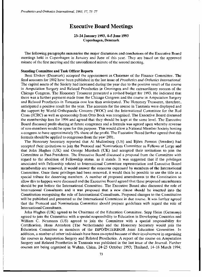
Göttingen Modular Knee Orthosis.* Rehabilitation step-by-step
O R T H O P E D I C I N D U S T R Y
A company of the Otto Bock Group PO, Box 12 60 D-37105 Duderetadl
Telephone (0 55 27) 8 48-0 Telefax (0 55 27) 7 23 30
Following knee surgery, joint
movement must often be restricted
to promote healing. The
Göttingen Modular Knee Orthosis
allows increases in range of motion
making it effective in the
rehabilitation. The orthosis consists
of anatomically shaped, plastic
shells with slotted connections and
polycentric joints that provide
stability and selectively restrict
movement. The prefabricated
P A R T N E R S 1
orthosis must be fitted by an
orthotist and is available as a
complete kit, under article number
28K10. Size 1 fits mid-thigh
circumferences of46-50 cm
(18-19 1/2 inches). Size 2 fits
mid-thigh circumferences of
42-46 cm (16-18 inches).
* Developed in cooperation with
the orthopedic clinic at the
University of Göttingen, Germany.
R E H A B I L I T A T I O N
Otto Bock 6/1993
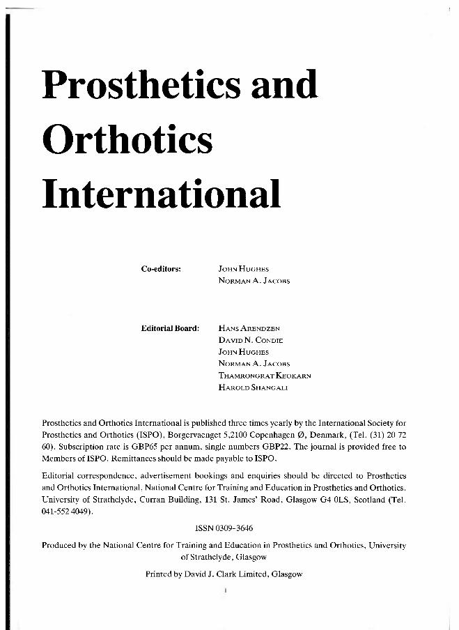
Prosthetics and Orthotics International
Co-editors: J O H N H U G H E S
N O R M A N A . JACOBS
Editorial Board: H A N S A R E N D Z E N
D A V I D N . C O N D I E
J O H N H U G H E S
N O R M A N A . JACOBS
T H A M R O N G R A T K E O K A R N
H A R O L D SHANGALI
Prosthetics and Orthotics International is published three times yearly by the International Society for Prosthetics and Orthotics ( ISPO), Borgervaenget 5,2100 Copenhagen 0 , Denmark , (Tel. (31) 20 72 60). Subscription rate is GBP65 per annum, single numbers GBP22. The journal is provided free to Members of ISPO. Remit tances should be made payable to ISPO.
Editorial correspondence, advertisement bookings and enquiries should be directed to Prosthetics and Orthotics International , National Centre for Training and Education in Prosthetics and Orthotics, University of Strathclyde, Curran Building, 131 St. James ' Road , Glasgow G4 0LS, Scotland (Tel. 041-552 4049).
ISSN 0309-3646
Produced by the National Centre for Training and Educat ion in Prosthetics and Orthotics, University of Strathclyde, Glasgow
Printed by David J . Clark Limited, Glasgow
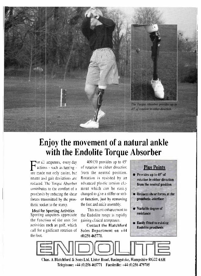
Enjoy the movement of a natural ankle with the Endolite Torque Absorber
F or all amputees, every day actions - such as turning -
are made not only easier, but neater and gait deviat ions are reduced. The Torque Absorber contributes to the comfort of a prosthesis by reducing the shear forces transmitted by the prosthetic socket to the stump. Ideal for Sporting Activities
Sport ing amputees appreciate the f u n d ion of the un i t for activi t ies such as golf, which call for s gnificant rotation of the foot,
409130 provides up to 45° of rotation in ei ther direction from the n e u t r a l p o s i t i o n , R o t a t i o n is r e s i s t e d by an advanced p las t ic tors ion e lem e n t w h i c h can be ea s i l y changed to give a stiffer or softer function, jus t by removing the foot and ankle assembly.
This recent enhancement to the Endol i te range is rapidly gaining clinical acceptance.
C o n t a c t the B la t ch fo rd Sales D e p a r t m e n t on +44 (0)256 465771.
Plus Points • Provides up lo 45" of
rotation in either direction from the neutral position
• Reduces shear forces at the prosthetic interface
• Variante degree of resistance
• Easily fitted to existing Endolite prosthesis
Chas. A Blatchford & Sons Ltd, Lister Road, Basingstoke, Hampshire RG22 4AH Telephone: +44 (0)256 465771 Facsimile: +44 (0)256 479705

The Journal of the International Society for Prosthetics and Orthotics
August 1993, Vol. 17, No. 2
Contents
Editorial 7 1
Executive Board Meetings 73
Walking speed of normal subjects and amputees: aspects of validity of gait analysis 7 8 A. M. B O O N S T R A , V. F I D L E R A N D W . H . E I S M A
Normative ground reaction force data for able-bodied and trans-tibial amputee children during running 83 J . R . E N G S B E R G , A . G . L E E , K . G . T E D F O R D A N D J . A . H A R D E R
Comparison of gait using a Multiflex foot versus a Quan tum foot in knee disarticulation amputees 90 A. M. B O O N S T R A , V . F I D L E R , G . M. A. SPITS, P . T U I L AND A. L . H O F
The C A T - C A M socket and quadrilateral socket: a comparison of energy cost during ambulation 95 R . S . G A I L E Y , D . L A W R E N C E , C. B U R D I T T , P. SPYROPOULOS, C. N E W E L L A N D M. S . N A S H
A comparison of paraplegic gait performance using two types of reciprocating gait orthoses 101 P. K . W I N C H E S T E R , J . J . C A R O L L O , R . N . P A R E K H , L . M. L U T Z AND J . W. A S T O N J R .
Enhancement of hémiplégie patient rehabilitation by means of functional electrical stimulation 107 A. K R A L J , R . A Č I M O V I C A N D U . STANIČ
Multi-adjustable post-operative orthosis for congenital muscular torticollis 1 1 5 C . Y. C H E N G , K . W. H O AND K . K . L E U N G
Technical note : automatic suspension device for gait training 1 2 0 J . K A W A M U R A , T. I D E , S . H A Y A S H I , H . O N O A N D T . H O N D A
Clinical note : lightweight prostheses for bilateral below-elbow amputees 1 2 6 S . N . R O U T
Technical note : continuous passive mot ion in hand rehabilitation 130 J . A . D E N T
Tor Hie ran 80 Years 136
I S P O Upda te Course on Lower Limb Amputa t ions and Related Prosthetics 137
Calendar of Events 139
m
ISPO Elected Members of Executive Board: M. L. Stills (President) S. Sawamura (President Elect) P. Christiansen (Vice President) J. Vaucher (Vice President) H- Arendzen D . N . Condie T. Keokarn H . Shangali W. H . Eisma ( Immedia te Past President) J. Steen Jensen (Hon . Treasurer) N. A . Jacobs (Hon . Secretary)
Standing Committee Chairmen and Task officers B. Ebsko \ (Finance) W. H . Eisma (Protocol and Nominations) S. Sawamura (Congress) .1 Hughes (Educat ion) S. Heim (Educat ion in Developing Countries) W. Neumann (Certification) H. Arendzen (Membership) D. N. Condie (Standards) P. Christiansen (Professional Register) Consultants to the Executive Board H. C. Chadder ton (Consumer) J. F . T. Bredie ( IVO) J. Van Rol leghem (Interbor) J. N . Wilson ( W O C ) T. Lagerwall (RI / ICTA) International Consultants to the Executive Board C. Marincek J. Craig and R. de Saez O . E . Feldman Chairmen of National Member Societies Australia Austria Belgium Canada Carr ibean China Denmark Finland Germany Hong Kong India Israel Japan Korea Nether lands New Zealand Norway Pakistan Sweden Switzerland U K U S A Past Presidents K.Jansen (1974-1977) G Murdoch (1977-1480) A. S ta ros(19S0-198: ) E . Lvquist (1982-1983) E . G. Marquardt (1983-198t.) J. H u e h e s ( 1 9 8 6 - 1 9 S ' J I W. H. Eisma (1989-1992) Secretary Aase Larsson
U S A Japan Denmark Switzerland Nether lands U K Thailand Tanzania Nether lands Denmark U K
D e n m a r k Nether lands Japan U K Germany U S A Netherlands U K Denmark
Canada Nether lands Belgium U K Sweden
Central and Eas te rn E u r o p e Central and South America Russia
G. Car ter W. Ott E . Schoolmeester G. Martel J. Mart ina Zhongzhe W u P . Christiansen L. Nummelin R. Baumgar tner K. Y. Lee M K. Goel M. Azaria S. Sawamura J. S. Shin J. H . B . Geer tzen A. W. Beasley O Johansen N. M. Akh ta r A . Stenström J. Vaucher R. A . Cooper M. Schuch
D e n m a r k U K U S A Denmark Germany U K
Nether lands
D e n m a r k IV
Prosthetics and Orthotics International, 1993, 17, 71-72
Editorial
Countries ravished by war will take generations to totally recover. The toll on human life receives international attention but the war-injured with physical disabilities are often forgotten in t ime. Vietnam still has an estimated 200,000 amputees with 15,000 under the age of 15. There are no clear estimates of the numbers of remaining spinal cord injured, head injured, those disabled as a result of mal-union or non-union of fractures or peripheral nerve injuries. There are certainly other disabilities secondary to the war and a host of disabilities that occur as a result of disease and birth defects. Road accidents, industrial injuries and accidental detonat ion of war munitions strewn throughout the country and towns often add to the list of the disabled on a daily basis and in some areas surpass the number of war-injured. There are simply hundreds of thousands of disabled to be dealt with and many could become functional members of their society given the chance. Many have received prosthetic or orthotic services in the past but will require replacement and repair on a regular basis. The average amputee in Vietnam for example might need a new prosthesis every five years and a new foot every year. This alone would account for one million prostheses required in the next 20 years and about four million feet.
This scenario describes Vietnam but can easily be applied to much of Southeast Asia, many countries in Africa, the Middle East , and now Eastern Europe .
There are numerous agencies and non-governmental organizations working in these areas to reduce suffering and trying to rebuild lives by providing orthotic and prosthetic services. Some are strictly humanitarian projects providing prostheses and orthoses which are simply fabricated and fitted. Others are service and education providers. They are teaching others so that long term dependency on outside support can be reduced. Others may approach the problem by introducing the latest in technology. "High tech" manufacturing, "high tech" imported material and components with good prosthetic fitting results, is viewed as research by many but is still an important element for the future.
I had the opportunity recently to view four such projects at six locations in Vietnam on behalf of the US Agency for International Development ( A I D ) . All were providing a much needed service at a varying degree of quality. Limb quality ranged from basic to "high tech"; and at costs ranging from realistic to unaffordable. The cost of a trans-tibial prosthesis ranged from $35 to $225, a foot from $2 locally made to $100 for an imported design. All patients generally seemed to benefit from their fitting but it is difficult to judge quality because everyone makes the best of the situation. I spoke with two expatriate prosthetist/orthotists, one from U S A working with World Vision and one from France working with the International Commit tee of the Red Cross ( ICRC) and both remarked how difficult it will be to return home after working with the Vietnamese patients who never complain, are totally accepting, and very appreciative of everything done for them. The highly trained could easily be lulled into complacency and the other service providers may not realize the need for continual upgrading,
Of particular interest was one project independent of US A I D funding. The I C R C project in H o Chi Minh City is using an innovative approach to delivering lower limb prosthetic services. They import polypropylene that has been pigmented to closely resemble skin tones in Vietnam. The entire lower limb prosthesis is fabricated from polypropylene. Polypropylene is used to make the socket and the exoskeleton of the prostheses. The scrap material is ground, heated, and moulded to produce knee joints, alignment fixtures, a t tachment pylons, and foot keels. The results are a very cosmetic and light-weight prosthesis that is not affected by the high heat and moisture common in that part of the world. Standard socket shapes are utilized and the prosthetic fit appeared to be very appropriate . There is no waste of material; everything is recycled; all components are fabricated on site. This technique is viewed by some as experimental , but similar techniques are utilized by I C R C in Cambodia and Mozambique , also with excellent results. What is most interesting is that from the developing world has come a technique that has clinical application in the developed world.
71
Editorial
The need continues for a more rapid exchange of information between practitioners regardless of where they are working. Improved methods of testing and evaluation are needed so that new concepts can be safely introduced. "High tech" does not need to cost more but by combining it with current concepts function may be further improved.
We applaud those individuals and organisations working to improve lives in areas we have all read about but may have started to forget.
Melvin L. Stills President
Prosthetics and Orthotics International, 1993, 17, 73-77
Executive Board Meetings 23-24 January 1993, 4-5 June 1993
Copenhagen, Denmark
The following paragraphs summarise the major discussions and conclusions of the Executive Board meetings held in Copenhagen in January and June of this year. They are based on the approved minute of the first meeting and the unconfirmed minute of the second meeting.
Standing Committee and Task Officer Reports Bent Ebskov (Denmark) accepted the appointment as Chairman of the Finance Commit tee . The
final accounts for 1992 have been published in the last issue of Prosthetics and Orthotics International. The capital assets of the Society had increased during the year due to the positive result of the course in Amputa t ion Surgery and Related Prosthetics in Groningen and the extraordinary success of the Chicago Congress. The Honorary Treasurer presented a revised budget for 1993. H e indicated that there was a further payment made from the Chicago Congress and the course in Amputa t ion Surgery and Related Prosthetics in Tanzania cost less than anticipated. The Honorary Treasurer , therefore, anticipated a positive result for the year. The accounts for the course in Tanzania were displayed and the support by World Orthopaedic Concern ( W O C ) and the International Commit tee for the Red Cross ( ICRC) as well as sponsorship from Ot to Bock was recognised. The Executive Board discussed the membership fees for 1994 and agreed that they should be kept at the same level. The Executive Board discussed profit-sharing at future congresses and a formula was agreed upon whereby revenue of non-members would be open for this purpose. This would allow a National Member Society hosting a congress to have approximately 5 % share of the profit. The Executive Board further agreed that this formula should be applied to congresses from the year 2001.
The Honorary Secretary reported that Al Muilenburg (US) and Björn Persson (Sweden) had accepted their invitations to join the Protocol and Nominations Commit tee as Fellows at Large and that John Hughes (UK) and George Murdoch (UK) had accepted their invitations to join the Commit tee as Past-Presidents. The Executive Board discussed a proposal from the Commit tee with regard to the abolition of Fellowship status, as it stands. It was suggested that if the privileges associated with Fellowship related to International Commit tee representat ion and Executive Board membership are removed, it would answer the concerns expressed by members of the International Commit tee . Once these privileges had been removed, it would then be possible to use the title as a special tr ibute for deserving members . A number of proposed amendments to the Constitution to allow this to happen were discussed and the Executive Board agreed that these proposed amendments should be put before the International Commit tee . The Executive Board also discussed the role of International Consultants and it was proposed that a new clause should be inserted into the Constitution recognising the role of International Consultants. Proposed changes to the Constitution will be published and presented to the International Commit tee in due course. It was further agreed that the Protocol and Nominat ions Commit tee should prepare guidelines with regard the role of International Consultants.
John Hughes (UK) agreed to be Chairman of the Education Commit tee . Sepp Heim (Germany) agreed to join the Commit tee with a special responsibility to Educat ion in Developing Countries and William C. Neumann (US) agreed to join the Commit tee with a special responsibility for Certification. Hans Arendzen (The Netherlands) and the Honorary Secretary would join the Education Commit tee as members of the I S P O / I N T E R B O R Joint Educat ion Commit tee . In addition, a number of o ther individuals have been co-opted because of their involvement in organising the courses in Amputa t ion Surgery and Related Prosthetics. A report of the course on Amputa t ion Surgery and Related Prosthetics in Tanzania was published in the last issue of the Journal . Fur ther courses are being organised in Wuhan , China, 24-25 October 1993; Thailand, 14-18 March 1994;
73
74 Executive Board Meeting
Ljubljana, Slovenia, 12-15 September 1994 and in Central America , sometime in November 1994 (Honorary Secretary's Note—Since the last Executive Board meeting, it has been necessary to postpone the course in China due to local difficulties). The Chairman of the Educat ion Commit tee reported that the American Board for Certification (ABC) held a trial of the written part of their certification examinations in collaboration with the Association of Prosthetists and Orthotists in the U K in March 1993. The results of the trial examination mirror those obtained in the corresponding examinations in the US . A post-examination critique with the 15 candidates established that they all felt it was a fair examination which reflected practice in the United Kingdom, apart from some local/regional-type questions which could be easily removed or amended. The Executive Board felt that these examinations may form the basis for International Certification and it was agreed that a further three trial examinations be conducted: in Australia which is another English-speaking country with a differing education system from the UK; in Tanzania to assess the performance of Category II graduates in such an examination; and in Germany to examine the results of non-native English-speaking Category I professionals sitting these examinations. The Education Commit tee was asked to examine the possibilities for development of International Certification for Category II personnel after the trial examinations in Tanzania were completed. The Honorary Secretary repor ted on his inspection of the I C R C course in Beira, Mozambique and the Executive Board agreed that ISPO recognise the course to be of Category II level. The Honorary Secretary also presented a list of criteria considered while inspecting the course in Mozambique . It was agreed that the Educat ion Commit tee put minimum requirements to each criterion, where applicable, and present it to the Executive Board in due course. Seishi Sawamura (Japan) repor ted on the proposed plan for an Asian Prosthetic and Orthotic Education Centre . Both Indonesia and Thailand were being considered as possible sites. The Executive Board agreed that the proposal formed a sound basis for Category II education which could be up-graded in the future if required to Category I and agreed to the proposal , in principle, and suggested that when the school is established, it would require a formal inspection to confirm these findings. The Honorary Secretary reported on an approach by the Cambodia Trust inviting the Society to provide advice for an Educat ion and Training Programme that the Cambodia Trust wishes to establish in the near future. The Executive Board agreed to give any advice and assistance it could in helping the Cambodia Trust formulate its plans. The Honorary Treasurer repor ted that during a recent visit to Kenya, he had some preliminary discussions with the Danish Embassy in Nairobi with regard to the Society applying to the Danish International Development Agency ( D A N I D A ) for support to help improve the education and training of orthopaedic technologists there. The Executive Board discussed this proposal and agreed that the Honorary Treasurer in collaboration with the Chairman of the Educat ion Commit tee should continue with processing this proposal to D A N I D A . The Executive Board discussed a proposal to produce videos on amputat ion techniques. It was agreed that the Society should participate in producing a pilot video on trans-tibial amputations.
The Honorary Secretary reported that at the end of 1992, there were 2,605 members of ISPO. A number of countries and regions were considering establishing National Member Societies. They included Panama, Colombia, Mexico, Argent ina , Slovenia, Mozambique , Malaysia, France and Indonesia. The Executive Board appointed Hans Arendzen (The Netherlands) as Task Officer for Membership .
The Publications Commit tee had initiated two proposals, firstly production of a flyer to promote the sale of the Journal and secondly, a promotional brochure for membership . The Executive Board agreed to both of these proposals. The Executive Board discussed the role of the Publications Commit tee and agreed that it should be disbanded, however, Hans Arendzen (The Netherlands) was appointed as Task Officer for Public Relat ions, Promot ion and Publicity and the Editors John Hughes (UK) and Norman A . Jacobs ( U K ) , were appointed as Task Officers for the Journal . As other tasks evolved, they would be allotted to other individuals.
David Condie (UK) reported on the work currently being carried out by the International Standards Organisation (ISO) and the European Community Standards Organisation (CEN) in the fields of prosthetics, orthotics and rehabilitation engineering. H e agreed to prepare a paper outlining the current situation with regards work on standards for publication in Prosthetics and Orthotics International.
Executive Board Meeting 75
It was agreed that Per Christiansen (Denmark) should be appointed as Task Officer for the Professional Register. He reported that the Professional Register had been successfully transferred to new equipment and reported that it was his intention to send the completed records to members for checking and to add any missing information, if possible. Members who have not yet completed the register will be sent a new questionnaire. Per Christiansen also informed the Executive Board that he would be at tempting to link the Professional Register with the Membership List and both he and the Honorary Secretary would examine the possibilities of combining the application to join the Society with the Professional Register Questionnaire and would report back to the next Executive Board meeting.
The Executive Board were examining the possibilities of holding Consensus Conferences on the Management of Poliomyelitis, Appropr ia te Prosthetic Technology for Developing Countries and the Orthotic Management of Cerebral Palsy. Proposals for these Consensus Conferences are presently being formulated and proposals will be discussed at the next Executive Board meeting.
Cliff Chadder ton (Canada) agreed to become Consumer Consultant to the Executive Board.
International Consultants Crt Marinček (Slovenia) had agreed to become International Consultant to the Executive Board for
Central and Eastern Eu rope ; John Craig (US) and Rosie de Sáez (Panama) had agreed to become International Consultants for Central and South America and Oleg E . Feldman (Russia) had agreed to be International Consultant for Russia. International Consultants for other regions were presently being considered.
International Organisations The arrangements for I N T E R B O R ' s Twelfth International Congress to be held in Lisbon,
Portugal, 22-25 September 1993, were well underway. Willem Eisma (The Netherlands) sits on the Organisation Commit tee representing ISPO as well as on the Scientific Commit tee together with Per Christiansen (Denmark) and David Condie (UK) .
The Honorary Secretary reported that the Executive Board of the World Heal th Organisation ( W H O ) had agreed to establish official relations with ISPO at its Twenty-First Session, 29 January 1993.
The Honorary Secretary repor ted on the at tendance by the President and himself at the Rehabili tation International (RI) Seventeenth World Congress in Kenya, 7-11 September 1992. ISPO had organised a one-day seminar on Prosthetics and Orthotics and the Society participated in a joint exhibit together with the Tanzanian Training Centre for Orthopaedic Technologists ( T A T C O T ) and the German Agency for Technical Cooperat ion ( G T Z ) . A n invitation had been received from the organisers of the R I Sixth European Regional Conference to be held in Budapest , Hungary, 4-9 September 1994 with regard organising a session on Amputa t ion and Prosthetics in Rehabili tation following Accidents. The Executive Board agreed to accept this invitation and Hans Arendzen (The Netherlands) was asked to organise this session on behalf of the Executive Board.
Tomas Lagerwall (Sweden) a t tended the June Executive Board meeting as representative of R I and the International Commission on Technology and Accessibility ( ICTA) . It is hoped that participation in future Executive Board meetings would allow for greater collaboration between the Society and ICTA.
Jan Bredie (The Netherlands) indicated that the Internationaler Verband der Orthopädie Schuteckniker ( IVO) have decided to form a European Section with the purpose of having bet ter access to the European Community in Brussels and also to be in a bet ter position to collaborate with Central and East European countries. The Eleventh International Congress of I V O will be held in Quebec , Canada, 3-6 September 1993.
The Executive Board agreed that collaboration with World Orthopaedic Concern (WOC) should be pursued through a t tendance at Board meetings. It was agreed that Thamrongra t Keokarn (Thailand) and Seishi Sawamura (Japan) should represent ISPO interests at W O C Asia meetings, the President should represent ISPO interests at Orthopaedics Overseas meetings in the US and that George Murdoch (UK) should act as ISPO's representative to W O C (UK) with John Hughes (UK) acting as al ternate .
76 Executive Board Meeting
The Honorary Secretary repor ted that the United Nations (UN) Commit tee on Non-Governmenta l Organisation at its session held from 22 March-2 April 1993 decided to recommend to the Economic and Social Council that ISPO be re-classified to Category II Consultative status. This recommendat ion is still subject to the approval of the Economic and Social Council which will examine and tćike action on the Commit tee ' s recommendat ion at its session to be held from 28 J u n e -30 July 1993. The Honorary Secretary reported that he had represented the Society at the Non-Governmental Organisations Consultative Meeting at the U N office in Vienna on 3-4 December 1992 during which he had reported on the activities of the Society over the past year.
Congresses The final report of the Chicago Congress was submitted to the Executive Board . The Congress had
been at tended by 2,220 people from 40 different countries which included participants, volunteers, complementary registrations and exhibitor registrations. The Scientific Programme had been an outstanding success. There were 20 overview sessions, 225 contr ibuted papers , 160 symposia papers , 50 posters and 40 video/films. Some 37 instructional courses and 20 commercial workshops had been presented. The exhibit was also very well supported. There were 98 commercial exhibitors and 16 scentific exhibitors. The total number of booths taken was 222. The social p rogramme had been very well received and the response from everyone who participated in the Congress had been favourable. The Congress was an extremely successful event and the final outcome will make a considerable contribution to the Society's reserves. The President , on behalf of the Executive Board and the Society expressed gratitude to Dudley Childress and all his colleagues for the hard work and enthusiasm which they had put into making the Congress such a great success.
Arrangements for the Eighth World Congress to be held in Melbourne , Austral ia, 2 -7 April 1995 were well underway. Detai led planning for the Congress, both as far as programme and organisation was at an advanced stage. The President repor ted that bo th he and David Condie (UK) had visited Australia in April and met with Valma Angliss, the Secretary General of the Eighth World Congress, together with the majority of members of the organising committees to discuss progress with arrangements The International Congress Commit tee had met prior to the Executive Board meetings and the ideas generated at the Commit tee are being incorporated into the programme and organisation procedures .
The Executive Board considered the bids to host the 1998 Congress submitted by the German National Member Society and The Netherlands National Member Society. The Executive Board discussed these bids in detail and after much deliberation decided that the 1998 Congress should be held in the The Netherlands.
Conferences and Meetings O n behalf of the Society, Rene Baumgarter (Germany) had organised a session on the Neuropathic
Foot at the first European Congress of Orthopaedics , Paris, France , 21-23 April 1993. Contributors to this session included Rene Baumgarter (Germany) , Jean Vaucher (Switzerland), Per Holstein (Denmark) and Pierre Bot ta (Switzerland).
The Society has been asked to organise a session at the World Federat ion of Occupational Therapists , Eleventh International Congress, London , U K , 17-22 April 1994.
The Executive Board agreed to collaborate with the organisers of D u n d e e '94 Clinical Gait Analysis, Dundee , U K , 5-8 July 1994. There would be no financial obligations on ISPO and members of the Society would be offered a reduced registration.
Meeting of International Committee Representatives The Executive Board discussed arrangements for the Interim Meeting of International Commit tee
Representat ives and agreed that it should be held on 21-22 January 1994 in Denmark in association with the next Executive Board meet ing. The Executive Board also agreed that in order to ensure full participation, the International Society would pay for all the costs of the Inter im Meeting. Travel
Executive Board Meeting 77
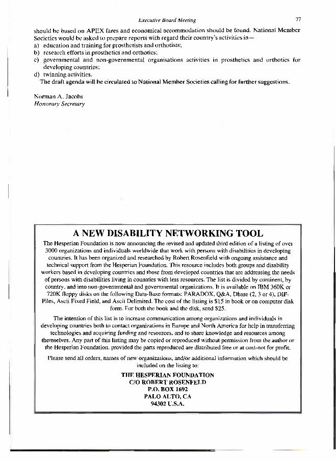
A NEW DISABILITY NETWORKING TOOL The Hesperian Foundation is now announcing the revised and updated third edition of a listing of ovei 3000 organizations and individuals worldwide that work with persons with disabilities in developing
countries. It has been organized and researched by Robert Rosenfield with ongoing assistance and technical support from the Hesperian Foundation. This resource includes both groups and disability
workers based in developing countries and those from developed countries that are addressing the needs of persons with disabilities living in countries with less resources. The list is divided by continent, by country, and into non-governmental and governmental organizations. It is available on IBM 360K or 720K floppy disks on the following Data-Base formats: PARADOX, Q&A, Dbase (2, 3 or 4), DIF-
Files, Ascii Fixed Field, and Ascii Delimited. The cost of the listing is $15 in book or on computer disk form. For both the book and the disk, send $25.
The intention of this list is to increase communication among organizations and individuals in developing countries both to contact organizations in Europe and North America for help in transferring
technologies and acquiring funding and resources, and to share knowledge and resources among themselves. Any part of this listing may be copied or reproduced without permission from the author or
the Hesperian Foundation, provided the parts reproduced are distributed free or at cost-not for profit.
Please send all orders, names of new organizations, and/or additional information which should be included on the listing to:
THE HESPERIAN FOUNDATION C/O ROBERT ROSENFELD
P.O. BOX 1692 PALO ALTO, CA
94302 U.S.A.
should be based on A P E X fares and economical accommodation should be found. National Member Societies would be asked to prepare reports with regard their country's activities in — a) education and training for prosthetists and orthotists; b) research efforts in prosthetics and orthotics; c) governmental and non-governmental organisations activities in prosthetics and orthotics for
developing countries; d) twinning activities.
The draft agenda will be circulated to National Member Societies calling for further suggestions.
Norman A . Jacobs Honorary Secretary

Prosthetics and Orthotics International, 1993, 17, 78-82
Walking speed of normal subjects and amputees: aspects of validity of gait analysis
A. M. B O O N S T R A * , V. F I D L E R * * and W. H . E ISMA*
*Department of Rehabilitation Medicine, University Hospital Groningen, The Netherlands **Groningen University, Groningen, The Netherlands
Abstract This study investigated some aspects of the validity of walking speed recording in 15 normal subjects, 16 trans-femoral amputees and 8 knee disarticulation amputees . The variability and test-retest reliability of walking speed and the influence of simultaneous recording of E M G and goniometry on comfortable and fast walking speeds were studied.
The variability between sessions was mainly determined by the variance within each session. The variance of speed within sessions while walking with East speed, was higher when walking without equipment than when walking with equipment . The variances of speed within sessions of the normal subjects were higher than those for both amputee groups. The test-retest reliability, expressed by the intra-class correlation coefficient, was good: between 0.83 and 0.98. The speed when walking without equipment was significantly higher both in normal subjects and amputees than the speed when walking with equipment .
Introduction Measurements of walking speed by means of
gait analysis are based on the assumption that walking speed is a basic gait parameter which, when measured objectively, can be useful in characterizing an individual's walking ability. In studies of prosthetic components , walking speed is often used as a parameter for the quality of performance (Barth et al., 1992;
Godfrey et al., 1975; Murray et al., 1983). Validity of speed recordings is essential. There are several aspects of validity. One of these is the test-retest reliability. Normal subjects show good reliability, but studies have been limited to the test-retest reliability of the natural walking speed (Kadaba et al., 1989; Waring and McLaurin, 1992; Winter , 1984) or have included only a few subjects (Winter , 1984). In neurologic patients (Holden et al., 1984; Wade et al., 1987) and post-polio patients (Waring and McLaurin, 1992) the test-retest reliability has been shown to be satisfactory.
Another aspect of the validity concerns the question about the relationship between speed recordings measured during gait analysis and the walking speed of the patient in his/her own surroundings. So far, nobody has answered this question. A n important problem in gait analysis, affecting the validity, is the simultaneous recording of speed, goniometry and E M G . It is unknown how the walking speed is influenced by the equipment and the wires attached to a patient for gait analysis.
In this paper , a study is presented of the variability of walking speed and the test-retest reliability, based on repeated recordings of comfortable and fast speed in normal subjects and amputees . The study also examined the influence of simultaneous recording of goniometry and E M G on walking speed.
Material and methods Subjects
The study included 15 normal subjects, 16 trans-femoral amputees and 8 knee
All correspondence to be addressed to A. M. Boonstra. Department of Rehabilitation Medicine. Universitv Hospital. Groningen, P.O. Box 30.001 , 9700 RB Groningen. The Netherlands.
78
Walking speed of normal subjects and amputees 7 9
disarticulation amputees . All gave informed consent.
The study of test-retest reliability was limited to the 15 normal subjects and 8 trans-femoral amputees .
The mean age of the normal subjects was 30 years (range 18-45, that of the trans-femoral amputees 40 years (range 15-63) and that of the knee disarticulation amputees 38 years (range 20-70). Ten of the trans-femoral amputees used a quadrilateral socket, six used a (modified) ischial containment socket (socket with narrow mediolateral dimension). All the trans-femoral prostheses had a 4-bar linkage knee with mechanical swing phase control (3R20, Ot to Bock); all but one had a Multiflex foot. One prosthesis was fitted with a Lager Bock foot.
All the knee disarticulation prostheses were fitted with an end-bearing socket, 4-bar linkage knee with mechanical or hydraulic swing phase control and a Multiflex foot.
Prosthetic component design and alignment of the amputees ' prostheses were all directed towards obtaining optimal gait. During the study neither the prostheses nor the shoes were changed.
Gait analysis The recordings were taken on a 10 metre
walkway. Two infra-red beams at the beginning and the end of a 7 metre trajectory started and stopped the measurements automatically. The normal subjects and amputees first walked without any goniometer or electrodes. The speed of comfortable walking and of fast walking ("as fast as possible") was measured. This was repeated once. Thereafter electrodes on the gluteal muscles, electrogoniometers (Penny & Giles) for hip, knee and ankle and aluminium strips on the shoes for stance phase and swing phase recordings were attached to the subject. The goniometry, E M G and stance/
swing phase recordings were used for a different study. The goniometers , electrodes and strips were connected to a box on the back of the subject. The box was connected by one cable to the computer . The investigator guided the subject while walking. The subject walked two more times at comfortable and fast speed.
The normal subjects were investigated on two days, with an interval of 2-7 days. Eight trans-femoral amputees were measured twice, with an interval of 3 to 6 weeks.
This meant that all 15 normal subjects and 8 trans-femoral amputees walked two sessions consisting of 4 runs each, two runs without and two runs with equipment . Eight trans-femoral amputees and all 8 knee disarticulation amputees walked one session consisting of 4 runs, two runs without and two runs with equipment.
Statistical analysis The variability of speed recordings was
studied separately in each of the three groups, for each of the two walking speeds and with and without equipment . The within-session variance, sd 2
w i t h i n , was calculated as the mean of the variances calculated within each session. The between-session variance component was estimated by sd 2
b e t w e e n = s d 2
s e s s i o n - 1 / 2 s d 2
w i t h i n . where s d 2
s e s s i o n is the variance of the session means.
The test-retest reliability can be described by the intra-class correlation coefficient between the means of the two sessions. This coefficient was estimated by ( s d 2 g r o u p - 1 / 2 s d 2
s e s s i o n ) / ( s d 2
g r o u p + 1 / 2 s d 2
s e s s i o n ) , where sd2 g r o u p denotes the variance of the subject means in a given group.
Group means of the repeated measures were compared using A N O V A . The effect of carrying equipment was evaluated by means of the paired t-test (comparison of means) and the signed-rank test (comparison of variances).
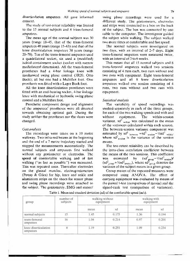
Table 1. Mean and standard deviation (sd) of the comfortable speed (m/s).
80 A. M. Boonstra, V. Fidler and W. H. Eisma
Tests were performed at 5% level of significance (two-sided if applicable).
Results The mean comfortable and fast walking
speeds and standard deviations are summarized in Tables 1 and 2.
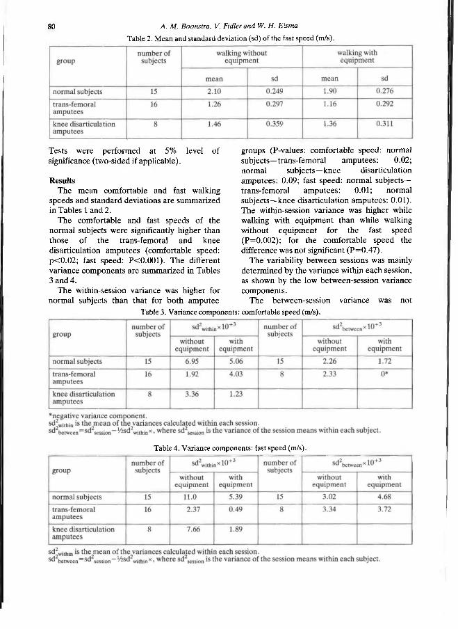
The comfortable and fast speeds of the normal subjects were significantly higher than those of the trans-femoral and knee disarticulation amputees (comfortable speed: p<0 .02 ; fast speed: P<0 .001) . The different variance components are summarized in Tables 3 and 4.
The within-session variance was higher for normal subjects than that for both amputee
groups (P-values: comfortable speed: normal subjects—trans-femoral amputees : 0.02; normal subjects—knee disarticulation amputees: 0.09; fast speed: normal subjects— trans-femoral amputees: 0 .01; normal subjects—knee disarticulation amputees: 0.01). The within-session variance was higher while walking with equipment than while walking without equipment for the fast speed (P=0.002) ; for the comfortable speed the difference was not significant (P=0 .47) .
The variability between sessions was mainly determined by the variance within each session, as shown by the low between-session variance components .
The between-session variance was not
Table 2. Mean and standard deviation (sd) of the fast speed (m/s).
Table 3. Variance components: comfortable speed (m/s).
Table 4. Variance components: fast speed (m/s).

Walking speed of normal subjects and amputees 81
significantly different while walking with equipment than while walking without equipment (P-values>0.20) . The between-session variance in normal subjects was not higher than in the amputee group (P-values: comfortable speed: without equipment: 0.52; with equipment: 0.95; fast speed: without equipment: 0.05; with equipment: 0.89).
In each session both normal subjects and amputees showed a tendency to walk slower in the first run than in the second run (mean comfortable speeds for all subjects while walking without equipment were , respectively, 1.17 m/s and 1.20 m/s), but the difference did not reach significance (P-values>0.1) .
The comfortable and fast speeds of the first session did not differ significantly from those of the second session, neither in normal subjects nor in amputees (P-values>0.1) .
The intra-class correlation coefficients of the data for both sessions are summarized in Table 5.
The speed while walking without equipment was significantly higher than while walking with equipment , both in normal subjects and amputees (P-values<0.01). The difference in fast speed with and without equipment was bigger than the difference in comfortable speed (see Tables 1 and 2).
Discussion and conclusion The study presented investigated the
variability and test-retest reliability of speed recordings both in normal subjects and amputees as well as the influence of equipment like goniometers and E M G electrodes on the walking speed. A good reliability was found for speed recordings both at comfortable speed and at fast speed. The variances of speed within each session and between two sessions were acceptably low. The low value of the between-session variance components indicates that the main part of the within-subject variability is already present within one session.
The within-session variance of speed recordings was lower for amputees than for normal subjects. This may be explained by the mechanical propert ies of the prosthesis, especially the knee-joint. Because of the almost fixed duration of the flexion-extension motion of the knee-joint, the amputee is forced to vary his/her walking speed only by means of one leg, the sound one . This may have led to the lower variation.
The test-retest reliability, as expressed by the intra-class correlation coefficient, was good (between 0.83 and 0.98). The correlation coefficient is comparable with the Pearson correlation coefficient found by others for speed recordings in patients with other diseases (Godfrey et al., 1975; Kadaba et al., 1989; Wade et al., 1987; Waring and McLaurin, 1992).
As has already been shown by others (James and Oberg , 1973), amputees walk slower than normal subjects.
The speed of both comfortable and fast walking is influenced by the equipment put on a patient for electrogoniometry and E M G ; it is reduced by about 8% when walking with equipment . Hence , the validity of speed recordings demands that the walking speed is measured without equipment for electrogoniometry and E M G .
REFERENCES
B A R T H DG, SCHUMACHER L . T H O M A S S S (1992). Gail analysis and energy cost of below-knee amputees wearing six different prosthetic feet. J Prosthet Orthol 4, 63-75.
G O D F R E Y CM, JOUSSE AT, B R E T T R, BUTLER JF (1975). A comparison of some gait characteristics with six knee joints Orthot Prosthetic, 33-38.
H O L D E N MK, GILL KM, MAGLIOZZI MR, N A T H A N J, P i e H L - B A K E R L (1984). Clinical gait assessment in the neurologically impaired. Reliability and meaningfullness. Phys Ther 64, 35-40.
Table 5. Intra-class correlation coefficients.

82 A. M. Boonstra, V. Fidler and W. H. Eisma
JAMES U , OBERG K (1973) . Prosthetic gait pattern in unilateral above-knee amputees. Scand J Rehabil Med 5, 3 5 - 5 0 .
KADABA MP, RAMAKRISHNAN H K , WOOTTEN ME, GAINEY J , GORTON G , COCHRAN G V B (1989) . Repeatability of kinematic, kinetic and electromyographic data in normal adult gait. J Orthop Res 7, 8 4 9 - 8 6 0 .
MURRAY MP, MOLLINGER L A , SEPIC S B , GARDNER GM, LINDER MT (1983) . Gait patterns in above-knee amputee patients: hydraulic swing control vs constant-friction knee components Arch Phys Med Rehabil 64, 3 3 9 - 3 4 5 .
WADE D T , WOOD V A , HELLER A , MAGGS J , HEWER R L (1987) . Walking after stroke: measurement and recovery over the first 3 months. Scand J Rehabil Med 19, 2 5 - 3 0 .
WARING WP, MCLAURIN T M (1992) . Correlation of creatine kinase and gait measurement in the postpolio population. Arch Phvs Med Rehabil 73, 3 7 - 3 9 .
WINTER D A (1984) . Kinematic and kinetic patterns in human gait: variability and compensating effects. Hum Move Sci 3, 5 1 - 7 6 .
Prosthetics and Orthotics International, 1993, 17, 83-89
Normative ground reaction force data for able-bodied and trans-tibial amputee children during running
J. R. ENGSBERG*, A. G. LEE*, K. G. TEDFORD,** and J. A. HARDER**
*Human Performance Laboratory, The University of Calgary, Alberta, Canada **Alberta Children's Hospital, Calgary, Alberta, Canada
Abstract The purpose of this investigation was to develop normative ground reaction force data for able-bodied (AB) and trans-tibial amputee (TTA) children during running. Two hundred AB (mean age 9.4 years, range 7-12) and 21 TTA (mean age 11.1 years, range 5-17) children ran (2.2 m/s±10%) over a force platform. Ground reaction force data were normalized, averaged within groups and plotted to produce force-time curves characterizing the different leg types (i.e. able-bodied, non-prosthetic and prosthetic). In addition, discrete variables characterizing the leg type differences were determined. One way ANOVA determined significant differences between variables and a TukeyB Post Hoc analysis defined which variables were significantly different (p<0.05). Results generally indicated differences between the three leg types with the non-prosthetic leg indicating greater forces than the prosthetic and AB legs. The results of this investigation provide normative ground reaction force data for both AB and TTA children during running and can be used for comparison with other groups of children.
Introduction In order to assess the benefit, detriment, or
irrelevance of a particular change in a prosthesis (e.g. new socket design or new terminal device) it is often desirable to compare measured variables with established norms. If
normative data do not exist then the effects of the particular change are more difficult to judge. In the case of a trans-tibial amputee (TTA) child, two sets of normative data appear desirable: data from able-bodied (AB) children and data from other TTA children. In both cases comparisons can be made to determine how closely the TTA child matches the respective groups. While normative data for both AB and TTA children exist for walking (Engsberg et al., 1993; Sutherland et al., 1988), none exist for running. The purpose of this investigation was to develop normative ground reaction force data for AB and TTA children during running.
Methods Two hundred AB (mean age 9.4 years, range
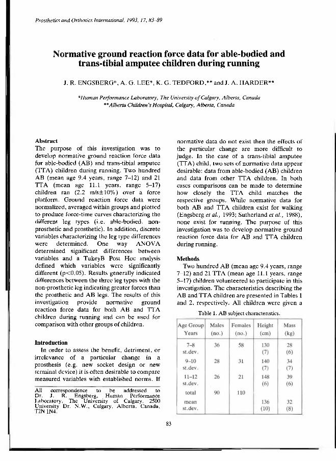
7-12) and 21 TTA (mean age 11.1 years, range 5-17) children volunteered to participate in this investigation. The characteristics describing the AB and TTA children are presented in Tables 1 and 2, respectively. All children were given a
Table 1. AB subject characteristics.
All correspondence to be addressed to Dr. J. R. Engsberg, Human Performance Laboratory, The University of Calgary, 2500 University Dr. N.W., Calgary, Alberta, Canada, T2N 1N4.
83
84 J R Engsberg, A, G Lee, K. G. Tedford and) A. Harder
physical examination by a pediatric orthopaedic surgeon and children not deemed able-bodied were excluded from the investigation. AB children were tested over a two week period during which approximately 20 children visited the Human Performance Laboratory each day. TTA children were tested over a one week period during which from 2-5 children visited the laboratory each day.
Ground reaction force data (1,000Hz) were collected during running support. For the AB children one trial was collected from each foot whereas for the I T A children at least three trials were collected from each foot. The same nominal rate of running (2.2 m/sec±10%) was enforced for all trials and was monitored by photocells spaced 2.4 m apart.
To allow for intersubject comparisons ground reaction force data were normalized by dividing by subject weight and contact time on the plate was normalized to a value of 1 (time 0 was touch down of the foot and time 1 was take off) (Engsberg et al., 1991). These data were then averaged within groups and plotted to produce force-time curves characterizing the different leg types (i.e. able-bodied, non-prosthetic and prosthetic). Since the time of occurrence of
relative maxima and minima forces for the leg types were not constant, the averaging process did not highlight the differences between leg types. Hence discrete variables characterizing these differences were determined (Andriacchi et al., 1977; Engsberg et al., 1991). For the vertical force-time curves, a slope, two local maxima, a local minimum, local impulses, total impulse, and local times of support were determined. For the anteroposterior force-time curves, two local maxima, impulses for the retarding (i.e. force applied by the foot in the anterior direction) and propulsive (i.e. force applied by the foot in the posterior direction) phases, associated time components, and total impulse were recorded. Impulses for the medial and lateral force components, respective maxima, and durations were compiled from the medio-lateral force-time traces.
One way ANOVA determined any significant differences between variables and a TukeyB Post Hoc analysis defined which variables were significantly different (p<0.05). Differences between variables for: 1) the right and left legs of the AB children; 2) ages of the AB children; 3) gender of the AB children;
Table 2. TTA subject characteristics
AB and TTA children during running 85
4) legs of the AB children and the non-prosthetic legs of the TTA children; 5) legs of the AB children and prosthetic legs of the TTA children; 6) non-prosthetic and prosthetic legs of the TTA children; and 7) foot type and suspension of the TTA children were compared.
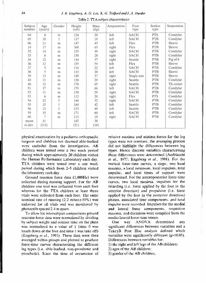
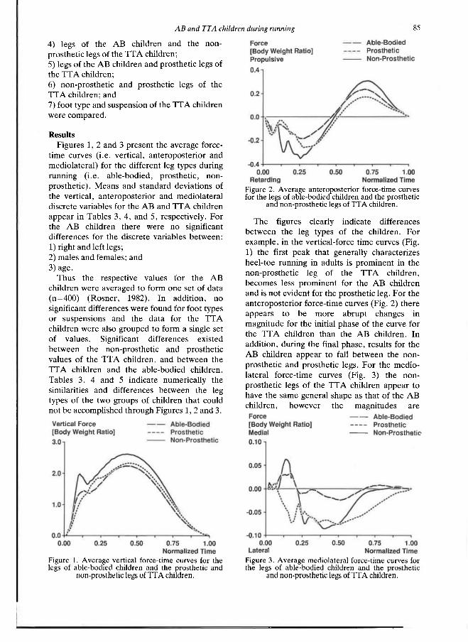
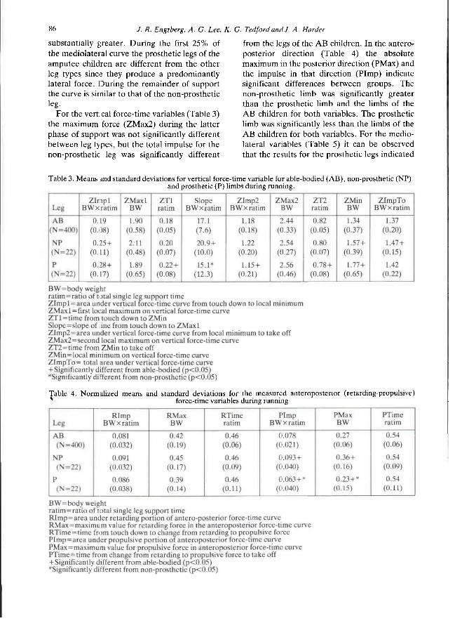
Results Figures 1, 2 and 3 present the average force-
time curves (i.e. vertical, anteroposterior and mediolateral) for the different leg types during running (i.e. able-bodied, prosthetic, non-prosthetic). Means and standard deviations of the vertical, anteroposterior and mediolateral discrete variables for the AB and TTA children appear in Tables 3, 4, and 5, respectively. For the AB children there were no significant differences for the discrete variables between: 1) right and left legs; 2) males and females; and 3) age.
Thus the respective values for the AB children were averaged to form one set of data (n=400) (Rosner, 1982). In addition, no significant differences were found for foot types or suspensions and the data for the TTA children were also grouped to form a single set of values. Significant differences existed between the non-prosthetic and prosthetic values of the TTA children, and between the TTA children and the able-bodied children. Tables 3, 4 and 5 indicate numerically the similarities and differences between the leg types of the two groups of children that could not be accomplished through Figures 1, 2 and 3.
The figures clearly indicate differences between the leg types of the children. For example, in the vertical-force time curves (Fig. 1) the first peak that generally characterizes heel-toe running in adults is prominent in the non-prosthetic leg of the I T A children, becomes less prominent for the AB children and is not evident for the prosthetic leg. For the anteroposterior force-time curves (Fig. 2) there appears to be more abrupt changes in magnitude for the initial phase of the curve for the TTA children than the AB children. In addition, during the final phase, results for the AB children appear to fall between the non-prosthetic and prosthetic legs. For the medio-lateral force-time curves (Fig. 3) the non-prosthetic legs of the TTA children appear to have the same general shape as that of the AB children, however the magnitudes are
Figure 1. Average vertical force-time curves for the legs of able-bodied children and the prosthetic and
non-prosthetic legs of TTA children.
Figure 2. Average anteroposterior force-time curves for the legs of able-bodied children and the prosthetic
and non-prosthetic legs of TTA children.
Figure 3. Average mediolateral force-time curves for the legs of able-bodied children and the prosthetic
and non-prosthetic legs of TTA children.
86 J. R. Engsberg. A. G. Lee. K. G. Tedford and J. A. Harder
substantially greater. During the first 25% of the mediolateral curve the prosthetic legs of the amputee children are different from the other leg types since they produce a predominantly lateral force. During the remainder of support the curve is similar to that of the non-prosthetic leg.
For the vertical force-time variables (Table 3) the maximum force (ZMax2) during the latter phase of support was not significantly different between leg types, but the total impulse for the non-prosthetic leg was significantly different
from the legs of the AB children. In the anteroposterior direction (Table 4) the absolute maximum in the posterior direction (PMax) and the impulse in that direction (PImp) indicate significant differences between groups. The non-prosthetic limb was significantly greater than the prosthetic limb and the limbs of the AB children for both variables. The prosthetic limb was significantly less than the limbs of the AB children for both variables. For the mediolateral variables (Table 5) it can be observed that the results for the prosthetic legs indicated
Table 3. Means and standard deviations for vertical force-time variable for able-bodied (AB), non-prosthetic (NP) and prosthetic (P) limbs during running.
Table 4. Normalized means and standard deviations for the measured anteroposterior (retarding-propulsive) force-time variables during running
AB and TTA children during running 87
significant differences when compared to the AB legs. Greater values were obtained for the maximum force in the lateral direction (LMax) and for the time spent applying force in the lateral direction (LTime).
Discussion The purpose of this investigation was to
determine normative ground reaction force data for AB and TTA children during running. Four limitations appear noteworthy. The first is that the externally measured ground reaction force data may not reflect the internal loading of the joints of the lower limbs. Other forces such as those of muscles and ligaments also contribute to this load. The second limitation was with respect to the running speed. A number of methods exist for comparing the running speeds of the subjects (e.g. fixed speed, fixed cadence, freely chosen speed) with each method having its own advantages and limitations. Since it has been shown that rate of walking can influence force variables for children with trans-femoral amputation, (Zernicke et al., 1985) the same rate of running was used for all subjects in the present investigation. This chosen speed was based upon preliminary pilot investigations performed in the laboratory.
The third limitation was that this investigation determined normative data for TTA children, disregarding inter-subject prosthetic differences (i.e. foot types or suspension). While the design of this study was
not well suited for considering the potential effects of these factors, the results were however, examined. In general, no significant differences were found between the variables for foot type and suspension.
The fourth limitation relates to the differences between the average force curves presented in Figures 1, 2 and 3 and the quantification of discrete variables derived from individual subject force curves presented in Tables 3, 4 and 5. The apparent discrepancy of results between the figures and the tables arises from the averaging process used to derive the force curves. Since the maximum values for a particular variable do not occur at the same instant in time for all subjects and leg types the averaged curves will not reflect the peak value displayed in the Table. On the other hand, the discrete variables determined from each individual curve and presented in the Tables will provide the true value since time of occurrence was not used in the determination of the value. For example, the ZMax1 variable in Table 3 reports values of 1.90, 2.11 and 1.89 for the AB, non-prosthetic and prosthetic legs, respectively. However, Figure 1 indicates a ZMax1 value for AB children to be about 1.5, ZMax1 value for non-prosthetic leg of the TTA to be about 2.0, and Zmax1 value for the prosthetic leg to be about 1.9 (i.e. if the levelling of the curve at about 0.25 of the cycle is used as the average ZMax1 value). Thus it is important to view the curves as providing average shapes characterizing general
Table 5. Normalized means and standard deviations for the measured mediolateral force-time variables during running
88 J. R. Engsberg, A. G. Lee, K. G. Tedford and J. A. Harder
differences between leg types and the tables as providing accurate discrete information for particular variables associated with the curves.
Prince et al. (1992) presented local maxima in the vertical, anterior, and posterior directions for a group of young TTA adults (n=9, mean age of 16 years). They found significantly greater values for the ZMax2 variable for the non-prosthetic leg when compared to both the prosthetic legs and legs of the AB. The present investigation did not find those differences and indicated no differences between the three leg types. Even though Miller (1987) did not present discrete values for the prosthetic and non-prosthetic limbs of TTA adults (n=4, mean age of 40 years) her figures concur with the results of the present investigation suggesting that approximately the same values occurred for the non-prosthetic and prosthetic legs. In support of the results presented by Prince et al. (1992) and in contrast to the results of the present investigation and of Miller (1987) the authors of this paper have reported greater ZMax2 values for the children of this investigation during walking (Engsberg et al., 1993). The results indicated that the non-prosthetic leg had a significantly greater ZMax2 value than the prosthetic leg and the legs of AB children. In addition, it was reported that the prosthetic leg force was significantly less than the AB leg force for the ZMax2 variable. Further investigation appears warranted in this regard.
In the anteroposterior directions Prince et al. (1992) reported significant differences for the RMax variable between the non-prosthetic legs and the AB legs and between the prosthetic legs and the non-prosthetic legs. The present investigation found no significant differences in RMax values between leg types. For the PMax variable Prince et al. (1992) reported significant differences between the prosthetic leg and both the non-prosthetic and AB legs. The present investigation supported these relationships and also found significant differences between the non-prosthetic and the AB legs. The differences in the results between the two investigations may be explained by subject age, subject numbers, and different prostheses.
Prince et al. (1992) reported that the ZMax2 and the ZImpTo variables were significantly greater (rigid keel only) in non-prosthetic legs when compared to similar values of AB
controls. Similar results for the same variables and for approximately the same group of subjects as in the present investigation have been reported for walking (Engsberg et al., 1991 and 1993). In contrast to these results the present investigation did not concur with these findings. A potential explanation could be related to the possible effects of foot types since Prince et al. (1992) reported no significant differences for the two variables for flexible keel feet. Further investigation is necessary in this regard.
Prince et al. (1992) reported that the PMax and Pimp variables were significantly different between the prosthetic legs and the legs of the AB subjects. These loading differences, also occurring in the present investigation, have been reported for walking (Engsberg et al., 1991 and 1993). The similarity of these results appears to indicate that despite the type of terminal device used, the prosthetic legs do not generate propulsive forces similar to those produced by intact legs. The objective of the authors' research in this area is to enable TTA children to walk and run in the same way as AB children. The accomplishment of this objective would however require the development of a prosthesis which allows the prosthetic leg to produce the same forces as those of intact legs.
Conclusions The results of this investigation provide
normative ground reaction force data for both AB and TTA children during running. The results for the AB children can be used for comparison with TTA children and with any other groups of children (e.g. children with cerebral palsy) if similar data are determined. The results for the TTA children can be used to determine if TTA children are functioning similarly to other TTA children. Since the results indicate basic differences between TTA and AB children during running, research should be directed towards eliminating these differences.
Acknowledgment Funding provided by the George Reed
Foundation for the Handicapped, Hospital for Sick Children Foundation, and the Variety Club of Southern Alberta-Tent 61.
AB and TTA children during running 89
REFERENCES
A n d r i a c c h i T P , OGLE JA, GALANTE JO (1977) . Walking speed as a basis for normal and abnormal gait measurements. J Biomech 10, 2 6 1 - 2 6 8 .
ENGSBERG JR, LEE AG, PATTERSON JL, HARDER JA (1991) . External loading comparisons between able-bodied and below-knee-amputee children during walking. Arch Phys Med Rehabil 12, 6 5 7 -6 6 1 .
ENGSBERG JR, LEE AG, TEDFORD KG, HARDER JA (1993) . Normative ground reaction force data for able-bodied and below-knee-amputee children during walking. J Pediatr Orthop 13, 1 6 9 - 1 7 3 .
MILLER DI (1987) . Resultant lower extremity joint moments in below-knee amputees during running stance. J Biomech 20, 5 2 9 - 5 4 1 .
PRINCE F, ALLARD P, THERRIEN RG, MCFADYEN B J (1992) . Running gait impulse asymmetries in below-knee amputees. Prosthet Orthot Int 16, 19-24 .
ROSNER B (1982) . Statistical methods in ophthalmology: an adjustment for the intraclass correlation between eyes. Biometrics 38, 1 0 5 - 1 1 4 .
SUTHERLAND DH, OLSHEN RA, BIDEN EN, WYATT MP (1988) . The Development of Mature Walking. — Oxford: MacKeith Press.
ZERNICKE RF, HOY MG, WHITING WC (1985) . Ground reaction forces and centre of pressure patterns in the gait of children with amputations: preliminary report. Arch Phys Med Rehabil. 66, 7 3 6 - 7 4 1 .
Prosthetics and Orthotics International, 1993, 17, 90-94
Comparison of gait using a Multiflex foot versus a Quantum foot in knee disarticulation amputees.
A. M. B O O N S T R A * , V. F I D L E R * * , G. M. A . SPITS*, P. T U IL * and A . L. H O F *
* Department of Rehabilitation Medicine, Groningen University Hospital, Department of Health Science, Groningen University, Groningen, The Netherlands
**Orthopaedic Workshop "Noord-Nederland", Haren, The Netherlands
Abstract The subjective responses and gait pat terns of unilateral knee disarticulation amputees wearing prostheses fitted first with the Multiflex foot and then with the Quan tum foot were studied. Nine amputees were included in the trial.
A questionnaire asked the amputees about their preference for one of the feet.
Gait analysis was performed measuring temporal parameters and goniometry of hips, knees and ankles in the sagittal and frontal planes.
There was a slight preference for the Quan tum foot. Preference seemed not to be related to physical characteristics of the amputees nor to gait parameters .
There were no differences in gait as far as the temporal factors were concerned.
The main differences in the range of motion of the joints were in the frontal plane: the eversion-inversion movement of the ankle and the adduction-abduction movement of the hip. During walking at comfortable speed with the Multiflex foot the ankle and hip range of motion averaged 2.1 and 3.1 degrees respectively, less than during walking with the Quan tum foot.
Introduction If a prosthesis is to be prescribed after an
amputat ion, the choice of a prosthetic foot is an important one, both for the amputee and for
the clinical team. Several studies have reported on differences in gait pat terns in trans-tibial (Barth et al., 1992, Culham et al., 1986; Mizuno et al., 1992; Wirta et al., 1991) and trans-femoral (Goh et al., 1984; James and Stein, 1986) amputees resulting from the use of different feet. However , it has so far been difficult to make a choice for the individual patient from the many available artificial feet. The same problem occurs in knee disarticulation amputees . The gait characteristics of the knee disarticulation amputee are difficult to compare with those of trans-tibial amputees because of the absence of a knee joint; neither are they comparable with trans-femoral amputees , because of the end weight-bearing principle of the socket. Hence , studies of trans-tibial or trans-femoral amputees cannot be generalized to knee disarticulation amputees .
This study investigated the gait pat terns of unilateral knee disarticulation amputees wearing prostheses fitted with either the Multiflex or Quan tum foot. The Multiflex foot is one of the most common prosthetic feet in the Netherlands. The Quan tum foot was used because it is one of the modern "energy-storing" feet and because it differs from the Multiflex foot in biomechanical propert ies such as hysteresis and stiffness (Jaarsveld et al., 1990).
Method Subjects
Nine subjects who met the following criteria were recruited for the study: unilateral knee disarticulation amputat ion fitted with an end
All correspondence to be addressed to A. M. Boonstia, Department of Rehabilitation Medicine, University Hospital Groningen, P.O. Box 30. 001, 9700 RB Groningen, The Netherlands.
90
Comparison Multiflex foot versus Quantum foot 91
bearing socket, relatively pain-free s tump with no skin abrasions, and residency in the north of the Nether lands. All gave informed consent.
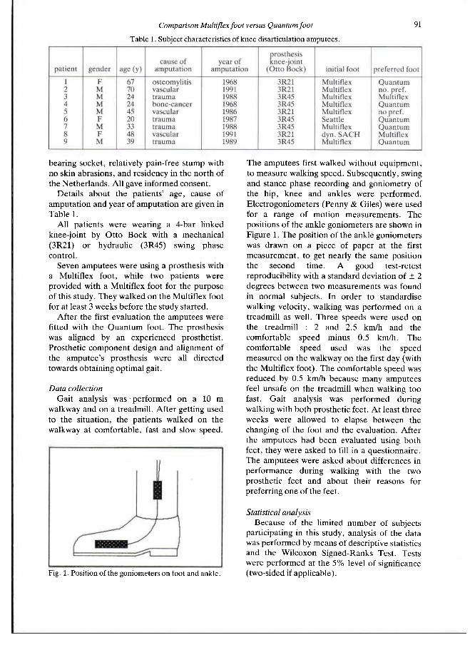
Details about the patients ' age, cause of amputation and year of amputat ion are given in Table 1.
All patients were wearing a 4-bar linked knee-joint by Ot to Bock with a mechanical (3R21) or hydraulic (3R45) swing phase control.
Seven amputees were using a prosthesis with a Multiflex foot, while two patients were provided with a Multiflex foot for the purpose of this study. They walked on the Multiflex foot for at least 3 weeks before the study started.
After the first evaluation the amputees were fitted with the Quan tum foot. The prosthesis was aligned by an experienced prosthetist. Prosthetic component design and alignment of the amputee 's prosthesis were all directed towards obtaining optimal gait.
Data collection Gait analysis was - performed on a 10 m
walkway and on a treadmill . After getting used to the situation, the patients walked on the walkway at comfortable, fast and slow speed.
The amputees first walked without equipment , to measure walking speed. Subsequently, swing and stance phase recording and goniometry of the hip, knee and ankles were performed. Electrogoniometers (Penny & Giles) were used for a range of motion measurements . The positions of the ankle goniometers are shown in Figure 1. The position of the ankle goniometers was drawn on a piece of paper at the first measurement , to get nearly the same position the second t ime. A good test-retest reproducibility with a standard deviation of ± 2 degrees between two measurements was found in normal subjects. In order to standardise walking velocity, walking was performed on a treadmill as well. Three speeds were used on the treadmill : 2 and 2.5 km/h and the comfortable speed minus 0.5 km/h. The comfortable speed used was the speed measured on the walkway on the first day (with the Multiflex foot). The comfortable speed was reduced by 0.5 km/h because many amputees feel unsafe on the treadmill when walking too fast. Gait analysis was performed during walking with both prosthetic feet. At least three weeks were allowed to elapse between the changing of the foot and the evaluation. After the amputees had been evaluated using both feet, they were asked to fill in a questionnaire. The amputees were asked about differences in performance during walking with the two prosthetic feet and about their reasons for preferring one of the feet.
Statistical analysis Because of the limited number of subjects
participating in this study, analysis of the data was performed by means of descriptive statistics and the Wilcoxon Signed-Ranks Test. Tests were performed at the 5% level of significance (two-sided if applicable).
Table 1. Subject characteristics of knee disarticulation amputees.
Fig. 1. Position of the goniometers on foot and ankle.
92 A. M. Boonstra, V. Fidler, G. M. A. Spits, P. TuilandA L Hof
Results Five amputees preferred the Quan tum foot,
while two preferred the Multiflex foot and two amputees had no preference. N o clear explanation of the preferences could be found, neither in characteristics of the amputees , nor in differences in gait parameters . The reasons for preference which the amputees gave on the questionnaire were not consistent either.
The results of the gait analysis are summarised in Tables 2 and 3 and Figure 2.
Temporal parameters of gait analysis. No difference in walking speed was found
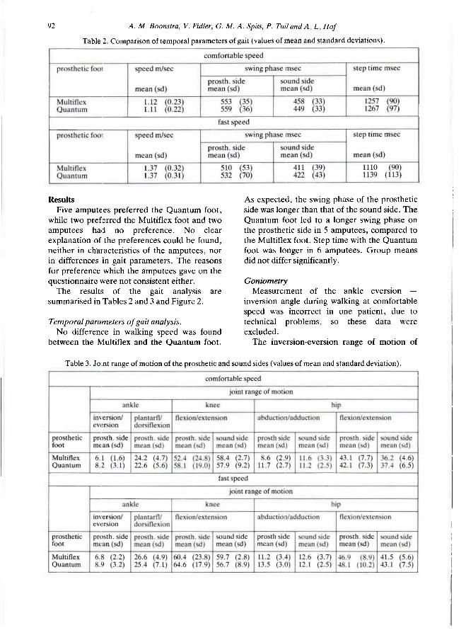
between the Multiflex and the Quan tum foot.
As expected, the swing phase of the prosthetic side was longer than that of the sound side. The Quantum foot led to a longer swing phase on the prosthetic side in 5 amputees , compared to the Multiflex foot. Step time with the Quan tum foot was longer in 6 amputees . Group means did not differ significantly.
Goniometry Measurement of the ankle eversion —
inversion angle during walking at comfortable speed was incorrect in one patient, due to technical problems, so these data were excluded.
The inversion-eversion range of motion of
Table 2. Comparison of temporal parameters of gait (values of mean and standard deviations).
Table 3. Joint range of motion of the prosthetic and sound sides (values of mean and standard deviation).
Comparison Multiflex foot versus Quantum foot 93
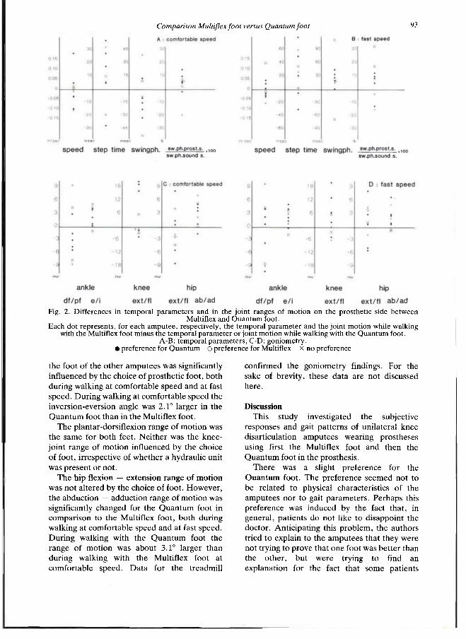
the foot of the other amputees was significantly influenced by the choice of prosthetic foot, both during walking at comfortable speed and at fast speed. During walking at comfortable speed the inversion-eversion angle was 2.1° larger in the Quan tum foot than in the Multiflex foot.
The plantar-dorsiflexion range of motion was the same for bo th feet. Neither was the knee-joint range of motion influenced by the choice of foot, irrespective of whether a hydraulic unit was present or not .
The hip flexion — extension range of motion was not altered by the choice of foot. However , the abduction — adduction range of motion was significantly changed for the Quan tum foot in comparison to the Multiflex foot, both during walking at comfortable speed and at fast speed. During walking with the Quan tum foot the range of motion was about 3.1° larger than during walking with the Multiflex foot at comfortable speed. Data for the treadmill
confirmed the goniometry findings. For the sake of brevity, these data are not discussed here.
Discussion This study investigated the subjective
responses and gait pat terns of unilateral knee disarticulation amputees wearing prostheses using first the Multiflex foot and then the Quan tum foot in the prosthesis.
There was a slight preference for the Quan tum foot. The preference seemed not to be related to physical characteristics of the amputees nor to gait parameters . Perhaps this preference was induced by the fact that , in general, patients do not like to disappoint the doctor. Anticipating this problem, the authors tried to explain to the amputees that they were not trying to prove that one foot was bet ter than the other , but were trying to find an explanation for the fact that some patients
Fig. 2. Differences in temporal parameters and in the joint ranges of motion on the prosthetic side between Multiflex and Quantum foot.
Each dot represents, for each amputee, respectively, the temporal parameter and the joint motion while walking with the Multiflex foot minus the temporal parameter or joint motion while walking with the Quantum foot.
A-B: temporal parameters, C-D: goniometry. • preference for Quantum o preference for Multiflex x no preference
94 A. M. Boonstra, V. Fidler, G. M. A, Spits, P. Tuil and A. L. Hof
preferred the Multiflex foot while others preferred the Quan tum foot. One of the two amputees who had not previously used the Multiflex foot, now preferred the Mutliflex foot, while the other preferred the Quan tum foot. There were no differences in gait as far as regards the temporal factors.
As expected, the swing phase of the prosthesis was longer than that of the sound leg: the difference W A S about 2 3 % during walking at comfortable speed. Only one patient showed a nearly symmetrical gait.
The only earlier study comparing Multiflex and Quan tum feet — as well as other types — was that by Mizuno et al. (1992) using trans-tibial amputees , but they studied other parameters . Most studies (Culham et al., 1986; D o a n e and Holt , 1983; Goh et al., 1984; MacFarlane, 1991; Wagner et al., 1987) of different feet found no differences in walking speed. Only Nielson et al. (1989) found that trans-tibial amputees walked faster when fitted with the Flex-foot than with the S A C H foot. Some studies of trans-tibial amputees (Culham et al., 1986; MacFarlane et al., 1991; Van Leeuwen et al., 1990) found differences in symmetry in the stance phase between the prosthetic and sound sides while walking with different feet. Other studies failed to find such differences (Doane and Holt , 1983; Goh et al., 1984).
Trie main differences in the range of motion of the joints were in the frontal plane; the eversion-inversion movement of the ankle and the adduction-abduction movement of the hip. During walking at comfortable speed using the Multiflex foot, the ankle and hip joint ranges of motion were an average of respectively 2.1° and 3.1° smaller than with the Quan tum foot.
It may be assumed that the difference in the ankle joint range of mot ion in the frontal plane was primary, while the difference in hip joint range of motion was secondary. Differences in ankle joint range of motion between different feet have been found by many authors (Barth et al., 1992; Doane and Holt , 1983; James and Stein, 1986; Wagner, 1987) in studies of trans-
tibial amputees . To what extent this increased transverse motion is reflected in the subjective preference, remains unclear.
REFERENCES
BARTH DG, SCHUMACHER L, THOMAS SS (1992). Gait analysis and energy cost of below-knee amputees wearing six different prosthetic feet. J Prosthet Orthot 4, 63-75.
CULHAM EG, PEAT M, NEWELL E (1986). Below-knee amputation: a comparison of the effect of the SACH foot and single axis foot on the electromyographic patterns during locomotion. Prosthet Orthot Int 10,15-22.
DOANE NE, HOLT L E (1983). A comparison of the SACH and single axis foot in the gait of unilateral below-knee amputees. Prosthet Orthot Int 7, 33-36.
GOH JCH, SOLOMONIDIS SE, SPENCE WD, PAUL JP (1984). Biornechanical evaluation of SACH and uniaxial feet. Prosthet Orthot Int», 147-154.
VAN JAARSVELD H W L , GROOTENBOER HJ, D E VRIES J, KOOPMAN HFJM (1990). Stiffness and hysteresis properties of some prosthetic feet. Prosthet Orthot Int 14, 117-124.
JAMES KB, STEIN RB (1986). Improved ankle-foot system for above-knee amputees. Am J Phys Med 65,301-314.
VAN LEEUWEN JL, SPETH LAWM, DAANEN HAM (1990). Shock absorbtion of below-knee prostheses: a comparison between the SACH and the Multiflex foot. J Biomech 2 3 , 441-446.
MACFARLANE PA, NIELSON DH, SHURR DG, MEIER K (1991). Gait comparisons for below-knee amputees using a Flex-foot versus a conventional prosthetic foot. J Prosthet Orthot 3 , 150-161.
MIZUNO N , AC>YAMA T , NAKAJIMA A, KASAHARA T , TAKAMI K (1992). Functional evaluation by gait analysis of various ankle-foot assemblies used by below-knee amputees. Prosthet Orthot Int 16, 174-182
NIELSON DH, SHURR DG, GOLDEN JC, MEIER K (1989). Comparison of energy cost and gait efficiency during ambulation in below-knee amputees using different prosthetic feet — a preliminary report. J Prosthet Orthot J, 24-31.
WAGNER J, SIENKO S, SUPAN T , BARTH D (1987). Motion analysis of SACH vs. Flex-foot in moderately active below-knee amputees. Clin Prosthet Orthot 11, 55-62.
WIRTA RW, MASON R, CALVO K, GOLBRANSON FL (1991). Effect on gait using various prosthetic ankle-foot devices. J Rehabil Res Dev 28,13-24.
Prosthetics and Orthotics International, 1993, 17, 95-100
The CAT-CAM socket and quadrilateral socket: a comparison of energy cost during ambulation
R. S. G A I L E Y , D. L A W R E N C E , C. B U R D I T T , P. SPYROPOULOS, C. NEWELL and M . S . NASH
Division of Physical Therapy, Dept. of Orthopaedics and Rehabilitation, University of Miami School of Medicine, Florida, USA
Abstract Twenty unilateral trans-femoral amputees fitted with either the Contoured Adducted Trochanteric-Controlled Alignment Method (CAT-CAM) socket (n=10) or the quadrilateral (QUAD) socket (n = 10), and a "non-amputee" control group (n=10) participated in the study. Subjects meeting the following criteria were studied: healthy males between the ages of 18 and 55 years, amputation due to non-vascular pathology, an unaffected sound limb, at least six months use of the test prosthesis, and a minimal stump length of 15 cm. Subjects ambulated in two randomized trials separated by 20 minutes of rest at 2 assigned speeds: a pace reflecting normal walking speed (97 m/min=2.5 mph) or a slower speed (48.5 m/min=1.25 mph). Heart rate (HR) and Oxygen uptake ( V O 2 ) measured during steady state walking were analyzed via two-way ANOVA. Differences among means were further analyzed using Tukey post hoc and simple effects tests. Significant differences were observed between the control group and CAT-CAM subjects with respect to V O 2 (p<0.05) and HR (p<0.01) at the slower speed. The control group and subjects using the QUAD socket also differed with respect to V O 2
(p<0.01) and HR (p<0.01) at the slower pace. Faster pace required more energy expenditure (p<0.01) and produced higher HR (p<0.01) than slower speeds. At faster pace, a significantly higher energy expenditure in the QUAD than the CAT-CAM group was
observed (p<0.01) . It is concluded that ambulating at normal pace using the CAT-. CAM socket design uses less energy than when using a QUAD socket design.
Introduction Interest in lower limb prosthetic research has
recently focused on reducing energy expenditure during ambulation. Different types of prostheses have been designed for trans-femoral amputees to improve biomechanical function and reduce energy used in ambulation, although empirical data demonstrating an energy advantage of one particular design during walking remains scant. Moreover, research has yet to show that trans-femoral amputees can ambulate at an energy cost commensurate with levels reported for individuals without disability. For example, James (1973) reported that 37 trans-femoral amputees fitted with a quadrilateral (QUAD) socket consumed 40% more oxygen than non-disabled persons while ambulating at a pace that was 30% slower (51 m/mins=1.9 mph). Similarly, Traugh (1975) found that 9 trans-femoral amputee subjects fitted with QUAD sockets expended 65% more energy than non-disabled controls while walking at 39 m/min (1.45 mph), half the normal walking speed of the control subjects. Waters (1976) compared energy cost in vascular (n=13) and traumatic amputees (n=15) ambulating at a self-selected speed. The amount of energy consumed during ambulation was similar in both groups, although pace differed as subjects self-adjusted the walking speed to maintain minimal energy expenditure. Subjects with vascular amputation ambulated at 36 m/min (1.34 mph) while those
All correspondence to be addressed to R. S. Gailey, University of Miami, Division of Physical Therapy, 5th Floor, Plumer Building, 5915 Ponce de Leon Blvd., Coral Gables, Florida 33146, USA.
95
96 R. S. Gailey, D. Lawrence, C. Burditt, P. Spyropoulos, C Newell and M. S. Nash
with traumatic amputation walked at 52 m/min (1.94 mph), 56% and 37% slower respectively than non-disabled control subjects (82 m/min= 3.02 mph). Similarly, Huang et al. (1979) determined that trans-femoral amputees wearing a QUAD socket expended 49% more energy than able-bodied individuals while ambulating at 47 m/min (1.75 mph). In an effort to improve biomechanical function and thus reduce metabolic cost of ambulation, John Sabolich designed the Contoured Adducted Trochanteric-Controlled Alignment Method (CAT-CAM) socket in the early 1980s. The CAT-CAM socket, more narrow in the mediolateral dimension than the QUAD socket, is designed to fit intimately with the ischial ramus, thus "locking" onto the pelvis, and encapsulating the ischial tuberosity. Additionally, the CAT-CAM socket has been claimed to improve muscular function, enhance pelvic motion, and maintain a more natural femoral adduction angle to a greater extent than the QUAD socket (Sabolich, 1985; Flandry et al., 1989). Consequently, proponents of the CAT-CAM socket have suggested that this design is associated with a decreased energy requirement during ambulation by trans-femoral amputees. To date, however, there has been limited research which has shown a reduction in energy cost for those using the CAT-CAM socket. For example, Flandry et al. (1989) reported that five trans-femoral amputees fitted with both Q U A D and CAT-CAM sockets used 56% less energy during ambulation with the latter. Ambulation speed of the CAT-CAM group was 44.5 m/min (1.66 mph), 16% faster than the pace of the subjects using the QUAD socket design (40.4 m/min= 1.5 mph), while sustaining reduced gait deviations and a slightly longer stride length. While this study suggests an energy and biomechanical advantage for the CAT-CAM socket during ambulation, the findings are compromised by the small study sample.
Therefore, the purpose of this study was to determine whether differences exist in energy expenditure during ambulation of trans-femoral amputees using Q U A D and CAT-CAM sockets.
Methods Subjects
Twenty unilateral trans-femoral amputees
wearing either the CAT-CAM (n=10) or quadrilateral (QUAD) (n=10) socket and 10 non-amputee (control) subjects participated in this study. Subjects who met the following selection criteria were randomly selected: healthy males between the ages of 18 and 58 years (see results), amputation due to nonvascular pathology, unaffected sound limb, at least six months experience with the tested prosthesis, and a minimum stump length of 15 cm, measured from the greater trochanter to the distal lateral end of the femur, with the amputation site proximal to the femoral condyles. All subjects were free from any stump soft tissue problems, pain, or limitations that would influence their gait to an appreciable degree.
All amputee subjects were selected based upon the type of socket they were currently wearing. Subjects were not asked the name of their prosthetist, and as a result the investigators were not aware of the socket fabrication techniques employed by the prosthetists. Therefore, subjects were selected and tested purely of the basis of their socket design and not a single prosthetist's fabrication or casting technique.
Subjective criteria The subjective criteria for the CAT-CAM
socket were true ischial containment medially with no lateral gapping between the lateral wall and stump, an appropriate mediolateral and anteroposterior dimension for each individual amputee, and an appropriate femoral adduction angle. Criteria for the QUAD socket were: a posterior brim which an ischial seat, equal height of the medial and posterior brim, Scarpa's bulge anteriorly. Essentially, the socket design had to be consistent with the classic description of the quadrilateral socket by Radcliffe (1955). The same investigator evaluated every subject to provide standardisation in subject selection.
Procedure Two trials were performed during which each
subject ambulated at both 33.5 m/min (1.25 mph) and 67 m/min (2.5 mph). The order of trial was randomised and separated by a 20 minute rest period. Heart rate (HR) for each subject was measured using a Vantage
CAT-CAM—QUAD socket comparison of energy cost 97
Performance Monitor.* Oxygen uptake ( V O 2 ) was quantified by open-circuit spirometry using a calibrated Horizon System II Metabolic Measurements Analyzer** and Hans-Rudolph non-rebreathing valve. Non-exercise V O 2 and HR measurements were obtained during a one-minute of quiet standing before ambulation. Thereafter, each subject walked at one of the two designated speeds for eight minutes around a 36 metre, L-shaped, industrial carpeted indoor track. A metronome and verbal cues were used to pace subjects.
Data were collected during steady-state ambulation in the last three minutes of each trial. Data analysis
Descriptive statistics (means and standard deviations) were generated for V O 2 and HR. A two-way ANOVA was peformed with time (pre-exercise, exercise) as the within subjects factor and ambulation pace (slow, fast) as the between subjects factor. In cases of significant F , post hoc testing was performed using Tukey and simple effects tests. In all cases, statistical significance was accepted at 0.05 level, or less.
Results Descriptive characteristics of the study
subjects (age, time after amputation, mass of prosthesis, stump length, time with present prosthesis) are shown in Tables 1 and 2. No significant age differences were found among the three study groups. The difference in mean time of socket wear prior to testing between the two groups (QUAD x = 4 . 6 and CAT-CAM x = 1.6) is attributed to one QUAD subject who reported using his tested socket for the past 30 years. The mean time of socket wear prior to testing of the QUAD group exclusive of this one subject would be x = 1 . 8 years. Eliminating this subject, there was no significant difference found in mean time of socket wear prior to testing between the two groups. The difference in mean age between the trans-femoral amputees did not differ from one another in any prosthetic or amputation-related characteristic studied as indentified in Table 2. All subjects were tested in their existing prosthetic componentry. Table 3 identifies the componentry worn by each subject.
*Polar Electro, Inc., USA P.O. Box 920, 300 Cottonwood Avenue, Hartland, WI 53029. **SensorMedics Corporation, 1630 South State College Boulevard, Anaheim, CA 92806.
Means and standard deviations for pre-ambulation and ambulation V O 2 and HR are shown in Tables 4 and 5, respectively. No significant differences were observed for pre-ambulation V O 2 or HR, regardless of group or ambulation trial. At the slower pace, both QUAD and CAT-CAM groups showed higher V O 2 (p<0.05) and H R (p<0.01) than control subjects. No significant effect of socket type on V O 2 or HR was observed at slower pace. At the faster pace, however, V O 2 was significantly lower in subjects using the CAT-CAM socket that those using a QUAD socket (p<0 .01) . No effect on HR for the same groups under the same condition was observed.
Discussion Two speeds of ambulation were chosen for
this study because of the wide variance in walking velocity reported in the literature for trans-femoral amputees. Several authors have suggested that normal walking pace in persons with or without disability is approximately 82 m/min (3.0 mph) (Finley and Cody, 1970; Smidt, 1990; Fisher and Gullickson, 1978). However, it has been well-documented that trans-femoral amputees ambulate at slower speeds than normal, with a reported range of 36 to 52 rn/min (James, 1973; Trough et al., 1975; Huang et al, 1979; Waters et al, 1976; Flandry et al., 1989; Fisher and Gullickson, 1978; Peizer et al., 1969). To eliminate the effects of self-selected speeds on V O 2 and HR, two speeds were selected for study in this project (67 m/min
Table 1. Age mean and range.
Table 2. Mean value comparison of prosthesis and residual limb.
98 R. S. Galley, D. Lawrence, C. Burditt, P. Spyropoulos, C. Newelland M. S. Nash
and 33.5 m/min). Smidt (1990) reported that "moderate" walking speed for persons without disability is betwen 60 an 79 m/min. Therefore, ambulation at 67 m/min in this study falls within the range of normal or moderate walking speed. The slower walking speed of 33.5 m/min represents 50% of this pace, and permits comparison of the two socket designs when amputees must ambulate at slow speeds because of high energy demand, challenging terrain and grade, or gait deviations.
The findings of this study support previous
claims that the CAT-CAM socket reduces the metabolic cost of ambulation for trans-femoral amputees. Direct comparison of energy used while ambulating at normal walking speed (67 m/min=2.5 mph) showed subjects using the CAT-CAM design to require 20% less energy than those using the QUAD socket design. Moreover, indirect comparison of trans-femoral amputee subjects with normal controls showed energy consumption in the QUAD group to average 42% more than those without amputation at normal walking pace. In
Table 3. Prosthetic componentry and mass of the limb worn by each subject.
Table 4. Means and standard deviations of non-exercise oxygen uptake and heart rate.
Table 5. Means and standard deviations of oxygen uptake and heart rate.

CAT-CAM— QUAD socket comparison of energy cost 99
contrast, subjects with CAT-CAM sockets used on 27% more energy at comparable walking speed.
While significant differences in V O 2 were observed while ambulating at 67 m/min, no significant effect of socket type on H R was observed at either walking speed. Others investigating the influences of socket design on metabolic responses to ambulation in trans-femoral amputees have either failed to report both V O 2 and H R data, or have observed significant differences in V O 2 without differences in H R (James, 1973; Waters et al., 1976; Flandry et al., 1989). While V O 2 and HR are known to rise in parallel during submaximal work, the disparate findings of the trans-femoral amputee subjects in this study with respect to V O 2 and HR responses to ambulation may be attributable to inter-individual differences in baseline levels of conditioning, or other unknown influence.
To date, the question as to why persons using the CAT-CAM socket use less energy during ambulation has yet to be meaningfully investigated, although proponents of this socket design suggest that: 1) by "locking" the medial wall to the ischial ramus, 2) containing the ischial tuberosity within the socket, and 3) maintaining the femur in an adducted position, energy may be conserved through optimisation of pelvic motion and gait mechanics. Additionally, the reduced energy cost of ambulation by trans-femoral amputees using a CAT-CAM design in the present study is consistent with previous speculation that this
design places the stump musculature in a more optimal length-tension ratio than can be achieved when wearing a Q U A D socket.
Conclusion Direct comparison of energy cost during
ambulation at normal speed showed that trans-femoral amputee users of a CAT-CAM socket design consumed less energy than amputees who used a QUAD socket. No energy advantage was observed for either socket design when subjects ambulated at slower pace. These findings have implications for optimal patient use of their prosthetic device and should assist prosthetists in selecting an appropriate socket design for their patients. Studies which compare the biomechanical characteristics of ambulation for these (and other) socket types and explain the observed energy advantage of the CAT-CAM socket are indicated.
Acknowledgements The authors would like to thank the Edwin
Hokin Foundation whose financial support made this research project possible. Additionally, they would like to thank National Handicapped Sports, National Amputee Golfers Association and each of the participants and prosthetists who assisted them throughout the study.
Table 6. Statistical significance and percentage difference between groups.
R E F E R E N C E S
BLESSEY R L , HISLOP H J , W A T E R S R L (1976). Metabolic energy cost of unrestrained walking. Phys Ther 56,1019-1024.

100 R. S. Gailey, D. Lawrence, C. Burditt, P. Spyropoulos, C Newell and M, S. Nash
FINLEY F R , C O D Y KA (1970). Locomotive characteristics of urban pedestrians. Arch Phys Med Rehabil 5 1 , 423-426.
FISHER S V , G u l l i c k s o n G (1978). Energy cost of ambulation in health and disability: a literature review. Arch Phys Med Rehabil 5 9 , 121-132.
FLANDRY E , BESKIN J , CHAMBERS R, P E R R Y J, WATERS R L , CHAVEZ R (1989). The effects of the CAT-CAM above-knee prosthesis on functional rehabilitation. Clin Orthop 2 3 9 , 249-262.
HUANG CT, JACKSON J R , M O O R E N B , FINE R, KUHLEMEIR KV, TRAUGH GH, SAUNDERS PT (1979). Amputation: energy cost of ambulation Arch Phys Med Rehabil 6 0 , 18-24.
JAMES U (1973). Oxygen uptake and heart rate during prosthetic walking in healthy male unilateral above-knee amputees. Scand J Rehabil Med 5 , 7 1 -80.
P E I Z E R E , WRIGHT D W , MASON C (1969). Human locomotion Bull Prosthet Res 1 0 ( 1 2 ) , 48-105.
RADCLIFFE CW (1955) Functional considerations in the fitting of above-knee prostheses. Artificial Limbs 2 ( 1 ) , 35-60.
SABOLICH J (1985). Contoured adducted trochanteric-controlled alignment method (CAT-CAM): introduction and basic principles. Orthot Prosthet 9 , 15-26.
SMIDT G L (1990). Clinics in physical therapy: gait in rehabilitation.—New York: Churchill Livingston Inc. p2-9.
TRAUGH G H , CORCORAN P J , RUDOLPHO L R (1975). Energy expenditure of ambulation in patients with above-knee amputations. Phys Med Rehabil 5 6 , 67-71.
WATERS RL. P E R R Y J , ANTONELLI E E , HISLOP H (1976). Energy cost of walking of amputees: the influence of level of amputation, J Bone Joint Surg 5 8 A , 42-46
WATERS R L , Y A K U R A J S (1989). The energy expenditure of normal and pathological gait. CRC Crit Rev Phys Rehabil Med 1 ,183-209 .

Prosthetics and Orthotics International, 1993, 17, 101-106
A comparison of paraplegic gait performance using two types of reciprocating
gait orthoses
P. K. WINCHESTER, J. J. CAROLLO, R. N. PAREKH, L. M. LUTZ and J. W. ASTON Jr. Mobility Research and Assessment Laboratory
Department of Orthopaedic Surgery The University of Texas Southwestern Medical Center, Dallas, USA
Abstract This study examined the energy cost of ambulation using the reciprocating gait orthosis (RGO) and the modified Isocentric RGO in paraplegic spinal cord injured subjects. In 4 subjects, the rates of O2 consumption per minute, O 2 cost per metre, heart rate (HR), respiratory exchange ratio, velocity, and physiologic cost index (PCI) were measured during ambulation with the two orthotic devices. PCI was calculated by dividing the difference between walking and resting HR by velocity. PCI was significantly lower during ambulation trials with the Isocentric RGO compared to the RGO, but was the only measurement that detected a significant difference beween the two orthotic devices. These results indicate that energy costs of ambulation at self-selected speeds were lower with the Isocentric RGO compared to the standard RGO. Furthermore, PCI could be used as a sensitive indicator of gait efficiency in spinal cord injury subjects.
Introduction In the last decade, considerable attention has
been directed towards the development of devices that would enable paralyzed people to achieve a reciprocal gait. The options available to the spinal cord injured individual include mechanical orthoses, functional electrical stimulation (FES), and a combination of these
two systems. Current systems using FES are limited due to the low reliability and safety of these systems (Yamaguchi and Zajac, 1990). Therefore, at this time only the systems using mechanical orthoses are practical in a clinical and home setting (Stallard et al., 1989).
The reciprocating gait orthosis (RGO) is one of the orthotic options available to spinal cord injured patients with paraplegia. The RGO consists of bilateral hip-knee-ankle-foot orthoses (HKAFO) connected by an extended pelvic band (Douglas et al., 1983). This custom moulded pelvic band functions as a lever, so that ipsilateral hip extension is achieved when the individual extends his back. Two Bowden cables cross-connected to opposite sides of each hip joint, mechanically couple hip extension on one side to hip flexion on the contralateral side. Rhythmic activation of the cable system causes hip flexion on alternating sides, thus producing a reciprocal gait pattern.
Although crossed cables are a simple and reasonably effective way to produce reciprocal hip joint motion, they may not be the most efficient mechanical coupling available. Since the cables are secured only at each end, some of the energy associated with active hip extension is wasted in unwanted cable flexion. In addition, since a cable must be in tension to effectively transmit large amounts of force, only half of the system is being used at a time. For these reasons, any orthotic system that can achieve reciprocal hip joint motion without the need for a crossed cable coupling may theoretically be more efficient and subsequently require less effort on the part of the spinal cord injured patient.
All correspondence to be addressed to Dr. P . K. Winchester , Mobility Research and Assessment Labora tory , 9705 Harry Hines Blvd. , Suite 105, Dal las , T X 75220, U S A .
101
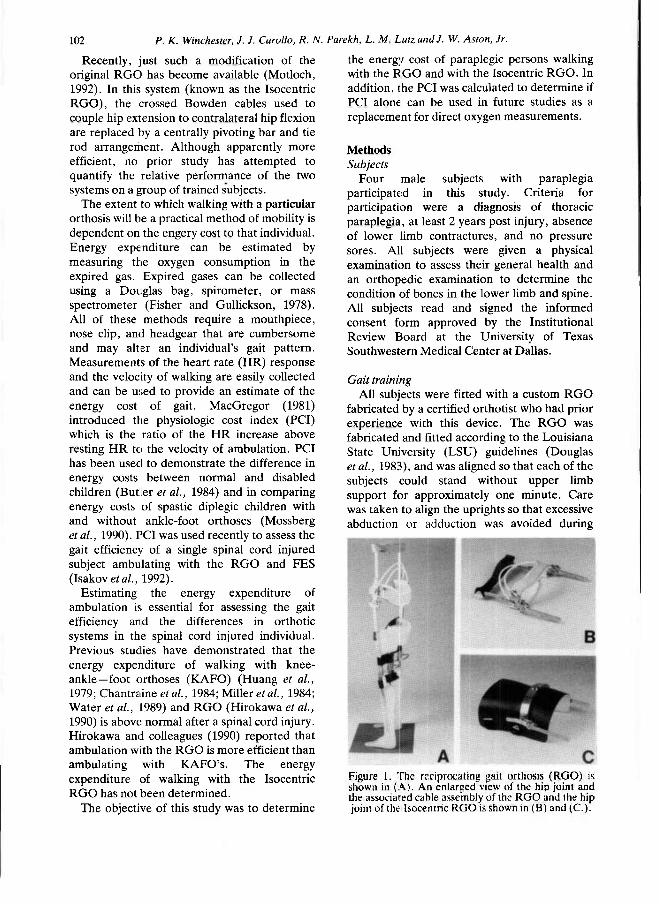
102 P. K. Winchester, J. J. Carollo, R. N. Parekh, L. M Lutz, and J. W. Aston, Jr.
Recently, just such a modification of the original RGO has become available (Motloch, 1992). In this system (known as the Isocentric RGO), the crossed Bowden cables used to couple hip extension to contralateral hip flexion are replaced by a centrally pivoting bar and tie rod arrangement. Although apparently more efficient, no prior study has attempted to quantify the relative performance of the two systems on a group of trained subjects.
The extent to which walking with a particular orthosis will be a practical method of mobility is dependent on the engery cost to that individual. Energy expenditure can be estimated by measuring the oxygen consumption in the expired gas. Expired gases can be collected using a Douglas bag, spirometer, or mass spectrometer (Fisher and Gullickson, 1978). All of these methods require a mouthpiece, nose clip, and headgear that are cumbersome and may alter an individual's gait pattern. Measurements of the heart rate (HR) response and the velocity of walking are easily collected and can be used to provide an estimate of the energy cost of gait. MacGregor (1981) introduced the physiologic cost index (PCI) which is the ratio of the HR increase above resting HR to the velocity of ambulation. PCI has been used to demonstrate the difference in energy costs between normal and disabled children (Butler et al., 1984) and in comparing energy costs of spastic diplegic children with and without ankle-foot orthoses (Mossberg et al., 1990). PCI was used recently to assess the gait efficiency of a single spinal cord injured subject ambulating with the RGO and FES (Isakov et al., 1992).
Estimating the energy expenditure of ambulation is essential for assessing the gait efficiency and the differences in orthotic systems in the spinal cord injured individual. Previous studies have demonstrated that the energy expenditure of walking with knee-ankle—foot orthoses (KAFO) (Huang et al., 1979; Chantraine et al., 1984; Miller et al., 1984; Water et al., 1989) and RGO (Hirokawa et al., 1990) is above: normal after a spinal cord injury. Hirokawa and colleagues (1990) reported that ambulation with the RGO is more efficient than ambulating with KAFO's. The energy expenditure of walking with the Isocentric RGO has not been determined.
The objective of this study was to determine
the energy cost of paraplegic persons walking with the RGO and with the Isocentric RGO. In addition, the PCI was calculated to determine if PCI alone can be used in future studies as a replacement for direct oxygen measurements.
Methods Subjects
Four male subjects with paraplegia participated in this study. Criteria for participation were a diagnosis of thoracic paraplegia, at least 2 years post injury, absence of lower limb contractures, and no pressure sores. All subjects were given a physical examination to assess their general health and an orthopedic examination to determine the condition of bones in the lower limb and spine. All subjects read and signed the informed consent form approved by the Institutional Review Board at the University of Texas Southwestern Medical Center at Dallas.
Gait training All subjects were fitted with a custom RGO
fabricated by a certified orthotist who had prior experience with this device. The RGO was fabricated and fitted according to the Louisiana State University (LSU) guidelines (Douglas et al., 1983), and was aligned so that each of the subjects could stand without upper limb support for approximately one minute. Care was taken to align the uprights so that excessive abduction or adduction was avoided during
Figure 1. The reciprocating gait orthosis ( R G O ) is shown in ( A ) . An enlarged view of the hip joint and the associated cable assembly of the R G O and the hip joint of the Isocentric R G O is shown in (B) and (C . ) .
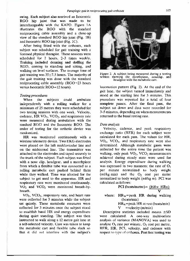
Paraplegic gait in reciprocating gait orthoses 1 0 3
swing. Each subject also received an Isocentric RGO hip joint that was made to be interchangeable with the KAFO. Figure 1A illustrates the RGO with the standard reciprocating cable assembly and a close-up view of the standard RGO hip joint (Fig. 1B) and Isocentric RGO hip joint (Fig. 1C).
After being fitted with the orthoses, each subject was scheduled for gait training with a licensed physical therapist. These sessions were scheduled for 2 hours, 2-3 times weekly. Training included donning and doffing the RGO, coming to standing and sitting, and walking on level surfaces. The average time of gait training was 35±7.5 hours. The majority of the gait training was done with the standard reciprocating cable assembly (RGO=23 hours versus Isocentric RGO=12 hours).
Testing procedures When the subjects could ambulate
independently with a rolling walker for a minimum of 25 metres they were scheduled for two testing sessions with one week. Velocity, cadence, HR VO 2 , VCO 2 , and respiratory rate were measured during ambulation with the sandard RGO and the Isocentric RGO. The order of testing for the orthotic device was randomized.
HR was monitored continuously with a wireless telemetry device. Two EKG electrodes were placed on the left midclavicular line and on the midsternal line. The transmitter was attached to the electrodes and taped securely to the trunk of the subject. Each subject was fitted with a nose clip, headgear, and a mouthpiece from which a flexible tube was connected to a rolling metabolic cart pushed behind them while they walked. Time was allotted for the subject to get used to the apparatus. HR and respiratory rate were monitored continuously. VO 2 and VCO 2 were monitored breath-by-breath.
VO 2 , VCO 2 , respiratory rate, and heart rate were collected for 5 minutes while the subject sat quietly. These metabolic measures were collected for 3 minutes once the subject stood to establish basal HR and energy expenditure during quiet standing. The subject was then instructed to walk along a 12 metre gait lane at a self-selected velocity. Care was taken to keep the metabolic cart and flexible tube slack so that it did not interfere with the subject's
locomotion pattern (Fig. 2). At the end of the gait lane, the subject turned immediately and stood at the starting line for 3 minutes. This procedure was repeated for a total of four complete passes. After the final pass, the subject sat down and data were recorded for 3-5 minutes, depending on when measurements returned to the basal resting rate.
Data analysis Velocity, cadence, and peak respiratory
exchange ratio (RER) for each subject were calculated for each pass. The values for HR, VO 2 , VCO2, and respiratory rate were also determined. Although metabolic gases were collected for the entire time the patient was walking, only peak VO 2 , VCO 2 measurements achieved during steady state were used for analysis. Energy expenditure during walking was expressed in two manners; the rate of O 2
per minute normalized to body weight (ml/kg min) and the O 2 cost per metre normalized to body weight (ml/kg m). PCI was calculated as follows:
PCI (beats/metre) = ( H R W - H R R ) / V
where: H R w = p e a k HR during walking (beats/min)
HRr=peak HR at rest (beats/min) V =velocity (m/min)
Descriptive statistics included means ±SD were calculated. A one-way multivariate analysis of variance (MANOVA) was used to analyze O 2 rate per minute, O 2 cost per metre, RER, HR, PCI, velocity, and cadence with respect to type of orthoses. Post hoc testing was
Figure 2. A subject being measured during a testing session showing the mouthpiece , noseclip, and
headgear with the metabol ic cart .
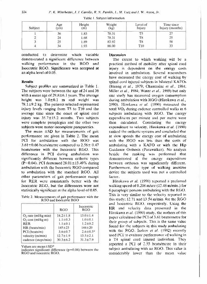
104 P. K. Winchester, J. J. Carollo, R N. Parekh, L. M. Lutz and J W. Aston, Jr.
conducted to determine which variable demonstrated a significant difference between walking performance in the RGO and Isocentric RCrO. Significance was accepted at an alpha level of 0.05.
Results Subject profiles are summarized in Table 1.
The subjects v/ere between the age of 24 and 36 with a mean age of 29.8±6.1 years. The average height was 1.8±0.1 m and weight was 78.1 ±9.2 kg. The patients selected represented injury levels ranging from T5 to T10 and the average time since the onset of spinal cord injury was 35.7±15.2 months. Two subjects were complete paraplegics and the other two subjects were motor incomplete paraparetics.
The mean ±SD for measurements of gait performance are given in Table 2. The mean PCI for ambulation with the RGO was 3.61±0.66 beats/metre compared to 2.56± 0.47 beats/metre with the Isocentric RGO. This difference in PCI during ambulation was significantly different between orthotic types (P=0.04). PCI decreased 28.01 ±13.49% during ambulation with the Isocentric RGO compared to ambulation with the standard RGO. All other parameters of gait performance except for RER were consistently better with the Isocentric RGO, but the differences were not statistically significant at the alpha level of 0.05.
Discussion The extent to which walking will be a
practical method of mobility after spinal cord injury is dependent on the energy costs involved in ambulation. Several researchers have measured the energy cost of walking by spinal cord injured subjects in bilateral KAFOs (Huang et al., 1979; Chantraine et al., 1984; Miller et al., 1984; Water e t al., 1989) but only one study has measured oxygen consumption during ambulation with RGO (Hirokawa et al., 1990). Hirokawa et al. (1990) measured the total VO 2 during cadence controlled walks in 6 subjects ambulating with RGO. The energy expenditures per minute and per metre were then calculated. Correlating the energy expenditure to velocity, Hirokawa et al. (1990) ranked the orthotic systems and concluded that at slow speeds the energy cost of ambulating with the RGO was less than the costs of ambulating with a KAFO or with the Hip Guidance Orthosis (Parawalker). No analysis beside the ranking was performed that demonstrated if the energy expenditure between orthoses was significantly different. Furthermore, the type of walking assistive device the subjects used was not a controlled factor.
Hirokawa et al. (1990) reported a preferred walking speed of 0.208 m/sec (12.48 m/min.) for 6 paraplegic persons ambulating with the RGO. This is very similar to the velocity reported in this study; 12.71 and 13.54 m/min. for the RGO and Isocentric RGO, respectively. Using the HR and velocity data presented in the Hirokawa et al. (1990) study, the authors of this paper calculated the PCI of 3 61 beats/metre for their group of subjects. This is the same value found for the subjects in this study ambulating with the RGO. Isakov et al. (1992) recently used PCI to evaluate performance of walking in a T4 spinal cord injured individual. They reported a PCI of 2.55 beats/metre in their subject ambulating with an RGO. This value is considerably lower than the mean value
Table 1. Subject information
Table 2. Measurements of gait performance with the R G O and Isocentric R G O
Paraplegic gait in reciprocating gait orthoses 105
reported in this study. This difference could be due to the length of training with the RGO. Their subject had been ambulating with the RGO for two years and averaged 8 hours/week of gait training.
A significant reduction in the PCI was found in the group of subjects in this study when the standard RGO hip joint was replaced by the Isocentric RGO hip joint. Caution must be exercised when a conclusion is based on a small sample size, however, subjective reports from the subjects included that they felt less fatigued when walking with the Isocentric RGO.
Although a decrease in the O 2 rate per minute and the O 2 cost per metre was observed, these two methods of expressing energy expenditure were not statistically different between orthotic devices. One reason for this may be the small sample size. However, an O 2
recording system that requires a face mask, nose clip, and tethered cart may be so disruptive to the subject that the oxygen consumption measurements may not be representative of the true energy requirements of walking. It has been observed that face masks can block a test subject's vision. This is not a problem for normal subjects, but in certain disabilities where proprioception and kinesthesia are absent or impaired, subjects may have to rely upon visual feedback for foot placement during gait. In the earlier stages of this study an attempt was made to use a face mask that covered both the nose and mouth but the collection apparatus had to be modified to allow the subjects a better visual field. The advantages of using measurements of HR and velocity to estimate energy expenditure in paraplegic individuals is that it eliminates the use of mouthpieces and nose clips that the patients may find cumbersome or functionally interfering. Furthermore, it does not require the use of expensive gas collection apparatus.
This study demonstrated that PCI can be used as a tool in the assessment of gait efficiency in spinal cord injured subjects. Combining a physiological measurement (HR) with a functional measurement (velocity) may make PCI a more sensitive measure for detecting small but significant differences in energy expenditure. Previous investigators have used PCI as an assessment tool in normal (MacGregor, 1981), disabled children (Butler et al., 1984; Mossberg et al., 1990), and paraplegic
subjects (Bowker et al., 1992; Isakov et al., 1992). Isakov et al. (1992) reported a dramatic reduction of PCI values in a spinal cord injured subject ambulating with RGO and FES compared to ambulating with just the RGO. Only minimal changes were observed in other parameters of gait performance such as cadence, velocity, and step length. A slight improvement is reported here in velocity and cadence with the Isocentric RGO, however, these measurements do not reflect changes in the energy demand of walking.
In this study, it is reported that modifying the standard RGO by exchanging the cables for an isocentric bar resulted in a significant reduction in the PCI of spinal cord injured individuals. Future studies incorporating kinematic analyses of gait with the RGO and the Isocentric RGO may provide insight that will continue to improve the design and function of this orthotic device.
Acknowledgements We thank all the individuals who took the
time to participate in this study and the cooperation of the spinal cord injury staff at Dallas Rehabilitation Institute in Dallas, TX.
We gratefully acknowledge the assistance of Bill Carlton, C O . of Dallas Prosthetic and Orthotic Center and The University of Texas Prosthetic and Orthotic Program in the fabrication and fitting of the orthoses.
This research was supported in part by the Texas Advanced Technology Program under Grant No. 003660-102 and a grant from the Southwestern Medical Foundation.
R E F E R E N C E S
BOWKER P , MESSENGER N . OGILVIE C . R O W L E Y D ( 1 9 9 2 ) . Energetics of paraplegic walking. J Biomed Eng 1 4 , 3 4 4 - 3 5 0 .
BUTLER P, ENGELBRECHT M, M A J O R R E , TAIT JH, STALLARD J, PATRICK J H ( 1 9 8 4 ) . Physiological cost index of walking for normal children and its use as an indicator of phvsical handicap. Dev Med Child Neurol 2 6 , 6 0 7 - 6 1 2 .
CHANTRAINE A, CRIELAARED JM, ONKELINX A , PIRNAY F ( 1 9 8 4 ) . Energy expendi ture of ambulat ion in paraplegics: effects of long term use of bracing. Parplegia 2 2 , 1 7 3 - 1 8 1 .
D O U G L A S R, LARSON PF, D'AMBROSIA R, M C C A L L R E ( 1 9 8 3 ) . The L S U reciprocating gait orthosis Orthopedics 6 , 8 3 4 — 8 3 8 .
106 P. K. Winchester, J. J. Carollo, R. N. Parekh, L. M. Lutz and J. W. Aston, Jr.
FISHER SV, GULLICKSON G (1978). Energy cost of ambulat ion in heal th and disability: a l i terature review. Arch Phys Med Rehabil 5 9 , 1 2 4 - 1 3 3 .
HIROKAWA S, GRIMM M , T H A N H L , SOLOMONOW M , B A R A T T A R V (1980). Energy consumption in paraplegic ambulat ion using the reciprocating gait orthosis and electric stimulation of the thigh muscles. Arch Phys Med Rehabil 71, 687-694.
H U A N G C T , KUHLEMEIER K V , M O O R E N B , FINE PR (1979). Energy cost of ambulat ion in paraplegic patients using Craig-Scott braces. Arch Phys Med Rehabil 60, 595-600.
ISAKOV E , D O U G L A S R , B E R N S P (1992). Ambula t ion using the reciprocating gait orthosis and functional electrical st imulation, Paraplegia 3 0 , 239-245.
M A C G R E G O R J (1981). The evaluation of pat ient performance using long-term ambulatory monitor ing technique in the domiciliary envi ronment . Physiotherapy 6 7 , 30 -33 .
MILLER N E , MEHRITT J L , M E R K E L K D , WESTBROOK P R (1984). Paraplegic energy expendi ture during negotiat ion of architectural barr iers . Arch Phys Med Rehabil 6 5 , 778-779.
MOSSBERG K A , LINTON K A , FRISKE K (1990). Ankle-foot or thoses: effect on energy expendi ture of gait in spastic diplegic children. Arch Phys Med Rehabil 7 1 , 4 9 0 - 4 9 4 .
MOTLOCH W (1992). Principles of orthotic management for child and adult paraplegia and clinical experience with the Isocentric R G O . In: Proceedings of the 7th World Congress of the Internat ional Society for Prosthetics and Orthot ics , Chicago, I I , June 28-July 3 , 1 9 9 2 - C o p e n h a g e n : ISPO. p28.
STALLARD J , PATRICK R E , M A J O R R E (1989). A review of the fundamental design problems of providing ambula t ion for paraplegic pat ients . Paraplegia 21, 70-75.
W A T E R S R L , Y A K U R A J S , A D K I N S R , B A R N E S G (1989). De te rminan t s of gait per formance following spinal cord injury. Arch Phys Med Rehabil70, 811-818.
Y A M A G U C H I G T , Z A J A C F E (1990). Restor ing unassisted natural gait to paraplegics via functional neuromuscular st imulation: a computer simulation study. IEEE Trans Biomed Eng 3 7 , 886-902.

Prosthetics and Orthotics International, 1993,17, 107-114
Enhancement of hemiplegic patient rehabilitation by means of
functional electrical stimulation
A. K R A L J * , R. A Č I M O V I Ć * * and U . STANIČ***
*Faculty of Electrical and Computer Engineering, University of Ljubljana, Slovenia **Rehabilitation Institute, University of Ljubljana, Slovenia **Institute Jozef Stefan, University of Ljubljana, Slovenia
Abstract This presentat ion will review briefly the current practice and state of the art in functional electrical stimulation (FES) as applied to s t roke, head injured or brain tumour opera ted patients. A similar application is used in paretic patients following t rauma or o ther aetiology. Over 20 years experience in the application of F E S , as practised in Ljubljana, will be highlighted and the devices currently in use will be described. The statistics show the results obtained on 2,500 hemiplegic patients examined for FES application during the last 10 years. The statistics and results of the Slovenian populat ion indicate 0.15-0.20% new cases annually or 1,500 new cases per million inhabitants. U p to 6 3 % of annual cases are candidates for an FES based therapeutic locomotion rehabilitation programme. Experience indicates that 60% of hemiplegic patients received single-channel stimulation to correct equinovarus or foot d rop , 30% obtained dual or even three channel stimulation t rea tment and only 10% of patients were involved in multichannel FES of four to six or even eight channels of stimulation. The benefits and outcome of rehabilitation will be presented and discussed in regard to current t rends in the field of F E S for hemiplegic and paretic patients. The partly inactive but very important field of FES application to the upper extremity in hemiplegic and paretic patients will be discussed and the relatively modest
achievements presented. Future developments will be presented together with advances foreseen by steadily improving technology.
Introduction For the last decade development in the field
of functional electrical stimulation (FES) of hemiplegic patients was concerned with indications, prescriptions, t rea tment and modality refinements with important hardware improvements but no basic innovative developments taking place. The steady refinements and advances were a consequence of general technology improvements . The vital developments in the field of F E S of spinal cord injured patients will dramatically, in the near future influence the field of hemiplegic patients. Due to the vast interest and overly optimistic expectations triggered in the early years of the last decade the major efforts of funding were focused on locomotion rehabilitation of spinal cord injured (SCI) patients utilising F E S . It took almost 10 years to recognise and adopt more realistic and achievable expectations. Interest was regained in FES rehabilitation and methodology advances in the field of applications t o s t roke, head injury, t rauma, surgical and other patients. The improved hemiplegia rehabilitation using FES is very important and promising. In te rms of numbers of pat ients it by far exceeds the SCI field. Stroke is common in the middle-aged and elderly and braln injury is
This paper was based on. a presentation made at Dundee '91—International Conference and Instructional Course on Orthotics, 16-20 September, 1991, Dundee, Scotland.
All correspondence to be addressed to Alojz Kralj, Faculty of Electrical and Computer Engineering, University of Ljubljana, Ljubljana, Slovenia.
107

108 A. Kralj, R Ačimović and U. Stanič
frequent in the younger and middle-aged population. Stroke affects two to three people per thousand and consequently dominates the field of neurologic rehabilitation. In the U S A the population of hemiplegic patients is of the order of 2 million (LaPlaute , 1988). It is also interesting to note that for the last 10 years no significant advance has been made in the application of FES in the upper limb in general and in hemiplegic hands in particular. In contrast the rehabilitation of grasping and hand movement advanced noticeably in the field of quadriplegic patients. Developing technology and other advances in neural recording may in the future influence FES application in the restoration of hand function in hemiparetic patients following s t roke, brain injury or operated brain tumour as described later in this presentation.
Before highlighting the advances and displaying the current state and trends in FES application for locomotion restoration in hemiplegic patients , the aims, goals and strategies in general will be presented since they are specific to the rehabilitation and use of FES in hemiplegic patients. Neurologic patients with upper motor neurone dysfunctions following stroke are considered as a stabilized or "complete s t roke" group including by definition patients with cerebral thrombosis , infarction, embolus with infarction, intracerebral haemorrhage , the sequence of subarachnoid haemorrhage from aneurysm or arterio-venous malformation. Brain injured and/or operated brain tumour patients are also candidates for FES therapeutic t reatment and later, if applicable, the long term use of FES devices for the restoration of lost function. All unstable or transient neurologic dysfunctions such as neurologic vascular episodes and stroke in progression are excluded from our consideration because by classical definitions (Davis, 1985) only patients suitable for rehabilitation are candidates for FES related rehabilitation t rea tment . After admission therapeutic FES application is focused on preserving existing function and augmenting recovery for restoration of lost function. A typical goal is to restrengthen weak muscles, maintain range; of motion at joints and prevent the development of very exaggerated synergistic pat terns such as extension or flexion pat terns. At this stage spasticity development is
reduced by FES t reatment and the voluntary recovery of movement is enhanced. In general the goal is to facilitate sensory and antigravity awareness, to use the lower limbs for standing and to initiate early walking by the patient. Multichannel FES is very efficient in this respect and also helpful in lessening the physical effort by the physiotherapist in raising the patient to a standing position in the early phase of t reatment . Early standing of patients is a good strategy ensuring faster recovery and preventing the development of severe spasticity with exaggerated synergism which is later very hard to modify. The described aims and goals of FES application follow the strategy of using inpatient hospital time efficiently to achieve a higher level of function in a shorter t ime. O n completion such t rea tment indicates the lost functions which can be , at present , only partially restored by continuing F E S . This is carried out in a programme of long term FES device prescription and patient training for independent home use. The following describes the current state of hemiplegic patient rehabilitation by means of FES as developed and practised in Ljubljana (Acimovic—Janezic, 1989; Malezic et al., 1984; Stanic et al., 1990). This will focus mainly on the lower limb programme utilising surface stimulation electrodes but also highlighting the upper limb applications.
Therapeutic FES applications For the past 20 years the Ljubljana
University Rehabilitation Institute has routinely practised FES as a t reatment module additional to the standard and established therapy for rehabilitation (Acimovic—Janezic. 1989; Stanic et al., 1990). At present about 80% of all patients admitted participate in the FES therapeutic or orthotic FES programme. With the help of electrical stimulation different muscles can be activated, restrengthened and the joint range of motion maintained actively, the blood circulation augmented and pain reduced. FES is a convenient and effective means of enhancing sensory awareness. In many instances mobilization is easier and achieved in a shorter t ime while also radically activating the patient 's remaining and developing resources because FES provides afferent and FES induced movement with proprioception and exteroceptive sensory
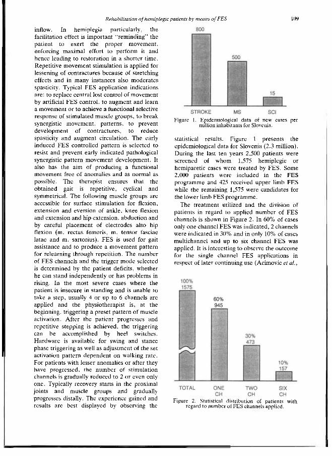
Rehabilitation of hemiplegic patients by means of FES 109
inflow. In hemiplegia particularly, the facilitation effect is important " reminding" the patient to exert the proper movement , enforcing maximal effort to perform it and hence leading to restoration in a shorter time. Repetitive movement stimulation is applied for lessening of contractures because of stretching effects and in many instances also moderates spasticity. Typical FES application indications are: to replace central lost control of movement by artificial FES control, to augment and learn a movement or to achieve a functional selective response of stimulated muscle groups, to break synergistic movement , pat terns, to prevent development of contractures, to reduce spasticity and augment circulation. The early induced FES controlled pat tern is selected to resist and prevent early indicated pathological synergistic pattern movement development. It also has the aim of producing a functional movement free of anomalies and as normal as possible. The therapist ensures that the obtained gait is repetit ive, cyclical and symmetrical. The following muscle groups are accessible for surface stimulation for flexion, extension and eversion of ankle, knee flexion and extension and hip extension, abduction and by careful placement of electrodes also hip flexion (m. rectus femoris, m. tensor fasciae latae and m. sartorius). FES is used for gait assistance and to produce a movement pattern for relearning through repetit ion. The number of FES channels and the trigger mode selected is determined by the patient deficits, whether he can stand independently or has problems in rising. In the most severe cases where the patient is insecure in standing and is unable to take a s tep, usually 4 or up to 6 channels are applied and the physiotherapist is, at the beginning, triggering a preset pat tern of muscle activation. After the patient progresses and repetitive stepping is achieved, the triggering can be accomplished by heel switches. Hardware is available for swing and stance phase triggering as well as adjustment of the set activation pat tern dependent on walking rate . For patients with lesser anomalies or after they have progressed, the number of stimulation channels is gradually reduced to 2 or even only one . Typically recovery starts in the proximal joints and muscle groups and gradually progresses distally. The experience gained and results are best displayed by observing the
statistical results. Figure 1 presents the epidemiological data for Slovenia (2.3 million). During the last ten years 2,500 pat ients were screened of whom 1,575 hemiplegic or hemiparetic cases were treated by F E S . Some 2,000 patients were included in the FES programme and 425 received upper limb FES while the remaining 1,575 were candidates for the lower limb FES programme.
The treatment utilized and the division of patients in regard to applied number of FES channels is shown in Figure 2. In 60% of cases only one channel FES was indicated, 2 channels were indicated in 30% and in only 10% of cases multichannel and up to six channel FES was applied. It is interesting to observe the outcome for the single channel FES applications in respect of later continuing use (Acimovic et al.,
Figure 1. Epidemiological data of new cases per million inhabitants for Slovenia.
Figure 2. Statistical distribution of patients with regard to number of FES channels applied.
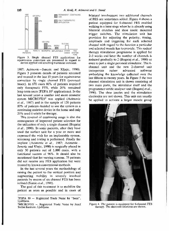
llo A. Kralj, R. Ačimovičand U. Stanic
1987; Acimovic—Janezic and Kljajic, 1990). Figure 3 presents details of patients screened and t reated in the last 10 years for equinovarus correction by single channel FES (peroneal brace) . In 670 cases 80% of patients required only therapeutic F E S , while 20% remained long-term users (FEPA 10 1 applications). In the last several years a smaller and more cosmetic system M I C R O F E S 2 was applied (Acimovic et al., 1987) and in the sample of 120 patients 80% of patients decided to use the system as a continuing assistive device in the home and only 20% used it solely for therapy.
This reversal of continuing usage is also the consequence of improved patient selection for the utilization of only a single channel (Bogataj et al., 1990). In some patients , after they have used the surface unit for a year or more and expressed the wish for an implantable system, screening and testing is performed. Finally the implant (Acimovic et al., 1987; Acimovic— Janezic and Klajic, 1990) is surgically placed in only 30 patients out of 1,000 cases, with a functional success of 9 6 % . It should also be ment ioned that for varying reasons, 75 patients did not receive any F E S application but were treated by known conventional methods .
In the last several years the methodology of raising the patient to the vertical position and augmenting mobility in severely involved patients by means of six channel FES has been refined (Stanic et al., 1990).
The goal of this t rea tment is to mobilize the patient as soon as possible and in cases of
bilateral involvement two additional channels of FES are sometimes added. Figure 4 shows a patient equipped for 6-channel F E S enabled walking in a later stage when he is already using bilateral crutches and shoe insole mounted trigger switches. The stimulation unit has provision for adjusting the polarity, timing, amplitude and triggering for each selected channel with regard to the function a particular and selected muscle has to provide. This radical therapy stimulation p rogramme is applied for 2 -3 weeks and later the number of channels is reduced gradually to 2 (Bogataj et al., 1990) or even to just a single peroneal stimulator. The 6-channel unit and the new 2-channel unit incorporate ra ther advanced softwear embodying the knowledge collected over the last fifteen to twenty years. In Figure 5 the two channel stimulation unit is shown consisting of two main par ts , the stimulator itself and the programmer-str ide analyser unit (Bogataj et al., 1990). The shoe insoles and the stimulation electrodes are not shown. This unit can usually be applied to activate a larger muscle group
Figure 3. Single channel FES applications for equinovarus correction are presented in regard to
device applied and resulting treatment outcome.
1FEPA 10 - Registered Trade Name by "Soca", Ljubljana. 2MICROFES - Registered Trade Name by Jozef Stefan Institute, Ljubljana.
Figure 4. The patient is equipped for 6-channel FES therapy. The electrode locations are shown.

Rehabilitation of hemiplegic patients by means of FES 111
such as the m. quadriceps for knee extension, hamstring muscles for knee flexion or m. glueteus maximus for hip extension and the second channel is applied for peroneal nerve stimulation or to the pretibial muscle group for ankle dorsiflexion.
The 6-channel and 2-channel microprocessor based units also monitor and store data about patient gait performance. The measuring is performed on the basis of shoe insole switches. The statistics and average data of performance can be recalled at any instance after or during gait. The number of steps average heel on and off time are measured and recorded for both legs including the standard deviations (Bogataj et al., 1990).
The programming unit enables user-friendly programming of the stimulator for the on/off t ime per channel in regard to the selected stance or swing phase trigger. The walking rate dependent ( W R D ) mode of stimulation or cyclical mode are available features. At the start of therapy the cyclical mode is applied while later the W R D mode allows the patient to choose his own preferred speed of gait with the stimulation sequence adjusted accordingly. The timing of the stimulation sequence of the patient 's gait is based on a linear or weighted extrapolation of the previous four stride phase times. Three gradient dependable extrapolation equations are utilized and automatically selected and applied (Bogataj et al., 1990). The patient 's regained and restored functional ability after the 6-channel and later 2-channel t reatment in most cases leads to the prescription of the single peroneal stimulator
even in cases where possibly a 2-channel or even 3-channel device would be preferred. This is because at present no such 3-channel or 2-channel units are available for long-term use. Of course, the 2-channel unit can be prescribed for skilled and motivated patients, but in general the daily multi-electrode placement and fixation is too clumsy and the donning and doffing time-consuming and impractical. It is expected that in the future a suitable implanted system will be developed.
The FES therapy and methodology described is effective and is carried out for 8-10 weeks. Comparative studies (Malezic et al., 1984) concluded that intense F E S therapy advanced patient recovery to a higher level of function for almost the same medical personnel cost. As depicted in Figure 6, it is evident that the group treated by F E S reaches a higher level of function, but on cessation of therapy slowly degenerates , gradually returning to the performance level of the control group treated by conventional rehabilitation methods . This indicates that FES devices such as the 2 or 3-channel continuous use units may be justified. Figure 6 also confirms that intensive FES locomotion restoration t reatment for hemiplegic patients is cost effective and functionally more efficient if compared to the conventional t reatment alone. It should be noted that FES intensive methodology shortens the time spent in hospital.
For the last ten years a rather passive stance was adopted in regard to FES application for hand and wrist function therapy with function restoration in hemiplegic patients. Recent interest in hemiplegic hand function restoration
Figure 5. The 2-channel FES unit is displayed consisting of the unit worn by the patient and the programmer. The shoe-insoles and electrodes are
omitted.
Figure 6. The course of functional progress utilizing the 6-channel FES system as compared in time of
treatment to the classical rehabilitation therapy.

112 A Kralj, R Ačimović and U. Stanic
observed between 1970-80 has mostly faded out during the last decade. FES was applied at the end of the 1970s as a therapeutic means of effective t reatment to improve the range of wrist and finger extension motions and to prevent the development of contractures caused by flexor spasticity. The application of cyclical isotonic F E S of wrist and finger extensors decreases not only the established spasticity but also wrist and finger flexion deformities (Baker et al., 1979). It was concluded that two to three 30-40 minute stimulation sessions per week could substitute for all other range of motion techniques. Patients gradually become accustomed to the feeling of discomfort caused by the t reatment and the majority later learned to tolerate the sensation produced by stimulation even at the range of settings activating isometrically the wrist and finger extensors for a good, visible motoric response. Bowman et al. (1979) has proposed position feedback for automated t reatment for the hemiplegic wrist by means of FES . The only marketed FES long-term wrist and finger extension system which is still available in Ljubljana was proposed nearly 15 years ago (Rebersek and Vodovnik, 1975). Because of the clumsiness in donning and doffing and the ra ther minor function of wrist and finger extension, the unit never reached comparable popularity when compared to the one-channel peroneal stimulator. The only FES system for continuing hand function restoration in hemiplegic patients is not a very successful unit. In spite of this the unit is still commercially available and is occasionally prescribed for therapeutic use. To our knowledge an implantable unit for hand function restoration in hemiplegic patients has not yet been produced or marketed . It is interesting, therefore, to present optimistic developments , because the future is going to progress first along the line of developing multichannel implants and these will also be very suitable for hand function restoration,
Trends and expectations The FES methodology applied to hemiplegic
patients for therapeutic t reatment and gait assistance with long-term use for gait restoration has proven its effectiveness and usefulness (Malezic et al., 1984; Stanic et al., 1990). At present , of patients requiring long-
term FES devices, only those who are candidates for simple equinovarus correction are reasonably well served. For this group of patients the single channel FES unit is available, being the only such unit in the market . In principle the equinovarus deficiency needs a balanced control of equinus and varus. This requires in essence a 2-channel unit. In addition up to now there has been negligible provision for the development of a total implanted system because the required technology was difficult to realize. The research results obtained within the last years have raised cumulative evidence that substantial progress can be expected towards the development of totally implantable systems. In principle the design of totally implanted systems is almost cosmetically ideal and eliminates patient donning and doffing.
In recent years refined recording techniques from sensory peripheral nerves (Johansson, 1991) indicate that with minimal circuitry for signal recognition it may be possible to develop very selective triggering and even provide control signals for arbitrary movement restoration by means of F E S . These developments are complemented by long-term implantable infrafascicular recording electrodes (Lefurge et al., 1991). These achievements are raising optimistic hopes that in the near future safe, reliable and good control using trigger signals can be obtained for hand and gait function restoration in hemiplegic patients, particularly with implanted systems. Continuous recording from peripheral nerves is very important , because these nerves are transmitting the information detected by natural sensors. For instance heel contact in gait can be detected and used for a trigger source.
Interesting developments have also begun to improve the motoric activation of muscles by FES and are also directly applicable to hemiplegic patients (Waters et al., 1988; Marsolais et al., 1990) Waters et al. (1988) are utilizing surgically inserted epimysial electrodes for therape utic electric stimulation of the lower limb. By percutaneous wires, as used for almost two decades in the Cleveland upper extremities programme (Hunter Peckham, 1987) external connection to the epimysial electrodes is established. In patients with inadequate hip stability preventing them from balancing on one
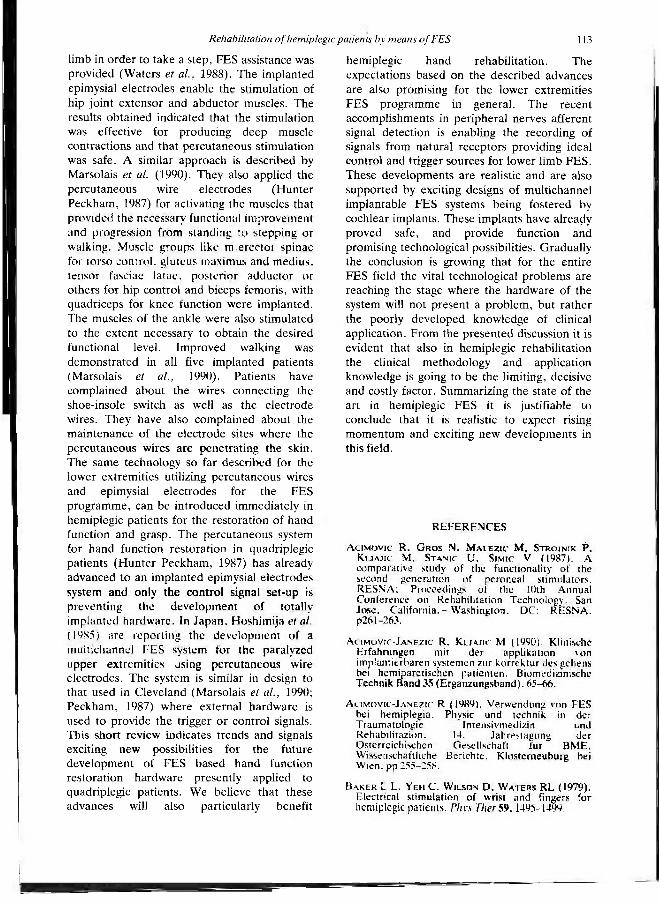
Rehabilitation of hemiplegic patients by means of FES 113
limb in order to take a step, FES assistance was provided (Waters et al., 1988). The implanted epimysial electrodes enable the stimulation of hip joint extensor and abductor muscles. The results obtained indicated that the stimulation was effective for producing deep muscle contractions and that percutaneous stimulation was safe. A similar approach is described by Marsolais et al. (1990). They also applied the percutaneous wire electrodes (Hunter Peckham, 1987) for activating the muscles that provided the necessary functional improvement and progression from standing to stepping or walking. Muscle groups like m. erector spinae for torso control, gluteus maximus and medius, tensor fasciae latae, posterior adductor or others for hip control and biceps femoris, with quadriceps for knee function were implanted. The muscles of the ankle were also stimulated to the extent necessary to obtain the desired functional level. Improved walking was demonstrated in all five implanted patients (Marsolais et al., 1990). Patients have complained about the wires connecting the shoe-insole switch as well as the electrode wires. They have also complained about the maintenance of the electrode sites where the percutaneous wires are penetrat ing the skin. The same technology so far described for the lower extremities utilizing percutaneous wires and epimysial electrodes for the FES programme, can be introduced immediately in hemiplegic patients for the restoration of hand function and grasp. The percutaneous system for hand function restoration in quadriplegic patients (Hunter Peckham, 1987) has already advanced to an implanted epimysial electrodes system and only the control signal set-up is preventing the development of totally implanted hardware. In Japan, Hoshimija et al. (1985) are reporting the development of a multichannel FES system for the paralyzed upper extremities using percutaneous wire electrodes. The system is similar in design to that used in Cleveland (Marsolais et al., 1990; Peckham, 1987) where external hardware is used to provide the trigger or control signals. This short review indicates trends and signals exciting new possibilities for the future development of F E S based hand function restoration hardware presently applied to quadriplegic patients. We believe that these advances will also particularly benefit
hemiplegic hand rehabilitation. The expectations based on the described advances are also promising for the lower extremities FES programme in general . The recent accomplishments in peripheral nerves afferent signal detection is enabling the recording of signals from natural receptors providing ideal control and trigger sources for lower limb FES . These developments are realistic and are also supported by exciting designs of multichannel implantable FES systems being fostered by cochlear implants. These implants have already proved safe, and provide function and promising technological possibilities. Gradually the conclusion is growing that for the entire FES field the vital technological problems are reaching the stage where the hardware of the system will not present a problem, but rather the poorly developed knowledge of clinical application. From the presented discussion it is evident that also in hemiplegic rehabilitation the clinical methodology and application knowledge is going to be the limiting, decisive and costly factor. Summarizing the state of the art in hemiplegic FES it is justifiable to conclude that it is realistic to expect rising momen tum and exciting new developments in this field.
REFERENCES
A C I M O V I C R, G R O S N, M A L E Z I C M, S T R O J N I K P, K U A J I C M, S T A N I C U, S I M I C V (1987). A comparative study of the functionality of the second generation of peroneal stimulators. RESNA; Proceedings of the 10th Annual Conference on Rehabilitation Technology, San Jose, California. - Washington. DC: RESNA. p261-263.
A c I M O V I C - J A N E Z I C R, Kljajic M (1990). Klinische Erfahrungen mit der applikation von implantierbaren s y s t e m e n zur korrektur des gehens bei hemiparetischen patienten. Biomedizinische Technik Band 35 (Erganzungsband). 65-66.
A c i m o v i c - J a n e z i c R (1989). Verwendung von FES bei hemiplegia. Physic und technik in der Traumatologic Intensivmedizin und Rehabilitazion. 14. Jahrestagung der Osterreichischen Gesellschaft fur BME. Wissenschaftliche Berichte. Klosterneuhure bei Wien, pp 255-258.
B A K E R L L, YEH C, W I L S O N D, W A T E R S RL (1979). Electrical S t imula t ion of wrist and fingers for hemiplegic patients. Phys Ther 59,1495-1499.
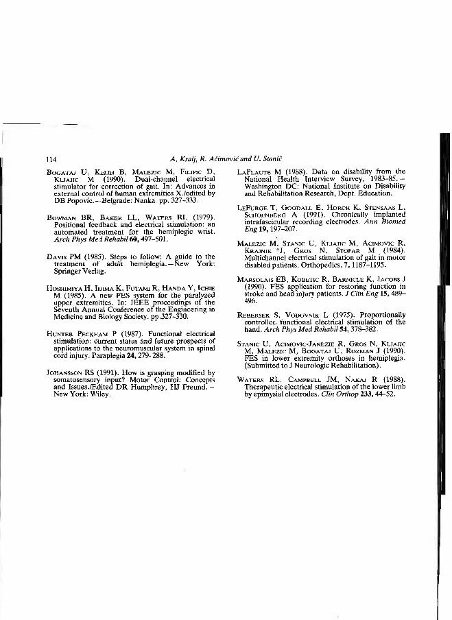
114 A. Kralj, R. Ačimovićand U. Stanic
B O G A T A J U, K E L I H B, M A L E Z I C M, F I L I P I C D, K l j A J I C M (1990). Dual-channel electrical stimulator for correction of gait. In: Advances in external control of human extremities X./edited by DB Popovic.-Belgrade: Nanka. pp. 327-333.
B O W M A N BR, B A K E R LL, W A T E R S RL (1979). Positional feedback and electrical stimulation: an automated treatment for the hemiplegic wrist. Arch Phys Med Rehabil 60,497-501.
DAVIS PM (198.5). Steps to follow: A guide to the treatment of adult hemiplegia.—New York: Springer Verlag.
Hoshimiya H, Iijima K, Futami R, HAnda Y, IChie M (1985). A new FES system for the paralyzed upper extremities. In: IEEE proceedings of the Seventh Annual Conference of the Engineering in Medicine and Biology Society, pp.327-330.
H U N T E R P E C K H A M P (1987). Functional electrical stimulation: current status and future prospects of applications to the neuromuscular system in spinal cord injury. Paraplegia 24,279-288.
J O H A N S S O N RS (1991). How is grasping modified by somatosensory input? Motor Control: Concepts and Issues./Edited DR Humphrey, HJ Freund.— New York: Wiley.
L A P L A U T E M (1988). Data on disability from the National Health Interview Survey, 1983-85.— Washington DC: National Institute on Disability and Rehabilitation Research, Dept. Education.
L E F U R G E T, G O O D A L L E, H O R C H K, S T E N S A A S L, S C H O E N B E R G A (1991). Chronically implanted intrafascicular recording electrodes. Ann Biomed Eng 19,197-207.
M A L E Z I C M, S T A N I C U, K L J A J I C M, A C I M O V I C R, K R A J N I K J, G R O S N, S T O P A R M (1984). Multichannel electrical stimulation of gait in motor disabled patients. Orthopedics, 7,1187-1195.
M A R S O L A I S EB, K o b e t i c R, B A R N I C L E K, J A C O B S J (1990). FES application for restoring function in stroke and head injury patients. J Clin Eng 15 ,489-496.
R E B E R S E K S, V O D O V N I K L (1975). Proportionally controlled functional electrical stimulation of the hand. Arch Phys Med Rehabil 54,378-382.
S T A N I C U, A C I M O V I C - J A N E Z I E R, G R O S N, K l j A J I C M, M A L E Z I C M, B O G A T A J U, R O Z M A N J (1990). FES in lower extremity orthoses in hemiplegia. (Submitted to J Neurologic Rehabilitation).
W A T E R S RL, C A M P B E L L JM, N A K A I R (1988). Therapeutic electrical stimulation of the lower limb by epimysial electrodes. Clin Orthop 233,44-52.
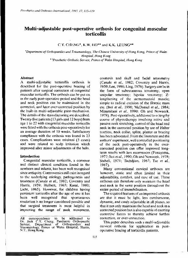
Prosthetics and Orthotics International, 1993, 17, 115-119
Multi-adjustable post-operative orthosis for congenital muscular torticollis
C.Y. CHENG*, K.W. HO** and K.K. LEUNG**
*Department of Orthopaedics and Traumatology, The Chinese University of Hong Kong, Prince of Wales Hospital, Hong Kong
**Prosthetic-Orthotic Service, Prince of Wales Hospital, Hong Kong
Abstract A multi-adjustable torticollis orthosis is described for the post-operative bracing of patients after surgical correction of congenital muscular torticollis. The orthosis can be put on in the early post-operative period and the head and neck position can be maintained in the corrected, and later over-corrected position by the built-in multi-adjustable joint-mechanism. The details of the manufacturing are described. Twenty-five patients ( 13 girls and 12 boys) from age 1 to 22 with congenital muscular torticollis were fitted with the orthosis post-operatively for an average duration of 10 weeks. Satisfactory compliance with the orthosis was found in 23 cases. Complications were minimal (3 cases) and were related to scalp irritation which improved after minor adjustments of the halo.
Introduction Congenital muscular torticollis, a common
and distinct clinical condition found in the newborn and infants, has been well recognised since antiquity. Controversies still exist in regard to the underlying etiology, pathogenesis and treatment (Canale et al., 1982; Coventry and Harris, 1959; Hulbert, 1965; Kasai, 1980; Licht, 1965). However, for children having persistent torticollis after the age of one it has been well recognised that spontaneous resolution is no longer considered possible and that surgical treatment is most helpful in improving the range of neck movement.
cosmesis and skull and facial asymmetry (Canale et al., 1982; Coventry and Harris, 1959; Lee, 1986; Ling, 1976). Surgery can be in the form of subcutaneous tenotomy; open unipolar tenotomy; bipolar tenotomy; Z-lengthening of the sternomastoid muscle: simple to radical excision of the fibrotic mass etc. (Itoi et al., 1990; McDaniel et al., 1984; Minamitani et al., 1990; Oh and Nowacek, 1978). Post-operatively, additional to a lengthy course of physiotherapy involving active and passive neck stretching, control of the head and neck in the corrected position by use of Halter traction, neck collar, splint, plaster or bracing has been advocated. From the literature and the authors' experience, well controlled positioning of the neck post-operatively in the over-corrected position can offer improved long-term results with less recurrences (Funayama, 1977; Itoi et al., 1990; Oh and Nowacek, 1978; Staheli, 1971; Tachdjian, 1967; Tse et al., 1987).
Many conventional cervical orthoses are however, static and often limited in their adjustability, comfort, and ease of use. These orthoses can therefore only maintain the head and neck in the same position throughout the entire period of immobilisation.
The required features of an improved orthosis are that it must be light, less cumbersome, dynamic, and easily adjustable in all planes, so that it not only maintains the head and neck in a corrected position but is also capable of exerting corrective forces to thereby achieve further correction, or over-correction.
This paper describes such a multi-adjustable cervical orthosis for application in postoperative bracing of torticollis patients.
All correspondence to be addressed to Dr. Jack C.Y, Cheng. Paediatric Orthopaedic Service, Department of Orthopaedics and Traumatology, Prince of Wales Hospital, Shatin, N.T., Hong Kong.
115
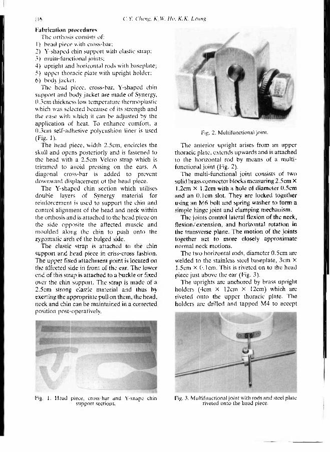
116 C.V. Cheng. K.W. Ho. K.K. Leung
Fabrication procedures The orthosis consists of:
1) head piece with cross-bar; 2) Y-shaped chin support with elastic strap: 3) multi-functional joints; 4) upright and horizontal rods with baseplate; 5) upper thoracic plate with upright holder; 6) body jacket.
The head piece, cross-bar, Y-shaped chin support and body jacket are made of Synergy, 0.3cm thickness low temperature thermoplastic which was selected because of its strength and the ease with which it can be adjusted by the application of heat. To enhance comfort, a 0.3cm self-adhesive polycushion liner is used (Fig 1).
The head piece, width 2.5cm, encircles the skull and opens posteriorly and is fastened to the head with a 2.5cm Velcro strap which is trimmed to avoid pressing on the ears. A diagonal cross-bar is added to prevent downward displacement of the head piece.
The Y-shaped chin section which utilises double layers of Synergy material for reinforcement is used to support the chin and control alignment of the head and neck within the orthosis and is attached to the head piece on the side opposite the affected muscle and moulded along the chin to push onto the zygomatic arch of the bulged side.
The elastic strap is attached to the chin support and head piece in criss-cross fashion. The upper fixed attachment point is located on the affected side in front of the ear. The lower end of this strap is attached to a buckle or fixed over the chin support. The strap is made of a 2.5cm strong elastic material and thus by exerting the appropriate pull on them, the head, neck and chin can be maintained in a corrected position post-operatively.
The anterior upright arises from an upper thoracic plate, extends upwards and is attached to the horizontal rod by means of a multifunctional joint (Fig. 2).
The multi-functional joint consists of two solid brass connector blocks measuring 2.5cm x 12cm X 1.2cm with a hole of diameter 0.5cm and an 0.1 cm slot. They are locked together using an M6 bolt and spring washer to form a simple hinge joint and clamping mechanism.
The joints control lateral flexion of the neck, flexion/extension, and horizontal rotation in the transverse plane. The motion of the joints together act to more closely approximate normal neck motions.
The two horizontal rods, diameter 0.5cm are welded to the stainless steel baseplate, 3cm X 1.5cm X 0.1cm. This is riveted on to the head piece just above the ear (Fig. 3).
The uprights are anchored by brass upright holders (4cm X 12cm X 12cm) which are riveted onto the upper thoracic plate. The holders are drilled and tapped M4 to accept
Fig. 1. Head piece, cross-bar and Y-shape chin support sections.
Fig. 2. Multifunctional joint.
Fig. 3. Multifunctional joint with rods and steel plate riveted onto the head piece.

Multi-adjustable torticollis orthosis 117
socket head type grub screws which when tightened effect locking of the uprights (Fig. 4).
The upper thoracic plate, 15cm X 4cm 2024-T2 aluminium, is attached to the body jacket by two M 6 screws and butterfly nuts (Figs. 4 and 5).
Clinical applications Prospective patients were measured for the
multi-adjustable cervical orthosis before operation and were fitted two to three days post-surgery. The head and neck were put into the orthosis and positioned in the maximally corrected while still tolerable position — i.e. with the neck laterally tilted to the opposite side, and also rotated to the side of the muscular torticollis with the neck in approximately about 20° of extension. The orthosis was only removed for the purpose of an intensive active mobilisation and passive stretching physiotherapy programme which was begun once the surgical wound had healed (five to seven days). For the rest of the time, the orthosis was kept on.
Regular adjustment of position and checking of compliance being done by the orthotist, physiotherapist and surgeon at a special follow-up clinic. The total period of orthosis wear ranged from 8 to 12 weeks.
Methods of assessment The results of the patient undergoing surgery
and post-operative bracing were assessed functionally and cosmetically according to the same criteria as Canale et al., 1982.
Functionally the results were classified as good when the range of active and passive rotational and side flexion movement was limited by less than 15° as measured by arthrodial protractor measurements. The cosmetic evaluation included the surgical scar, the persistence of head tilt, facial asymmetry and the presence of residual bands or tight sternomastoid muscle. The presence of any two or more of these criteria was considered unsatisfactory.
The overall results of treatment were considered good when both functional and cosmetic results were satisfactory, fair when either one was unsatisfactory and poor when both were unsatisfactory.
Results Between 1984 and 1991, 25 patients with
congenital muscular torticollis were treated surgically in the Prince of Wales Hospital. The age at operation ranged from 14 months to 22 years with an average age of 6.8 years. The detailed breakdown is listed in Table 1. Surgery included either a unipolar tenotomy for the younger patient, or a bipolar release to radical excision in the older patient (over 8 years of age).
Thirteen girls and 12 boys were treated with the orthosis post-operatively for an average of
Fig. 4. Upright with the upright holder attached to the body jacket.
Fig. 5. The multiadjustable cervical orthosis applied post-operatively on a patient,
a) Frontal view b) Posterior view
Table 1. Ages of patients using the orthosis.
l 18 C. Y. Cheng, K. W. Ho, K.K. Leung
10 weeks. The follow-up ranged from 1 to 7 years (average 3.9 years).
No major complications resulted only from the use of orthosis. Minor scalp irritation due to tightness of the halo necessitated readjustment in 3 cases. Orthosis compliance was good except in 2 cases where an earlier removal of the halo was necessary. Both cases were children less than 2 years of age and the parents found the child was not tolerating the orthosis and repeatedly pulling it off.
Follow-up analysis of the improvement in the range of active and passive neck movement, scar, facial asymmetry and head tilt have been analysed showing that 72% achieved a good result, 20% a fair result and 8% a poor result. The age at operation was found to affect the results significantly. Those operated before the age of 6 all showed good results, those between 7-11 of age had 60% good result, 40% fair result and those after 11 had 50% poor and 50% fair result.
Discussion There are diverging views regarding the
treatment of muscular torticollis in infants less than 1 year old ranging from simple observation, to aggressive manual myotomy in certain centres. Most centres would however recommend surgical treatment for children with persistent torticollis beyond the age of one, in conjunction with some form of post-operative immobilisation together with an appropriate physiotherap>y rehabilitation programme (Funayama, 1977; Itoi et al., 1990; Lee, 1986; Oh and Nowacek, 1978; Tse et al., 1987).
The use of Halter's traction, a soft cervical collar, a plastazote or orthoplast collar have all been described. However, the compliance with such orthoses is usually poor and the orthoses cannot effectively maintain the head and neck in the corrected, or over-corrected position. More rigid forms of immobilization using plaster halo-body cast or halo-body vest with skull pins have also been described but these are often neither well tolerated nor accepted by the child or parents, and moreover the essential physiotherapy cannot be instituted appropriately.
The major advantage of the orthosis described here is that it has an adjustable mechanism which facilitates controlling of the head and neck in the most desirable position.
Adjustments can be carried out simply and quickly with good positional control effected by the locking screws. The brace is easily handled, lightweight, offers good compliance, and can be easily removed and reapplied to permit daily rehabilitation physiotherapy. To minimize discomfort at the time of fitting, fabrication is done pre-operatively and the orthosis then only requires application and adjustment post-operatively. As post-operative pain diminishes, the initial correction attained can be gradually increased to an over-corrected position. To avoid compliance problems for children of school age the operation and subsequent bracing period are usually scheduled during the extended summer vacation period. The experience of the authors' also shows that it is difficult and probably not necessary to fit this orthosis for children operated before the age of 2 since the 2 cases who did not tolerate it were found on follow-up to have good results.
With the use of this multi-adjustable torticollis orthosis the authors have been able to maintain good control of surgical correction in 25 patients from age 1 to 22 with a follow-up period of 1 to 7 years, averaging 3.9 years. The results overall have been very satisfactory with 72% of patients fitted attaining good correction and for those treated before the age of 6 all having good results.
R E F E R E N C E S
CANALE ST, GRIFFIN D W , HUBBARD C N ( 1 9 8 2 ) . Congenital muscular torticollis: a long-term follow-up. J Bone Joint Surg 64A, 8 1 0 - 8 1 6 .
COVENTRY M B , HARRIS LE ( 1 9 5 9 ) . Congenital muscular torticollis in infancy: some observations regarding treatment. J Bone Joint Surg 41A, 8 1 5 -8 2 2 .
FUNAYAMA K ( 1 9 7 7 ) . Congenital muscular torticollis. Operative points and how to make the postoperative brace. In: Two pages of orthopaedic know-how./ edited by K Sato, Y Itami. — Tokyo: Kanehara. p 1 2 9 - 1 3 1 .
HULBERT K F ( 1 9 6 5 ) . Torticollis. Postgrad Med J 41, 6 9 9 - 7 0 1 .
ITOI E, FUNAYAMA K, SUZUKI T, KAMIO K, SAKURAI M ( 1 9 9 0 ) . Tenotomy and postoperative brace treatment for muscular torticollis. Contemp Orthop 20, 5 1 5 -5 2 3 .
KASAI J ( 1 9 8 0 ) . Experience of 2 4 0 0 cases of manual myotomy. Seikeigeka MOOK 12, 52.
Multi-adjustable torticollis orthosis 119
LEE EH, KANG Y K , BOSE K (1986). Surgical correction of muscular torticollis in the older child. J Pediat Orthop 6, 585-589.
Licht S ( ed ) (1965). Therapeutic exercise./2nd ed. — New Haven: Elizabeth Licht Publ.
LING C M (1976). The influence of age on the result of open stemomastoid tenotomy in muscular torticollis. Clin Orihop 116, 142-148.
MCDANIEL A , HIRSCH B E , KORNBLUT A D , ARMBrUStmACHER V M (1984), Torticollis in infancy and adolescence. Ear Nose Throat J 63, 478-487.
MiNAMITANI K, INOUE A , OKUNO T (1990). Results of surgical treatment of muscular torticollis for patients > 6 years of age. J Pediat Orthop 10 , 754-759.
OH I, NOWACEK C J (1978). Surgical release of congenital torticollis in adults. Clin Orthop l31,141-145.
STAHEli LT(1971) . Muscular torticollis: late results of operative treatment. Surgery 6 9 , 469-473.
TACHDJIAN M O (1967). Diagnosis and treatment of congenital deformities of the musculoskeletal system in the newborn and the infant. Pediatr Clin North Am 14, 307-358.
TSE P , CHENG J , CHOW Y, LEUNG P C (1987). Surgery for neglected congenital torticollis, Acta Orthop Scand 5 8 , 270-272.
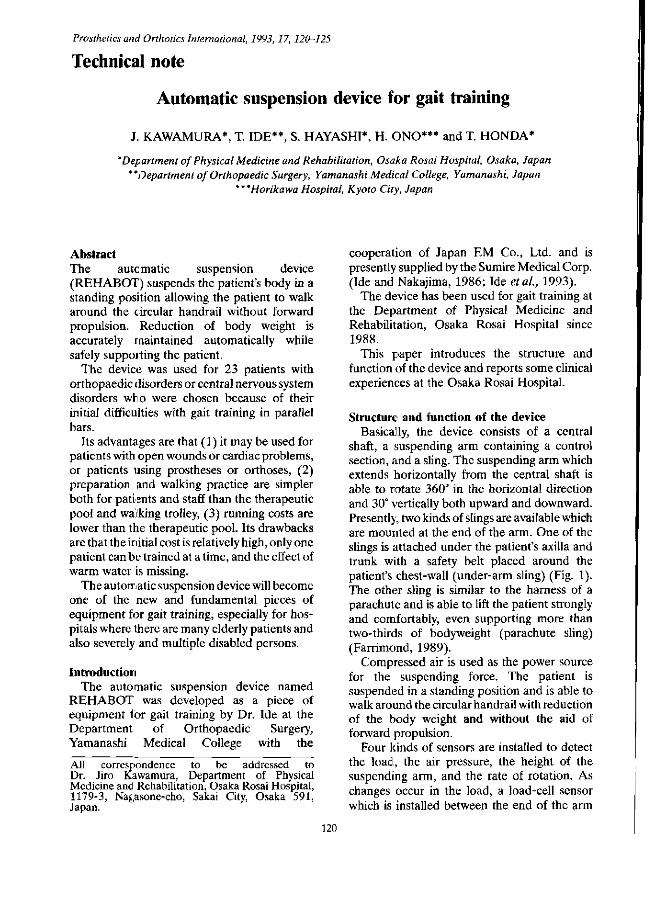
Prosthetics and Orthotics International, 1993,17, 120-125
Technical note
Automatic suspension device for gait training
J. KAWAMURA*, T. IDE**, S. HAYASHI*, H. ONO*** and T. HONDA*
*Department of Physical Medicine and Rehabilitation, Osaka Rosai Hospital, Osaka, Japan **Department of Orthopaedic Surgery, Yamanashi Medical College, Yamanashi, Japan
***Horikawa Hospital, Kyoto City, Japan
Abstract The automatic suspension device (REHABOT) suspends the patient's body in a standing position allowing the patient to walk around the circular handrail without forward propulsion. Reduction of body weight is accurately maintained automatically while safely supporting the patient.
The device was used for 23 patients with orthopaedic disorders or central nervous system disorders who were chosen because of their initial difficulties with gait training in parallel bars.
Its advantages are that (1) it may be used for patients with open wounds or cardiac problems, or patients using prostheses or orthoses, (2) preparation and walking practice are simpler both for patients and staff than the therapeutic pool and walking trolley, (3) running costs are lower than the therapeutic pool. Its drawbacks are that the initial cost is relatively high, only one patient can be trained at a time, and the effect of warm water is missing.
The automatic suspension device will become one of the new and fundamental pieces of equipment for gait training, especially for hospitals where there are many elderly patients and also severely and multiple disabled persons.
Introduction The automatic suspension device named
REHABOT was developed as a piece of equipment for gait training by Dr. Ide at the Department of Orthopaedic Surgery, Yamanashi Medical College with the
cooperation of Japan EM Co., Ltd. and is presently supplied by the Sumire Medical Corp. (Ide and Nakajima, 1986; Ide etal, 1993).
The device has been used for gait training at the Department of Physical Medicine and Rehabilitation, Osaka Rosai Hospital since 1988.
This paper introduces the structure and function of the device and reports some clinical experiences at the Osaka Rosai Hospital.
Structure and function of the device Basically, the device consists of a central
shaft, a suspending arm containing a control section, and a sling. The suspending arm which extends horizontally from the central shaft is able to rotate 360° in the horizontal direction and 30° vertically both upward and downward. Presently, two kinds of slings are available which are mounted at the end of the arm. One of the slings is attached under the patient's axilla and trunk with a safety belt placed around the patient's chest-wall (under-arm sling) (Fig. 1). The other sling is similar to the harness of a parachute and is able to lift the patient strongly and comfortably, even supporting more than two-thirds of bodyweight (parachute sling) (Farrimond, 1989).
Compressed air is used as the power source for the suspending force. The patient is suspended in a standing position and is able to walk around the circular handrail with reduction of the body weight and without the aid of forward propulsion.
Four kinds of sensors are installed to detect the load, the air pressure, the height of the suspending arm, and the rate of rotation. As changes occur in the load, a load-cell sensor which is installed between the end of the arm
All correspondence to be addressed to Dr. Jiro Kawamura, Department of Physical Medicine and Rehabilitation, Osaka Rosai Hospital, 1 1 7 9 - 3 , Nagasone-cho, Sakai City, Osaka 5 9 1 , Japan.
120

Suspension device for gait training 121
and the sling perceives them and controls the suspending force with a microprocessor. Once the suspending force has been set, it will automatically be maintained by these sensors and a microprocessor. The patient is prevented from falling down by an emergency locking system of the lifting arm which is activated immediately after detection of any sudden change in load or height of the arm (Fig. 2).
Clinical experiences Patients
There were 1,793 patients who received medical rehabilitation at the Department of Physical Medicine and Rehabilitation of the Osaka Rosai Hospital from August 1988 to April 1991, of which 1,030 patients received gait training because of orthopaedic or central nervous system disorders. The device was used for 23 patients who were chosen from 1,030
patients because of their difficulties with gait training in parallel bars. There were 12 female and 11 male patients ranging in age from 15 to 79 years (mean age 54 years).
Sixteen patients had orthopaedic disorders, of which 4 patients had bilateral surgery simultaneously (total knee replacement or high tibial osteotomy), 9 patients unilateral surgery (femoral head replacement, total hip replacement, or total knee replacement) with previous disorders (osteoarthritis of hip or knee joint, trans-femoral amputation or hemiparesis on the contralateral side, or rheumatoid arthritis), 2 patients juvenile rheumatoid arthritis with severe osteoporosis of the spine, and one patient was a bilateral trans-femoral amputee. Of these 16 patients, 8 were able to initiate gait training both in the therapeutic pool and with the automatic suspension device, but the other 8 patients were not able to initiate gait training in the therapeutic pool because of percutaneous fixation after high tibial osteotomy (3 patients), use of a lower limb prosthesis (2 patients), use of a spinal orthosis for severe osteoporosis (2 patients), hemiparesis on the contralateral side (1 patient).
Seven patients had central nervous system disorders, and of these 2 patients had hemiparesis with other difficulties (knee contracture or obesity) and 5 patients incomplete quadriplegia. Furthermore, one patient of the latter 5 patients had also cardiac insufficiency. None of them was able to undertake pool therapy.
Gait training using the robotic device was started after completion of a tilting table routine
Fig. 1. Automatic suspension device ( R E H A B O T ) .
Fig. 2 . Block diagram of the automatic suspension device.
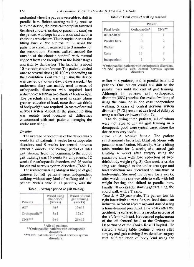
1 2 2 J. Kawamura, T. Ide, S. Hayashi, H. Ono and T. Honda
and ended when the patients were able to shift to parallel bars. Before starting walking practice with the device, the physical therapists fastened the sling (under-arm sling or parachute sling) on the patient, who kept his clothes on and sat on a chair or a wheelchair. The therapist then set the lifting force of the control unit to assist the patient to stand.. It required 2 to 3 minutes for the preparation. Patients walked around the outside of the circular handrail with manual support from the therapists in the initial stages and later by themselves. The handrail is about 10 metres in circumference. The patients circled once to several times (10-100m) depending on their condition. Gait training using the device was carried out once a day, 5 days a week. The under-arm sling was mainly used in cases of orthopaedic disorders who required load reduction of less than two-thirds of bodyweight. The parachute sling was used in cases where greater reduction of load, more than two thirds of bodyweight, was required. In cases of central nervous system disorders, the parachute sling was mainly used because of difficulties encountered with such patients managing the under-arm sling.
Results The average period of use of the device was 5
weeks for all patients, 3 weeks for orthopaedic disorders and 9 weeks for central nervous system disorders. The average period of total gait training (from the beginning to the end of gait training) was 16 weeks for all patients, 12 weeks for orthopaedic disorders and 26 weeks for central nervous system disorders (Table 1).
The levels of walking ability at the end of gait training for all patients were independent walking without any kind of walking aid in 1 patient, with a cane in 15 patients, with the
walker in 4 patients, and in parallel bars in 2 patients. One patient could not shift to the parallel bars until the end of gait training. Although 14 patients with orthopaedic disorders (88%) reached the levels of walking of using the cane, or in one case independent walking, 5 cases of central nervous system disorders (71 %) remained at a level of ability of using a walker or lower (Table 2).
The following three patients, all of whom were not able to initiate gait training in a therapeutic pool, were typical cases where the device was very useful. Case 1: A 69-year female. The patient simultaneously had a high tibial osteotomy with percutaneous fixation, bilaterally. After a tilting table routine for 2 weeks, she started gait training 4 weeks after surgery using the parachute sling with load reduction of two-thirds body weight (Fig. 3). One week later, the sling was changed to the under-arm type and load reduction was decreased to one-third of bodyweight. She used the device for 2 weeks, after which time she was able to walk with full weight bearing and shifted to parallel bars. Finally, 10 weeks after starting gait training, she could walk with a T cane. Case 2: A 25-year male. The patient lost his right lower limb at trans-femoral level due to an industrial accident 4 years ago and started using a trans-femoral prosthesis. Five years after the accident, he suffered from a vascular necrosis of the left femoral head. He received replacement of the left femoral head at the Orthopaedic Department of the Osaka Rosai Hospital. He started a tilting table routine 3 weeks after surgery and gait training 5 weeks after surgery with half reduction of body load using the
Table 1. Average period of gait training
Table 2 : Final levels of walking reached
Suspension device for gait training 123
under-arm sling (Fig. 4). After using the device for 2 weeks, he could walk with full weight bearing on the left lower limb, and was then shifted to parallel bars. The period of total gait training was 10 weeks. Finally, he could walk with a trans-femoral prosthesis and a crutch. Case 3: A 73-year male. This patient fell down the stairs, sustaining a cervical cord injury with incomplete quadriplegia. After the tilting table routine for 7 weeks which took place 5 weeks after the injury, he started gait training with the device using the parachute sling, because of insufficient muscle strength and improper proprioceptive sensation of the upper and lower limbs. Reduction of load was equivalent to 10-20 kg. He continued walking practice with the device for 16 weeks, after which time he was able to walk with a walker. Finally he could move with a walker at home, although still wheelchair dependent outdoors.
Discussion Gait training usually begins with the tilting
table followed by parallel bars, walkers, and various kinds of cane (Corcoran and Peszczynski, 1984). However, there are some patients who arc not able to stand in parallel bars for several reasons such as the need of restricted weight-bearing, on both limbs, insufficient muscle strength of both lower limbs, and difficulties in supporting a standing position using the upper limbs, etc
Pool treatment has been used for such kinds of patients. However, patients having open wounds or cardiac problems and patients wearing orthoses or prostheses cannot use the pool. Some patients are afraid of the water. Furthermore, it requires substantial time and work for both patients and staff for this form of treatment, including the task of taking off and putting on clothes. The initial cost and
Fig. 3. A patient using the parachure sling: high tibial osteotomy with percutaneous fixation bilaterally and
simultaneously.
Fig. 4. A right t rans-femoral a m p u t e e using underarm sling: left femoral head replacement.

124 /. Kawamura, T. 1de, S. Hayashi, H. Ono and T. Honda
maintenance of a pool are very high and it involves strenuous work for the staff during the treatment (Skinner and Thompson, 1983).
The automatic suspension device enables gait training even for patients who have open wounds, percutaneous fixation, prostheses, orthoses, etc The preparation before the walking practice with the device is simpler than for the therapeutic pool for both patients and staff. Staff costs using the device also are low. The running cost of the device is merely the cost of electricity.
Formerly, the walking trolley was used for patients with orthopaedic disorders and central nervous system disorders. However, the problems of the walking trolley were the difficulty of setting the sling and lifting the patient and the imprecise control of the reduction of load bearing. This caused patients to lose balance occasionally (Hattorf and Hara, 1964; Hosokawa, 1964; Sugiyama et ai, 1964; Boyle, 1965).
Although the purpose of the automatic suspension device is similar to that of the walking trolley, the automatic suspension device can control the reduction of load precisely and with security. The adjustment and fitting is easier than the walking trolley and there is no chance of the patient falling down.
Walking around the circular handrail enables the patients to walk uninterruptedly and thus reduce the frequency of manual support by therapists to permit turning around. Dizziness did not occur from walking around the circular handrail because the patients could not walk swiftly. Compared with pool therapy, patients infrequently were afraid of the device. None of the patients refused to use the device. There was no accident, such as a patient falling down during training with the device.
Although patients with central nervous system disorders needed longer periods of gait training and also final levels of walking ability were lower than for patients with orthopaedic disorders, work by staff during training with the device was easier and safer in comparison with the walking trolley.
Stubbies have been used for prosthetic training often in cases of bilateral trans-femoral amputation before training with full-length articulated prostheses. Using the device, a bilateral trans-femoral amputee could begin walking practice without the help of stubbies.
When gait will be achieved by combining assistive devices, such as reciprocating gait orthoses (Beckman, 1987) and functional electrical stimulation (Kralj and Bajd, 1989); or when other new technology is intended, the device could be very useful.
The number of patients trained with the device was equivalent to about 1.3% of the total number who received medical rehabilitation at the department and about 2.2% of the number of patients who received gait training during the period of the study. There were 156 patients who received pool treatment for the same period. If there were no therapeutic pool the number of patients trained by the device would have increased.
Concerning the drawbacks of the device, (1) it does not have the thermal and buoyancy effects of warm water (Walsh, 1990), (2) only one patient can be trained at a time, and (3) the frequency of its use is not high.
Conclusion The automatic suspension device is useful for
patients who are not able to initiate gait training in parallel bars, especially in cases involving difficulties of entering the therapeutic pool. Training is easier and safer and staff costs also are lower than for pool treatment and walking trolley. The running cost of the device is low.
The authors believe that the automatic suspension device will become one of the new and fundamental pieces of equipment for gait training, especially for hospitals where there are increasing numbers of elderly patients and also severely and multiply disabled persons.
Acknowledgements The authors wish to thank Dr. S. Sawamura
the Director of the Hyogo Prefectural Rehabilitation Center for his advice for the contribution.
Development of the device was carried out at the Department of Orthopaedic Surgery, Yamanashi Medical College. Clinical evaluation was carried out at the Department of Physical Medicine and Rehabilitation, Osaka Rosai Hospital.
REFERENCES
BECKMAN J (1987) . The Louisiana State University reciprocating gait orthosis. Physiotherapy 7 3 , 386-392 .
Suspension device for gait training 125
BOYLE R W (1965) . The therapeutic gymnasium. In: Therapeutic Exercise . /2nd ed . /ed i ted by Secht. — N e w Haven: Elizabeth Licht Puhl, p 2 5 7 - 2 8 4 .
CORCORAN PJ, PESZCZYNSKI M ( 1 9 8 4 ) . Gait and gait retraining. In: Therapeutic exercise. /4th ed. /edtted by JV Basnagian. — Baltimore: Williams & Wilkins. p 2 8 5 - 3 0 2 .
FARRIMOND K (1989) . Parachute harness. Physiotherapy 7 5 , 740 .
HATTORI I, HARA T ( 1 9 6 4 ) . Necessary equipment for medical rehabilitation and their usage. Shindan To Chiryo 5 2 , 4 3 2 - 4 4 3 .
HOSOKAWA T (1964) . Gymnastic equipment for medical rehabilitation. Ika Kikaigaku Zashi34,641-6 4 5 .
IDE T , NAKAJIMA I ( 1 9 8 6 ) . Development of a gait training robot ( A I D - 1 ) . Seikei Saigai Ceka 2 9 , 8 2 5 -
IDE T, SIDDIOE N A , AKAMATSU N (1993) . Expectations for medical and healthcare robotics. Advanced Robotics. (In press.)
KRAU A R , BAJD T ( 1 9 8 9 ) . FES-Assisted walking in complete SCI patients. In: Functional electrical stimulation: standing and walking after spinal cord injury./edited by A K Kralj, T Bajd. — Boca Raton: C R C Press, p 139 -191 .
SKINNER AT, THOMSON A M ( 1 9 8 3 ) . Duffield's exercise in water. /3rd ed. — London: Bailliere Tindall. p 39-53 .
SUGIYAMA T, KAYABA M, HANAKAGO R (1964) . Rehabilitation of cerebral vascular disorder. Nippon Rinsho27, 1 5 4 9 - 1 5 6 2 .
WALSH M T (1990) . Hydrotherapy: the use of water as a therapeutic agent. In: Thermal agents in rehabilitation./2nd ed . /ed i ted by SL Michlovitz. — Philadelphia: FA Davis, p 109-133 .

Prosthetics and Orthotics International, 1993, 17, 126-129
Clinical note
Lightweight prostheses for bilateral below-elbow amputees
S. N. R O U T
Dept. of Bio-Engineering, National Institute of Rehabilitation Training and Research (NIRTAR), Orissa, India
Abstract In view of the anticipated activity of the patient and working environment , lightweight prostheses were designed for an adult female, bilateral below-elbow (BE) amputee at N I R T A R to provide the greatest degree of function. The prostheses were fabricated using lightweight materials and new techniques. Depending on the s tump length there were two different types of lightweight prostheses designed and successfully used, (1) an endoskeletal B E prosthesis and (2) an exoskeletal BE prosthesis. After periodic follow-up and evaluation the function of the prostheses was found to be most satisfactory. By reducing the weight considerably compared to other available alternatives, it is more likely that the amputee will make use of the prostheses to efficiently perform various activities. The new prosthesis designs may counteract the high rejection rate of old conventional ones and the principle may be applied to the fabrication of all BE prostheses.
Introduction Bilateral BE amputees are fully dependent
upon prostheses to perform most of their activities in contrast to unilateral amputees . Therefore proper prosthetic fitting and suitable prosthetic components are necessary to optimise their performance in various activities. The design must anticipate the activity of the
patient and the working environment to provide the greatest function. Reducing the weight of the prosthesis will encourage the patient to perform most activities and also facilitate execution with less expenditure of energy. Taking the above points into consideration new types of lightweight prostheses were designed at N I R T A R for a bilateral B E amputee in an at tempt to provide maximum function.
Methods Subjects
The patient was an adult female bilateral BE amputee who presented at N I R T A R for fitting of prostheses. On the right side she had a very short B E stump of about 6.35 cm (2.5 ins) length and on the left side a long BE stump of about 16.5 cm (6.5 ins) length. Previously she had been provided with bilateral B E prostheses at other limb fitting centres using traditional exoskeletal construction. She had discarded these due to the heavy weight and high energy expenditure incurred in prosthetic operat ion. The same patient was successfully fitted with the lightweight prostheses developed at N I R T A R .
Fabrication techniques The prostheses are fabricated using new
techniques and lightweight materials.
Right side The right very short BE prosthesis was
designed based on a modular principle to reduce weight.
All correspondence to be addressed to S. N. R o u t , Lecturer in Prosthetics Dep t . of Bio-Engineer ing, N I R T A R , Bairoi-754010, Cuttack (Orissa) , India.
126
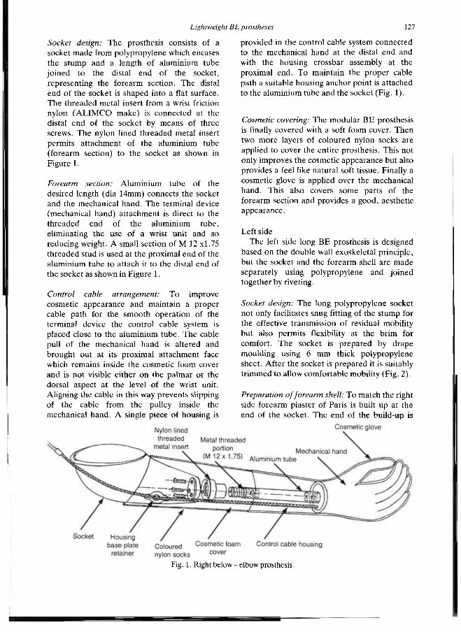
Lightweight BE prostheses 127
Socket design: The prosthesis consists of a socket made from polypropylene which encases the s tump and a length of aluminium tube joined to the distal end of the socket, representing the forearm section. The distal end of the socket is shaped into a flat surface. The threaded metal insert from a wrist friction nylon ( A L I M C O make) is connected at the distal end of the socket by means of three screws. The nylon lined threaded metal insert permits at tachment of the aluminium tube (forearm section) to the socket as shown in Figure 1.
Forearm section: Aluminium tube of the desired length (dia 14mm) connects the socket and the mechanical hand. The terminal device (mechanical hand) at tachment is direct to the threaded end of the aluminium tube, eliminating the use of a wrist unit and so reducing weight. A small section of M 12 x1.75 threaded stud is used at the proximal end of the aluminium tube to attach it to the distal end of the socket as shown in Figure 1.
Control cable arrangement: To improve cosmetic appearance and maintain a proper cable path for the smooth operat ion of the terminal device the control cable system is placed close to the aluminium tube . The cable pull of the mechanical hand is altered and brought out at its proximal a t tachment face which remains inside the cosmetic foam cover and is not visible either on the palmar or the dorsal aspect at the level of the wrist unit. Aligning the cable in this way prevents slipping of the cable from the pulley inside the mechanical hand. A single piece of housing is
provided in the control cable system connected to the mechanical hand at the distal end and with the housing crossbar assembly at the proximal end. To maintain the proper cable path a suitable housing anchor point is attached to the aluminium tube and the socket (Fig. 1).
Cosmetic covering: The modular B E prosthesis is finally covered with a soft foam cover. Then two more layers of coloured nylon socks are applied to cover the entire prosthesis. This not only improves the cosmetic appearance but also provides a feel like natural soft tissue. Finally a cosmetic glove is applied over the mechanical hand. This also covers some parts of the forearm section and provides a good, aesthetic appearance.
Left side The left side long B E prosthesis is designed
based on the double wall exoskeletal principle, but the socket and the forearm shell are made separately using polypropylene and joined together by riveting.
Socket design: The long polypropylene socket not only facilitates snug fitting of the s tump for the effective transmission of residual mobility but also permits flexibility at the br im for comfort. The socket is prepared by drape moulding using 6 mm thick polypropylene sheet. After the socket is prepared it is suitably tr immed to allow comfortable mobility (Fig. 2).
Preparation of forearm shell: To match the right side forearm plaster of Paris is built up at the end of the socket. The end of the build-up is
Fig. 1. Right below - elbow prosthesis

128 S. IV. Rout
shaped into a flat surface to match the diameter of the plate at tachment . Using drape moulding the polypropylene is moulded over the build-up to prepare the extended forearm shell. It is finally t r immed slightly proximal to the expected joint with the socket. At the distal end of the shell a suitable diameter hole is drilled at the centre, to insert the stud of the plate at tachment. The plate at tachment is firmly fixed with the polypropylene shell using screws and nuts as shown in Figure 2. The plate at tachment is used for the terminal device to eliminate the use of a wrist unit. The forearm shell prepared by this method is very light in weight.
The shell is then firmly united with the socket by riveting (using press rivets) on four sides i.e. anterior, posterior, medial and lateral.
Control cable arrangements: The control cable was positioned inside the forearm shell and brought out near the junction between the socket and shell to maintain the proper cable path while improving cosmetic appearance. A single piece of housing is provided in the control cable system which is anchored distally by the housing baseplate retainer fixed on the antero-lateral aspect of the socket and proximally by the housing crossbar assembly fixed on the triceps pad. By using a single housing the control cable path is properly maintained during the full range of elbow flexion and greater cable excursion is available for terminal device operat ion.
Cosmetic covering: After assembling the mechanical hand the cosmetic glove is finally applied over the prosthesis covering the mechanical hand and also a port ion of the forearm shell to improve the aesthetic appearance. The control cable remains inside the forearm shell and is not visible near the wrist joint on either the palmar or dorsal aspect.
Results and conclusions Because the wrist unit is eliminated and
lightweight materials are used for fabrication of both prostheses, the weight of each prosthesis is about 0.610 kg as compared to 1 kg for a conventional prosthesis. Because the stump length is not the same on both sides two different types of lightweight prostheses were designed. Depending on stump length either type may be fitted to the bilateral B E amputee . After fitting the patient was able to open the terminal device at any desired position of elbow flexion and to peform the following activities efficiently:
1) donning and doffing the prostheses easily and independently;
2) drinking water while holding the glass in one hand and operating the water tap with the other;
3) controlling the movement of a spoon and eating freely as normal;
4) manipulating the prosthesis while writing with one hand and stabilizing the paper
Fig. 2. Left below - elbow prosthesis.
Lightweight BE prostheses 129
with the other.- (She graduated using these prostheses);
5) (since she is interested in becoming a teacher) holding the chalk and writing freely on a blackboard by lifting the prosthesis above her head for long periods;
6) exercising good control for manipulating the prostheses to perform various daily living activities i .e. , using cosmetics, lipsticks etc;
7) performing ordinary household activities like sweeping etc;
The patient acceptance is very good and encouraging.
Advantages 1 ) They are very light in weight, more
efficient in function and have a superior cosmetic appearance.
2) By using the lightweight prostheses, the patient activity sphere increases and thereby is helped to perform various manipulative activities efficiently with minimum energy expendi ture .
3) Fabrication time required for the lightweight prosthesis is less compared to the conventional and also there are economies in eliminating the use of a wrist unit and avoiding laminating procedures .
4) The fabrication method is very simple, therefore care and maintenance is also very easy.
Disadvantages During follow-up and evaluation after using
the prostheses for a period of about 6 months , no specific disadvantage has been noticed except some minor adjustment in the harness system to improve function, Interchanging the terminal devices is not possible, however, it is believed that most of the amputees particularly ladies, do not prefer to interchange terminal devices.
Acknowledgement I would like to extend my gratitude to my
Director , Wing Commander S.N. Mohanty , and my Head of the Depar tment , Mr L. T. Iyer, who always inspired and encouraged me to take up new challenges in design and to develop suitable prostheses for problem cases.
R E F E R E N C E S
STURUP J, T H Y R E G O D H C , JENSEN JS, RETPEN JB , BoBERG G , RAMUSSEN E , JENSEN S ( 1 9 8 8 ) . Traumat ic amputa t ion of the upper limb: the use of body-powered prostheses and employment consequences . Prosthet Orthot Int 12, 5 0 - 5 2 .
ROESCHLEIN R A , D O M H O L D T E ( 1 9 8 9 ) . Factors related to successful upper extremity prosthetic use. Prosthet Orthot Int 13 , 1 4 - 1 8 .

Prosthetics and Orthotics International, 1993, 17, 130-135
Technical note Continuous passive motion
in hand rehabilitation
J. A. DENT
Dept. of Orthopaedic and Trauma Surgery, University of Dundee, UK
Abstract This paper reviews the literature comparing the results obtained in applying regimes involving motion with those involving rest following injury or surgery. The deleterious effects of immobilisation are compared to those obtained under conditions of passive motion and intermittent passive motion. It is concluded that continuous passive motion (CPM) represents an improvement on intermittent motion. Models of CPM machines are described and some results presented.
It is proposed that "intelligent" CPM would represent a further improvement in technique and a prototype machine for this purpose and some preliminary results are described.
Motion is better than no motion In 1982 Bob Salter from Toronto delivered
his presidential address to the Canadian Orthopaedic Association entitled, "Motion versus rest—why immobilise joints?". He observed that from the very beginnings of Western medicine there have been two schools of thought about the best way of treating injured limbs, namely the "resters" and the "movers". In the fourth century BC Hippocrates taught that in illness "it is especially needful for the body to be at rest and to lie up" while Aristotle, in the same century wrote, "the principle is that movement is life". From this time, throughout the subsequent development of orthopaedic surgery in the western world, the "resters" have maintained their popularity.
It was Hugh Owen Thomas, whose dictum that rest should be "complete, prolonged, uninterrupted and enforced", who influenced generations of orthopaedic surgeons all over the world. This opinion remained unchallenged until the beginning of the twentieth century when Champonniere wrote "every movement, which is not injurious by reason of its amplitude, favours repair". He believed that "massage and motion help to relieve pain rather than aggravate it".
Such teaching remained controversial, however, and Thomas's principles continued to be practised by Sir Robert Jones and Sir Reginald Watson-Jones until again challenged by Professor George Perkins from St. Thomas's Hospital who wrote that "in making a choice between rest and motion we are largely biased by tradition" adding that the "training of a doctor is such that it is not easy for him to break the tradition". In the face of that tradition he boldly proclaimed "movement is often better that rest" a sentiment succinctly summarised by Alan Graham Apley as "plaster is a disaster!".
The original work of Salter (1989) had shown the deleterious efffects of immobilisation. He demonstrated pressure necrosis of cartilage in immobilised joints and obliterative degeneration of articular cartilage in non-contact areas secondary to adherence of synovial membrane to the joint surface. The microscopic changes described include matrix fibrillation, cleft formation and ulceration. Palmoski et al. (1979) demonstrated interference with normal proteoglycan
This paper was based on a presentation made at Dundee '91—International Conference and Instructional Course on Orthotics, 16-20 September, 1991, Dundee , Scotland.
All correspondence to be addressed to Mr. John A. Dent , Department of Orthopaedic & Trauma Surgery, University of Dundee, Caird Block, Royal Infirmary, Dundee , UK.
130
Continuous passive motion in hand rehabilitation 131
synthesis and the aggregation of large molecular complexes following immobilisation. The immobilisation of joints as we know leads to stiffness, swelling, pain, muscle atrophy and disuse osteoporosis. Such observations plus his laboratory work led Salter to believe that there should be beneficial local effects to be gained from the early active mobilisation of joints. He argued that other disciplines such as open heart surgery demonstrate that tissues do not have to be put to rest in order to heal. Such principles are followed in general surgery by the advocates of early ambulation after abdominal operations and Dr. Earl Shouldice's clinic for day case hernia repair under local anaesthesia. Similar thinking is also found in the modern treatment of fractures whether by functional bracing as put forward by Augusto Sarmiento or by open reduction and internal fixation followed by early active mobilisation as proposed by Muller and Allgower and the AO group.
So much for the benefits of motion as opposed to immobilisation on the rehabilitation of joints. A similar conclusion had also been reached in the upper limb in the field of rehabilitation of flexor tendon repairs. Within the flexor fibro-osseous tunnel, or zone 2 of the hand two tendons run together. Their normal excursion is in the order of 20 mm. In other zones more proximal or distal an excursion of only 5 mm is necessary for normal finger function. Adhesion of the tendon in the fibrous flexor tunnel after injury in zone 2 will occur between 5 and 21 days post-operatively and cause limitation of movement. Early motion after tendon repair was, therefore, advocated by a number of surgeons in the early 1900's when Harmer (1917) developed a special suture technique allowing early active motion,
The most well known method of mobilisation is probably the rubber band traction introduced by Kleinert et al. (1967). The back shell allows controlled active extension of the finger with passive flexion being achieved by means of the rubber band (Lister et al., 1977). An alternative method of mobilisation was described by Duran and Houser (1975). They reported a system of controlled passive exercises which mobilised the individual joints of the injured digit separately.
This idea of intermittent passive motion was further studied by Strickland and Glogovac
(1980). They looked at 50 digits with zone 2 injuries. Twenty-five were treated by three and a half weeks of immobilisation and the second group of 25 by intermittent passive motion. The results were graded according to the percentage of return of motion at the PIP and DIP joints. In the group treated by immobilisation there were no excellent results and only 12% good results compared to 36% excellent and 20% good results in the early passive motion group. Furthermore increased tendon strength was demonstrated as an effect of early passive motion in that there was only one tendon rupture in this group compared to four ruptures in the group which had been treated by immobilisation. They concluded that "early passive motion appeared to be an effective technique to improve the results of flexor tendon repairs in this area".
Gelbermann et al. (1980) studying the effect of mobilisation on tendon repairs in dogs found that the mobilised group showed less vessel density and a quicker return to the normal longitudinal orientation of vessels than the group treated by immobilisation. These changes occurred within three weeks of surgery. They also noticed a significant increase in tendon strength and excursion with mobilisation. Hitchcock et al. (1987) showed that by five days after surgery there was a significant difference in tendon strength between an immobilised group and a group allowed immediate active motion. They concluded that immediate active motion allows progressive tendon healing without an intervening phase of tendon softening.
The concept of controlled motion instead of immobilisation for the post-operative treatment of tendon repairs continues to be popular (Creekmore et al., 1985; Cullen et al. 1989: Matthews. 1989; Morris. 1987: Small et al., 1989). Various modifications of Kleinert's original elastic band traction have more recently been described (Edinburg et al., 1987). A recent modification from Kleinert's own group incorporates a coiled lever on the forearm to regulate the traction force and has a spring-loaded roller bar in the palm to improve DIP joint flexion (Werntz et al. 1989). Sometimes a combination of dynamic splint and intermittent passive motion are being used as described by Chow et al. (1988).
132 J. A. Dent
Continuous passive motion is better than intermittent passive motion
Salter's original work (1989) led him to ask the question "If intermittent motion is good for articular cartilage, would continuous passive motion be even better?". Continuous active motion of course would not be possible because of muscle fatigue. The benefit of continuous passive motion to joints was originally proposed by Salter in 1970. He described the beneficial effects of this on the healing of articular cartilage after a variety of intra-articular defects (Salter, 1989). He stated that continuous passive motion (CPM) stimulated the pluripotential mesenchymal cells to differentiate into articular cartilage instead of fibrous tissue. It also enhanced the nutrition of articular cartilage and accelerated the healing of extra-articular tissues. Treatment was continued for four weeks at the rate of one complete cycle every 45 seconds. Cartilage regeneration with both cartilage cell proliferation and matrix production was found to be superior in the CPM group compared to the group treated with intermittent active motion. Neochondrogenesis from periosteal autograft was seen in 80% of the group with CPM and only 10% of the group with intermittent active motion. Of a group of intraarticular fractures treated by intermittent passive motion, 50% had not healed, 20% had healed with fibrous tissue and only 30% had healed with cartilage. In a group of intraarticular fractures treated by CPM 80% healed with cartilage within the first week. This superior repair tissue was found to be maintained in the experimental animals a year later.
Beneficial effects of CPM on rabbit tendons was reported by Loitz et al. (1989). Cyclical tensile loading led to increased tendon strength, the results being better in this group than in those treated by immobilisation. In other experimental animal work Salter ( 1989) showed that CPM led to thicker tendon callus and better alignment of tendon fibres than immobilisation or intermittent active motion in rabbits with partial lacerations of the patellar tendon. The breaking strength of the tendon was also higher.
On the basis of this laboratory evidence Salter, in collaboration with Saringer, introduced a continuous passive motion
machine in 1979 for use with their patients. This has subsequently been developed in Toronto for use in treating a variety of joints and conditions which will be illustrated later.
In 1972. however, Ketchum, Clark, Robinson and Masters had introduced an electronically controlled, driven hand splint. The machine consisted of nylon lines attached to adjustable thimbles on the fingertips. Gentle, rhythmic, passive motions of 2.3 kg force at varying frequencies could be applied in both extension and flexion of the fingers. This machine was subsequently modified to a safer and simpler model in which the range of motion applied to each finger could be adjusted independently. Ketchum et al. (1979) reported a gain in total active and passive motion in stiff fingers using this technique compared to manual passive exercises performed by a therapist. They found a statistically significant improvement after one month in both total active motion and total passive motion in the group treated by CPM compared to the group treated by the hand therapist. The time taken to use the electronically controlled, driven hand splint was half that of the time needed by a hand therapist to treat a patient by passive mobilisation.
Two models of CPM machines are currently available, the Toronto Mobilimb and the Stryker Hand Exerciser. The Toronto Mobilimb is a relatively rigid device capable of five different stoke lengths and giving both passive flexion and extension. The Stryker Hand Exerciser has three stroke lengths and speeds and is attached to the fingers by cables. Both devices are battery operated and lightweight. The applications for CPM suggested by Salter (1989) include its use after MCP joint arthroplasty, intra-articular fractures, synovectomy and arthrolysis. It can also be used after tendon repair, ligament reconstructions and other intra-articular procedures. Other authors have suggested additional uses. Giudice (1990) found it more effective than elevation alone in reducing hand oedema. Shaw and Kasser (1990) used it in the management of septic arthritis. Blauth et al. (1990), Soffer and Yahiro (1990) and Letsch et al. (1989) have reported it in the management of fractures and arthroplasties in the elbow.
However, not much has been written about the use of CPM in the hand (Bentham et al.,
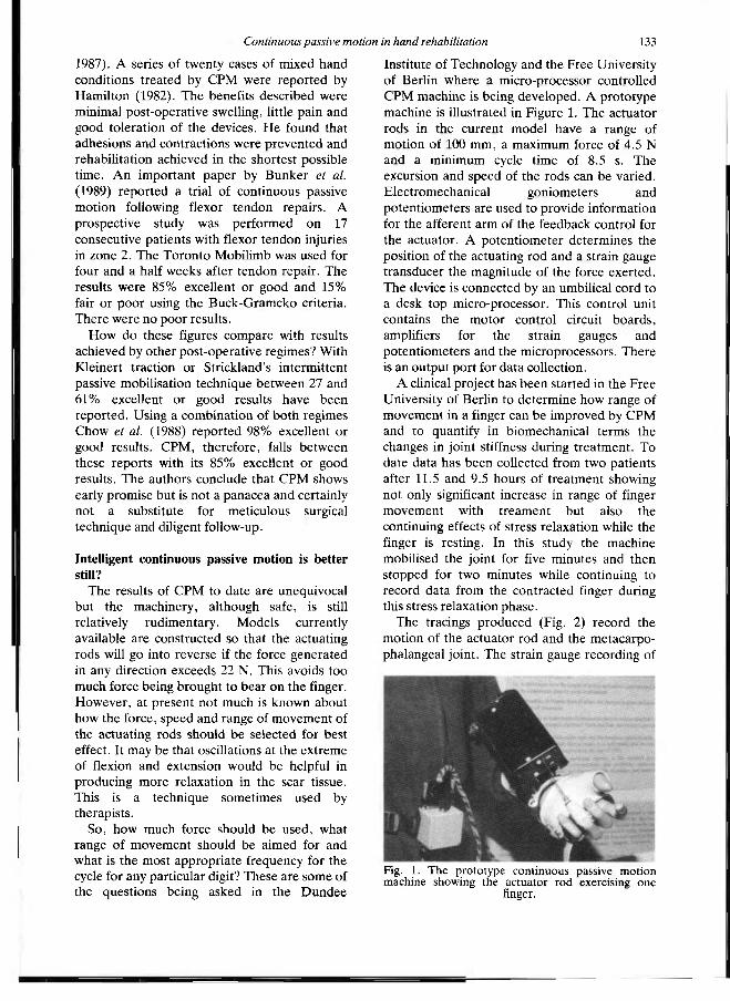
Continuous passive motion in hand rehabilitation 133
1987). A series of twenty cases of mixed hand conditions treated by CPM were reported by Hamilton (1982). The benefits described were minimal post-operative swelling, little pain and good toleration of the devices. He found that adhesions and contractions were prevented and rehabilitation achieved in the shortest possible time. An important paper by Bunker et al. (1989) reported a trial of continuous passive motion following flexor tendon repairs. A prospective study was performed on 17 consecutive patients with flexor tendon injuries in zone 2. The Toronto Mobilimb was used for four and a half weeks after tendon repair. The results were 85% excellent or good and 15% fair or poor using the Buck-Gramcko criteria. There were no poor results.
How do these figures compare with results achieved by other post-operative regimes? With Kleinert traction or Strickland's intermittent passive mobilisation technique between 27 and 61% excellent or good results have been reported. Using a combination of both regimes Chow et al. (1988) reported 98% excellent or good results. CPM, therefore, falls between these reports with its 85% excellent or good results. The authors conclude that CPM shows early promise but is not a panacea and certainly not a substitute for meticulous surgical technique and diligent follow-up.
Intelligent continuous passive motion is better still?
The results of CPM to date are unequivocal but the machinery, although safe, is still relatively rudimentary. Models currently available are constructed so that the actuating rods will go into reverse if the force generated in any direction exceeds 22 N. This avoids too much force being brought to bear on the finger. However, at present not much is known about how the force, speed and range of movement of the actuating rods should be selected for best effect. It may be that oscillations at the extreme of flexion and extension would be helpful in producing more relaxation in the scar tissue. This is a technique sometimes used by therapists.
So, how much force should be used, what range of movement should be aimed for and what is the most appropriate frequency for the cycle for any particular digit? These are some of the questions being asked in the Dundee
Institute of Technology and the Free University of Berlin where a micro-processor controlled CPM machine is being developed. A prototype machine is illustrated in Figure 1. The actuator rods in the current model have a range of motion of 100 mm, a maximum force of 4.5 N and a minimum cycle time of 8.5 s. The excursion and speed of the rods can be varied. Electromechanical goniometers and potentiometers are used to provide information for the afferent arm of the feedback control for the actuator. A potentiometer determines the position of the actuating rod and a strain gauge transducer the magnitude of the force exerted. The device is connected by an umbilical cord to a desk top micro-processor. This control unit contains the motor control circuit boards, amplifiers for the strain gauges and potentiometers and the microprocessors. There is an output port for data collection.
A clinical project has been started in the Free University of Berlin to determine how range of movement in a finger can be improved by CPM and to quantify in biomechanical terms the changes in joint stiffness during treatment. To date data has been collected from two patients after 11.5 and 9.5 hours of treatment showing not only significant increase in range of finger movement with treament but also the continuing effects of stress relaxation while the finger is resting. In this study the machine mobilised the joint for five minutes and then stopped for two minutes while continuing to record data from the contracted finger during this stress relaxation phase.
The tracings produced (Fig. 2) record the motion of the actuator rod and the metacarpophalangeal joint. The strain gauge recording of
Fig. 1. The prototype continuous passive motion machine showing the actuator rod exercising one
finger.
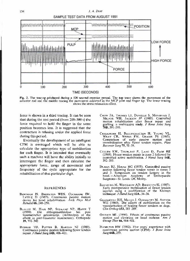
134 J A. Dent
force is shown in a third tracing. It can be seen that during the rest period (from 200-300 s) the force required to hold the finger in the same position becomes less. It is suggested that the contracture is relaxing under the applied force during this period.
Eventually the development of an intelligent CPM is envisaged which will be able to calculate the appropriate type of mobilisation for each finger. It is intended that eventually such a machine will have the ability initially to interrogate the finger and then calculate the appropriate force, range of movement and frequency of the cycle appropriate for the rehabilitation of that particular digit.
Fig. 2. The tracing produced during a 520 second exercise period. The top trace shows the movement of the actuator rod and the middle tracing the movement achieved in the MCP joint and finger tip. The lower tracing
shows the stress relaxation effect.
REFERENCES
BENTHAM JS, BRERETON W D S , C O C H R A N E I W , LYTTLE D (1987). Continuous passive motion device for hand rehabilitation. Arch Phys Med Rehabil 6 8 , 248-250.
B L A U T H M, H A S S NP, SUDKAMP NP, H A P P E T (1990). Die; ellbogenarthrolyse bei post traumatischer gelenksteije. (Arthrolysis of the elbow in post-traumatic contracture). Orthopade 1 9 , 332-342.
B U N K E R T D , POTTER B , B A R T O N NJ (1989). Continuous passive motion following flexor tendon repair. J Hand Surg 1 4 B , 406-411.
CHOW JA, THOMES LJ, D O V E L L E S, MONSIVAIS J, MILNOR W H , JACKSON JP (1988). Controlled motion rehabilitation after flexor repair and grafting: a multicentre study. J Bone Joint Surg 7 0 B , 591-595.
CREEKMORE H, SELLINGHAUSEN H, Y O U N G VL, W R A Y C R , W E E K S PM, G R A S S E PS (1985). Comparison of early passive motion and immobilisation after flexor tendon repairs. Plast Reconstr Surg 7 5 , 55-59.
CULLEN KW, TOLHURST P, L A N G D , P A G E RE (1989). Flexor tendon repair in zone 2 followed by controlled active mobilisation. J Hand Surg 1 4 B , 392-395.
D U R A N RJ, H O U S E RG (1975). Controlled passive motion following flexor tendon repair in zones 2 and 3. Symposium on tendon surgery in the hand.—American Academy of Orthopaedic Surgeons -S t . Louis: D C Mosby.
EDINBURG M, W I D G E R O W A D , BIDDULPH SL (1987). Early postoperative mobilisation of flexor tendon injuries using a modification of the Kleinen technique J Hand Surg 1 2 A . 34-38.
GELBERMAN R H , M E N O N J, GONSALVES M, A K E S O N W H (1980). The effects of mobilization on the vascularisation of healing flexor tendons in dogs. Clin Orthop 1 5 3 , 283-289.
G I U D I C E M L (1990). Effects of continuous passive motion and elevation on hand oedema. Am J Occup Ther 4 4 , 9 1 4 - 9 2 1 .
HAMILTON H W (1982). Five years' experience with continuous passive motion (CPM). J Bone Joint Surg 6 4 B , 259.

Continuous passive motion in hand rehabilitation 1 3 5
H A R M E R TW ( 1 9 1 7 ) . Tendon suture. Boston Med Surg J 167, 8 0 8 .
HITCHCOCK TF, LIGHT TR, B U N C H WH, KNIGHT GW, SARTORI MJ, PATWARDHAN A G , HOLLYFIELD RL ( 1 9 8 7 ) . The effect of immediate constrained digital motion on the strength of flexor tendon repairs in chickens. J Hand Surg 12A, 5 9 0 -5 9 5 .
KETCHUM L D , CLARK LC, ROBINSON D W , MASTERS FW ( 1 9 7 2 ) . A new electric hand splint. J Bone Joint Surg 5 4 A , 9 0 2 .
KETCHUM L D , H I B B A R D A , HASSANEIN KM ( 1 9 7 9 ) . Follow-up report on the electrically driven hand splint. J Hand Surg 4 , 4 7 4 — 4 8 1 .
KLEINERT H E , K U T Z JE, A S H B E L L T S , M A R T I N E E ( 1 9 6 7 ) . Primary repair of flexor tendons in "no man's land". J Bone Joint Surg 4 9 A , 5 7 7 .
LETSCH R, SCHMIT-NEUERBURG KP, STURMER KM, W A L Z M ( 1 9 8 9 ) . Intra-articular fractures of the distal humerus: surgical treatment and results. Clin Orthop 241,238-244.
LISTER G D , KLEINERT H E , K U T Z JE, A T A S O Y E ( 1 9 7 7 ) . .Primary flexor tendon repair followed by immediate controlled mobilization. J Hand Surg 2 . 4 4 1 - 4 5 1 .
L o i t z B J , ZERNICKE RF, VAILAS A C , K O D Y MH, M E A L S R A ( 1 9 8 9 ) . Effects of short-term immobilisation versus continuous passive motion on the biomechanical and biochemical properties of the rabbit tendon. Clin Orthop 2 4 4 , 2 6 5 - 2 7 1 .
MATTHEWS JP ( 1 9 8 9 ) . Early mobilization after flexor tendon repair. J Hand Surg 1 4 B , 3 6 3 - 3 6 7 .
MORRIS JS ( 1 9 8 7 ) . Continuous passive motion in post-operative rehabilitation of the hand: a prospective study (abstract). J Bone Joint Surg 6 9 B , 5 0 1 .
PALMOSKI MJ, B R A N D T KD ( 1 9 7 9 ) . Effect of salicylate on proteoglycan metabolism in normal canine articular cartilage in vitro. Arthritis Rheum 2 2 , 7 4 6 - 7 5 4 .
SALTER RB ( 1 9 8 2 ) . Motion vs. rest: why immobilise joints? Presidential address to the Canadian Orthopaedic Associations. J Bone Joint Surg 64B, 2 5 1 - 2 5 4 .
SALTER RB ( 1 9 8 9 ) . The biologic concept of continuous passive motion of synovial joints. Clin Orthop 242, 1 2 - 2 5 .
SMALL JO, B R E N N A N M D , COLVILLE J ( 1 9 8 9 ) . Early active mobilization following flexor tendon repair in zone 2 . J Hand Surg 14B, 3 8 3 - 3 9 1 .
SOFFER SR, Y A H I R O M A ( 1 9 9 0 ) . Continuous passive motion after internal fixation of distal humerus. Orthop Rev 19, 8 8 - 9 3 .
STICKLAND JW, G L O G O V A C SV ( 1 9 8 0 ) . Digital function following flexor tendon repair in zone II: a comparison of immobilization and controlled passive motion techniques. J Hand Surg 5, 5 3 7 -5 4 3 .
SHAW B A , KASSER JR ( 1 9 9 0 ) . Acute septic arthritis in infancy and childhood. Clin Orthop 2 5 7 , 2 1 2 -2 1 5 .
WERNTZ JR, CHESHER SP, B R E I D E N B A C H WC, KLEINERT H E , BISSONNETTE M A ( 1 9 8 9 ) . A new dynamic splint for post-operative treatment of flexor tendon injury. J Hand Surg 1 4 A , 5 5 8 - 5 6 6 .
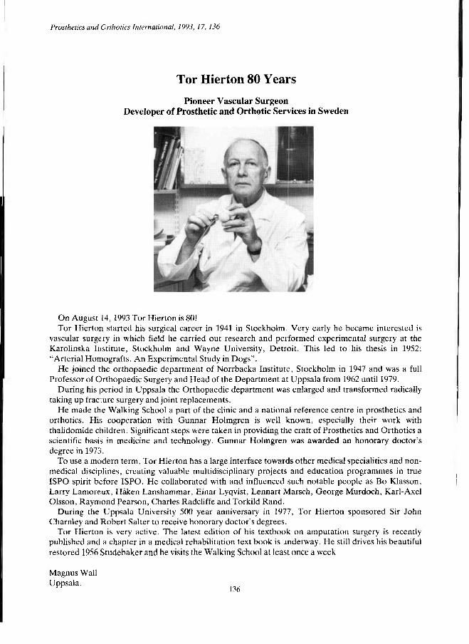
Prosthetics and Orthotics International, 1993, 17, 136
Tor Hierton 80 Years
Pioneer Vascular Surgeon Developer of Prosthetic and Orthotic Services in Sweden
On August 14, 1993 Tor Hierton is 80! Tor Hierton started his surgical career in 1941 in Stockholm. Very early he became interested is
vascular surgery in which field he carried out research and performed experimental surgery at the Karolinska Institute, Stockholm and Wayne University, Detroit. This led to his thesis in 1952: "Arterial Homografts. An Experimental Study in Dogs".
He joined the orthopaedic department of Norrbacka Institute, Stockholm in 1947 and was a full Professor of Orthopaedic Surgery and Head of the Department at Uppsala from 1962 until 1979.
During his period in Uppsala the Orthopaedic department was enlarged and transformed radically taking up fracture surgery and joint replacements.
He made the Walking School a part of the clinic and a national reference centre in prosthetics and orthotics. His cooperation with Gunnar Holmgren is well known, especially their work with thalidomide children. Significant steps were taken in providing the craft of Prosthetics and Orthotics a scientific basis in medicine and technology. Gunnar Holmgren was awarded an honorary doctor's degree in 1973.
To use a modern term, Tor Hierton has a large interface towards other medical specialities and nonmedical disciplines, creating valuable multidisciplinary projects and education programmes in true ISPO spirit before ISPO. He collaborated with and influenced such notable people as Bo Klasson, Larry Lamoreux, Håken Lanshammar, Einar Lyqvist, Lennart Marsch, George Murdoch, Karl-Axel Olsson, Raymond Pearson, Charles Radcliffe and Torkild Rand.
During the Uppsala University 500 year anniversary in 1977, Tor Hierton sponsored Sir John Charnley and Robert Salter to receive honorary doctor's degrees.
Tor Hierton is very active. The latest edition of his textbook on amputation surgery is recently published and a chapter in a medical rehabilitation text book is underway. He still drives his beautiful restored 1956 Studebaker and he visits the Walking School at least once a week
Magnus Wall Uppsala.
136

Prophéties and Orthotics International, 1993, 17, 137-138
ISPO Update Course on Lower Limb Amputations and Related Prosthetics Compound Fractures and the Neuropathic Foot
14th-18th March 1994 Pattaya, Thailand
Introduction The format of this third update course
following the Consensus Conference on Amputation Surgery, in Glasgow, October 1990 will be review lectures based on the presentations at the consensus conference and the points from the discussions leading to contemporary recommendations. Consideration will also be given to the treatment of compound fractures.
Ample time will be allocated for elucidation and discussion with the participants. The course is aimed at orthopaedic surgeons, general surgeons, rehabilitation specialists, prosthetists, orthopaedic technologists and other members of the amputation team, primarily from South East Asia.
Venue
The Merlin Pattaya, Pattaya, Thailand.
Course Fee The course fee is US$100 which covers lectures,
Consensus Conference Report, coffee, tea and lunches. Accommodation
Accommodation can be arranged at the Merlin Pattaya Hotel at a cost of US$40 per night, for
single occupancy and US$60 per night for double occupancy, including breakfast and dinner.
Transport By plane to Bangkok International Airport.
Local transportation between Bangkok and Pattaya is US$25 per person.
Sponsorship Prospective participants are encouraged to seek
sponsorship from local offices of such agencies as Ministry of Health, Ministry of Social Services, British Council, World Health Organisation and any other local agencies.
Collaborators The course is organised in collaboration with the
World Health Organisation, the Thai Orthopaedic Association, the Thai Physical Medicine and Rehabilitation Association, World Orthopaedic Concern and the International Committee of the Red Cross.
Registration Form Mail or fax to Dr. I. Steen Jensen, ISPO,
Polymers Reconstructive A/S, Rugmarken 24-26, 3520 Farum, DENMARK. Fax No: (45) 45 87 30 36. Bank drafts should be made payable to ISPO.
(continued overleaf)
Name - I wish to attend: Male/Female (Delete as appropriate) Update Course Mailing address I require accommodation in Merlin Pattaya [ |
Expected date of arrival Time Flight No.
. . . . , , Expected date of departure Institution address . r
Time . Flight No. Registration Fee for Update Course US$100
_ _ Payment enclosed as bank draft Tel: Fax:. Payment to be made on arrival Telex: Date . Profession Signature
137
138 ISPO Update Course
Preliminary Programme Monday Working Environment in Developing Countries Compound Fractures
Tuesday Amputation Surgery - General Considerations Basic Biomechanics
Wednesday Transfemoral (Above-Knee) Amputations and Prosthetics
Thursday Through-Knee Amputations and Prosthetics Transtibial (Below-Knee) Amputations and Prosthetics
Friday Ankle and Foot Amputations and Prosthetics Prosthetics Supply in South East Asia
Provisional Denmark: England: Japan:
Scotland:
Switzerland:
Thailand: Vietnam:
Faculty J. Steen Jensen (Surgeon)
(Surgeon) (Surgeon)
(Prosthetist) (Bioengineer) (Biocngineer)
(Surgeon) (Surgeon)
(Prosth/Orthotist) T. Keokarn (Surgeon) J.-C. Vessan (Prosth/Orthotist) D. Watkins (Prosth/Orthotist)
R. Merry weather S. Sawamura N. A. Govan J. Hughes N. A. Jacobs A. Jain R. K. Coupland J. Gehrels
Further Information Should you require further information contact: Dr. J. Steen Jensen, ISPO, Polymers Reconstructive A/S, Rugmarken 24-26, 3520 Farum, Denmark. Fax: (45) 45 87 30 36
Local Organising Committee Honorary Chairman: Thamrongrat Keokarn Over all Chairman: Suprija Mokkavesa Chair, of Organising Cmte: Charoen Chotigavanich Vice-Chair, of Organising Cmte: Prasit Gonggetyai
Chusilp Kunathai Secretary-General: Sahachart Pipithkul Assistant Secretary: Banchong Mahaisaviriya Congress Facility: Ananpat Impoolsub Promotion and Social Event: Somchai Prichasuk Publication: Manoch Chantarasom Treasurer: Sufhisak Ratanatumawat Members: Theodchai Jivacate
Panupan Songcharoen Ekachai Chulasaevok
Chuslip Kunathai Thamrongrat Keokarn
Yium Manopoop Anapat Impoolsub
Please send application form to:
Dr. J. Steen Jensen ISPO Polymers Reconstructive A/S Rugmarken 24-26 3520 Farum Denmark
Prosthetics and Orthotics International, 1993, 17, 139-142
Calendar of Events National Centre for Training and Education in Prosthetics and Orthotics
Short Term Courses and Seminars 1993-94
Courses for Physicians, Surgeons and Therapists
NC512 Orthotic Management of the Foot; 25-26 October, 1993 NC502 Upper Limb Prosthetics and Orthotics; 1-5 November, 1993 NC504 Lower Limb Orthotics ; 8-12 November, 1993 NC503 Introductory Biomechanics; 22-26 November, 1993 NC505 Lower Limb Prosthetics; 17-21 January, 1994 NC801 CAD CAM; 8-10 March, 1994 NC510 Wheelchairs and Seating; 12-14 April, 1994 NC511 Clinical Gait Analysis; 11-13 May, 1994 NC506 Fracture Bracing; 23-27 May, 1994
Courses for Prosthetics
NC221 Trans-Tibial Suction Socket; Date to be announced NC205 Trans-Femoral Prosthetics; 4-15 October, 1993 NC211 PTB Prosthetics; 29 November-10 December, 1993 NC218A Ischial Containment Prosthetics; 5-14 January, 1994 NC212 Hip Disarticulation Prosthetics; 21 February^! March, 1994 NC218B Ischial Containment Prosthetics; 21 April-6 May, 1994
Course for Orthotists and Therapists
NC217 Ankle-Foot Orthoses for the Management of the Cerebral Palsied Child; 20-22 April, 1994
Course for Rehabilitation Engineers
NC801 CAD CAM; 8-10 March, 1994
Seminar
NC719 CAD CAM; 7 March, 1994
Further information may be obtained by contacting Prof. J. Hughes, Director, National Centre for Training and Education in Prosthetics and Orthotics, University of Strathclyde, Curran Building, 131 St. James' Rd., Glasgow G4 0LS, Scotland. Tel: 041-552 4400 ext. 3298.
28 August-3 September, 1993 19th World Congress of SICOT, Seoul, Korea. Information: SICOT 93 Seoul Secretariat, c/o Korea Exhibition Centre, KWTC PO Box 4, Seoul 135-650, Korea.
13-15 September, 1993 Autumn Scientific Meeting of the British Orthopaedic Association, Torquay, England. Information: BOA, 35-43 Lincoln's Inn Fields, London WC2A 3PN, England.
139
140 Calendar of Events
13-15 September, 1993 Annual Scientific Meeting of the Biological Engineering Society, Bath, England. Information: Mrs. B. Freeman, BES, RCS, 35 Lincoln's Inn Fields, London, England.
15-17 September, 1993 Orthopaedica Belgica Annual Congress (in collaboration with the Belgian Ameroscopy Association on the Belguim Society for Foot Medicine and Surgery). Information: Medicongress, Waalpool 28, B-9960 Assende, Belgium.
22-25 September, 1993 12th International Congress of Interbor, Lisbon, Portugal. Information: Mundicongressos, Edif. Alcantara-Tejo, R. Maria Luisa Holstein 15,1300 Lisbon, Portugal.
29 September-1 October, 1993 2nd International Conference on Computers in Biomedicine, Bath, England. Information: Audrey Lampard, Wessex Institute of Technology, Ashurst Lodge, Ashurst, Southampton S04 2AA, England.
6-10 October, 1993 REHA '93: Disability aids exhibition, Düsseldorf, Germany. Information: Düsseldorfer, Messegesellschaft mbH-NO WE A-, Postfach 101006, D-40061 Düsseldorf, Germany.
7 October, 1993 Chair-Aware '93, Wheelchair awareness seminar, Independent Living Exhibition, Stoke Mandiville Hospital, Aylesbury HP21 8AL, UK.
12-16 October, 1993 Annual National Assembly of the American Orthotic and Prosthetic Association, Reno, USA. Information: AOPA, 1650 King Street, Suite 500, Alexandria, VA 22314, USA.
17-22 October, 1993 10th International Conference of Neurological Surgery, Acapulco, Mexico. Information: Dr. F. Rueda-Franco, Secretariat Office, PO Box 101-108, Col. Insurgentes Cuicuiclo Deleg. Coyoacan Mexico, DF-04530, Mexico,
21-23 October, 1993 17th Annual Meeting of the American Society of Biomechanics, Iowa City. Information: Prof. V. K. Goel, Dept. of Biomechanical Engineering. The University of Iowa, 1202 Engineering Building, Iowa City, IA 52242. USA.
23-29 October, 1993 1st North American Regional Conference of Rehabilitation International, Atlanta, USA. Information: 1st North American Regional Conference of Rehabilitation International, c/o U.S. Council on International Rehabilitation, International Square, 1825 I Street N.W., Suite 400, Washington, D.C. 20006, USA.
25-30 October, 1993 American Orthotic and Prosthetic Association Annual National Assembly, Reno, Nevada, USA. Information: AOPA, 1650 King Street, Suite 500, Alexandria, VA 22314, USA.
Calendar of Events 141
31 October-5 November, 1993 Joint Meeting of the American Congress of Rehabilitation Medicine and American Academy of Physical Medicine and Rehabilitation, Miami, USA.
Information: AAPMR, 122 South Michigan Ave., Suite 1300, Chicago, IL 60603, USA.
8-11 November, 1993 International Workshop on Cerebral Palsy and Other Severe Disabilities, Frankfurt, Germany. Information: Joanna Large, Workshop Secretariat, Community Publishing, Pamwell House, 160 Pennywell Rd., Bristol B55 0TX, England. 21-24 November, 1993 International Congress on Stroke Rehabilitation, Berlin, Germany.
Information: Prof. dr. K. H. Mauritz, Klinik Berlin, Kladower Damm 223, D-1000 Berlin 22.
7-12 December, 1993 17th Annual Convention of the American Academy of Neurological and Orthopaedic Surgery, Las Vegas, USA. Information: Dr. Michael R. Rask, 2320 Rancho Drive, Suite 108, Las Vegas, Nevada 89102-4592, USA.
1994
9-11 February, 1994 ISPO (UK) Annual Scientific Meeting, Blackpool, England. Information: Mr. D. Simpson, ISPO Blackpool '94, NCTEPO, University of Strathclyde, 131 St. James' Rd. Glasgow G4 0LS, Scotland.
5-6 March, 1994 10th Annual Conference of the Association of Prosthetists and Orthötists, Liverpool, England. Information: Mr. W. Dykes, APO Conference Co-ordinator, NCTEPO, University of Strathclyde, Curran Building, 131 St. James' Rd. Glasgow G4 0LS, Scotland.
15-20 March 1994 American Orthotic & Prosthetic Association: Annual Meeting and Scientific Symposium, Nashville, Tennessee, USA.
Information: AOPA, 1650 King Street, Suite 500, Alexandria, VA 22314, USA.
7-9 April, 1994 BME '94 International Conference on Biomedical Engineering, Hong Kong. Information: Conference Secretary, BME '94, Rehabilitation Engineering Centre, Hong Kong Polytechnic, Hunghom, Kowloon, Hong Kong. 9-16 April, 1994 7th World Congress of the International Rehabilitation Medicine Association, Washington, USA. Information: Ms. D. Jones, 1333 Moursund A-221, Houston, Texas 77030, USA.
13-14 April, 1994 Combined Meeting of the British, Dutch and Scandinavian Orthopaedic Associations, London, England. Information: BOA, 35-43 Lincoln's Inn Fields, London WC2A 3PN, England.
142 Calendar of Events
17-22 April, 1994 11th Congress of the World Federation of Occupational Therapists, London, England. Information: British Association of Occupational Therapists, 6-8 Marshalsea Rd., London SEI 1HL, England.
31 May-1 June, 1994 8th World Congress of Orthopädie + Reha Technik International, Essen, Germany. Information: Verlag Orthopädie Technik, 4600 Dortmund 1, Reinoldestrasse 7-9, Postfach 10 06 51, Germany.
5-8 July, 1994 Dundee '94—International Conference on Clinical Gait Analysis, Dundee, Scotland. Information: Dundee '94 Secretariat, Dundee Limb Fitting Centre, 133 Queen St., Broughty Ferry, Dundee DD5 1 AG, Scotland.
10-15 July, 1994 2nd World Congress of Biomechanics, Amsterdam. Information: Biomechanics Section, Institute of Orthopaedics, University of Nijmegen, PO Box 9101, 6500 HB Nijmegen, Netherlands.
20-26 August, 1994 17th International Conference on Medical and Biomedical Engineering, Rio de Janeiro, Brazil. Information: Dr. C. G. Orton, International Organization for Medical Physics, Gershenson Radiation Oncology Center, Harper-Grace Hospitals, 3990 John R., Detroit, MI 48201, USA.
4-9 September, 1994 6th European Regional Conference of Rehabilitation International, Budapest, Hungary. Information: Rehabilitation Secretariat, ISM Ltd., The Old Vicarage, Haley Hill, Halifax HX3 6DR, England.
11-15 October, 1994 American Orthotic & Prosthetic Association: Annual National Assembly, Washington. Information: AOPA, 1650 King Street, Suite 500, Alexandria, VA 22314, USA.
1995
2-7 April, 1995 8th World Congress of the International Society for Prosthetics and Orthotics, Melbourne, Australia. Information: Congress Secretariat, 8th World Congress of the International Society for Prosthetics and Orthotics, PO Box 29, Parkville 3052, Victoria, Australia.