Embed Size (px)
Citation preview
Human Cancer Biology
Prostate Cancer Progression Correlates with IncreasedHumoral Immune Response to a Human EndogenousRetrovirus GAG Protein
Bernardo Sgarbi Reis1, Achim A. Jungbluth1, Denise Frosina1, Megan Holz1, Erika Ritter1, Eiichi Nakayama8,Toshiaki Ishida8, Yuichi Obata9, Brett Carver2, Howard Scher3, Peter T. Scardino2, Susan Slovin3,Sumit K. Subudhi3, Victor E. Reuter4, Caroline Savage5, James P. Allison6, Jonathan Melamed7,Elke J€ager10, Gerd Ritter1, Lloyd J. Old1,†, and Sacha Gnjatic1
AbstractPurpose:Human endogenous retroviruses (HERV) encode 8%of the human genome.WhileHERVsmay
play a role in autoimmune and neoplastic disease, no mechanistic association has yet been established. We
studied the expression and immunogenicity of a HERV-K GAG protein encoded on chromosome 22q11.23
in relation to the clinical course of prostate cancer.
Experimental Design: In vitro expression of GAG-HERV-K was analyzed in panels of normal and
malignant tissues, microarrays, and cell lines, and effects of demethylation and androgen stimulation were
evaluated. Patient serawere analyzed for seroreactivity toGAG-HERV-Kandother self-antigens byELISA and
seromics (protein array profiling).
Results: GAG-HERV-K expression was most frequent in prostate tissues and regulated both by
demethylation of the promoter region and by androgen stimulation. Serum screening revealed that
antibodies to GAG-HERV-K are found in a subset of patients with prostate cancer (33 of 483, 6.8%) but
rarely in male healthy donors (1 of 55, 1.8%). Autoantibodies to GAG-HERV-K occurred more
frequently in patients with advanced prostate cancer (29 of 191 in stage III–IV, 21.0%) than in early
prostate cancer (4 of 292 in stages I–II, 1.4%). Presence of GAG-HERV-K serum antibody was correlated
with worse survival of patients with prostate cancer, with a trend for faster biochemical recurrence in
patients with antibodies to GAG-HERV-K.
Conclusions:Preferential expressionofGAG-HERV-K ch22q11.23 inprostate cancer tissue and increased
frequencyof autoantibodies observed inpatientswith advancedprostate cancermake this protein one of the
first bona fide retroviral cancer antigens in humans, with potential as a biomarker for progression and
biochemical recurrence rate of prostate cancer. Clin Cancer Res; 19(22); 6112–25. �2013 AACR.
IntroductionHuman endogenous retroviruses (HERV) are genome
modifiers of exogenous origin that became integrated inthe human genomemillions of years ago andnow represent8%of total DNA sequences. They are classified into 30 to 50
families, family K being the most recently identified (1).HERVs contain genes that encode polyproteins flanked by 2long terminal repeats (LTR; ref. 2). Most HERV genes havemutations or deletions in their coding and promotersequence, which compromise their gene and/or protein
Authors' Affiliations: 1Ludwig Institute for Cancer Research, New YorkBranch at Memorial Sloan-Kettering Cancer Center; Departments of2Surgery, 3Medicine, 4Pathology, 5Biostatistics, and 6Immunology,Memorial Sloan-Kettering Cancer Center; 7NYU Langone Medical Cen-ter, New York; 8Department of Immunology, Okayama University Grad-uate School of Medicine, Dentistry and Pharmaceutical Sciences,Okayama; 9RIKEN Bioresource Center, Tsukuba, Ibaraki, Japan; and10Klinik f€ur Onkologie und H€amatologie, Krankenhaus Nordwest, Frank-furt, Germany
Note: Supplementary data for this article are available at Clinical CancerResearch Online (http://clincancerres.aacrjournals.org/).
Current address for B.S. Reis: Laboratory of Mucosal Immunology, Rock-efeller University, 1248York Avenue, NewYork, NY10021; current addressfor J.P. Allison: Department of Immunology, The University of Texas, MDAnderson Cancer Center, PO Box 1301402, Houston, Texas 77030-1903;current address for G. Ritter: Ludwig Institute for Cancer Research Ltd,
New York Office, 666 Third Avenue, New York, NY 10017; and currentaddress for S. Gnjatic: Department of Medicine, Hematology/Oncology,TischCancer Institute, Icahn School ofMedicine atMount Sinai, NewYork,NY 10021.
†Deceased.
Corresponding Authors: Bernardo Sgarbi Reis, Laboratory of MucosalImmunology, Rockefeller University, 1248 York Avenue, Smith Hall Build-ing, Room 106, New York, NY 10065. Phone: 212-327-7591; E-mail:[email protected]; and Sacha Gnjatic, Department of Medicine,Division of Hematology/Oncology, Tisch Cancer Institute, Icahn School ofMedicine at Mount Sinai, 1470 Madison Avenue, Box 1129, Room 5-105,New York, NY 10029. Phone: 212-824-8438; Fax: 646-537-9577; E-mail:[email protected]
doi: 10.1158/1078-0432.CCR-12-3580
�2013 American Association for Cancer Research.
ClinicalCancer
Research
Clin Cancer Res; 19(22) November 15, 20136112
on February 3, 2019. © 2013 American Association for Cancer Research. clincancerres.aacrjournals.org Downloaded from
Published OnlineFirst September 30, 2013; DOI: 10.1158/1078-0432.CCR-12-3580
expression. In addition,HERVexpression innormal tissue isusually suppressed by DNA methylation (3–6).Endogenous retroviruses have been closely linked as the
etiologic agent to a wide variety of animal cancers. One ofthe best examples is the MuLV, a mouse endogenous ret-rovirus associated with leukemia in murine hosts (7, 8).Serologic techniques have been critical in establishing thecomplex relation between these viruses and their hosts(9–12). Our group developed a powerful approach fordetecting cancer antigens based on patient humoralimmune response (13, 14). Taking advantage of this tech-nique and by using a prostate cancer serum sample, wepreviously identified a GAG protein derived from humanendogenous retrovirus K located on chromosome22q11.23(GAG-HERV-K ch22q11.23, NGO-Pr-54; ref. 15). Thisendogenous retrovirus has been associated with chromo-somal rearrangements occurring in prostate cancer thatcreate fusion genes in which the promoter region ofGAG-HERV-K ch22q11.23 translocates downstream ofETV1 gene leading to abnormal expression of ETS transcrip-tional factor (16, 17).Despite the fact thatmostHERVs havemutated or deleted
coding sequences, intact open reading frames (ORF) existand give rise to transcripts that can be detected by real-timePCR (qPCR; refs. 15, 18, 19). Although no expression atthe protein level has been clearly identified, immuneresponses against various HERV components have beenwidely shown indifferent types of cancer, includingprostatecancer (20–24). However, a clear correlation betweenimmune response to HERV components and clinical stagehas not been intently addressed in prostate cancer.Prostate cancer is one of themost frequent causes ofmale
cancer-related deaths (25). Clinical staging of prostate can-cer is important in assessing the risk of the disease andtherefore for treatment recommendations (26). Still,
improved clinical staging biomarkers are required for morereliable prediction of pathologic stage in prostate cancer(27–30). Thediscovery of an endogenous retrovirus antigenhighly expressed in prostate cancer and able to elicit humor-al immune responses related to disease progression makesthis an attractive potential new biomarker for clinical stag-ing and a novel target for immunotherapy. In this work, weexplore the correlation between humoral immune responsetoHERV antigen and cancer progression and showmechan-isms that could be contributing to HERV activity in prostatecancer.
Materials and MethodsPatient samples and cell lines
Sera were collected with informed consent under proto-cols approved by the Institutional Review Board of Memo-rial Sloan-Kettering Cancer Center (New York, NY) and bythe Ethics Review Board from Krankenhaus Nordwest(Frankfurt, Germany). Serum samples were collected atMemorial Sloan-Kettering Cancer Center or provided byDr. Elke J€ager fromKrankenhausNordwest, typically at timeof diagnosis. Prostate cancer patient sera were dividedaccording to clinical stage of the disease (I–IV) using theTNM classification system (American Joint Committee onCancer), as well as according to Gleason score wheneveravailable. Patient characteristics for a subset of patients areshown in Table 1. Cell lines were derived at MemorialSloan-Kettering Cancer Center or purchased fromAmericanType Culture Collection. All human cell lines were main-tained in RPMI containing 10% FBS, 1% L-glutamine at37�C in a 5% CO2 atmosphere.
Probes and monoclonal antibodiesThe IgG1 monoclonal antibody to anti-GAG-HERV-K,
clone TI-35, was previously generated by our group (15).Antibodies for human androgen receptor (AR-441 - cloneab9474, 1:1,000) and actin (clone ab8227, 1:10,000) wereobtained from Abcam. TaqMan primers and probes forKLK3 (PSA), BCR, ZDHHC8P1, IGGL1, RGL4, and TFRCgenes were obtained from Amersham Bioscience. GAG-HERV-K primers sequences were GAG22q11.23-F (50-CGCAGG TTA GAC AAG CAC AA-30) and GAG22q11.23-R(50-CTC AAG ATC GCC CTG TTT TC-30).
RNA extractionRNA extraction was conducted using TRIzol Reagent
(Gibco), according to manufacturer’s instructions with fewmodifications. Briefly, 5�106 to 10� 106 cells or 100mgofcancer tissue were homogenized with 1 mL TRIzol reagentand incubated for 5 minutes at room temperature. Twodrops of OCT and 100 mL BCP were added to the solutionand vortexed. The samples were centrifuged for 15 minutesat 4�C, and the aqueous phase was collected for RNAprecipitation by isopropanol. The resuspended RNA wassubmitted to DNase treatment according to manufacturer’sinstructions (Ambion). The concentration was determinedusingNanoPhotometer (IMPLEM). Total RNAderived fromnormal tissues was obtained from Clontech and Ambion.
Translational RelevancePrediction of prostate cancer progression and mortality
has relied on several known biomarkers, including Glea-son score, serum prostate-specific antigen (PSA), andprostate specific membrane antigen (PSMA) levels. Here,we explore the expression and immunogenicity of a newendogenous retroviral antigen that correlates with progres-sion of prostate cancer. This antigen was found in primaryandmetastatic prostate cancer tissues and cell lines, andwedescribed mechanisms driving its specific expression.Moreover, the spontaneous serum antibody response tothis antigen in a large number of patients with prostatecancer revealed an association with clinical progressionof cancer. As a result, we propose that measuring expres-sion and serological responses for this new antigen couldhave a rapid impact on the clinical course and manage-ment of prostate cancer. In a long-term perspective, mod-ulating the immunogenicity to a retroviral endogenousantigen would be a novel immunointervention strategy.
HERV-K Immunogenicity and Poor Prognosis of Prostate Cancer
www.aacrjournals.org Clin Cancer Res; 19(22) November 15, 2013 6113
on February 3, 2019. © 2013 American Association for Cancer Research. clincancerres.aacrjournals.org Downloaded from
Published OnlineFirst September 30, 2013; DOI: 10.1158/1078-0432.CCR-12-3580
Real-time PCRcDNA was generated from 2 mg of total RNA using
the SuperScript First Strand Synthesis System for RT-PCR(Invitrogen), according to manufacturer’s instructions.Real-time PCR was carried out using Sybr-green MasterMix (Invitrogen) for GAG-HERV-K chr22q11.23 or TaqManMaster Mix (ABI) for AR and KLK3 (PSA) on an ABI Prism7500 Fast real-time PCR system machine (Applied Biosys-tems) using specific primers and probes. Relative PCRanalysis was completed using ABI Prism SDS software.Real-time PCR products were analyzed by electrophoresisand by sequencing. GAPDH (Sybr-green system) and TFRC(TaqMan system) were used as housekeeping genes.
TI-35 mAb characterizationThe generation and characterization of mAb TI-35 was
previously described by our group (15). In addition, trans-fection of HeLa cells with GFP-fused GAG-HERV-K con-struct or controlGFPwas conducted to assess the subcellularlocalization of TI-35 staining by confocal microscopy andimmunofluorescence. TI-35 specificity was further assess-ed by immunohistochemistry (IHC) with formalin-fixed,snap-frozen pellets of Sf9 insect cells infected with GAG-HERV-K recombinant or control baculovirus. In addition,TI-35 reactivity was assessed byWestern blotting and ELISAagainst GAG-HERV-K synthetic overlapping long peptides,recombinant GAG-HERV-K proteins produced in Escheri-chia coli and baculovirus as well as control proteins, and cellextracts from GAG-HERV-K–positive VCaP cells before andafter treatment with androgen (see details below).
ImmunohistochemistryFor the immunohistochemical detection of GAG-HERV-
K, monoclonal antibody TI-35 was used. TI-35 immunos-taining was done in snap-frozen specimens as well as informalin-fixed, paraffin-embedded tissue. For paraffin sec-tions, 0.5mg/mLof TI-35 using EDTA (pH9.0, 1mmol/L) asantigen retrieval solution achieved best staining results. Forprimary detection, the Powervision Kit (Leica Biosystems)was used. Immunohistochemical staining was done on 5-mmtissue sections applied to slides for IHC (Superfrost Plus,Menzel). Slides were heated for proper attachment at 60�Cfor 3hours. For immunostaining, slideswere deparaffinizedin xylene and rehydrated in a series of graded alcoholsfollowed by antigen retrieval. The latter consisted of heatingslides in EDTA solution at 97�C for 30 minutes. Primaryincubation was done overnight at 4�C followed by thepowervision secondary. Diaminobenzidine (liquid-DAB,Biogenex) served as a chromogen, and Gill’s #2 hematox-ylin was used as a counterstain.
Because mAb TI-35 stained many tissues with differentintensity, intensity of staining was scored in addition to themore classic extent of staining. The immunoreactivity ofTI-35 was graded on the basis of extent of immunopositivetumor areas as follows: negative (0), focal, that is, <5% (1),5%–25% (2), 25%–50% (3), 50%–75% (4), and >75% (5)of TI-35–positive tumor cells. Intensity of staining wasgraded as weak (1), moderate (2), and intense (3). On thebasis of our experience with other mAb to cancer antigens,there was more weight given to the extent of staining (on ascale of 0–5) than to intensity of staining (1–3). Cases with
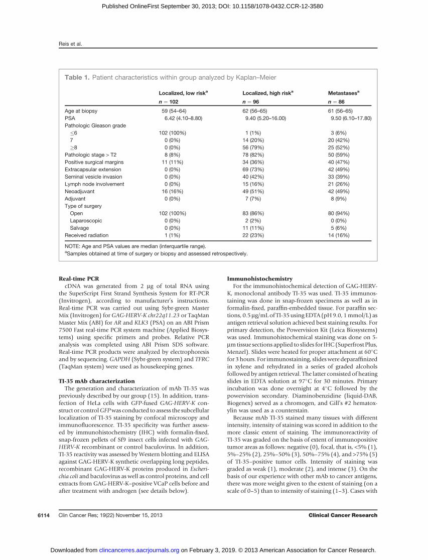
Table 1. Patient characteristics within group analyzed by Kaplan–Meier
Localized, low riska Localized, high riska Metastasesa
n ¼ 102 n ¼ 96 n ¼ 86
Age at biopsy 59 (54–64) 62 (56–65) 61 (56–65)PSA 6.42 (4.10–8.80) 9.40 (5.20–16.00) 9.50 (6.10–17.80)Pathologic Gleason grade�6 102 (100%) 1 (1%) 3 (6%)7 0 (0%) 14 (20%) 20 (42%)�8 0 (0%) 56 (79%) 25 (52%)
Pathologic stage > T2 8 (8%) 78 (82%) 50 (59%)Positive surgical margins 11 (11%) 34 (36%) 40 (47%)Extracapsular extension 0 (0%) 69 (73%) 42 (49%)Seminal vesicle invasion 0 (0%) 40 (42%) 33 (39%)Lymph node involvement 0 (0%) 15 (16%) 21 (26%)Neoadjuvant 16 (16%) 49 (51%) 42 (49%)Adjuvant 0 (0%) 7 (7%) 8 (9%)Type of surgeryOpen 102 (100%) 83 (86%) 80 (94%)Laparoscopic 0 (0%) 2 (2%) 0 (0%)Salvage 0 (0%) 11 (11%) 5 (6%)
Received radiation 1 (1%) 22 (23%) 14 (16%)
NOTE: Age and PSA values are median (interquartile range).aSamples obtained at time of surgery or biopsy and assessed retrospectively.
Reis et al.
Clin Cancer Res; 19(22) November 15, 2013 Clinical Cancer Research6114
on February 3, 2019. © 2013 American Association for Cancer Research. clincancerres.aacrjournals.org Downloaded from
Published OnlineFirst September 30, 2013; DOI: 10.1158/1078-0432.CCR-12-3580
moderate (2) and intense (3) staining in more than 50% ofthe tumor (4 and 5) were considered "strong" expressers;"moderate/weak" expressers were cases withweak (1) stain-ing irrespective of extent and cases with staining in less than50% of the tumor (1–3) irrespective of intensity.
Induction of GAG-HERV-K ch22q11.23 expression by4,5a-dihydrotestosteroneCancer cell lines were submitted to 4,5a-dihydrotestos-
terone (DHT; Sigma-Aldrich) stimulation as describedbefore (31). Briefly, 2 � 105 cells were cultured in a 35-mm culture dish for 48 hours in RPMI 10% fetal calf serum(FCS) media at 37�C. The cells were then treated with 0.1,1, or 10 nmol/L of DHT for another 48 hours. Cells werethen harvested for RNA extraction and qPCR evaluationof GAG-HERV-K expression, as described previously. Theresults were expressed as fold increase relative to untreatedcells. Cells were also harvested for protein extraction andWestern blot analysis.
Luciferase assayCancer cell lines VCaPwere plated at 5�104 cells perwell
in a 96-well plate in RPMI 10% FCS medium at 37�C.Twenty-four hours later, cells were transfected withpGL3-promoter plasmid (Promega) carrying the 50LTR pro-moter region of GAG-HERV-K. Cells were incubated foran extra 8 hours and then treated for 48 hours with orwithout 1 nmol/L DHT. The empty pGL3-promoter plas-mid was used as control (mock). Cells were harvested andassayed for luminescence count according to manufac-turer’s protocol (Promega).
Methylation status of theHERV-K ch22q11.23 promoterregion (50LTR)The gDNA from cancer cell lines was extracted using
DNeasy kit (Qiagen) according to manufacturer’s instruc-tions. Themethylation status was evaluated by adapting theMethyl-Profiler DNA Methylation PCR Array System pro-tocol (SA Bioscience). Briefly, gDNA from cancer cell lineswas incubated with methylation-sensitive restrictionenzyme (Ms), methylation-dependent restriction enzyme(Md), both (Msd) or any (Mo) and the promoter regionwasamplified by real-time PCRusing specific primers (methyl-F50-ATG TGCCTTGTT AACAATGTGTTT A-30 andmethyl-R50-CTC AAC TGC AAG AGG CCT TC-30). The methylationstatus was calculated by the following formula: PMS ¼ 1 �FUM, where PMS ¼ promoter methylation status; FUM
(unmethylated fraction) ¼ 2V�(Ct_Md � Ct_Mo). The
enzyme digestion efficiency (W) was calculated as W ¼100 � [100 � (2
V�(Ct_Mo � Ct_Msd))]. Seventy per centcutoff digestion efficiency was adopted for experimentvalidation.
Induction of GAG-HERV-K ch22q11.23 expression by5-aza-20-deoxycitidine treatmentThe influence of DNA demethylation in GAG-HERV-K
expression was evaluated by 5-aza-20-deoxycitidine treat-ment. Briefly, 2�105 cells were cultured in a 35-mmculture
dish for 48 hours in RPMI 10% FCS medium at 37�C. Thecells were then treated with 0.1 or 1 mmol/L of 5-aza-20-deoxycitidine for another 48 hours. Cells were then washedwith PBS and cultured for another 48 hours at 37�C inRPMI10%FCS in the presence or absence ofDHT.Cells were thenharvested for RNA extraction and qPCR evaluation ofGAG-HERV-K expression, as described previously. The resultswere expressed as fold increase relative to untreated cells.
SDS-PAGE and Western blotCell lines were cultured in RPMI 10% FCS and subjected
to protein extraction using modified radioimmunoprecipi-tation assay (RIPA) buffer. The protein extract was submit-ted to continuous electrophoresis using NuPAGE 4%–12%Bis–Tris gel (Invitrogen), under reducing conditions. Theseparated proteins were transferred to polyvinylidenedifluoride (PVDF) membrane and incubated with blockingsolution [3% bovine serum albumin (BSA), 0.15 mol/LPBS, pH 7.4] for 1 hour at room temperature and thenincubated with human sera diluted 1:10,000 or TI-35mouse monoclonal antibody diluted 1:1,000 in dilutionbuffer (1% BSA, 0.15 mol/L PBS, pH 7.4) for 16 hours at4�C. The membranes were then washed (0.2% Tween-20,0.15 mol/L PBS, pH 7.4) and incubated with goat anti-human IgG peroxidase-conjugated diluted 1:1,000 (South-ern Biotech) or rabbit anti-mouse peroxidase-conjugated(Sigma) diluted 1:20,000 in dilution buffer, for 1 hour atroom temperature. Membranes were developed with West-ern Lighting Plus-ECL solution (Perkin Elmer Inc).
ELISAELISA assays were conducted as previously described
(32). Briefly, plasma was serially diluted from 1:100 to1:2,500, added to low-volume 96-well plates (Corning)coated with 0.25 mg/mL full-length recombinant GAG-HERV-K protein or negative control protein DHFR, or with1 mmol/L GAG-HERV-K overlapping 20mer peptides, andblocked with PBS containing 5% non-fat milk. After incu-bation, plates were washed by automatic plate washer (Bio-Tek) with PBS containing 0.2% Tween and rinsed with PBS.Total IgG bound to antigens was detected with alkalinephosphatase–conjugated anti-human IgG monoclonalantibody (Southern Biotech). Following addition of ATTO-PHOS substrate (Fisher Scientific), absorbance was mea-sured. A reciprocal titerwas extrapolated by determining theintersection of a linear trend regression with a cutoff value.The cutoff was defined as 7.5� the average of the OD valuesfrom the 3 dilutions of a negative control pool of 4 healthydonor sera. Sera with reciprocal titers >100 toGAG-HERV-Kbut without reactivity to DHFR were considered significantafter confirmation in repeat titration assays.
GAG-HERV-K_GFP fusion protein expression in HeLacells and baculovirus
GAG-HERV-K gene was amplified using specific primers(pIC113-F 50-AACTCGAGATGGGGCAAACTGAAAGTAAA-T-30 and pIC113-R 50-AAAAGCTTCTACTGCGGTGCTG-CCTG-30). The amplified product was digested with XhoI
HERV-K Immunogenicity and Poor Prognosis of Prostate Cancer
www.aacrjournals.org Clin Cancer Res; 19(22) November 15, 2013 6115
on February 3, 2019. © 2013 American Association for Cancer Research. clincancerres.aacrjournals.org Downloaded from
Published OnlineFirst September 30, 2013; DOI: 10.1158/1078-0432.CCR-12-3580
and HindIII restriction enzymes and cloned into plasmidpIC113 containing GFP and G418 resistance genes (33).The plasmid was then transfected into HeLa cells usingEffectene reagent (Qiagen) and 48 hours after the cellswere split 1:10 in a 30-cm2 plate with RPMI 10% FCSmedia containing 500 mg/mL G418 selection reagent(Gibco). Two days after selection, positive transfectedcells were fixed with 4% formaldehyde and permeabilizedwith 10% Triton-X. Cells were then stained with TI-35antibody and analyzed by confocal microscopy (LSM710Zeiss). Recombinant baculovirus preparation using GAG-HERV-K plasmid or control was conducted followingmanufacturer’s instructions (Invitrogen) and used toinfect Sf9 insect cells (MOI ¼ 1).
Protein microarrayArrays were custom-made from version 4 ProtoArrays by
Invitrogen with the addition of GAG-HERV-K recombinantprotein and used per manufacturer’s instructions. For anti-body hybridization, arrays were blocked for 1 hour andincubated for 90minutes at 4�Cwith individual sera diluted1:500 in 4 mL buffer [1 mol/L HEPES, pH 7.5, 5 mol/LNaCl, 0.1% Triton X100 (v/v), 25% glycerol (v/v), 20mmol/L reduced glutathione, 1 mmol/L dithiothreitol(DTT) 10% Synthetic Block solution (Invitrogen) in H2O]in dishes placed on a horizontal shaker (50 rpm). After 3washes [0.1%Tween20 (v/v), 10%Synthetic Block solution(Invitrogen) in PBS], total bound IgG was detected byincubation with Alexa-Fluor 647 goat anti-human IgG(Invitrogen) diluted 1:2,000 in assay buffer for 90 minutesat 4�C. Arrays were washed again and dried by centrifuga-tion. The slides were scanned at 10 mm resolution using amicroarray scanner (Axon 4200AL with GenePix Pro Soft-ware, Molecular Devices) and fluorescence detected accord-ing to themanufacturer’s instructions. Images were saved as16-bit tif files, and analysis was conducted using GenePix.Local backgrounds were subtracted automatically, and themedian net intensity in relative fluorescence units (rfu) wasreported for each spot. The results were calculated as pre-viously described (34).
Statistical analysesStatistical analyses for comparingmultiple columns were
done by one-way ANOVA and with unpaired t test for 2-column comparisons. Frequency of patients with seropos-itive versus seronegative responses was compared using 2-tailed Fisher exact test. Analyses of overall survival andrecurrence rates were conducted using the Kaplan–Meiermethod and analyzed by log-rank or by Gehan–Breslow–Wilcoxon.
ResultsExpression of GAG-HERV-K ch22q11.23 in cancer cellline and cancer tissues
We evaluated the expression pattern of GAG-HERV-Kch22q11.23 in normal and cancer tissues as well as in cancercell lines by qPCR. As this gene has no introns, RNA waspretreated with DNase to remove gDNA before reverse
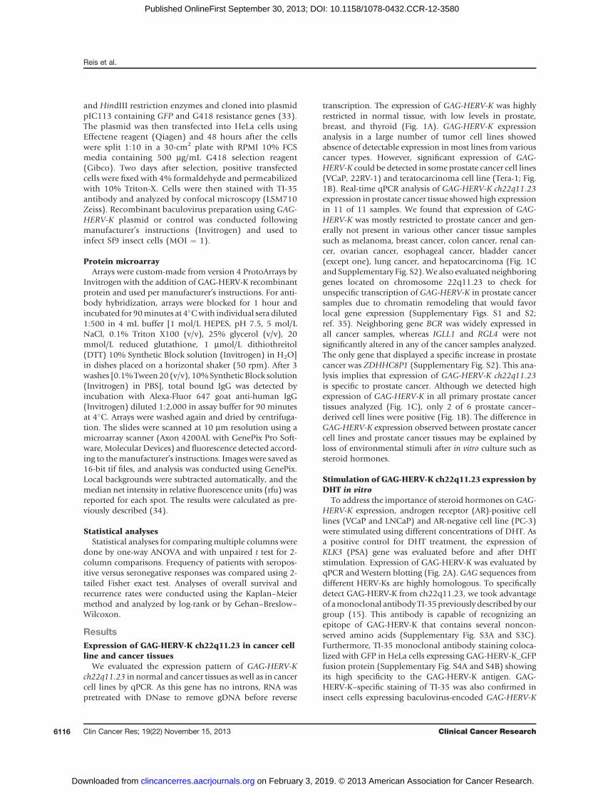
transcription. The expression of GAG-HERV-K was highlyrestricted in normal tissue, with low levels in prostate,breast, and thyroid (Fig. 1A). GAG-HERV-K expressionanalysis in a large number of tumor cell lines showedabsence of detectable expression in most lines from variouscancer types. However, significant expression of GAG-HERV-K could be detected in some prostate cancer cell lines(VCaP, 22RV-1) and teratocarcinoma cell line (Tera-1; Fig.1B). Real-time qPCR analysis of GAG-HERV-K ch22q11.23expression in prostate cancer tissue showed high expressionin 11 of 11 samples. We found that expression of GAG-HERV-K was mostly restricted to prostate cancer and gen-erally not present in various other cancer tissue samplessuch as melanoma, breast cancer, colon cancer, renal can-cer, ovarian cancer, esophageal cancer, bladder cancer(except one), lung cancer, and hepatocarcinoma (Fig. 1Cand Supplementary Fig. S2).We also evaluated neighboringgenes located on chromosome 22q11.23 to check forunspecific transcription of GAG-HERV-K in prostate cancersamples due to chromatin remodeling that would favorlocal gene expression (Supplementary Figs. S1 and S2;ref. 35). Neighboring gene BCR was widely expressed inall cancer samples, whereas IGLL1 and RGL4 were notsignificantly altered in any of the cancer samples analyzed.The only gene that displayed a specific increase in prostatecancer was ZDHHC8P1 (Supplementary Fig. S2). This ana-lysis implies that expression of GAG-HERV-K ch22q11.23is specific to prostate cancer. Although we detected highexpression of GAG-HERV-K in all primary prostate cancertissues analyzed (Fig. 1C), only 2 of 6 prostate cancer–derived cell lines were positive (Fig. 1B). The difference inGAG-HERV-K expression observed between prostate cancercell lines and prostate cancer tissues may be explained byloss of environmental stimuli after in vitro culture such assteroid hormones.
Stimulation of GAG-HERV-K ch22q11.23 expression byDHT in vitro
To address the importance of steroid hormones on GAG-HERV-K expression, androgen receptor (AR)-positive celllines (VCaP and LNCaP) and AR-negative cell line (PC-3)were stimulated using different concentrations of DHT. Asa positive control for DHT treatment, the expression ofKLK3 (PSA) gene was evaluated before and after DHTstimulation. Expression of GAG-HERV-K was evaluated byqPCR and Western blotting (Fig. 2A). GAG sequences fromdifferent HERV-Ks are highly homologous. To specificallydetect GAG-HERV-K from ch22q11.23, we took advantageof amonoclonal antibodyTI-35previously describedbyourgroup (15). This antibody is capable of recognizing anepitope of GAG-HERV-K that contains several noncon-served amino acids (Supplementary Fig. S3A and S3C).Furthermore, TI-35 monoclonal antibody staining coloca-lized with GFP in HeLa cells expressing GAG-HERV-K_GFPfusion protein (Supplementary Fig. S4A and S4B) showingits high specificity to the GAG-HERV-K antigen. GAG-HERV-K–specific staining of TI-35 was also confirmed ininsect cells expressing baculovirus-encoded GAG-HERV-K
Reis et al.
Clin Cancer Res; 19(22) November 15, 2013 Clinical Cancer Research6116
on February 3, 2019. © 2013 American Association for Cancer Research. clincancerres.aacrjournals.org Downloaded from
Published OnlineFirst September 30, 2013; DOI: 10.1158/1078-0432.CCR-12-3580
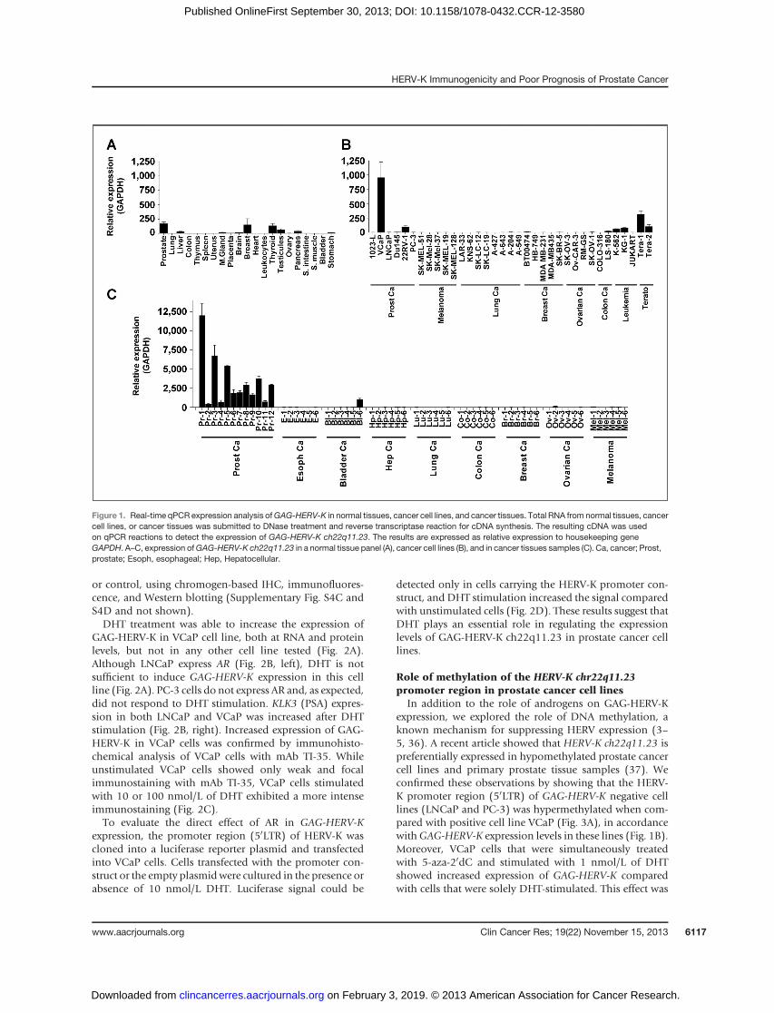
or control, using chromogen-based IHC, immunofluores-cence, and Western blotting (Supplementary Fig. S4C andS4D and not shown).DHT treatment was able to increase the expression of
GAG-HERV-K in VCaP cell line, both at RNA and proteinlevels, but not in any other cell line tested (Fig. 2A).Although LNCaP express AR (Fig. 2B, left), DHT is notsufficient to induce GAG-HERV-K expression in this cellline (Fig. 2A). PC-3 cells do not express AR and, as expected,did not respond to DHT stimulation. KLK3 (PSA) expres-sion in both LNCaP and VCaP was increased after DHTstimulation (Fig. 2B, right). Increased expression of GAG-HERV-K in VCaP cells was confirmed by immunohisto-chemical analysis of VCaP cells with mAb TI-35. Whileunstimulated VCaP cells showed only weak and focalimmunostaining with mAb TI-35, VCaP cells stimulatedwith 10 or 100 nmol/L of DHT exhibited a more intenseimmunostaining (Fig. 2C).To evaluate the direct effect of AR in GAG-HERV-K
expression, the promoter region (50LTR) of HERV-K wascloned into a luciferase reporter plasmid and transfectedinto VCaP cells. Cells transfected with the promoter con-struct or the empty plasmidwere cultured in the presence orabsence of 10 nmol/L DHT. Luciferase signal could be
detected only in cells carrying the HERV-K promoter con-struct, and DHT stimulation increased the signal comparedwith unstimulated cells (Fig. 2D). These results suggest thatDHT plays an essential role in regulating the expressionlevels of GAG-HERV-K ch22q11.23 in prostate cancer celllines.
Role of methylation of the HERV-K chr22q11.23promoter region in prostate cancer cell lines
In addition to the role of androgens on GAG-HERV-Kexpression, we explored the role of DNA methylation, aknown mechanism for suppressing HERV expression (3–5, 36). A recent article showed that HERV-K ch22q11.23 ispreferentially expressed in hypomethylated prostate cancercell lines and primary prostate tissue samples (37). Weconfirmed these observations by showing that the HERV-K promoter region (50LTR) of GAG-HERV-K negative celllines (LNCaP and PC-3) was hypermethylated when com-pared with positive cell line VCaP (Fig. 3A), in accordancewithGAG-HERV-K expression levels in these lines (Fig. 1B).Moreover, VCaP cells that were simultaneously treatedwith 5-aza-20dC and stimulated with 1 nmol/L of DHTshowed increased expression of GAG-HERV-K comparedwith cells that were solely DHT-stimulated. This effect was
Figure 1. Real-time qPCR expression analysis ofGAG-HERV-K in normal tissues, cancer cell lines, and cancer tissues. Total RNA from normal tissues, cancercell lines, or cancer tissues was submitted to DNase treatment and reverse transcriptase reaction for cDNA synthesis. The resulting cDNA was usedon qPCR reactions to detect the expression of GAG-HERV-K ch22q11.23. The results are expressed as relative expression to housekeeping geneGAPDH. A–C, expression ofGAG-HERV-K ch22q11.23 in a normal tissue panel (A), cancer cell lines (B), and in cancer tissues samples (C). Ca, cancer; Prost,prostate; Esoph, esophageal; Hep, Hepatocellular.
HERV-K Immunogenicity and Poor Prognosis of Prostate Cancer
www.aacrjournals.org Clin Cancer Res; 19(22) November 15, 2013 6117
on February 3, 2019. © 2013 American Association for Cancer Research. clincancerres.aacrjournals.org Downloaded from
Published OnlineFirst September 30, 2013; DOI: 10.1158/1078-0432.CCR-12-3580
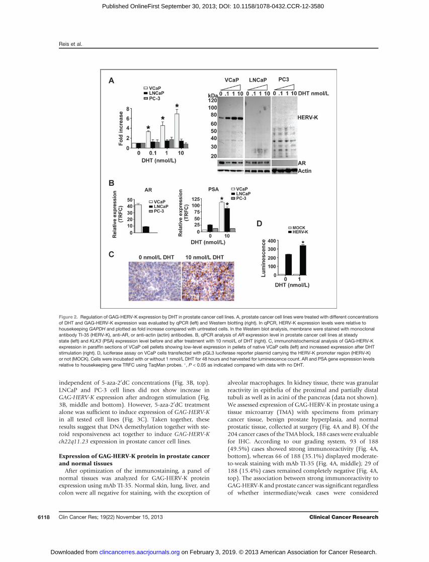
independent of 5-aza-20dC concentrations (Fig. 3B, top).LNCaP and PC-3 cell lines did not show increase inGAG-HERV-K expression after androgen stimulation (Fig.3B, middle and bottom). However, 5-aza-20dC treatmentalone was sufficient to induce expression of GAG-HERV-Kin all tested cell lines (Fig. 3C). Taken together, theseresults suggest that DNA demethylation together with ste-roid responsiveness act together to induce GAG-HERV-Kch22q11.23 expression in prostate cancer cell lines.
Expression of GAG-HERV-K protein in prostate cancerand normal tissues
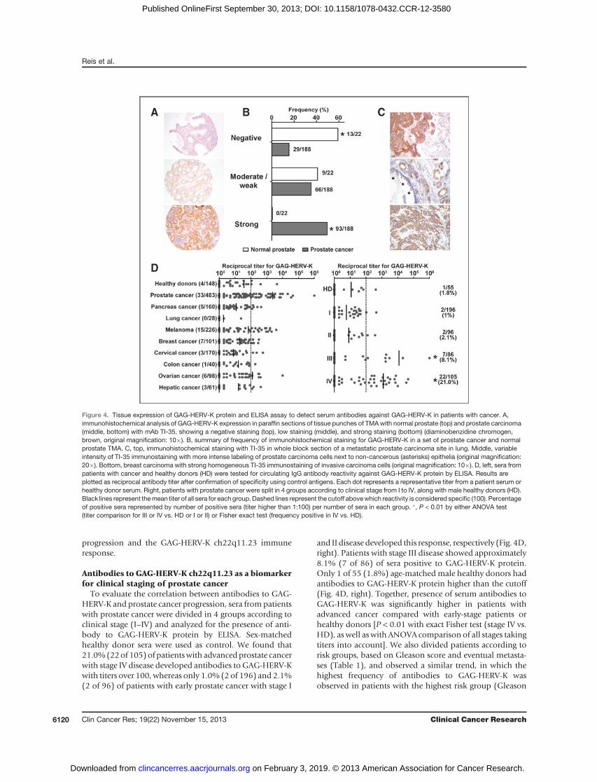
After optimization of the immunostaining, a panel ofnormal tissues was analyzed for GAG-HERV-K proteinexpression using mAb TI-35. Normal skin, lung, liver, andcolon were all negative for staining, with the exception of
alveolar macrophages. In kidney tissue, there was granularreactivity in epithelia of the proximal and partially distaltubuli as well as in acini of the pancreas (data not shown).We assessed expression of GAG-HERV-K in prostate using atissue microarray (TMA) with specimens from primarycancer tissue, benign prostate hyperplasia, and normalprostatic tissue, collected at surgery (Fig. 4A and B). Of the204 cancer cases of the TMAblock, 188 cases were evaluablefor IHC. According to our grading system, 93 of 188(49.5%) cases showed strong immunoreactivity (Fig. 4A,bottom), whereas 66 of 188 (35.1%) displayed moderate-to-weak staining with mAb TI-35 (Fig. 4A, middle); 29 of188 (15.4%) cases remained completely negative (Fig. 4A,top). The association between strong immunoreactivity toGAG-HERV-K and prostate cancer was significant regardlessof whether intermediate/weak cases were considered
Figure 2. Regulation of GAG-HERV-K expression by DHT in prostate cancer cell lines. A, prostate cancer cell lines were treated with different concentrationsof DHT and GAG-HERV-K expression was evaluated by qPCR (left) and Western blotting (right). In qPCR, HERV-K expression levels were relative tohousekeeping GAPDH and plotted as fold increase compared with untreated cells. In the Western blot analysis, membrane were stained with monoclonalantibody TI-35 (HERV-K), anti-AR, or anti-actin (actin) antibodies. B, qPCR analysis of AR expression level in prostate cancer cell lines at steadystate (left) and KLK3 (PSA) expression level before and after treatment with 10 nmol/L of DHT (right). C, immunohistochemical analysis of GAG-HERV-Kexpression in paraffin sections of VCaP cell pellets showing low-level expression in pellets of native VCaP cells (left) and increased expression after DHTstimulation (right). D, luciferase assay on VCaP cells transfected with pGL3 luciferase reporter plasmid carrying the HERV-K promoter region (HERV-K)or not (MOCK). Cells were incubated with or without 1 nmol/L DHT for 48 hours and harvested for luminescence count. AR and PSA gene expression levelsrelative to housekeeping gene TRFC using TaqMan probes. �, P < 0.05 as indicated compared with data with no DHT.
Reis et al.
Clin Cancer Res; 19(22) November 15, 2013 Clinical Cancer Research6118
on February 3, 2019. © 2013 American Association for Cancer Research. clincancerres.aacrjournals.org Downloaded from
Published OnlineFirst September 30, 2013; DOI: 10.1158/1078-0432.CCR-12-3580
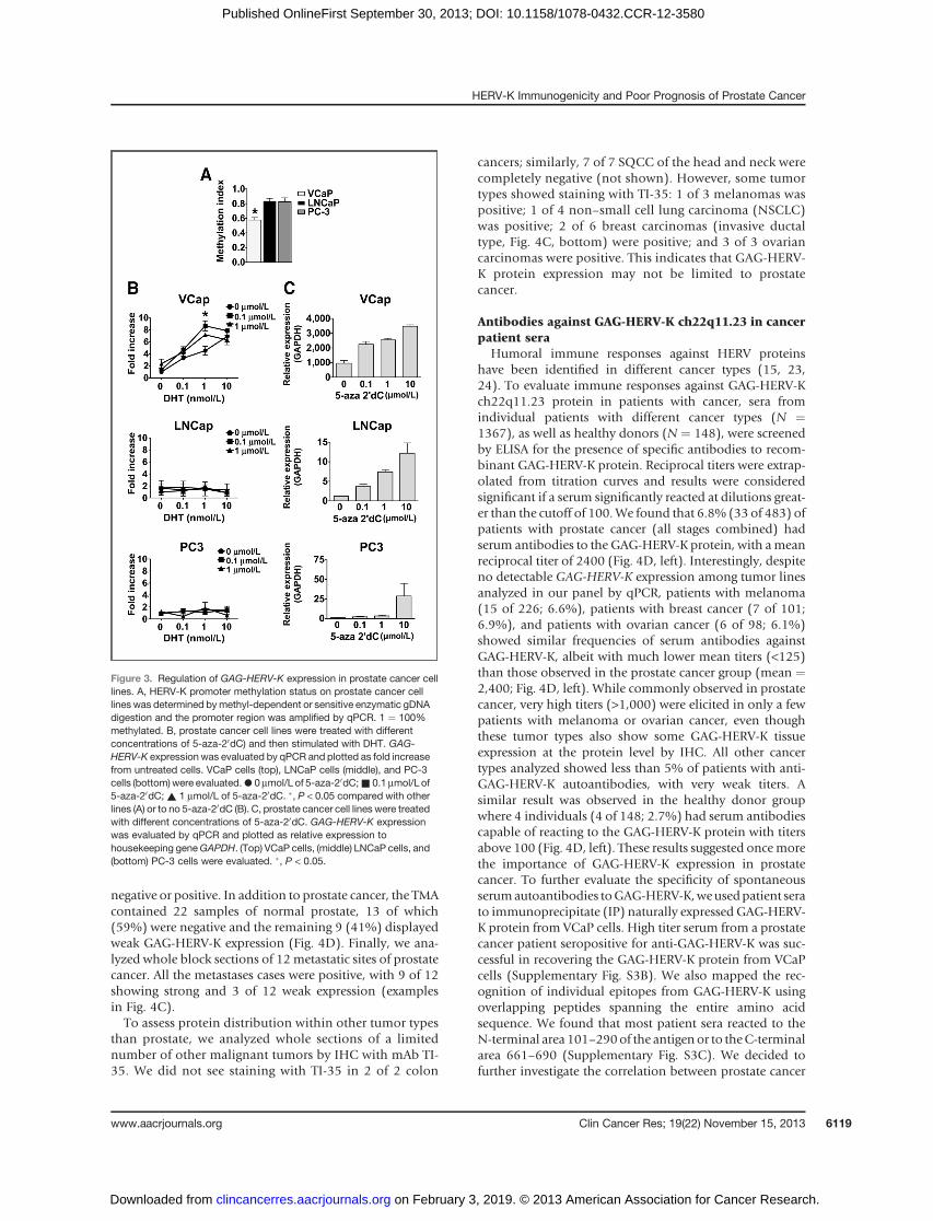
negative or positive. In addition to prostate cancer, the TMAcontained 22 samples of normal prostate, 13 of which(59%) were negative and the remaining 9 (41%) displayedweak GAG-HERV-K expression (Fig. 4D). Finally, we ana-lyzed whole block sections of 12 metastatic sites of prostatecancer. All the metastases cases were positive, with 9 of 12showing strong and 3 of 12 weak expression (examplesin Fig. 4C).To assess protein distribution within other tumor types
than prostate, we analyzed whole sections of a limitednumber of other malignant tumors by IHC with mAb TI-35. We did not see staining with TI-35 in 2 of 2 colon
cancers; similarly, 7 of 7 SQCC of the head and neck werecompletely negative (not shown). However, some tumortypes showed staining with TI-35: 1 of 3 melanomas waspositive; 1 of 4 non–small cell lung carcinoma (NSCLC)was positive; 2 of 6 breast carcinomas (invasive ductaltype, Fig. 4C, bottom) were positive; and 3 of 3 ovariancarcinomas were positive. This indicates that GAG-HERV-K protein expression may not be limited to prostatecancer.
Antibodies against GAG-HERV-K ch22q11.23 in cancerpatient sera
Humoral immune responses against HERV proteinshave been identified in different cancer types (15, 23,24). To evaluate immune responses against GAG-HERV-Kch22q11.23 protein in patients with cancer, sera fromindividual patients with different cancer types (N ¼1367), as well as healthy donors (N ¼ 148), were screenedby ELISA for the presence of specific antibodies to recom-binant GAG-HERV-K protein. Reciprocal titers were extrap-olated from titration curves and results were consideredsignificant if a serum significantly reacted at dilutions great-er than the cutoff of 100.We found that 6.8% (33 of 483) ofpatients with prostate cancer (all stages combined) hadserum antibodies to the GAG-HERV-K protein, with ameanreciprocal titer of 2400 (Fig. 4D, left). Interestingly, despiteno detectable GAG-HERV-K expression among tumor linesanalyzed in our panel by qPCR, patients with melanoma(15 of 226; 6.6%), patients with breast cancer (7 of 101;6.9%), and patients with ovarian cancer (6 of 98; 6.1%)showed similar frequencies of serum antibodies againstGAG-HERV-K, albeit with much lower mean titers (<125)than those observed in the prostate cancer group (mean ¼2,400; Fig. 4D, left). While commonly observed in prostatecancer, very high titers (>1,000) were elicited in only a fewpatients with melanoma or ovarian cancer, even thoughthese tumor types also show some GAG-HERV-K tissueexpression at the protein level by IHC. All other cancertypes analyzed showed less than 5% of patients with anti-GAG-HERV-K autoantibodies, with very weak titers. Asimilar result was observed in the healthy donor groupwhere 4 individuals (4 of 148; 2.7%) had serum antibodiescapable of reacting to the GAG-HERV-K protein with titersabove 100 (Fig. 4D, left). These results suggested oncemorethe importance of GAG-HERV-K expression in prostatecancer. To further evaluate the specificity of spontaneousserumautoantibodies toGAG-HERV-K,weusedpatient serato immunoprecipitate (IP) naturally expressed GAG-HERV-K protein from VCaP cells. High titer serum from a prostatecancer patient seropositive for anti-GAG-HERV-K was suc-cessful in recovering the GAG-HERV-K protein from VCaPcells (Supplementary Fig. S3B). We also mapped the rec-ognition of individual epitopes from GAG-HERV-K usingoverlapping peptides spanning the entire amino acidsequence. We found that most patient sera reacted to theN-terminal area 101–290of the antigen or to theC-terminalarea 661–690 (Supplementary Fig. S3C). We decided tofurther investigate the correlation between prostate cancer
Figure 3. Regulation of GAG-HERV-K expression in prostate cancer celllines. A, HERV-K promoter methylation status on prostate cancer celllines was determined bymethyl-dependent or sensitive enzymatic gDNAdigestion and the promoter region was amplified by qPCR. 1 ¼ 100%methylated. B, prostate cancer cell lines were treated with differentconcentrations of 5-aza-20dC) and then stimulated with DHT. GAG-HERV-K expression was evaluated by qPCR and plotted as fold increasefrom untreated cells. VCaP cells (top), LNCaP cells (middle), and PC-3cells (bottom)were evaluated.*0mmol/L of 5-aza-20dC;& 0.1mmol/L of5-aza-20dC;~ 1 mmol/L of 5-aza-20dC. �, P < 0.05 compared with otherlines (A) or to no 5-aza-20dC (B). C, prostate cancer cell lines were treatedwith different concentrations of 5-aza-20dC. GAG-HERV-K expressionwas evaluated by qPCR and plotted as relative expression tohousekeeping geneGAPDH. (Top) VCaP cells, (middle) LNCaP cells, and(bottom) PC-3 cells were evaluated. �, P < 0.05.
HERV-K Immunogenicity and Poor Prognosis of Prostate Cancer
www.aacrjournals.org Clin Cancer Res; 19(22) November 15, 2013 6119
on February 3, 2019. © 2013 American Association for Cancer Research. clincancerres.aacrjournals.org Downloaded from
Published OnlineFirst September 30, 2013; DOI: 10.1158/1078-0432.CCR-12-3580
progression and the GAG-HERV-K ch22q11.23 immuneresponse.
Antibodies to GAG-HERV-K ch22q11.23 as a biomarkerfor clinical staging of prostate cancer
To evaluate the correlation between antibodies to GAG-HERV-K and prostate cancer progression, sera frompatientswith prostate cancer were divided in 4 groups according toclinical stage (I–IV) and analyzed for the presence of anti-body to GAG-HERV-K protein by ELISA. Sex-matchedhealthy donor sera were used as control. We found that21.0%(22of 105) of patientswith advancedprostate cancerwith stage IV disease developed antibodies to GAG-HERV-Kwith titers over 100, whereas only 1.0% (2of 196) and 2.1%(2 of 96) of patients with early prostate cancer with stage I
and II disease developed this response, respectively (Fig. 4D,right). Patients with stage III disease showed approximately8.1% (7 of 86) of sera positive to GAG-HERV-K protein.Only 1 of 55 (1.8%) age-matched male healthy donors hadantibodies to GAG-HERV-K protein higher than the cutoff(Fig. 4D, right). Together, presence of serum antibodies toGAG-HERV-K was significantly higher in patients withadvanced cancer compared with early-stage patients orhealthy donors [P < 0.01 with exact Fisher test (stage IV vs.HD), as well as with ANOVA comparison of all stages takingtiters into account]. We also divided patients according torisk groups, based on Gleason score and eventual metasta-ses (Table 1), and observed a similar trend, in which thehighest frequency of antibodies to GAG-HERV-K wasobserved in patients with the highest risk group (Gleason
Figure 4. Tissue expression of GAG-HERV-K protein and ELISA assay to detect serum antibodies against GAG-HERV-K in patients with cancer. A,immunohistochemical analysis of GAG-HERV-K expression in paraffin sections of tissue punches of TMAwith normal prostate (top) and prostate carcinoma(middle, bottom) with mAb TI-35, showing a negative staining (top), low staining (middle), and strong staining (bottom) (diaminobenzidine chromogen,brown, original magnification: 10�). B, summary of frequency of immunohistochemical staining for GAG-HERV-K in a set of prostate cancer and normalprostate TMA. C, top, immunohistochemical staining with TI-35 in whole block section of a metastatic prostate carcinoma site in lung. Middle, variableintensity of TI-35 immunostaining with more intense labeling of prostate carcinoma cells next to non-cancerous (asterisks) epithelia (original magnification:20�). Bottom, breast carcinoma with strong homogeneous TI-35 immunostaining of invasive carcinoma cells (original magnification: 10�). D, left, sera frompatients with cancer and healthy donors (HD) were tested for circulating IgG antibody reactivity against GAG-HERV-K protein by ELISA. Results areplotted as reciprocal antibody titer after confirmation of specificity using control antigens. Each dot represents a representative titer from a patient serum orhealthy donor serum. Right, patients with prostate cancer were split in 4 groups according to clinical stage from I to IV, along with male healthy donors (HD).Black lines represent themean titer of all sera for each group. Dashed lines represent the cutoff abovewhich reactivity is considered specific (100). Percentageof positive sera represented by number of positive sera (titer higher than 1:100) per number of sera in each group. �, P < 0.01 by either ANOVA test(titer comparison for III or IV vs. HD or I or II) or Fisher exact test (frequency positive in IV vs. HD).
Reis et al.
Clin Cancer Res; 19(22) November 15, 2013 Clinical Cancer Research6120
on February 3, 2019. © 2013 American Association for Cancer Research. clincancerres.aacrjournals.org Downloaded from
Published OnlineFirst September 30, 2013; DOI: 10.1158/1078-0432.CCR-12-3580
score �7 and eventual metastases) when compared withsex-matched healthy donor sera and other risk groups(Supplementary Fig. S5A). Independently of metastases,when considering Gleason score alone, serum antibodiesto GAG-HERV-K were also significantly more frequent inpatients with a Gleason score � 8, compared with patientswith score � 6, even though not all presenting metastaticcases could be graded (Supplementary Fig. S5B). Theseresults show a clear correlation between eventual prostatecancer progression and detection of antibody to GAG-HERV-K in patients with prostate cancer at time of biopsy,highlighting the importance of HERV-K ch22q11.23 as apredictive biomarker of tumor burden and progression.
Antibodies to self-antigens in prostate cancerGAG-HERV-K Abþ groupAs the previous results unveiled, only a subset of patients
with advanced prostate cancer develops antibodies to GAG-HERV-K protein. To evaluate whether patients seropositivefor GAG-HERV-K may have overall differences in theirimmunologic response to prostate cancer and/or againstself-antigens compared with GAG-HERV-K antibody-nega-tive patients, sera from28patients with prostate cancer withstage IV disease were divided in 2 groups according topresence of antibodies to GAG-HERV-K (HERV-Kþ: n ¼28 andHERV-K�: n¼ 14) and analyzed for serum antibodyprofiling using custom-made protein microarrays (Proto-arrayswithGAG-HERV-K andother cancer antigens added).Sera fromsex- andage-matchedhealthy donorswere used ascontrol (n ¼ 14). By applying statistical analyses describedpreviously (32), we were able to identify several antigensthat were co-recognized by the HERV-Kþ group (Supple-mentary Table S1). By comparing results from ELISA andseromic profiling, we could validate the microarray results:89% of samples expected positive for GAG-HERV-K byELISAwere also reactive toGAG-HERV-K on themicroarray,missing only 3 patients with low titer samples (data notshown). NY-ESO-1 (18%; 5 of 28) and CCNB1 (18%; 5 of28) were the most frequently recognized antigens by theHERV-Kþ group. NY-ESO-1 expression has been associatedwith advanced prostate cancer (38) and CCNB1 autoanti-
body has been described in several other cancer types (39,40). On the other hand, patients in the HERV-K� grouppreferentially recognized antigens such as TMP4 (29%; 4 of14), TLE4 (29%; 4 of 14), and ARID3A (29%; 4 of 14),which have been linked to leukemias or lymphomas (41–43). Moreover, ARID3A is regulated by p53 and is a pro-posed target for cancer immunotherapy (43). This analysisshows heterogeneity of the immune response to cancerantigens among patients with prostate cancer with stage IVdisease. Understanding the correlation between prostatecancer progression and antigen-specific immune responsescould unveil individual etiologies in patients with prostatecancer.
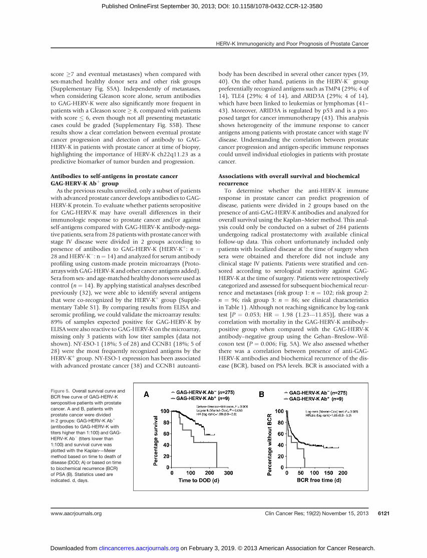
Associations with overall survival and biochemicalrecurrence
To determine whether the anti-HERV-K immuneresponse in prostate cancer can predict progression ofdisease, patients were divided in 2 groups based on thepresence of anti-GAG-HERV-K antibodies and analyzed foroverall survival using the Kaplan–Meier method. This anal-ysis could only be conducted on a subset of 284 patientsundergoing radical prostatectomy with available clinicalfollow-up data. This cohort unfortunately included onlypatients with localized disease at the time of surgery whensera were obtained and therefore did not include anyclinical stage IV patients. Patients were stratified and cen-sored according to serological reactivity against GAG-HERV-K at the time of surgery. Patients were retrospectivelycategorized and assessed for subsequent biochemical recur-rence and metastases (risk group 1: n ¼ 102; risk group 2:n ¼ 96; risk group 3: n ¼ 86; see clinical characteristicsin Table 1). Although not reaching significance by log-ranktest [P ¼ 0.053; HR ¼ 1.98 (1.23---11.85)], there was acorrelation with mortality in the GAG-HERV-K antibody–positive group when compared with the GAG-HERV-Kantibody–negative group using the Gehan–Breslow–Wil-coxon test (P ¼ 0.006; Fig. 5A). We also assessed whetherthere was a correlation between presence of anti-GAG-HERV-K antibodies and biochemical recurrence of the dis-ease (BCR), based on PSA levels. BCR is associated with a
Figure 5. Overall survival curve andBCR free curve of GAG-HERV-Kseropositive patients with prostatecancer. A and B, patients withprostate cancer were dividedin 2 groups: GAG-HERV-K Abþ
(antibodies to GAG-HERV-K withtiters higher than 1:100) and GAG-HERV-K Ab� (titers lower than1:100) and survival curve wasplotted with the Kaplan-¼Meiermethod based on time to death ofdisease (DOD; A) or based on timeto biochemical recurrence (BCR)of PSA (B). Statistics used areindicated. d, days.
HERV-K Immunogenicity and Poor Prognosis of Prostate Cancer
www.aacrjournals.org Clin Cancer Res; 19(22) November 15, 2013 6121
on February 3, 2019. © 2013 American Association for Cancer Research. clincancerres.aacrjournals.org Downloaded from
Published OnlineFirst September 30, 2013; DOI: 10.1158/1078-0432.CCR-12-3580
high likelihood of metastatic progression or prostate can-cer–specific mortality (44). Although not statistically sig-nificant, there was a trend for greater BCR in prostate cancerpatients seropositive for GAG-HERV-K (P ¼ 0.085, by log-rank) compared with the seronegative group (Fig. 5B).The association of GAG-HERV-K serum antibodies withworse survival or greater BCRwas explained by the exclusivepresence of antibody to GAG-HERV-K in the high-riskgroups and none in the lower risk group. However, therewas no significant correlation between seropositivity forGAG-HERV-K and metastasis-free survival. Together withthe observed higher frequency of antibody responses toGAG-HERV-K in stage IV patients, these results suggest thatthe presence of antibodies to GAG-HERV-K may be associ-atedwith aworse prognosis for patientswithprostate cancerand may be indicative of relapse of disease.
DiscussionIn the last decade, several studies have shown correlations
between HERV expression and human disease such asmultiple sclerosis (15, 19, 45). Moreover, immuneresponses to endogenous retrovirus derived proteins havealso been correlated to cancer (20, 21, 23, 46), but no clearclinical significance has yet been suggested. Here, we iden-tify an HERV-K GAG protein, located on chromosome22q11.23, highly expressed in prostate cancer and capableof generating a specific immune response in advanced stagesof prostate cancer.
The HERV-K located on ch22q11.23 is a complete retro-virus element, with GAG, POL, and ENV genes, flanked byan LTR sequence. The POL and ENV genes have mutationsand/or deletions that prevent the proteins from beingtranslated. In contrast, the GAG gene has a complete ORFthat translates to a protein of 715 amino acids. Restrictedexpression of GAG-HERV-K ch22q11.23 in prostate cancertissue suggests this molecule as a suitable biomarker andtarget for prostate cancer. The expression analysis of thesurrounding genes located on chromosome 22q11.23showed that expression of GAG-HERV-K is not due tounspecific region activation in prostate cancer.
The transcriptional program of androgenic signaling inthe prostate consists of thousands of gene targets whoseproducts play a role in almost all cellular functions, includ-ing cellular proliferation, survival, lipid metabolism, anddifferentiation (47). Here, we showed that androgen hor-mone DHT was able to upregulate the expression of GAG-HERV-K in VCaP cell line. Although VCaP and LNCaP areAR-positive, we were only able to stimulate GAG-HERV-Kexpression in VCaP cells. One possible explanation is thatVCaP cells have higher levels of AR than LNCaP cells, thusrendering the cells more sensitive to DHT. It is well knownthat prostate cancer cells can overexpress AR and be moresensitive to androgen hormones (48, 49), which couldcontribute to the increased expression of GAG-HERV-Kobserved in prostate cancer samples. Using PROMO, avirtual laboratory for the identification of putative tran-scription factor–binding sites in DNA sequences, we found2 potential AR-binding site at positions 328 to 336 and 496
to 504 of the promoter of GAG-HERV-K (from its 990nucleotide upstream sequence). We observed that the DNAmethylation status of the HERV-K promoter region (LTR)contributes to GAG-HERV-K expression in cancer cell lines.In accordance with our findings, HERV-K from chromo-some 22q11.23 was recently observed to be highlyexpressed in prostate cancer samples and the expressionwas dependent on LTR region demethylation (37). Never-theless, methylation is just one of several mechanisms thatimpact chromatin structure and gene expression, and it ispossible that some of the heterogeneity in GAG-HERV-Kexpression observed among cancer cell lines may relate todifferences in other possible pathways (acetylation, etc.)used to regulate the chromatin structure in the promoterregion of GAG-HERV-K. Such mechanisms might reflectdifferent subtypes or stages of prostate cancer, and someof the differences in the induction of GAG-HERV-Kobserved by androgen treatment may be related to trans-formation in cell cultures.
Recently, an interesting translocation event in prostatecancer has brought attention to the promoter region ofHERV-K. The HERV-K ch22q11.23 50LTR region was shownto be fused with ETV1 leading to an overexpression of thegene in patients with prostate cancer (17). Importantly,ETV1 overexpression in prostate cells confers invasiveness.It is not clear whether the HERV-K 50LTR region alone issufficient to activate EVT1 expression in these patients.However, our results suggest that the 50LTRpromoter regionfrom HERV-K not only is active but can also increaseexpression in response to androgen hormone stimulation,in accordance with previous findings (37). Taken together,these findings highlight the importance of HERV-K activityin prostate cancer.
Specific immune response to endogenous retrovirus com-ponents is well described in the field (20, 21, 23, 46), but noclear correlation to cancer progression has yet been deter-mined. In thiswork,we showed that patients with advancedprostate cancer have ahigher incidence of humoral immuneresponse to GAG-HERV-K protein, suggesting an increase ingene expression due to prostate cancer progression. Unfor-tunately, none of the cohorts that we had access to allowedus to ask whether there is a direct correlation betweenantigen expression and antibody response, as we couldonly collect patient serum or tumor, but not both simulta-neously. It is known that during prostate cancer progres-sion, hypomethylation of DNA occurs and this event cul-minates in the expression of endogenous retrovirus (50,51). Such increase in gene expression could lead to a specificimmune response in patients with prostate cancer with theprogression of the disease. Indeed, our result suggests thatprogression of prostate cancer is directly correlated toincrease of humoral response to GAG-HERV-K protein.
Clinical staging of prostate cancer is important in asses-sing the risk of the disease and therefore for treatmentrecommendations (26). Our results indicate that GAG-HERV-K could be a useful tool for diagnostic and/or clinicalstage biomarker in prostate cancer. Although we could notdetect significant levels of GAG-HERV-K expression in other
Reis et al.
Clin Cancer Res; 19(22) November 15, 2013 Clinical Cancer Research6122
on February 3, 2019. © 2013 American Association for Cancer Research. clincancerres.aacrjournals.org Downloaded from
Published OnlineFirst September 30, 2013; DOI: 10.1158/1078-0432.CCR-12-3580
cancer type,we detected serumantibody toGAG-HERV-K ina number of patients with melanoma, breast, and ovariancancer, albeit at lower titers compared with patients withprostate cancer. Expression of GAG-HERV-K was also seenby IHC inother tumor tissues thanprostate, although itmayhave been missed by qPCR in the small panel of cancersamples we used to assess it. Although the presence ofautoantibodies to endogenous retrovirus proteins has beencorrelated to a worse clinical prognostic in patients withmelanoma (23), we could not determine any correlationbetween presence of antibody toGAG-HERV-K ch22q11.23and clinical staging in melanoma or ovarian cancer (datanot shown).Protein microarray profiling analysis of serum from
patients with advanced prostate cancer suggests that thepresence of antibodies to GAG-HERV-K is linked to otherantigen-specific immune responses. An increased humoralimmune response to cancer testis antigen NY-ESO-1 wasobserved in the GAG-HERV-K Abþ group. The presence ofantibodies to cancer testis (CT) antigens has been associatedwith a worse prognosis in several cancer types includingprostate cancer (32, 38, 52). The evidence of a co-immuneresponse to GAG-HERV-K and NY-ESO-1 in a subset ofpatients with advanced prostate cancer suggests a potentialbiologic association between these 2 genes during prostatecancer progression and deserves to be further investigated.We believe that specific expression of GAG-HERV-K in
prostate cancer occurs by the combination of epigeneticmodification, such as demethylation, and androgen hor-mone stimulation.Whether the expression ofGAG-HERV-Kis a causal event or abiologic consequenceof prostate cancerprogression still needs to be addressed. Nevertheless, weshow here a clear correlation between the tissue-specificGAG-HERV-K protein expression and development of aspecific immune response with progression of prostatecancer. Moreover, patients who developed antibodies toGAG-HERV-K also had a lower survival rate and appeared tohave higher incidence of disease relapse (BCR). BCR iscorrelatedwith an increasedmortality andmetastatic eventsin prostate cancer and the early detection of disease relapseis crucial for an efficient treatment (44).In conclusion, we characterized a GAG protein from a
human endogenous retrovirus (HERV-K) located on chro-mosome 22q11.23 highly expressed in prostate cancer. Theexpression of GAG-HERV-K is capable of inducing humoralimmune response in patients with prostate cancer. This
immune response was correlated to advanced stages of thedisease possibly due to increase in androgen hormonestimulation and demethylation events triggered by prostatecancer progression. The presence of serum antibodies toGAG-HERV-K was also indicative of worse prognosis andhigher BCR among patient with advanced prostate cancer.Because of its restricted expression and its ability to elicit astrong humoral response, GAG-HERV-K antigen might beused as an important biomarker for prognostic purposes aswell as serving as a target for immunotherapy of advancedprostate cancer. Future directions should address whetherGAG-HERV-K serumantibodies changewith treatment suchas androgen deprivation, or during natural evolution inpatients with hormone-refractory disease.
Disclosure of Potential Conflicts of InterestH.I. Scher is a consultant/advisory boardmember of Veridex.Nopotential
conflicts of interest were disclosed by the other authors.
Authors' ContributionsConception and design: B.S. Reis, A.A. Jungbluth, Y. Obata, H.I. Scher,G. Ritter, L.J. Old, S. GnjaticDevelopment of methodology: B.S. Reis, E. Nakayama, Y. Obata, H.I.Scher, J. Melamed, S. GnjaticAcquisitionofdata (provided animals, acquired andmanagedpatients,provided facilities, etc.): B.S. Reis, A.A. Jungbluth, M. Holz, T. Ishida, B.S.Carver, H.I. Scher, P.T. Scardino, S.F. Slovin, J. Melamed, E. J€ager, G. RitterAnalysis and interpretation of data (e.g., statistical analysis, biosta-tistics, computational analysis): B.S. Reis, A.A. Jungbluth, E. Ritter, B.S.Carver, H.I. Scher, S.F. Slovin, V.E. Reuter, C. Savage, J.P. Allison, G. Ritter,S. GnjaticWriting, review, and/or revision of the manuscript: B.S. Reis, A.A.Jungbluth, Y. Obata, B.S. Carver, H.I. Scher, P.T. Scardino, S.F. Slovin,S.K. Subudhi, V.E. Reuter, E. J€ager, G. Ritter, S. GnjaticAdministrative, technical, or material support (i.e., reporting or orga-nizing data, constructing databases): D. Frosina, E. Ritter, T. Ishida, H.I.Scher, S.F. Slovin, V.E. Reuter, J. Melamed, S. GnjaticStudy supervision: H.I. Scher, G. Ritter, S. Gnjatic
AcknowledgmentsThe authors thankDr. Jianda Yuan for access to serum samples at MSKCC
andAngel Cronin for helpwith the analysis of the clinical data fromMSKCC.
Grant SupportThis study was funded by the Ludwig Institute for Cancer Research with
additional grant support from the Cancer Research Institute. B.S. Reis waspartially funded by a grant from the Brazilian agency CNPq.
The costs of publication of this article were defrayed in part by thepayment of page charges. This article must therefore be hereby markedadvertisement in accordance with 18 U.S.C. Section 1734 solely to indicatethis fact.
Received November 20, 2012; revised August 26, 2013; acceptedSeptember 9, 2013; published OnlineFirst September 30, 2013.
References1. Mayer J, Meese E. Human endogenous retroviruses in the primate
lineage and their influence on host genomes. Cytogenet Genome Res2005;110:448–56.
2. Tonjes RR, Lower R, Boller K, Denner J, Hasenmaier B, Kirsch H, et al.HERV-K: the biologically most active human endogenous retrovirusfamily. J Acquir Immune Defic Syndr Hum Retrovirol 1996;13 Suppl 1:S261–7.
3. Lavie L, Kitova M, Maldener E, Meese E, Mayer J. CpG methyl-ation directly regulates transcriptional activity of the humanendogenous retrovirus family HERV-K(HML-2). J Virol 2005;79:876–83.
4. Schulz WA, Steinhoff C, Florl AR. Methylation of endogenous humanretroelements in health and disease. Curr Top Microbiol Immunol2006;310:211–50.
5. Szpakowski S, Sun X, Lage JM, Dyer A, Rubinstein J, Kowalski D, et al.Loss of epigenetic silencing in tumors preferentially affects primate-specific retroelements. Gene 2009;448:151–67.
6. Stengel S, Fiebig U, Kurth R, Denner J. Regulation of human endog-enous retrovirus-K expression in melanomas by CpG methylation.Genes Chromosomes Cancer 2010;49:401–11.
7. Stoye JP, Moroni C, Coffin JM. Virological events leading to sponta-neous AKR thymomas. J Virol 1991;65:1273–85.
HERV-K Immunogenicity and Poor Prognosis of Prostate Cancer
www.aacrjournals.org Clin Cancer Res; 19(22) November 15, 2013 6123
on February 3, 2019. © 2013 American Association for Cancer Research. clincancerres.aacrjournals.org Downloaded from
Published OnlineFirst September 30, 2013; DOI: 10.1158/1078-0432.CCR-12-3580
8. Coffin JM, Stoye JP, Frankel WN. Genetics of endogenous murineleukemia viruses. Ann N Y Acad Sci 1989;567:39–49.
9. Obata Y, Stockert E, DeLeo AB,O'Donnell PV, Snyder HWJr, Old LJ. Acell surface antigen of the mouse related to xenotropic MuLv definedby naturally occurring antibody and monoclonal antibody. Relation toGix G(rada1), G(aksl2) systems of MuLV-related antigens. J Exp Med1981;154:659–75.
10. Stockert E, O'Donnell PV, Obata Y, Old LJ. Inhibition of AKR leuke-mogenesis by SMX-1, a dualtropic murine leukemia virus. Proc NatlAcad Sci U S A 1980;77:3720–4.
11. Stockert E,DeLeoAB,O'Donnell PV,ObataY,Old LJ.G(AKSL2): a newcell surface antigen of the mouse related to the dualtropic mink cellfocus-inducing class of murine leukemia virus detected by naturallyoccurring antibody. J Exp Med 1979;149:200–15.
12. DeLeo AB, ShikuH, Takahashi T, JohnM,Old LJ. Cell surface antigensof chemically induced sarcomas of the mouse. I. Murine leukemiavirus-related antigens and alloantigens on cultured fibroblasts andsarcoma cells: description of a unique antigen on BALB/c Meth Asarcoma. J Exp Med 1977;146:720–34.
13. Chen YT. Cancer vaccine: identification of human tumor antigens bySEREX. Cancer J 2000;6 Suppl 3:S208–17.
14. Jager D. Potential target antigens for immunotherapy identified byserological expression cloning (SEREX). Methods Mol Biol 2007;360:319–26.
15. Ishida T, Obata Y, Ohara N, Matsushita H, Sato S, Uenaka A, et al.Identification of the HERV-K gag antigen in prostate cancer by SEREXusing autologous patient serum and its immunogenicity. CancerImmun 2008;8:15.
16. Hermans KG, van der Korput HA, van Marion R, van de Wijngaart DJ,Ziel-van der Made A, Dits NF, et al. Truncated ETV1, fused to noveltissue-specific genes, and full-length ETV1 in prostate cancer. CancerRes 2008;68:7541–9.
17. Tomlins SA, Laxman B, Dhanasekaran SM, Helgeson BE, Cao X,Morris DS, et al. Distinct classes of chromosomal rearrangementscreate oncogenic ETS gene fusions in prostate cancer. Nature 2007;448:595–9.
18. Lee YN, Bieniasz PD. Reconstitution of an infectious human endog-enous retrovirus. PLoS Pathog 2007;3:e10.
19. Herbst H, Sauter M,Mueller-Lantzsch N. Expression of human endog-enous retrovirus K elements in germ cell and trophoblastic tumors. AmJ Pathol 1996;149:1727–35.
20. Boller K, Janssen O, Schuldes H, Tonjes RR, Kurth R. Characterizationof the antibody response specific for the humanendogenous retrovirusHTDV/HERV-K. J Virol 1997;71:4581–8.
21. Rakoff-Nahoum S, Kuebler PJ, Heymann JJ, E Sheehy M, Ortiz GM, SOgg G, et al. Detection of T lymphocytes specific for human endog-enous retrovirusK (HERV-K) in patientswith seminoma.AIDSResHumRetroviruses 2006;22:52–6.
22. Schiavetti F, Thonnard J, Colau D, Boon T, Coulie PG. A humanendogenous retroviral sequence encoding an antigen recognized onmelanoma by cytolytic T lymphocytes. Cancer Res 2002;62:5510–6.
23. Hahn S, Ugurel S, Hanschmann KM, Strobel H, Tondera C, Schaden-dorfD, et al. Serological response tohumanendogenous retrovirusK inmelanoma patients correlates with survival probability. AIDS Res HumRetroviruses 2008;24:717–23.
24. Wang-Johanning F, Radvanyi L, Rycaj K, Plummer JB, Yan P, SastryKJ, et al. Human endogenous retrovirus K triggers an antigen-specificimmune response in breast cancer patients. Cancer Res 2008;68:5869–77.
25. Jemal A, Siegel R, Ward E, Murray T, Xu J, Smigal C, et al. Cancerstatistics, 2006. CA Cancer J Clin 2006;56:106–30.
26. Borley N, FeneleyMR. Prostate cancer: diagnosis and staging. Asian JAndrol 2009;11:74–80.
27. Ilic D, Green S. Prostate specific antigen for detecting early prostatecancer. BMJ 2009;339:b3572.
28. Stark JR, Mucci L, Rothman KJ, Adami HO. Screening for prostatecancer remains controversial. BMJ 2009;339:b3601.
29. Bensalah K, Lotan Y, Karam JA, Shariat SF. New circulating biomar-kers for prostate cancer. Prostate Cancer Prostatic Dis 2008;11:112–20.
30. Steuber T, O'Brien MF, Lilja H. Serum markers for prostate cancer: arational approach to the literature. Eur Urol 2008;54:31–40.
31. LeeMS, Igawa T, ChenSJ, Van Bemmel D, Lin JS, Lin FF, et al. p66Shcprotein is upregulated by steroid hormones in hormone-sensitivecancer cells and in primary prostate carcinomas. Int J Cancer 2004;108:672–8.
32. Gnjatic S, Old LJ, Chen YT. Autoantibodies against cancer antigens.Methods Mol Biol 2009;520:11–9.
33. Cheeseman IM, Desai A. A combined approach for the localization andtandem affinity purification of protein complexes from metazoans. SciSTKE 2005;2005:pl1.
34. Gnjatic S, Ritter E, Buchler MW, Giese NA, Brors B, Frei C, et al.Seromic profiling of ovarian and pancreatic cancer. Proc Natl Acad SciU S A 2010;107:5088–93.
35. Cremer T, Kreth G, Koester H, Fink RH, Heintzmann R, Cremer M,et al. Chromosome territories, interchromatin domain compart-ment, and nuclear matrix: an integrated view of the functionalnuclear architecture. Crit Rev Eukaryot Gene Expr 2000;10:179–212.
36. Gimenez J, Montgiraud C, Pichon JP, Bonnaud B, Arsac M, Ruel K,et al. Custom human endogenous retroviruses dedicated microarrayidentifies self-induced HERV-W family elements reactivated in testic-ular cancer upon methylation control. Nucleic Acids Res 2010;38:2229–46.
37. Goering W, Ribarska T, Schulz WA. Selective changes of retro-element expression in human prostate cancer. Carcinogenesis2011;32:1484–92.
38. Nakada T, Noguchi Y, Satoh S, Ono T, Saika T, Kurashige T, et al. NY-ESO-1 mRNA expression and immunogenicity in advanced prostatecancer. Cancer Immun 2003;3:10.
39. HoffmannTK, Trellakis S,OkuliczK,Schuler P,Greve J,Arnolds J, et al.Cyclin B1 expression and p53 status in squamous cell carcinomas ofthe head and neck. Anticancer Res 2011;31:3151–7.
40. Covini G, Chan EK, Nishioka M, Morshed SA, Reed SI, Tan EM.Immune response to cyclin B1 in hepatocellular carcinoma. Hepatol-ogy 1997;25:75–80.
41. Cools J, Wlodarska I, Somers R, Mentens N, Pedeutour F, Maes B,et al. Identification of novel fusion partners of ALK, the anaplasticlymphoma kinase, in anaplastic large-cell lymphomaand inflammatorymyofibroblastic tumor. Genes Chromosomes Cancer 2002;34:354–62.
42. Greif PA, Eck SH, Konstandin NP, Benet-Pages A, Ksienzyk B, DufourA, et al. Identification of recurring tumor-specific somatic mutations inacute myeloid leukemia by transcriptome sequencing. Leukemia2011;25:821–7.
43. Liu G, Huang YJ, Xiao R, Wang D, Acton TB, Montelione GT. SolutionNMR structure of the ARID domain of human AT-rich interactivedomain-containing protein 3A: a human cancer protein interactionnetwork target. Proteins 2010;78:2170–5.
44. Stephenson AJ, KattanMW, Eastham JA, Dotan ZA, Bianco FJ Jr, LiljaH, et al. Defining biochemical recurrence of prostate cancer afterradical prostatectomy: a proposal for a standardized definition. J ClinOncol 2006;24:3973–8.
45. Sauter M, Schommer S, Kremmer E, Remberger K, Dolken G, Lemm I,et al. Human endogenous retrovirus K10: expression of Gag proteinand detection of antibodies in patients with seminomas. J Virol1995;69:414–21.
46. Wang-Johanning F, Rycaj K, Plummer JB, Li M, Yin B, Frerich K, et al.Immunotherapeutic potential of anti-human endogenous retrovirus-kenvelope protein antibodies in targeting breast tumors. J Natl CancerInst 2012;104:189–210.
47. Lamont KR, Tindall DJ. Androgen regulation of gene expression. AdvCancer Res 2010;107:137–62.
48. Linja MJ, Savinainen KJ, Saramaki OR, Tammela TL, Vessella RL,Visakorpi T. Amplification and overexpression of androgen receptorgene in hormone-refractory prostate cancer. Cancer Res 2001;61:3550–5.
49. Ford OH 3rd, Gregory CW, Kim D, Smitherman AB, Mohler JL. Andro-gen receptor gene amplification and protein expression in recurrentprostate cancer. J Urol 2003;170:1817–21.
Reis et al.
Clin Cancer Res; 19(22) November 15, 2013 Clinical Cancer Research6124
on February 3, 2019. © 2013 American Association for Cancer Research. clincancerres.aacrjournals.org Downloaded from
Published OnlineFirst September 30, 2013; DOI: 10.1158/1078-0432.CCR-12-3580
50. Florl AR, Steinhoff C, Muller M, Seifert HH, Hader C, Engers R, et al.Coordinate hypermethylation at specific genes in prostate carcinomaprecedes LINE-1 hypomethylation. Br J Cancer 2004;91:985–94.
51. Yegnasubramanian S, Haffner MC, Zhang Y, Gurel B, Cornish TC, WuZ, et al. DNA hypomethylation arises later in prostate cancer progres-
sion than CpG island hypermethylation and contributes to metastatictumor heterogeneity. Cancer Res 2008;68:8954–67.
52. Suyama T, Shiraishi T, Zeng Y, Yu W, Parekh N, Vessella RL, et al.Expression of cancer/testis antigens in prostate cancer is associatedwith disease progression. Prostate 2010;70:1778–87.
HERV-K Immunogenicity and Poor Prognosis of Prostate Cancer
www.aacrjournals.org Clin Cancer Res; 19(22) November 15, 2013 6125
on February 3, 2019. © 2013 American Association for Cancer Research. clincancerres.aacrjournals.org Downloaded from
Published OnlineFirst September 30, 2013; DOI: 10.1158/1078-0432.CCR-12-3580
2013;19:6112-6125. Published OnlineFirst September 30, 2013.Clin Cancer Res Bernardo Sgarbi Reis, Achim A. Jungbluth, Denise Frosina, et al. ProteinImmune Response to a Human Endogenous Retrovirus GAG Prostate Cancer Progression Correlates with Increased Humoral
Updated version
10.1158/1078-0432.CCR-12-3580doi:
Access the most recent version of this article at:
Material
Supplementary
http://clincancerres.aacrjournals.org/content/suppl/2013/09/30/1078-0432.CCR-12-3580.DC1
Access the most recent supplemental material at:
Cited articles
http://clincancerres.aacrjournals.org/content/19/22/6112.full#ref-list-1
This article cites 52 articles, 19 of which you can access for free at:
Citing articles
http://clincancerres.aacrjournals.org/content/19/22/6112.full#related-urls
This article has been cited by 3 HighWire-hosted articles. Access the articles at:
E-mail alerts related to this article or journal.Sign up to receive free email-alerts
Subscriptions
Reprints and
To order reprints of this article or to subscribe to the journal, contact the AACR Publications Department at
Permissions
Rightslink site. Click on "Request Permissions" which will take you to the Copyright Clearance Center's (CCC)
.http://clincancerres.aacrjournals.org/content/19/22/6112To request permission to re-use all or part of this article, use this link
on February 3, 2019. © 2013 American Association for Cancer Research. clincancerres.aacrjournals.org Downloaded from
Published OnlineFirst September 30, 2013; DOI: 10.1158/1078-0432.CCR-12-3580