Embed Size (px)
Citation preview
Prostaglandin E2 Regulates Renal Cell Carcinoma Invasionthrough the EP4 Receptor-Rap GTPase Signal TransductionPathway*
Received for publication, September 21, 2010, and in revised form, August 2, 2011 Published, JBC Papers in Press, August 10, 2011, DOI 10.1074/jbc.M110.187344
Juanjuan Wu‡, Yushan Zhang§, Nicole Frilot‡, Jae I. Kim§, Wan-Ju Kim§, and Yehia Daaka‡§¶1
From the ‡Department of Pathology, Georgia Health Sciences University, Augusta, Georgia 30912 and the Departments of§Urology and Prostate Disease Center and ¶Anatomy and Cell Biology, University of Florida College of Medicine,Gainesville, Florida 32610
Prognosis for patients with early stage kidney cancer hasimproved, but the treatment options for patients with locallyadvanced disease andmetastasis remain few.Understanding themolecular mechanisms that regulate invasion and metastasisis critical for developing successful therapies to treat thesepatients. Proinflammatory prostaglandin E2 plays an importantrole in cancer initiation and progression via activation of cog-nate EP receptors that belong to the superfamily of G protein-coupled receptors. Here we report that prostaglandin E2 pro-motes renal cancer cell invasion through a signal transductionpathway that encompasses EP4 and small GTPase Rap. Inacti-vation of Rap signaling with Rap1GAP, like inhibition of EP4signaling with ligand antagonist or knockdown with shRNA,reduces the kidney cancer cell invasion.Human kidney cells evi-dence increased EP4 and decreased Rap1GAP expression levelsin the malignant compared with benign samples. These resultssupport the idea that targeted inhibition of EP4 signaling andrestoration of Rap1GAP expression constitute a new strategy tocontrol kidney cancer progression.
Kidney cancer is increasingly common, and the number ofpatients dying of this disease has also increased over the pastseveral years (1). Kidney cancer accounts for roughly 4% of allcancer cases, and it occurs more often inmales than in females.Common causes of kidney cancer have largely been attributedto genetic (e.g. inactivation of the von Hippel-Lindau geneproduct), environmental, and behavioral (e.g. smoking and diet)factors. Renal cell carcinoma (RCC)2 accounts for the majority(90%) of kidney cancer cases (2) and mostly originates in prox-imal renal tubules. RCC comprises several distinct histologicalsubtypes that are traditionally classified by light microscopy,including clear cell (80% of all RCC cases), papillary, chromo-phobe, and oncocytoma (3). Although RCC presents as a local-ized disease in the majority of cases, more than one-third of
patients exhibit metastatic lesions (4) that produce the highestmortality of any adult urological cancer (5).Kidney tumor mass is sustained by the release of circulating
and locally produced factors acting through cellular receptorsthat can switch the susceptible quiescent kidney cells to an acti-vated state. Prostaglandins are naturally occurring lipids thatare produced from cyclooxygenase (COX)-mediated metabo-lism of arachidonic acid (6, 7). The prostaglandins are abun-dantly expressed in the kidney and act locally to regulate renalfunction and systemic blood pressure (6). Notably, COX-2expression is up-regulated in many human malignancies,including RCC (8–10) and correlates with poor prognosis.Clinical practice-based outcomes reveal a drawback to the safeuse of drugs that target COX-2 as a result of increasing renaland cardiovascular risk (11, 12).PGE2 is the predominant prostaglandin in the kidney, and a
large body of evidence demonstrates that its levels are increasedin patients diagnosed with cancer (13–16). PGE2 exerts itseffects on target cells through activation of cognate receptorsnamed EP1, EP2, EP3, and EP4 (6, 17) that belong to the super-family of G protein-coupled receptors. Stimulation with PGE2activates at least three distinct subfamilies of heterotrimeric Gproteins, namely Gq, Gi, andGs. Inmost cells, PGE2-bound EP1couples to Gq and induces the activation of protein kinase Cthrough the releaseofCa2� ions from intracellular stores (18). EP3couples predominantly toGi and inhibits the accumulation of sec-ondmessenger cAMP (19, 20). Stimulated EP2 and EP4 couple toGs leading to synthesis of cAMP and activation of the cAMP-de-pendent protein kinase (PKA) (21, 22). Hence, PGE2 transducesthe multiple receptor-specific signaling events in target cells.Emerging evidence implicates prostaglandins in cancer cell
migration (23, 24). In this study, we explored the possibleinvolvement of EPs and their downstream effectors in kidneycancer cell invasion. The results show that PGE2 promotes kid-ney cancer cell invasion through activation of EP4 and smallGTPase Rap proteins. Interference of EP4-to-Rap signalingwith complementary pharmacologic and biologic reagentsreduces invasion of the kidney cancer cells. EP4 protein expres-sion is increased in malignant compared with benign human kid-ney cells and inversely correlates with Rap1GAP protein expres-sion.These studies identify EP4 andRap1GAPproteins as positiveand negative regulators, respectively, of kidney cancer cell inva-sion, and suggest their utility as prognosticmarkers and therapeu-tic targets to limit patient morbidity andmortality.
* This work was supported, in whole or in part, by National Institutes of HealthPublic Health Service Grant CA129155 from the NCI.
1 Supported by the Georgia Cancer Coalition Distinguished Cancer Scholarfund. To whom correspondence should be addressed: CGRC, Rm. 462,2033 Mowry Rd., Gainesville, FL 32610. Tel.: 352-273-8112; Fax: 352-273-8335; E-mail: [email protected].
2 The abbreviations used are: RCC, renal cell carcinoma; PGE, prostaglandin E;PKA, protein kinase A; EP, prostaglandin E receptors; RBD, Ras-bindingdomain; GAP, GTPase activating protein; EV, empty vector; VASP, Vasodi-lator-stimulated phosphoprotein.
THE JOURNAL OF BIOLOGICAL CHEMISTRY VOL. 286, NO. 39, pp. 33954 –33962, September 30, 2011© 2011 by The American Society for Biochemistry and Molecular Biology, Inc. Printed in the U.S.A.
33954 JOURNAL OF BIOLOGICAL CHEMISTRY VOLUME 286 • NUMBER 39 • SEPTEMBER 30, 2011
by guest on August 17, 2018
http://ww
w.jbc.org/
Dow
nloaded from
MATERIALS AND METHODS
Reagents—The mammalian expression plasmids were ob-tained as follows: FLAGepitope-taggedRap1GAP fromL.Quil-liam (Indiana University) and YFP-Epac1-CFP from V. Niko-laev (University of Wuerzburg). Bacterial GST-RalGDS plas-mid was obtained from J. Bos (University Medical CenterUtrecht). HA epitope-tagged GAP domains of Rap1GAP werecloned by PCR amplification using the FLAG-Rap1GAP cDNAas a template, and all cDNA clones were verified by sequencing.Antibodies were obtained as follows: anti-EP1, anti-EP2, anti-EP3, and anti-EP4 from Cayman Chemical; anti-VASP fromMillipore; anti-HA from Sigma; anti-Rap and anti-Rap1GAPfrom Santa Cruz; anti-GAPDH from Chemicon; and secondaryantibodies fromJackson ImmunoResearchLaboratories.Reagentswere obtained as follows: PGE2, AH23848, GW627368, H89, andanti-EP4 antibody (C terminus) blocking peptide fromCaymanChemical. Human embryonic kidney (HEK)-293 cells stablyoverexpressing EP4 were kind gift of J. Regan (University ofArizona).Cell Culture—Human RCC7 and Caki-1 cells were main-
tained in RPMI1640, and HEK-293 cells in DMEM supple-mented with 10% fetal bovine serum (FBS), 1% penicillin/streptomycin, and 10 mM HEPES buffer. The HK-2 cellswere maintained in keratinocyte/SFM medium with EGF,bovine pituitary extract, and 1% penicillin/streptomycin. Cellswere transfected with the appropriate cDNA and Lipo-fectamine 2000 (Invitrogen), and experiments were performed1–2 days after transfection. For stable overexpression of indi-vidual HA-tagged wild-type (GAP-WT) or mutated (GAP-K194A, GAP-K285A, and GAP-N290A) GAP domain ofRap1GAP, transfected RCC7 cells were generated and culturedin the presence of G418 (500 �g/ml). Control RCC7 cells (EV)were similarly generated using pcDNA3.1 empty vector. EP4knockdown with shRNA was peformed exactly as described inan earlier publication (24). Knockdown of endogenous Epac1and -2 was achieved with Dharmacon ON-TARGETplusSMARTpool. Cells grown on 6-well plates to 40% confluencewere transfected with 50–100 nM siRNA and 5 �l of Dharma-FECT 2 reagent. For all assays, pooled transfected cells wereequally divided to ensure the identical cell populations, and theagonist-regulated cell proliferation was determined by count-ing cells using trypan blue and hemocytometer.Reverse Transcription Quantitative PCR Assay—Total RNA
was isolated using TRIzol reagent (Invitrogen) according to themanufacturer’s instructions. Reverse transcription was con-ducted using SuperScriptTM III First-strand Synthesis System(Invitrogen). Gene levels were determined by two-step quanti-tative real-time PCR (SYBRGreen I) with gene-specific primersamplification followed by melting curve analysis. The primersdesigned for PTGER1–4 were as follows: PTGER1 (sense),5�-ACC TTC TTTGGCGGC TCT C-3�, PTGER1 (antisense),5�-GCA CGA CAC CAC CAT GAT AC-3�; PTGER2 (sense),5�-CAG TCT CCC TGC TCT TCT GC-3�, PTGER2 (anti-sense), 5�-GCA CCG AGA CAA TGA GAA GC-3�; PTGER3(sense), 5�-TCA TCG TCG TGT ACC TGT CC-3�, PTGER3(antisense), 5�-CGA TGA ACA ACG AGG AGA GC-3�; andPTGER4 (sense), 5�-TGC TCT TCT TCA GCC TGT CC-3�;
PTGER4 (antisense), 5�-AGA CTG CAA AGA GCG TGAGG-3�.GAPDH primers were (sense) 5�-GGT CATGAG TCCTTC CAC GAT-3� and (antisense) 5�-CAT GGG TGT GAACCA TGA GAA-3�. Calculation of relative mRNA of theprobed genes were carried out by taking the normalized thresh-old cycle value (�Ct) for each sample (�Ct � Ct of Queriedgene � Ct GAPDH) � the �Ct of the control samples (��Ct),and converting the difference to fold-expression using the fol-lowing equation: fold � 2∧(���Ct). Experiments were re-peated at least three times, each in triplicate.FRET Assay—Cells were transfected with a cDNA encoding
CFP-Epac1-YFP fusion protein and seeded onto glass cover-slips. After 48 h, cells were inspected with a SP2 scanning con-focal microscope (Leica) and imaged using a 63 � 1.4 NA oilimmersion objective. Cells were excited at 425 nm, and emis-sion of CFP and YFP was detected simultaneously through470 � 20- and 530 � 25-nm bandpass filters. Cells were con-tinuously perfused with a solution (150mMNaCl, 5mMKCl, 10mM HEPES, 10 mM glucose, 1.5 mM CaCl2, and 2.5 mM MgCl2)containing PGE2 in the absence or presence ofAH23848 (5�M).Solution changes were made by using a multiport attachmentand perfusion capillary positioned directly in front of the cellunder study. Exposure time was 200–500 s and images weretaken every 10–30 s. Fluorescent images were background cor-rected by subtracting autofluorescence intensities of back-ground with no cells. Data were digitized and the ratio of YFP/CFP emissions were calculated at different time points andnormalized by dividing all ratios by the emission ratio justbefore stimulation.Western Blot Analysis—Appropriately treated cells were
lysed in RIPA buffer (150 mM NaCl, 50 mM Tris-HCl, pH 8, 1mM EDTA, 0.25% (w/v) sodium deoxycholate, 0.1% (v/v) Non-idet P-40, 1 mM NaF, 1 mM sodium pyrophosphate, 100 �M
Na3VO4, 1 mM phenylmethylsulfonyl fluoride, 10 �g/ml of leu-peptin, 10 �g/ml of aprotinin, and 0.7 �g/ml of pepstatin) andanalyzed by SDS-PAGE andWestern blotting. All primary anti-bodies were used at a dilution of 1:1,000 except Rap1GAP,which was used at a dilution of 1:500. For anti-EP4 antibodyneutralization, blocking peptides (that target the C terminus ofEP4) were pre-mixed with the anti-EP4 antibodies in blockingsolution for 1 h prior to incubation with filter. Peroxidase-con-jugated secondary antibodies were used at a dilution of1:10,000, and membranes were developed using an ECL plusWestern blotting Detection System (GE Healthcare).Rap Activation Assay—Rap activation was determined
using an established pulldown method based on the specificbinding of a GST fusion protein containing the Ras-bindingdomain of RalGDS (GST-RalGDS-RBD) to the active GTP-bound form of Rap (25, 26). Briefly, whole cell lysates werecentrifuged at 14,000 � g for 10 min at 4 °C, and the super-natant was removed and assayed for protein concentration.The GST-tagged RalGDS-RBD protein was expressed inBL21 cells and purified using glutathione-Sepharose 4Bbeads and an equal amount was added to 500 �g of cellextracts. Mixtures were incubated for 1 h at 4 °C, washedwith PBS, and boiled in Laemmli sample buffer. The precip-itated proteins were resolved by SDS-PAGE and analyzed byWestern blotting to detect Rap1.
Rap1GAP Suppresses RCC Invasion
SEPTEMBER 30, 2011 • VOLUME 286 • NUMBER 39 JOURNAL OF BIOLOGICAL CHEMISTRY 33955
by guest on August 17, 2018
http://ww
w.jbc.org/
Dow
nloaded from

Cell Adhesion Assay—Cells were seeded in fibronectin-coated 96-well plates at 2.0 � 104 cells/well and treated, or not,with PGE2 or FBS for 1 h at 37 °C. Wells were washed threetimeswith PBS to remove non-adherent cells, followed by addi-tion of ice-coldmethanol for 10min. Fixed cells were incubatedwith crystal violet (0.5%) solution for additional 10min at ambi-ent temperature, followed by repeated washing with water.Wells were allowed to dry and adherent stained cells were sol-ubilizedwith SDS (1%) solution. Absorbancewas determined ata wavelength of 570 nm.Cell Invasion Assay—Cells were starved overnight in
RPMI1640 containing 0.1% FBS, washed with PBS, anddetached. A total of 2 � 105 cells in 100 �l were placed into atranswell chamber containing collagen-coated filters, placed onthe feeder tray that contained RPMI1640 supplemented, or not,with PGE2 at 37 °C in a humidified atmosphere. For experimentsusingEP4antagonistAH23848 (1 and5�M)orPKA inhibitorH89(5–20�M), reagent was added to cells 10 and 30min, respectively,prior to stimulation with PGE2. Cells in the upper well wereremovedwith cotton swabs. Themembraneswere then fixedwithethanol andstainedwithcrystal violet. Invadingcellswerecountedusing a phase-contrast microscope and for each membrane, fiverandomly selected fields were counted.Statistical Analysis—The significance of agonist-induced
cellular response was analyzed by one-way analysis of variancewith Tukey post-test or two-way analysis of variance with Bon-ferroni post-test and implied at p � 0.05. For quantitative PCRexperiments, mean � S.E. for final gene pool analysis was cal-culated by propagation of error (addition). All statistical analy-ses were done, and all graphs were generated, using GraphPadPrism 5.0 software (GraphPad). The x and y labels of all pre-sented data were prepared using Adobe Illustrator CS5 suite(Adobe).
RESULTS
EP4 Is Expressed in Renal Cancer Cells—G protein-coupledreceptor signaling arrays were used to compare the geneexpression profile in renal cancer RCC7 cells to that in benignhuman kidneyHK-2 cells. The results evidenced a distinct geneexpression pattern between the malignant RCC7 and normalHK-2 cells. The RCC7 cells expressed elevated gene levels forprostaglandin E and D receptors, in comparison to the HK-2cells. Four distinct genes encode for the prostaglandin E recep-tors (PTGER) and reverse transcription followed by real timequantitative PCR analysis confirmed expression of all fourPTGER subtypes in both the RCC7 and HK-2 cells (Fig. 1A).The expression results also revealed a significant increase onlyin the PTGER4 gene level in the RCC7, compared with theHK-2 cells (Fig. 1A). The PTGER3 gene levels appeared to bemore in the RCC7 compared with HK-2 cells, but the increasedid not reach statistical significance. In comparison toGAPDHgene expression, PTGER2 gene levels were expressed most inHK-2 andRCC7 cells (Fig. 1B).Western blot analysis evidencedthe expression of bands corresponding to the reported EP1 (44kDa), EP2 (53 kDa), EP3 (52 and 62 kDa), and EP4 (52 kDa)protein masses (Fig. 1C). The results also confirmed the con-clusion that EP4 protein expression is elevated in cancer RCC7,compared with benign HK-2 cells (Fig. 1C). To ascertain spec-
ificity of the anti-EP4 antibodies, EP4 antibody blocking pep-tides were pre-mixed with the antibodies followed by Westernblot analysis. Results show that neutralization of anti-EP4 anti-bodies yielded a specific decrease in the intensity of theexpected EP4 (52 kDa) protein band (Fig. 1D, compare arrowpointing band in upper and lower panels). Increased intensity ofprotein bands that migrated with apparent molecular massespredicted for EP4 in HEK-EP4 cells that stably overexpress theEP4 gene or inHEK�HA-EP4 cells thatwere transiently trans-fected withHA-EP4 cDNA, in comparison to control HEK cells(Fig. 1D, upper panel), further suggested specificity of the EP4antibody used. Because EP4 has been implicated in transduc-tion of mitogenic signals, we determined its expression in four
FIGURE 1. Expression of EPs in kidney cancer cells. A and B, expression ofPTGER genes in HK-2 and RCC7 cells. Isolated RNA was reverse transcribedand equal amounts of cDNA was subjected to real-time PCR amplificationto establish relative expression of the PTGER1 (EP1), PTGER2 (EP2), PTGER3(EP3), or PTGER4 (EP4) genes. The gene expression was calculated asdescribed, and the data were expressed as RNA levels in RCC7 relative toHK-2 cells (A) and EP relative to GAPDH in RCC7 and HK-2 cells (B). Eachpoint represents the mean � S.E. of values obtained from three experi-ments each performed in triplicate. *, p � 0.05 versus corresponding HK-2samples. C, expression of the EP4 protein is increased in RCC7 comparedwith HK-2 cells. Equal amounts of cell lysates were analyzed by immuno-blotting using specific anti-EP1, -EP2, -EP3, -EP4, or -GAPDH antibodies.GAPDH was used to calibrate total protein loading. D, EP4 antibody block-ing peptides decrease the intensity of the detected EP4 protein band. Twosets of equal amounts of cell lysate from HEK, HEK-EP4, and HEK � HA-EP4were fractionated on SDS-PAGE and transferred to filters. After blocking,one filter was incubated with anti-EP4 antibodies alone and the other withanti-EP4 antibodies that were pre-mixed with blocking peptides. The fil-ters were analyzed by immunoblotting to detect EP4 and Actin (used asloading control) proteins. HEK-EP4, HEK293 cells stably overexpressingEP4; HEK � HA-EP4, HEK293 cells transiently transfected with HA-EP4cDNA. Arrows on left indicate EP4 protein. E, expression of EP4 protein inkidney cancer cell lines. Equal amounts of cell lysates were analyzed byimmunoblotting to detect EP4 and GAPDH (used as loading control) pro-teins. Arrows on left indicate EP4 protein. For C and E, contemporaneousshort and long exposures of the same filter are shown to provide visualevidence for the relative expression of the EP4 protein.
Rap1GAP Suppresses RCC Invasion
33956 JOURNAL OF BIOLOGICAL CHEMISTRY VOLUME 286 • NUMBER 39 • SEPTEMBER 30, 2011
by guest on August 17, 2018
http://ww
w.jbc.org/
Dow
nloaded from
additional human kidney cancer cell lines (obtained from theNCI) and found it to be expressed at high levels in aggressiveSN12C and lower in less aggressive 786-O, TK10, and Caki-1(27–30) cells (Fig. 1E). These findings support the idea that EP4expression is increased in human renal cancer compared withbenign cells.EP4 Mediates the PGE2-induced RCC7 Cell Invasion—Stim-
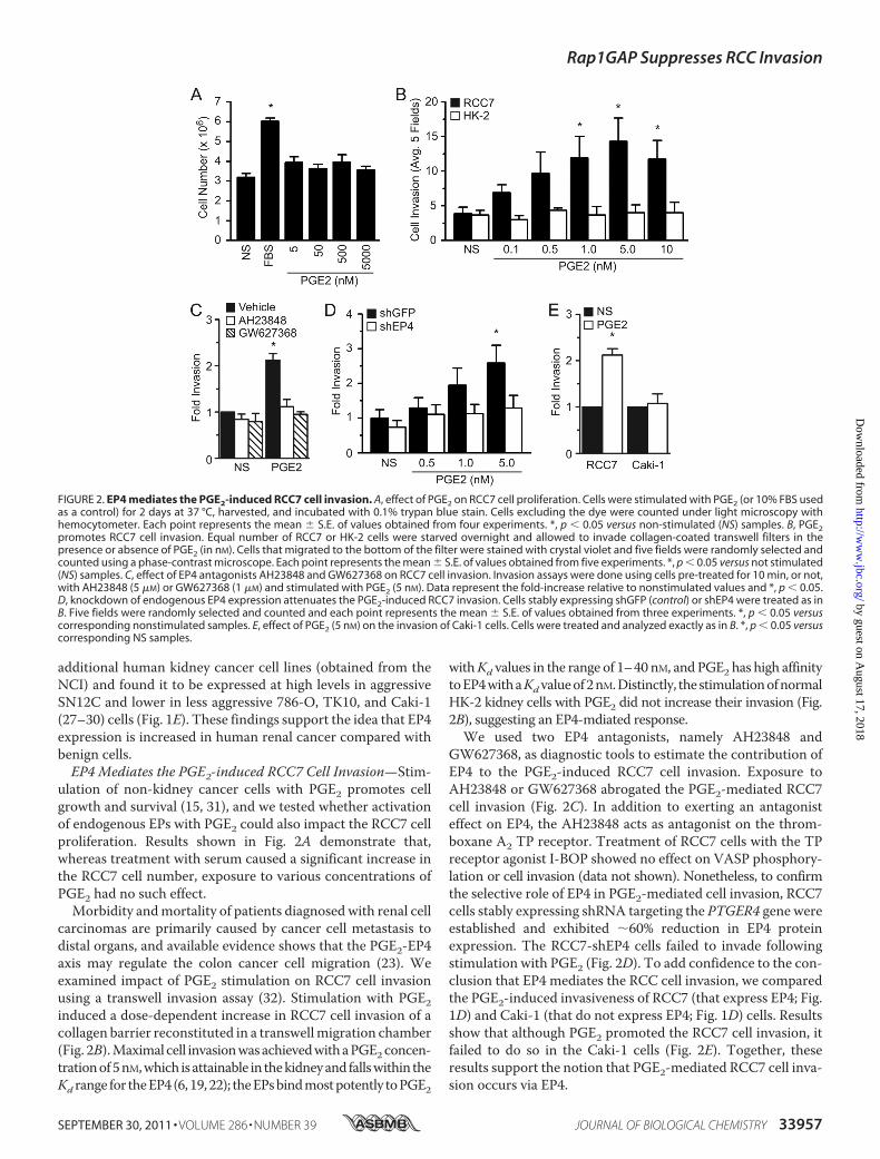
ulation of non-kidney cancer cells with PGE2 promotes cellgrowth and survival (15, 31), and we tested whether activationof endogenous EPs with PGE2 could also impact the RCC7 cellproliferation. Results shown in Fig. 2A demonstrate that,whereas treatment with serum caused a significant increase inthe RCC7 cell number, exposure to various concentrations ofPGE2 had no such effect.
Morbidity andmortality of patients diagnosed with renal cellcarcinomas are primarily caused by cancer cell metastasis todistal organs, and available evidence shows that the PGE2-EP4axis may regulate the colon cancer cell migration (23). Weexamined impact of PGE2 stimulation on RCC7 cell invasionusing a transwell invasion assay (32). Stimulation with PGE2induced a dose-dependent increase in RCC7 cell invasion of acollagen barrier reconstituted in a transwellmigration chamber(Fig. 2B).Maximal cell invasionwasachievedwithaPGE2concen-trationof5nM,which isattainable in thekidneyand fallswithin theKd range for theEP4(6,19,22); theEPsbindmostpotently toPGE2
withKd values in the range of 1–40 nM, andPGE2 has high affinitytoEP4withaKdvalueof2nM.Distinctly, thestimulationofnormalHK-2 kidney cells with PGE2 did not increase their invasion (Fig.2B), suggesting an EP4-mdiated response.We used two EP4 antagonists, namely AH23848 and
GW627368, as diagnostic tools to estimate the contribution ofEP4 to the PGE2-induced RCC7 cell invasion. Exposure toAH23848 or GW627368 abrogated the PGE2-mediated RCC7cell invasion (Fig. 2C). In addition to exerting an antagonisteffect on EP4, the AH23848 acts as antagonist on the throm-boxane A2 TP receptor. Treatment of RCC7 cells with the TPreceptor agonist I-BOP showed no effect on VASP phosphory-lation or cell invasion (data not shown). Nonetheless, to confirmthe selective role of EP4 in PGE2-mediated cell invasion, RCC7cells stably expressing shRNA targeting the PTGER4 gene wereestablished and exhibited 60% reduction in EP4 proteinexpression. The RCC7-shEP4 cells failed to invade followingstimulation with PGE2 (Fig. 2D). To add confidence to the con-clusion that EP4 mediates the RCC cell invasion, we comparedthe PGE2-induced invasiveness of RCC7 (that express EP4; Fig.1D) and Caki-1 (that do not express EP4; Fig. 1D) cells. Resultsshow that although PGE2 promoted the RCC7 cell invasion, itfailed to do so in the Caki-1 cells (Fig. 2E). Together, theseresults support the notion that PGE2-mediated RCC7 cell inva-sion occurs via EP4.
FIGURE 2. EP4 mediates the PGE2-induced RCC7 cell invasion. A, effect of PGE2 on RCC7 cell proliferation. Cells were stimulated with PGE2 (or 10% FBS usedas a control) for 2 days at 37 °C, harvested, and incubated with 0.1% trypan blue stain. Cells excluding the dye were counted under light microscopy withhemocytometer. Each point represents the mean � S.E. of values obtained from four experiments. *, p � 0.05 versus non-stimulated (NS) samples. B, PGE2promotes RCC7 cell invasion. Equal number of RCC7 or HK-2 cells were starved overnight and allowed to invade collagen-coated transwell filters in thepresence or absence of PGE2 (in nM). Cells that migrated to the bottom of the filter were stained with crystal violet and five fields were randomly selected andcounted using a phase-contrast microscope. Each point represents the mean � S.E. of values obtained from five experiments. *, p � 0.05 versus not stimulated(NS) samples. C, effect of EP4 antagonists AH23848 and GW627368 on RCC7 cell invasion. Invasion assays were done using cells pre-treated for 10 min, or not,with AH23848 (5 �M) or GW627368 (1 �M) and stimulated with PGE2 (5 nM). Data represent the fold-increase relative to nonstimulated values and *, p � 0.05.D, knockdown of endogenous EP4 expression attenuates the PGE2-induced RCC7 invasion. Cells stably expressing shGFP (control) or shEP4 were treated as inB. Five fields were randomly selected and counted and each point represents the mean � S.E. of values obtained from three experiments. *, p � 0.05 versuscorresponding nonstimulated samples. E, effect of PGE2 (5 nM) on the invasion of Caki-1 cells. Cells were treated and analyzed exactly as in B. *, p � 0.05 versuscorresponding NS samples.
Rap1GAP Suppresses RCC Invasion
SEPTEMBER 30, 2011 • VOLUME 286 • NUMBER 39 JOURNAL OF BIOLOGICAL CHEMISTRY 33957
by guest on August 17, 2018
http://ww
w.jbc.org/
Dow
nloaded from
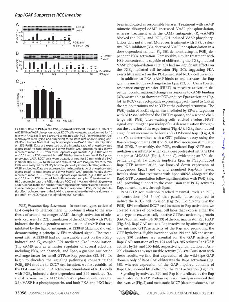
PGE2 Promotes Rap Activation—Inmost cell types, activatedEP4 couples to heterotrimeric Gs proteins leading to the syn-thesis of second messenger cAMP through activation of ade-nylyl cyclases (19, 22). Stimulation of the RCC7 cells with PGE2induced the dose-dependent accumulation of cAMP that wasinhibited by the ligand antagonist AH23848 (data not shown),demonstrating a principally EP4-mediated signal. The treat-ment with AH23848 had no measurable effect on the PGE2-induced and Gq-coupled EP1-mediated Ca2� mobilization.The cAMP acts as a master regulator of several effectors,including PKA, ion channels, and Epac, a guanine nucleotideexchange factor for small GTPase Rap proteins (33, 34). Tobegin to elucidate the signaling pathway(s) connecting thePGE2-EP4 module to RCC7 cell invasion, we first establishedthe PGE2-mediated PKA activation. Stimulation of RCC7 cellswith PGE2 induced a dose-dependent and EP4-mediated (i.e.signal is sensitive to AH23848) VASP phosphorylation (Fig.3A). VASP is a phosphoprotein, and both PKA and PKG have
been implicated as responsible kinases. Treatment with cAMPmimetic dibutyryl-cAMP increased VASP phosphorylation,whereas treatment with the cAMP antagonist (Rp)-cAMPSblocked the PGE2- and PGE1-OH-induced VASP phosphory-lation (data not shown).Moreover, treatmentwithH89, a selec-tive PKA inhibitor (35), decreased VASP phosphorylation in adose-dependentmanner (Fig. 3B), demonstrating the PGE2-de-pendent PKA activation. Remarkably, similar treatment withH89 concentrations capable of obliterating the PGE2-inducedVASP phosphorylation (Fig. 3B) had no significant effects onthe PGE2-mediated cell invasion (Fig. 3C), suggesting PKAexerts little impact on the PGE2-mediated RCC7 cell invasion.
In addition to PKA, cAMP binds to and activates the Rapguanine nucleotide exchange factor Epac (33, 36). Using Forsterresonance energy transfer (FRET) to measure activation-de-pendent conformational changes in response to cAMP binding(37), we are able to show that PGE2 induces Epac activation (Fig.4A) in RCC7 cells ectopically expressing Epac1 (fused to CFP atthe amino terminus and to YFP at the carboxyl terminus). ThePGE2-induced FRET signal was mediated by EP4; antagonismwithAH23848 inhibited the FRET response, and a second chal-lenge with PGE2 (after washing cells) elicited a robust FRETsignal, excluding the possibility of EP4 desensitization through-out the duration of the experiment (Fig. 4A). PGE2 also induceda significant increase in the levels of GTP-bound Rap1 (Fig. 4, Band C), as determined with a GST pulldown assay using theRas-binding domain (RBD) of Ral GDP-dissociation stimulator(Ral-GDS). Remarkably, the PGE2-mediated Rap1�GTP accu-mulationwas inhibitedwhen cells were pretreatedwith the EP4antagonist AH23848 (Fig. 4, B and C), evidencing an EP4-de-pendent signal. To directly implicate Epac in PGE2-inducedRap1�GTP accumulation, we knocked down expression ofendogenous Epac1 and -2 and examined Rap�GTP levels.Results show that treatment with Epac siRNA abrogated theRap1�GTP accumulation following stimulation with PGE2 (Fig.4D), providing support to the conclusion that PGE2 activatesRap, at least in part, through Epac.Rap1�GTP accumulation reached maximal levels at PGE2
concentrations (0.5–5 nM) that parallel doses required toinduce the RCC7 cell invasion (Fig. 2B). To directly link thePGE2-EP4-mediated RCC7 cell invasion to Rap activation, wecreated a series of polyclonal cell lines that express either thewild-type or enzymatically inactive GTPase activating protein(GAP) domain only (34, 38, 39) of the Rap inactivator Rap1GAP(Fig. 5A). Rap1GAP acts as a Rap inactivator by stimulating thelow intrinsic GTPase activity of the Rap and promoting theGTP hydrolysis. Highly invariant lysine 194 and 285 and aspar-agine 290 residues are essential for the GAP activity ofRap1GAP: mutation of Lys-194 and Lys-285 reduces Rap1GAPactivity by 25- and 100-fold, respectively, andmutation of Asn-290 eliminates anymeasurable activity (38, 39). Consistent withthese results, we find that expression of the wild-type GAPdomain only of Rap1GAP obliterates the Rap1 activation (Fig.5B), whereas expression of the GAP-impaired domains ofRap1GAP showed little effect on the Rap1 activation (Fig. 5B).Signaling by activated EP4 and Rap is interdicted by the Rap
inactivator Rap1GAP. Protein expression analysis revealed thatthe invasive (Fig. 2) andmetastatic RCC7 (data not shown), like
FIGURE 3. Role of PKA in the PGE2-induced RCC7 cell invasion. A, effect ofAH23848 on VASP phosphorylation. RCC7 cells were pretreated, or not, for 10min with AH23848 (1 �M, 5 �M) and stimulated with PGE2 (in nM) for 5 min. Cellmonolayers were lysed and subjected to Western blot analysis using anti-VASP antibodies. Note that the phosphorylation of VASP retards its migrationon SDS-PAGE. Data are expressed as the intensity ratio of phosphorylated(upper band) to total (upper and lower bands) VASP protein. Values shownrepresent mean � S.E. from three separate experiments. *, p � 0.05 and **,p � 0.01 versus PGE2-treated, but AH23848-untreated samples. B, PKA phos-phorylates VASP. RCC7 cells were treated, or not, for 30 min with the PKAinhibitor H89 (0.1 �M to 10 �M) and stimulated with PGE2 (in nM) for 5 min.Cells were analyzed for VASP phosphorylation by immunoblotting with anti-VASP antibodies. Data are expressed as the intensity ratio of phosphorylated(upper band) to total (upper and lower bands) VASP protein. Values shownrepresent mean � S.E. from three separate experiments. *, p � 0.05 and **,p � 0.01 versus PGE2-treated, but H89-untreated samples. C, treatment withH89 does not impact the PGE2-induced RCC7 cell invasion. H89 (5–20 �M) wasadded, or not, to the top and bottom compartments and cells were allowed toinvade collagen-coated transwell filters in response to PGE2 (5 nM) stimula-tion. Each point represents the fold-increase relative to the vehicle alone sam-ple and *, p � 0.05 versus not stimulated (NS) samples.
Rap1GAP Suppresses RCC Invasion
33958 JOURNAL OF BIOLOGICAL CHEMISTRY VOLUME 286 • NUMBER 39 • SEPTEMBER 30, 2011
by guest on August 17, 2018
http://ww
w.jbc.org/
Dow
nloaded from
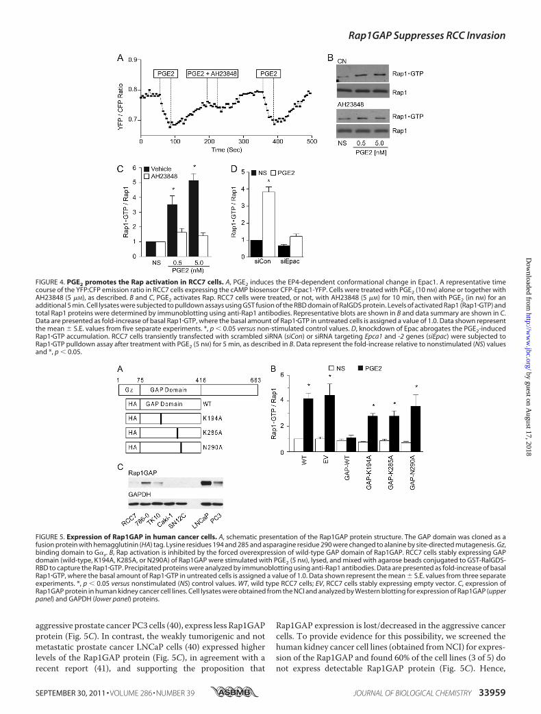
aggressive prostate cancer PC3 cells (40), express less Rap1GAPprotein (Fig. 5C). In contrast, the weakly tumorigenic and notmetastatic prostate cancer LNCaP cells (40) expressed higherlevels of the Rap1GAP protein (Fig. 5C), in agreement with arecent report (41), and supporting the proposition that
Rap1GAP expression is lost/decreased in the aggressive cancercells. To provide evidence for this possibility, we screened thehuman kidney cancer cell lines (obtained fromNCI) for expres-sion of the Rap1GAP and found 60% of the cell lines (3 of 5) donot express detectable Rap1GAP protein (Fig. 5C). Hence,
FIGURE 4. PGE2 promotes the Rap activation in RCC7 cells. A, PGE2 induces the EP4-dependent conformational change in Epac1. A representative timecourse of the YFP:CFP emission ratio in RCC7 cells expressing the cAMP biosensor CFP-Epac1-YFP. Cells were treated with PGE2 (10 nM) alone or together withAH23848 (5 �M), as described. B and C, PGE2 activates Rap. RCC7 cells were treated, or not, with AH23848 (5 �M) for 10 min, then with PGE2 (in nM) for anadditional 5 min. Cell lysates were subjected to pulldown assays using GST fusion of the RBD domain of RalGDS protein. Levels of activated Rap1 (Rap1�GTP) andtotal Rap1 proteins were determined by immunoblotting using anti-Rap1 antibodies. Representative blots are shown in B and data summary are shown in C.Data are presented as fold-increase of basal Rap1�GTP, where the basal amount of Rap1�GTP in untreated cells is assigned a value of 1.0. Data shown representthe mean � S.E. values from five separate experiments. *, p � 0.05 versus non-stimulated control values. D, knockdown of Epac abrogates the PGE2-inducedRap1�GTP accumulation. RCC7 cells transiently transfected with scrambled siRNA (siCon) or siRNA targeting Epca1 and -2 genes (siEpac) were subjected toRap1�GTP pulldown assay after treatment with PGE2 (5 nM) for 5 min, as described in B. Data represent the fold-increase relative to nonstimulated (NS) valuesand *, p � 0.05.
FIGURE 5. Expression of Rap1GAP in human cancer cells. A, schematic presentation of the Rap1GAP protein structure. The GAP domain was cloned as afusion protein with hemagglutinin (HA) tag. Lysine residues 194 and 285 and asparagine residue 290 were changed to alanine by site-directed mutagenesis. Gz,binding domain to G�z. B, Rap activation is inhibited by the forced overexpression of wild-type GAP domain of Rap1GAP. RCC7 cells stably expressing GAPdomain (wild-type, K194A, K285A, or N290A) of Rap1GAP were stimulated with PGE2 (5 nM), lysed, and mixed with agarose beads conjugated to GST-RalGDS-RBD to capture the Rap1�GTP. Precipitated proteins were analyzed by immunoblotting using anti-Rap1 antibodies. Data are presented as fold-increase of basalRap1�GTP, where the basal amount of Rap1�GTP in untreated cells is assigned a value of 1.0. Data shown represent the mean � S.E. values from three separateexperiments. *, p � 0.05 versus nonstimulated (NS) control values. WT, wild type RCC7 cells; EV, RCC7 cells stably expressing empty vector. C, expression ofRap1GAP protein in human kidney cancer cell lines. Cell lysates were obtained from the NCI and analyzed by Western blotting for expression of Rap1GAP (upperpanel) and GAPDH (lower panel) proteins.
Rap1GAP Suppresses RCC Invasion
SEPTEMBER 30, 2011 • VOLUME 286 • NUMBER 39 JOURNAL OF BIOLOGICAL CHEMISTRY 33959
by guest on August 17, 2018
http://ww
w.jbc.org/
Dow
nloaded from

Rap1GAP expression (in both prostate and kidney cancer celllines) coincides with weakmetastatic character, and absence ofRap1GAP expression correlates with the metastatic potential.Rap Mediates the PGE2-induced RCC7 Cell Invasion—Tak-
ing advantage of the reagents that we generated (i.e. the RCC7cell lines that stably express wild-type or mutated GAP do-mains of Rap1GAP), we sought to implicate Rap signaling in thePGE2-mediated RCC7 cell invasion. First, we determined theeffect of expressing the various GAP domains of Rap1GAP onthe RCC7 cell proliferation in serum. The RCC7 cell clonesexhibited similar fold-increases in their growth rates, suggest-ing Rap signaling does not impact proliferation of the RCC7cells (Fig. 6A). However, PGE2-induced cell invasion was dra-matically impaired in the RCC7 cells that express wild-typeGAP domain of Rap1GAP (GAP-WT), but not in the cells thatexpress the inactive forms of theGAPdomain of Rap1GAP (Fig.6B). Together, these results demonstrate that PGE2-mediatedRCC7 cell invasion is controlled, at least in part, by a EP43Rapsignal.
DISCUSSION
Majority of kidney cancer-related deaths result from cancermetastasis to distal organs and the prognosis of patients withmetastatic disease is poor with a median survival of 10 months(4, 42, 43). Although surgery is highly effective for the treatmentof localized low-grade RCC (43), current management optionsof patients diagnosed with locally advanced or metastatic kid-ney cancer are not curative, reinforcing the need to identifymechanisms involved in kidney cancer initiation, survival, andmetastasis as a prerequisite for discovering effective therapeu-tics. Themajor finding of this study is that PGE2 promotes clearcell RCC cell invasion by activating cognate receptor EP4,thereby leading to the activation of small GTPase Rap. Asdepicted schematically in Fig. 7A, the signaling cascade con-necting stimulated EP4 to Rap may include heterotrimeric G�sprotein, cAMP, and the Rap activator Epac. Under physiologicconditions, this proinvasive signal is counterbalanced by theRap inactivator Rap1GAP, whose expression is lost in the clearcell RCC.
PGE2 is a product of the COX-2 enzyme that is overex-pressed under pathophysiologic conditions, including kidneycancer (8–10), and is associated with poor prognosis andreduced survival time. Specific COX-2 inhibitors have beentried as therapeutics to treat cancer patients but unwanted car-diovascular and renal (11, 12) side effects limited their applica-tion and emphasized a need to identify COX-2 effectors fortherapeutic intervention. Expression of the PGE2 receptor EP4is elevated in RCC cells, and it mediates RCC7 cell invasion.Hence, the targeting of EP4 with specific ligand antagonists orneutralizing antibodies may harness the benefits to interferewith the RCC cell invasion, whereas circumventing the healthsafety concerns associated with specific COX-2 inhibitors.Activated EP4 initiates multiple signaling pathways that are
transduced by activated G�s (Fig. 7A), and our gene arrayresults demonstrated the increased expression of G�s in malig-nant RCC7 compared with benign HK-2 kidney epithelial cells(data not shown). Activatingmutations ofGNAS (referred to asgsp oncogene) have been detected in a number of endocrinemalignancies, including pituitary and thyroid adenomas (44,45) and Leydig cell tumors (46). More recent work has revealedactivating G�s mutations in kidney (47) and colorectal andbreast (48–50) cancers, and increased G�s expression in breastcancer associates with poor prognosis (49). Somatic (tumorspecific) activating mutations of the GNAS were found in asignificant portion of clear cell RCC cases, and it was reportedthat 16% of the patients (5 of 30) diagnosed with clear cell RCCexpressed mutations of the GNAS (47), rendering the enzymeconstitutively active. These results link G�s signaling to kidneycarcinogenesis and are consistent with the conclusion that acti-vated EP4 (that signals through G�s) associates with kidneycancer.Activated G�s transduces signals mainly through adenylyl
cyclases that produce cAMP (Fig. 7A). The best studied effectorof cAMP is PKA, which has been demonstrated to exert celltype- and context-dependent responses ranging from induc-tion of cell proliferation to inhibition of cell survival. Ourresults show that PKA does not significantly impact the PGE2-
FIGURE 6. Rap mediates the PGE2-induced RCC7 cell invasion. A, role of Rap signaling in RCC7 cell proliferation. Cells stably expressing wild-type or mutatedGAP domains of Rap1GAP were grown in starvation medium, or in medium containing 10% fetal bovine serum. The cells were cultured for 3 days, harvested,and stained with trypan blue. The cells excluding the dye were counted under light microscopy with a hemocytometer. Each point represents the mean � S.E.of values obtained from three experiments. *, p � 0.05 versus same cell type in starvation medium. NS, non-stimulated; WT, control wild-type RCC7 cells; EV,RCC7 cells transfected with empty vector. B, PGE2 induces the Rap-mediated RCC7 cell invasion. RCC7 cells stably expressing the GAP domain (wild-type,K194A, K285A, or N290A) of Rap1GAP were allowed to invade collagen-coated transwell filters in response to stimulation with PGE2 (5 nM). Cells that migratedto the bottom side of the filter were stained and inspected using a phase-contrast microscope. Cells in randomly selected five fields were counted and eachexperiment was repeated three times. *, p � 0.05 versus not stimulated (NS) samples. WT, wild-type RCC7 cells; EV, RCC7 cells stably expressing empty vector.
Rap1GAP Suppresses RCC Invasion
33960 JOURNAL OF BIOLOGICAL CHEMISTRY VOLUME 286 • NUMBER 39 • SEPTEMBER 30, 2011
by guest on August 17, 2018
http://ww
w.jbc.org/
Dow
nloaded from

regulatedRCC7 cell invasion. Rather, we find that Rapmediatesthe effects of EP4-induced RCC7 cell invasion. Data from sev-eral in vitro and in vivo cell-based and mouse models provideevidence that aberrant Rap1 activation, either through activat-ing mutations in its exchange factors or by inactivating muta-tions in its GAPs, contribute to several types of malignancies(51).Of note, Rapmay be activated by several guanine exchangefactors, including Epac, C3G, PDZ-GEF, RasGRP, phospho-lipase C�, and DOCK4 (52).
Althoughmany Rap1 functions are attributed to its ability toregulate integrins and cell adhesion dynamics (53, 54), theresponsible mechanisms involved in Rap-mediated cancer pro-gression remain to be elucidated. Our results show that stimu-lation with PGE2 promotes the RCC7 cell adhesion to collagen(data not shown) and fibronectin (Fig. 7B) and increases thenumber of focal adhesions (data not shown). In addition, E-cad-herin has been suggested as another Rap target, and our datashow that stimulation with PGE2 decreases the E-cadherinexpression in the RCC7 cells (Fig. 7C). Hence, PGE2-inducedRCC7 cell invasion and metastasis may proceed via the activa-tion of multiple Rap effectors that converge to activate theintegrins and focal adhesions.Rap signaling is negatively impacted by the inactivator
Rap1GAP (Fig. 7A) and previous studies suggest Rap1GAP andits family members Rap1GAPII, Spa1/SIPA1, and E6TP1 pos-sess tumor metastasis suppressor activities. Rap1GAP proteinlevels are decreased in several types of cancer, including pan-creatic adenocarcinoma (55), papillary thyroid carcinoma (56,57), colorectal carcinoma (58), and melanoma (59). The impli-cation of Rap1GAP as a tumor suppressor was largely deducedbased upon experimental results using forced overexpression offull-length Rap1GAP (60, 61). In addition to its GAP domain,the Rap1GAP protein encompasses regulatory N terminus andC terminus domains (34, 38), rendering it capable of exertingother functions, like regulation of heterotrimeric Gz protein
signaling (62) or serving as a docking site for binding partnerproteins due to phosphorylation-dependent modification of itsC terminus (63, 64). In these experiments, we employed onlythe GAP domain of Rap1GAP, allowing us to confidently con-clude that Rap signals mediate the PGE2-regulated RCC cellinvasion.In summary, we have uncovered a PGE2-controlled signaling
pathway that regulates the clear cell RCC cell invasion. Theresponsible signal pathway contains drug targetable intermedi-ates, including EP4, Epac, and Rap1GAP that may be used tobenefit patients with progressive disease. The absence ofRap1GAP together with increased EP4 protein expression inlocalized tumor cells may also serve as early markers for thedevelopment of amore aggressive, invasive phenotype. EP4 sig-naling may be interdicted with specific ligand antagonists as Gprotein-coupled receptors have proven to be viable drug tar-gets, accounting for 40–60% of all therapeutic drugs. Hence,the combined targeted inhibition of EP4 activation and rescuedexpression of Rap1GAP may improve prognosis of patientsdiagnosed with advanced kidney cancer.
Acknowledgments—We thank our colleagues for providing valuablereagents and the NCI for providing the NCI-60 cancer cell lysate. Wealso thank Drs. N. Lambert, Z. Nie, P. Arora, and T. Polascik fortechnical assistance and advice.
REFERENCES1. Jemal, A., Siegel, R., Ward, E., Hao, Y., Xu, J., and Thun, M. J. (2009) CA
Cancer J. Clin. 59, 225–2492. Biswas, S., and Eisen, T. (2009) Nat. Rev. Clin. Oncol. 6, 478–4873. Young, A. N., Dale, J., Yin-Goen, Q., Harris, W. B., Petros, J. A., Datta,
M. W., Wang, M. D., Marshall, F. F., and Amin, M. B. (2006) Urology 67,873–880
4. Schlesinger-Raab, A., Treiber, U., Zaak, D., Holzel, D., and Engel, J. (2008)Eur. J. Cancer 44, 2485–2495
5. Murai, M., and Oya, M. (2004) Curr. Opin. Urol. 14, 229–233
FIGURE 7. Rap mediates the cell adhesion. A, schematic presentation of the signal relay from activated EP4 to Rap and cell invasion. AA, arachidonic acid; AC,adenylyl cyclase. B, effect of Rap activity on cell adhesion. RCC7 cells stably expressing empty vector (EV) or the GAP domain (GAP-WT) of Rap1GAP weresuspended in starvation medium supplemented, or not, with PGE2 or FBS. Adherent cells were stained with crystal violet and dye absorbance was measuredat a wavelength of 570 nm. Data are presented as fold-increase above basal, where the basal absorbance in untreated cells is assigned a value of 1.0. Datashown represent the mean � S.E. from three separate experiments. *, p � 0.05 versus nonstimulated (NS) control values. EV and GAP-WT denote, respectively,RCC7 cells stably expressing empty vector and GAP domain of Rap1GAP. C, effect of PGE2 on E-cadherin expression. RCC7 cells were treated, or not, with PGE2for 24 h and lysates were analyzed by Western blotting for expression of E-cadherin (upper panel) and GAPDH (lower panel) proteins.
Rap1GAP Suppresses RCC Invasion
SEPTEMBER 30, 2011 • VOLUME 286 • NUMBER 39 JOURNAL OF BIOLOGICAL CHEMISTRY 33961
by guest on August 17, 2018
http://ww
w.jbc.org/
Dow
nloaded from

6. Hao, C. M., and Breyer, M. D. (2008) Annu. Rev. Physiol. 70, 357–3777. Zeldin, D. C. (2001) J. Biol. Chem. 276, 36059–360628. Chen, Q., Shinohara, N., Abe, T., Harabayashi, T., and Nonomura, K.
(2004) J. Urol. 172, 2153–21579. Mungan, M. U., Gurel, D., Canda, A. E., Tuna, B., Yorukoglu, K., and
Kirkali, Z. (2006) Eur. Urol. 50, 92–9710. Tawfik, O. W., Kramer, B., Shideler, B., Danley, M., Kimler, B. F., and
Holzbeierlein, J. (2007) Arch. Pathol. Lab. Med. 131, 261–26711. Cheng, H. F., and Harris, R. C. (2005) Curr. Pharm. Des. 11, 1795–180412. Lanas, A., and Hunt, R. (2006) Ann. Med. 38, 415–42813. Asano, T., Shoda, J., Ueda, T., Kawamoto, T., Todoroki, T., Shimonishi,
M., Tanabe, T., Sugimoto, Y., Ichikawa, A., Mutoh, M., Tanaka, N., andMiwa, M. (2002) Clin. Cancer Res. 8, 1157–1167
14. Pugh, S., and Thomas, G. A. O. (1994) Gut 35, 675–67815. Wang, D., Wang, H., Brown, J., Daikoku, T., Ning, W., Shi, Q., Richmond,
A., Strieter, R., Dey, S. K., and DuBois, R. N. (2006) J. Exp. Med. 203,941–951
16. Muller, M., Sales, K. J., Katz, A. A., and Jabbour, H. N. (2006) Endocrinol-ogy 147, 3356–3365
17. Sugimoto, Y., and Narumiya, S. (2007) J. Biol. Chem. 282, 11613–1161718. Funk, C. D., Furci, L., FitzGerald, G. A., Grygorczyk, R., Rochette, C.,
Bayne,M.A., Abramovitz,M., Adam,M., andMetters, K.M. (1993) J. Biol.Chem. 268, 26767–26772
19. Namba, T., Sugimoto, Y., Negishi, M., Irie, A., Ushikubi, F., Kakizuka, A.,Ito, S., Ichikawa, A., and Narumiya, S. (1993) Nature 365, 166–170
20. Sonnenburg,W. K., Zhu, J. H., and Smith,W. L. (1990) J. Biol. Chem. 265,8479–8483
21. Bastien, L., Sawyer, N., Grygorczyk, R., Metters, K. M., and Adam, M.(1994) J. Biol. Chem. 269, 11873–11877
22. Regan, J. W., Bailey, T. J., Pepperl, D. J., Pierce, K. L., Bogardus, A. M.,Donello, J. E., Fairbairn, C. E., Kedzie, K. M., Woodward, D. F., and Gil,D. W. (1994)Mol. Pharmacol. 46, 213–220
23. Buchanan, F. G., Gorden, D. L., Matta, P., Shi, Q., Matrisian, L. M., andDuBois, R. N. (2006) Proc. Natl. Acad. Sci. U.S.A. 103, 1492–1497
24. Kim, J. I., Lakshmikanthan, V., Frilot, N., andDaaka, Y. (2010)Mol. CancerRes. 8, 569–577
25. Herrmann, C., Horn, G., Spaargaren, M., and Wittinghofer, A. (1996)J. Biol. Chem. 271, 6794–6800
26. Franke, B., Akkerman, J. W., and Bos, J. L. (1997) EMBO J. 16, 252–25927. Bisignani, G. J., McLaughlin, P. J., Ordille, S. D., Beltz, M. S., Jarowenko,
M. V., and Zagon, I. S. (1999) J. Urol. 162, 2186–219128. Nomura, T., Huang, W. C., Seo, S., Zhau, H. E., Mimata, H., and Chung,
L. W. (2007) J. Urol. 178, 292–30029. An, Z., Jiang, P., Wang, X., Moossa, A. R., and Hoffman, R. M. (1999)Clin.
Exp. Metastasis 17, 265–27030. Kadhim, S. A., Bowlin, T. L., Waud, W. R., Angers, E. G., Bibeau, L.,
DeMuys, J. M., Bednarski, K., Cimpoia, A., and Attardo, G. (1997) CancerRes. 57, 4803–4810
31. Castellone, M. D., Teramoto, H., Williams, B. O., Druey, K. M., and Gut-kind, J. S. (2005) Science 310, 1504–1510
32. Kelly, P., Moeller, B. J., Juneja, J., Booden, M. A., Der, C. J., Daaka, Y.,Dewhirst, M.W., Fields, T. A., and Casey, P. J. (2006) Proc. Natl. Acad. Sci.U.S.A. 103, 8173–8178
33. Bos, J. L., de Rooij, J., and Reedquist, K. A. (2001) Nat. Rev. Mol. Cell Biol.2, 369–377
34. de Rooij, J., Zwartkruis, F. J., Verheijen, M. H., Cool, R. H., Nijman, S. M.,Wittinghofer, A., and Bos, J. L. (1998) Nature 396, 474–477
35. Chijiwa, T., Mishima, A., Hagiwara, M., Sano, M., Hayashi, K., Inoue, T.,Naito, K., Toshioka, T., and Hidaka, H. (1990) J. Biol. Chem. 265,5267–5272
36. Bos, J. L. (2007) Trends Biochem. Sci. 31, 680–68637. Ponsioen, B., Zhao, J., Riedl, J., Zwartkruis, F., van der Krogt, G., Zaccolo,
M., Moolenaar, W. H., Bos, J. L., and Jalink, K. (2004) EMBO Rep. 5,1176–1180
38. Brinkmann, T., Daumke, O., Herbrand, U., Kuhlmann, D., Stege, P., Ah-madian, M. R., and Wittinghofer, A. (2002) J. Biol. Chem. 277,12525–12531
39. Daumke, O., Weyand, M., Chakrabarti, P. P., Vetter, I. R., and Witting-hofer, A. (2004) Nature 429, 197–201
40. Sobel, R. E., and Sadar, M. D. (2005) J. Urol. 173, 342–35941. Bailey, C. L., Kelly, P., and Casey, P. J. (2009) Cancer Res. 69, 4962–496842. Atkins, M. B., Regan, M., andMcDermott, D. (2004) Clin. Cancer Res. 10,
6342S–6346S43. Belldegrun, A. S., Klatte, T., Shuch, B., LaRochelle, J. C., Miller, D. C., Said,
J. W., Riggs, S. B., Zomorodian, N., Kabbinavar, F. F., Dekernion, J. B., andPantuck, A. J. (2008) Cancer 113, 2457–2463
44. Landis, C. A., Masters, S. B., Spada, A., Pace, A. M., Bourne, H. R., andVallar, L. (1989) Nature 340, 692–696
45. Vallar, L., Spada, A., and Giannattasio, G. (1987) Nature 330, 566–56846. Fragoso, M. C., Latronico, A. C., Carvalho, F. M., Zerbini, M. C., Mar-
condes, J. A., Araujo, L. M., Lando, V. S., Frazzatto, E. T., Mendonca, B. B.,and Villares, S. M. (1998) J. Clin. Endocrinol. Metab. 83, 2074–2078
47. Kalfa, N., Lumbroso, S., Boulle, N., Guiter, J., Soustelle, L., Costa, P.,Chapuis, H., Baldet, P., and Sultan, C. (2006) J. Urol. 176, 891–895
48. Chin, K., DeVries, S., Fridlyand, J., Spellman, P. T., Roydasgupta, R., Kuo,W. L., Lapuk, A., Neve, R. M., Qian, Z., Ryder, T., Chen, F., Feiler, H.,Tokuyasu, T., Kingsley, C., Dairkee, S.,Meng, Z., Chew, K., Pinkel, D., Jain,A., Ljung, B.M., Esserman, L., Albertson,D.G.,Waldman, F.M., andGray,J. W. (2006) Cancer Cell 10, 529–541
49. Neve, R.M., Chin, K., Fridlyand, J., Yeh, J., Baehner, F. L., Fevr, T., Clark, L.,Bayani, N., Coppe, J. P., Tong, F., Speed, T., Spellman, P. T., DeVries, S.,Lapuk, A., Wang, N. J., Kuo, W. L., Stilwell, J. L., Pinkel, D., Albertson,D. G., Waldman, F. M., McCormick, F., Dickson, R. B., Johnson, M. D.,Lippman,M., Ethier, S., Gazdar, A., and Gray, J.W. (2006)Cancer Cell 10,515–527
50. Sjoblom, T., Jones, S., Wood, L. D., Parsons, D. W., Lin, J., Barber, T. D.,Mandelker, D., Leary, R. J., Ptak, J., Silliman, N., Szabo, S., Buckhaults, P.,Farrell, C.,Meeh, P.,Markowitz, S. D.,Willis, J., Dawson, D.,Willson, J. K.,Gazdar, A. F., Hartigan, J., Wu, L., Liu, C., Parmigiani, G., Park, B. H.,Bachman, K. E., Papadopoulos, N., Vogelstein, B., Kinzler, K. W., andVelculescu, V. E. (2006) Science 314, 268–274
51. Hattori, M., and Minato, N. (2003) J. Biochem. 134, 479–48452. Pannekoek, W. J., Kooistra, M. R., Zwartkruis, F. J., and Bos, J. L. (2009)
Biochim. Biophys. Acta 1788, 790–79653. Katagiri, K., Ohnishi, N., Kabashima, K., Iyoda, T., Takeda, N., Shinkai, Y.,
Inaba, K., and Kinashi, T. (2004) Nat. Immunol. 5, 1045–105154. Boettner, B., and Van Aelst, L. (2009) Curr. Opin. Cell Biol. 21, 684–69355. Zhang, L., Chenwei, L., Mahmood, R., van Golen, K., Greenson, J., Li, G.,
D’Silva, N. J., Li, X., Burant, C. F., Logsdon, C. D., and Simeone, D. M.(2006) Cancer Res. 66, 898–906
56. Zuo, H., Gandhi, M., Edreira, M. M., Hochbaum, D., Nimgaonkar, V. L.,Zhang, P., Dipaola, J., Evdokimova, V., Altschuler, D. L., and Nikiforov,Y. E. (2010) Cancer Res. 70, 1389–1397
57. Nellore, A., Paziana, K., Ma, C., Tsygankova, O. M., Wang, Y., Puttas-wamy, K., Iqbal, A. U., Franks, S. R., Lv, Y., Troxel, A. B., Feldman, M. D.,Meinkoth, J. L., and Brose, M. S. (2009) J. Clin. Endocrinol. Metab. 94,1026–1032
58. Tsygankova, O. M., Ma, C., Tang, W., Korch, C., Feldman, M. D., Lv, Y.,Brose, M. S., and Meinkoth, J. L. (2010)Mol. Cell Biol. 30, 3262–3274
59. Zheng, H., Gao, L., Feng, Y., Yuan, L., Zhao, H., andCornelius, L. A. (2009)Cancer Res. 69, 449–457
60. Mitra, R. S., Goto, M., Lee, J. S., Maldonado, D., Taylor, J. M., Pan, Q.,Carey, T. E., Bradford, C. R., Prince, M. E., Cordell, K. G., Kirkwood, K. L.,and D’Silva, N. J. (2008) Cancer Res. 68, 3959–3969
61. Zhang, Z.,Mitra, R. S., Henson, B. S., Datta, N. S.,McCauley, L. K., Kumar,P., Lee, J. S., Carey, T. E., and D’Silva, N. J. (2006) Am. J. Pathol. 168,585–596
62. Meng, J., Glick, J. L., Polakis, P., and Casey, P. J. (1999) J. Biol. Chem. 274,36663–36669
63. Polakis, P., Rubinfeld, B., and McCormick, F. (1992) J. Biol. Chem. 267,10780–10785
64. McAvoy, T., Zhou, M. M., Greengard, P., and Nairn, A. C. (2009) Proc.Natl. Acad. Sci. U.S.A. 106, 3531–3536
Rap1GAP Suppresses RCC Invasion
33962 JOURNAL OF BIOLOGICAL CHEMISTRY VOLUME 286 • NUMBER 39 • SEPTEMBER 30, 2011
by guest on August 17, 2018
http://ww
w.jbc.org/
Dow
nloaded from
Juanjuan Wu, Yushan Zhang, Nicole Frilot, Jae I. Kim, Wan-Ju Kim and Yehia DaakaReceptor-Rap GTPase Signal Transduction Pathway
Regulates Renal Cell Carcinoma Invasion through the EP42Prostaglandin E
doi: 10.1074/jbc.M110.187344 originally published online August 10, 20112011, 286:33954-33962.J. Biol. Chem.
10.1074/jbc.M110.187344Access the most updated version of this article at doi:
Alerts:
When a correction for this article is posted•
When this article is cited•
to choose from all of JBC's e-mail alertsClick here
http://www.jbc.org/content/286/39/33954.full.html#ref-list-1
This article cites 64 references, 30 of which can be accessed free at
by guest on August 17, 2018
http://ww
w.jbc.org/
Dow
nloaded from