Embed Size (px)
Citation preview
PROPOSAL TO ESTABLISH A PROGRAM IN GRADUATE STUDIES IN BIOMEDICAL IMAGING
FOR THE MS DEGREE AT THE UNIVERSITY OF CALIFORNIA, SAN FRANCISCO
This proposal was developed by a group of faculty from the Department of Radiology and Biomedical Imaging, with faculty representation from QB3, and the Bioengineering Graduate Group UCSF and UC Berkeley. Committee Members Ella Jones, PhD: Asst Adj Prof in Radiology & Biomedical Imaging Sharmila Majumdar, PhD (Co-Chair): Prof in Res in Radiology & Biomedical Imaging *Alastair Martin, PhD: Adj Prof in Radiology & Biomedical Imaging Tracy Richmond McKnight, PhD: Assoc Prof in Res in Radiology & Biomedical Imaging Susan Noworolski, PhD: Asst Adj Prof in Radiology & Biomedical Imaging David Saloner, PhD (Co-Chair): Prof in Res in Radiology & Biomedical Imaging Youngho Seo, PhD: Asst Adj Prof in Radiology & Biomedical Imaging Henry VanBrocklin, PhD: Prof in Res in Radiology & Biomedical Imaging *Proposed Director of Graduate Studies Home Department: Department of Radiology and Biomedical Imaging Contact: Sharmila Majumdar, Ph.D. [email protected] Campus Box 2520 QB3 Building, 2nd Floor, Suite 203 1700 - 4th Street, University of California, San Francisco San Francisco, CA 94158 David Saloner, Ph.D. [email protected] Radiology (114D) VAMC 4150 Clement St San Francisco, CA 94121
MS in Biomedical Imaging: Revision Date: 4/29/10 1
Table of Contents
SECTION 1: INTRODUCTION.......................................................................................................... 4 1.A. Aims and Objectives ........................................................................................................... 4 1.B. Historical Development of the Field and Institutional Strengths .................................... 4
1.B.1. Interdisciplinary Background........................................................................................... 5 1.B.2 Institutional Strengths....................................................................................................... 6
1.C. Relationship of the Proposed Program to Existing Programs at UCSF ........................ 7 1.C.1. Department of Radiology and Biomedical Imaging......................................................... 8 1.C.2. UCSF/UCB Graduate Group in Bioengineering.............................................................. 8 1.C.3. Department of Bioengineering and Therapeutic Sciences.............................................. 8 1.C.4. Master of Advanced Study in Clinical Research ............................................................. 9 1.C.5. Graduate Education in Medical Sciences ....................................................................... 9
1.D. Relationship of the Proposed Program with Other UC Institutions................................ 9 1.E. Relationship of the Proposed Program with Emerging Programs................................ 11 1.F. Administration and Governance of the MS Program...................................................... 12 1.G. Plan for the Evaluation of the Program ........................................................................... 13 1.H. Timetable of Development of MS in Biomedical Imaging Program .............................. 14
SECTION 2: MS IN BIOMEDICAL IMAGING................................................................................. 15 2.A. Candidates for the Master’s Degree in Biomedical Imaging ......................................... 15 2.B. Foreign Language.............................................................................................................. 15 2.C. Program of Study............................................................................................................... 15
2.C.1 Unit Requirements ......................................................................................................... 15 2.C.2 Residency Requirements............................................................................................... 16 2.C.3 Advancement to Candidacy .......................................................................................... 16 2.C.4. Transfer Credits ............................................................................................................ 16 2.C.5. Required Courses ......................................................................................................... 16
2.D. Proposed Core Course and Existing Courses to be Cross-Listed ............................... 18 2.D.1. Proposed Core Courses ............................................................................................... 18 2.D.2 Existing Courses (for Elective Credit) to be Cross-Listed .............................................. 18 2.D.3. Proposed New Elective Courses for MS in Biomedical Imaging................................... 19 2.D.4. Grading ......................................................................................................................... 19
2.E. Research/Imaging Study Design ...................................................................................... 21 2.F. Symposium and Presentation........................................................................................... 21 2.G. MS with Thesis Option ...................................................................................................... 22
2.G.1. Advising for Students in the MS with Thesis Option ..................................................... 22 2.G.2. Thesis Committee......................................................................................................... 22
2.H. Normative Time from Matriculation to Degree................................................................ 22
SECTION 3: PROJECTED NEED .................................................................................................. 23 3.A. Student Demand for the Program .................................................................................... 23 3.B. Opportunities for Placement of Graduates ..................................................................... 27 3.C. Importance and Impact of the MS in Biomedical Imaging ............................................. 28 3.D Ways in which the program will meet the needs of society ........................................... 29 3.E Relationship of the program to research and/or professional interests of the faculty 29 3.F Program Differentiation ...................................................................................................... 29
SECTION 4: LIST OF CORE FACULTY MEMBERS, RANKS AND HIGHEST DEGREES.......... 30
SECTION 5: COURSES ................................................................................................................. 35
MS in Biomedical Imaging: Revision Date: 4/29/10 2
5.A Proposed Core Courses..................................................................................................... 35 5.B Existing Courses (for Core or Elective Credit) to be Cross-Listed ................................ 36 5.C Proposed New Elective Courses for MS in Biomedical Imaging.................................... 37 5.D Evaluation of the Courses ................................................................................................. 38
SECTION 6: RESOURCE REQUIREMENTS................................................................................. 39 6.A. Faculty and Staff Support ................................................................................................. 39 6.B. UCSF Program Costs for Self-Supporting Programs..................................................... 39
6.B.1 Library Acquisitions ........................................................................................................ 39 6.B.2 Computing Equipment Costs ......................................................................................... 39 6.B.3 Imaging Equipment Costs .............................................................................................. 40 6.B.4 Space and Other Capital Facilities................................................................................. 40
6.C Method of Funding: Tuition and Fees............................................................................... 40
SECTION 7: GRADUATE STUDENT SUPPORT .......................................................................... 43
SECTION 8: CHANGES IN SENATE REGULATIONS.................................................................. 43
Appendix.1 Letters of Support from Industry Partners who Reviewed the Proposal............. 44
Appendix.2 Letters of Support from UCSF and other UC campuses....................................... 49
Appendix.3 Information Required by CPEC ............................................................................... 64
MS in Biomedical Imaging: Revision Date: 4/29/10 3
SECTION 1: INTRODUCTION
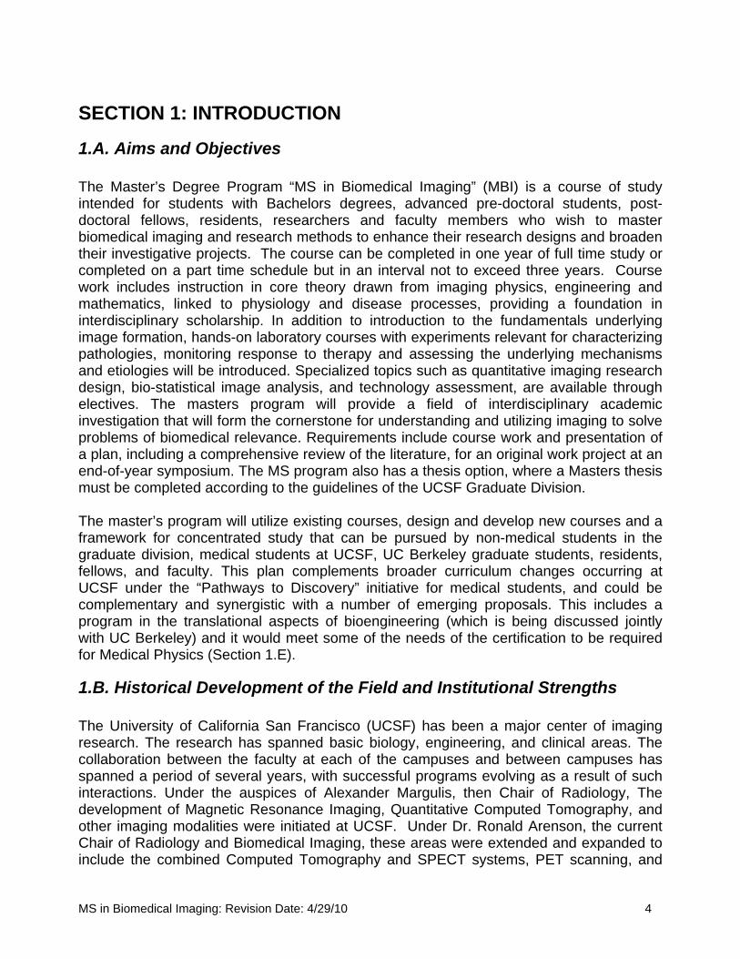
1.A. Aims and Objectives The Master’s Degree Program “MS in Biomedical Imaging” (MBI) is a course of study intended for students with Bachelors degrees, advanced pre-doctoral students, post-doctoral fellows, residents, researchers and faculty members who wish to master biomedical imaging and research methods to enhance their research designs and broaden their investigative projects. The course can be completed in one year of full time study or completed on a part time schedule but in an interval not to exceed three years. Course work includes instruction in core theory drawn from imaging physics, engineering and mathematics, linked to physiology and disease processes, providing a foundation in interdisciplinary scholarship. In addition to introduction to the fundamentals underlying image formation, hands-on laboratory courses with experiments relevant for characterizing pathologies, monitoring response to therapy and assessing the underlying mechanisms and etiologies will be introduced. Specialized topics such as quantitative imaging research design, bio-statistical image analysis, and technology assessment, are available through electives. The masters program will provide a field of interdisciplinary academic investigation that will form the cornerstone for understanding and utilizing imaging to solve problems of biomedical relevance. Requirements include course work and presentation of a plan, including a comprehensive review of the literature, for an original work project at an end-of-year symposium. The MS program also has a thesis option, where a Masters thesis must be completed according to the guidelines of the UCSF Graduate Division. The master’s program will utilize existing courses, design and develop new courses and a framework for concentrated study that can be pursued by non-medical students in the graduate division, medical students at UCSF, UC Berkeley graduate students, residents, fellows, and faculty. This plan complements broader curriculum changes occurring at UCSF under the “Pathways to Discovery” initiative for medical students, and could be complementary and synergistic with a number of emerging proposals. This includes a program in the translational aspects of bioengineering (which is being discussed jointly with UC Berkeley) and it would meet some of the needs of the certification to be required for Medical Physics (Section 1.E).
1.B. Historical Development of the Field and Institutional Strengths The University of California San Francisco (UCSF) has been a major center of imaging research. The research has spanned basic biology, engineering, and clinical areas. The collaboration between the faculty at each of the campuses and between campuses has spanned a period of several years, with successful programs evolving as a result of such interactions. Under the auspices of Alexander Margulis, then Chair of Radiology, The development of Magnetic Resonance Imaging, Quantitative Computed Tomography, and other imaging modalities were initiated at UCSF. Under Dr. Ronald Arenson, the current Chair of Radiology and Biomedical Imaging, these areas were extended and expanded to include the combined Computed Tomography and SPECT systems, PET scanning, and
MS in Biomedical Imaging: Revision Date: 4/29/10 4
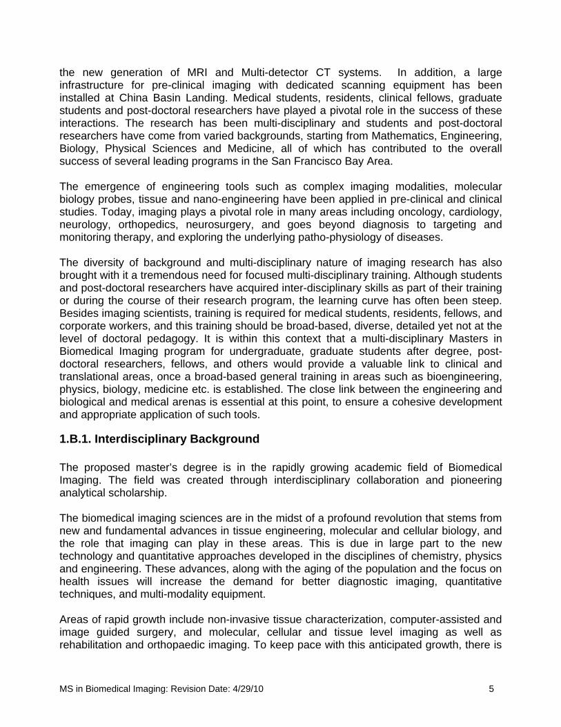
the new generation of MRI and Multi-detector CT systems. In addition, a large infrastructure for pre-clinical imaging with dedicated scanning equipment has been installed at China Basin Landing. Medical students, residents, clinical fellows, graduate students and post-doctoral researchers have played a pivotal role in the success of these interactions. The research has been multi-disciplinary and students and post-doctoral researchers have come from varied backgrounds, starting from Mathematics, Engineering, Biology, Physical Sciences and Medicine, all of which has contributed to the overall success of several leading programs in the San Francisco Bay Area. The emergence of engineering tools such as complex imaging modalities, molecular biology probes, tissue and nano-engineering have been applied in pre-clinical and clinical studies. Today, imaging plays a pivotal role in many areas including oncology, cardiology, neurology, orthopedics, neurosurgery, and goes beyond diagnosis to targeting and monitoring therapy, and exploring the underlying patho-physiology of diseases. The diversity of background and multi-disciplinary nature of imaging research has also brought with it a tremendous need for focused multi-disciplinary training. Although students and post-doctoral researchers have acquired inter-disciplinary skills as part of their training or during the course of their research program, the learning curve has often been steep. Besides imaging scientists, training is required for medical students, residents, fellows, and corporate workers, and this training should be broad-based, diverse, detailed yet not at the level of doctoral pedagogy. It is within this context that a multi-disciplinary Masters in Biomedical Imaging program for undergraduate, graduate students after degree, post-doctoral researchers, fellows, and others would provide a valuable link to clinical and translational areas, once a broad-based general training in areas such as bioengineering, physics, biology, medicine etc. is established. The close link between the engineering and biological and medical arenas is essential at this point, to ensure a cohesive development and appropriate application of such tools.
1.B.1. Interdisciplinary Background The proposed master’s degree is in the rapidly growing academic field of Biomedical Imaging. The field was created through interdisciplinary collaboration and pioneering analytical scholarship. The biomedical imaging sciences are in the midst of a profound revolution that stems from new and fundamental advances in tissue engineering, molecular and cellular biology, and the role that imaging can play in these areas. This is due in large part to the new technology and quantitative approaches developed in the disciplines of chemistry, physics and engineering. These advances, along with the aging of the population and the focus on health issues will increase the demand for better diagnostic imaging, quantitative techniques, and multi-modality equipment. Areas of rapid growth include non-invasive tissue characterization, computer-assisted and image guided surgery, and molecular, cellular and tissue level imaging as well as rehabilitation and orthopaedic imaging. To keep pace with this anticipated growth, there is
MS in Biomedical Imaging: Revision Date: 4/29/10 5

a critical need to expand training programs for individuals to serve as instructors and researchers in clinical and basic sciences departments in institutions of higher learning as well as to work in the growing industrial sector, which supports imaging research. Traditional curricula in the life sciences have not included quantitative methods nor have they provided instruction in the technology that is required in many areas of the current biological enterprise. An academic and intellectual environment that fosters seamless interaction between imaging and life sciences and that trains students to solve complex biological and clinical problems using modern imaging tools is emerging. The field has over 50 national and international associations and specialized journals dedicated to its scholarship. The National Institutes of Health has formed an independent institution, the National Institute of Biomedical Imaging and Bioengineering (NIBIB), largely to promote these imaging developments. These institutions and journals help to provide national educational standards for course development, opportunities for employment, peer review, publication in this area, and funding. In sum, this interdisciplinary field has provided innovative analytic tools to investigate disease, and has proven to be an attractive field of study at the intersection between engineering, physical, chemical and biological science.
1.B.2 Institutional Strengths The vision for the degree program at UCSF is to adapt the approach referred to above to teach fundamentals of imaging with hands on laboratory courses, preparing the Masters graduate with a deeper understanding of the biomedical imaging sciences.
We believe UCSF is well positioned in several ways for developing such a training program. As a health-science campus with no undergraduates, our teaching focus is on graduate students and post-doctoral researchers. UCSF’s historically strong biological focus and its biomedical community provide the advantage of a deep understanding of the problems that will continue to drive future clinical and research areas. Our location within the San Francisco Bay area provides us unequaled opportunities for interactions with biotechnology and high-tech communities, UC Berkeley and Stanford, and the new California Institute for Regenerative Medicine. This environment attracts outstanding students and faculty, as well as allows for placement of our Masters graduates. We already have significant experience in interdisciplinary training: the Joint Bio-engineering Graduate Group between UCSF and UCB, the Biophysics, Biological and Medical Informatics (BMI) and Chemistry and Chemical Biology (CCB) graduate programs at UCSF are all excellent examples. Thus from the very beginning we have had to work to bring these engineering-based approaches into a biological environment.
UCSF has been a leader in taking quantitative sciences into the field of medicine. At UCSF, to further foster the application of the “hard sciences” to biology, an umbrella organization, the Quantitative Biosciences Consortium (QBC) was recently formed. QBC seeks to enhance enrollment of physicists, mathematicians and engineers at UCSF. Graduate groups in, Bioengineering, Biophysics, Chemistry and Chemical Biology,
MS in Biomedical Imaging: Revision Date: 4/29/10 6

Biological Informatics, Complex Biological Systems and Pharmaceutical Sciences and Pharmacogenomics are participating the QBC umbrella. The Joint Bioengineering Graduate Group (JGGB) has been a focus for collaboration between the University of California at San Francisco (UCSF) and the University of California at Berkeley (UCB) for over 20 years. During that period it has stimulated numerous interactions between the two campuses and has enriched the opportunities for graduate students to experience how engineering principles can be brought to bear upon important problems in biology and medicine. The current NIH training grant, now in its 19th year, has been critical to the program’s past and present successes and its renewal is crucial for the future. The recent increase in the number of students who are seeking to enter the discipline has led to the assignment of major new resources for bioengineering research and education. At UCSF this need was met by the formation of a multi-disciplinary Department of Bioengineering and Therapeutic Sciences. In addition, other programs such as the Program in Craniofacial and Mesenchymal Biology (CMB), Molecular Medicine and others bring together faculty, research labs and investigators, many of whom have laboratories extensively using imaging methodologies and whose students and fellows may benefit from a one year didactic, hands-on course of biomedical imaging. An interdisciplinary approach is the crux of academic inquiry at UCSF. This is evident foremost in its encouragement of translational research. Obtaining an insight into the complexity of many of the disease processes that the health care system now seeks to manage demands the engagement of specialists from multiple areas, and the ability to provide these constituents with the most appropriate and highest quality imaging capabilities to address their biomedical questions. Thus, there are numerous faculty and PhD programs in place at UCSF that provide the structural integrity of the proposed one-year master’s program (see below, 1.C. for relationship to existing programs). As explained below (see section 3.A), there is strong reason to believe that the nature of instruction and research offered by a program in Biomedical Imaging would be appealing to students who elect to pursue a master’s degree. Students may be drawn from a number of sources. This includes undergraduate UC or Cal State programs, graduate programs at UCSF, joint UCSF and UCB programs, fellows and researchers in areas utilizing imaging as a major tool, medical students, commercial companies, and others.
1.C. Relationship of the Proposed Program to Existing Programs at UCSF Whereas the establishment of masters programs often acts as a building block toward creating a PhD program, the proposed program finds a niche under the umbrella of other successful PhD programs already active at UCSF. However, the proposed program serves the interests of a number of prospective students who either do not want to commit to a PhD program, or are under-qualified for admission to a PhD program and would benefit from an intermediate MS degree. The following list reflects the most closely related forms of intellectual inquiry to the activities of the proposed MS in Biomedical Imaging degree at UCSF.
MS in Biomedical Imaging: Revision Date: 4/29/10 7
1.C.1. Department of Radiology and Biomedical Imaging The host department does not currently offer graduate degrees. Establishing this degree program gives a new definition to Biomedical Imaging as a discipline at UCSF. Furthermore, the emerging requirements for Imaging Medical Physics residency programs that train Medical Physicists engaged in providing support in a hospital setting (Sect. 1E), underlines the need for developing a didactic curriculum in imaging. The faculty engaging in biomedical imaging research will be available for mentoring master’s research projects.
1.C.2. UCSF/UCB Graduate Group in Bioengineering UCSF and UC Berkeley offer a joint graduate program in Bioengineering. This program primarily admits students for a PhD though it may grant terminal MS degrees to some students. While there may be overlap in coursework between the proposed MS program and Bioengineering, Bioengineering is a broader discipline and has a focus on independent research being completed with a PhD dissertation. In addition, a MS in Bioengineering with an option to do Translational Medicine is possible for a two-year period. This is not an in-depth course in a specific focus area, but more a professional course with a few scientific and some management and technology oriented courses. This does not overlap with the Masters in Biomedical Imaging (MBI) proposed here. In addition, the expertise in terms of the number of faculty, as well resources as regards equipment is at UCSF, and there are no plans for providing similar training at UC Berkeley as underlined by Dr. Tirrell’s letter of support (See attached). The program has over 150 active graduate students, and offers a range of elective courses of relevance to the proposed MS in Biomedical Imaging that can be cross-listed (see below, Section 2.E.2). The faculty engaging in biomedical imaging research within the Joint Graduate Group in Bioengineering (JGGB) will be available for mentoring master’s research projects. The MS in Bioengineering is offered via Plan I defined in the Graduate Handbook that requires a thesis and formal coursework as outlined below.
Plan I Requirements:
1. Completion of 20 semester units, eight of which are graded graduate level courses in the major field of study, not including seminars. Of the remaining 12 units, up to three may be individual research, while the remaining must be advanced undergraduate or graduate courses in the major or other fields of study.
2. Completion of a Masters Thesis. The Masters Thesis must be read and approved by at least two Group faculty members who may come from either or both campuses.
1.C.3. Department of Bioengineering and Therapeutic Sciences This department is the administrative home of three multidisciplinary PhD programs, including the Graduate Group in Bioengineering. Research disciplines for the PhD
MS in Biomedical Imaging: Revision Date: 4/29/10 8
programs in this department are much broader than the proposed MS in Biomedical Imaging. The Department also has a Memorandum of Understanding with the Department of Radiology and Biomedical Imaging which outlines that major Biomedical Imaging related activities are carried out under the auspices of DRBI as the home department. Dr. Nelson and Giacommini’s letters of support (attached) attest to this.
1.C.4. Master of Advanced Study in Clinical Research Department of Epidemiology and Biostatistics runs an MAS program in Clinical Research. This program provides a two-year course of study for advanced pre-doctoral students, post-doctoral fellows, and faculty members. Although this program has a similar target population for the student enrollment, the proposed MS in Biomedical Imaging program does not have any overlap with this MS program because of the difference in disciplines.
1.C.5. Graduate Education in Medical Sciences The UCSF Graduate Education in Medical Sciences Training Program (http://physio.ucsf.edu/GEMS/index.asp) is a new initiative made possible through a grant from the Howard Hughes Medical Institute that provides UCSF graduate students enhanced knowledge of medical science and opportunities in disease-relevant research areas. The program aims to promote interest in this research among UCSF graduate students and will provide them with tools that they need to pursue interactive investigations with clinical investigators, either at UCSF or during their future careers. At this time there is no overlap between this program and MBI. If an imaging track is desired for such a program, then the MBI infra-structure could readily be adapted to meet the needs of this program.
1.D. Relationship of the Proposed Program with Other UC Institutions This program will be the only Masters with specific focus on biomedical imaging within the UC system that can be completed in one year. Of the programs and areas of study at other UC institutions, the following list reflects the most closely related forms of intellectual inquiry to the activities of the proposed MS in Biomedical Imaging degree at UCSF. Other UC Institutions (Berkeley, Davis, Irvine, Los Angeles, Merced, Riverside, San Diego, Santa Barbara, and Santa Cruz) offer traditional Bioengineering, Biomedical Engineering, Biological Sciences, Bioinformatics and/or Electrical Engineering programs. While the proposed coursework and research could fall under the scope of these umbrella programs, these traditional programs are much more broad, generally requiring coursework beyond medical imaging and admit students into PhD programs as opposed to the proposed program designed to provide one year of study focused on medical imaging. UC Berkeley runs a joint PhD program in Bioengineering with UCSF. This graduate group enrolls students only for a Ph.D., and a Masters is a terminal Masters, often given to students who cannot meet the expectations of the program. Berkeley also runs a separate, traditional bioengineering program at an undergraduate level leading to a BS degree. The focused group of faculty with their research emphasis in medical imaging
MS in Biomedical Imaging: Revision Date: 4/29/10 9
whose main affiliation is the host department of the proposed MS program at UCSF (Radiology and Biomedical Imaging) will have close relationship to the proposed core courses and elective courses. UC Davis runs a graduate program in Biomedical Engineering. The Davis program currently has active biomedical imaging research components. This program, however, does not provide critical experience necessary to learn practical medical imaging implementations and applications because the Davis Medical Center is not actually in Davis (but in Sacramento). In addition, this traditional MS/PhD program lacks flexibility in course selections and a concentrated master’s coursework in medical imaging as found in the proposed MBI program. UC Irvine offers MS/PhD programs in Biomedical Engineering. Imaging is not one of the focus areas for these degrees. UC Los Angeles offers traditional graduate degrees (MS/PhD) through the Biomedical Engineering Interdepartmental Program and the Biomedical Physics Interdepartmental Graduate Program. Both programs are broad in their disciplines, and may not be suitable for the target population that the proposed MS program would like to attract because these programs are usually a part of, or on the way to, respective PhD programs. The interdepartmental programs at UCLA offer MS degrees with elements of medical imaging. There is little overlap with the Biomedical Engineering program at UCLA, which is limited to the area of Biomedical signal/image processing and Image Informatics, and that program does not have an emphasis on the underlying principles of medical imaging acquisition and clinical applications, the main emphasis of the proposed UCSF program. The Biomedical Physics Interdepartmental Program covers many of the same areas of interest that will be offered in the UCSF program. The UCLA program is a traditional MS program, consisting of 48 credits of coursework and a thesis or comprehensive exam. The proposed UCSF program is more concentrated in time, with a corresponding reduced range in coursework and permits those who want to complete this in a year an opportunity to do so. The MS with thesis option in our program is very demanding for a three quarter program and we are recommending an additional quarter for summer research. Thus, our program is partly different, focused only on imaging, and a good complement to the UCLA program, and fills the need in Northern California . UC Merced runs graduate degree programs (MS and PhD) in Bioengineering through the School of Engineering. These degree programs are not yet formed within a home department because of the status of UC Merced as a new campus with new graduate programs. The Merced Bioengineering graduate programs do not currently have a medical imaging focus. UC Riverside offers MS and PhD programs in Bioengineering and a PhD in Biomedical Sciences. The graduate degrees at these programs do not have an emphasis in medical imaging.
MS in Biomedical Imaging: Revision Date: 4/29/10 10
UC Santa Cruz runs traditional MS/PhD programs in Bioinformatics and in Electrical Engineering. Neither focus on medical imaging. UC San Diego offers the MS, MEng, and PhD degrees in Bioengineering and the MS and PhD degrees in the area of Signal and Imaging Processing within the Electrical and Computer Engineering department and a PhD in Bioinformatics. These two programs are traditional bioengineering and electrical engineering graduate programs that do not offer degrees with specialization in medical imaging although some specific applications in medical imaging are part of the curriculum. The MS and MEng in Bioengineering in UCSD are totally different from our proposed program, the course work involves general physiology, tissue engineering, biophotonics, fluid mechanics, and only two general courses are available for Biomedical Imaging. Our coursework in focussed on Biomedical Imaging and complements the UCSD program. UC Santa Barbara offers PhD degrees in Biochemistry and Molecular Biology with emphasis in Biophysics and Bioengineering through its Biomolecular Science and Engineering program. There is no focused area of medical imaging from this graduate program at Santa Barbara.
1.E. Relationship of the Proposed Program with Emerging Programs
There are two major programmatic efforts that have emerged in the past year that would be highly complementary to the Master of Science in Biomedical Imaging. Although these programs do not yet exist, there are compelling reasons for their formation, substantial interest from many sectors, and preliminary efforts in place to promote them. We believe that the MBI would be able to provide a component of instruction that would be attractive to these other programs, would reduce the burden on them to offer that instruction, and would provide a source of revenue to the MBI – although we stress that we do not rely on those programs being implemented in the budgetary considerations herein.
The first program was the subject of a recent high profile symposium with leaders from Bioengineering and Translational Sciences at UC Berkeley and UCSF, including Matt Tirrell (Chair of UC Berkeley Bioengineering), Clay Johnston (Director of the CTSI at UCSF), Tejal Desai (Vice Chair, UCSF Bioengineering and Therapeutic Sciences) and Andy Grove (Founder of Intel Corp.). These advocates propose a Masters degree that would have a focus on scaling up advances in bioengineering associated with healthcare technology following the model of the semiconductor industry. This program is envisaged as a very large effort with ~150 students per year and with a broad portfolio of interests including elements from the Business School and other Engineering disciplines. There was recognition in that forum for including a component in clinical imaging, and the course curriculum offered by MBI program, including the exposure to clinical and translational aspects of imaging would complement the offerings of the proposed program. The formation of this degree would create a large pool of potential students who would take courses in the MBI program. This has led to the concept of a collaborative program in translational science and bioengineering that would link the University of California, San Francisco and Berkeley, and the students from other programs under this umbrella may take some of the courses offered under MBI.
MS in Biomedical Imaging: Revision Date: 4/29/10 11

The second program arises from a recent effort by CAMPEP, the Commission on Accreditation of Medical Physics Education Programs. This commission is requiring new accreditation standards for Medical Physicists who are involved in the operation, maintenance, and certification of any equipment that delivers radiation to patients, and are involved in imaging in hospitals nationally. The thrust of their recommendation is that any of these individuals will need to undergo training in a certified Medical Physics Residency Education program. Given the large number of hospitals and clinics that currently employ Health Physicists, there is a strong need for the establishment of new programs in this field, as there are only two such imaging programs in the US. The radiological Society of North America is urging Radiology departments to take the lead to establish these programs. The residency programs require education pertaining to radiation dosimetry, imaging fundamentals, image quality and laboratory experience. The Department of Radiology and Biomedical Imaging at UCSF is considering developing such a program. Students in a Medical Physics Residency program will require education in many areas related to radiation exposure other than those that are proposed in this current MBI program, specifically topics related to quality assurance and imaging equipment specifications, performance testing for CAMPEP certification. The MBI is not designed cover all of these aspects, however, the core didactic courses proposed as part of the MBI would fulfill some of the requirements for such a residency program. Once the residency program is established, the students in the residency could be in a different track under the same general umbrella, thereby increasing our pool of students.
1.F. Administration and Governance of the MS Program The master’s degree program in Biomedical Imaging will be administered through the Department of Radiology and Biomedical Imaging (DRBI) at UCSF. The Department has administrative space and staffing support, shared cubicle workspace for students, access to computers, and access to conference and meeting rooms for seminars and classes at China Basin Landing, and at the Mission Bay and Parnassus campuses of UCSF. The administration of student applications, funding issues, and student affairs will be under the auspices of the Postgraduate education office in DRBI. The students’ academic and community affairs will be overseen by the MBI Committee, Chaired by the Director of Graduate Studies in Radiology and Biomedical Imaging. The MBI Committee will be comprised of a minimum of four faculty with primary appointments or affiliations with Radiology and Biomedical Imaging (see Section 4.A List of Faculty Members for initial committee structure), with the Chair/Co-Chair of MBI a fifth, ex officio, member. The Chair of the MBI Committee will be responsible for committee membership, record keeping, and ensuring proper evaluation of each student’s performance. The Chair of the MBI Committee (Director of Graduate Studies in Radiology and Biomedical Imaging) will also ensure that each master’s student is assigned an appropriate advisor as a liaison for curricular or personal concerns. If the student chooses the MS with Thesis option, he/she will also have a thesis committee (See Section 2.G)
MS in Biomedical Imaging: Revision Date: 4/29/10 12

The MBI Committee composition and membership roster will be reviewed, every two years. Inactive members may be removed from the membership by a majority vote of the MBI Committee. Criteria to be considered when reviewing the faculty membership to the MBI Committee shall include:
1. Prior expertise and experience in serving on graduate student qualifying exams and/or dissertation committees.
2. Established record of scholarly, peer-reviewed publications. 3. Willingness and expertise to teach at least one course, seminar, or tutorial at the
graduate level in Biomedical Imaging. 4. Attendance and participation in MBI Committee meetings and regular
communications. 5. An associate or affiliate in good standing with the DRBI.
The Chair of the MBI Committee will report to and liaise with the Executive Committee of DRBI and the Graduate Division. The MBI Committee will oversee the development and implementation of all new master’s degree courses, maintain the coherency of the course catalog and ensure the maintenance of the online course materials and course evaluation procedures. The MBI Committee will also be responsible for overseeing the recruitment, applications, and admissions to the master’s degree program.
1.G. Plan for the Evaluation of the Program Courses that comprise the MS in Biomedical Imaging Program (core and elective) will be evaluated by the students using the new course review platform that can be accessed though Myaccess (Academic Senate Course Review). A longitudinal study will be developed to assess regularly the impact of the MS program on career development, productivity, and placement of graduates. Learning portfolios based on the University selected platform (Mahara.org) will be used to showcase student learning, provide a framework for assessing academic progress, and demonstrate how skills have developed over time. This will include for each student, their educational background, basic scores, grades, and training information, for the MBI program, it will include a plan of study for each student, progress in accomplishing the goals, final projects, and ultimate placement ultimately. The development of this platform will take advantage of the portfolios already set up by the Collaborative Learning Environment at UCSF. The Director of Graduate Studies for the MS in Biomedical Imaging degree program will be responsible for keeping records and data that will provide the basis for annual reports on the performance of the master’s program presented to the MS Committee. This information will be used for the Academic Program Review conducted by the Graduate Division and the Graduate Council, every 5 years. The Academic Program Review Committee will be comprised of a minimum of three experts from outside UCSF who are faculty in existing academic programs in Imaging Science, Medical Physics, and/or Bioengineering or any related academic fields that would provide familiarity with current scholarship and professional standards (the list of programs listed in 1.D. above provides a pool of colleagues who could participate in a five-year review).
MS in Biomedical Imaging: Revision Date: 4/29/10 13
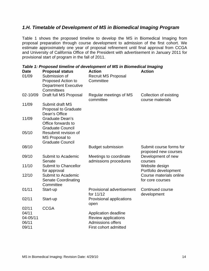
1.H. Timetable of Development of MS in Biomedical Imaging Program Table 1 shows the proposed timeline to develop the MS in Biomedical Imaging from proposal preparation through course development to admission of the first cohort. We estimate approximately one year of proposal refinement until final approval from CCGA and University of California Office of the President with advertisement in January 2011 for provisional start of program in the fall of 2011. Table 1: Proposed timeline of development of MS in Biomedical Imaging Date Proposal status Action Action 01/09 Submission of
Proposed Action to Department Executive Committees
Recruit MS Proposal Committee
02-10/09 Draft full MS Proposal Regular meetings of MS committee
Collection of existing course materials
11/09 Submit draft MS Proposal to Graduate Dean’s Office
11/09 Graduate Dean’s Office forwards to Graduate Council
05/10 Resubmit revision of MS Proposal to Graduate Council
08/10 Budget submission Submit course forms for proposed new courses
09/10 Submit to Academic Senate
Meetings to coordinate admissions procedures
Development of new courses
11/10 Submit to Chancellor for approval
Website design Portfolio development
12/10 Submit to Academic Senate Coordinating Committee
Course materials online for core courses
01/11 Start-up Provisional advertisement for 11/12
Continued course development
02/11 Start-up Provisional applications open
02/11 CCGA 04/11 Application deadline 04-05/11 Review applications 06/11 Admissions offers 09/11 First cohort admitted
MS in Biomedical Imaging: Revision Date: 4/29/10 14

SECTION 2: MS IN BIOMEDICAL IMAGING
2.A. Candidates for the Master’s Degree in Biomedical Imaging Students who have graduated with an undergraduate degree in the basic sciences will be considered for admission to the master’s degree program. Medical students, residents and fellows who are permitted time to pursue a professional degree, and professionals who wish to pursue the MS will be considered for admission to the program. For professionals, a background in the health sciences, clinical or basic science training, will be a prerequisite for admission to the program. In addition to the transcripts from the Bachelor’s degree, GRE scores will be required. Applicants who have been admitted to medical school or a fellowship program can submit their MCAT scores in lieu of GRE scores. Applicants with advanced professional degrees or in advanced degree programs do not need to take the GRE. Foreign applicants will be eligible under the same conditions, but they must take the Test of English as a Foreign Language (TOEFL) with a minimum acceptable score of 550 (paper version) or 213 (computer version), or the IELTS exam with a minimum score of 7, or who have demonstrated proficiency in English by completing one year of full-time study with a minimum GPA of 3.2 in an accredited university in the United States.
2.B. Foreign Language There will be no requirement for any foreign language proficiency in this program. The lingua franca of Imaging Science is English and has been since the inception of the field so there is currently little literature that is pertinent to the field that is not available in English. Most scientific meetings of relevance are conducted in English.
2.C. Program of Study The existing Radiology and Bioengineering programs at UCSF provide the impetus for the establishment of this MBI program. There are a few courses that already exist as part of the bioengineering curriculum that provide the foundation for the development of the Biomedical Imaging curriculum. The MS in Biomedical Imaging will conform to the Masters of Science Plan I as outlined by the UCSF Graduate Council Regulations and Procedures.
2.C.1 Unit Requirements MS Option:
i. Thirty-six units will required. MS With Thesis Option:
i. Thirty units of course work and an additional 12 units of BI 250 (Research) will be required.
This will be accomplished over 4 quarters, or three quarters and summer research.
MS in Biomedical Imaging: Revision Date: 4/29/10 15

2.C.2 Residency Requirements Three quarters of academic residence are required for the Master’s degree. A student who wishes a leave of absence must submit a written request to the Director of Graduate Studies of Biomedical Imaging for initial approval and then to the graduate dean or departmental chair for final approval. The granting of a leave of absence does not automatically change the time limit for advancement to candidacy or completion of degree. 2.C.3. Advancement to Candidacy Advancement to Candidacy must take place not later than the first day of the last quarter during which the student will be registered.
i. At least one quarter in registered student status must elapse between advancement to candidacy and conferral of the degree.
ii. Candidacy for the Master’s degree lapses if a student has not completed requirements for the degree within five quarters after advancement to candidacy.
2.C.4. Transfer Credits Up to six quarter units of credit for work taken elsewhere may be applied towards a master’s degree. For course work completed at another campus of the University of California, up to one-half of the program (18 units) may be accepted for transfer. Otherwise, all course work for the Master’s degree must be done in residence.
i. A student must be registered as a graduate student for at least one quarter before petitioning for transfer of credit.
ii. Units accepted for transfer must have been earned in graduate status. iii. Students enrolled in an articulated BS-MS program may transfer up to six units of
200 series course work taken during the quarter immediately prior to graduate standing for credit toward the master's degree.
iv. Work that formed part of the program for a degree previously conferred may not be applied toward a current degree program.
v. Courses taken in a university extension division may not be accepted for transfer.
2.C.5. Required Courses The Biomedical Imaging master’s degree program will consist of three quarters of didactic instruction. All students enrolled for the master’s degree in Biomedical Imaging will be required to complete the five-part Core Course in Biomedical Imaging (i.e., BI 201 through BI 205), and 4 units of a laboratory based course, or alternatively an approved laboratory research project. The remaining MS coursework will consist of elective interdisciplinary courses. See 2.D below for description of courses. Courses will be selected from an approved catalog of courses for major subject concentration. New course development in the major subject concentration will be reviewed by the MS Committee and follow the New Course proposal procedures (“General Course Form”) for UCSF approval.
Calculation of Course Units: 1 unit = 1 lecture hour per week 1 unit = 3 hours per week of:
Independent study; Conference; Seminar; Project; Web-Based Course Work
MS in Biomedical Imaging: Revision Date: 4/29/10 16
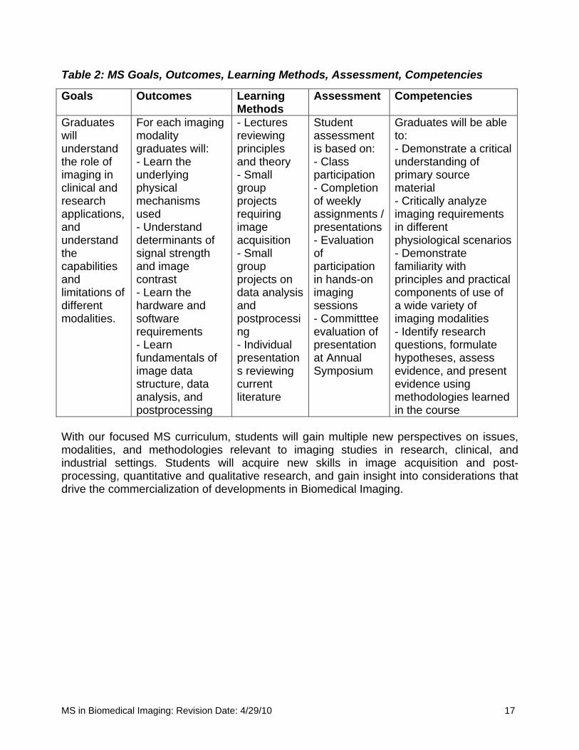
Table 2: MS Goals, Outcomes, Learning Methods, Assessment, Competencies
Goals Outcomes Learning Methods
Assessment Competencies
Graduates will understand the role of imaging in clinical and research applications, and understand the capabilities and limitations of different modalities.
For each imaging modality graduates will: - Learn the underlying physical mechanisms used - Understand determinants of signal strength and image contrast - Learn the hardware and software requirements - Learn fundamentals of image data structure, data analysis, and postprocessing
- Lectures reviewing principles and theory - Small group projects requiring image acquisition - Small group projects on data analysis and postprocessing - Individual presentations reviewing current literature
Student assessment is based on: - Class participation - Completion of weekly assignments / presentations - Evaluation of participation in hands-on imaging sessions - Committtee evaluation of presentation at Annual Symposium
Graduates will be able to: - Demonstrate a critical understanding of primary source material - Critically analyze imaging requirements in different physiological scenarios - Demonstrate familiarity with principles and practical components of use of a wide variety of imaging modalities - Identify research questions, formulate hypotheses, assess evidence, and present evidence using methodologies learned in the course
With our focused MS curriculum, students will gain multiple new perspectives on issues, modalities, and methodologies relevant to imaging studies in research, clinical, and industrial settings. Students will acquire new skills in image acquisition and post-processing, quantitative and qualitative research, and gain insight into considerations that drive the commercialization of developments in Biomedical Imaging.
MS in Biomedical Imaging: Revision Date: 4/29/10 17
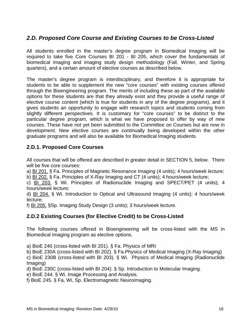
2.D. Proposed Core Course and Existing Courses to be Cross-Listed All students enrolled in the master’s degree program in Biomedical Imaging will be required to take five Core Courses BI 201 - BI 205, which cover the fundamentals of biomedical imaging and imaging study design methodology (Fall, Winter, and Spring quarters), and a certain amount of elective courses as described below. The master’s degree program is interdisciplinary, and therefore it is appropriate for students to be able to supplement the new “core courses” with existing courses offered through the Bioengineering program. The merits of including these as part of the available options for these students are that they already exist and they provide a useful range of elective course content (which is true for students in any of the degree programs), and it gives students an opportunity to engage with research topics and students coming from slightly different perspectives. It is customary for “core courses” to be distinct to the particular degree program, which is what we have proposed to offer by way of new courses. These have not yet been submitted to the Committee on Courses but are now in development. New elective courses are continually being developed within the other graduate programs and will also be available for Biomedical Imaging students.
2.D.1. Proposed Core Courses All courses that will be offered are described in greater detail in SECTION 5, below. There will be five core courses: a) BI 201. § Fa. Principles of Magnetic Resonance Imaging (4 units); 4 hours/week lecture; b) BI 202. § Fa. Principles of X-Ray Imaging and CT (4 units); 4 hours/week lecture; c) BI 203. § Wi. Principles of Radionuclide Imaging and SPECT/PET (4 units); 4 hours/week lecture; d) BI 204. § Wi. Introduction to Optical and Ultrasound Imaging (4 units); 4 hours/week lecture; f) BI 205. §Sp. Imaging Study Design (3 units); 3 hours/week lecture.
2.D.2 Existing Courses (for Elective Credit) to be Cross-Listed The following courses offered in Bioengineering will be cross-listed with the MS in Biomedical Imaging program as elective options. a) BioE 240 (cross-listed with BI 201). § Fa. Physics of MRI b) BioE 230A (cross-listed with BI 202). § Fa.Physics of Medical Imaging (X-Ray Imaging) c) BioE 230B (cross-listed with BI 203). § Wi. Physics of Medical Imaging (Radionuclide Imaging) d) BioE 230C (cross-listed with BI 204). § Sp. Introduction to Molecular Imaging. e) BioE 244. § Wi. Image Processing and Analysis. f) BioE 245. § Fa, Wi, Sp. Electromagnetic Neuroimaging.
MS in Biomedical Imaging: Revision Date: 4/29/10 18
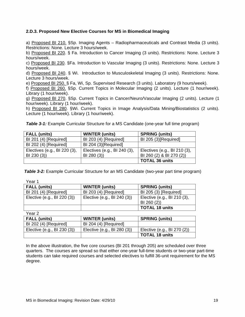
2.D.3. Proposed New Elective Courses for MS in Biomedical Imaging a) Proposed BI 210. §Sp. Imaging Agents – Radiopharmaceuticals and Contrast Media (3 units). Restrictions: None. Lecture 3 hours/week. b) Proposed BI 220. § Fa. Introduction to Cancer Imaging (3 units). Restrictions: None. Lecture 3 hours/week. c) Proposed BI 230. §Fa. Introduction to Vascular Imaging (3 units). Restrictions: None. Lecture 3 hours/week. d) Proposed BI 240. § Wi. Introduction to Musculoskeletal Imaging (3 units). Restrictions: None. Lecture 3 hours/week. e) Proposed BI 250. § Fa, Wi, Sp. Supervised Research (3 units). Laboratory (9 hours/week). f) Proposed BI 260. §Sp. Current Topics in Molecular Imaging (2 units). Lecture (1 hour/week). Library (1 hour/week). g) Proposed BI 270. §Sp. Current Topics in Cancer/Neuro/Vascular Imaging (2 units). Lecture (1 hour/week). Library (1 hour/week). h) Proposed BI 280. §Wi. Current Topics in Image Analysis/Data Mining/Biostatistics (2 units). Lecture (1 hour/week). Library (1 hour/week). Table 3-1: Example Curricular Structure for a MS Candidate (one-year full time program) FALL (units) WINTER (units) SPRING (units) BI 201 (4) [Required] BI 203 (4) [Required] BI 205 (3)[Required] BI 202 (4) [Required] BI 204 (3)[Required] Electives (e.g., BI 220 (3), BI 230 (3))
Electives (e.g., BI 240 (3), BI 280 (3))
Electives (e.g., BI 210 (3), BI 260 (2) & BI 270 (2))
TOTAL 36 units
Table 3-2: Example Curricular Structure for an MS Candidate (two-year part time program) Year 1 FALL (units) WINTER (units) SPRING (units) BI 201 (4) [Required] BI 203 (4) [Required] BI 205 (3) [Required] Elective (e.g., BI 220 (3)) Elective (e.g., BI 240 (3)) Elective (e.g., BI 210 (3),
BI 260 (2)) TOTAL 18 units Year 2 FALL (units) WINTER (units) SPRING (units) BI 202 (4) [Required] BI 204 (4) [Required] Elective (e.g., BI 230 (3)) Elective (e.g., BI 280 (3)) Elective (e.g., BI 270 (2)) TOTAL 18 units In the above illustration, the five core courses (BI 201 through 205) are scheduled over three quarters. The courses are spread so that either one-year full-time students or two-year part-time students can take required courses and selected electives to fulfill 36-unit requirement for the MS degree.
MS in Biomedical Imaging: Revision Date: 4/29/10 19
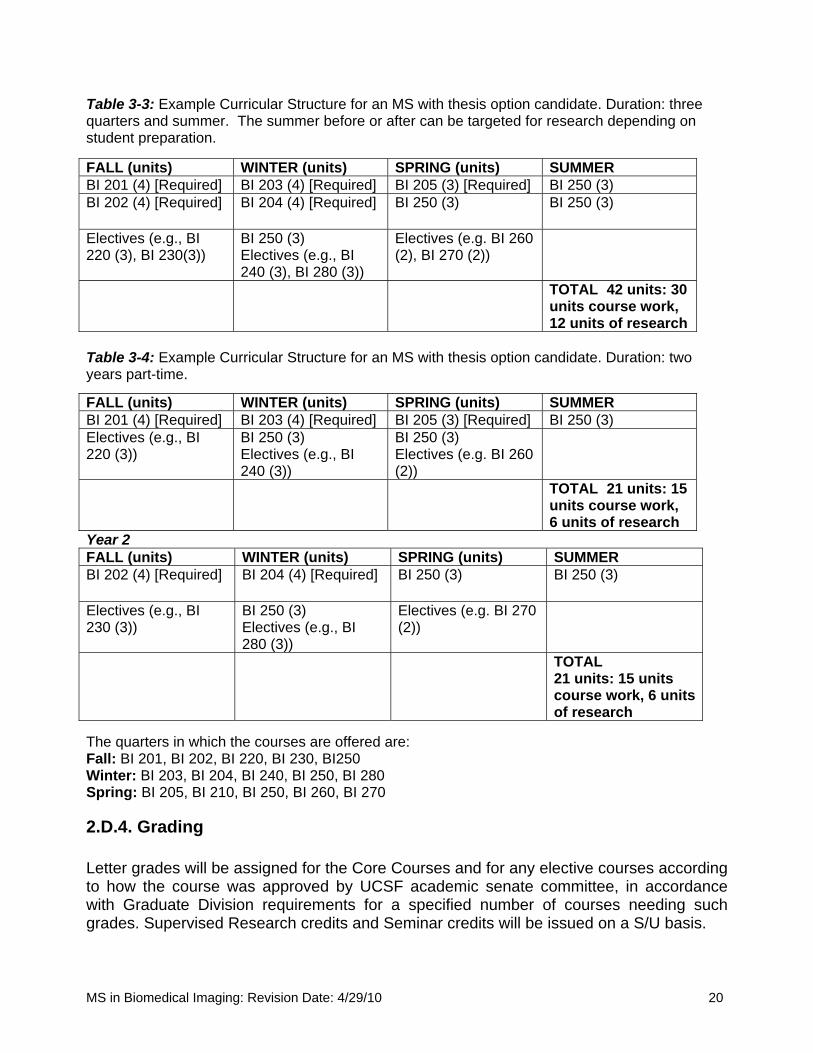
Table 3-3: Example Curricular Structure for an MS with thesis option candidate. Duration: three quarters and summer. The summer before or after can be targeted for research depending on student preparation. FALL (units) WINTER (units) SPRING (units) SUMMER BI 201 (4) [Required] BI 203 (4) [Required] BI 205 (3) [Required] BI 250 (3) BI 202 (4) [Required] BI 204 (4) [Required] BI 250 (3)
BI 250 (3)
Electives (e.g., BI 220 (3), BI 230(3))
BI 250 (3) Electives (e.g., BI 240 (3), BI 280 (3))
Electives (e.g. BI 260 (2), BI 270 (2))
TOTAL 42 units: 30 units course work, 12 units of research
Table 3-4: Example Curricular Structure for an MS with thesis option candidate. Duration: two years part-time.
FALL (units) WINTER (units) SPRING (units) SUMMER BI 201 (4) [Required] BI 203 (4) [Required] BI 205 (3) [Required] BI 250 (3) Electives (e.g., BI 220 (3))
BI 250 (3) Electives (e.g., BI 240 (3))
BI 250 (3) Electives (e.g. BI 260 (2))
TOTAL 21 units: 15 units course work, 6 units of research
Year 2 FALL (units) WINTER (units) SPRING (units) SUMMER BI 202 (4) [Required] BI 204 (4) [Required] BI 250 (3)
BI 250 (3)
Electives (e.g., BI 230 (3))
BI 250 (3) Electives (e.g., BI 280 (3))
Electives (e.g. BI 270 (2))
TOTAL 21 units: 15 units course work, 6 units of research
The quarters in which the courses are offered are: Fall: BI 201, BI 202, BI 220, BI 230, BI250 Winter: BI 203, BI 204, BI 240, BI 250, BI 280 Spring: BI 205, BI 210, BI 250, BI 260, BI 270
2.D.4. Grading Letter grades will be assigned for the Core Courses and for any elective courses according to how the course was approved by UCSF academic senate committee, in accordance with Graduate Division requirements for a specified number of courses needing such grades. Supervised Research credits and Seminar credits will be issued on a S/U basis.
MS in Biomedical Imaging: Revision Date: 4/29/10 20
Graduate students must maintain a cumulative grade point average of 3.0 or better and must make satisfactory progress toward the requirements of the degree program. Students who fail to maintain a 3.0 GPA or fail to make satisfactory progress toward the degree are subject to dismissal by the Graduate Division Dean after consultation with the MS Committee and the Director of Graduate Studies. The graduate program will establish a regular mechanism for reviewing students’ satisfactory progress toward the degree. Completion of specific program requirements will be documented and maintained in the graduate program’s student files. Any deficiency or failure to meet the standards of the program should be discussed with the student and confirmed in writing.
2.E. Research/Imaging Study Design As the Core Courses for Biomedical Imaging demonstrate, the theoretical framework and methodological approaches to investigating these subjects are interdisciplinary. It is anticipated that a high percentage of the students will not have previous training in biomedical imaging research, though it is anticipated that many will have training in medicine, basic science, health policy and/or epidemiology. The acquisition of skills to identify appropriate imaging modality for disease detection, imaging probe design, imaging assisted drug development, image reconstructions and analyses, and to be positioned to present a written argument according to scholarly conventions defined in the basic and clinical science is a major outcome of this MS program. Therefore, much emphasis is placed on techniques and procedures for acquiring these skills, both through core courses, electives, and hands-on research. The capstone for the course will be a final project. The rigor of the projects will be similar for all students but the format will differ depending on the student’s background and the amount of hands-on research they are able to accomplish during the course year. The particular kind of research project a student may wish to pursue can vary. Some projects may be more oriented toward imaging hardware or software, but some may focus on chemistry or biology of imaging agents and their interaction with disease targets. Students may choose an appropriate research project under the supervision of participating investigators. Examples of project formats are:
- the outline of a grant proposal based on preliminary data obtained during the course - a journal article suitable for publication - an invention disclosure describing the design of imaging hardware or software
Although the projects will be formatted in a practical manner for communication to the scientific community, formal submissions of the work will not be required for acceptance by the MBI graduate committee.
2.F. Symposium and Presentation At the end of the academic year, each MS candidate will present a final paper (20 minute oral presentation, followed by 15 minute Q&A) at a day-long Medical Imaging Science
MS in Biomedical Imaging: Revision Date: 4/29/10 21

Research Symposium, sponsored by the Department of Radiology and Biomedical Imaging. This event acts as a mechanism to gauge students’ understanding of the research topic and their ability to present the components of an original research project with a scientifically sound rationale and in a logical manner.
2.G. MS with Thesis Option The thesis option requires the completion of a Master's Thesis in accordance with the rules of the Graduate Division. The thesis constitutes the results of an original investigation of a problem. It should be carried out in the same systematic and scholarly way as investigations of greater magnitude, such as a doctoral dissertation.
2.G.1. Advising for Students in the MS with Thesis Option The Director of Graduate Studies will ensure that entering students who wish to participate in this option, are assigned a faculty adviser who can direct them to appropriate research laboratories to pursue their thesis option. Students are expected and encouraged to meet with their adviser immediately and thereafter quarterly regarding their academic program, particularly at the beginning of each quarter to prepare and approve the study list and assistance with selecting the thesis area. Students must select their area of research, and decide on their thesis advisor by the second quarter. The research advisor serves as the Chair of the thesis committee.
2.G.2. Thesis Committee The thesis/research advisor (different from the faculty advisor on entrance to the program) serves as the Chair of the thesis committee. In addition, at least two other committee members must be selected from faculty in the Department of Radiology and Biomedical Imaging. An additional outside member familiar with the research area should complement the committee but is not mandatory. Authorship of a master’s thesis by more than one degree candidate is not allowed. Upon completion of the master’s thesis, two copies shall be deposited in the Graduate Division Office by the date specified in the degree calendar for that term. Specific information regarding the form in which the master’s thesis manuscripts are to be prepared must be obtained from the Graduate Division Office.
2.H. Normative Time from Matriculation to Degree The time from Matriculation to degree for full-time students will be one year. Generally, students will complete all course unit requirements by the end of the spring quarter. The degree will be awarded only after successful completion of the coursework (full unit requirements) and acceptance of the final project. The final project should be undertaken after consultation and following approval by the MBI Committee, or a designated academic advisor appointed by the MBI Committee prior to the spring quarter. For the two-year part-time students, this final project should be initiated prior to the spring quarter of the second year. This is a rigorous master’s degree program, but not so difficult as to be overly challenging to complete in a single year of full-time study. The thesis option can be completed over three quarters and with summer research, and can also be accomplished over a two year period.
MS in Biomedical Imaging: Revision Date: 4/29/10 22

SECTION 3: PROJECTED NEED
3.A. Student Demand for the Program Growth of Biomedical Imaging: As suggested in sections 1.B.1 and 1.B.2 above, the interdisciplinary academic field of Medical Imaging has rapidly emerged as a vibrant field over the last twenty years. Growth is indicated institutionally, marked by increasing academic undergraduate and graduate degree programs across the country (including UCLA, UC Berkeley, UC Davis, Stanford, Washington University St. Louis, Harvard/Mass General Hospital, Duke, Wisconsin, University of Washington, Johns Hopkins, etc.), by growth in membership to its scholarly societies and the establishment of two new societies this decade (particularly SNM – Society of Nuclear Medicine, International Society for Magnetic Resonance in Medicine –ISMRM, Academy of Molecular Imaging – est. ~2000 and the Society of Molecular Imaging – est. ~ 2000 ), and publication in its specialized journals (Journal of Nuclear Medicine, Molecular Imaging, Molecular Imaging and Biology, Magnetic Resonance in Medicine and Journal of Magnetic Resonance Imaging). There is an increasing recognition of the need for formal education in the field of Biomedical Imaging, and the Clinical and Translational Science Awards group of the National Center for Research Resources, NCRR, at the NIH has formed an Education in Imaging Group to promote education in the field of Biomedical Imaging. Additionally, a growing number of biotechnology companies, small drug companies and large pharmaceutical companies (Genentech, Merck, Lilly, Pfizer, etc.) have purchased imaging devices for preclinical drug development or have incorporated imaging applications into their core drug development schema. Program scarcity and enrollment statistics for similar programs in UK, Netherlands, UCLA and Boston University: Several major imaging centers have been established over the last decade that offer access to multiple imaging modalities including UCSF and many of those listed above. While these centers are offering undergraduate courses and an increasing numbers of graduate degree programs, there is a lack of programs that are specifically related to different imaging modalities, probe/ contrast agent development, medical physics, instrumentation development or clinical research. There are programs in the UK, at Oxford University, University of Kent and Imperial College. In Europe, University of Utrecht, Netherlands has a similar program. Despite differences in the academic system between the US and the European programs, we have been in communication with the program at Oxford University and in the Netherlands. Oxford has a constant enrollment ranging from 9-12 students a year, from both the UK and other countries. The number of applicants for these positions this past year totaled 24. The program at Utrecht has an enrollment ranging from 12-15, and they received a total of 20 applications. In order to confirm if other Masters’ programs in Biomedical Imaging existed in the US as well as to seek feedback on the concept and gauge the potential interest in graduates from this program we performed an informal survey of our colleagues in imaging programs at several academic institutions. It is notable that the one program on the West coast, that is
MS in Biomedical Imaging: Revision Date: 4/29/10 23
most similar in course content to the proposed program, the Masters in Biomedical Physics at UCLA (a two to three year course), reports having an average of 84 applicants for 12 slots in their program. A second program in Boston University has just been launched this year, and in their program, for this year they received 20 applicants for 10-15 available slots. Most of the individuals that applied, were bound for medical imaging industry positions, some were foreign MD scholars. Target Students: Post-Baccalaurete and Physician trainees: Our target enrollees, outlined above (1.A), include post-baccalaurete students, medical students, residents and fellows interested in pursuing a master’s option and who wish to expand their research skills, and pre-doctoral graduate students who wish to earn a Biomedical Imaging Master’s degree. Given the applicant pool for the programs listed above, we are expecting a post-Baccalaurete pool of applicants, individuals interested in specializing in Medical Imaging, wishing to go to jobs in industry, join research teams in academia and national laboratories, and also individuals uncertain about pursuing a research career. Others include, individuals who are currently working in the biotechnology or pharmaceutical industry requiring training to meet job expectations, and foreign students from countries where imaging programs are just emerging (Asia, India, South America, etc.). Many students enter medical school with backgrounds in a variety of non-physical science disciplines and are eager to have the opportunity to pursue a higher level of training in the field, but who are not intending to commit to a PhD program. Post-Residency Physicians: There is strong interest in imaging approaches among many physicians who emerge from residency programs and wish to pursue more focused specialization. Examples of this include Neurologists who are increasingly utilizing advanced imaging techniques such as functional MRI, or functional nuclear medicine studies to explore both neurological and psychological disorders. Cardiologists are actively involved in using imaging modalities to evaluate the underlying physiology of cardiac disorders in vivo, both in pre-clinical and clinical settings. Similarly, among the surgical specialties, there are compelling reasons to better understand and utilize imaging methods. We list a few examples: Cardiothoracic surgeons who wish to develop improved methods for different surgical repairs of ischemia and valvular disorders place heavy reliance on ultrasound, MR, and CT studies; Neurosurgery is interested in improved methods for identifying fiber tracts in planning surgical treatment of tumors; Vascular surgeons wish to use imaging methods to investigate the response of blood vessels in patients with atherosclerosis to pharmacologic interventions; and; Orthopedic surgeons who wish to obtain an improved prognosis of which individuals with musculoskeletal disorders will best respond to surgical interventions. Although these individuals may not wish to become Radiologists, it is clear that their participation in their own areas of specialization would be enhanced by completing the Master of Science in Biomedical Imaging. This would also make them more competitive for placement in fellowship programs in their field.
MS in Biomedical Imaging: Revision Date: 4/29/10 24

Research Specialists: The Department of Radiology and Biomedical Imaging conducts a large number of research projects both those initiated by investigators in the Department, as service to colleagues from other disciplines, and as contracts to industry. Generally, many facets of these projects are conducted by junior individuals who do not have an advanced degree but are eager to participate in research. Aspects of research that these individuals perform include: subject (patient, animal, or specimen) preparation for imaging; operation of imaging equipment; data handling and image processing; preparation and delivery of imaging agents; and data analysis, such as statistics and figure preparation. There are a large number of academic institutions around the nation and throughout the world that similarly employ this category of worker. Many of these individuals are keenly interested in bolstering their expertise to enable them to work more independently, to provide a higher level of service, and to permit them to move into positions of greater responsibility in the management and conduct of imaging projects. This type of need is also experienced increasingly in the pharmaceutical and biotechnology industries, and in companies that use imaging either as a biomarker for assessing the efficacy of therapeutic interventions, in the evaluation of imaging agents, or in the development of devices used in patient care. However, they are generally not interested in establishing their own independent research programs that require involvement in activities such as securing funding, or establishing program research goals and collaborations. All of the constituencies mentioned above would be interested in recruiting workers who have a rigorous training in a broad range of imaging modalities, and a deep understanding for how best to use those tools in clinical and research applications. Clinical technologists: In addition to the areas already noted, every large imaging center employs a large number of technologists to perform routine clinical imaging. These technologists receive dedicated training for their field of work, which includes extensive training on the operation of imaging systems, in patient handling, and in imaging-relevant anatomy. They generally only receive a rudimentary introduction to the underlying principles of the different imaging modalities, and are therefore limited in their ability to modify and improve imaging approaches, to identify causes of image degradation, and to streamline examinations by taking such factors into account. There is a need for these institutions to have a subset of these technologists who are trained at an advanced level, commensurate with what would be provided by this masters program. Such individuals would become very marketable as lead technologists, who, in addition to providing financial advantages to their employers, would also provide benefits to patient care by implementing more effective, and accurate imaging studies with reduced patient discomfort, and lower exposure to any risk factors that might be related to undergoing an imaging study. There are currently no training programs for these “super technologists”, and while some individuals have been able to acquire some of the needed skills from their local environment or by personal experience, this is typically not possible at most institutions. Students from Emerging Programs: As discussed in Section 1.D, there are major complementary programmatic efforts that are being planned: (i) a program in bioengineering studies that are associated with healthcare technology, which will be joint
MS in Biomedical Imaging: Revision Date: 4/29/10 25
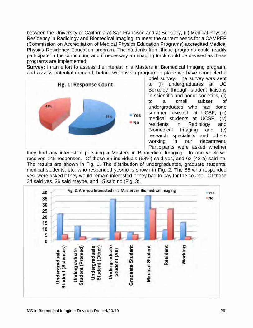
between the University of California at San Francisco and at Berkeley, (ii) Medical Physics Residency in Radiology and Biomedical Imaging, to meet the current needs for a CAMPEP (Commission on Accreditation of Medical Physics Education Programs) accredited Medical Physics Residency Education program. The students from these programs could readily participate in the curriculum, and if necessary an imaging track could be devised as these programs are implemented. Survey: In an effort to assess the interest in a Masters in Biomedical Imaging program, and assess potential demand, before we have a program in place we have conducted a
brief survey. The survey was sent to (i) undergraduates at UC Berkeley through student liaisons in scientific and honor societies, (ii) to a small subset of undergraduates who had done summer research at UCSF, (iii) medical students at UCSF, (iv) residents in Radiology and Biomedical Imaging and (v) research specialists and others working in our department. Participants were asked whether
they had any interest in pursuing a Masters in Biomedical Imaging. In one week we received 145 responses. Of these 85 individuals (58%) said yes, and 62 (42%) said no. The results are shown in Fig. 1. The distribution of undergraduates, graduate students, medical students, etc. who responded yes/no is shown in Fig. 2. The 85 who responded yes, were asked if they would remain interested if they had to pay for the course. Of these 34 said yes, 36 said maybe, and 15 said no (Fig. 3).
MS in Biomedical Imaging: Revision Date: 4/29/10 26
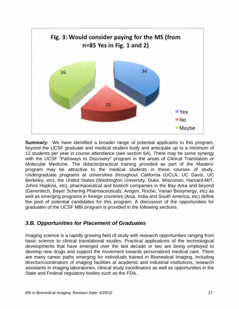
Summary: We have identified a broader range of potential applicants to this program, beyond the UCSF graduate and medical student body and anticipate up to a minimum of 12 students per year in course attendance (see section 6A). There may be some synergy with the UCSF “Pathways to Discovery” program in the areas of Clinical Translation or Molecular Medicine. The didactic/practical training provided as part of the Masters’ program may be attractive to the medical students in these courses of study. Undergraduate programs at universities throughout California (UCLA, UC Davis, UC Berkeley, etc), the United States (Washington University, Duke, Wisconsin, Harvard-MIT, Johns Hopkins, etc), pharmaceutical and biotech companies in the Bay Area and beyond (Genentech, Bayer Schering Pharmaceuticals, Amgen, Roche, Varian Biosynergy, etc) as well as emerging programs in foreign countries (Asia, India and South America, etc) define
e pool of potential candidates for this program. A discussion of the opportunities for provided in the following sections.
ssistants in imaging laboratories, clinical study coordinators as well as opportunities in the State and Federal regulatory bodies such as the FDA.
thgraduates of the UCSF MBI program is
3.B. Opportunities for Placement of Graduates Imaging science is a rapidly growing field of study with research opportunities ranging from basic science to clinical translational studies. Practical applications of the technological developments that have emerged over the last decade or two are being employed to develop new drugs and support the movement towards personalized medical care. There are many career paths emerging for individuals trained in Biomedical Imaging, including director/coordinators of imaging facilities at academic and industrial institutions, research a
MS in Biomedical Imaging: Revision Date: 4/29/10 27
We envision that the master’s program will provide essential training for students who wish to pursue PhD level research in Chemistry (contrast agent development), Physics (instrumentation development), Medical Physics, Bioengineering or one of the PhD programs offered in imaging programs elsewhere. However, not every student will intend to pursue a PhD, nor will every student wish this to provide a springboard to an academic career in this specified field. We envision a number of students with primary training in medicine or science who wish to expand the scope of their analytical skills and knowledge of Biomedical Imaging to enhance their investigations and work in their primary rofessional field.
s intractable medical problems and the implementation of ersonalized medicine.
response from the corporate entities underlines the importance of this type of training.
.C. Importance and Impact of the MS in Biomedical Imaging
skills and knowledge to immediately apply to their research and development fforts.
p The placement depends on the student, and as stated above there is reason to believe that we will admit other than medical students. Graduates may go on to pursue a PhD in one of a number of PhD programs, with ultimate placement in an academic department or a pharmaceutical, biotechnology or medical imaging device company. There may be candidates who already are working in a corporate position with the desire or need to receive training that will enhance job performance. This program will provide the skills and knowledge that will allow the graduates to design and implement new research projects, apply for research funding and to implement new translation imaging paradigms to address delivery and efficacy of new therapeutics. The “value added” applies to all students, not only medical students. This program cultivates critical thinking on the infusion of imaging technologies to addresp In Appendix 1, we provide a set of responses from leaders in industry who were sent our proposal for review, and, as can be seen, the
3 The rapid expansion of Biomedical Imaging over the past decade has created shortages of trained professionals to fill the positions available in academic institutions. The added application of molecular imaging in the pharmaceutical and biotechnology industry has placed even more pressure on the limited number of training programs to increase the numbers of trained professionals to meet the demand. The increased demand has created positions for trained post-Baccalaureate and Masters’ degree candidates. Additionally, MD candidates may wish to augment their medical school training with specialized training in Biomedical Imaging that will create opportunities for future translational research in academia or the corporate sectors. These candidates may additionally benefit professionally from the Master’s in Biomedical Imaging and be more marketable or enhance their current position. This specialized training will produce professionals that will have thee There are similar programs in the UK, at Oxford University, Kent University and Imperial College. There is a program at the University of Utrecht, Netherlands as well. With the
MS in Biomedical Imaging: Revision Date: 4/29/10 28
exception of these programs in Europe, and the UCLA program there are no Masters’ programs in Biomedical Imaging. Therefore, establishment of a UCSF based program offering access to graduate students, medical students and pharmaceutical/ biotech scientists would have significant impact on the dearth of trained scientists. The Master’s program would also provide broad-based training with introduction of all students to the breadth of the field. This would be a perfect portal for students desiring to pursue a PhD as their ultimate goal as their thesis will more than likely be multimodality and multidisciplinary. The consensus from a poll of colleagues in the field (as seen by the letters attached as Appendix 2) is that this MBI program is needed, would be welcome, and provide a means of increasing the numbers of competent individuals who will bear the torch of future imaging research and applications.
.D Ways in which the program will meet the needs of society
al diagnostic systems should be implemented to meet the needs of the global community.
he program to research and/or professional terests of the faculty
the approaches, tools, and methodology that are routinely used by faculty in the DRBI.
.F Program Differentiation
an verage more than two years to complete and serves students in Southern California.
3 Students who receive an MS in Biomedical Imaging will be knowledgeable and skilled in the safe and effective use of the broad range of imaging modalities that are routinely, and increasingly, used in the delivery of health care to the broad population in the United States and throughout the world. They will contribute to the ability to deliver safer, more reliable, and more cost effective methods of imaging diagnostics to evaluate health, disease, and the efficacy of pharmacologic and interventional therapies. Not only will they understand the implementation of the most sophisticated imaging modalities, they will also understand when cheaper and more practic
3.E Relationship of tin The MS in Biomedical Imaging proposes a course of study that would train students in many of
3 To our knowledge, there is no similar program on any of the campuses of the UC system apart from the Biomedical Physics Interdepartmental Program at UCLA, which takesa
MS in Biomedical Imaging: Revision Date: 4/29/10 29
SECTION 4: LIST OF CORE FACULTY MEMBERS, RANKS AND HIGHEST DEGREES Alphabetical List of UCSF faculty members who currently teach and/or mentor students in a related Department and degree program that maybe aligned with the MBI. This is a shortened list including only those courses that form the core and elective infra-structure of the program. The DRBI has a number of other talented, well-renowned faculty who will be added to the list if the student learning needs require advanced courses in topics such as Spectroscopic Imaging, etc. Yanjun Fu, PhD, Course Instructor Assistant Research Scientist in Radiology and Biomedical Imaging Dr. Fu received his PhD from Wuhan University, China, in the field of Organic and Polymer Chemistry. His work has been focused on the field of Contrast Media research and molecular imaging. He has teaching experience on polymer chemistry and molecular imaging (particularly molecular design, synthesis and characterization of imaging contrast agents/probes) for graduates in the joint Bioengineering program between UCSF and UC Berkeley (as one of the instructors in 2008). He also has enthusiastically involved in informal laboratory teaching on chemistry for research fellows/postdocs, technologist staff and medical students in the contrast media lab at UCSF Radiology department. Ella Fung Jones, PhD, Faculty Mentor and Course Instructor, Adjunct Assistant Professor in Radiology and Biomedical Imaging Dr. Jones obtained her PhD in Chemistry from University of California at Davis and completed two years of post-doctoral training at the Oxford University in UK. She has over 15 years of research experience in cancer imaging. At UCSF, Dr. Jones is a NIH funded Principal Investigator researching molecularly targeted probes to delineate diseases using nuclear and optical imaging. Dr. Jones teaches the chemistry and biology aspects of molecular imaging to graduate students and radiology residents. She currently serves as the Director of Preclinical Optical Imaging Laboratory at the Center for Molecular and Functional Imaging in the Department of Radiology. Dr. Jones will develop the new course BI 204 “Introduction to Optical Imaging”. She will also participate in BI 260, “Current Topics in Molecular Imaging” Jiang He, PhD, Course Instructor Assistant Adjunct Professor of Radiology and Biomedical Imaging Dr. He obtained a PhD in radiopharmaceutical chemistry from Peking University. He has been actively participating in teaching and mentoring students, postdoctoral fellows, research staff and junior faculty members in different settings by providing technical expertise and career development advice in molecular imaging and radiopharmaceutical
MS in Biomedical Imaging: Revision Date: 4/29/10 30
research. He served as a counselor for students and postdoctoral fellows who were just starting their post-graduate careers in imaging science as well as for postdoctoral fellows and junior faculty member with many years experience in other fields who are interested in transitioning into imaging research. He has taught lectures in imaging probe development for medical residents and PhD students in the UCSF/UC Berkeley joint bioengineering program. Roland Henry, PhD, Course Instructor Associate Professor in Residence, Radiology and Biomedical Imaging Dr. Henry has been a member of the Bioengineering Graduate Group for a number of years, and teaches the graduate level course in Magnetic Resonance Imaging. His research involves quantitative MRI measurements in the brain. These new and exciting techniques yield physiological information about normal and diseased brain. His focus is on the improvement and development of these techniques in order to understand the relevance of these measures and their potential for tissue characterization. In particular his group has been working on developing diffusion MRI techniques. Diffusion MRI is the only technique that can delineate white matter pathways in the brain. This relatively new technique is growing rapidly in its scope of application and utility in neurological studies. Thomas Lang, PhD, Course Instructor Professor in Residence, Department of Radiologyand Biomedical Imaging Thomas Lang, Ph.D., is a Professor in Residence in the UCSF Department of Radiology and the UCSF/UC Berkeley Joint Bioengineering Graduate Group, and the course instructor for a graduate level course in x-ray based imaging techniques. He received his B.A. in Chemistry from the University of Chicago in 1983 and a Ph.D. in Chemistry from UC Berkeley in 1990. After completing a postdoctoral fellowship with Professor Bruce Hasegawa in the UCSF Department of Radiology, Dr. Lang worked as a Nuclear Medicine Physicist at ADAC Laboratories in Milpitas, Calif. Dr. Lang joined the UCSF faculty as an Assistant Adjunct Professor in 1994. As a faculty member at UCSF, Dr. Lang's core interest is the use of quantitative computed technology and other clinically available imaging modalities in the study of human musculoskeletal biology. His group is currently funded by the National Institutes of Health, National Space Biomedical Research Institute and pharmaceutical industry. In addition to his position at UCSF, he is also Leader of the Musculoskeletal Alterations Team at the National Space Biomedical Research Institute, where he coordinates a research team comprising investigators from eight institutions. Sharmila Majumdar, PhD, Faculty Mentor and Course Instructor, Professor in Residence in Radiology and Biomedical Imaging with appointments in the Dept. of Orthopedic Surgery, Bioengineering and Therapeutic Sciences at UCSF, and UC Berkeley, Department of Bioengineering. Proposed member of MBI Committee. Sharmila Majumdar has taught courses such as Clinical Concepts of Bioengineering and courses on imaging and image processing techniques, at UCSF and UCB; she also served as Co-Chairman of the Bioengineering graduate admissions committee for the year 2000,
MS in Biomedical Imaging: Revision Date: 4/29/10 31

and 2004-2007. She brings to this program considerable expertise in high-resolution imaging and quantitative image analysis techniques. She trained in magnetic resonance imaging at Yale University, and in the last twenty years at the University of California, San Francisco, she has concentrated on adapting and developing techniques to image and quantify trabecular bone structure in the study of osteoporosis, arthritis and other musculo-skeletal disorders. She served as the Director for Translational Research in the Department of Radiology, UCSF, where her charter was to facilitate the transfer of research tools to the clinical arena, coordinate and run educational sessions for faculty, staff and technologists in upcoming areas of research, and facilitate the use of advanced and interdisciplinary tools in a clinical setting. More recently she has taken on a pivotal role in building the resources for the Musculo-skeletal and Quantitative Imaging Research Group (MQIR), and serves as Vice-Chair for research in the Department of Radiology. Dr. Majumdar will develop the new course BI 240 “Introduction to Musculoskeletal Imaging” which will provide an overview of multi-modality imaging of musculoskeletal disease. She will also provide supervision for BI 250, “Supervised Research” and will participate in BI 280, “Current Topics in Image Analysis/Data Mining/Biostatistics” Alastair Martin, PhD, Faculty Mentor, Course Instructor, proposed Director of Graduate Studies, Adjunct Professor in Radiology and Biomedical Imaging. Proposed ex officio member of MBI Committee. Dr Martin received his BSc in Physics from the University of British Columbia in 1987. He did his graduate work at the University of Toronto in the Department of Medical Biophysics, receiving an MSc in 1989 and PhD in 1994. His research focuses on magnetic resonance imaging and he did a 2-year post-doctoral fellowship at the University of Western Ontario after completing his PhD. Dr Martin then spent nine years working as a Clinical Scientist for Philips Medical Systems, where he worked on-site in the Departments of Radiology at the University of Minnesota (1996-2000) and at the University of California – San Francisco (2000-2005). During this time he developed magnetic resonance methods for guiding neurosurgical and endovascular procedures. Dr Martin joined the faculty of the University of California in 2005 and is presently an Associate Adjunct Professor in the Department of Radiology and Biomedical Imaging. His research focus remains on the use of magnetic resonance methods to guide and evaluate novel therapeutic techniques. He regularly interacts with students, residents and fellows and is co-organizer and a principle lecturer of a course entitled “Advanced Cardiovascular Imaging” (Bioengineering 297). Tracy Richmond McKnight, PhD, Faculty Mentor and Course Instructor Associate Professor in Residence in Radiology and Biomedical Imaging. Proposed member of MBI Committee. She received her PhD in Biomedical Engineering from the University of California, Davis. She is currently an Associate Professor in the Department of Radiology and Biomedical Imaging and her research focus is magnetic resonance imaging and spectroscopy of brain tumors. Dr. McKnight is a member of the UCSF Brain Tumor Research Center, the UCSF Helen Diller Cancer Center, and the UCSF/UCB Joint Bioengineering Graduate Group. She is also actively involved in mentoring and advising students interested in science,
MS in Biomedical Imaging: Revision Date: 4/29/10 32
particularly students that are underrepresented in the science and medical fields. In addition to teaching a Bioengineering course in Biomedical Image Processing and Analysis, she also sits on several UCSF committees that provide research opportunities for pre- and post-doctoral trainees. Srikantan Nagarajan, PhD, Course Instructor Professor in Residence of Radiology and Biomedical Imaging at the University of California San Francisco (UCSF) Dr. Srikantan Nagarajan obtained his MS and PhD in Biomedical Engineering from Case Western Reserve University and a postdoctoral fellowship at the Keck Center for Integrative Neuroscience at the University of California, San Francisco (UCSF). Currently, he is a Professor in the Department of Radiology and Biomedical Imaging at UCSF, and a faculty member in the UCSF/UCB Joint Graduate Program in Bioengineering. His research interests in the areas of Neural Engineering are to better understand brain dynamics in health and disease and its relationship to behavior, through the development of algorithms and tools for improved functional brain imaging and biomedical signal processing. His neuroscience interests include understanding neural mechanisms of brain plasticity associated with sensorimotor learning and of the role of sensory feedback in speech motor control. Susan Noworolski, PhD, Faculty Mentor and Course Instructor Assistant Adjunct Professor in Radiology and Biomedical Imaging Affiliate Faculty Member, UCSF/UCB Joint Graduate Group in Bioengineering Dr. Noworolski received her PhD from the Joint Graduate Group in Bioengineering from UC Berkeley and UC San Francisco. She has taught magnetic resonance imaging for the past thirteen years. She has organized the MR Discussion Series, the Seminars for Summer Students Series, and Cell Biology for Non-biologists all taught within the Department of Radiology and Biomedical Imaging Department. In addition to lecturing in these courses, she has lectured in a number of other MR courses at UCSF, UC Berkeley and SF State. Dr. Noworolski is also involved in science education from preschool to high school and beyond through her outreach programs and as part of the UCSF/San Francisco public schools’ Science Education Partnership, the Take our Daughters and Sons to Work Days and as chair of a Science Night at a local public elementary school. For the Masters in Medical Imaging Program, Dr. Noworolski will develop the new course BI 220 called “Introduction to Cancer Imaging” with an emphasis on magnetic resonance imaging in cancer detection, treatment planning, and evaluation of therapy. David Saloner, PhD, Faculty Mentor and Course Instructor Professor in Residence of Radiology and Biomedical Imaging with a joint appointment in the Department of Surgery, and is an Affiliate Faculty Member, UCSF/UCB Joint Graduate Group in Bioengineering. Proposed member of MBI Committee. Dr. Saloner received his PhD in Nuclear Physics from the University of Heidelberg. Over the past 20 years, he has provided instruction on physical principles of medical imaging. He is Co-Director of the Departmental Neurovascular Research Interest Group. He chairs
MS in Biomedical Imaging: Revision Date: 4/29/10 33

the Departmental Committee on Seminars and Presentations. He has taught a course on Imaging and Image Processing Techniques at UC Berkeley, and a course on Advanced Cardiovascular Imaging at UCSF. He provides regular instruction to residents and fellows on the principles of Medical Imaging. He has served on the Academic Senate Graduate Council and on the Academic Senate Committee on Research. His research interests are in the area of vascular disease, including atherosclerosis, aneurysmal disease, cardiovascular disorders, and hemodynamics. Dr. Saloner will develop the new course BI 230 “Introduction to Vascular Imaging” which will provide an overview of multi-modality imaging of vascular disease. He will also provide supervision for BI 250, “Supervised Research” and will participate in BI 270, “Current Topics in Cancer/Neuro/Vascular Imaging” Youngho Seo, PhD, Faculty Mentor and Course Instructor Assistant Adjunct Professor, Department of Radiology and Biomedical Imaging; Faculty Member, UCSF/UCB Joint Graduate Group in Bioengineering Dr. Seo obtained a PhD in Astroparticle Physics from UCLA. After his doctorate work and one year of postdoctoral training at UCLA, he came to UCSF to concentrate his efforts on research in the development of quantitative nuclear medicine imaging techniques. He teaches nuclear medicine physics to graduate students and both diagnostic radiology and nuclear medicine residents. He has once served the Faculty Council of School of Medicine at UCSF. He currently serves as a Group Leader to the renowned UCSF Physics Research Laboratory, a large group of nuclear medicine physics researchers at the Center for Molecular and Functional Imaging. Dr. Seo's main research interest is in developing hardware and software to perform quantitative dynamic PET/CT and SPECT/CT imaging studies for cardiovascular and oncological applications in both human subjects and laboratory animals. Dr. Seo will co-develop the course BI 203, “Principles of Radionuclide Imaging and SPECT/PET”. He will also participate in BI 260, “Current Topics in Molecular Imaging” Colin Studholme, PhD, Course Instructor Associate Professor in Residence of Radiology and Biomedical Imaging at the University of California San Francisco (UCSF) Dr. Studholme’s research interests are in the area of computational imaging methods which bridge the gap between in-vivo brain imaging and the areas of neuroscience and clinical research. His current work focuses on developing and applying novel methodology to study variations and changes in brain anatomy at a macroscopic level from human imaging data. At the most basic level he is seeking to develop physically meaningful mathematical representations of brain structure, which capture properties useful in clinical diagnosis, progression monitoring and fundamental neuroscience. Such work falls under the emerging field of computational anatomy. Henry F. VanBrocklin, PhD, Course Instructor Professor in Residence of Radiology and Biomedical Imaging at the University of California San Francisco (UCSF) Proposed member of MBI Committee.
MS in Biomedical Imaging: Revision Date: 4/29/10 34

Dr. VanBrocklin received his Ph.D. in Radiopharmaceutical Chemistry in 1990 from Washington University St. Louis. In 1992, following a DOE sponsored Alexander Hollander Postdoctoral Fellowship at the University of Illinois, Dr. VanBrocklin moved to Lawrence Berkeley National Laboratory where he was a Staff Scientist and Radiopharmaceutical Chemistry Group Leader in the Department of Functional Imaging for 13 years. He is currently Director of Radiopharmaceutical Research in the Center for Molecular and Functional Imaging (cmfi.ucsf.edu). His research in the field spans many disciplines from short-lived radioisotope production to the creation of fluorine-18 and carbon-11 labeling chemistry strategies for new radiotracer preparation and their application. Current interests include development of radiopharmaceutical probes for PET and SPECT cancer imaging, blood flow measurement and translational applications in drug development. Dr. VanBrocklin will participate in the new course BI 207 “Imaging Agents – Radiopharmaceuticals and Contrast Media” which will provide an overview of the use of contrast agents with different modalities. He will also participate in BI 260, “Current Topics in Molecular Imaging”
SECTION 5: COURSES 5.A Proposed Core Courses a) BI 201. § Fa. Principles of Magnetic Resonance Imaging (4 units); 4 hours/week lecture. Roland Henry (Cross-listed BioE240) This course is designed to provide the basic knowledge base to understand the physical principles of magnetic resonance imaging. Through “real” examples of how MRI is used in medical diagnosis and disease management, we will combine physical science and mathematical foundations of MRI with practical applications for thorough understanding of the principles of MRI. We will emphasize how MRI and various MR-based imaging techniques have become so useful in everyday medicine. b) BI 202. § Fa. Principles of X-Ray Imaging and CT (4 units); 4 hours/week lecture. T. Lang (Cross listed BioE230A) This course is designed to provide the basic knowledge base to understand the physical principles of x-ray imaging and x-ray computed tomography (CT). Through “real” examples of how radiography and x-ray CT are used in medical diagnosis and disease management, we will combine physical and mathematical foundations of x-ray imaging with actual applications for thorough understanding of the principles of these imaging techniques. In this course, we will also describe principles of x-ray based screening imaging examinations such as mammography for breast cancer screening. c) BI 203. § Wi. Principles of Radionuclide Imaging and SPECT/PET (4 units); 4 hours/week lecture. Y. Seo (Cross listed BioE 230B) Radionuclide imaging namely planar scintigraphy and radionuclide computed tomography methods such as single photon emission computed tomography (SPECT) and positron
MS in Biomedical Imaging: Revision Date: 4/29/10 35
emission tomography (PET) are discussed in this course. Principles and developments of advanced SPECT and PET imaging technologies will be emphasized. In addition, introduction to basic applications of functional and physiological imaging using SPECT and PET and radiopharmaceuticals will be presented in this course. d) BI 204. § Wi. Introduction to Optical and Ultrasound Imaging (4 units); 4 hours/week lecture. E. Jones and D. Saloner (Cross-listed BioE 230C) Optical imaging techniques, particularly using bioluminescence and fluorescence signals are important imaging tools in biomedical research. We will introduce concepts and basic principles of optical imaging for laboratory biomedical research and its potential in clinical translations. Molecular imaging based on optical imaging techniques is also a focus of this course. Ultrasound imaging is one of the major everyday imaging procedures in medical practices. In this course, we will provide the fundamental knowledge base for an understanding of ultrasound imaging techniques. Physics of ultrasound imaging, and advanced high-frequency ultrasound techniques will be described. Ultrasound contrast enhancement media such as microbubbles will be described. e) BI 205. §Sp. Imaging Study Design (3 units); 3 hours/week lecture. Program Faculty This course will focus on specific issues related to imaging study design, imaging data analysis, specifically dealing with multi-center imaging studies, cross-calibration of imaging equipment, quality control of imaging data, establishment of reproducibility, role of validation studies, etc. The course will cover aspects of manuscript preparation. The course will be designed to enable the student to outline a viable imaging research project to be presented at the end-of-year symposium. Options for this research project could be: design of a grant proposal including hypotheses, specific aims, and description of imaging methods; description of a proposal to commercialize new imaging instrumentation with consideration of patent and licensing issues; or detailed description of all the elements required in conducting an imaging trial including regulatory body approvals.
5.B Existing Courses (for Core or Elective Credit) to be Cross-Listed The following is a list of courses currently on offer under the aegis of a PhD program (UCSF/UCB Joint Graduate Group in Bioengineering) that will be cross-listed with the MS in Biomedical Imaging program as core or elective. a) BioE 240 (cross-listed with BI 201). Physics of MRI § Fa. Additional 1 hour/week will be required through an arrangement with the instructor for the 4-unit equivalency with BI 201. R. Henry This Bioengineering graduate course discusses physical foundations of nuclear magnetic resonance imaging. b) BioE 230A (cross-listed with BI 202). Physics of Medical Imaging (X-Ray Imaging) § Fa. Additional 1 hour/week will be required through an arrangement with the instructor for the 4-unit equivalency with BI 202. T. Lang
MS in Biomedical Imaging: Revision Date: 4/29/10 36
This Bioengineering graduate course provides the basic knowledge base of x-ray imaging and computed tomography techniques in biomedical research. c) BioE 230B (cross-listed with BI 203). Physics of Medical Imaging (Radionuclide Imaging) § Wi. Additional 1 hour/week will be required through an arrangement with the instructor for the 4-unit equivalency with BI 203. Y. Seo This Bioengineering graduate course provides the basic knowledge base of physics in radionuclide imaging with a special focus on single photon emission computed tomography and positron emission tomography imaging techniques d) BioE 230C (cross-listed with BI 204). Introduction to Molecular Imaging. § Sp. Additional 1 hour/week will be required through an arrangement with the instructor for the 4-unit equivalency with BI 204. E. Jones This Bioengineering graduate course introduces molecular imaging in the context of imaging techniques such as optical imaging and radionuclide imaging with a special focus on small animal imaging applications. e) BioE 244. Image Processing and Analysis. § Wi. Additional 1 hour/week will be required through arrangement with the instructor for the 4-unit equivalency with BI 207. T. McKnight/C. Studholme This Bioengineering graduate course provides overview on digital biomedical imaging basics and the use of programming tools such as MATLAB to manipulate and analyze imaging data. Basics of image enhancement, restoration, filtering, segmentation, image object classification, and image registration will be introduced. f) BioE 245. Electromagnetic Neuroimaging. § Fa, Wi, Sp. Additional 1 hour/week will be required through arrangement with the instructor for the 4-unit equivalency with BI 210. S. Nagarajan This Bioengineering course provides a mathematically rigorous introduction to human electromagnetic neuroimaging using electroencephalography (EEG) and magnetoencephalography (MEG). Topics include neuronal sources of EEG/MEG signals, electric head modeling, data acquisition, dynamical analysis and techniques for solving the ill-posed inverse problem.
5.C Proposed New Elective Courses for MS in Biomedical Imaging a) Proposed BI 210. §Sp. Imaging Agents – Radiopharmaceuticals and Contrast Media (3 units). Restrictions: None. Lecture 3 hours/week. H. VanBrocklin/Y. Fu This course introduces principles of developing radiopharmaceuticals for radionuclide imaging and contrast media for enhancing CT and MRI imaging. b) Proposed BI 220. § Fa. Introduction to Cancer Imaging (3 units). Restrictions: None. Lecture 3 hours/week. S. Noworolski
MS in Biomedical Imaging: Revision Date: 4/29/10 37

This course will introduce various imaging techniques in cancer research. Topics will include magnetic resonance imaging techniques in cancer detection and therapy monitoring, and other imaging modalities that are being utilized in cancer management. c) Proposed BI 230. §Fa. Introduction to Vascular Imaging (3 units). Restrictions: None. Lecture 3 hours/week. D. Saloner This course will describe physical principles of cardiovascular and cerebrovascular imaging using magnetic resonance imaging and computed tomography. Special topics can include vascular imaging techniques using radiopharmaceuticals. d) Proposed BI 240. § Wi. Introduction to Musculoskeletal Imaging (3 units). Restrictions: None. Lecture 3 hours/week. S. Majumdar/X. Li This course will elaborate imaging techniques in musculoskeletal research. Topics will include quantitative MR, CT, and SPECT/PET imaging methods in musculoskeletal investigations. e) Proposed BI 250. § Fa, Wi, Sp. Supervised Research (3 units). Laboratory (9 hours/week). f) Proposed BI 260. §Sp. Current Topics in Molecular Imaging (2 units). Lecture (1 hour/week). Library (1 hour/week). E. Jones/J. He This course is a weekly seminar series with speakers to present current topics in molecular imaging research. Registered students will be required to provide seminar review reports after literature search using the library resources after each presentation. g) Proposed BI 270. §Sp. Current Topics in Cancer/Neuro/Vascular Imaging (2 units). Lecture (1 hour/week). Library (1 hour/week). Y. Seo This course is a weekly seminar series with speakers to present current topics in cancer, neuro, or vascular imaging research. Registered students will be required to provide seminar review reports after literature search using the library resources after each presentation. h) Proposed BI 280. §Wi. Current Topics in Image Analysis/Data Mining/Biostatistics (2 units). Lecture (1 hour/week). Library (1 hour/week). T. McKnight, S. Majumdar, Program Faculty This course is a weekly seminar series with speakers to present current topics in image analysis, data mining, and biostatistics, with focussed problems relevant to student prohjects and thesis options.
5.D Evaluation of the Courses The course evaluation will be covered by the new on-line Academic Senate Course Review mechanism that has recently been launched at UCSF.
MS in Biomedical Imaging: Revision Date: 4/29/10 38

SECTION 6: RESOURCE REQUIREMENTS
6.A. Faculty and Staff Support The Department has an administrative infrastructure that supports teaching programs for residents and for CME credit. There is also a comprehensive administrative program for visiting students, fellow, and postdoctoral researchers, both from within the US and from abroad. In addition, there is an extensive administrative and financial office that provides support for all grant applications and other financial issues. While use of the existing infrastructure will be available during the start-up period, it is planned that after three years, all administrative functions for the MS in Biomedical Imaging will be supported by the MS program itself. A Director of Graduate Studies for the MS in Biomedical Imaging will be appointed to administer the program and will provide 10% effort to these duties. The Program will provide support for instructors to teach the required core courses and the elective courses (the BI courses). In total these courses will provide 36 units of instruction per year. Support is budgeted at approximately 12.5% annual effort to teach one 4 unit course during one quarter term. The total support for all instruction is therefore equivalent to 112.5% of an FTE. We also budget for 80% effort for an administrative assistant II to support duties related to the management of the MS program. The Chair of DRBI has allocated a percentage of staff resources to support the administration of student affairs within DRBI for the start-up of the program. While 0.8 FTE will be supported by the program, it is anticipated that a pooling of administrative resources will occur within the Department in future years. This will permit, e.g., administration of applications from foreign applicants, which requires specialized knowledge, using the extensive experience that already exists within the Department for foreign students issues, such as visas, fees, etc. The MS program will then pick up a portion of that AA’s support with a commensurate transfer of effort of the program AA.
6.B. UCSF Program Costs for Self-Supporting Programs
6.B.1 Library Acquisitions There will be no additional costs for library acquisitions, as the books and journals necessary for the graduate program are already available in the library or on-line.
6.B.2 Computing Equipment Costs DRBI has allocated access to computers within the Department in the research offices at China Basin Landing. These desktop computers are maintained by IT staff on a Departmental contract.
MS in Biomedical Imaging: Revision Date: 4/29/10 39
6.B.3 Imaging Equipment Costs Expenses are budgeted for the use of Radiological imaging equipment, including MRI, CT, Ultrasound, MEG, PET, SPECT, and small animal imaging equipment. Hands-on instruction on the use of this equipment is an essential component in this MS program. Each imaging resource is managed by a recharge operation at a rate that must conform with federal guidelines. That rate is the same as that charged to research users and is substantially less than charges associated with the use of equivalent clinical equipment owned by the hospital. Imaging sessions will be conducted in small groups (generally 3 students per session) to maximize practical learning opportunities while keeping costs manageable.
6.B.4 Space and Other Capital Facilities Cubicle space for each of the students in the MBI program will be provided using the usual rental agreement for such space in the research space at China Basin Landing. The space that will be used by this program is space that is leased by the Department of Radiology and Biomedical Imaging and no state-supported space will be used. There are also other resources that are available for use by the MBI program, including conference rooms, videoconferencing, and teleconferencing on an occasional basis. Classrooms are booked through the DRBI staff at no cost for rooms at China Basin Landing for seminar and class meetings.
6.C Method of Funding: Tuition and Fees Tuition: Based on the expenses below and as the program is self-supporting the tuition for the courses will be $30.069. For registered students who wish to take selected courses, the cost will be $600 per unit based on our expenses for faculty salaries, equipment use etc. as shown below. For programs, such as the Joint Bioengineering Graduate group, other emerging programs, where our courses maybe cross-registered, or those that come under the proposed UCSF/UCB Collaborative Educational Program in Translational Science and Bioengineering there will be a reciprocity mechanism, and students will be allowed to take our courses free of charge. Projected Income (Scenario with twelve students registered in MS program): The chart on the following page provides budget information and is structured on the template provided by the UCSF Budget Office. “Faculty salaries”: This line accounts for 10% effort for the MS Program Director and for the equivalent of 112.5% effort to support instructors. All the instructors combined will provide 36 units of courses. This is calculated based on an estimated 12.5% effort per calendar year to an instructor to teach one 4 unit course in this MS program.
MS in Biomedical Imaging: Revision Date: 4/29/10 40

“Staff salaries”: This line budgets for an administrative assistant II to manage all program-related matters for the 12 students enrolled. Sundry Expenses are detailed: This figure reflects anticipated costs for computer support, office supplies, space costs, and imaging equipment costs. “Projected Revenue”: We have indicated above that we expect to enroll twelve students a year to complete this one-year master’s program. We have set student fees at $30,069 per year for full-time study. Students may choose to spread the course load over two years and would pay an additional $5,743 to cover the costs of annual fees for items such as Health services and Milberry Union and Library rights, however that will be revenue neutral to the program. This program is intended to be entirely self supporting. Offerings and enrollment will be modified/limited if we do not meet projected revenues. “Student and Other Central Services”, “Direct Services”, and “Other Direct Services”: The total amount of student service costs are estimates automatically generated by the Budget Office template and represent recharges from tuition income administered by UCSF Student Academic Affairs for master degree programs. All graduate students will be provided with a comprehensive health plan and other benefits in accordance with UCSF Graduate Division policies, paid for through the student service costs recharged to and administered by Student Academic Affairs.
MS in Biomedical Imaging: Revision Date: 4/29/10 41

UCSF Cost of Education Model Self-supporting Degree Programs
Program: MS in Biomedical Imaging Department: Radiology and Biomedical imaging Projected FY 2010-11 Budget
Number of Students (First year) 12 Number of Students (Second year) Total Enrollment 12
Annual Fee Level (First year) $ 30,069 Annual Fee Level (Second year)
Projected Revenue Fee Revenue (First year) $ 360,828 Fee Revenue (First year) Total Annual Revenue: $ 360,828
Support Model Support Model Support Model Components Components Projected Expenses FTE Components $ Per Student % of Total Faculty Salaries
1.23 $ 139,400
Staff Salaries 0.80 $ 38,400
Benefits (faculty, staff and IAP) $ 39,116 GAEL S & E (Advertising, Brochures/Syllabus) $ 2,000 Travel Equipment $ 32,000 Other-space rental/supplies for clinical training $ 10,000 Subtotal Department Support: $ 260,916 $ 21,567 73% Student & Other Central Services (Estimates Only- Requires update from the SAA Office) Administration & Program Review $ 2,592 $ 216 Admission & Registration $ 4,032 $ 336 Student Information System $ 3,636 $ 303 Graduate Division $ 5,268 $ 439 International Students & Scholars $ 960 $ 80 Office of Student Relations $ 2,628 $ 219 Other Student Services $ 2,220 $ 185 EMR (IRTS) $ 2,472 $ 206 Student Financial Services $ 7,188 $ 599 Subtotal Student Services: $ 30,996 $ 2,583 8% Direct Services to Students (Estimates Only- Requires update from SHS) Student Health Services $ 9,264 $ 772 Student Health Insurance Fee $ 44,856 $ 3,738 Subtotal Direct Support Services: $ 54,120 $ 4,510 15% Other Direct Services to Students (Estimates Only- Requires update from the SAA Office) Millberry Union Membership $ 1,656 $ 138 UCSF Library $ 13,140 $ 1,095
Subtotal Other Direct Sppt
Services: $ 14,796 $ 1,233 4%
Total Costs: $ 360,828 100% Variance Revenue less Est. Expense: $ -
MS in Biomedical Imaging: Revision Date: 4/29/10 42

SECTION 7: GRADUATE STUDENT SUPPORT This program is intended to be an intensive course of instruction designed to lead to the MS degree. As such, there will be no opportunity for those students that are taking the course on a full-time basis to participate in teaching duties or to assume research duties. However, the situation would be different for students who choose to take the program on a part-time basis. There are a significant number of job opportunities within the DRBI for individuals with profiles similar to those of the intended student pool in the MS in Biomedical Imaging. We also anticipate that some of the students in this program will come from local industry where they will have ongoing employment. The program aims to fund positions for 10% of students (this would amount to 1 student in the start up phase). These funds will come from Departmental support and from training grants that will be applied for in the future.
SECTION 8: CHANGES IN SENATE REGULATIONS None.
MS in Biomedical Imaging: Revision Date: 4/29/10 43
MS in Biomedical Imaging: Revision Date: 4/29/10 44
Appendix.1 Letters of Support from Industry Partners who Reviewed the Proposal Paul McCracken, Senior Research Physicist in Imaging, Merck Research Laboratories
Karthik Kuppusamy, General Manager, Sales and Marketing, GE Medical Systems
Cesar Libanati, Director, Global Development, Bone Therapeutic Area, Amgen Inc
Gerhard Laub, Director, MR West, Siemens Medical Systems

From: "McCracken, Paul J." <[email protected]> Date: March 3, 2010 11:32:14 AM PST To: "Majumdar, Sharmila 1000026320" <[email protected]>, "Dardzinski, Bernard J." <[email protected]> Subject: RE: Help Dear Sharmila, Thank you for asking my feedback on your Biomedical Imaging MS Program proposal. This is great to see. I think that this training would provide a hands-on and theoretical background necessary to be successful in an industry imaging program such as ours. Over the past several years, there has been a shift in headcount to entry-level MS positions, as opposed to higher band levels (PhD or mid-career positions). As such, our and other departments have positions available at this level, at several of our sites. This training is critical as many people in the field come from diverse backgrounds such as biology, chemistry, and physiology, and take several years to gain familiarity with imaging. Few have been trained specifically in imaging and those that have tend to make significant impact. The diversity of coursework from functional and anatomical imaging to molecular imaging will prove valuable, and specifically hands-on thesis training. I would only recommend that either part of the disease area introductions (Cancer Imaging, Vascular Imaging, etc.) or separate courses provide an introduction to anatomy and integrated physiologic systems. Model development and an understanding of healthy and diseased states go a long way to planning the right study and identifying the relevant biomarkers. Best of luck! Kind regards, Paul _______________________________________ Dr. Paul J. McCracken Imaging Department - MRI Merck Research Laboratories Merck & Co., Inc. 770 Sumneytown Pike, WP44C-2 West Point, PA 19486 Office: 215-652-3359 Fax: 215-652-3667
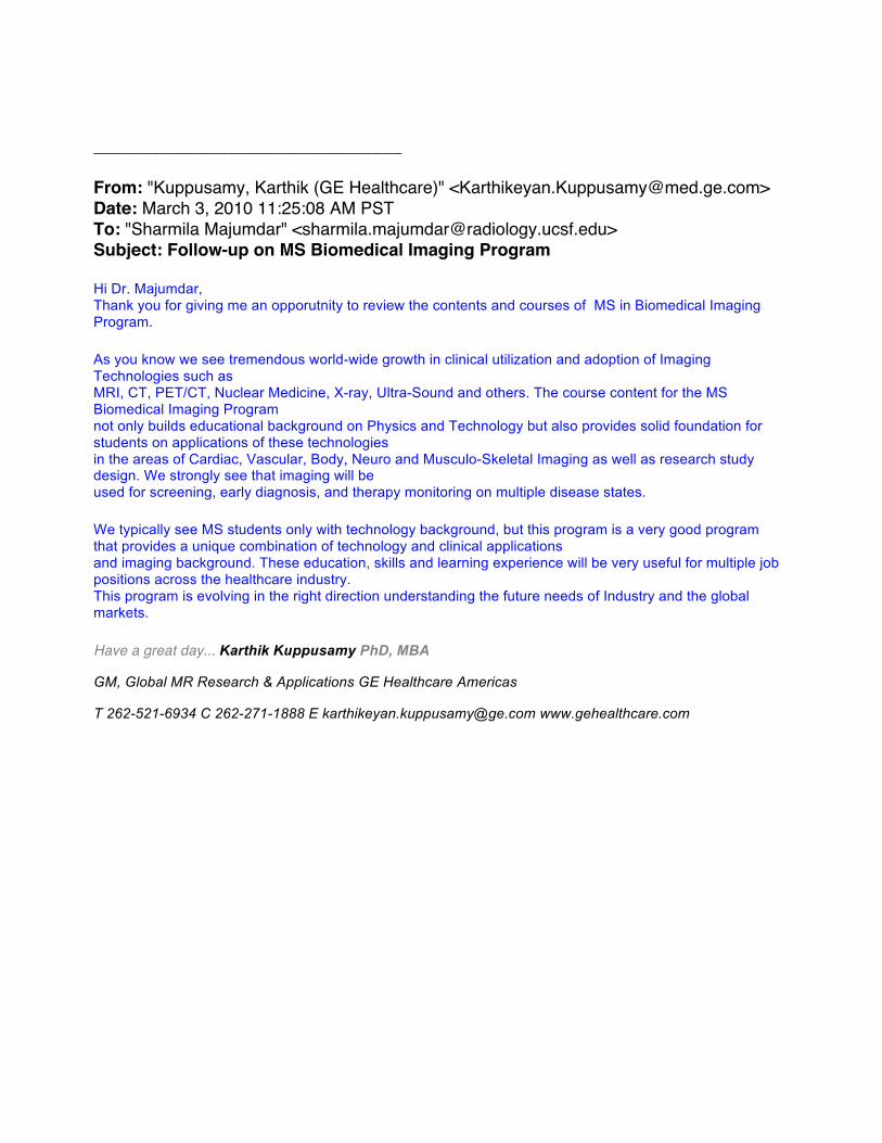
________________________________ From: "Kuppusamy, Karthik (GE Healthcare)" <[email protected]> Date: March 3, 2010 11:25:08 AM PST To: "Sharmila Majumdar" <[email protected]> Subject: Follow-up on MS Biomedical Imaging Program Hi Dr. Majumdar, Thank you for giving me an opporutnity to review the contents and courses of MS in Biomedical Imaging Program. As you know we see tremendous world-wide growth in clinical utilization and adoption of Imaging Technologies such as MRI, CT, PET/CT, Nuclear Medicine, X-ray, Ultra-Sound and others. The course content for the MS Biomedical Imaging Program not only builds educational background on Physics and Technology but also provides solid foundation for students on applications of these technologies in the areas of Cardiac, Vascular, Body, Neuro and Musculo-Skeletal Imaging as well as research study design. We strongly see that imaging will be used for screening, early diagnosis, and therapy monitoring on multiple disease states. We typically see MS students only with technology background, but this program is a very good program that provides a unique combination of technology and clinical applications and imaging background. These education, skills and learning experience will be very useful for multiple job positions across the healthcare industry. This program is evolving in the right direction understanding the future needs of Industry and the global markets. Have a great day... Karthik Kuppusamy PhD, MBA
GM, Global MR Research & Applications GE Healthcare Americas
T 262-521-6934 C 262-271-1888 E [email protected] www.gehealthcare.com
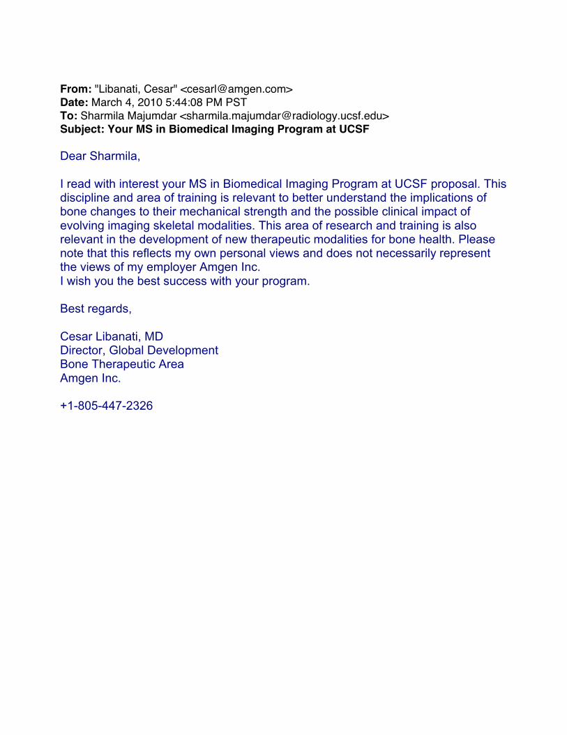
From: "Libanati, Cesar" <[email protected]> Date: March 4, 2010 5:44:08 PM PST To: Sharmila Majumdar <[email protected]> Subject: Your MS in Biomedical Imaging Program at UCSF Dear Sharmila, I read with interest your MS in Biomedical Imaging Program at UCSF proposal. This discipline and area of training is relevant to better understand the implications of bone changes to their mechanical strength and the possible clinical impact of evolving imaging skeletal modalities. This area of research and training is also relevant in the development of new therapeutic modalities for bone health. Please note that this reflects my own personal views and does not necessarily represent the views of my employer Amgen Inc. I wish you the best success with your program. Best regards, Cesar Libanati, MD Director, Global Development Bone Therapeutic Area Amgen Inc. +1-805-447-2326

SIEMENSMarch 12,2010
David Saloner, PhD
Department of Radiology and Biomedical lmaging
University of California San Francisco
San Francisco, CA 94121
Dear Dr. Saloner,
thank you for the opportunity to review the program that your Department is planning that will
develop a Master's of Science in Biomedical lmaging. I am very impressed with the scope and
depth of this program. As you know, Siemens Medical Solutions is a major provider of medical
imaging equipment across all imaging modalities. We firmly believe that the pathway to improved
imaging care in the healthcare system is not only through better equipment but through improved
education. This crosses all levels of practice. There is a need for better training for research
scientists, for individuals who in turn provide training to clinical practitioners, and to the end-users of
the equipment - both physicians and technologists.
I am sure that your program will be an important example for other Departments of Radiology across
the country. I can assure you that Siemens Medical Solutions would be interested in employing
individuals with the type of training that you detailed in your proposal.
I wish you much success with your program.
Sincerely yours,
M/*#Director, MR R&D West
Siemens MedicalSolutions USA, lnc
Siemens Medical Solutions USA' Inc.6700 Koll Center Parkway, Pleasonton, C494566
Tel. (310) 721-2s48gerhard. I au b@ggl]gnseotx

Appendix.2 Letters of Support from UCSF and other UC campuses Kathleen Giacomini, Sarah Nelson co-Chairs, UCSF Dept of Bioengineering and
Therapeutic Sciences Tejal Desai, Chair, UCSF/UC Berkeley Joint Graduate Group in Bioengineering Matthew Tirrell, Chair, Dept of Bioengineering, UC Berkeley Ronald Arenson, Chair, Dept of Radiology and Biomedical Imaging, UCSF Josh Adler, Director, Pathways to Discovery, UCS David Irby, Vice Dean for Education, UCSF William Tang, Acting Chair, Dept of Biomedical Engineering, UC Irvine Kyriacos Athanasiou, Chair, Dept of Biomedical Engineering, UC Davis Michael McNitt-Gray, Director, Interdepartmental Program in Biomedical Physics, UCLA Jerome Schuz, Chair, Department of Bioengineering, UC Riverside Shankar Subramaniam, Chair, Department of Bioengineering, UC San Diego Fyl Pincus, Chair, Program on Biomolecular Science and Engineering, UC Santa Barbara
MS in Biomedical Imaging: Revision Date: 4/29/10
49

March 1, 2010 Sharmila Majumdar, Ph.D. Vice Chair of Research, Professor and Director MQIR Radiology and Biomedical Imaging Campus Box 2520 RE: PROPOSAL FOR A MASTERS IN BIOMEDICAL IMAGING (BMI) Dear Sharmila: We are delighted to endorse your proposal for a Masters of Science in Biomedical Imaging (BMI). We believe that this focused effort is an excellent development and that it will attract a large number of students. Imaging has become a critical tool for many basic science and clinical applications. Having the program that you describe will allow many non-imaging specialists to get up to speed and to determine which methods would be most appropriate for their studies. The Department of Bioengineering and Therapeutic Sciences is actively involved in developing new degree options that will apply engineering principles to medical problems, but we were not including imaging as a focus area. These new programs will most likely be jointly developed with UC Berkeley and will consider micro- and nano-scale technologies, tissue engineering and mechanical devices that will be very complementary to the MBI degree. We anticipate that we may be able to share some elective courses with you but will definitely not be in competition. We were particularly impressed with the breadth of the coursework that you are proposing and the faculty who have agreed to participate. This will provide an important educational experience for a large group of professional students. It is an innovative concept and is likely to be a model for other institutions. Please let us now if we can do anything to help in developing the proposal. Sincerely,
Sarah Nelson, PhD Kathy Giacomini, PhD Professor and Co-Chair Professor and Co-Chair Bioengineering and Therapeutic Sciences Bioengineering and Therapeutic Sciences
Kathleen M. Giacomini, PhD Professor and Co-Chair UCSF Mail Code 2911 Rock Hall Room RH-584F 1550 4th Street San Francisco, CA 94158-2324 tel: 415/ 476-1936 fax: 415/ 514-4361 [email protected] Sarah J. Nelson, Ph.D. Professor and Co-Chair Margaret Hart Surbeck Distinguished Professor, Radiology & Biomedical Imaging 1700 4th Street, Byers Hall, Suite 303 San Francisco, CA 94158-2330 tel: 415/ 476-6383 fax: 415/ 514-2550 [email protected]
http://ucsf.bts.edu
Schools of Pharmacy and Medicine Bioengineering and Therapeutic Sciences
February 11, 2010 Dr. Sharmila Majumdar Professor of Radiology Byers Hall 203 UCSF San Francisco, CA 94158 Re: Support for Masters in Biomedical Imaging Dear Sharmila, I am writing to express my enthusiastic support of the proposed masters program in biomedical imaging. I am excited about the potential training opportunities and the synergy with the existing UCSF/UCB joint graduate group in bioengineering. The ability to provide innovative training opportunities for not just graduate students, but also residents, fellows, and medical students, will be critical in fostering new interdisciplinary research and training activities. This is complimentary and will be a great resource to the proposed Translational Medicine program that I am working on with Clay Johnston (CTSI – UCSF) and Matt Tirrell (UC Berkeley).
I believe the new program will enhance the visibility of bioengineering and imaging activities occurring on the UCSF campus. There is tremendous need to train clinicians in established and emerging aspects of biomedical imaging. The proposed new courses will also be attractive to the students in the joint bioengineering program and will serve to enhance UCSF’s reputation in developing translational technologies. I will ensure that our students are fully aware of the program activities and encouraged to expand their horizons by participating in its seminars and other activities. We look forward to collaborating and interacting with you to ensure the future success of this master’s program. Sincerely yours, Tejal Desai, Ph.D. Chair, UCSF/UC Berkeley Joint Graduate Group in Bioengineering
University of California, Berkeley Department of Bioengineering 306 Stanley Hall #1762 College of Engineering Berkeley, California 94720‐1762 http://bioeng.berkeley.edu (510) 642‐5833
SANTA BARBARA • SANTA CRUZ
BERKELEY • DAVIS • IRVINE • LOS ANGELES • MERCED • RIVERSIDE • SAN DIEGO • SAN FRANCISCO
February 9, 2010 Sharmila Majumdar,Ph.D. Vice Chair for Research, Professor, and Director Musculoskeletal and Quantitative Imaging Research Group (MQIR), Department of Radiology and Biomedical Imaging Campus Box 2520 QB3 Building, 2nd Floor, Suite 203 1700 - 4th Street, University of California, San Francisco San Francisco, CA 94158 Dear Sharmila, I have read the proposal to establish a MS Degree Program in Biomedical Imaging at UCSF with interest. I am convinced that this program meets an under-served need. It is well-designed to introduce the degree candidates to a broad variety of imaging methods and bring them to a practical understanding of the state-of-the-art of the field. I see this new program as a valuable addition to the spectrum of offerings in biomedical technologies. It certainly is not duplicative of any current program on the Berkeley campus. Though we have several people, e.g., Steve Conolly, who are developing advanced imaging methodologies, we do not currently have the breadth on our campus that a full view of the field of biomedical imaging requires.
As we work jointly with UCSF within our Joint Graduate Group in Bioengineering, I see the establishment of this proposed Program as a very positive development. Best wishes for a successful launch.
Sincerely,
Matthew Tirrell Arnold and Barbara Silverman Professor & Chair

University of CaliforniaSan Francisco
U%FDepartment of Radiology and Biomedical Imaging
Ronald L. Arenson, M.D.
Alexander R. MargullsDistinguished Professor February 11, 2010and chairmanDepartment of Radiology andBiomedical Imaging Sharmila Majumdar, PhD505 Parnassus Avenue, M391 Professor In ResidenceSan Francisco, CA 94143-0628 v~°~ Ch~r Rtel: 415/476-1537fax: 415/[email protected] David Saloner, PhD
Professor In Residence
Dear Sharmila and David,
I am delighted to express my enthusiastic endorsement of this proposal fora focused training in Biomedical Imaging, and the development of a Mastersprogram. Our Department provides a broad array of educational opportunities forresidents, fellows, and medical students, but has not had a commensurate formaldegree-granting program for our non-MD students. This Masters program wouldfill that important need, and I am sure, will serve as a model for other Departmentsof Radiology across the country.
The Biomedical Imaging faculty in our department are extraordinary andhave a long-standing history of graduate education, as evidenced by the largenumber of graduate students in our laboratories. In addition, the summer programwith undergraduate students and high school students that has evolved over the lastseveral years is unique and a testimony to the teaching skills of the faculty. Thestudents we have are of the highest quality and are very much in demand amongstour faculty, in corporations and in laboratories. I have played an active role insupporting the program, worked with the faculty to improve their interactions withother groups and encouraged the institution to move forward with a more formalstructure for education. I am pleased to see that this is now coming to fruition.
The proposal that is outlined in this submission is a major step forward inexpanding the educational and training program at UCSF. It is an exciting programthat takes advantage of the resources on the campus and offers opportunities to awide range of students.
As you know, there is a tremendous need for didactic imaging education inthe field of radiological imaging. One example is the new requirement that, startingin 2014, medical physicists who work in hospitals will need certification fromprograms accredited under the Commission on Accreditation of Medical PhysicsEducation Programs (CAMPEP). Our Department will need to be poised to start
Page 2Sharmila Majumdar, PhDDavid Saloner, PhDFebruary 11, 2010
our own CAMPEP-accredited residency program. The course content of this MSprogram will be tremendously useful for our residents by providing a structurededucational lecture and laboratory series, and will facilitate a pathway to establisha CAMPEP-certified residency.
I am willing to provide support from our departmental infra-structure toassist you in implementing your educational and training program. We haveestablished expertise in our Radiology Learning Center, Post-Graduate EducationSection and in the offices of our post-doctoral coordinator for dealing with many ofthe logistical issues that the Master’s program will face. In addition, support fordeveloping the web page for the program, advertising for the first year, andrecruitment resources will be provided by the Department of Radiology andBiomedical Imaging. The use of the classroom and lockers, and a break room forthe students will be available at China Basin Landing in the Center for Functionaland Molecular Imaging. The Departmental recharge process will also be madeavailable to manage costs associated with the laboratory aspects of the courses.
In addition to these specific resources, the University has been extremelyactive in developing programs to help with the academic and professionaldevelopment of students and post-doctoral scholars at UCSF. I am sure that thegraduates of the Master’s program will benefit from that support. The placementof your own trainees in the recent years demonstrates the tremendous demand thatwell-trained scholars have in academia and industry.
Finally, I would like to stress the strong partnership that we have with theCollege of Engineering and the Department of Bioengineering at UC Berkeley, andthe Department of Bioengineering and Therapeutic Sciences at UCSF. Theeducatipnal program that the two institutions have developed has a high degree ofsynergy and has attracted a large population of undergraduate and graduatestudents to UCSF. The enhancements that are proposed as part of this applicationwill have a major impact upon the quality and flexibility of the scholars.
Sincerely,
Ronald L. Arenson, MD
U%F Medical Center
Administration500 Parnassus AvenueSan Francisco, CA 94143-0296 p i,tel: 415/353-2733 ~ e~ruary ~fax: 415)353-2765flV: 415)885-3889www.ucsfhealth.org
University of California David Saloner, Ph.D.San Francisco Co-chair Committee on Masters of Science in Biomedical Imaging Department of
Radiology and Biomedical ImagingUCSF505 Pamassus AvenueSan Francisco, CA 94 143-0628
Dear David:
I have reviewed the proposal for a Masters of Science in Biomedical Imaging thatyour committee has developed. The proposal appears to be solid andcomprehensive. I am particularly excited about the opportunities that this formaleducational structure would provide to students in the Pathways to Discoveryprogram who would like to bolster their understanding of the role of imaging indiagnosing and monitoring disease and the response of disease to therapy. Suchinsights will be important in planning and evaluating the delivery of improvedhealth care both across the U.S. and globally. I believe that students in thePathways to Discovery program can benefit from many of the courses that will beprovided by your proposed program and I therefore would like to express mystrong support for this proposal.
Sincerely,
Adler, M.D.Professor of Clinical MedicineChief Medical OfficerUCSF Medical Center
David M. Irby, Ph.D. Vice Dean for Education
Office of Medical Education 521 Parnassus Avenue Room C-254 San Francisco, CA 94143-0410 tel. 415/502-1633 fax 415/514-0468 email: [email protected] http://medschool.ucsf.edu/education/
March 16, 2010 Ron Arenson, MD Professor and Chair Department of Radiology UCSF School of Medicine Dear Dr. Arenson, I strongly support the concept of a Master’s degree program in Biomedical Imaging. I know that this is a growing field and many physicians will need to have an in-depth understanding of imaging. As you undoubtedly know, many of our medical students come in with engineering and basic science degrees, and this program would be an ideal option for those who want to utilize their prior scientific background in medicine. As we continue to develop the Pathways to Discovery program, we are looking for master’s degree program options for our students. I am sure that this program would be of interest to a number of our students. As we convert to a competency-based curriculum, we are requiring our medical students to complete an electronic Portfolio, utilize the Collaborative Learning Environment (an on-line learning resource) and rate all courses using electronic course evaluations. I am delighted to see that all of these tools will be utilized in your proposed program. I wish you the best in your efforts to get this Masters Program approved and launched. Sincerely yours, David M. Irby, PhD Vice Dean for Education Professor of Medicine
U N I V E R S I T Y O F C A L I F O R N I A , I R V I N E
BERKELEY • DAVIS • IRVINE • LOS ANGELES • MERCED • RIVERSIDE • SAN DIEGO • SAN FRANCISCO SANTA BARBARA • SANTA CRUZ
DEPARTMENT OF BIOMEDICAL ENGINEERING HENRY SAMUELI SCHOOL OF ENGINEERING
3120 Natural Sciences II Irvine, CA 92697-2715
William C. Tang, Ph.D.
William J. Link Professor and Acting ChairVoice: (949) 824-8155
Fax: (949) 824-1727E-mail: [email protected]
February 18, 2010
David Saloner, Ph.D. Co‐chair Committee on Masters of Science in Biomedical Imaging Department of Radiology and Biomedical Imaging UC San Francisco 505 Parnassus Avenue San Francisco, CA 94143‐0628
Dear Professor Saloner,
I am writing to express my support for the proposed graduate study program in Biomedical Imaging for the MS Degree at the University of California, San Francisco. After reviewing the proposal, I believe it will be a positive contribution to the educational missions of the UC system. It appears that this is a training program designed for a broad range of students having finished bachelor’s degree, including post‐doctoral fellows, residents, researchers, and faculty members. This one‐year (full‐time) course of study aims at providing the skills and knowledge in the rapidly growing interdisciplinary imaging tools for biomedical investigative projects. This should be a key element of the Program in Quantitative Biology, and should be intended as a supplement to the other components that span across Physical Sciences, Medicine, and Engineering. In particular, since this is structured to be a transitional MS degree that does not necessarily lead to a doctoral program, there would be minimal overlap with existing programs that do. UCSF is also well positioned to offer this program, given the strengths and experiences in graduate trainings across the health‐science campus. As your program evolves in future years, you may wish to consider adding a requirement that your students file a master’s thesis. Our experience has shown hands‐on research to be valuable to our students and to their future employers. However, I appreciate the constraints that your program will have to work under in order to provide a comprehensive educational experience in one year even without a thesis requirement, and, as it stands, it is clear that your proposed program will provide solid training to your students. Please feel free to contact me if I can be of further assistance.
Sincerely,
William C. Tang, PhD
D E P A R T M E N T O F B I O M E D I C A L E N G I N E E R I N G U N I V E R S I T Y O F C A L I F O R N I A , D A V I S
O N E S H I E L D S A V E N U E D A V I S , C A 9 5 6 1 6
T E L E P H O N E : ( 5 3 0 ) 7 5 4 - 6 6 4 5
F A X : ( 5 3 0 ) 7 5 4 - 5 7 3 9 E M A I L : a t h a n a s i o u @ u c d a v i s . e d u
February 16, 2010 David Saloner, Ph.D. Co-Chair, Committee on Masters of Science in Biomedical Imaging Department of Radiology and Biomedical Imaging UC San Francisco 505 Parnassus Avenue San Francisco, CA 94143-0628 Dear Dr. Saloner, Thank you for forwarding me the draft proposal for a Master of Science in Biomedical Imaging that your committee proposes to establish in the Department of Radiology and Biomedical Imaging at UC San Francisco. Your proposed program will make a significant contribution to the education of individuals who wish to develop strong expertise in the use of the important imaging modalities that are playing an increasing role in translational research. The courses described in the program will provide a sound basis for understanding the physical principles and practical steps needed to conduct rigorous and systematic investigations in imaging studies both in the pre-clinical and in the clinical setting. In my estimation, this program would provide a useful educational resource to the University of California system. I have discussed the elements of your proposal with my imaging/optics colleagues in the BME Department at UC Davis, and we support your efforts. I appreciate that you have kept me apprised of this development and wish you the best of luck with this proposal. Sincerely,
Kyriacos Athanasiou, Ph.D., P.E. Distinguished Professor Chair, Department of Biomedical Engineering Editor-in-Chief, Annals of Biomedical Engineering
BIOMEDICAL PHYSICS INTERDEPARTMENTAL GRADUATE PROGRAM DAVID GEFFEN SCHOOL OF MEDICINE AT UCLA
LOS ANGELES, CALIFORNIA 90095-7319
March 1, 2010 David Saloner, PhD Co-chair Committee on Masters of Science in Biomedical Imaging Department of Radiology and Biomedical Imaging UC San Francisco 505 Parnassus Avenue San Francisco, CA 94143-0628 SUBJECT: Letter of Support for Creation of UCSF Masters of Science in Biomedical Imaging Program Dear Dr. Saloner: I am writing this letter in support of your efforts to create a Masters of Science in Biomedical Imaging program at UCSF. I have been the Director of the Biomedical Physics IDP Graduate Program since 2004 and so I am familiar with some of the opportunities and challenges you face. That said, there are tremendous resources available to you for this program. The faculty you have recruited into this effort are outstanding and the facilities you have at your disposal at UCSF are also outstanding. These are the foundation for an excellent training program. There is certainly a need for increased training in biomedical imaging techniques. The advances in technology in each of the imaging modalities is exceptional, which has led to an explosion of clinical applications. It is difficult to keep up with the demand for increased training at all levels – physicists, radiologists, technologists, researchers, etc. Your program gives an opportunity for training in several imaging modalities and also exposes your trainees to quantitative imaging techniques, which are also very important to the future of our field. The program that we offer at UCLA is quite different. While we do offer MS and PhD degrees, our primary focus at the moment is on research and therefore the training of students who wish to pursue the PhD degree. Our students tend to come from physics or biology or engineering backgrounds; we attract very few medical students (or even MDs) into our program. Because our program is CAMPEP (Commission on Medical Physics Education Programs) accredited, our program is necessarily broader than just biomedical imaging. We have four tracks: Medical Imaging (most similar to your program), Molecular Imaging (also some similarities), Therapeutic Medical Physics and Molecular and Cellular Oncology. Therefore, we have additional course requirements (such as the fundamentals of dosimetry, basics of radiation therapy, radiation biology) that would be different from your program. Our MS degree track (rarely used) typically takes two years to complete. This is so that these graduates will be competitive for CAMPEP approved residency programs (which currently are very competitive, even for our PhD graduates).
In summary, I think that the training program you and your colleagues have proposed fills a very important need with respect to biomedical imaging. I think the foundation of the program is strong with the faculty and facilities available at UCSF. I wish you all the best in this very important endeavor. Sincerely,
Michael F. McNitt-Gray, Ph.D., DABR Professor Department of Radiology Director, IDP in Biomedical Physics David Geffen School of Medicine at UCLA Email: [email protected] Phone: +1 310-794-8979
Jerome S. Schultz Chairman Department of Bioengineering 900 University Avenue A321 Bourns Hall Riverside CA 92521
February 16, 2010 David Saloner, PhD Co-chair Committee on Masters of Science in Biomedical Imaging Department of Radiology and Biomedical Imaging UC San Francisco 505 Parnassus Avenue San Francisco, CA 94143-0628 Re: Propose New M.S. Degree In Biomedical Imaging Dear Dr. Saloner: Thank you for the opportunity to review your proposed new masters degree program in Biomedical Imaging. First let me complement you and your associates on your thorough and persuasive presentation of the need and value of this proposed program. I fully agree that biomedical imaging is a critical component of modern medicine for diagnosis, cure, and rehabilitation. Your new training program in this area would notably enhance the capability of professionals who share the responsibility of conducting these procedures in both the clinical and research setting. Your program would certainly enhance the service that the University of California provides to the medical community and the population it serves. Your group has the unique capability to provide clinically related training that is scarce in other institutions. We would be delighted to recommend your new program to our undergraduates with an interest in furthering their careers in biomedical imaging. I am highly supportive of your program and wish you success in this endeavour. Sincerely yours,
Jerome S. Schultz Member, National Academy of Engineering
Shankar Subramaniam Joan and Irwin Jacobs Professor of Bioengineering & Systems Biology Chair, Department of Bioengineering Tel: (858) 822-0986 Professor of Bioengineering, Chemistry & Biochemistry, Cellular & Molecular Medicine [email protected] and Nanoengineering University of California at San Diego 9500 Gilman Drive La Jolla, CA 92093-0427 February 18, 2010 Professor Sharmila Majumdar, Ph.D. Campus Box 2520 QB3 Building, 2nd Floor, Suite 203 1700 - 4th Street, University of California, San Francisco San Francisco, CA 94158 Sub: Your Proposal to establish a program in Graduate Studies in Biomedical Imaging Dear Dr. Majumdar:
I am writing to congratulate you and strongly support your proposal to establish a Graduate (Master’s) Program in Biomedical Imaging.
With the advent of very sophisticated imaging modalities, it is exceptionally timely to train our science and medical graduates with strong foundations in imaging and your proposed Program will address this need.
I have gone through your proposal in detail and it is very clear that substantial thought and planning has gone into it and I commend you and colleagues on the preparation. The proposed course load is appropriate with the 6 core courses. I also would recommend that your M.D. students be allowed to take some of these courses if they so desire.
The job opportunities for students getting a Master’s degree in Biomedical Imaging is likely to be very significant with medical institutions, radiology laboratories and other research institutions competing for hiring the graduates. Having spoken to our large Undergraduate Student body in UCSD, I can assure you that our students would love to join your program after graduating with a BS degree in Bioengineering.
I would like also to note that UCSD does not have a comparable program in Biomedical Imaging. Neither our Bioengineering nor our Ph.D. only Bioinformatics Program address Imaging.
I am looking forward to the establishment and success of your program and it will help us in the future share your curriculum for our students.
With best regards,
Sincerely,
Shankar Subramaniam, Ph.D.
UNIVERSITY OF CALIFORNIA, SANTA BARBARA
BERKLEY ∙ DAVIS ∙ IRVINE ∙ LOS ANGELES ∙ RIVERSIDE ∙ SAN DIEDO ∙ SAN FRANCISCO SANTA BARBARA ∙ SANTA CRUZ
PHILIP PINCUS
MATERIALS RESEARCH LABORATORY SANTA BARBARA, CALIFORNIA 93106‐5121
FAX: (805) 893‐8797
TEL: (805) 893‐4685
February 21, 2010
David Saloner, PhD Co-chair Committee on Masters of Science in Biomedical Imaging Department of Radiology and Biomedical Imaging UC San Francisco 505 Parnassus Avenue San Francisco, CA 94143-0628
Dear David,
On behalf of the Program on Biomolecular Science & Engineering at UCSB, I would like to lend my support to your proposal Masters of Science program in Biomedical Imaging at UCSF. It is very well thought out with an emphasis on medical imaging. The necessary personnel are in place and the curriculum is excellent. The emphasis on imaging is certainly timely with many technological advances engendering new techniques with improved resolution.
The proposed Program is a welcome addition to our increasing emphasis at UC in interdisciplinary modern biology.
Best regards,
Philip Pincus
Appendix.3 Information Required by CPEC
This questionnaire is to be completed by sponsoring faculty (department or group). It will be used by Systemwide Administration to prepare a report to the California Postsecondary Education Commission. If more space is required, please attach as many additional sheets as necessary. Attach to full proposal.
1. Name of Program: Masters of Science in Biomedical Imaging
2. Campus:
UCSF
3. Degree/Certificate:
Master of Science
4. CIP Classification (to be completed by Office of the President):
5. Date to be started:
September 2011
I6. If modification of existing program, identify that program and explain changes.
N/A
7. Purpose (academic or professional training) and distinctive features (how does this program differ from others, if any, offered in California?):
The “Master of Science in Biomedical Imaging” Master's Degree Program is a one-year academic course of study for students who wish to master imaging-oriented research methods to enhance their research designs and broaden their investigative projects. Course work includes instruction in core theory and provides a foundation in interdisciplinary scholarship. The master’s program will provide a field of interdisciplinary academic investigation that examines the many facets of imaging, both at the molecular and cellular level and also translational aspects from the whole organ to clinical applications.
8. Type(s) of students to be served:
Students with Bachelor’s degrees, medical students pursuing a master’s option, advanced pre-doctoral students, residents, and others who wish to augment their professional qualifications.
MS in Biomedical Imaging: Revision Date: 4/29/10
64

9. If program is not in current campus academic plan, give reason for proposing program now:
The proposed program offers an integrated, in-depth training opportunity for individuals who wish to develop expertise in the technical and practical aspects of cutting-edge Radiological imaging techiques. Imaging methodologies have evolved rapidly over the past few decades and are used increasingly in multi-center studies as surrogate markers of disease state. There is an increasing demand for individuals with rigorous trainig in these methods to conduct clinical research studies. The program will be housed in the Department of Radiology and Biomedical Imaging at UCSF. The department has a total of 40 Ph.D. faculty specializing in Biomedical Imaging, in addition there are 47 non-faculty academic appointees in the Researcher and Specialist series, all of whom can participate in course instruction.
10. If program requires approval of a licensure board, what is the status of such approval?
N/A
11. Please list special features of the program (credit for experience, internships, lab requirements, unit requirements, etc.)
N/A
12. List all new courses required: Department, Course Number, Title, Hours/Week Lecture Lab.
Proposed New Required Courses BI 201. Principles of Magnetic Resonance Imaging; 4 hours/week lecture; BI 202. Principles of X-Ray Imaging and CT; 4 hours/week lecture; BI 203. Principles of Radionuclide Imaging and SPECT/PET; 4 hours/week lecture; BI 204. Introduction to Optical and Ultrasound Imaging; 4 hours/week lecture; BI 205. Imaging Study Design ; 3 hours/week lecture.
Proposed New Elective Courses BI 210.Imaging Agents – Radiopharmaceuticals and Contrast Media; 3 hours/week lecture; BI 220. Introduction to Cancer Imaging; 3 hours/week lecture; BI 230. Introduction to Vascular Imaging; 3 hours/week lecture; BI 240. Introduction to Musculoskeletal Imaging; 3 hours/week lecture; BI 250. Supervised Research. 9 hours/week; Laboratory; BI 260. Current Topics in Molecular Imaging; 1 hours/week lecture; 1 hour/week library; BI 270. Current Topics in Cancer/Neuro/Vascular Imaging; 1 hour/week lecture; 1 hour/week library; BI 280. Current Topics in Image Analysis/Data Mining/Biostatistics; 1 hour/week lecture; 1 hour/week library
MS in Biomedical Imaging: Revision Date: 4/29/10
65
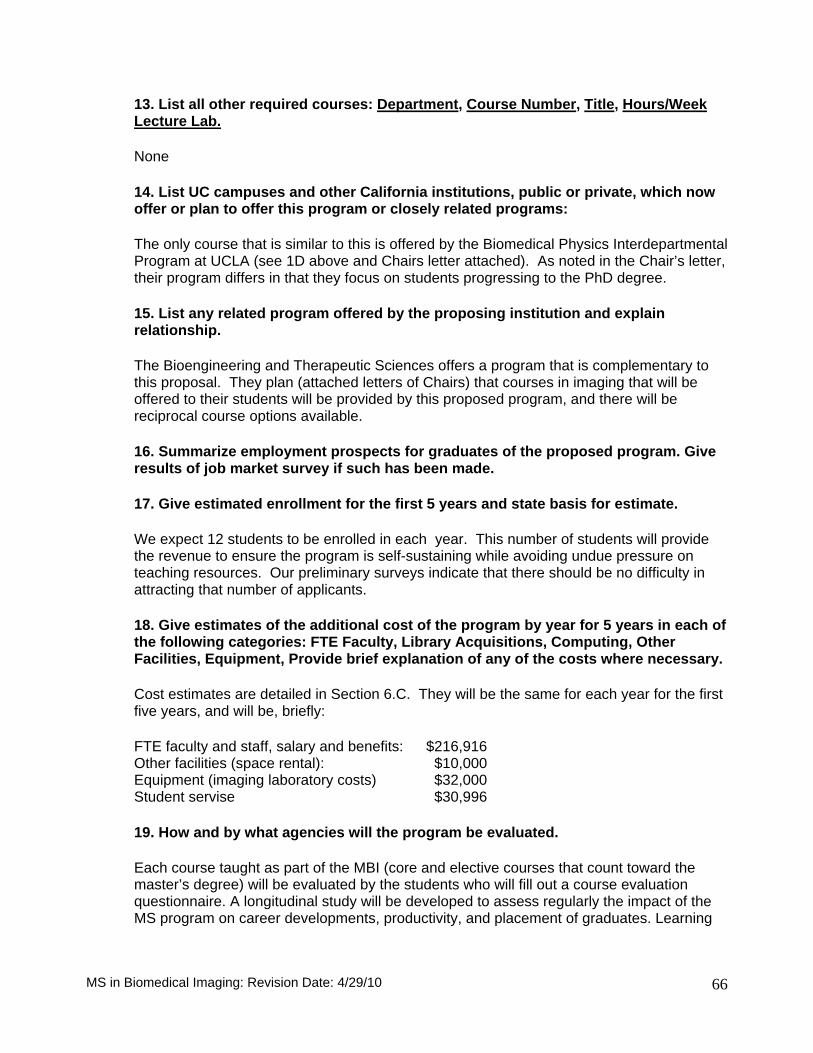
13. List all other required courses: Department, Course Number, Title, Hours/Week Lecture Lab.
None
14. List UC campuses and other California institutions, public or private, which now offer or plan to offer this program or closely related programs:
The only course that is similar to this is offered by the Biomedical Physics Interdepartmental Program at UCLA (see 1D above and Chairs letter attached). As noted in the Chair’s letter, their program differs in that they focus on students progressing to the PhD degree.
15. List any related program offered by the proposing institution and explain relationship.
The Bioengineering and Therapeutic Sciences offers a program that is complementary to this proposal. They plan (attached letters of Chairs) that courses in imaging that will be offered to their students will be provided by this proposed program, and there will be reciprocal course options available.
16. Summarize employment prospects for graduates of the proposed program. Give results of job market survey if such has been made.
17. Give estimated enrollment for the first 5 years and state basis for estimate.
We expect 12 students to be enrolled in each year. This number of students will provide the revenue to ensure the program is self-sustaining while avoiding undue pressure on teaching resources. Our preliminary surveys indicate that there should be no difficulty in attracting that number of applicants.
18. Give estimates of the additional cost of the program by year for 5 years in each of the following categories: FTE Faculty, Library Acquisitions, Computing, Other Facilities, Equipment, Provide brief explanation of any of the costs where necessary.
Cost estimates are detailed in Section 6.C. They will be the same for each year for the first five years, and will be, briefly:
FTE faculty and staff, salary and benefits: $216,916 Other facilities (space rental): $10,000 Equipment (imaging laboratory costs) $32,000 Student servise $30,996
19. How and by what agencies will the program be evaluated.
Each course taught as part of the MBI (core and elective courses that count toward the master’s degree) will be evaluated by the students who will fill out a course evaluation questionnaire. A longitudinal study will be developed to assess regularly the impact of the MS program on career developments, productivity, and placement of graduates. Learning
MS in Biomedical Imaging: Revision Date: 4/29/10
66

MS in Biomedical Imaging: Revision Date: 4/29/10
67
portfolios (“ePortfolio”) will be used to showcase student learning, provide a framework for assessing academic progress, and demonstrate how skills have developed over time.
The Director of Graduate Studies for the MBI degree program will be responsible for keeping records and data that will provide the basis for annual reports on the performance of the master’s program presented to the MS Committee. This information will also be used to facilitate a quinquenniel review performed by a peer review committee.
The peer review committee will be comprised of a minimum of four colleagues from outside UCSF who are faculty in existing academic programs in Biomedical Imaging or any related academic fields that would provide familiarity with current scholarship and professional standards.