Embed Size (px)
Citation preview
Prone breast dosimetry Kirby page 1
Prone versus supine positioning for whole and partial breast radiotherapy: a
comparison of non-target tissue dosimetry
Kirby AM1, Evans PM2, Donovan EM1, Convery HM1, Haviland JS2, Yarnold JR2
1Royal Marsden NHS Foundation Trust, Sutton, UK and 2Institute of Cancer Research, Sutton, UK
Corresponding author: Dr Anna M. Kirby FRCR, Department of Academic Radiotherapy, Royal
Marsden NHS Foundation Trust, Downs Road, Sutton, Surrey, SM2 5PT, United Kingdom.
Telephone: +44 208 661 3169. Fax: +44 208 661 3107. E-mail: [email protected]
Acknowledgements: Dr A Kirby was funded by a Royal College of Radiologists’ Clinical Oncology
Research Fellowship and by the Breast Cancer Research Trust. The authors are grateful to Mr
Craig Cummings in the Institute of Cancer Research Workshop for his assistance in the design
and manufacture of the prone breast platform. The work was undertaken in The Royal Marsden
NHS Foundation Trust which receives a proportion of its funding from the NHS Executive; the
views expressed in this publication are those of the authors and not necessarily those of the NHS
Executive.
Key words: Breast cancer; Prone breast radiotherapy; Partial breast irradiation; Cardiac dosimetry
Conflict of interest: None declared.
Prone breast dosimetry Kirby page 2
Abstract
Purpose: To compare non-target tissue (including left-anterior-descending coronary-artery (LAD))
dosimetry of whole (WBI) and partial-breast irradiation (PBI) planned in prone versus supine
positions.
Methods and materials: Sixty-five post-lumpectomy breast cancer patients underwent CT-
imaging supine and prone. On each dataset, whole-breast clinical-target-volume (CTV) was
defined using wire and partial-breast CTV as tumour-bed+15mm. Heart and LAD were outlined in
left-breast-affected patients (n=30), and ipsilateral lung and chest-wall in all patients. Tangential-
field WBI and PBI plans were generated for each position. Mean LAD, heart, and ipsilateral lung
doses (NTDmean), maximum LAD (LADmax) doses, and chest-wall V50Gy were compared.
Results: 260 plans were generated. Prone positioning reduced heart and LAD doses in 19/30 WBI
cases (mean reduction in LAD-NTDmean=7.5Gy) and 7/30 PBI cases (mean reduction in LADmax
=27.6Gy). However, prone positioning increased cardiac doses in 8/30 WBI (mean increase in
LAD-NTDmean=8.4Gy) and 19/30 PBI cases (mean increase in LADmax=23.7Gy). WB-
CTV>1000cm3 was associated with improved cardiac dosimetry in the prone position for WBI
(p=0.04) and PBI (p=0.02). Prone positioning reduced ipsilateral-lung NTDmean in 65/65 WBI and
61/65 PBI cases, and chest-wall V50Gy in all WBI cases. PBI reduced normal-tissue doses
compared to WBI in all cases, regardless of treatment position.
Conclusions: In the context of tangential-field WBI and PBI, prone positioning is likely to benefit
left-breast-affected women of cup-size ≥D, and most right-breast-affected women, but to be
detrimental in left-breast-affected women of smaller cup size. PBI reliably reduces normal-tissue
doses compared to WBI such that eligible women should be encouraged to participate in PBI
studies.

Prone breast dosimetry Kirby page 3
Introduction
Whole-breast irradiation (WBI) following breast-conserving surgery (BCS) improves local control
and survival from breast cancer but increases non-breast-cancer-related deaths by 1% at 15 years
(1). The majority of these deaths are cardiovascular in origin (2) and irradiation of the left-anterior-
descending coronary-artery (LAD) is implicated in pathogenesis (3-5). Irradiation of lung, chest-
wall and other tissues also contributes to late mortality and morbidity (1, 2). Improvements in
radiotherapy techniques have resulted in reduced normal-tissue doses (6), correlating with
reduced non-breast-cancer-related mortality (2). Nonetheless doses to the heart, LAD and lung
from current standard supine WBI remain significant (7).
Methods by which normal-tissue doses could be decreased include optimization of patient
positioning and use of partial-breast irradiation (PBI). Prone positioning for WBI improves dose
homogeneity within breast-tissue (8, 9), reduces lung doses (10-12), and reduces wedge
requirements with consequent reduction of scattered dose, particularly in women of larger cup-size
(10). However, reports comparing cardiac dosimetry from supine versus prone WBI are conflicting.
One study using IMRT (13) reported that prone positioning reduced within-field heart volume in
85% of women. However, studies using conventional tangential-field arrangements have failed to
show an overall benefit of prone positioning on cardiac dosimetry (11, 14). Furthermore, a study
comparing distances between anterior pericardium and chest-wall on supine CT-images and prone
MRI (15) found prone positioning to systematically displace supero-lateral aspects of heart-tissue
closer to chest-wall, such that prone positioning might be detrimental where target tissues include
chest-wall and/or deeply-lying breast tissue. Thus far, LAD doses from prone positioning have not
been documented.
PBI could also reduce normal-tissue doses by restricting higher radiation doses to the volume of
breast tissue at highest risk of tumour relapse (16). The combination of prone positioning and PBI
is particularly attractive in terms of reducing normal-tissue toxicity. A dosimetric comparison of
accelerated PBI techniques (14) found that brachytherapy & prone tomotherapy resulted in least
Prone breast dosimetry Kirby page 4
dose to lung and non-target ipsilateral breast tissue (compared with supine 3D-conformal-RT and
tomotherapy). However, heart doses again varied between patients, prone tomotherapy benefiting
only women with larger breasts and/or lesions further from chest-wall.
Cardiac and, in particular, LAD dosimetry from prone WBI and PBI require further evaluation
before prone positioning can be adopted into routine practice. As the majority of centres worldwide
continue to use conventional tangential-field arrangements, this study prospectively evaluates non-
target tissue exposure from WBI and PBI planned conventionally in supine and prone positions
using within-patient comparison. The resulting four-way comparison of prone and supine, WBI and
PBI aims to clarify optimal approaches to reducing normal-tissue exposure applicable to the
majority of current practices.
Methods and materials
This study was approved by the Royal Marsden Committee for Clinical Research and the Regional
Ethics Committee. All women had undergone BCS for unifocal T1-2 G1-3 invasive-ductal
carcinoma or high-grade ductal-carcinoma-in-situ, at which time titanium-clips were placed in the
tumour-bed (TB) according to a UK protocol (17).
Patient positioning and image acquisition
Patients underwent non-contrast CT-imaging (slice-thickness 1.5mm, C6 to below diaphragm) in
the standard supine position at which time radio-opaque wire was placed at the palpable edge of
breast-tissue circumferentially. Patients were repositioned and CT-imaged prone using an in-
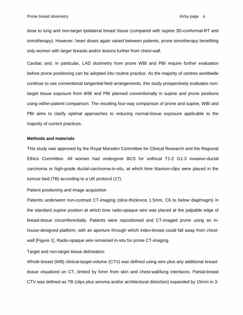
house-designed platform, with an aperture through which index-breast could fall away from chest-
wall [Figure 1]. Radio-opaque wire remained in-situ for prone CT-imaging.
Target and non-target tissue delineation
Whole-breast (WB) clinical-target-volume (CTV) was defined using wire plus any additional breast-
tissue visualized on CT, limited by 5mm from skin and chest-wall/lung interfaces. Partial-breast
CTV was defined as TB (clips plus seroma and/or architectural distortion) expanded by 15mm in 3-

Prone breast dosimetry Kirby page 5
D (limited circumferentially by WB-CTV). Planning-target-volumes (PTV) were generated by
addition of 3-D 10mm margins to CTV, limited by 5mm from skin.
Heart and LAD were defined according to published criteria (6). Where LAD was difficult to
visualise, its location was inferred from the course of the anterior-interventricular groove (6).
Consistent with previous practice, an 10mm axial margin was added to the LAD to allow for
delineation-uncertainty, respiratory-motion and cardiac-motion (7). Ipsilateral-lung was outlined
using an autocontour tool (edited to exclude major airways). Chest-wall was defined as ipsilateral
ribcage and intercostal musculature.
Radiotherapy-planning
For each position, standard opposing tangential-fields were employed such that for WBI, ≥90% of
WB-CTV was encompassed by the 95%-isodose and, for PBI, ≥95% of partial-breast CTV was
encompassed by the 95%-isodose (according to UK National-Cancer-Research-Institute Intensity-
Modulated Partial Organ Radiotherapy (IMPORT) study criteria (18)). Plans fulfilled ICRU dose-
homogeneity criteria (19). Dose-distributions were reviewed in 3D and using dose-volume-
histogram (DVH) data. 50Gy in 25 fractions over 5 weeks (6-10MV photons) was prescribed to the
100% isodose. MLC leaves were used as required to reduce cardiac doses whilst maintaining
satisfactory coverage of WB and PB-CTVs as defined above.
Analysis
NTDmean (a biologically-weighted mean of dose to normal-tissue normalised to 2Gy fractions (20))
was calculated for heart, LAD, and lung. Maximum-dose to LAD (LADmax) was read from DVH data.
The volume of chest-wall receiving 50Gy (V50Gy) was calculated.
Differences between heart, LAD and lung-NTDmean, LADmax, and chest-wall V50Gy for supine versus
prone WBI, supine versus prone PBI, and supine WBI versus PBI plans were calculated for each
patient and compared using the paired t-test or Wilcoxon signed-rank test, as appropriate. Lung
and chest-wall doses were compared in all patients. Cardiac doses were compared for left-breast-
affected patients only (as a population and by individual patient). Individual-patient cardiac data

Prone breast dosimetry Kirby page 6
were pooled by WB-CTV into tertiles (≤500cm3, 501-1000cm3, >1000cm3) and the likelihood of
benefit from prone positioning compared between tertiles using analysis-of-variance.
Results
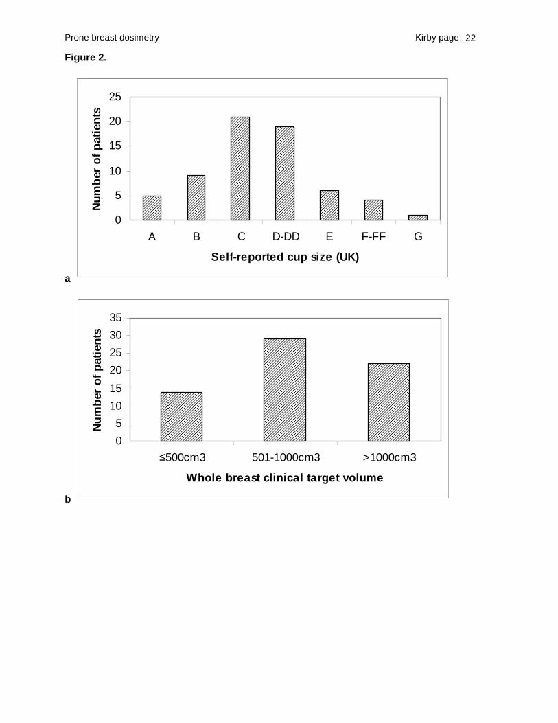
65 women were recruited (30 left-sided: 35 right-sided BC). Mean age was 57 years (range 34-79
years). Self-reported cup-sizes and WB-CTVs are illustrated in figure 2. There was no difference
between WB-CTVs delineated in supine and prone positions (p=0.15). Satisfactory coverage of
target-volumes was achieved in 100% of plans.
Cardiac dosimetry
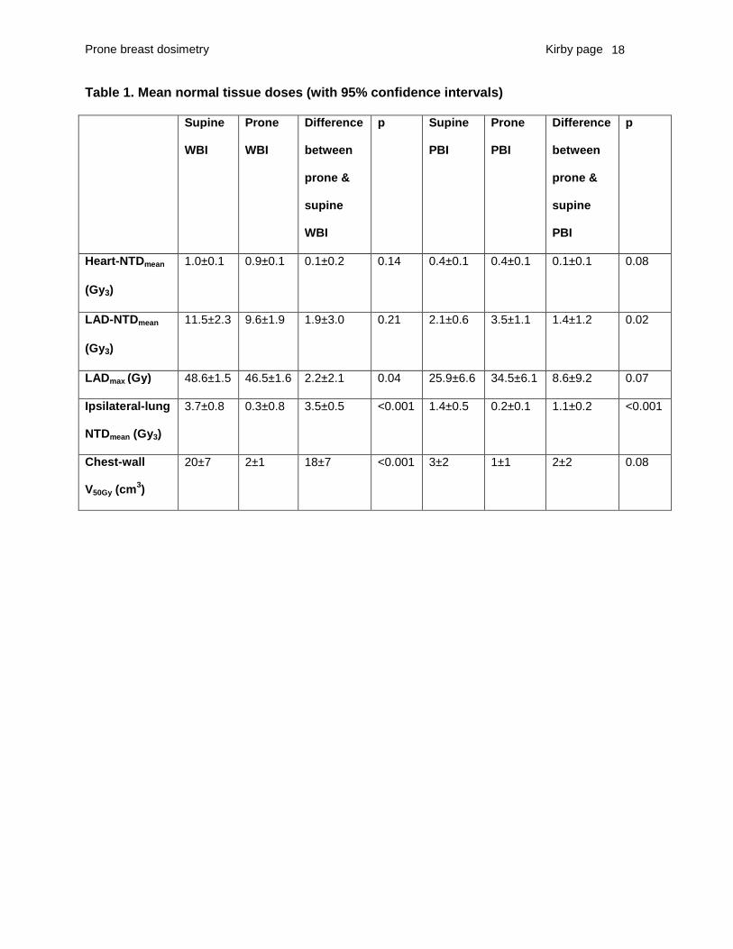
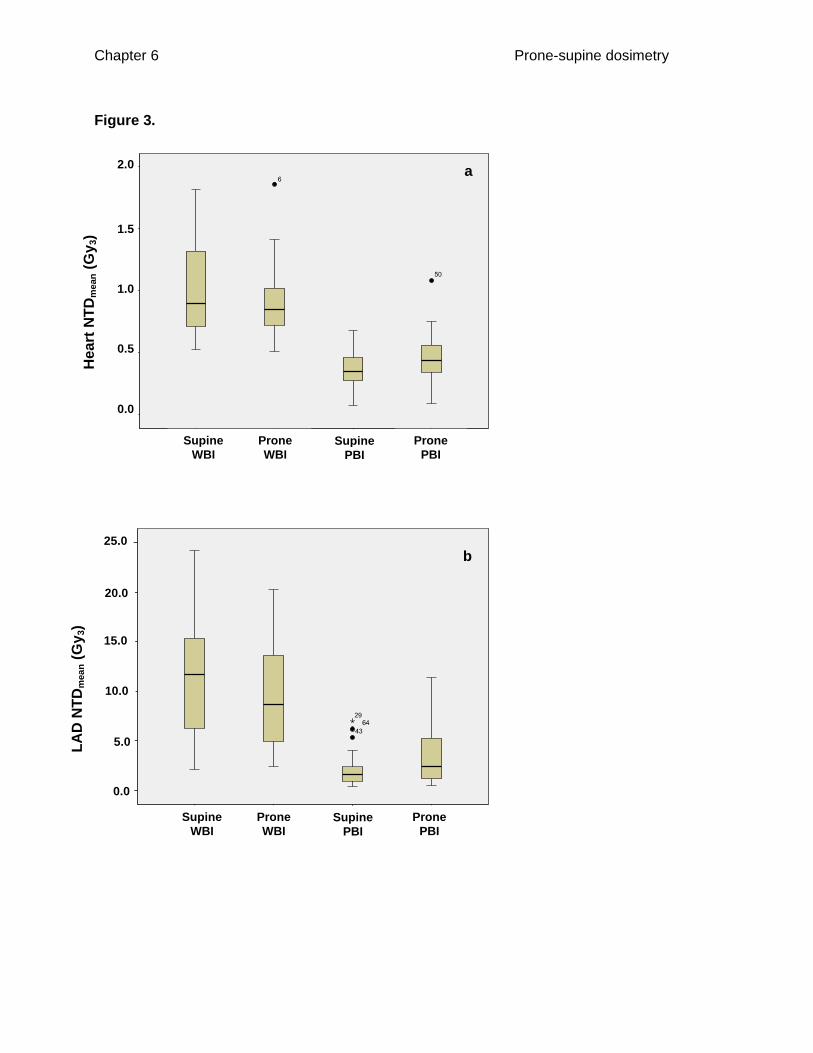
Heart and LAD doses for the patients with left BC (30 cases, 120 plans) are summarized in table 1
and figure 3. Overall, for the population of left-breast-affected women, there was no significant
difference in cardiac parameters between prone and supine positions for either WBI or PBI.
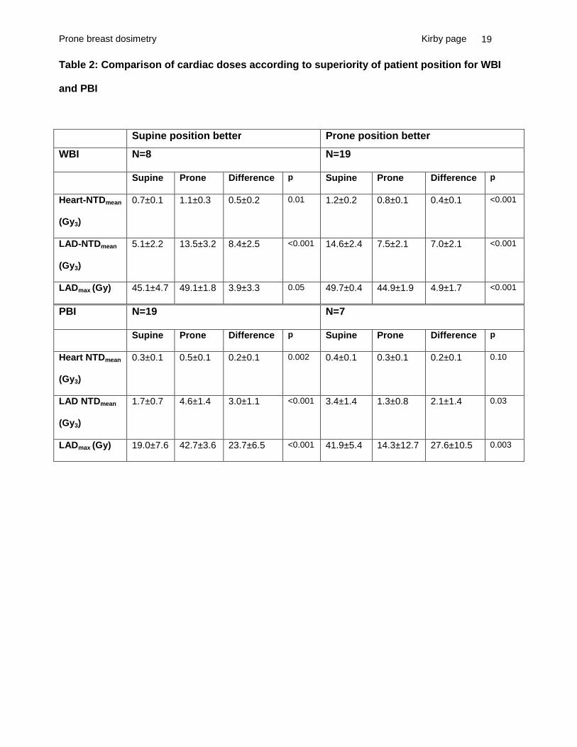
Comparing doses within patients, for WBI prone positioning was advantageous in 19/30 patients
but disadvantageous in 8/30 patients. The magnitude of the difference between LAD-NTDmean
doses was similar whether prone positioning improved or worsened dosimetry [table 2]. In 3/30
patients, prone positioning increased LAD-NTDmean but decreased LADmax. If heart-NTDmean had
been the only comparator, 20/30 would have been judged to benefit from prone and 10/30 from
supine positioning.
For PBI, prone positioning was advantageous in 7/30 patients, and disadvantageous in 19/30
patients. The magnitude of the difference between LADmax doses was similar whether prone
positioning improved or worsened dosimetry. In 4/30 cases, parameters failed to agree on optimal
position. If heart NTDmean had been the only comparator, 13/30 patients would have been judged to
benefit from prone and 17/30 from supine positioning.
Across all 60 WBI-plans for left-breast-affected cases, LADmax was high (≥29.9Gy) irrespective of
treatment position. The mean difference in LADmax for prone versus supine WBI was 4.5±2.5Gy.
There was greater inter-patient variability in LAD NTDmean (mean difference in prone versus supine
LAD-NTDmean=7.7±2.3Gy3). Conversely, across all 60 PBI plans, LAD-NTDmean was low (mean
Prone breast dosimetry Kirby page 7
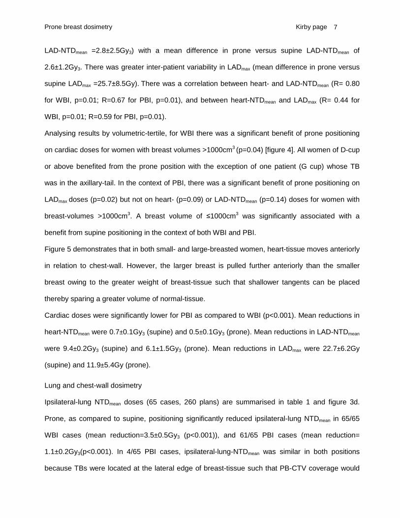
LAD-NTDmean =2.8±2.5Gy3) with a mean difference in prone versus supine LAD-NTDmean of
2.6±1.2Gy3. There was greater inter-patient variability in LADmax (mean difference in prone versus
supine LADmax =25.7±8.5Gy). There was a correlation between heart- and LAD-NTDmean (R= 0.80
for WBI, p=0.01; R=0.67 for PBI, p=0.01), and between heart-NTDmean and LADmax (R= 0.44 for
WBI, p=0.01; R=0.59 for PBI, p=0.01).
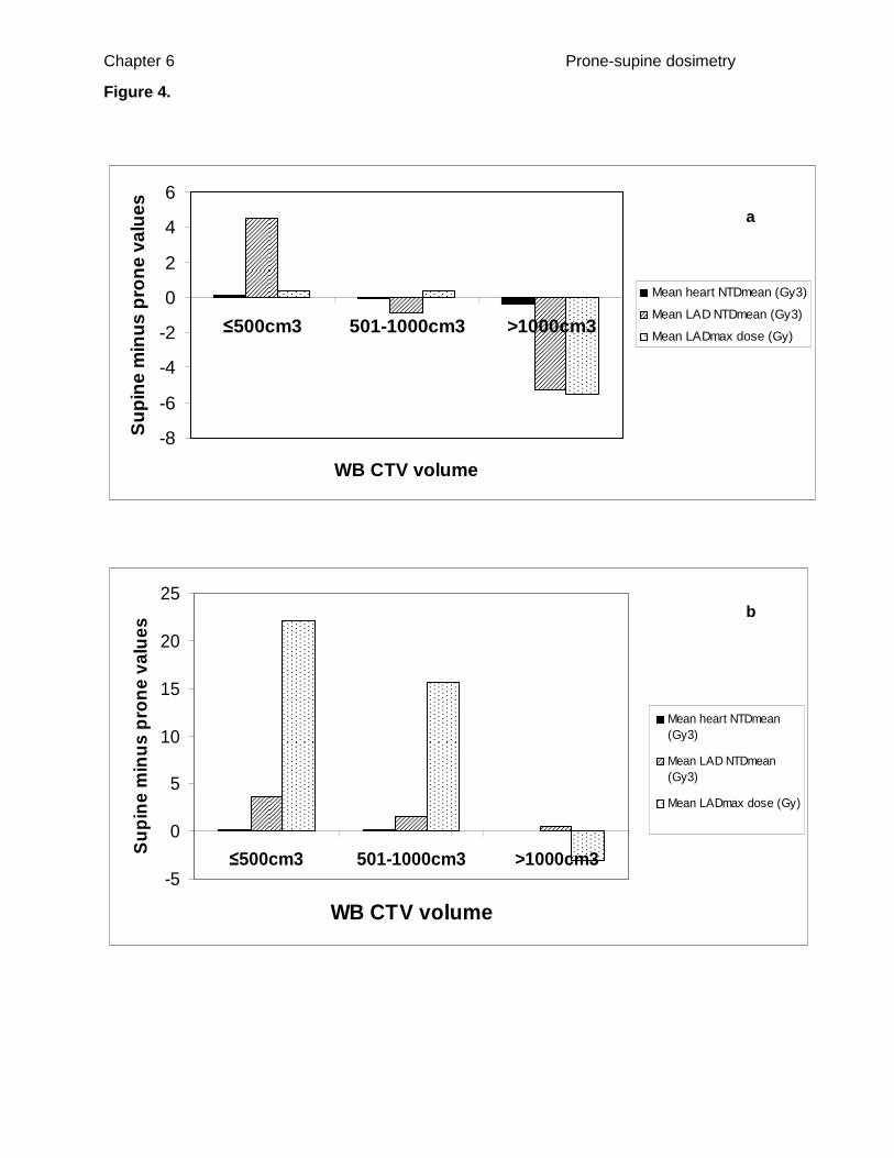
Analysing results by volumetric-tertile, for WBI there was a significant benefit of prone positioning
on cardiac doses for women with breast volumes >1000cm3 (p=0.04) [figure 4]. All women of D-cup
or above benefited from the prone position with the exception of one patient (G cup) whose TB
was in the axillary-tail. In the context of PBI, there was a significant benefit of prone positioning on
LADmax doses (p=0.02) but not on heart- (p=0.09) or LAD-NTDmean (p=0.14) doses for women with
breast-volumes >1000cm3. A breast volume of ≤1000cm3 was significantly associated with a
benefit from supine positioning in the context of both WBI and PBI.
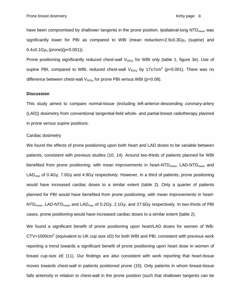
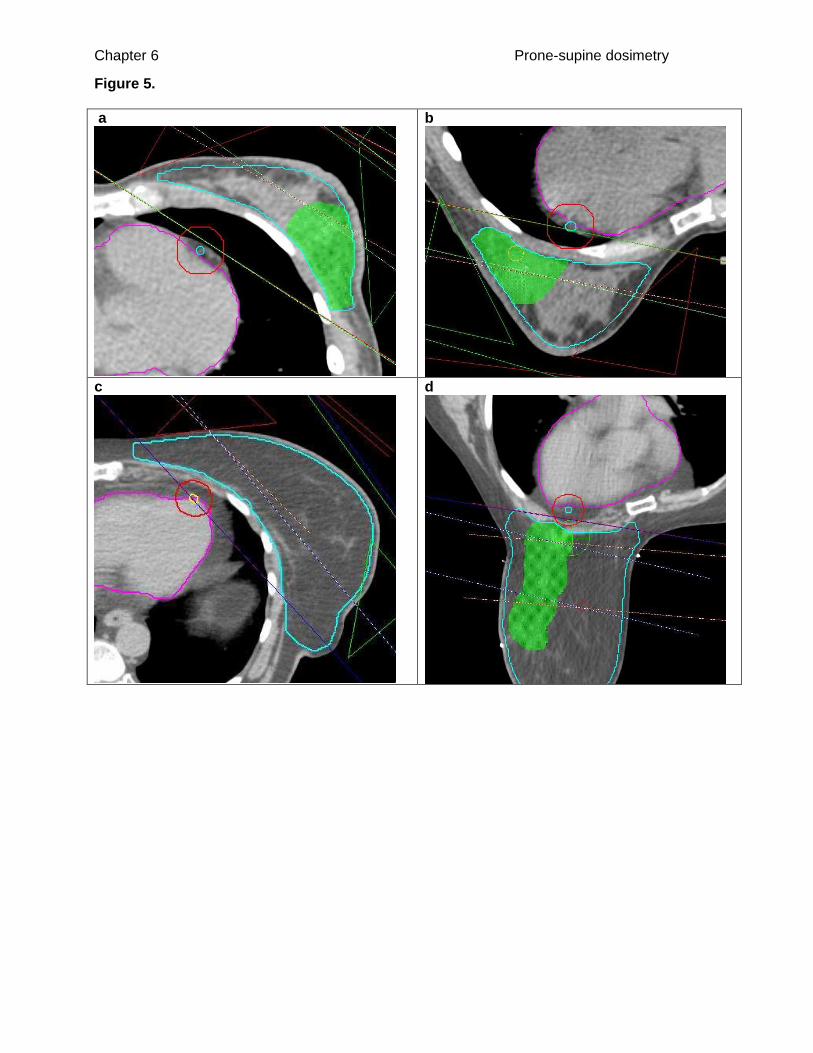
Figure 5 demonstrates that in both small- and large-breasted women, heart-tissue moves anteriorly
in relation to chest-wall. However, the larger breast is pulled further anteriorly than the smaller
breast owing to the greater weight of breast-tissue such that shallower tangents can be placed
thereby sparing a greater volume of normal-tissue.
Cardiac doses were significantly lower for PBI as compared to WBI (p<0.001). Mean reductions in
heart-NTDmean were 0.7±0.1Gy3 (supine) and 0.5±0.1Gy3 (prone). Mean reductions in LAD-NTDmean
were 9.4±0.2Gy3 (supine) and 6.1±1.5Gy3 (prone). Mean reductions in LADmax were 22.7±6.2Gy
(supine) and 11.9±5.4Gy (prone).
Lung and chest-wall dosimetry
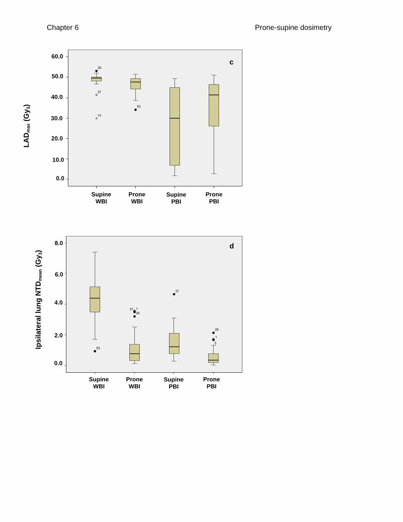
Ipsilateral-lung NTDmean doses (65 cases, 260 plans) are summarised in table 1 and figure 3d.
Prone, as compared to supine, positioning significantly reduced ipsilateral-lung NTDmean in 65/65
WBI cases (mean reduction=3.5±0.5Gy3 (p<0.001)), and 61/65 PBI cases (mean reduction=
1.1±0.2Gy3(p<0.001). In 4/65 PBI cases, ipsilateral-lung-NTDmean was similar in both positions
because TBs were located at the lateral edge of breast-tissue such that PB-CTV coverage would
Prone breast dosimetry Kirby page 8
have been compromised by shallower tangents in the prone position. Ipsilateral-lung NTDmean was
significantly lower for PBI as compared to WBI (mean reduction=2.9±0.3Gy3 (supine) and
0.4±0.1Gy3 (prone)(p<0.001)).
Prone positioning significantly reduced chest-wall V50Gy for WBI only (table 1, figure 3e). Use of
supine PBI, compared to WBI, reduced chest-wall V50Gy by 17±7cm3 (p<0.001). There was no
difference between chest-wall V50Gy for prone PBI versus WBI (p=0.08).
Discussion
This study aimed to compare normal-tissue (including left-anterior-descending coronary-artery
(LAD)) dosimetry from conventional tangential-field whole- and partial-breast radiotherapy planned
in prone versus supine positions.
Cardiac dosimetry
We found the effects of prone positioning upon both heart and LAD doses to be variable between
patients, consistent with previous studies (10, 14). Around two-thirds of patients planned for WBI
benefited from prone positioning, with mean improvements in heart-NTDmean, LAD-NTDmean and
LADmax of 0.4Gy, 7.0Gy and 4.9Gy respectively. However, in a third of patients, prone positioning
would have increased cardiac doses to a similar extent (table 2). Only a quarter of patients
planned for PBI would have benefited from prone positioning, with mean improvements in heart-
NTDmean, LAD-NTDmean and LADmax of 0.2Gy, 2.1Gy, and 27.6Gy respectively. In two-thirds of PBI
cases, prone positioning would have increased cardiac doses to a similar extent (table 2).
We found a significant benefit of prone positioning upon heart/LAD doses for women of WB-
CTV>1000cm3 (equivalent to UK cup size ≥D) for both WBI and PBI, consistent with previous work
reporting a trend towards a significant benefit of prone positioning upon heart dose in women of
breast cup-size ≥E (11). Our findings are also consistent with work reporting that heart-tissue
moves towards chest-wall in patients positioned prone (15). Only patients in whom breast-tissue
falls anteriorly in relation to chest-wall in the prone position (such that shallower tangents can be
Prone breast dosimetry Kirby page 9
placed) are likely to gain from prone treatment. Otherwise, where smaller breasts are not pulled
anteriorly, tangents are likely to encompass more cardiac tissue in the prone position.
Not all patients of large breast size in our study had better cardiac dosimetry in the prone position.
One patient (cup size G) had a tumour-bed in the axillary tail which could not be encompassed by
WBI-tangents without increasing their depth and therefore normal-tissue doses. There may be
other predictive factors including heart-size, chest-wall breadth and curvature that our study is
underpowered to detect.
The proportion of women benefitting from prone positioning in our study differed from interim
reports of the largest ongoing comparative study of prone versus supine WBI, which suggest that
prone positioning reduces in-field heart volume in the majority (85%) of left-sided BC patients
(n=200)(13). Discrepancies between results could be due to use of volumetric rather than
dosimetric comparators and/or to use of IMRT rather than conventional tangential-fields. Using
IMRT, an in-field heart volume of 0cm3 is likely to be achievable in a proportion of patients in both
supine and prone positions. In this case, the effect of prone positioning upon heart is assumed to
be neutral but the prone position might still be judged “optimal” based on reduced in-field lung
volume compared to the supine position. This approach does not however detect differences in
lower-dose irradiation of cardiac tissues thereby overestimating the clinical benefits of prone
positioning in comparison to our study. Our additional use of LAD-dosimetry to discern optimal
treatment position might also have lead to differing results. Although heart- and LAD-NTDmean
correlated reasonably well, there was disagreement between heart and LAD doses over optimal
treatment position in 7/60 plans. Had heart NTDmean been the only comparator, 20/30 and 13/30
patients would have benefited from prone treatment in the context of WBI and PBI respectively.
Another source of discrepancies could be the method by which WB-CTV is defined. Clinicians’
decisions on where to place the posterior RT-field border in order to achieve target-volume
coverage will significantly impact upon doses to tissues close to chest-wall. Our study used a
method of WB-CTV definition agreed to be more representative of the true volume than standard
Prone breast dosimetry Kirby page 10
anatomical landmarks (21). Wire-delineated breast tissue shifts anteriorly in the prone position but
is consistently included as WB-CTV. Our WB-CTVs were comparable between positions with no
significant difference in percentage volume covered. Additionally, all of our cases had titanium-clip-
defined TB volumes, without which, coverage of PB-CTV at depth cannot be ensured (22).
The clinical impact of differences in cardiac doses of the magnitude described above is difficult to
quantify as radiation parameters determining excess cardiovascular disease (CVD) risk are poorly
understood. Gagliardi (23) used the relative-seriality model to quantitatively describe the dose-
response relationship for excess cardiac mortality and found a low dependence of this endpoint
upon irradiated-heart-volume, concluding that cardiac mortality is more likely to be reduced by
decreasing dose than by restricting irradiated volume. Borger et al (24) also found no relationship
between maximum heart distance (MHD) (a correlate of irradiated-heart-volume) and risk of CVD
but reported that, even where MHD=0mm, more cardiotoxic effects occurred following left-sided as
compared to right-sided-RT suggesting that differences in doses <25Gy may be important. Other
data supporting the hypothesis that low-dose radiation increases CVD risk come from atomic-
bomb survivors (4Gy single exposure) (25), patients treated with RT for peptic-ulcer disease (mean
heart dose 1.6-3.9Gy) (26), patients treated with para-aortic irradiation for testicular cancer (~1Gy
scattered heart dose) (27) and radiation workers (28). Cardiac variables which encompass
volumes of cardiac tissue irradiated to low doses such as heart-NTDmean have shown a strong
correlation with mortality (29). Optimal positioning in our study decreased mean heart-NTDmean
from ~1.2 to ~0.8Gy3 for WBI, and from ~0.5 to ~0.3Gy3 for PBI. Based on the evidence above, the
risks of low-dose cardiac irradiation are not negligible. However, the dose-effect relationship at
these dose-levels is difficult to define and the clinical consequences of such small differences
unquantifiable.
Meanwhile, other studies suggest that LAD dose is the most relevant exposure variable (3-5).
Retrospective review of patients irradiated between 1977-95 found a significantly higher
prevalence of cardiac stress-test abnormalities amongst left- versus right-side-irradiated patients,
Prone breast dosimetry Kirby page 11
70% of which were in LAD territory (3) CHECK Others correlate a fall in mean LAD doses from
breast RT over the last 30 years (6, 7) with a decrease in CVD over the same period (2).
Furthermore, it may be that Gagliardi’s finding of a low dependence of cardiac mortality upon
irradiated-heart-volume (23) relates to the fact that the LAD is likely to remain within the high-dose
volume from a tangential-field arrangement even at low irradiated-heart-volumes. Optimal
positioning in our study decreased mean LAD-NTDmean from ~14Gy to 6Gy for WBI, and from ~4Gy
to 1.5Gy for PBI. Reductions in dose of these magnitudes could be associated with a significant
reduction in CVD (7). As atherosclerosis anywhere along the LAD could cause CVD, LADmax is a
relevant additional variable. Optimal positioning in our study decreased mean LADmax from ~49Gy
to 45Gy for WBI, and from 42Gy to 15Gy for PBI. The latter could be particularly significant
depending partly upon the threshold dose for atherosclerosis, Gagliardi’s work suggesting that the
risk of cardiac mortality rises steeply above doses of around 20Gy regardless of the volume
irradiated (23). Whether dosimetric differences of these magnitudes continue to be relevant in the
context of 5mm set-up errors and physiological changes in heart and intra-thoracic volume with
respiration (each of which may cause interfraction variations in normal-tissue doses) remains to be
determined.
Lung and chest-wall dosimetry
Our study confirms previous reports (10-12) that prone positioning reduces mean lung doses for
both WBI and PBI and furthermore demonstrates that benefits are applicable to women of all cup-
sizes. The main threat of death in relation to irradiation of lung-tissue is from low-dose stochastic
effects rather than from high-dose deterministic effects, the relative-risk of death from second-
primary lung cancer ranging from 1.5 to 2.8 at 15 years (30, 31), with odds ratios of up to 37.6
reported in smokers (32). Data on lung-cancer deaths in ~9000 women irradiated in 1935-1971
(30) suggest a dose-response relationship with an incremental RR of 0.2 per Gy to ipsilateral-lung
(equating to 9 cases of RT-induced lung cancer/ year/10,000 women receiving a lung dose of
10Gy and living to 10 years). The SEER registry cohort demonstrates a similar relationship
Prone breast dosimetry Kirby page 12
between mean lung dose and risk of second-primary lung-cancer (2) in women irradiated in 1973-
2001. Our mean lung-NTDmean for supine WBI was 3.7Gy. Prone positioning reduced this to 0.3Gy.
Based on evidence above, this reduction in dose might prevent around 3 lung cancers/year/10000
women living to 10 years post-RT. The effect may be larger however in women who smoke, in
whom prone treatment might be particularly beneficial.
The START trial suggests that 40% of women experience chest-wall discomfort at 10 years post-
RT (33), whilst the incidence of rib-fracture following WBI is reported to be 0.3-2.2% (34, 35). A
recent study of external-beam-accelerated-PBI found the incidence of chest-wall pain and rib
fracture to relate to the volume of chest-wall receiving 35Gy or more (based on 38.5Gy/10 fractions
5 days (36)). This is equivalent to around 48Gy in 2Gy fractions and is in keeping with tolerance
doses published by Emami (37) (TD5/5 ribcage ~50Gy). Therefore chest-wall V50Gy could be
considered a reasonable parameter by which to compare radiotherapeutic approaches in terms of
late chest-wall discomfort. Prone positioning significantly reduces chest-wall V50Gy in WBI and
therefore warrants consideration as a technique by which chest-wall morbidity might be reduced.
The future of prone breast RT
In departments where conventional tangential-field WBI is standard, prone positioning is likely to
benefit most left-breast-affected women of cup size ≥D, and nearly all right-breast-affected women.
A current priority is to establish whether or not the position is reproducible in order that dosimetric
benefits can be realized. In the context of tangential-field PBI, prone positioning benefited fewer
left-breast-affected women but still reduced the LADmax by over 20Gy in many large-breasted
women. Further work in this setting might be helpful in establishing predictive factors for deciding
optimal treatment position in left-breast-affected women. For right-breast-affected women,
reductions in lung and chest wall doses are small and a change of treatment technique may not
therefore be warranted. Based on our results, prone positioning for either WBI or PBI is not
recommended in left-breast-affected women of cup-size<C as cardiac doses may be significantly
increased in comparison to supine treatment.
Prone breast dosimetry Kirby page 13
PBI versus WBI
The normal-tissue dosimetric advantages of PBI have been assumed but not proven. Indeed a
recent study reported that 3D-conformal PBI increased the volume of lung exposed to low-dose
radiation whilst decreasing the volume of tissue exposed to higher-dose radiation (38). We found
that supine PBI reduced mean heart-NTDmean (by 0.6Gy), mean LAD-NTDmean (9Gy), mean LADmax
(23Gy), mean ipsilateral-lung-NTDmean (3Gy) and mean chest-wall V50Gy (17cm3) compared to
supine WBI. With dose-sparing of this magnitude, it seems likely that PBI will reduce long-term
cardiovascular side-effects of breast RT, reduce second-primary lung malignancies by around 2
lung cancers/year/10000 women at 10 years post-RT, and reduce the incidence of late chest-wall
discomfort. PBI should be considered the optimal strategy for reducing late morbidity of breast RT
but is currently only available in trials for which many women are ineligible. Prone positioning still
has a role in reducing normal-tissue toxicity in women requiring adjuvant WBI.
Conclusions
In the context of tangential-field WBI and PBI, prone positioning is likely to benefit left-breast-
affected women of cup-size ≥D, and most right-breast-affected women, but to be detrimental in
left-breast-affected women of smaller cup size. PBI reliably reduces normal-tissue doses
compared to WBI such that eligible women should be encouraged to participate in PBI studies.
Prone breast dosimetry Kirby page 14
References
1. Clarke M, Collins R, Darby S, et al. Effects of radiotherapy and of differences in the extent
of surgery for early breast cancer on local recurrence and 15-year survival: an overview of
the randomised trials. Lancet 2005;366:2087-2106.
2. Darby SC, McGale P, Taylor CW, et al. Long-term mortality from heart disease and lung
cancer after radiotherapy for early breast cancer: prospective cohort study of about 300,000
women in US SEER cancer registries. Lancet Oncol 2005;6:557-565.
3. Correa CR, Das IJ, Litt HI, et al. Association between tangential beam treatment parameters
and cardiac abnormalities after definitive radiation treatment for left-sided breast cancer. Int
J Radiat Oncol Biol Phys 2008;72:508-516.
4. Storey MR, Munden R, Strom EA, et al. Coronary artery dosimetry in intact left breast
irradiation. Cancer J 2001;7:492-497.
5. Lind PA, Pagnanelli R, Marks LB, et al. Myocardial perfusion changes in patients irradiated
for left-sided breast cancer and correlation with coronary artery distribution. Int J Radiat
Oncol Biol Phys 2003;55:914-920.
6. Taylor CW, Nisbet A, McGale P, et al. Cardiac exposures in breast cancer radiotherapy:
1950s-1990s. Int J Radiat Oncol Biol Phys 2007;69:1484-1495.
7. Taylor CW, Povall JM, McGale P, et al. Cardiac dose from tangential breast cancer
radiotherapy in the year 2006. Int J Radiat Oncol Biol Phys 2008;72:501-507.
8. Merchant TE, McCormick B. Prone position breast irradiation. Int J Radiat Oncol Biol Phys
1994;30:197-203.
9. Grann A, McCormick B, Chabner ES, et al. Prone breast radiotherapy in early-stage breast
cancer: a preliminary analysis. Int J Radiat Oncol Biol Phys 2000;47:319-325.
10. Griem KL, Fetherston P, Kuznetsova M, et al. Three-dimensional photon dosimetry: a
comparison of treatment of the intact breast in the supine and prone position. Int J Radiat
Oncol Biol Phys 2003;57:891-899.
Prone breast dosimetry Kirby page 15
11. Buijsen J, Jager JJ, Bovendeerd J, et al. Prone breast irradiation for pendulous breasts.
Radiother Oncol 2007;82:337-340.
12. DeWyngaert JK, Jozsef G, Mitchell J, et al. Accelerated intensity-modulated radiotherapy to
breast in prone position: dosimetric results. Int J Radiat Oncol Biol Phys 2007;68:1251-
1259.
13. Formenti S, Lymberis S, Parhar P, Fenton-Kerimian M, Magnolfi C, Wen B, Chang J,
DeWyngaert J. Results of NYU 05-181: A prospective trial to determine optimal position
(prone versus supine) for breast radiotherapy. Int J Radiat Biol 2009;75:S203-S204.
14. Patel RR, Becker SJ, Das RK, et al. A dosimetric comparison of accelerated partial breast
irradiation techniques: multicatheter interstitial brachytherapy, three-dimensional conformal
radiotherapy, and supine versus prone helical tomotherapy. Int J Radiat Oncol Biol Phys
2007;68:935-942.
15. Chino JP, Marks LB. Prone positioning causes the heart to be displaced anteriorly within the
thorax: implications for breast cancer treatment. Int J Radiat Oncol Biol Phys 2008;70:916-
920.
16. Smith TE, Lee D, Turner BC, et al. True recurrence vs. new primary ipsilateral breast tumor
relapse: an analysis of clinical and pathologic differences and their implications in natural
history, prognoses, and therapeutic management. Int J Radiat Oncol Biol Phys
2000;48:1281-1289.
17. Coles CE, Wilson CB, Cumming J, et al. Titanium clip placement to allow accurate tumour
bed localisation following breast conserving surgery: audit on behalf of the IMPORT Trial
Management Group. Eur J Surg Oncol 2009;35:578-582.
18. Yarnold, J, Coles, C. On behalf of the IMPORT-Low Trial Management Group. Intensity-
Modulated and Partial Organ Radiotherapy. Randomised trial testing intensity-modulated
and partial organ radiotherapy following breast conservation surgery for early breast cancer.
Trial Protocol, version 4. Institute of Cancer Research, Sutton, UK. p.1-74.

Prone breast dosimetry Kirby page 16
19. ICRU Report 62. Prescribing, Recording and Reporting Photon Beam Therapy (Supplement
to ICRU Report 50). Maryland:Bethseda; 1999.
20. Scrimger RA, Tome WA, Olivera GH, et al. Reduction in radiation dose to lung and other
normal tissues using helical tomotherapy to treat lung cancer, in comparison to conventional
field arrangements. Am J Clin Oncol 2003;26:70-78.
21. Valdagni R, Italia C, Montanaro P, et al. Clinical target volume localization using
conventional methods (anatomy and palpation) and ultrasonography in early breast cancer
post-operative external irradiation. Radiother Oncol 1997;42:231-237.
22. Algan O, Fowble B, McNeeley S, et al. Use of the prone position in radiation treatment for
women with early stage breast cancer. Int J Radiat Oncol Biol Phys 1998;40:1137-1140.
23. Gagliardi G, Lax I, Ottolenghi A, et al. Long-term cardiac mortality after radiotherapy of
breast cancer--application of the relative seriality model. Br J Radiol 1996;69:839-846.
24. Borger JH, Hooning MJ, Boersma LJ, et al. Cardiotoxic effects of tangential breast
irradiation in early breast cancer patients: the role of irradiated heart volume. Int J Radiat
Oncol Biol Phys 2007;69:1131-1138.
25. Preston DL, Shimizu Y, Pierce DA, et al. Studies of mortality of atomic bomb survivors.
Report 13: Solid cancer and noncancer disease mortality: 1950-1997. Radiat Res
2003;160:381-407.
26. Carr ZA, Land CE, Kleinerman RA, et al. Coronary heart disease after radiotherapy for
peptic ulcer disease. Int J Radiat Oncol Biol Phys 2005;61:842-850.
27. van den Belt-Dusebout AW, Nuver J, de Wit R, et al. Long-term risk of cardiovascular
disease in 5-year survivors of testicular cancer. J Clin Oncol 2006;24:467-475.
28. McGale P, Darby SC. Low doses of ionizing radiation and circulatory diseases: a systematic
review of the published epidemiological evidence. Radiat Res 2005;163:247-257.
29. Perman M, Johanson, I, Ohlson, B, Johansson, KA, Karlsson, P. Death from ischaemic
heart disease 10-19 years after treatment for early breast cancer: a population-based
Prone breast dosimetry Kirby page 17
nested case-control study regarding absorbed dose to the heart and 11 anatomical
substructures of the heart. Radiother Oncol 2008;x:x.
30. Inskip PD, Stovall M, Flannery JT. Lung cancer risk and radiation dose among women
treated for breast cancer. J Natl Cancer Inst 1994;86:983-988.
31. Roychoudhuri R, Evans H, Robinson D, et al. Radiation-induced malignancies following
radiotherapy for breast cancer. Br J Cancer 2004;91:868-872.
32. Kaufman EL, Jacobson JS, Hershman DL, et al. Effect of breast cancer radiotherapy and
cigarette smoking on risk of second primary lung cancer. J Clin Oncol 2008;26:392-398.
33. Bentzen SM, Agrawal RK, Aird EG, et al. The UK Standardisation of Breast Radiotherapy
(START) Trial A of radiotherapy hypofractionation for treatment of early breast cancer: a
randomised trial. Lancet Oncol 2008;9:331-341.
34. Pierce SM, Recht A, Lingos TI, et al. Long-term radiation complications following
conservative surgery (CS) and radiation therapy (RT) in patients with early stage breast
cancer. Int J Radiat Oncol Biol Phys 1992;23:915-923.
35. Meric F, Buchholz TA, Mirza NQ, et al. Long-term complications associated with breast-
conservation surgery and radiotherapy. Ann Surg Oncol 2002;9:543-549.
36. Reeder R, Carter DL, Howell K, et al. Predictors for clinical outcomes after accelerated
partial breast intensity-modulated radiotherapy. Int J Radiat Oncol Biol Phys 2009;74:92-97.
37. Emami B, Lyman J, Brown A, et al. Tolerance of normal tissue to therapeutic irradiation. Int
J Radiat Oncol Biol Phys 1991;21:109-122.
38. Jain AK, Vallow LA, Gale AA, et al. Does three-dimensional external beam partial breast
irradiation spare lung tissue compared with standard whole breast irradiation? Int J Radiat
Oncol Biol Phys 2009;75:82-88.
Prone breast dosimetry Kirby page 18
Table 1. Mean normal tissue doses (with 95% confidence intervals)
Supine
WBI
Prone
WBI
Difference
between
prone &
supine
WBI
p Supine
PBI
Prone
PBI
Difference
between
prone &
supine
PBI
p
Heart-NTDmean
(Gy3)
1.0±0.1 0.9±0.1 0.1±0.2 0.14 0.4±0.1 0.4±0.1 0.1± 0.1 0.08
LAD-NTDmean
(Gy3)
11.5±2.3 9.6±1.9 1.9±3.0 0.21 2.1±0.6 3.5±1.1 1.4±1 .2 0.02
LADmax (Gy) 48.6±1.5 46.5±1.6 2.2±2.1 0.04 25.9±6.6 34.5±6.1 8.6±9.2 0.07
Ipsilateral-lung
NTDmean (Gy3)
3.7±0.8 0.3±0.8 3.5±0.5 <0.001 1.4±0.5 0.2±0.1 1.1± 0.2 <0.001
Chest-wall
V50Gy (cm3)
20±7 2±1 18±7 <0.001 3±2 1±1 2±2 0.08
Prone breast dosimetry Kirby page 19
Table 2: Comparison of cardiac doses according to superiority of patient position for WBI
and PBI
Supine position better Prone position better
WBI N=8 N=19
Supine Prone Difference p Supine Prone Difference p
Heart-NTDmean
(Gy3)
0.7±0.1 1.1±0.3 0.5±0.2 0.01 1.2±0.2 0.8±0.1 0.4±0.1 <0.001
LAD-NTDmean
(Gy3)
5.1±2.2 13.5±3.2 8.4±2.5 <0.001 14.6±2.4 7.5±2.1 7.0±2.1 <0.001
LADmax (Gy) 45.1±4.7 49.1±1.8 3.9±3.3 0.05 49.7±0.4 44.9±1.9 4.9±1.7 <0.001
PBI N=19 N=7
Supine Prone Difference p Supine Prone Difference p
Heart NTDmean
(Gy3)
0.3±0.1 0.5±0.1 0.2±0.1 0.002 0.4±0.1 0.3±0.1 0.2±0.1 0.10
LAD NTDmean
(Gy3)
1.7±0.7 4.6±1.4 3.0±1.1 <0.001 3.4±1.4 1.3±0.8 2.1±1.4 0.03
LADmax (Gy) 19.0±7.6 42.7±3.6 23.7±6.5 <0.001 41.9±5.4 14.3±12.7 27.6±10.5 0.003
Prone breast dosimetry Kirby page 20
Figure Captions
Figure 1. Prone platform including polycarbonate centrepiece with aperture,
polystyrene head and body supports, and polystyrene wedge to support
contralateral breast
Figure 2. Distributions of patients by a) cup size and b) whole-breast clinical-target
volume
Figure 3a-e. Box and whiskers plots displaying normal tissue dosimetry a) Heart-
NTDmean b) LAD-NTDmean c) LADmax (figures 3a-c include left-breast-affected
women only, n=30) d) Ipsilateral-lung NTDmean (n=65) e) Chest-wall V50Gy
(n=65). Black dots= outliers (numbers represent trial-numbers)
Figure 4. Relationship of breast-tissue (cyan-outline), heart (pink-outline), and left-
anterior-descending coronary-artery (red-outline with cyan-bullseye) to
chest-wall for: i) woman of cup-size B and ii) woman of cup-size F a) supine
and b) prone
Figure 5. Mean difference in cardiac variables (supine minus prone) expressed by
whole-breast clinical-target volume tertile for a) WBI and b) PBI. (Negative
values= benefit of prone position)

Prone breast dosimetry Kirby page 21
Figure 1.
Prone breast dosimetry Kirby page 22
Figure 2.
a
0
5
10
15
20
25
A B C D-DD E F-FF G
Self-reported cup size (UK)
Nu
mb
er o
f p
atie
nts
b
0
5
10
15
20
25
30
35
≤500cm3 501-1000cm3 >1000cm3
Whole breast clinical target volume
Nu
mb
er o
f p
atie
nts
Chapter 6 Prone-supine dosimetry
Figure 3.
Supine WBI
Prone WBI
Supine PBI
Prone PBI
0.0
a
0.5
1.0
1.5
2.0
Hea
rt N
TD
mea
n (
Gy 3
)
Supine WBI
Prone WBI
Supine PBI
Prone PBI
0.0
5.0
10.0
15.0
20.0
LA
D N
TD
mea
n (
Gy 3
)
25.0 b
Chapter 6 Prone-supine dosimetry
Supine WBI
Prone WBI
Supine PBI
Prone PBI
0.0
10.0
20.0
30.0
40.0
LA
Dm
ax (
Gy 3
)
50.0
60.0
Supine WBI
Prone WBI
Supine PBI
Prone PBI
0.0
2.0
4.0
6.0
8.0
Ipsi
late
ral l
un
g N
TD
mea
n (
Gy 3
)
c
d

Chapter 6 Prone-supine dosimetry
Supine WBI
Prone WBI
Supine PBI
Prone PBI
0.0
10.0
20.0
30.0
40.0
Ch
est-
wal
l V50
Gy(
cm3 ) 50.0
60.0 e
Chapter 6 Prone-supine dosimetry
Figure 4.
-8
-6
-4
-2
0
2
4
6
≤500cm3 501-1000cm3 >1000cm3
WB CTV volume
Su
pin
e m
inu
s p
ron
e va
lues
Mean heart NTDmean (Gy3)
Mean LAD NTDmean (Gy3)
Mean LADmax dose (Gy)
-5
0
5
10
15
20
25
≤500cm3 501-1000cm3 >1000cm3
WB CTV volume
Su
pin
e m
inu
s p
ron
e va
lues
Mean heart NTDmean(Gy3)
Mean LAD NTDmean(Gy3)
Mean LADmax dose (Gy)
a
b
Chapter 6 Prone-supine dosimetry
Figure 5.
a
b
c
d