Embed Size (px)
Citation preview

Promotion of Testa Rupture during Garden CressGermination Involves Seed Compartment-SpecificExpression and Activity of Pectin Methylesterases1[OPEN]
Claudia Scheler2, Karin Weitbrecht2, Simon P. Pearce2, Anthony Hampstead, Annette Büttner-Mainik,Kieran J.D. Lee, Antje Voegele, Krystyna Oracz, Bas J.W. Dekkers, Xiaofeng Wang,Andrew T.A. Wood, Leónie Bentsink, John R. King, J. Paul Knox, Michael J. Holdsworth3,Kerstin Müller3, and Gerhard Leubner-Metzger3*
Botany and Plant Physiology, Institute for Biology II, Faculty of Biology, University of Freiburg, D–79104Freiburg, Germany (C.S., K.W., A.B.-M., K.O., G.L.-M.); Institute of Biochemical Plant Pathology, HelmholtzZentrum München, Deutsches Forschungszentrum für Gesundheit und Umwelt, D–85764 Neuherberg,Germany (C.S.); Staatliches Weinbauinstitut Freiburg, D–79104 Freiburg, Germany (K.W.); Centre for PlantIntegrative Biology (S.P.P., A.H., A.T.A.W., J.R.K., M.J.H.) and Division of Plant and Crop Science (S.P.P.,M.J.H., K.M.), School of Biosciences, University of Nottingham, Sutton Bonington Campus, Sutton Bonington,Leicestershire LE12 5RD, United Kingdom; School of Mathematical Sciences, University of Nottingham,University Park, Nottingham NG7 2RD, United Kingdom (S.P.P., A.H., A.T.A.W., J.R.K.); Agroscope, Institutefor Plant Production Sciences, Seed Quality, CH–8046 Zurich, Switzerland (A.B.-M.); Centre for Plant Sciences,Faculty of Biological Sciences, University of Leeds, Leeds LS2 9JT, United Kingdom (K.J.D.L., J.P.K.);National Institute for Health Research Trainees Coordinating Centre, Leeds Innovation Centre, Leeds LS2 9DF,United Kingdom (K.J.D.L.); School of Biological Sciences, Plant Molecular Science and Centre for Systems andSynthetic Biology, Royal Holloway, University of London, Egham, Surrey TW20 0EX, United Kingdom(A.V., G.L.-M.); Department of Plant Physiology, Warsaw University of Life Sciences, 02–776, Warsaw, Poland(K.O.); Wageningen Seed Laboratory, Laboratory of Plant Physiology, Wageningen University and ResearchCentre, NL–6708 PB Wageningen, The Netherlands (B.J.W.D., L.B.); College of Life Sciences, South ChinaAgricultural University, Guangzhou 510642, China (X.W.); and Laboratory of Growth Regulators, Faculty ofScience, Palacký University and Institute of Experimental Botany, CZ–783 71 Olomouc, Czech Republic (G.L.-M.)
Pectin methylesterase (PME) controls the methylesterification status of pectins and thereby determines the biophysical propertiesof plant cell walls, which are important for tissue growth and weakening processes. We demonstrate here that tissue-specific andspatiotemporal alterations in cell wall pectin methylesterification occur during the germination of garden cress (Lepidium sativum).These cell wall changes are associated with characteristic expression patterns of PME genes and resultant enzyme activities in thekey seed compartments CAP (micropylar endosperm) and RAD (radicle plus lower hypocotyl). Transcriptome and quantitativereal-time reverse transcription-polymerase chain reaction analysis as well as PME enzyme activity measurements of separated seedcompartments, including CAP and RAD, revealed distinct phases during germination. These were associated with hormonal andcompartment-specific regulation of PME group 1, PME group 2, and PME inhibitor transcript expression and total PME activity.The regulatory patterns indicated a role for PME activity in testa rupture (TR). Consistent with a role for cell wall pectin methylesterificationin TR, treatment of seeds with PME resulted in enhanced testa permeability and promoted TR. Mathematical modeling of transcriptexpression changes in germinating garden cress and Arabidopsis (Arabidopsis thaliana) seeds suggested that group 2 PMEs make amajor contribution to the overall PME activity rather than acting as PME inhibitors. It is concluded that regulated changes in thedegree of pectin methylesterification through CAP- and RAD-specific PME and PME inhibitor expression play a crucial role duringBrassicaceae seed germination.
Mature seeds of members of the Brassicaceae familysuch as Arabidopsis (Arabidopsis thaliana) and gardencress (Lepidium sativum) are endospermic (i.e. the embryois surrounded by a thin living cell layer, the endosperm,and a dead outer layer, the testa). Many angiospermseeds, including those of garden cress and Arabidopsis,germinate in a two-step process: after the initial phase ofwater uptake by the dry seeds (imbibition), testa rupture(TR) occurs and is subsequently followed by endospermrupture (ER), which marks the completion of germina-tion (Liu et al., 2005; Müller et al., 2006). Germination is
controlled by two opposing forces, the increasing growthpotential of the radicle and the resistance of the testa andendosperm tissues covering it (Bewley, 1997; Schopfer,2006; Linkies and Leubner-Metzger 2012). After TR, en-dosperm resistance decreases through tissue softening, aprocess called endosperm weakening (Müller et al., 2006;Linkies et al., 2009). Both radicle elongation and endospermweakening require cell wall modifications (Schopfer, 2006;Müller et al., 2009; Morris et al., 2011).
Plant cell walls are the main determinants for theshape and biomechanical properties of plant tissues,
200 Plant Physiology�, January 2015, Vol. 167, pp. 200–215, www.plantphysiol.org � 2014 American Society of Plant Biologists. All Rights Reserved. www.plantphysiol.orgon April 5, 2018 - Published by Downloaded from
Copyright © 2015 American Society of Plant Biologists. All rights reserved.
organs, and even the whole plant body. They controlturgor-driven water uptake to allow cell growth throughchanges in extensibility, which depend on wall compo-sition and the interaction between their components(Thompson, 2005; Cosgrove and Jarvis, 2012; Yoshidaet al., 2014). One of the most abundant groups of poly-saccharides in primary cell walls is pectins. Pectins arecomplex polysaccharides that are characterized by a-1,4-linked galacturonic acid (Willats et al., 2001; Mohnen,2008; Tan et al., 2013); they are present in the middlelamellae and are key polymers in cell separationprocesses. The most abundant plant cell wall pectin ishomogalacturonan (HG). HG is a linear polymer of (1,4)-linked-a-D-galacturonic acid that can be modified bymethylesterification at the C-6 carboxyl position to formmethylesterified homogalacturonan (Me-HG; Wolf et al.,2009a). The degree of methylesterification is variablebetween developmental stages, tissues, and even regionsof the wall of an individual cell and strongly affects themechanical properties of cell walls (Braybrook et al.,2012). After synthesis in the endomembrane system, HGis secreted in a highly methylesterified form into theplant cell wall of growing cells (Mohnen, 2008).Pectin methylesterases (PMEs; EC 3.1.1.11) catalyze
the demethylesterification of HG (Wolf et al., 2009a).PMEs are ubiquitous cell wall-associated enzymes thatare found in all higher plants as well as in some bacteriaand fungi. PMEs can act linearly to deesterify stretchesof Me-HG to give rise to blocks of free carboxyl groupsthat can be cross-linked by calcium ions. These calciumbridges influence cell wall porosity and may enhancethe overall firmness of tissues. PMEs also can act in anonlinear fashion and deesterify only individual gal-acturonate residues or short stretches, which does notallow for calcium bridges to form, leading to a loosercell wall matrix structure. PME activity might promotethe subsequent action of cell wall hydrolases such asendopolygalacturonases (Wakabayashi et al., 2000, 2003),
which contribute to cell wall weakening and/or cellseparation (González-Carranza et al., 2007). PMEs havebeen shown to be involved in pectin remodeling atdifferent developmental stages, such as pollen tubegrowth (Eckardt, 2005), root elongation and its reac-tion to soil aluminum concentrations (Yang et al., 2013),hypocotyl elongation (Pelletier et al., 2010), fruit ripening(Hyodo et al., 2013), and seed germination (Müller et al.,2013). PMEs are encoded by a large multigene familythat has been classified into two groups (Wang et al.,2013): all PMEs have a conserved pectinesterase domain(Pfam01095), but only group 2 has in addition a PMEinhibitory domain (Pfam04043). PME activity is regulatedby pectin methylesterase inhibitor (PMEI) proteins (Giovaneet al., 2004; Wolf et al., 2009a).
In our integrative study, we discovered that duringthe seed germination of garden cress, changes in thetranscript abundance of specific PMEs and PMEIs arereflected in seed compartment-specific changes in PMEactivity and accompanied by spatiotemporal changesin cell wall pectin methylesterification. We mathemati-cally modeled the contribution of the different groups ofPMEs and of PMEIs to the degree of methylesterificationin the garden cress seed cell walls. Based on these mo-lecular, physiological, histochemical, and biophysicalanalyses, we propose that PME activity is involved in thegermination process of garden cress and is differentiallyregulated in a spatial and temporal manner.
RESULTS
Seed Compartment-Specific Transcriptome Analysis ofGarden Cress Germination
We utilized the fact that garden cress has a two-stepgermination process, with TR and ER separated by severalhours, to conduct a dense spatiotemporal transcriptomeanalysis during the germination process (Fig. 1). We in-vestigated transcriptome changes in the seed compart-ments that are directly involved in ER, the micropylarendosperm (CAP; Fig. 1B) and the radicle with the lowerpart of the hypocotyl (RAD; embryo growth zone;Fig. 1B), at key time points during the germinationprocess from early imbibition to the completion of ER(Fig. 1, A and B). At the time points when approximately50% of the seed population had reached TR and ER,respectively, we divided the sampling population be-tween the seeds that had already undergone rupture(+TR or +ER) and those that had not (2TR or 2ER), sothat we could compare samples that would have un-dergone TR within the next 1 or 2 h with those thatalready had ruptured testas. We also sampled cotyle-dons (COTs) and nonmicropylar endosperms (NMEs)at three time points (Fig. 1). Seed compartment-specificgene expression analyses for garden cress have beenperformed successfully before (Linkies et al., 2009) usingComplete Arabidopsis Transcriptome Microarray-spottedPCR-amplified gene-specific tag-based chips, butonly for a small number of samples that allowed for a
1 This work was supported by the ERA-NET Plant GenomicsvSEED project through the Deutsche Forschungsgemeinschaft (grantnos. DFG LE720/8 and DFG LE720/6 to G.L.-M.), the U.K. Biotech-nology and Biosciences Research Council (grant no. BB/G024898/1to J.P.K. and grant no. BBG02488X1 to M.J.H. and J.R.K.), the Neth-erlands Organization for Scientific Research (grant no. 855.50.011to L.B.), the Wissenschaftliche Gesellschaft Freiburg (to G.L.-M.),the Guangdong Natural Science Foundation (grant no. 07006658 toX.W.), a Marie Curie International Outgoing Fellowships for CareerDevelopment fellowship (to K.M.), an Alexander von HumboldtFoundation research fellowship (to K.O.), and the Royal Societyand the Wolfson Foundation (to J.R.K.).
2 These authors contributed equally to the article.3 These authors contributed equally to the article.* Address correspondence to [email protected] author responsible for distribution of materials integral to the
findings presented in this article in accordance with the policy de-scribed in the Instructions for Authors (www.plantphysiol.org) is:Gerhard Leubner-Metzger ([email protected]). ‘The SeedBiology Place’ www.seedbiology.eu.
[OPEN] Articles can be viewed without a subscription.www.plantphysiol.org/cgi/doi/10.1104/pp.114.247429
Plant Physiol. Vol. 167, 2015 201
Spatiotemporal Actions of Seed Pectin Methylesterases
www.plantphysiol.orgon April 5, 2018 - Published by Downloaded from Copyright © 2015 American Society of Plant Biologists. All rights reserved.

limited analysis of the garden cress germination process.Our sampling concept in this work with garden cress(Fig. 1) led to a time course with sufficient spatiotemporalresolution to investigate the distinct phases of germina-tion and make physiologically relevant comparisons.
We performed microarrays by hybridizing gardencress RNA to Affymetrix ATH1 microarrays designedfor Arabidopsis. Heterologousmicroarrays on ArabidopsisATH1 chips have been employed successfully by severalgroups to elucidate transcriptome changes in unsequencedBrassicaceae species or in species without commerciallyavailable microarrays (Hammond et al., 2006; Slotte et al.,
2007). To further improve the method for garden cress, wedeveloped a sophisticated masking approach that allowedus to extract a maximum of information from the arrays(see “Materials andMethods”). Using themaskingmethodpresented here, with a false discovery rate of 0.01, 36.6% ofprobes and 65.1% of probe sets were retained, leading to5,793 genes identified as being differentially expressedbetween 1 and 16 h after sowing in the CAP, and 6,098genes in the RAD. Conversely, using the method ofHammond et al. (2005), the maximal number of differen-tially expressed genes was 1,712 at a cutoff of 100. Thus,our method retained amuch larger number of differentially
Figure 1. Spatiotemporal transcriptome analysis of garden cress ‘FR14’ seed compartments during germination. A, The kineticsof garden cress TR and ER at 24˚C in continuous light. Arrows indicate sampling time points for RNA extraction at which theseeds were dissected into CAP, RAD, COT, and NME, as indicated. Note that at 0 h (dry seed stage), CAP plus RAD as well asNME plus COT were sampled together. Mean values 6 SE of n = 4 plates each with 100 seeds are presented for TR and ER. B,Garden cress seed compartments (CAP, RAD, COT, and NME) at different stages during germination as related to the TR and ERkinetics. C, Principal component analysis (PCA) of the garden cress microarray results with four biological RNA replicates foreach time point. Eigenrow 1 separates transcriptomes in time, while eigenrow 2 separates the seed compartments.
202 Plant Physiol. Vol. 167, 2015
Scheler et al.
www.plantphysiol.orgon April 5, 2018 - Published by Downloaded from Copyright © 2015 American Society of Plant Biologists. All rights reserved.

expressed genes that could then be used for further anal-ysis. Therefore, normalized expression values for gardencress were obtained for 13,895 transcripts (SupplementalData Set S1), with garden cress gene transcripts referringto the putative Arabidopsis orthologs defined by havingan Arabidopsis Genome Initiative identifier such asAt1g62380. Supplemental Data Sets S2 and S3 providethe normalized log2 values for the mean and SD expressionresults, respectively.
Seed Compartment-Specific Differentially RegulatedGarden Cress Transcriptome during Germination ShowsOverrrepresentation of Cell Wall-Related Transcripts at theTR Stage Transition
A principal component analysis (PCA) shows the gen-eral distribution of values calculated into eigenvectors andmakes it possible to identify the eigenvectors with thegreatest influence on sample variance. In our arrays, theeigenvectors with the biggest influence appear to corre-spond to time and seed compartment (Fig. 1C). In dryseeds and at 1 h after sowing, RAD and CAP samples stillcluster together, separating from 3 h after sowing. In theRAD, distinct clusters formed at the phase transition timepoint at 7 h, where we segregated for 2TR and +TR, andat 16 h, where we segregated for seeds that had under-gone ER and thus completed the germination process(+ER) and those whose endosperm had not rupturedyet (2ER). The same was true for the CAP at 7 h (+TRversus 2TR), although the 16-h CAP with ER clusterswith the CAP samples without ER.The COT samples exhibited a higher variance throughout
the time course (Fig. 1). The NME for the two time pointswithout TR clustered apart from the CAP samplesbut was still closer to them than to the RAD and COTsamples derived from the embryo. At 13 h, the NMEsamples varied more and clustered closer still to the CAPsamples (Fig. 1). The finding that transcriptomes differbetween the distinct seed compartments and over time isfurther supported by a cluster analysis of the transcriptabundance patterns, which showed five clearly distinctclusters (Supplemental Fig. S1). Together with the prin-cipal component analysis (Fig. 1), it supports the viewthat TR is a decisive step during the germination pro-cess that is associated with abundant transcriptomeschanges (+TR versus 2TR) in both the CAP and theRAD.The Gene Ontology (GO) overrepresentation analyses
of the TR rupture transition time point (SupplementalTables S1 and S2) indicate that both hormone regulationand cell wall modifications are overrepresented in thedifferentially regulated transcripts in the different seedcompartments. We decided to look in more detail atPME and PMEI, as those cell wall-modifying enzymesand inhibitors have been shown to be tied in with hor-monal regulations and to play a role in seed germinationof Arabidopsis (Müller et al., 2013; Saez-Aguayo et al.,2013), and pectinesterase activity and inhibition was oneof the activities whose GO terms were specifically over-represented in the RAD (Supplemental Table S2).
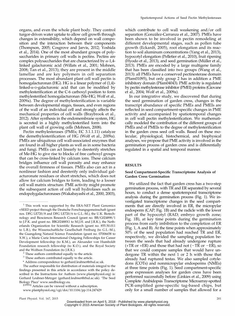
Spatiotemporal Patterns of HG and Me-HG Pectin CellWall Epitopes during Garden Cress Seed Germination
In order to observe if, where, and at which stage duringthe garden cress seed germination process changes in thedegree of pectin methylesterification actually happen inthe RAD and CAP, we studied HG epitopes in situ.To distinguish pectic HG in its methylesterified form(Me-HG) from the demethylesterified form (HG), weused a set of well-characterized monoclonal antibodies(www.plantcellwalls.net) in conjunction with fluores-cence imaging (Fig. 2). Antibody LM19 is specific fordemethylesterified HG, and its epitope was detected inseeds 3 h after sowing. The LM19 epitope was ubiqui-tously distributed in the cell walls of the RAD as well asin the CAP (E in Fig. 2), the testa, and the testa-derivedmucilage layer. In contrast, the JIM7 (Fig. 2) and LM20(data not shown) Me-HG epitopes were restricted to thetesta and mucilage and were present at reduced levelsin the RAD but absent from the CAP. The pattern of theLM19 HG epitope did not change between our samplingtimes. However, the spatial distribution of Me-HG (JIM7)was altered. The JIM7 signal increased in the RAD andappeared in the inner cell wall of the CAP upon TR(Fig. 2). This suggests that there is new deposition ofpectin into the cell wall, as HG is methylesterified inthe Golgi and secreted as Me-HG. In testa and mu-cilage, the JIM7 epitope was detectable in all phasesduring seed germination (Fig. 2).
Molecular Phylogenetic Analyses of Arabidopsis andGarden Cress PMEs and Their Inhibitors
We mined our microarrays for the expression pat-terns of putative PMEs and PMEIs in garden cressseeds (Supplemental Data Sets S1–S3). As the gardencress genome has not been sequenced, we started byidentifying 136 Arabidopsis sequences for PMEs andPMEIs by annotation and similarity searches in publicdatabases, with subsequent verification of the exis-tence of specific domains in the predicted proteins.According to our molecular phylogenetic analysis oftheir full-length predicted protein sequences, theseArabidopsis PMEs and PMEIs cluster into three largegroups (Fig. 3): group 1 PME (22 members) and group 2PME (45members) contain the PME domain (Pfam01095),which harbors five characteristic sequence motifs impor-tant for PME activity. The group 2 PMEs additionallypossess a PMEI domain (Pfam04043) with conserved Cysresidues, and both domains are separated by a processingmotif that is a putative target for subtilisin-like proteases.The third cluster consists of 69 PMEIs (Fig. 3).
We used the seed-specific Electronic Fluorescent Pic-tograph browser and the eNorthern tool at www.bar.utoronto.ca (Winter et al., 2007) for an in silico analysisof the PME/PMEI transcript expression patterns inimbibed whole Arabidopsis seeds (Fig. 3). This analysisyielded a general pattern in which many PME group 2transcripts are down-regulated by cold stratification(1 in Fig. 3). However, a small number of group 2 PMEs
Plant Physiol. Vol. 167, 2015 203
Spatiotemporal Actions of Seed Pectin Methylesterases
www.plantphysiol.orgon April 5, 2018 - Published by Downloaded from Copyright © 2015 American Society of Plant Biologists. All rights reserved.
and PMEIs was strongly up-regulated during the firstfew hours of the germination process (2 in Fig. 3). PMEsfrom both groups as well as PMEIs are up-regulated inthe course of the germination process in the presence ofabscisic acid (ABA; 3 in Fig. 3).
We then compared the expression of PME and PMEItranscripts that we identified in our arrays for garden
cress seeds (four group 1 PMEs, 28 group 2 PMEs, and25 PMEIs) in CAP and RAD (Fig. 4) with their ho-mologs in Arabidopsis (data extracted from the tran-scriptome of Dekkers et al. [2013]) as defined by thetranscripts that bind to the same probe sets. There wasan overall stronger differential regulation visible in theArabidopsis data, which may be due to the heterolo-gous nature of the garden cress arrays or to the fact thatthe Arabidopsis seed compartments were less confinedand contained additional tissues (Fig. 4). However, wecould clearly observe that the genes that were stronglydifferentially regulated in garden cress seed compart-ments were also strongly regulated in Arabidopsis in asimilar manner in the same compartments (Fig. 4).
PME Activity in Garden Cress Seed Compartments inRelation to TR and ER
With the large number of PMEs and PMEIs and theirdiverse expression patterns, it is hard to predict whetherthere is a net PME activity at any given stage and seedpart and how it changes over the course of germination.Therefore, we measured the total PME activity in gardencress RADs and CAPs. The total PME enzyme activitywas roughly 10-fold higher in the CAP compared withthe RAD during garden cress seed germination (Fig. 5).The PME activities in the CAP were highest during im-bibition and in the very early phase of germination (3–8 h),declined around the time of TR, and then stabilized ata lower activity as the population reached the comple-tion of TR followed by ER (16 h; Fig. 5A). The activity inthe RAD also decreased around TR, then increasedagain as the seeds neared ER, and peaked at a stagewhere the whole population had reached TR but notyet progressed to ER (16 h).
Endosperm weakening and rupture of garden cressare known to be delayed by exogenous ABA, while thekinetics of TR is unaffected by its presence. In accor-dance with this, when ABA was added to the germi-nation medium, the population only completed ERafter about 70 h, but the timing of TR did not changesignificantly (Fig. 5B). PME activity in the CAP duringthe early germination phase before TR was similar inseed populations with and without ABA. However,when this seed population neared its delayed ER, PMEactivity was significantly lower than at the physio-logically equivalent time point without ABA (Fig. 5B).PME activity in the RAD was relatively constant overtime and approximately 2-fold lower in the ABA seriescompared with the control (Fig. 5B).
Mathematical Model of PME Activity in the GardenCress CAP and RAD
As mentioned above, PMEs fall into two groups, oneof which (group 2) contains a PMEI domain. In orderto explore the significance of the two PME groups, alongwith the action of the PMEIs, on the total PME activity and,therefore, to the pectin demethylesterification process, we
Figure 2. Immunolocalization of the LM19 HG and JIM7 Me-HG pectincell wall epitopes in longitudinal sections of germinating garden cressseeds 3 h after imbibition (3 h), at TR, and at ER. Immunodetection withLM19 indicates the ubiquitous distribution of HG in all cell walls of theembryo, the endosperm (E), at the testa surface (T), and in mucilage (M).By contrast, immunodetection with JIM7 indicates that the occurrence ofMe-HG is restricted to the mucilage and testa surface, with reducedlevels detectable in the radicle at 3 h. Comparative high-magnificationmicrographs of endosperm tissue at 3 h indicate the absence of the JIM7epitope and its appearance in endosperm cell walls by TR, suggestingthe deposition of newly synthesized HG. Arrowheads with E and T in-dicate ruptured endosperm and testa, respectively.
204 Plant Physiol. Vol. 167, 2015
Scheler et al.
www.plantphysiol.orgon April 5, 2018 - Published by Downloaded from Copyright © 2015 American Society of Plant Biologists. All rights reserved.

Figure 3. Phylogeny of Arabidopsis PMEs and PMEIs and transcript expression in germinating seeds. Phylogenetic analysis ofthe predicted full-length amino acid sequences of 136 PMEs and PMEIs (Supplemental Data Set S4) reveals three distinctphylogenetic groups: PME group 1 (yellow) with a specific PME domain; PME group 2 (rose) with a PME domain and an in-hibitory domain; and PMEI (blue) with just the inhibitory domain. We used the MUSCLE algorithm for the amino acid multiplesequence alignment, and a maximum likelihood tree was constructed using PYHML. A similar tree topology (i.e. the samemajor clusters and subclusters) was also obtained when a different alignment algorithm was used (MAFFT). We used severalPMEs of Paenibacillus mucilaginosus as an outgroup. Note that in our phylogenetic analysis, three sequences of PME enzymeswithout PMEI domains are localized within PME group 2 (highlighted with white background) due to the high similarity of their
Plant Physiol. Vol. 167, 2015 205
Spatiotemporal Actions of Seed Pectin Methylesterases
www.plantphysiol.orgon April 5, 2018 - Published by Downloaded from Copyright © 2015 American Society of Plant Biologists. All rights reserved.

constructed a biologically informed network of reactions(Fig. 6A) and converted it into a system of ordinary dif-ferential equations. Cumulative transcript accumulationfor each PME group (Fig. 6B) was used as a proxy fortheir protein accumulation. The network (Fig. 6) that wasused as a basis for our set of ordinary differential equa-tions centers on the demethylesterification of Me-HG,caused by either of the PME groups, with PMEI inhib-iting both groups of PMEs and group 2 PMEs able toinhibit themselves. Several assumptions were made tosimplify the system for the purpose of modeling: (1)spatial variations are neglected; (2) negligible deposi-tion of additional pectin occurs over the time scale ofinterest (germination process until approximately 16 h);(3) interactions between PME proteins and their inhib-itors irreversibly remove the proteins from the system;(4) protein production rates are proportional to thelevels of the relevant mRNA, the latter being obtainedfrom the transcriptomic data; and (5) a group 2 PMEmolecule is able to inactivate itself, since it containsboth the PME and PMEI domains. These assumptionsand the network (Fig. 6A) lead to Equations 1 to 8(Fig. 6C); for further explanation, see “Materials andMethods.” The model was fitted to the PME enzymedata shown in Figure 5A, with the resulting param-eters listed in Supplemental Table S3. The resultingPME activity predicted by the model after fitting isshown in Figure 6D in comparison with the measuredPME enzyme activity data.
PME and PMEI cumulative transcript accumulationwithin garden cress seed compartments, our proxy forprotein production, appears to be phasic in both CAPand RAD (Fig. 6B): first, the group 1 PMEs are produced,then the PMEI proteins, and finally the group 2 PMEs.This is especially striking in the RAD, since the group 2PMEs do not have the same escalation in the CAP, wheregroup 2 PME production peaks around the time of TRbefore declining. Parameter sensitivity analysis was car-ried out on the model, and it was noted that altering theactivating reaction rate, ai, has a greater impact on themodel than varying the inhibiting reaction rate, zi.Therefore, our parameter (Supplemental Table S3) sen-sitivity analysis gives insight into which processes aremost significant in governing the overall activity andopen the way for subsequent application of the modeland for its refinement.
Our mathematical model implies that inhibitingprocesses, in particular by the group 2 PMEs, are lessimportant for the overall activity than their demethy-lesterification function. This suggests that the primary
importance of the group 2 PMEs is in their PME actionrather than their PMEI behavior (see “Discussion”).Therefore, analyzing the group 2 PME expression pattern(see below) is relevant to the PME enzyme activitypattern (Fig. 5).
Hormonal and Seed Compartment-Specific Regulation ofGarden Cress Group 2 PMEs
With the large number of differentially regulatedgroup 2 PMEs (Fig. 4) in our arrays for both RAD andCAP, as well as the insight from the model (Fig. 6) thatthe group 2 PMEs are predicted to play a role mostlyas PMEs and not as inhibitors, we decided to clone andlook in detail at several garden cress group 2 PMEs(Fig. 7). Several group 2 PMEs were dramatically up-regulated in whole Arabidopsis seeds (Fig. 3): At1g11580increased more than 100-fold, but we did not obtain datacorresponding to this transcript from our heterologous ar-ray analysis. At2g26440 and At3g14310 were more than100-fold up-regulated during the first 24 h of imbibitionin Arabidopsis, but they did not show changes in theL. sativum arrays in either seed compartment under theconditions we used for our arrays.
We fully cloned and analyzed the complementaryDNAs (cDNAs) for the garden cress PME group 2homolog of Arabidopsis At1g11580, which we namedLesaPME11580. All other cloned garden cress PMEcDNAs were named following the same principle,including PME group 2 LesaPME26440, LesaPME14310,and LesaPME51490 (Supplemental Table S4). Takentogether, the sequence comparisons (for details, seeSupplemental Fig. S2) considering known domains(Markovic and Janecek, 2004; Pelloux et al., 2007)strongly suggest that LesaPME11580 is a functionalPME group 2 of garden cress. We also cloned the full-length cDNA of LesaPMEI14890, which shows thetypical PMEI domain and other characteristic featuresof this class of inhibitors (Supplemental Fig. S2), andtherefore is most likely a functional PMEI.
Using quantitative real-time reverse transcription(qRT)-PCR, we analyzed the transcript expression ofthe garden cress group 2 PMEs (Fig. 7) and a smallselection of group 1 PMEs and PMEIs (SupplementalFig. S3) in the CAP and RAD with and without ex-posure to ABA. In vitro PME activities were measuredin the CAP and RAD at two sampling time points,seeds that had just undergone TR (Early ER0%) andseeds just before ER (Late ER50%), for the control andABA treatments (Fig. 7A). In all conditions tested,
Figure 3. (Continued.)PME domains to the PME domain of group 2. One of these three PMEs, At3g10720, generates two different RNAs by alternativesplicing. If both RNAs are translated, they would produce a group 1 PME (At3g10720.1) and a group 2 PME (At3g10720.2)protein. Columns at the right next to the Arabidopsis Genome Initiative numbers show results from eNorthern analysis forArabidopsis seed cold stratification (1), imbibition (2), and ABA treatment (3); red indicates up-regulation, blue indicates down-regulation, and white indicates no regulation in entire seeds. Gray genes are not present in the data sets used. The transcriptomeanalysis is available via the seed-specific Electronic Fluorescent Pictograph browser at www.bar.utoronto.ca, on which thisanalysis was based, with nondormant, after-ripened wild-type seeds. Structural motifs are according to Pelloux et al. (2007):PM, processing motif; SP, signal peptide; TM, transmembrane domain.
206 Plant Physiol. Vol. 167, 2015
Scheler et al.
www.plantphysiol.orgon April 5, 2018 - Published by Downloaded from Copyright © 2015 American Society of Plant Biologists. All rights reserved.

Figure 4. Seed compartment-specific expression patterns of PMEs and PMEIs in garden cress and Arabidopsis. At top, heat mapsshow the expression patterns of PME and PMEI transcripts in our microarrays of the garden cress CAP (left) compared with theexpression of the putative ortholog in the Arabidopsis CAP plus chalazal endosperm (right). At bottom, the expression patternsof PME and PMEI transcripts in our microarrays of the garden cress RAD (left) are compared with the expression patterns of the
Plant Physiol. Vol. 167, 2015 207
Spatiotemporal Actions of Seed Pectin Methylesterases
www.plantphysiol.orgon April 5, 2018 - Published by Downloaded from Copyright © 2015 American Society of Plant Biologists. All rights reserved.

PME activity in the RAD was significantly lower thanin the CAP. In both seed compartments, ABA inhibitedPME activity compared with the untreated control(Fig. 7A). Supplemental Figure S3 contains additionaldata for exposure to 1-aminocyclopropane-1-carboxylicacid (ACC), the precursor of the plant hormone ethyl-ene, which acts antagonistically to ABA in the germi-nation process of garden cress (Linkies et al., 2009). ACChad no effect on total activity in the presence or absenceof ABA in the RAD but led to a strong increase in ac-tivity in the CAP at the TR time point (SupplementalFig. S3).
All transcripts we investigated showed a response toABA that differed between RAD and CAP, confirmingthe importance of investigating the seed compartmentsseparately. LesaPME11580 was expressed more abun-dantly in the RAD compared with the CAP (Fig. 7B).The transcript stayed at the same level at the two timepoints we investigated in the RAD but declined in theCAP between the early and late time points. Treatmentwith ABA down-regulated LesaPME11580 in all condi-tions tested and in both seed compartments (Fig. 7B),while ACC caused a down-regulation only in the CAPand not in the RAD (Supplemental Fig. S3). In situmRNA hybridization (Supplemental Fig. S4) confirmedthat LesaPME11580 transcripts localized to the RAD andwere hardly detectable in the CAP. LesaPME26440 wasalso predominantly expressed in the RAD (Fig. 7C), butcontrary to LesaPME11580, it was down-regulated inthe presence of ABA specifically at the early time pointin the CAP and the late time point in the RAD. Inaddition, its abundance in the CAP was lower at thelater time point than it was at the early time point.LesaPME51490 showed a predominant expression inthe CAP (Fig. 7D), while LesaPME14310 was expressedin both seed compartments at a low level (Fig. 7E).LesaPME14310 expression was sensitive to ABA in themedium only at the late time point in the RAD, whereasABA had no effect on LesaPME14130 at the other timepoints. Compared with the four group 2 PMEs that weinvestigated, the group 1 LesaPME29090 transcript abun-dance was around 10-fold higher, and there was noappreciable down-regulation by ABA (SupplementalFig. S3).
Exogenous Treatment with PME Enhances TestaPermeability and Promotes TR
Having investigated the endogenous PME transcriptabundances, activities, and pectin methylesterificationpatterns in the garden cress CAP and RAD duringgermination, we focused on the question of whether
treatment of seeds with exogenous PME affects theirgermination. Interestingly, addition of 0.2 units oforange (Citrus sinensis) peel PME to the seed incubationmedium (0.03 units PME mL21) promoted their TR (Fig.8A) but did not appreciably affect ER (Supplemental Fig.S5A). In contrast, relatively high PME amounts (ap-proximately 20 units) delayed TR and ER (SupplementalFig. S5). Earlier work (Linkies et al., 2009) showed that,while treatment of garden cress seeds with ABA or theethylene precursor ACC affected ER, it did not affect thekinetics of TR. Addition of 0.2 units of PME plus ABA orACC to the seed incubation medium also promoted TR(Fig. 8A), which suggests that the PME action is a directeffect of the enzyme action and could be associated withan increased testa and/or mucilage permeability.
To assay for testa permeability, we imbibed seeds intetrazolium assay solution, a method used to analyzeArabidopsis transparent testamutants (Debeaujon et al.,2000) and the effect of myrigalone A on garden cressseeds (Voegele et al., 2012). We imbibed garden cressseeds in tetrazolium salt solution in the absence (CON)or presence of 0.2 units of PME, thus at a concentrationof PME that led to earlier TR. After 9 h, the embryoswere excised from the seeds, photographed, and cat-egorized (Fig. 8B). For CON, 88% of the embryos wereunstained (i.e. the testa was impermeable to the dye)and 12% were yellow, showing a low staining intensitythat indicates the low testa permeability. Thus, thetesta permeability of CON-imbibed seeds for tetrazo-lium salts was very low. In contrast, for seeds treatedwith 0.2 units of PME, only 46% of the embryos werepale (impermeable) and 20% stained yellow (low per-meability), while 34% stained partly (either at the COTbase or the radicle tip) or almost fully red (highlypermeable; Fig. 8B; Supplemental Fig. S6).
PME-mediated demethylesterification of Me-HG in-creases the cell wall HG content. Polygalacturonase(PG) is a pectin-degrading enzyme that cleaves thea-1,4-D-galacturonosidic linkages of HG chains. Con-certed action of PME and PG, therefore, is known tocause extensive pectin depolymerization (Wakabayashiet al., 2000, 2003). Therefore, we assumed that, if testaand mucilage permeability depend on the state ofpectin, PG should further enhance the testa permea-bility when exogenously applied in combination with0.2 units of PME. This was indeed the case: only 18%of the embryos were pale (impermeable), 20% werestained yellow, and 62% were stained partly or almostfully red upon treatment with PME plus PG (Fig. 8B;Supplemental Fig. S6). We conclude that the promotingeffect on TR and ER by low concentrations of PME is atleast partially achieved through an enhanced perme-ability of the testa and/or mucilage layer (Fig. 8).
Figure 4. (Continued.)putative Arabidopsis ortholog in the Arabidopsis RAD (right). The Arabidopsis results were extracted from the microarray databasepublished by Dekkers et al. (2013). Note that, in contrast to garden cress (Fig. 1B), the Arabidopsis CAP compartment contains thechalazal endosperm in addition to the micropylar endosperm and the Arabidopsis RAD compartment contains the upper hypocotylin addition to the lower hypocotyl (embryo growth zone) and radicle (Dekkers et al., 2013).
208 Plant Physiol. Vol. 167, 2015
Scheler et al.
www.plantphysiol.orgon April 5, 2018 - Published by Downloaded from Copyright © 2015 American Society of Plant Biologists. All rights reserved.

DISCUSSION
TR Constitutes a Transition between Phases of GeneExpression and Enzyme Activities in the Garden CressCAP and RAD
Our microarray analysis provides a high-resolutionpicture of seed compartment-specific transcriptomechanges during garden cress seed germination. Theadded density of sampling time points made possibleby this technical advance supports the identificationof the importance of TR as a transitional event duringgarden cress seed germination. Many transcripts showedan increase in abundance shortly before or more oftenjust after the TR event. One group of genes strongly up-regulated around TR are cell wall-modifying enzymes,and cell wall-related genes were evident in our GO over-representation analysis. This indicates the importance of
Figure 5. PME enzyme activity during the seed germination of gardencress. Seeds were imbibed in water (control [CON]; A) or 5 mM ABA(B). The kinetics of garden cress TR and ER at 18˚C in continuous lightis shown. PME enzyme activity was measured for protein extracts ofCAP and RAD excised from imbibed seeds at the times indicated.Mean values6 SE for four biological replicates with 100 seeds each arepresented.
Figure 6. Mathematical model of the contributions of group 1 PMEs, group2 PMEs, and PMEIs to overall PME activity in RAD and CAP during gardencress seed germination. A, Network diagram, where ai indicates the rates atwhich Me-HG is demethylesterified and zi represents the rates at which aPMEI domain binds with a PME domain. dHG, Demethylesterified HG. B,Plots of the b-functions: cumulative transcript levels for each group withinthe CAP and RAD of garden cress during germination; these levels are usedas approximations of protein production. C, Ordinary differential equationsbased on our network model and the law of mass action. D, Predicted PMEactivities when using the garden cress PME model and fitting to the gardencress data (Fig. 5A) for PME enzyme activity within the CAPand RAD. For adetailed description of the modeling, see “Materials and Methods”; for theparameter values for the mathematical model, see Supplemental Table S3.
Plant Physiol. Vol. 167, 2015 209
Spatiotemporal Actions of Seed Pectin Methylesterases
www.plantphysiol.orgon April 5, 2018 - Published by Downloaded from Copyright © 2015 American Society of Plant Biologists. All rights reserved.

cell wall remodeling in the CAP and RAD around andafter the time of TR. This increased level of expression isthen maintained or further increased for the remaininggermination process. While specific groups of genes suchas the cell wall-modifying enzymes are up-regulated atTR, the overall number of differentially regulated genesdrops drastically once TR is complete. These observa-tions fit with those made using microarrays with seedcompartment-specific RNA of Arabidopsis, where TRalso emerged as a central event for transcriptionalregulation in seeds (Dekkers et al., 2013). The transi-tional nature of the TR time point was also evident inour principal component analysis (Fig. 1): up to andincluding TR, the CAP and NME endosperm samplesare clearly distinguishable, but after TR from 10 to 16 hwith and without ER, the samples cluster together asone group.
As TR is not influenced by the presence of ABA in thegermination medium, but ER is strongly delayed byABA, and the processes that begin at TR are clearlysubject to further hormonal regulation once they havebeen initiated (Linkies et al., 2009). For cell wall-modifying enzymes, this has been observed for PMEenzyme activities during Arabidopsis seed germination(Müller et al., 2013). Whole-seed total PME activity in-creased until TR was reached and then declined. WhenABA was added to the medium, TR still constituted thehighest point of PME activity, but the activity only de-creased with a delay after a plateau phase (Müller et al.,2013).
Cell Wall-Modifying Enzymes Are DifferentiallyRegulated at the Time of TR in Garden Cress in aSeed Compartment-Specific Manner
Cell wall modifications are necessary to allow radi-cle elongation and endosperm weakening, the twoprocesses that eventually lead to ER (Schopfer, 2006;Linkies and Leubner-Metzger, 2012). It is also possiblethat cell wall modifications contribute to TR, as cellwall loosening in the RAD and CAP and a subsequent
Figure 7. Spatial and temporal analysis of transcript abundances ofnovel garden cress (Lesa) group 2 PMEs in germinating seeds (18˚C andcontinuous light) by qRT-PCR. A, PME enzyme activities as determined
in Figure 5. Note that the scales for CAP and RAD are different; notefurther that only intact CAPs (and corresponding RADs) were used atboth time points. B to E, Normalized transcript abundances of selectedgroup 2 PMEs. Seeds were imbibed without (control [CON]) or withABA (5 mM). CAP and RAD were excised from seeds; results for CAP(left) and RAD (right) are displayed on identical scales. Early germi-nation indicates seeds after TR but prior to ER (16 h). Late germinationindicates seeds at ER50%, which was approximately 22 h for controltreatment and approximately 65 h for ABA treatment. Only unrupturedCAPs were sampled. Lesa17210, Lesa04320, and Lesa20000 (Graeberet al., 2011) were used as references genes for the qRT-PCR normali-zation as described in “Materials and Methods.” Mean values 6 SE forfour biological replicates are shown. The statistical significance of CAPand RAD results was analyzed separately by one-way ANOVA withTukey’s multiple comparison test performed using GraphPad Prismsoftware (version 4.0; GraphPad Software; www.graphpad.com). Meanvalues labeled with different letters differ significantly from each otherat P , 0.05.
210 Plant Physiol. Vol. 167, 2015
Scheler et al.
www.plantphysiol.orgon April 5, 2018 - Published by Downloaded from Copyright © 2015 American Society of Plant Biologists. All rights reserved.

volume increase through water uptake could providethe additional force necessary to overcome the break-ing resistance of the testa. The testa is dead tissue, butcell wall-modifying enzymes could be secreted fromthe underlying endosperm to cause modifications inthe inner testa cell walls, which could lead to site-specific weakening. A number of cell wall-modifyinggenes, and the enzyme activities of their products,have been shown to be differentially regulated beforeER in the seeds of various endospermic species, suchas b-1,3-glucanase in tobacco (Nicotiana tabacum;Leubner-Metzger et al., 1995; Manz et al., 2005), b-1,4-mannanase in tomato (Solanum lycopersicum; Nonogakiet al., 2000), garden cress (Morris et al., 2011), andArabidopsis (Iglesias-Fernández et al., 2011), andxyloglucan endotransglycosylases/hydrolases in gar-den cress (Voegele et al., 2011; Graeber et al., 2014) andArabidopsis (Endo et al., 2012), and species-specificchanges in cell wall composition have been observedduring the later germination process (Lee et al., 2012),supporting the importance of the cell wall remodelingduring seed germination.
We found that PME activity during germination was1 order of magnitude higher in the CAP than in theRAD. A higher activity in the seed-covering layersthan the radicle was also observed in germinatingseeds of the conifer yellow cypress (Chamaecyparisnootkatensis [formerly yellow cedar]; Ren and Kermode,2000). We observed the highest PME activity in thegarden cress CAP in the first hours after the start ofimbibition (Fig. 5A), which possibly prevents pretermCAP weakening, similar to the observation by Mülleret al. (2013) that high PME activity in whole seeds wasassociated with a delay of ER in Arabidopsis. Contraryto the CAP, the RAD showed an increase of PME activityonly after TR.
Our modeling approach (Fig. 6) that used the CAP-and RAD-specific PME activities we measured as wellas the compartment-specific transcriptomes predictedthat group 2 PMEs, although they possess a PMEIdomain, mainly contribute their PME activity, ratherthan an inhibitory effect on themselves or other PMEs,to the overall PME activity in the distinct seed com-partments. Beyond seed, our mathematical modelingprovides support with a completely independent ap-proach for the experimental evidence provided byothers (Wolf et al., 2009b) that the PMEI domains ofgroup 2 PMEs are cleaved off during protein maturationand, thus, are not active as inhibitors.
Changes in Pectin Methylesterification around TRMight Account for Transcriptome Changes throughMechanosensing Processes
In situ analyses indicated that deesterified HG waspresent throughout the garden cress seed cell wallsduring the whole germination process and was partic-ularly abundant in the seed-covering layers, indicatingthat PMEs are widely active in seed cell walls. Me-HG
Figure 8. Treatment of garden cress seeds with low amounts of PMEpromotes TR and enhances testa permeability. A, Treatment of imbibedseeds with low amounts (0.2 units; i.e. 0.03 units mL21) of orange peelPME promoted but did not affect ER. This promotion of TR by low PMEamounts was not affected by simultaneous treatment with ABA or ACC.Note that, in contrast to low amounts, relatively high amounts (ap-proximately 20 units) of PME delayed TR and ER (Supplemental Fig. S5).Seeds were imbibed at 18˚C in continuous light; mean values 6 SE offour biological replicates are shown. B, Treatment of seeds with PME andpectin degradation by PG enhances testa permeability as determinedusing the tetrazolium assay. Seeds were imbibed for 9 h in tetrazoliumsalt assay solution without (control [CON]) or with 0.2 units of PME orPME plus PG added. Embryos were excised and classified into fivestaining groups: pale (no staining, testa impermeable for tetrazoliumsalts), yellow (low testa permeability), and three categories of red (frompartly to almost fully red; Supplemental Fig. S6). Relative numbers basedon 50 embryos for each series are presented. For different subcategoriesof red-stained embryos, see Supplemental Figure S6. Red embryostaining is indicative for increased testa permeability.
Plant Physiol. Vol. 167, 2015 211
Spatiotemporal Actions of Seed Pectin Methylesterases
www.plantphysiol.orgon April 5, 2018 - Published by Downloaded from Copyright © 2015 American Society of Plant Biologists. All rights reserved.

epitopes were only detected in the CAP around the timeof TR. It is likely that this indicates the addition of newcell wall material in the endosperm, as there is currentlyno knownmechanism by which methylester groups canbe added to HG in muro. The fact that there are changesin PME activity as well as in the degree of methylester-ification around the time of TR suggests that this con-tributes to the biomechanical changes that ultimately leadto ER (Linkies et al., 2009; Martínez-Andújar et al., 2012;Dekkers et al., 2013). It has been shown that changes inelasticity caused by changes in pectin methylesterificationin the apical meristem are crucial for phyllotaxis (Peaucelleet al., 2011; Braybrook and Peaucelle, 2013). PME actionalso has been connected with brassinosteroid signaling,as several of the strong phenotypes caused by the over-expression of AtPMEI5 could be suppressed when abrassinosteroid receptor was mutated in the overexpressor(Wolf et al., 2012). Brassinosteroids are also known topromote germination in Arabidopsis (Steber andMcCourt,2001) and tobacco (Leubner-Metzger, 2001); thus, achange in their signaling caused by the regulation ofPME activity might contribute to changes in germinationbehavior.
Exposure to PMEs Changes Seed Coat Permeability andGermination Speed
Low levels of exogenous PME promoted TR and ER,while they was delayed by higher PME concentrations(Fig. 8). The opposing effects of different concentrationsof PME might result from differences in the degree andpattern of demethylesterification caused by the differentconcentrations. It is possible that low PME concentra-tions accelerated water uptake and swelling of the seeddue to increased seed coat permeability through cellwall loosening. Indeed, we found an increase in testaand/or mucilage permeability to tetrazolium in thepresence of germination-accelerating concentrations ofPME (Fig. 8). That this was a direct effect of the PME onthe cell wall was demonstrated by a further increase inpermeability when PG was also added. Treatment ofgarden cress seeds with the allelochemical myrigaloneA increases the coat permeability (Voegele et al., 2012).In Arabidopsis, mutants with an increased testa per-meability (transparent testa mutants) germinate fasterthan the wild type (Debeaujon et al., 2000). Moreover,methanol, which is released by PME action, is known tobe a hydroxyl radical scavenger. Hydroxyl radicalshave been shown to promote cell wall loosening asso-ciated with ER during garden cress seed germination(Müller et al., 2009). Hence, radical-mediated looseningof the CAP may be interfered with by the methanolproduced through high PME activity, as would be ex-pected from exposure to high concentrations of PME.
Altogether, on the basis of our findings, we concludethat garden cress seed germination entails tightly regu-lated changes in the degree of pectin methylesterificationthrough seed compartment-specific differential expressionof PMEs and PMEIs.
MATERIALS AND METHODS
Plant Material and Germination Kinetics
For all experiments, after-ripened seeds of garden cress (Lepidium sativum)‘FR14’ were used (Graeber et al., 2010). Seeds were incubated in petri disheswith two layers of filter paper containing 6 mL of distilled and autoclavedwater, sealed with Parafilm, and placed in a climate chamber with continuouslight (approximately 100 mmol m22 s21) at 24°C for the microarrays and at 18°Cfor the PME experiments. TR and ER were scored at the indicated times. Whereindicated, orange (Citrus sinensis) peel PME (P5400; Sigma), (+)cis-trans-ABA(Duchefa), or ACC (Sigma) was added in the indicated concentrations.
RNA Extraction and Microarrays
Garden cress seed compartments (100 RADs, 100 CAPs, 200 NMEs, or 50COTs) were homogenized in liquid nitrogen with a Precellys homogenizatorfor two cycles at 6,100 rpm, thawed in 1 mL of CTAB buffer (2% [w/v]hexadecyl-trimethyl-ammonium bromide, 2% [w/v] polyvinylpyrrolidone[molecular weight = 40,000], 100 mM Tris-HCl, pH 8, 25 mM EDTA, pH 8, 2 M
NaCl, and 2% [v/v] b-mercaptoethanol), homogenized again, and then incu-bated at 65°C for 15 min. After two extraction steps with 1 mL of chloroform:isoamyl alcohol (24:1 [v/v]), the volume of the hydrophilic phase was deter-mined, and one-fourth of that volume LiCl (10 M) was added. The RNA wasprecipitated at 4°C overnight. After centrifugation at 13,000 rpm and 4°C, thepellet was resuspended in 600 mL of SSTE buffer (1 M NaCl, 0.5% [w/v] SDS,10 mM Tris-HCl, pH 8, and 1 mM EDTA), and two more chloroform extractionswere performed in PhaseLock vials. The RNA was precipitated with sodiumacetate/ethanol at 220°C, and the pellet was washed with ethanol, air dried,and resuspended in RNase-free water. An RNeasy column cleanup was per-formed according to the manufacturer’s instructions including the optionalDNase. The quality of the resulting RNA was assessed via nanodrop measure-ment, analytical gel electrophoresis, and integrity measurement on a bioanalyzerwith a nanochip. RNAs were concentrated to 100 ng mL21 and shipped on dryice to ServiceXS, which performed the antisense RNA synthesis and hybridi-zations to the Arabidopsis (Arabidopsis thaliana) GeneChip ATH1 Genome Array(Affymetrix).
Probe Masking and Normalization
Masking determines how many probes of each probe set are retained in theanalysis. The commonly used method for cross-species masking is to performexperiments hybridizing genomic DNA instead of RNA to the microarray andthen masking probes whose DNA spot intensity is below an arbitrary cutoff(Hammond et al., 2005). This method assumes that any probe that shows alow binding signal in the DNA hybridization has no orthologous transcript ofsufficient sequence similarity expressed in the species of interest, withouttaking into account that different probes have differing binding affinities. In-stead, we performed an ANOVA on each probe individually to determinewhether it is receiving signal or noise across conditions, allowing us to removeprobes with no signal prior to normalization. To support that performing thisANOVA did not introduce an undesirable bias, most of the reference genesidentified from Arabidopsis and garden cress seed transcriptomes (Graeberet al., 2011; Dekkers et al., 2012) have been kept in our masking process. Toallocate the probes to probe sets before masking, the Arabidopsis custom chipdefinition file (CDF) from the CustomCDF project (Ath1121501_At_TAIRG.cdfv14.0.0; Dai et al., 2005) was used. This CDF maps the individual probes totheir genes in Arabidopsis, using recent sequencing information from TheArabidopsis Information Resource (Lamesch et al., 2012); this eliminates themany-many relationship that exists in the original CDF. The probe-level spotintensities for all 107 chips, using the CustomCDF, were read into a matrix andwere then normalized to have equal medians across each chip; this helpscorrect for the fact that the overall intensity of the chips varies prior toattempting to determine whether each probe represents signal. For each probe,a one-way ANOVA was used to test whether the mean expression across allconditions was the same, generating a P value for each probe. A false dis-covery rate was then applied to these P values, with threshold 0.01, to give alist of probes that show significant differences across the conditions. Theprobes that failed this test were removed from consideration, as were anyprobe sets with fewer than three probes remaining. To support that thisANOVA masking did not introduce an undesirable bias, of the 24 referencegenes identified using Arabidopsis seed transcriptomes (Dekkers et al., 2012),all but one have been kept in our masking process. Furthermore, of the 15
212 Plant Physiol. Vol. 167, 2015
Scheler et al.
www.plantphysiol.orgon April 5, 2018 - Published by Downloaded from Copyright © 2015 American Society of Plant Biologists. All rights reserved.

reference gene candidates identified using the garden cress seed compartment-specific Complete Arabidopsis Transcriptome Microarray microarrays (Graeberet al., 2011), one is not on the ATH1 microarrays and nine were kept(Supplemental Fig. S7). These nine reference gene candidates include the threemost stable reference genes for which the geometric mean was used for nor-malizing our qRT-PCR analysis; these were validated as being stable in the RNAsamples used in this work. The chips were then background corrected andnormalized using robust multiarray averaging (Irizarry et al., 2003) with theCDF resulting from the above masking method. The microarray data includingthe normalized intensity values for each microarray of our garden cress workwere deposited in the National Center for Biotechnology Information’s GeneExpression Omnibus with accession number GSE55702. Using the maskingmethod presented here, with a false discovery rate of 0.01, 36.6% of probes and65.1% of probe sets were retained, leading to 5,793 genes identified as beingdifferentially expressed between 1 and 16 h after sowing in the CAP, and 6,098genes in the RAD. Conversely, using the method of Hammond et al. (2005), themaximal number of differentially expressed genes was 1,712 at a cutoff of 100.Thus, our method retained a much larger number of differentially expressedgenes that could then be used for further analysis.
Immunofluorescence Microscopy
The preparation of plant materials and subsequent immunofluorescencemicroscopywith antibodies specific for specific cell wall epitopeswere conductedas described (Lee et al., 2012).
PME Activity Assay
Activity assays were performed as described (Downie et al., 1998) usingesterified pectin from citrus fruit (P9561; Sigma) with more than 85% esteri-fication with the following modifications: we used 4% (w/v) agarose, theincubation took place at 32°C, and after Ruthenium Red staining, an addi-tional wash step was carried out overnight. For protein extraction, 100 RADsor 150 CAPs were ground in liquid nitrogen, and 200 mL of extraction buffer(40 mM sodium acetate, pH 5.2) was added. The samples were incubated on ashaker at 4°C for 10 min and then centrifuged at 10,000 rpm for 5 min at 4°C.For RADs and CAPs, 10 and 20 mL, respectively, of the supernatant wereloaded directly into the wells of the agarose plate. For each time or treatment,three biological replicates were used. To normalize enzyme activities, proteinconcentration in the extracts was determined using the Bio-Rad Protein AssaySolution. A standard curve for PME activity was carried out with commercialpectinesterase from orange peel (P5400; Sigma). Stained zones on the agaroseplates were analyzed with ImageJ software, and enzyme activity was calculatedbased on the standard curve (Supplemental Fig. S8).
Mathematical Model of PME Activity inSeed Compartments
Amodel was developed to describe the action of PME and PMEIs in alteringMe-HG into demethylesterified homogalacturonan. The two groups of PMEs(G1 and G2) and the PMEIs may form irreversible complexes (PMEI:G1 andPMEI:G2) as well as the self-inactivation of group 2 PMEs to form an inactiveversion (iG2). The PME demethylesterification rates of group i PMEs isdenoted ai, and the binding rates of the PMEI proteins with group i PMEs aredenoted zi. Group 2 PME molecules may inactivate themselves, in a unimo-lecular way, with binding rate z3, whereas the binding between PMEs andPMEIs is obviously bimolecular, as reflected in the nonlinear terms in Equations1 to 8 (Fig. 6C). Transcript accumulation as measured from the microarrays forthe two groups of PMEs and the PMEIs was used as a proxy for protein accu-mulation, with bp(t) being the production of the mRNA corresponding to pro-tein p, and C is a standardizing constant, to describe the relationship betweenprotein and mRNA. To determine the values of the constants, the results fromthe model were fitted to the garden cress PME enzyme activity data for the RADand CAP shown in Figure 5A using Matlab’s genetic algorithm. The resultingPME activity predicted by the model after fitting is shown in Figure 6D. Theresulting fitted parameters are shown in Supplemental Table S3.
Testa Permeability Assay Using Tetrazolium
Entire garden cress seeds were incubated for 9 h in continuous light at 18°Cwith tetrazolium staining solution (Graeber et al., 2010; Voegele et al., 2012)
containing the indicated concentrations of PME (P5400; Sigma) or PG (17389;Sigma). The embryos were subsequently extracted, classified according to theirstaining intensity and patterns, and photographed.
In Situ mRNA Hybridization
For LesaPME11580, a forward primer with an additional BamHI restrictionsite (59-ATGGATCCAGCAGTGACTGCAGCACCG-39) and a reverse primerwith an EcoRI site (59-ATGAATTCCGTGTGAGTGTAGAGCGTGT-39) wereused to amplify a probe of 353 bp from reverse-transcribed RNA isolated fromgarden cress seeds. The probe spanned the pectinesterase domain. After digestionwith BamHI and EcoRI, the product was cloned into the pBluescript II KS+ vector.For the sense probe, the plasmid was linearized with XbaI and transcribed withT3 polymerase (Promega), while for the antisense probe, HindIII was used forlinearization and T7 polymerase (Promega) was used for transcription with thedigoxigenin labeling kit (Roche). Finally, the in situ hybridization was performedas described previously using 400 ng of probe per slide (Mayer et al., 1998).
Sequence Alignments, Molecular Phylogenetic Analysis,and eNorthern Analysis
For all sequence and phylogenetic analyses, the bioinformatic softwareGeneious 5.0.4. (Biomatters) was used. Multiple sequence alignment on thebasis of amino acid sequences of PMEs and PMEIs from different species wasperformed using the MUSCLE algorithm with default settings. For the phylo-genetic tree, two different algorithms, MAFFT and MUSCLE, were used; twomethods also were used for the tree construction, maximum likelihood (PHYMLsoftware) and Bayesian inference (MrBayes software). The eNorthern tool basedon global transcriptome analysis of Arabidopsis at www.bar.utoronto.ca (Winteret al., 2007) was used for the visualization of transcript expression patterns ofArabidopsis PMEs and PMEI.
Analyses of the Relative Transcript Abundanceby qRT-PCR
cDNA synthesis and qRT-PCR were performed as described previouslyusing LesaG17210, LesaG20000, and LesaG04320 as reference genes (Graeberet al., 2011). Analysis of all raw data and calculation of the efficiency (E) andcycle threshold (CT) values were carried out with the PCR Miner software.The relative transcript abundance for every well was calculated as (1 + E)2CT andnormalized against the geometric mean of the reference genes. The mean valuesof four biological replicates 6 SE are shown. All primers were designed with theGeneious 5.0.4. software. Cloned cDNAs were sequenced and have been de-posited as ESTs or full-length sequences to GenBank. Accession numbers andprimer sequences are listed in Supplemental Table S4.
Sequence data from this article can be found in the GenBank/EMBL datalibraries under accession numbers provided in Supplemental Table S4.
Supplemental Data
The following supplemental materials are available.
Supplemental Figure S1. Cluster analysis of the garden cress differentiallyregulated transcriptome over time and seed compartments.
Supplemental Figure S2. Amino acid sequence alignment of garden cressPMEs and PMEIs.
Supplemental Figure S3. Spatial and temporal qRT-PCR analyses of PMEand PMEI transcript abundances in germinating garden cress seeds.
Supplemental Figure S4. In situ mRNA hybridization detection of gardencress PME group 2 LesaPME11580 transcripts.
Supplemental Figure S5. Effect of exogenous treatments of garden cressseeds with PME on TR and ER.
Supplemental Figure S6. Effect of exogenous treatments of garden cressseeds with PME and pectin degradation by PG on testa permeability.
Supplemental Figure S7. Expression of garden cress reference gene candi-dates.
Supplemental Figure S8. PME enzyme activity assay method.
Plant Physiol. Vol. 167, 2015 213
Spatiotemporal Actions of Seed Pectin Methylesterases
www.plantphysiol.orgon April 5, 2018 - Published by Downloaded from Copyright © 2015 American Society of Plant Biologists. All rights reserved.
Supplemental Table S1. Overrepresentation analysis of GO terms for CAPgenes differentially up-regulated during TR.
Supplemental Table S2. Overrepresentation analysis of GO terms for RADgenes differentially up-regulated during TR.
Supplemental Table S3. Parameter values for the mathematical model.
Supplemental Table S4. GenBank accession numbers and primer sequences.
Supplemental Data Set S1. Normalized log2 expression values from individualmicroarrays for the garden cress seed compartments (13,895 transcripts).
Supplemental Data Set S2. Normalized log2 expression mean values forthe garden cress seed compartments (13,895 transcripts).
Supplemental Data Set S3. Normalized log2 expression SD values for thegarden cress seed compartments (13,895 transcripts).
Supplemental Data Set S4. Arabidopsis group 1 and 2 PMEs and PMEIs.
Received July 25, 2014; accepted November 23, 2014; published November 26,2014.
LITERATURE CITED
Bewley JD (1997) Seed germination and dormancy. Plant Cell 9: 1055–1066Braybrook SA, Hofte H, Peaucelle A (2012) Probing the mechanical con-
tributions of the pectin matrix: insights for cell growth. Plant SignalBehav 7: 1037–1041
Braybrook SA, Peaucelle A (2013) Mechano-chemical aspects of organformation in Arabidopsis thaliana: the relationship between auxin andpectin. PLoS ONE 8: e57813
Cosgrove DJ, Jarvis MC (2012) Comparative structure and biomechanics ofplant primary and secondary cell walls. Front Plant Sci 3: 204
Dai M, Wang P, Boyd AD, Kostov G, Athey B, Jones EG, Bunney WE,Myers RM, Speed TP, Akil H, et al (2005) Evolving gene/transcriptdefinitions significantly alter the interpretation of GeneChip data. NucleicAcids Res 33: e175
Debeaujon I, Léon-Kloosterziel KM, Koornneef M (2000) Influence of thetesta on seed dormancy, germination, and longevity in Arabidopsis. PlantPhysiol 122: 403–414
Dekkers BJ, Pearce S, van Bolderen-Veldkamp RP, Marshall A, Widera P,Gilbert J, Drost HG, Bassel GW, Müller K, King JR, et al (2013)Transcriptional dynamics of two seed compartments with opposingroles in Arabidopsis seed germination. Plant Physiol 163: 205–215
Dekkers BJ, Willems L, Bassel GW, van Bolderen-Veldkamp RP, Ligterink W,Hilhorst HW, Bentsink L (2012) Identification of reference genes for RT-qPCR expression analysis in Arabidopsis and tomato seeds. Plant Cell Physiol53: 28–37
Downie B, Dirk LM, Hadfield KA, Wilkins TA, Bennett AB, Bradford KJ(1998) A gel diffusion assay for quantification of pectin methylesteraseactivity. Anal Biochem 264: 149–157
Eckardt NA (2005) VANGUARD1: at the forefront of pollen tube growth.Plant Cell 17: 327–329
Endo A, Tatematsu K, Hanada K, Duermeyer L, Okamoto M, Yonekura-Sakakibara K, Saito K, Toyoda T, Kawakami N, Kamiya Y, et al (2012)Tissue-specific transcriptome analysis reveals cell wall metabolism, flavonolbiosynthesis and defense responses are activated in the endosperm of ger-minating Arabidopsis thaliana seeds. Plant Cell Physiol 53: 16–27
Giovane A, Servillo L, Balestrieri C, Raiola A, D’Avino R, Tamburrini M,Ciardiello MA, Camardella L (2004) Pectin methylesterase inhibitor.Biochim Biophys Acta 1696: 245–252
González-Carranza ZH, Elliott KA, Roberts JA (2007) Expression of pol-ygalacturonases and evidence to support their role during cell separationprocesses in Arabidopsis thaliana. J Exp Bot 58: 3719–3730
Graeber K, Linkies A, Müller K, Wunchova A, Rott A, Leubner-MetzgerG (2010) Cross-species approaches to seed dormancy and germination:conservation and biodiversity of ABA-regulated mechanisms and theBrassicaceae DOG1 genes. Plant Mol Biol 73: 67–87
Graeber K, Linkies A, Steinbrecher T, Mummenhoff K, Tarkowská D,Ture�cková V, Ignatz M, Sperber K, Voegele A, de Jong H, et al (2014)Delay of germination 1 mediates a conserved coat-dormancy mechanismfor the temperature- and gibberellin-dependent control of seed germination.Proc Natl Acad Sci USA 111: E3571–E3580
Graeber K, Linkies A, Wood ATA, Leubner-Metzger G (2011) A guidelineto family-wide comparative state-of-the-art quantitative RT-PCR analysisexemplified with a Brassicaceae cross-species seed germination case study.Plant Cell 23: 2045–2063
Hammond JP, Bowen HC, White PJ, Mills V, Pyke KA, Baker AJM,Whiting SN, May ST, Broadley MR (2006) A comparison of the Thlaspicaerulescens and Thlaspi arvense shoot transcriptomes. New Phytol 170:239–260
Hammond JP, Broadley MR, Craigon DJ, Higgins J, Emmerson ZF,Townsend HJ, White PJ, May ST (2005) Using genomic DNA-basedprobe-selection to improve the sensitivity of high-density oligonucleotidearrays when applied to heterologous species. Plant Methods 1: 10
Hyodo H, Terao A, Furukawa J, Sakamoto N, Yurimoto H, Satoh S,Iwai H (2013) Tissue specific localization of pectin-Ca2+ cross-linkagesand pectin methyl-esterification during fruit ripening in tomato (Solanumlycopersicum). PLoS ONE 8: e78949
Iglesias-Fernández R, Rodríguez-Gacio MC, Barrero-Sicilia C, Carbonero P,Matilla A (2011) Three endo-b-mannanase genes expressed in the micropylarendosperm and in the radicle influence germination of Arabidopsis thalianaseeds. Planta 233: 25–36
Irizarry RA, Hobbs B, Collin F, Beazer-Barclay YD, Antonellis KJ, ScherfU, Speed TP (2003) Exploration, normalization, and summaries of highdensity oligonucleotide array probe level data. Biostatistics 4: 249–264
Lamesch P, Berardini TZ, Li D, Swarbreck D, Wilks C, Sasidharan R,Muller R, Dreher K, Alexander DL, Garcia-Hernandez M, et al (2012)The Arabidopsis Information Resource (TAIR): improved gene annota-tion and new tools. Nucleic Acids Res 40: D1202–D1210
Lee KJD, Dekkers BJW, Steinbrecher T, Walsh CT, Bacic A, Bentsink L,Leubner-Metzger G, Knox JP (2012) Distinct cell wall architectures inseed endosperms in representatives of the Brassicaceae and Solanaceae.Plant Physiol 160: 1551–1566
Leubner-Metzger G (2001) Brassinosteroids and gibberellins promote tobaccoseed germination by distinct pathways. Planta 213: 758–763
Leubner-Metzger G, Fründt C, Vögeli-Lange R, Meins F Jr (1995) Class Iß-1,3-glucanases in the endosperm of tobacco during germination. PlantPhysiol 109: 751–759
Linkies A, Leubner-Metzger G (2012) Beyond gibberellins and abscisicacid: how ethylene and jasmonates control seed germination. Plant CellRep 31: 253–270
Linkies A, Müller K, Morris K, Turecková V, Wenk M, Cadman CSC,Corbineau F, Strnad M, Lynn JR, Finch-Savage WE, et al (2009) Eth-ylene interacts with abscisic acid to regulate endosperm rupture duringgermination: a comparative approach using Lepidium sativum and Ara-bidopsis thaliana. Plant Cell 21: 3803–3822
Liu PP, Koizuka N, Martin RC, Nonogaki H (2005) The BME3 (Blue MicropylarEnd 3) GATA zinc finger transcription factor is a positive regulator of Arab-idopsis seed germination. Plant J 44: 960–971
Manz B, Müller K, Kucera B, Volke F, Leubner-Metzger G (2005) Wateruptake and distribution in germinating tobacco seeds investigated in vivo bynuclear magnetic resonance imaging. Plant Physiol 138: 1538–1551
Markovic O, Janecek S (2004) Pectin methylesterases: sequence-structuralfeatures and phylogenetic relationships. Carbohydr Res 339: 2281–2295
Martínez-Andújar C, Pluskota WE, Bassel GW, Asahina M, Pupel P,Nguyen TT, Takeda-Kamiya N, Toubiana D, Bai B, Górecki RJ, et al(2012) Mechanisms of hormonal regulation of endosperm cap-specific geneexpression in tomato seeds. Plant J 71: 575–586
Mayer KF, Schoof H, Haecker A, Lenhard M, Jürgens G, Laux T (1998)Role of WUSCHEL in regulating stem cell fate in the Arabidopsis shootmeristem. Cell 95: 805–815
Mohnen D (2008) Pectin structure and biosynthesis. Curr Opin Plant Biol11: 266–277
Morris K, Linkies A, Müller K, Oracz K, Wang X, Lynn JR, Leubner-Metzger G, Finch-Savage WE (2011) Regulation of seed germination inthe close Arabidopsis relative Lepidium sativum: a global tissue-specifictranscript analysis. Plant Physiol 155: 1851–1870
Müller K, Levesque-Tremblay G, Bartels S, Weitbrecht K, Wormit A,Usadel B, Haughn G, Kermode AR (2013) Demethylesterification of cellwall pectins in Arabidopsis plays a role in seed germination. PlantPhysiol 161: 305–316
Müller K, Linkies A, Vreeburg RAM, Fry SC, Krieger-Liszkay A, Leubner-Metzger G (2009) In vivo cell wall loosening by hydroxyl radicals duringcress seed germination and elongation growth. Plant Physiol 150: 1855–1865
214 Plant Physiol. Vol. 167, 2015
Scheler et al.
www.plantphysiol.orgon April 5, 2018 - Published by Downloaded from Copyright © 2015 American Society of Plant Biologists. All rights reserved.

Müller K, Tintelnot S, Leubner-Metzger G (2006) Endosperm-limitedBrassicaceae seed germination: abscisic acid inhibits embryo-inducedendosperm weakening of Lepidium sativum (cress) and endosperm ruptureof cress and Arabidopsis thaliana. Plant Cell Physiol 47: 864–877
Nonogaki H, Gee OH, Bradford KJ (2000) A germination-specific endo-b-mannanase gene is expressed in the micropylar endosperm cap oftomato seeds. Plant Physiol 123: 1235–1246
Peaucelle A, Louvet R, Johansen JN, Salsac F, Morin H, Fournet F,Belcram K, Gillet F, Höfte H, Laufs P, et al (2011) The transcriptionfactor BELLRINGER modulates phyllotaxis by regulating the expressionof a pectin methylesterase in Arabidopsis. Development 138: 4733–4741
Pelletier S, Van Orden J, Wolf S, Vissenberg K, Delacourt J, Ndong YA,Pelloux J, Bischoff V, Urbain A, Mouille G, et al (2010) A role for pectin de-methylesterification in a developmentally regulated growth acceleration indark-grown Arabidopsis hypocotyls. New Phytol 188: 726–739
Pelloux J, Rustérucci C, Mellerowicz EJ (2007) New insights into pectinmethylesterase structure and function. Trends Plant Sci 12: 267–277
Ren C, Kermode AR (2000) An increase in pectin methyl esterase activityaccompanies dormancy breakage and germination of yellow cedar seeds.Plant Physiol 124: 231–242
Saez-Aguayo S, Ralet MC, Berger A, Botran L, Ropartz D, Marion-Poll A,North HM (2013) PECTIN METHYLESTERASE INHIBITOR6 promotesArabidopsis mucilage release by limiting methylesterification of homo-galacturonan in seed coat epidermal cells. Plant Cell 25: 308–323
Schopfer P (2006) Biomechanics of plant growth. Am J Bot 93: 1415–1425Slotte T, Holm K, McIntyre LM, Lagercrantz U, Lascoux M (2007) Dif-
ferential expression of genes important for adaptation in Capsella bursa-pastoris (Brassicaceae). Plant Physiol 145: 160–173
Steber CM, McCourt P (2001) A role for brassinosteroids in germination inArabidopsis. Plant Physiol 125: 763–769
Tan L, Eberhard S, Pattathil S, Warder C, Glushka J, Yuan C, Hao Z, Zhu X,Avci U, Miller JS, et al (2013) An Arabidopsis cell wall proteoglycan consistsof pectin and arabinoxylan covalently linked to an arabinogalactan protein.Plant Cell 25: 270–287
Thompson DS (2005) How do cell walls regulate plant growth? J Exp Bot56: 2275–2285
Voegele A, Graeber K, Oracz K, Tarkowská D, Jacquemoud D, Ture�ckováV, Urbanová T, Strnad M, Leubner-Metzger G (2012) Embryo growth,
testa permeability, and endosperm weakening are major targets for theenvironmentally regulated inhibition of Lepidium sativum seed germi-nation by myrigalone A. J Exp Bot 63: 5337–5350
Voegele A, Linkies A, Müller K, Leubner-Metzger G (2011) Members ofthe gibberellin receptor gene family GID1 (GIBBERELLIN INSENSITIVEDWARF1) play distinct roles during Lepidium sativum and Arabidopsisthaliana seed germination. J Exp Bot 62: 5131–5147
Wakabayashi K, Chun JP, Huber DJ (2000) Extensive solubilization anddepolymerization of cell wall polysaccharides during avocado (Perseaamericana) ripening involves concerted action of polygalacturonase andpectinmethylesterase. Physiol Plant 108: 345–352
Wakabayashi K, Hoson T, Huber DJ (2003) Methyl de-esterification as amajor factor regulating the extent of pectin depolymerization duringfruit ripening: a comparison of the action of avocado (Persea americana)and tomato (Lycopersicon esculentum) polygalacturonases. J Plant Physiol160: 667–673
Wang M, Yuan D, Gao W, Li Y, Tan J, Zhang X (2013) A comparativegenome analysis of PME and PMEI families reveals the evolution ofpectin metabolism in plant cell walls. PLoS ONE 8: e72082
Willats WGT, McCartney L, Mackie W, Knox JP (2001) Pectin: cell biologyand prospects for functional analysis. Plant Mol Biol 47: 9–27
Winter D, Vinegar B, Nahal H, Ammar R, Wilson GV, Provart NJ (2007)An “Electronic Fluorescent Pictograph” browser for exploring and analyzinglarge-scale biological data sets. PLoS ONE 2: e718
Wolf S, Mouille G, Pelloux J (2009a) Homogalacturonan methyl-esterificationand plant development. Mol Plant 2: 851–860
Wolf S, Mravec J, Greiner S, Mouille G, Höfte H (2012) Plant cell wallhomeostasis is mediated by brassinosteroid feedback signaling. Curr Biol22: 1732–1737
Wolf S, Rausch T, Greiner S (2009b) The N-terminal pro region mediatesretention of unprocessed type-I PME in the Golgi apparatus. Plant J58: 361–375
Yang XY, Zeng ZH, Yan JY, Fan W, Bian HW, Zhu MY, Yang JL, Zheng SJ(2013) Association of specific pectin methylesterases with Al-inducedroot elongation inhibition in rice. Physiol Plant 148: 502–511
Yoshida S, Barbier de Reuille P, Lane B, Bassel GW, Prusinkiewicz P,Smith RS, Weijers D (2014) Genetic control of plant development byoverriding a geometric division rule. Dev Cell 29: 75–87
Plant Physiol. Vol. 167, 2015 215
Spatiotemporal Actions of Seed Pectin Methylesterases
www.plantphysiol.orgon April 5, 2018 - Published by Downloaded from Copyright © 2015 American Society of Plant Biologists. All rights reserved.


![In Vivo Cell Wall Loosening by Hydroxyl Radicals …...In Vivo Cell Wall Loosening by Hydroxyl Radicals during Cress Seed Germination and Elongation Growth1[W][OA] Kerstin Mu¨ller,](https://img.pdfslide.us/doc/110x75/5fa270bcf76a9306b149591c/in-vivo-cell-wall-loosening-by-hydroxyl-radicals-in-vivo-cell-wall-loosening.jpg)