Embed Size (px)
Citation preview

0
Promoter-proximal chromatin domain insulator protein BEAF mediates local and long-
range communication with a transcription factor and directly activates a housekeeping
promoter in Drosophila
Yuankai Dong,* S. V. Satya Prakash Avva,* Mukesh Maharjan,*,1 Janice Jacobi,† and Craig M.
Hart*,2
*Department of Biological Sciences, Louisiana State University, Baton Rouge, Louisiana, 70803
†Hayward Genetics Center, Tulane University, New Orleans, Louisiana 70112
1Present address: Department of Pediatrics, Baylor College of Medicine, Houston, Texas, 77030
Genetics: Early Online, published on March 17, 2020 as 10.1534/genetics.120.303144
Copyright 2020.

1
Running title: Transcriptional effects of BEAF insulator proteins
Key words: BEAF; Insulators; Chromatin domains; Gene regulation; Enhancer-promoter
looping; Drosophila
2Corresponding author: Department of Biological Sciences, Louisiana State University, 202 Life
Sciences Bldg, Baton Rouge, Louisiana, 70803
E-mail: [email protected]

2
ABSTRACT
BEAF (Boundary Element-Associated Factor) was originally identified as a Drosophila
melanogaster chromatin domain insulator binding protein, suggesting a role in gene regulation
through chromatin organization and dynamics. Genome-wide mapping found that BEAF usually
binds near transcription start sites, often of housekeeping genes, suggesting a role in promoter
function. This would be a nontraditional role for an insulator binding protein. To gain insight into
molecular mechanisms of BEAF function, we identified interacting proteins using yeast 2-hybrid
assays. Here we focus on the transcription factor Sry-δ. Interactions were confirmed in pull-
down experiments using bacterially expressed proteins, by bimolecular fluorescence
complementation, and in a genetic assay in transgenic flies. Sry-δ interacted with promoter-
proximal BEAF both when bound to DNA adjacent to BEAF or over 2 kb upstream to activate a
reporter gene in transient transfection experiments. The interaction between BEAF and Sry-δ
was detected using both a minimal developmental promoter (y) and a housekeeping promoter
(RpS12), while BEAF alone strongly activated the housekeeping promoter. These two functions
for BEAF implicate it in playing a direct role in gene regulation at hundreds of BEAF-associated
promoters.

3
Introduction
Chromatin domain insulator binding proteins are thought to link nuclear architecture to
gene regulation. There is evidence they can separate chromosomal topologically associating
domains (TADs), and can either block or facilitate enhancer-promoter communication
depending on context (ALI et al. 2016; CHETVERINA et al. 2017). The main known vertebrate
insulator binding protein is CTCF, often found with the protein complex Cohesin (BELL et al.
1999; VIETRI RUDAN et al. 2015). CTCF usually binds in intergenic regions and introns (KIM et al.
2007). Pairwise mapping of chromatin interactions by Hi-C has found that many CTCF sites are
found at TAD boundaries, with convergently-oriented motifs at opposite boundaries interacting
to form loop domains (RAO et al. 2014). While this contributes to nuclear architecture, it should
be noted that CTCF also localizes within TADs, and not all TAD boundaries are associated with
CTCF. Nonetheless, TADs play a role in gene regulation and CTCF plays a role in establishing
or maintaining many TAD boundaries (GUO et al. 2015; LUPIANEZ et al. 2016).
In contrast, many DNA sequence-specific binding proteins have been identified as
insulator proteins for the gene-dense genome of Drosophila melanogaster ((PAULI et al. 2016)
and references therein). The Drosophila homolog of CTCF (dCTCF) does not pair to form loop
domains, and is not preferentially found at TAD boundaries (ROWLEY et al. 2017). In fact, many
fly TADs appear to be separated by regions of active chromatin containing clustered
housekeeping genes that form inter-TAD regions (ULIANOV et al. 2016; CUBENAS-POTTS et al.
2017; HUG et al. 2017). Multiple Drosophila insulator binding and associated proteins often
colocalize at inter-TADs (VAN BORTLE et al. 2014). The DNA binding insulator protein with the
strongest correlation with these regions is the Boundary Element-Associated Factor of 32 kDa,
BEAF (ULIANOV et al. 2016). Like their different associations with inter-TADs, the various
insulator binding proteins differ from each other with respect to their localization relative to
genes. As examples, roughly 85% of BEAF peaks (JIANG et al. 2009), 35% of dCTCF peaks
(BUSHEY et al. 2009), 30% of GAGA factor (GAF) peaks (LEE et al. 2008), 25% of Zw5 peaks
4
(modENCODE 3303 and 3304), and 5% of Su(Hw) peaks (BUSHEY et al. 2009) are within 300
bp of a transcription start site (TSS). These differences suggest that there are differences in
molecular mechanisms between vertebrate and insect insulator binding proteins, as well as
differences between the various Drosophila proteins.
Our focus is on BEAF, as a model insulator binding protein. BEAF was discovered
based on its binding to the Drosophila scs’ insulator (ZHAO et al. 1995). Other BEAF binding
sites have subsequently been shown to be associated with insulator activity, supporting the idea
that it plays a role in insulator function (CUVIER et al. 1998; CUVIER et al. 2002; SULTANA et al.
2011). Consistent with the view that insulators play roles in nuclear architecture, a dominant
negative transgene and a null mutation in BEAF affect chromatin (GILBERT et al. 2006; ROY et
al. 2007a). Both disrupt polytene chromosome structure and affect position effect variegation, in
addition to affecting scs’ insulator function. Yet genome-wide mapping of BEAF binding found
that it is normally found within a few hundred base pairs of transcription start sites (BUSHEY et
al. 2009; JIANG et al. 2009; NEGRE et al. 2010; LIANG et al. 2014). It is unclear if BEAF is
primarily an insulator protein or a promoter factor, or if these two functions are somehow linked.
Molecular mechanisms by which insulator binding proteins function are generally
unclear. To gain insight into BEAF function, we screened for physical interactions with other
proteins. There are two 32 kDa BEAF isoforms encoded by one gene, BEAF-32A and BEAF-
32B (HART et al. 1997). These proteins differ by 80 amino acids at their amino termini, both of
which contain a DNA-binding zinc finger. The remaining 200 amino acids are identical, and their
carboxy termini have a BESS domain that mediates BEAF-BEAF interactions (AVVA AND HART
2016). BEAF-32B is essential while BEAF-32A is not (ROY et al. 2007a), and genome-wide
mapping found that the DNA binding of BEAF-32B is dominant (JIANG et al. 2009). Therefore we
focused on BEAF-32B or the portion of the protein common to both isoforms. We identified a
transcription factor that interacts with BEAF: Sry-δ. This suggested that one function of
promoter-proximal BEAF could be to facilitate communication, including enhancer-promoter

5
looping, with specific transcription factors. Here we characterize the interaction between BEAF
and Sry-δ. We find synergistic activation when both proteins bind near the two promoters tested,
and that gene activation by distantly bound Sry-δ is facilitated by promoter-proximal BEAF.
There are differences between developmental and housekeeping promoters (ZABIDI et al. 2015).
We previously reported that BEAF is usually found near housekeeping promoters (JIANG et al.
2009; SHRESTHA et al. 2018). In the course of these experiments, we found that promoter-
proximal BEAF can activate two housekeeping promoters on its own but does not have this
effect on a developmental promoter. Our results provide insights into possible roles of BEAF at
hundreds of housekeeping promoters in Drosophila.
Materials and Methods
Plasmid construction
Yeast 2-hybrid (Y2H): All cDNAs were PCR amplified using appropriate primers and
fused in-frame as EcoRI-SalI restriction fragments on the 3’ side of sequences encoding the
GAL4-AD in pOAD. Sources of the cDNAs are given in Table 1. The AbdB cDNA (Drosophila
Genomics Resource Center clone RE47096) had a 1 bp deletion in the middle of the
homeodomain coding sequences, which was corrected by QuikChange mutagenesis
(Stratagene). BEAF-32B full length and parts were similarly fused to the GAL4-BD in pOBD2, as
previously described (AVVA AND HART 2016). Gibson Assembly (New England Biolabs) was
used to insert sequences encoding the N-half or C-half of Sry-δ into the EcoRI site of pOAD. All
plasmids were confirmed by sequencing. The GAL4-AD Y2H library was from Clontech, made in
pGADT7 using equal quantities of Drosophila melanogaster polyA RNA isolated from 20 hour
embryos, larvae, and adults.
Pull-down: The Sry-δ gene lacks introns, so the coding sequence was directly PCR
amplified from genomic DNA. Sequences encoding Sry-δ or its N-terminal or C-terminal halves
were PCR amplified such that each had an N-terminal Myc epitope tag. PCR products were

6
cloned into a pET3 expression vector through Gibson Assembly, using a unique KpnI site in the
plasmid. Construction of a pET plasmid encoding N-terminally FLAG epitope-tagged 32B was
previously described (AVVA AND HART 2016). All plasmids were confirmed by sequencing.
Bimolecular fluorescence complementation: Plasmids using modified genomic BEAF
sequences so expression from endogenous BEAF promoters leads to the production of 32B-
mRFP or 32B-delBESS-mRFP (deletion of the BESS domain) have been described (ROY et al.
2007a; AVVA AND HART 2016). Fluorescent protein coding sequences were excised with KpnI
and NotI and replaced by Gibson assembly with PCR-amplified coding sequences of the Venus
yellow fluorescent protein from the pTWV Drosophila gateway vector, incorporating a 7 amino
acid spacer (GTRSAIT) between BEAF and Venus sequences. Amino acids 1-173 were used
for the N-terminal part of Venus (nV) and amino acids 155-239 were used for the C-terminal part
(cV) (HUDRY et al. 2011) to make plasmids capable of producing 32B-nV, 32B-cV and 32B-
delBESS-cV proteins in Drosophila cells. For Sry-δ, Gibson assembly was used to modify the
Act5C promoter plasmid described below with cV sequences. The C-terminal cV fusion was
done as described below for VP16 activation domain tagging.
Luciferase: Renilla luciferase (from pGL4.70; Promega) and Sry-δ coding sequences
were PCR amplified and cloned by Gibson assembly into the BamHI site of pPac, which is
located between a 2.6 kb Act5C promoter fragment and a 1.2 kb Act5C polyadenylation
fragment in pUC18 (KRASNOW et al. 1989). VP16 activation domain coding sequences were
PCR amplified (from DD594; kind gift of D. Donze) and fused at the carboxy end of Sry-δ by
Gibson assembly using DraIII (4 C-terminal amino acids of Sry-δ were removed). Looping test
plasmids were built in pBSKS- (Stratagene). A PCR-amplified 225 bp SV40 polyadenylation
region from pEGFP-N3 (Clontech) was inserted into the XbaI and SacI sites, followed by
insertion of PCR-amplified firefly luciferase coding sequences from pGEM-luc (Promega) into
the HindIII and BamHI sites. Gene blocks (IDT) with a 43 bp wild-type or mutant BEAF binding
site from scs’ (ZHAO et al. 1995) connected to a minimal -69 to +71 y promoter (MORRIS et al.

7
2004; MELNIKOVA et al. 2008) or -33 to +67 RpS12 promoter (ZABIDI et al. 2015) were then
inserted into the SalI and HindIII sites. Finally, a 2.3 kb lambda phage HindIII fragment was
PCR amplified with or without 4 tandem Sry-δ binding sites on the 5’ or 3’ end and inserted into
the SalI site by Gibson assembly. The following sequence was used for the 4 tandem binding
sites, with the binding sites underlined:
AGATCTTCGCGCGTATTAGAGATGGAAACGATCGCGCGTATTAGAGATGGAAACGATCGC
GCGTATTAGAGATGGAAACGATCGCGCGTATTAGAGATGGAAACCAAGATCT (PAYRE AND
VINCENT 1991; KRYSTEL AND AYYANATHAN 2013). The BEAF binding site used is near the aurA
TSS in scs’. To test the effects of the BEAF binding site on aurA promoter function, a 215 bp
scs’ fragment without or with the BEAF binding site mutated (CUVIER et al. 1998) was inserted
into the firefly luciferase-SV40 polyadenylation plasmid.
Yeast 2-Hybrid
Y2H assays were carried out using standard methods, as previously described (AVVA
AND HART 2016). Yeast strain Y2H-Gold (Clontech) or DDY2937 (MATα; trp1-901; leu2-3, 112;
ura3-52; his3Δ200; gal4Δ; gal80Δ; LYS2::GAL1-HIS3; GAL2-ADE2; met2::GAL7-lacZ; kind gift
of D. Donze) was transformed by the lithium acetate method with plasmids derived from pOAD
and pOBD2 and plated on media lacking tryptophan and leucine (2-drop, selects for plasmids).
After 3-5 days of growth at 30°C, individual colonies were patched onto 2-drop and 4-drop
(lacking tryptophan, leucine, adenine and histidine; selects for reporter gene expression) plates.
Colonies of interest were grown in liquid 2-drop medium for 2 days and diluted to an OD600 of
0.1. Four 5-fold serial dilutions were made in a 96 well plate and 5 µl from each well was spotted
onto 2-drop and 4-drop plates. Growth was compared after 2–3 days.
Library screening was done using the mate-and-plate method as described by the
manufacturer (Clontech). The GAL4-AD plasmid library was in the Y187 yeast strain (MATα),
and the GAL4-BD-BEAF-32B plasmid was in Y2H-Gold (MATa). Mated cells were plated on

8
media containing X-α-Gal and Aureobasidin and lacking Trp and Leu. Blue colonies were picked
onto similar plates additionally lacking His and Ade. Blue colonies from these plates had their
inserts PCR amplified and sequenced. Over 2.5e6 mated yeast were screened.
Pull-down assay
Proteins were expressed in E. coli strain BL21, pLysS by growth at 25 °C for 24 hours in
autoinduction medium ZYM-5052 (1% N-Z-amine, 0.5% yeast extract, 2 mM MgSO4, 25 mM
Na2HPO4, 25 mM KH2PO4, 50 mM NH4Cl, 5 mM Na2SO4, 0.5% glycerol, 0.05% glucose, 0.2%
lactose, 100 mg/l ampicillin, 34 mg/l chloramphenicol), and protein extracts were prepared by
standard methods (STUDIER et al. 1990; STUDIER 2005). Extracts containing Myc-tagged
transcription factors and FLAG-tagged 32B were mixed and immunoprecipitated using anti-
FLAG M2 beads (Sigma-Aldrich), followed by protein detection on Western blots using anti-Myc
(Santa Cruz Biotechnology) or anti-BEAF antibodies (ZHAO et al. 1995), as previously described
(AVVA AND HART 2016).
Genetic interaction assay
Genetic interaction between BEAF and Sry-δ was tested using the rough-eye assay that
was previously used to show genetic interactions between BEAF and other proteins (ROY et al.
2007b). This assay uses a GAL4-inducible, dominant-negative BEAF transgene called BID for
BEAF self-Interaction Domain (GILBERT et al. 2006).The mutant Sry-δSF2, kindly provide by A.
Vincent (CROZATIER et al. 1992), and 2 UAS-RNAi stocks (VDRC 102786 and 41094) were
tested. Briefly, ey-GAL4/CyO (BDSC 5535) or ey-GAL4/CyO; UAS-BID flies were crossed to
Sry-δSF2/TM3 and UAS-RNAi flies. Flies of the desired genotypes were collected, processed and
photographed using a JEOL JSM-6610 LV scanning electron microscope at 10kV under high
vacuum as previously described (ROY et al. 2007b).

9
Bimolecular Fluorescence Complementation (biFC) assay
Drosophila S2 cells were grown at 25°C in Shields and Sang M3 medium (M3; Sigma
S8398) with 10% fetal bovine serum (FBS; Gibco) and antibiotic/antimycotic (anti/anti; 100 u/ml
penicillin, 0.1 mg/ml streptomycin, 250 ng/ml Amphotericin B; Gibco) from 5x10⁵ to 107 cells/ml.
For transfection, 1.5x10⁶ cells in 1 ml medium were grown per well in a 24-well plate for 24
hours. Cells were washed with serum-free medium and transfected using Lipofectamine 2000
(Invitrogen). Briefly, 3 µl Lipofectamine 2000 was mixed with 500 µl M3 plus anti/anti and added
to a mix of 250 ng N-Venus plasmid plasmid and 250 ng C-Venus plasmid. After 10 minutes,
this was added to the washed cells and placed at 25°C for 4.5 hours. The medium with DNA
was removed and replaced by 1 ml M3 with 10% FBS and anti/anti. After two days, cells were
resuspended in the medium plus 10 µg/ml Hoechst 33342 and placed on a slide with a Secure
Seal Spacer (Invitrogen), covered with a coverslip, and a Leica DM6B fluorescence microscope
was programed to scan and capture 50 images per slide. Venus-positive and total nuclei
(Hoechst staining) were counted using CellProfiler (cellprofiler.org). Signal in the Venus channel
had to overlap with signal in the Hoechst channel (i.e. had to be nuclear) to be counted. Values
for the 50 images were added together to calculate the fraction of cells showing biFC. Three
biological replicates were done.
Luciferase assay
Transfections were done as for the biFC assays. The plasmid DNAs used were a mix of
400 ng firefly luciferase (looping) plasmid, 5 ng pPac-Renilla luciferase (control) plasmid, and
100 ng pPac-transcription factor plasmid. After replacing the medium plus DNA by 1 ml M3 with
10% FBS and anti/anti, cells were grown an additional 60 hours. Cells were lysed and assayed
for luciferase activity using the dual-luciferase assay system (Promega E1910) and a GloMax
20/20 luminometer (Promega). For each transfection, experimental firefly luciferase was divided
by the control Renilla luciferase activity to control for transfection efficiency. For each plasmid

10
set, values were then normalized to the BEAF-associated promoter without transcription factor
binding sites. Three biological replicates were done.
Data availability
Strains and plasmids are available upon request. Primer sequences are in
Supplementary Table 1. The authors affirm that all data necessary for confirming the
conclusions of the article are present within the article, figures, and tables.
Results
Identification of BEAF-interacting proteins
BEAF was originally identified as a chromatin domain insulator binding protein, while
subsequent genome-wide mapping found that it usually binds near transcription start sites
(ZHAO et al. 1995; BUSHEY et al. 2009; JIANG et al. 2009; NEGRE et al. 2010). This raises
questions about the function of promoter-proximal BEAF, as well as whether it is a traditional
insulator protein. To gain insight into how BEAF works, we decided to identify proteins that
physically interact with BEAF-32B using yeast 2-hybrid assays (Y2H). BEAF-32B was used
because it is essential, while BEAF-32A is not (ROY et al. 2007a), possibly because BEAF-32B
has the dominant DNA binding activity (JIANG et al. 2009). Both proteins are identical over about
200 amino acids since they are produced from the same gene, differing only over their 80
amino-terminal amino acids that encode DNA binding domains. So similar results should be
obtained if BEAF-32A were used, unless interactions occur with the DNA binding domain
portion of the protein.
We had previously identified genetic interactions between BEAF and several proteins
using a rough-eye assay (ROY et al. 2007b). To see if any genetic interactions reflected physical
interactions, we started by testing these proteins. In addition, we tested a few other proteins of
interest. Two proteins interacted with 32B in our Y2H assays (Table 1 and Figure 1A), the

11
homeodomain-containing transcription factors Bcd and Scr. Interestingly, we did not detect
interactions with proteins that have previously been reported to interact with BEAF: Zw5
(BLANTON et al. 2003), CP190 (VOGELMANN et al. 2014), and D1 (CUVIER et al. 2002). Both Y2H
interactions were atypical in the sense that only around 30% of colonies containing both Y2H
plasmids grew on 4-drop plates selecting for HIS3 and ADE2 reporter gene expression. Growth
on the 4-drop plates was delayed and only a few colonies grew rather than the entire patch.
When repatched onto a fresh 4-drop plate, the entire patch would grow. Additionally, a third
reporter gene was also activated (URA3::MEL1UAS-Mel1TATA encoding secreted alpha-
galactosidase 1). GAL4-BD-BEAF-32B and GAL4-AD-transcription factor coding sequences
were PCR amplified from yeast growing on 4-drop plates and sequenced to look for mutations.
No mutations were found, eliminating this as an explanation for the interactions. In contrast,
using self-interactions of 32B or its leucine zipper plus BESS domain or its BESS domain alone
we found that 100% of colonies containing both Y2H plasmids grew on 4-drop plates (Figure
1A) (AVVA AND HART 2016).
Next we screened a Drosophila cDNA library to identify additional proteins that interact
with 32B. Over 2.5 million colonies were screened, resulting in 188 positive colonies that were
sequenced and identified (Table 2). BEAF interacts with itself via a C-terminal BESS domain
(AVVA AND HART 2016), and 56 of the identified clones encoded BEAF. Of these, 16 had coding
sequences only for the common part of BEAF, 32 also had sequences unique to 32B, and 8
also had sequences unique to 32A. The remaining cDNAs encoded 20 different proteins, with
most identified once or twice. Annotations in FlyBase indicated that 5 of these proteins are
nuclear. Like Bcd and Scr, one of these is a transcription factor, although Serendipity δ (Sry-δ)
has multiple zinc-fingers rather than a homeodomain. Of the rest, 8 proteins have unknown
cellular locations and functions; 6 are found in the cytoplasm or non-nuclear organelles; and 1 is
extracellular.

12
We decided to focus our attention on the 3 transcription factors identified. Further Y2H
testing of Sry-δ found that 100% of colonies containing the GAL4-BD-BEAF-32B and GAL4-AD-
Sry-δ plasmids grew on 4-drop plates. Because of the atypical Y2H results for Bcd and Scr,
here we will focus only on Sry-δ. To check its interaction with BEAF, we tested for co-pull-down
after expression in E. coli. Protein extracts containing N-terminal Myc-tagged Sry-δ and FLAG-
tagged 32B were mixed and proteins were pulled down using anti-FLAG beads. Sry-δ was
pulled down with 32B, while as a negative control Myc-tagged Abd-B was not (Fig. 1B).
Mapping interaction regions
To further validate interactions between Sry-δ and BEAF, we mapped regions that
interact by Y2H and pull-down assays. First we tested parts of BEAF for interactions with Sry-δ,
using BESS-BESS domain interactions and full length 32B-Sry-δ interactions as positive
controls (Fig. 2). The parts of BEAF tested are present in both 32A and 32B. Sry-δ interacted
with the MID region. For unknown reasons possibly related to polypeptide folding or stability, the
MID region with the putative leucine zipper (MLZ) did not interact. A point of interest is that
roughly the first 75 amino acids of this 120 amino acid region are highly conserved among
Drosophila species (AVVA AND HART 2016). There is no reliable structural information for this
region, so we split it into 3 overlapping 60 amino acid segments. M1-60 is sufficient for
interactions with Sry-δ (Fig. 2). The interaction is weaker than for the entire MID region,
suggesting additional sequences contribute to the interaction, or the proper folding or stability of
M1-60.
Next we determined if 32B interacts with the N-terminal part of Sry-δ lacking zinc fingers
(amino acids 1-181), or the C-terminal part with 7 zinc fingers (amino acids 182-433). Sry-δ was
split and fused either to an N-terminal GAL4-AD for Y2H assays or to an N-terminal Myc tag for
pull-down assays (Fig. 3). The half of Sry-δ that has an acidic domain (amino acids 96-174) but
lacks zinc fingers interacted with 32B in both Y2H and pull-down assays. These results are

13
summarized in Fig. 3D, and could be useful for future experiments designed to disrupt the
interaction.
Testing interactions by biFC
As a further test of interactions with BEAF, we used biFC (Fig. 4). N-terminal Venus
(amino acids 1-173; nV) was fused to the carboxy terminus of 32B. As positive and negative
controls, C-terminal Venus (amino acids 155-239; cV) was fused to the carboxy terminus of 32B
or 32B with the BESS domain deleted (32B-delBESS) respectively. The fraction of cells showing
biFC of 32B-cV with 32B-nV was around 9 times more than for 32B-delBESS-cV. A C-terminal
cV fusion was made for Sry-δ and Abd-B. Abd-B-cV showed less interaction with 32B-nV than
did delBESS-cV, indicating that little artifactual interaction with 32B-nV is driven by cV
expression from the Act5C promoter (not shown). A higher fraction of cells showed biFC of 32B-
nV with Sry-δ-cV than with 32B-cV, clearly showing that Sry-δ and BEAF interact.
Genetic interaction between BEAF and Sry-δ
A genetic interaction between BEAF and other chromatin proteins was previously shown
(ROY et al. 2007b), and guided our above selection of specific proteins to test for physical
interactions. The assay utilized UAS-BID, a transgene encoding a dominant-negative form of
BEAF lacking a DNA binding domain. When produced under GAL4 control in eyes, it caused a
rough eye phenotype that was enhanced in the presence of heterozygous mutations in other
proteins including several transcription factors. We used the same assay to test for genetic
interactions between BEAF and Sry-δ. Driving heterozygous UAS-BID expression using ey-
GAL4 leads to a mild rough-eye phenotype, while combining ey-GAL4 and Sry-δSF2 does not
affect eye development. The combination of heterozygous ey-GAL4, UAS-BID and Sry-δSF2 has
a dramatic effect on eye development, all flies have eyes with only a few ommatidia (Fig. 5). We
also tested two Sry-δ UAS-RNAi lines. Both gave a rough eye phenotype with the ey-GAL4

14
driver, complicating the genetic interaction analysis. However, in both cases the rough eye was
clearly more extreme when a copy of UAS-BID was also present (Fig. 5). We conclude that Sry-
δ shows a genetic interaction with BEAF. While no mechanistic conclusions can be drawn from
these results, the genetic interaction is consistent with our data showing a physical interaction
between BEAF and Sry-δ.
Promoter-proximal BEAF facilitates Sry-δ action locally and from a distance
Genome-wide mapping has found that BEAF usually binds near transcription start sites
(BUSHEY et al. 2009; JIANG et al. 2009; NEGRE et al. 2010; LIANG et al. 2014). This suggested to
us that BEAF could facilitate long distance enhancer-promoter communication with enhancers
that utilize Sry-δ. We tested this using luciferase assays in transiently transfected S2 cells,
similar to other studies (NOLIS et al. 2009). As shown in Fig. 6A, the high affinity BEAF binding
site from the scs’ insulator (HART et al. 1997) was placed next to a minimal promoter from the
yellow (y) gene (MORRIS et al. 2004; MELNIKOVA et al. 2008), with or without mutations that
abrogate BEAF binding. Although this promoter is not normally active in S2 cells, a large body
of evidence, including high-throughput studies (ARNOLD et al. 2017), show that minimal
promoters can be activated in any cell type by adjacent transcription factors. Upstream of this
promoter was a 2.3 kb spacer sequence from a bacteriophage lambda HindIII fragment. Four
tandem Sry-δ transcription factor binding sites were placed either in a promoter-proximal
position adjacent to the BEAF binding site, or in a promoter-distal position upstream of the
spacer sequence. Promoter-proximal Sry-δ binding without and with BEAF will show if it can
locally interact with BEAF to activate the reporter gene (normalized to co-transfected Renilla
luciferase activity driven by an Act5C promoter, and then normalized to the BEAF-associated
promoter without Sry-δ binding sites). If BEAF facilitates activation by Sry-δ looping, this should
be apparent by comparing the luciferase activity for the promoter-distal transcription factor
binding sites in the presence and absence of BEAF binding. Although Sry-δ is present in S2

15
cells (GRAMATES et al. 2017), we also made a plasmid to produce it from an Act5C promoter
without and with a VP16 activation domain.
Activation of the y promoter by promoter-proximal Sry-δ binding sites increased from
around 3-fold to 8-fold when ectopic Sry-δ was provided, and to 50-fold by ectopic Sry-δ-VP16
(Fig. 6B-D). In all cases, activation doubled when promoter-proximal BEAF also bound. Since
BEAF binding alone did not activate, this demonstrates a synergistic interaction between BEAF
and Sry-δ when they bind next to each other. In contrast, promoter-distal Sry-δ binding sites did
not activate when the promoter-proximal BEAF binding site was mutated. However, promoter-
proximal BEAF binding facilitated activation by promoter-distal Sry-δ binding sites. Activation
was 3 to 5-fold with and without ectopic Sry-δ, and increased to 13-fold with ectopic Sry-δ-
VP16. This provides evidence for long-range communication between BEAF and distal Sry-δ.
Promoter-proximal BEAF activates a housekeeping promoter but not a developmental
promoter
We expanded our analysis to include a minimal RpS12 housekeeping promoter (ZABIDI
et al. 2015). There are differences between promoters for developmental and housekeeping
genes (ZABIDI et al. 2015), and the y promoter is a developmental promoter with a TATA box, an
Initiator element, and a Downstream Promoter Element (MORRIS et al. 2004; MELNIKOVA et al.
2008). We previously found that BEAF usually localizes near promoters of housekeeping genes
(JIANG et al. 2009). We extended this by compiling lists of genes with a TSS within 300 bp of the
center of BEAF peaks from various additional sources (BUSHEY et al. 2009; NEGRE et al. 2010;
LIANG et al. 2014) and compared them to lists of housekeeping genes as defined by low
variance in expression levels in various tissues, cell types and developmental stages (LAM et al.
2012; ULIANOV et al. 2016). We found that roughly 85% of BEAF-associated genes are
housekeeping genes (SHRESTHA et al. 2018). Note that the minimal RpS12 promoter has the

16
DREF binding site (HIROSE et al. 1993) deleted, and presumably so are the sequences
responsible for a BEAF peak near this promoter.
Surprisingly, BEAF alone activated the minimal RpS12 promoter over 100-fold (Fig. 6E-
G). Aside from that, once again evidence for proximal and long-range communication between
Sry-δ and BEAF was obtained. Promoter-proximal Sry-δ binding activated the RpS12 promoter
in the absence of BEAF binding. As for the y promoter, there was synergistic activation together
with BEAF binding, without or with ectopic Sry-δ or Sry-δ-VP16. Again as for the y promoter,
Sry-δ alone did not activate from promoter-distal binding sites, but interacted with promoter-
proximal BEAF to provide higher activation relative to BEAF alone. For some reason the long-
range communication gave 3 to 4-fold higher activation than local interactions between
promoter-proximal Sry-δ and BEAF for endogenous Sry-δ and ectopic Sry-δ-VP16 (Fig 6E, G).
To summarize, these results show that local and long-range communication between
Sry-δ and promoter-proximal BEAF facilitates gene activation. Unexpectedly, we also found that
BEAF is a powerful activator of the housekeeping promoter we used, but not the developmental
promoter. To expand this analysis, we examined the ability of BEAF to activate another
promoter. The BEAF binding site we used comes from near the aurA TSS, which is in the scs’
insulator. Although aurA is not on the list of housekeeping genes, it must be expressed in all
dividing cells because it encodes a protein essential for mitosis (GLOVER et al. 1995).
Furthermore, our binding site mutations are in the natural promoter context. Promoter activity
dropped around 50-fold when the BEAF binding site was mutated (Fig. 6H). This provides
strong evidence that BEAF can directly participate in the activation of some promoters.
Discussion
BEAF was initially discovered as an insulator binding protein, and transgenic assays
demonstrated that genomic sequences with BEAF binding sites have insulator activity (ZHAO et
al. 1995; CUVIER et al. 1998; CUVIER et al. 2002; SULTANA et al. 2011). Additionally, interfering

17
with BEAF function with a dominant negative protein or null mutation affects scs’ insulator
activity (GILBERT et al. 2006; ROY et al. 2007a). Yet genome-wide mapping found that BEAF is
usually found near TSSs, suggesting it could play a role in promoter activity (BUSHEY et al.
2009; JIANG et al. 2009; NEGRE et al. 2010). To gain insight into molecular mechanisms of
BEAF function we conducted a Y2H screen for interacting proteins. We found a robust
interaction between BEAF and the transcription factor Sry-δ. The interaction was confirmed by
mapping interaction regions, pull-down experiments using bacterially expressed proteins, and
biFC. A genetic interaction between BEAF and Sry-δ was shown using a previously described
rough eye assay (ROY et al. 2007b). Three other studies also found an interaction between
BEAF and Sry-δ. One expressed 459 epitope-tagged chromatin proteins in S2 cells, immuno-
affinity purified the proteins, and did proteomic mass spectrometry to identify co-purifying
proteins (RHEE et al. 2014). BEAF co-immunoprecipitated with epitope-tagged Sry-δ and vice
versa, finding multiple peptides for both proteins. We also detected Sry-δ by mass spectrometry
of proteins that co-immunoprecipitated with BEAF from embryo nuclear protein extracts (MM
and CMH, in preparation). Second, an unpublished large-scale Y2H study found an interaction
of BEAF with Sry-δ (http://flybi.hms.harvard.edu/results.php). Third, another large-scale Y2H
study focused on transcription factors also found an interaction between BEAF and Sry-δ
(SHOKRI et al. 2019).
Sry-δ has 7 zinc fingers, binds DNA as a dimer, and was shown to be a transcriptional
activator in transient transfection experiments (PAYRE et al. 1997). It is closely related to, but
functionally distinct from, Sry-β that is encoded by a neighboring gene (PAYRE et al. 1994; RUEZ
et al. 1998). Like BEAF, Sry-δ is maternally provided and ubiquitous throughout development
(PAYRE et al. 1990). Mutations are recessive embryonic lethal, although certain alleles allow
development of some adults when hemizygous over a deficiency (CROZATIER et al. 1992).
Almost all of these adults are small, sterile males, and some have phenotypes including rough
eyes, extra humeral bristles and missing thoracic macrochaetes. A dominant negative form of

18
BEAF is also embryonic lethal (GILBERT et al. 2006), and the few adults obtained from embryos
lacking maternal and zygotic BEAF are nearly all males with rough eyes, although they are
fertile (ROY et al. 2007a). Heterozygous mutations in sry-δ can suppress sterility caused by a
piwi mutation, although Sry-δ does not appear to regulate piwi (SMULDERS-SRINIVASAN AND LIN
2003). At this point, only the expression of bcd during oogenesis has been shown to require
Sry-δ (PAYRE et al. 1994; RUEZ et al. 1998; SCHNORRER et al. 2000). However, the pleiotropic
effects of sry-δ mutations during embryogenesis and later development indicate that many
genes are regulated by Sry-δ.
The interaction with a transcription factor suggested that BEAF might be playing an
activating role at BEAF-associated promoters, rather than insulating promoters. In support of
this, we found higher activation when Sry-δ bound next to promoter-proximal BEAF than for
either protein binding alone. We also tested the ability of promoter-proximal BEAF to facilitate
gene activation by Sry-δ bound 2.3 kb upstream. We call this a looping assay because,
although various models have been proposed (FURLONG AND LEVINE 2018), there is strong
evidence that looping is a key component of enhancer-promoter communication (DE LAAT AND
GROSVELD 2003; DENG et al. 2012; WEINTRAUB et al. 2017). Evidence includes similar transient
transfection experiments (NOLIS et al. 2009). This has been confirmed at the genome-wide
scale using methods such as Hi-C and ChIA-PET (JIN et al. 2013; ZHANG et al. 2013). Promoter-
distal Sry-δ binding alone did not activate the reporter gene even with a VP16 activation
domain. We obtained convincing evidence for looping between Sry-δ and BEAF leading to
reporter gene activation.
There are prior demonstrations of a role for BEAF in activating BEAF-associated genes.
Previous experiments found that many BEAF-associated genes are downregulated 2- to 4-fold
after knockdown of BEAF in cultured S2 cells or in the absence of BEAF in embryos (EMBERLY
et al. 2008; JIANG et al. 2009; LHOUMAUD et al. 2014). In contrast, another study found that
BEAF knockdown had minimal effects on gene expression in BG3 cells, with only 6 genes

19
showing significant downregulation and none showing upregulation (SCHWARTZ et al. 2012).
These reports did not examine the effects of mutating BEAF binding sites on gene expression.
Further, they could not determine if the effects were direct or indirect, or if effects on gene
regulation were due to activation by BEAF or insulation from repressive effects. By mutating a
BEAF binding site, we clearly show that BEAF can interact with the transcription factor Sry-δ to
activate a promoter.
There are also earlier demonstrations that BEAF can participate in DNA looping
interactions. It was shown that BEAF can interact with CP190 and Chromator, and
homodimerization of either of these proteins can then act as bridges between BEAF binding
sites or BEAF and binding sites for other proteins these bridge proteins interact with, such as
the insulator proteins dCTCF, Su(Hw) and GAGA factor (VOGELMANN et al. 2014). In the case of
CP190, it was shown that interactions with BEAF lead to looping interactions with genomic sites
lacking BEAF binding sites that are detected as indirect peaks by ChIP-seq. These indirect
peaks often have binding sites for dCTCF or GAGA factor. Mutating BEAF so that it does not
interact with CP190 eliminated the indirect peaks and also affected the expression of genes
associated with BEAF and indirect peaks, suggesting that the CP190-mediated looping
interactions are important for gene regulation (LIANG et al. 2014). It is not known what effect the
BEAF mutation has on interactions with other proteins such as Chromator. We did not detect
interactions between BEAF and CP190 by Y2H either by a direct test or in our cDNA library
screen, although we more recently detected an interaction between BEAF and Chromator (data
not shown). The coIP-mass spectrometry study mentioned above also did not detect an
interaction between BEAF and CP190, but did detect an interaction between BEAF and
Chromator (RHEE et al. 2014). We have similar coIP-MS results (MM and CMH, in preparation),
and an earlier report also found that BEAF coIPed with Chromator (GAN et al. 2011).
Regardless of the contradictory CP190 results, Chromator could be mediating long-range
looping between BEAF and other chromatin proteins. However, neither CP190 nor Chromator

20
are typical transcription factors. They do not directly bind DNA (VOGELMANN et al. 2014), and
how they affect gene regulation is not clear. Here we show DNA looping interactions between
BEAF and Sry-δ, a typical transcription factor, leading to reporter gene activation without a need
for bridging proteins.
An unexpected finding was that BEAF strongly activated the RpS12 housekeeping
promoter and the aurA cell cycle-related promoter. It was previously found that sequences with
BEAF binding sites do not activate an hsp27 or hsp26 promoter after transient transfection
(ZHAO et al. 1995; CUVIER et al. 1998) or a w or hsp70 promoter in transgenic flies (KELLUM AND
SCHEDL 1991; KELLUM AND SCHEDL 1992; CUVIER et al. 1998). This led to the idea that BEAF is
not a transcriptional activator. We obtained a similar result with the y promoter after transient
transfection, supporting this idea. These are all regulated promoters. There are differences
between regulated and housekeeping promoters (ZABIDI et al. 2015), and we noticed that BEAF
is usually found near the latter. Our results with the RpS12 promoter suggest that BEAF could
be a transcriptional activator that is specific for housekeeping promoters, or a subset of these
promoters. This could include the special class of ribosomal protein gene promoters (WANG et
al. 2014), at least one-third of which (such as RpS12) are BEAF-associated. Although aurA was
not on the list of housekeeping genes that we used, it has a BEAF-associated promoter (located
in the scs’ insulator) and encodes an essential cell cycle protein (GLOVER et al. 1995). Thus, it
must be expressed in all cycling cells and so could be considered a type of housekeeping gene.
It will be interesting to expand the number of promoters tested, and to determine the mechanism
behind the promoter-type specificity.
One question is whether the transcription factor DREF (HIROSE et al. 1993; TUE et al.
2017) rather than BEAF might account for the effects we observed. The consensus motif for
DREF (TATCGATA) is related to that for BEAF (clustered CGATA motifs). However, their
binding sites do not always overlap. We previously found that DREF does not bind to the BEAF
binding site used here, and BEAF and DREF compete rather than cooperate for binding when

21
their binding sites overlap (HART et al. 1999). We did not detect an interaction between BEAF
and DREF in our Y2H screen. As mentioned in Results, the minimal RpS12 promoter lacks the
DREF motif present at the endogenous promoter. It is unlikely that DREF influenced our results.
Metazoan chromosomes are organized into TADs. Vertebrate TAD boundaries often
have convergent CTCF sites that interact to form TAD loops. In contrast, fly TADs appear to be
separated by regions of active chromatin containing clustered housekeeping genes that form
inter-TAD regions (ULIANOV et al. 2016; CUBENAS-POTTS et al. 2017; HUG et al. 2017). BEAF is
found near the TSSs of hundreds of housekeeping genes. By contributing to the activation of
these promoters BEAF could contribute to nuclear organization by helping to establish and
maintain active genes that form inter-TAD regions. This could explain why BEAF is found at
TAD boundaries and inter-TADs. The interaction with Sry-δ could be important at a subset of
sites.
Here we demonstrate two functions for the BEAF insulator protein: activating a gene
through local or long-range communication with a transcription factor, and directly activating a
housekeeping promoter. It should be noted that nucleosomes form on nonreplicating transfected
DNA, although with irregular density and positioning on most plasmid copies (REEVES et al.
1985; ARCHER et al. 1992; JEONG AND STEIN 1994). Future experiments testing chromosomally
integrated reporter genes would be informative to determine if normal chromatin affects these
functions. This provides insight into BEAF, although it is currently unclear how these functions
relate to insulator activity. It will be interesting to determine if BEAF can mediate long-range
interactions with additional transcription factors, and what characteristics allow direct activation
of a promoter by BEAF. Integrating this information with understanding of insulator activity and
the potential role of BEAF in helping to establish or maintain genomic TAD organization remain
challenges for the future.
Funding:

22
This work was supported by NSF grant 1244100 from the Division of Molecular and Cellular
Biosciences (www.nsf.gov).
Acknowledgements:
The authors would like to thank the Drosophila Genomics Resource Center (NIH grant
2P40OD010949) for cDNA clones; the Bloomington Drosophila Stock Center (NIH
P40OD018537) and Alain Vincent for fly stocks; FlyBase as an essential Drosophila resource;
David Donze for plasmids, help with Y2H, and discussions; and Jamie Wood for advice on
luciferase assays.
References
Ali, T., R. Renkawitz and M. Bartkuhn, 2016 Insulators and domains of gene expression. Curr Opin Genet Dev 37: 17-26.
Archer, T. K., P. Lefebvre, R. G. Wolford and G. L. Hager, 1992 Transcription factor loading on the MMTV promoter: a bimodal mechanism for promoter activation. Science 255: 1573-1576.
Arnold, C. D., M. A. Zabidi, M. Pagani, M. Rath, K. Schernhuber et al., 2017 Genome-wide assessment of sequence-intrinsic enhancer responsiveness at single-base-pair resolution. Nat Biotechnol 35: 136-144.
Avva, S. V., and C. M. Hart, 2016 Characterization of the Drosophila BEAF-32A and BEAF-32B Insulator Proteins. PLoS One 11: e0162906.
Bell, A. C., A. G. West and G. Felsenfeld, 1999 The protein CTCF is required for the enhancer blocking activity of vertebrate insulators. Cell 98: 387-396.
Blanton, J., M. Gaszner and P. Schedl, 2003 Protein:protein interactions and the pairing of boundary elements in vivo. Genes Dev 17: 664-675.
Bushey, A. M., E. Ramos and V. G. Corces, 2009 Three subclasses of a Drosophila insulator show distinct and cell type-specific genomic distributions. Genes Dev 23: 1338-1350.
Chetverina, D., M. Fujioka, M. Erokhin, P. Georgiev, J. B. Jaynes et al., 2017 Boundaries of loop domains (insulators): Determinants of chromosome form and function in multicellular eukaryotes. Bioessays 39.
Crozatier, M., K. Kongsuwan, P. Ferrer, J. R. Merriam, J. A. Lengyel et al., 1992 Single amino acid exchanges in separate domains of the Drosophila serendipity delta zinc finger protein cause embryonic and sex biased lethality. Genetics 131: 905-916.
Cubenas-Potts, C., M. J. Rowley, X. Lyu, G. Li, E. P. Lei et al., 2017 Different enhancer classes in Drosophila bind distinct architectural proteins and mediate unique chromatin interactions and 3D architecture. Nucleic Acids Res 45: 1714-1730.
Cuvier, O., C. M. Hart, E. Kas and U. K. Laemmli, 2002 Identification of a multicopy chromatin boundary element at the borders of silenced chromosomal domains. Chromosoma 110: 519-531.

23
Cuvier, O., C. M. Hart and U. K. Laemmli, 1998 Identification of a class of chromatin boundary elements. Mol Cell Biol 18: 7478-7486.
de Laat, W., and F. Grosveld, 2003 Spatial organization of gene expression: the active chromatin hub. Chromosome Res 11: 447-459.
Deng, W., J. Lee, H. Wang, J. Miller, A. Reik et al., 2012 Controlling long-range genomic interactions at a native locus by targeted tethering of a looping factor. Cell 149: 1233-1244.
Emberly, E., R. Blattes, B. Schuettengruber, M. Hennion, N. Jiang et al., 2008 BEAF regulates cell-cycle genes through the controlled deposition of H3K9 methylation marks into its conserved dual-core binding sites. PLoS Biol 6: 2896-2910.
Furlong, E. E. M., and M. Levine, 2018 Developmental enhancers and chromosome topology. Science 361: 1341-1345.
Gan, M., S. Moebus, H. Eggert and H. Saumweber, 2011 The Chriz-Z4 complex recruits JIL-1 to polytene chromosomes, a requirement for interband-specific phosphorylation of H3S10. J Biosci 36: 425-438.
Gilbert, M. K., Y. Y. Tan and C. M. Hart, 2006 The Drosophila Boundary Element-Associated Factors BEAF-32A and BEAF-32B Affect Chromatin Structure. Genetics 173: 1365-1375.
Glover, D. M., M. H. Leibowitz, D. A. McLean and H. Parry, 1995 Mutations in aurora prevent centrosome separation leading to the formation of monopolar spindles. Cell 81: 95-105.
Gramates, L. S., S. J. Marygold, G. D. Santos, J. M. Urbano, G. Antonazzo et al., 2017 FlyBase at 25: looking to the future. Nucleic Acids Res 45: D663-D671.
Guo, Y., Q. Xu, D. Canzio, J. Shou, J. Li et al., 2015 CRISPR Inversion of CTCF Sites Alters Genome Topology and Enhancer/Promoter Function. Cell 162: 900-910.
Hart, C. M., O. Cuvier and U. K. Laemmli, 1999 Evidence for an antagonistic relationship between the boundary element-associated factor BEAF and the transcription factor DREF. Chromosoma 108: 375-383.
Hart, C. M., K. Zhao and U. K. Laemmli, 1997 The scs' boundary element: characterization of boundary element-associated factors. Mol Cell Biol 17: 999-1009.
Hirose, F., M. Yamaguchi, H. Handa, Y. Inomata and A. Matsukage, 1993 Novel 8-base pair sequence (Drosophila DNA replication-related element) and specific binding factor involved in the expression of Drosophila genes for DNA polymerase alpha and proliferating cell nuclear antigen. J Biol Chem 268: 2092-2099.
Hudry, B., S. Viala, Y. Graba and S. Merabet, 2011 Visualization of protein interactions in living Drosophila embryos by the bimolecular fluorescence complementation assay. BMC Biol 9: 5.
Hug, C. B., A. G. Grimaldi, K. Kruse and J. M. Vaquerizas, 2017 Chromatin Architecture Emerges during Zygotic Genome Activation Independent of Transcription. Cell 169: 216-228 e219.
Jeong, S., and A. Stein, 1994 Micrococcal nuclease digestion of nuclei reveals extended nucleosome ladders having anomalous DNA lengths for chromatin assembled on non-replicating plasmids in transfected cells. Nucleic Acids Res 22: 370-375.
Jiang, N., E. Emberly, O. Cuvier and C. M. Hart, 2009 Genome-wide mapping of boundary element-associated factor (BEAF) binding sites in Drosophila melanogaster links BEAF to transcription. Mol Cell Biol 29: 3556-3568.
Jin, F., Y. Li, J. R. Dixon, S. Selvaraj, Z. Ye et al., 2013 A high-resolution map of the three-dimensional chromatin interactome in human cells. Nature 503: 290-294.
Kellum, R., and P. Schedl, 1991 A position-effect assay for boundaries of higher order chromosomal domains. Cell 64: 941-950.
Kellum, R., and P. Schedl, 1992 A group of scs elements function as domain boundaries in an enhancer-blocking assay. Mol Cell Biol 12: 2424-2431.

24
Kim, T. H., Z. K. Abdullaev, A. D. Smith, K. A. Ching, D. I. Loukinov et al., 2007 Analysis of the vertebrate insulator protein CTCF-binding sites in the human genome. Cell 128: 1231-1245.
Krasnow, M. A., E. E. Saffman, K. Kornfeld and D. S. Hogness, 1989 Transcriptional activation and repression by Ultrabithorax proteins in cultured Drosophila cells. Cell 57: 1031-1043.
Krystel, J., and K. Ayyanathan, 2013 Global analysis of target genes of 21 members of the ZAD transcription factor family in Drosophila melanogaster. Gene 512: 373-382.
Lam, K. C., F. Muhlpfordt, J. M. Vaquerizas, S. J. Raja, H. Holz et al., 2012 The NSL complex regulates housekeeping genes in Drosophila. PLoS Genet 8: e1002736.
Lee, C., X. Li, A. Hechmer, M. Eisen, M. D. Biggin et al., 2008 NELF and GAGA factor are linked to promoter-proximal pausing at many genes in Drosophila. Mol Cell Biol 28: 3290-3300.
Lhoumaud, P., M. Hennion, A. Gamot, S. Cuddapah, S. Queille et al., 2014 Insulators recruit histone methyltransferase dMes4 to regulate chromatin of flanking genes. EMBO J 33: 1599-1613.
Liang, J., L. Lacroix, A. Gamot, S. Cuddapah, S. Queille et al., 2014 Chromatin immunoprecipitation indirect peaks highlight long-range interactions of insulator proteins and Pol II pausing. Mol Cell 53: 672-681.
Lupianez, D. G., M. Spielmann and S. Mundlos, 2016 Breaking TADs: How Alterations of Chromatin Domains Result in Disease. Trends Genet 32: 225-237.
Melnikova, L., M. Kostuchenko, M. Silicheva and P. Georgiev, 2008 Drosophila gypsy insulator and yellow enhancers regulate activity of yellow promoter through the same regulatory element. Chromosoma 117: 137-145.
Morris, J. R., D. A. Petrov, A. M. Lee and C. T. Wu, 2004 Enhancer choice in cis and in trans in Drosophila melanogaster: role of the promoter. Genetics 167: 1739-1747.
Negre, N., C. D. Brown, P. K. Shah, P. Kheradpour, C. A. Morrison et al., 2010 A comprehensive map of insulator elements for the Drosophila genome. PLoS Genet 6: e1000814.
Nolis, I. K., D. J. McKay, E. Mantouvalou, S. Lomvardas, M. Merika et al., 2009 Transcription factors mediate long-range enhancer-promoter interactions. Proc Natl Acad Sci U S A 106: 20222-20227.
Pauli, T., L. Vedder, D. Dowling, M. Petersen, K. Meusemann et al., 2016 Transcriptomic data from panarthropods shed new light on the evolution of insulator binding proteins in insects : Insect insulator proteins. BMC Genomics 17: 861.
Payre, F., P. Buono, N. Vanzo and A. Vincent, 1997 Two types of zinc fingers are required for dimerization of the serendipity delta transcriptional activator. Mol Cell Biol 17: 3137-3145.
Payre, F., M. Crozatier and A. Vincent, 1994 Direct control of transcription of the Drosophila morphogen bicoid by the serendipity delta zinc finger protein, as revealed by in vivo analysis of a finger swap. Genes Dev 8: 2718-2728.
Payre, F., S. Noselli, V. Lefrere and A. Vincent, 1990 The closely related Drosophila sry beta and sry delta zinc finger proteins show differential embryonic expression and distinct patterns of binding sites on polytene chromosomes. Development 110: 141-149.
Payre, F., and A. Vincent, 1991 Genomic targets of the serendipity beta and delta zinc finger proteins and their respective DNA recognition sites. EMBO J 10: 2533-2541.
Rao, S. S., M. H. Huntley, N. C. Durand, E. K. Stamenova, I. D. Bochkov et al., 2014 A 3D map of the human genome at kilobase resolution reveals principles of chromatin looping. Cell 159: 1665-1680.
Reeves, R., C. M. Gorman and B. Howard, 1985 Minichromosome assembly of non-integrated plasmid DNA transfected into mammalian cells. Nucleic Acids Res 13: 3599-3615.

25
Rhee, D. Y., D. Y. Cho, B. Zhai, M. Slattery, L. Ma et al., 2014 Transcription factor networks in Drosophila melanogaster. Cell Rep 8: 2031-2043.
Rowley, M. J., M. H. Nichols, X. Lyu, M. Ando-Kuri, I. S. M. Rivera et al., 2017 Evolutionarily Conserved Principles Predict 3D Chromatin Organization. Mol Cell 67: 837-852 e837.
Roy, S., M. K. Gilbert and C. M. Hart, 2007a Characterization of BEAF Mutations Isolated by Homologous Recombination in Drosophila. Genetics 176: 801-813.
Roy, S., Y. Y. Tan and C. M. Hart, 2007b A genetic screen supports a broad role for the Drosophila insulator proteins BEAF-32A and BEAF-32B in maintaining patterns of gene expression. Mol Genet Genomics 277: 273-286.
Ruez, C., F. Payre and A. Vincent, 1998 Transcriptional control of Drosophila bicoid by Serendipity delta: cooperative binding sites, promoter context, and co-evolution. Mech Dev 78: 125-134.
Schnorrer, F., K. Bohmann and C. Nusslein-Volhard, 2000 The molecular motor dynein is involved in targeting swallow and bicoid RNA to the anterior pole of Drosophila oocytes. Nat Cell Biol 2: 185-190.
Schwartz, Y. B., D. Linder-Basso, P. V. Kharchenko, M. Y. Tolstorukov, M. Kim et al., 2012 Nature and function of insulator protein binding sites in the Drosophila genome. Genome Res 22: 2188-2198.
Shokri, L., S. Inukai, A. Hafner, K. Weinand, K. Hens et al., 2019 A Comprehensive Drosophila melanogaster Transcription Factor Interactome. Cell Rep 27: 955-970 e957.
Shrestha, S., D. H. Oh, J. K. McKowen, M. Dassanayake and C. M. Hart, 2018 4C-seq characterization of Drosophila BEAF binding regions provides evidence for highly variable long-distance interactions between active chromatin. PLoS One 13: e0203843.
Smulders-Srinivasan, T. K., and H. Lin, 2003 Screens for piwi suppressors in Drosophila identify dosage-dependent regulators of germline stem cell division. Genetics 165: 1971-1991.
Studier, F. W., 2005 Protein production by auto-induction in high density shaking cultures. Protein Expr Purif 41: 207-234.
Studier, F. W., A. H. Rosenberg, J. J. Dunn and J. W. Dubendorff, 1990 Use of T7 RNA polymerase to direct expression of cloned genes. Methods Enzymol 185: 60-89.
Sultana, H., S. Verma and R. K. Mishra, 2011 A BEAF dependent chromatin domain boundary separates myoglianin and eyeless genes of Drosophila melanogaster. Nucleic Acids Res 39: 3543-3557.
Tue, N. T., Y. Yoshioka, M. Mizoguchi, H. Yoshida, M. Zurita et al., 2017 DREF plays multiple roles during Drosophila development. Biochim Biophys Acta 1860: 705-712.
Ulianov, S. V., E. E. Khrameeva, A. A. Gavrilov, I. M. Flyamer, P. Kos et al., 2016 Active chromatin and transcription play a key role in chromosome partitioning into topologically associating domains. Genome Res 26: 70-84.
Van Bortle, K., M. H. Nichols, L. Li, C. T. Ong, N. Takenaka et al., 2014 Insulator function and topological domain border strength scale with architectural protein occupancy. Genome Biol 15: R82.
Vietri Rudan, M., C. Barrington, S. Henderson, C. Ernst, D. T. Odom et al., 2015 Comparative Hi-C reveals that CTCF underlies evolution of chromosomal domain architecture. Cell Rep 10: 1297-1309.
Vogelmann, J., A. Le Gall, S. Dejardin, F. Allemand, A. Gamot et al., 2014 Chromatin insulator factors involved in long-range DNA interactions and their role in the folding of the Drosophila genome. PLoS Genet 10: e1004544.
Wang, Y. L., S. H. Duttke, K. Chen, J. Johnston, G. A. Kassavetis et al., 2014 TRF2, but not TBP, mediates the transcription of ribosomal protein genes. Genes Dev 28: 1550-1555.
Weintraub, A. S., C. H. Li, A. V. Zamudio, A. A. Sigova, N. M. Hannett et al., 2017 YY1 Is a Structural Regulator of Enhancer-Promoter Loops. Cell 171: 1573-1588 e1528.
26
Zabidi, M. A., C. D. Arnold, K. Schernhuber, M. Pagani, M. Rath et al., 2015 Enhancer-core-promoter specificity separates developmental and housekeeping gene regulation. Nature 518: 556-559.
Zhang, Y., C. H. Wong, R. Y. Birnbaum, G. Li, R. Favaro et al., 2013 Chromatin connectivity maps reveal dynamic promoter-enhancer long-range associations. Nature 504: 306-310.
Zhao, K., C. M. Hart and U. K. Laemmli, 1995 Visualization of chromosomal domains with boundary element-associated factor BEAF-32. Cell 81: 879-889.

27
Table 1 Proteins tested in Y2H assays for interactions with BEAF
Protein cDNA source
Y2H result
Protein cDNA source
Y2H result
From Roy et al. 2007b Other proteins
Abd-A RE04174 - CP190 LD02352 -
Abd-B RE47096 - dCTCF GH14774 -
Bcd LD36304 (+) D1 RE39218 -
Dfd A - DREF CMH -
Dll IP14437 - GAF F -
Ftz IP01266 - NELF-A F -
lab RE63854 - NELF-B F -
MRTF B - NELF-D F -
Pb C - NELF-E F -
Scr D (+)
SpnE IP03663 -
Su(Hw) LD15893 -
Taf6 LD24529 -
zen E -
Zw5 LD45751 -
cDNA sources are Drosophila Genomics Resource Center clone IDs except: A: W McGinnis (1988 Cell
55:477); B: EN Olson (2004 PNAS 101:12567); C: DL Cribbs (1997 Mech Dev 62:51); D: DJ Andrew
(1993 Development 118:339); E: C Rushlow (1987 Genes Dev 1:1268); F: DS Gilmour (2008 MCB
28:3290); CMH: 1999 Chromosoma 108:375. (+) signifies an ambiguous Y2H result, as described in
Results.

28
Table 2 Results of yeast 2-hybrid cDNA library screening for interactions with BEAF
Gene FlyBase ID Hits Location
BEAF-32 FBgn0015602 16 Nucleus
BEAF-32A FBgn0015602 8 Nucleus
BEAF-32B FBgn0015602 32 Nucleus
CG11164 FBgn0030507 15 Nucleus
Sry-δ FBgn0003512 2 Nucleus
Bin1, dSAP18 FBgn0024491 1 Nucleus
Polybromo, bap180 FBgn0039227 1 Nucleus
EAChm FBgn0036470 1 Nucleus
mRpL44 FBgn0037330 48 Mitochondria
CG32276 FBgn0047135 7 Endoplasmic Reticulum
CG3625 FBgn0031245 3 Endomembrane System
Tango9 FBgn0260744 1 Golgi
Pfdn1 FBgn0031776 1 Cytoplasm
Tailor FBgn0037470 1 Cytoplasm
Lcp3 FBgn0002534 1 Extracellular
CkIIα-i3 FBgn0025676 37 Unknown
CG30424 FBgn0050424 4 Unknown
CG14317 FBgn0038566 2 Unknown
CG13285 FBgn0035611 2 Unknown
CG43088 FBgn0262534 2 Unknown
CG9947 FBgn0030752 1 Unknown
CG13083 FBgn0032789 1 Unknown
CG17162 FBgn0039944 1 Unknown
29
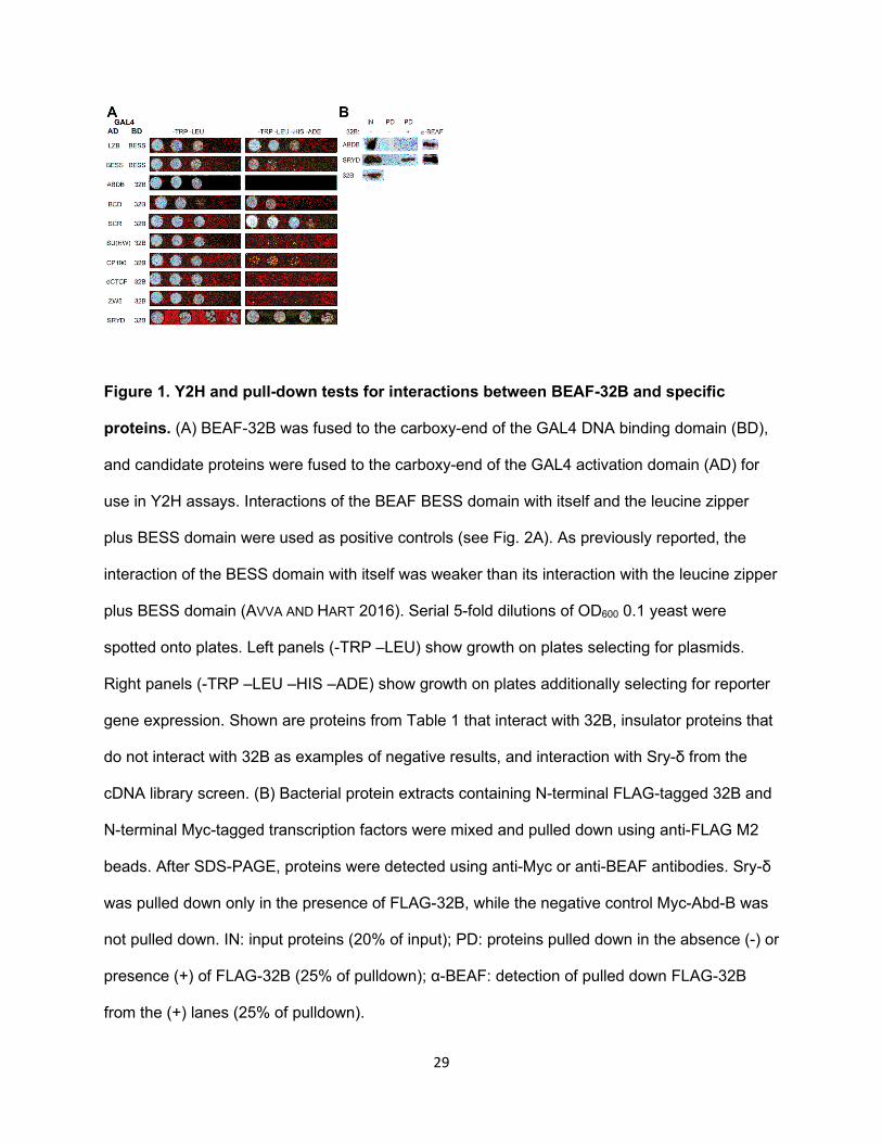
Figure 1. Y2H and pull-down tests for interactions between BEAF-32B and specific
proteins. (A) BEAF-32B was fused to the carboxy-end of the GAL4 DNA binding domain (BD),
and candidate proteins were fused to the carboxy-end of the GAL4 activation domain (AD) for
use in Y2H assays. Interactions of the BEAF BESS domain with itself and the leucine zipper
plus BESS domain were used as positive controls (see Fig. 2A). As previously reported, the
interaction of the BESS domain with itself was weaker than its interaction with the leucine zipper
plus BESS domain (AVVA AND HART 2016). Serial 5-fold dilutions of OD600 0.1 yeast were
spotted onto plates. Left panels (-TRP –LEU) show growth on plates selecting for plasmids.
Right panels (-TRP –LEU –HIS –ADE) show growth on plates additionally selecting for reporter
gene expression. Shown are proteins from Table 1 that interact with 32B, insulator proteins that
do not interact with 32B as examples of negative results, and interaction with Sry-δ from the
cDNA library screen. (B) Bacterial protein extracts containing N-terminal FLAG-tagged 32B and
N-terminal Myc-tagged transcription factors were mixed and pulled down using anti-FLAG M2
beads. After SDS-PAGE, proteins were detected using anti-Myc or anti-BEAF antibodies. Sry-δ
was pulled down only in the presence of FLAG-32B, while the negative control Myc-Abd-B was
not pulled down. IN: input proteins (20% of input); PD: proteins pulled down in the absence (-) or
presence (+) of FLAG-32B (25% of pulldown); α-BEAF: detection of pulled down FLAG-32B
from the (+) lanes (25% of pulldown).

30
Figure 2. Mapping the region of BEAF that interacts with Sry-δ. (A) Schematic of the parts
of BEAF that were fused to the GAL4 BD for Y2H assays. BED ZnF: 32B unique sequences,
encompassing the DNA-binding BED finger (blue rectangle). M, MID: middle region. LZ: putative
leucine zipper (purple rectangle). BESS: BESS domain (green rectangle). Numbers indicate the
first and last amino acid present in the truncated proteins. (B) Results of Y2H assays, as in Fig.
1A. BESS-BESS and Sry-δ-32B interactions were included as positive controls. Sry-δ interacts
with M1-60.

31
Figure 3. Mapping the region of Sry-δ that interacts with 32B. (A) Schematic of the parts of
Sry-δ that were fused to the GAL4 AD or a Myc tag. An acidic region and zinc fingers are
indicated. (B) Results of Y2H assays, as in Fig. 1A. The N-terminal half of Sry-δ, which lacks the
zinc fingers, interacted with 32B. (C) Results of 32B pull-down assays, as in Fig. 1B (IN: 20% of
input; PD: 50% of the pulled down proteins; α-BEAF: 25% of the pulled down proteins). The half
of Sry-δ lacking the zinc fingers was pulled down with 32B. (D) Summary of interactions
between BEAF and Sry-δ.

32
Figure 4. Testing the interaction between Sry-δ and 32B using biFC. (A) Graph showing the
fraction of cells showing biFC of indicated cV-tagged proteins with 32B-nV, normalized to 32B-
cV with 32B-nV. A minimum of 50 images were counted per sample per experiment, and results
are an average of three biological replicates with error bars showing the standard deviation of
the normalized replicates. Results for Sry-δ-cV were variable, but it clearly interacted with 32B-
nV. (B) Representative micrographs for the indicated proteins. Nuclei were stained with Hoechst
and false colored red, while Venus is shown in green. All images were acquired using the same
settings, but the images shown for delBESS-cV had the green channel enhanced to better show
the Venus signal.

33
Figure 5. A rough-eye assay shows a strong genetic interaction between BEAF and Sry-
δ. Shown are SEM images from eyes of 3-5 day old females. Negative control ey-GAL4/+; Sry-
δSF2/+ flies have normal eyes. A mild rough-eye phenotype is seen in ey-GAL4/+; UAS-BID/+
flies expressing a dominant-negative form of BEAF. The rough-eye phenotype is much stronger
when the Sry-δSF2 mutation is also present (ey-GAL4/+; UAS-BID/ Sry-δSF2). This phenotype is
100% penetrant. Sry-δ UAS RNAi transgenes give a clear rough eye phenotype when
heterozygous with ey-GAL4 (ey-GAL4/Sry-δ-RNAi1 and ey-GAL4/Sry-δ-RNAi2). The phenotype
is more extreme in combination with heterozygous UAS-BID (ey-GAL4/Sry-δ-RNAi1; BID/+ and
ey-GAL4/Sry-δ-RNAi2; BID/+). RNAi1: VDRC 41094; RNAi2: VDRC 102786.

34
Figure 6. Promoter-proximal BEAF facilitates local and long-range interactions between
Sry-δ and promoters, and directly activates a housekeeping promoter. (A) Schematic of
constructs used to drive firefly luciferase expression in transfected S2 cells. All transfections
also had a plasmid with an Act5C promoter driving Renilla luciferase expression to normalize for
transfection efficiency, with or without a plasmid with an Act5C promoter driving expression of
Sry-δ or Sry-δ-VP16 (endogenous Sry-δ is expressed in S2 cells). Transfections with each set
of plasmids was further normalized to firefly luciferase expression when only the BEAF binding
site was present. Error bars indicate standard deviations of 3 biological replicates. Test

35
plasmids had either a minimal y (developmental) or RpS12 (housekeeping) promoter. mBF or
BF: promoter-proximal mutant or wild-type BEAF binding site. ppTF: promoter-proximal 4
tandem Sry-δ transcription factor binding sites. pdTF: promoter-distal 4 tandem Sry-δ
transcription factor binding sites. Also shown is a model of Sry-δ interacting with BEAF to
facilitate long-range activation of the promoter. The y promoter was tested (B) without an Sry-δ-
expressing plasmid; (C) with an Sry-δ plasmid; and (D) with an Sry-δ-VP16 plasmid. Average
luminometer readings for BF.y were 102,245 compared to a mock transfection background of
99. The RpS12 promoter was tested (E) without an Sry-δ-expressing plasmid; (F) with an Sry-δ
plasmid; and (G) with an Sry-δ-VP16 plasmid. Average luminometer readings for BF.R were
1,145,492 compared to a mock transfection background of 102. (H) Testing the aurA promoter
with and without mutations in the BEAF binding site. Average luminometer readings for aurA
were 924,577 compared to a mock transfection background of 122. In (B) through (G),
comparison of the BF, ppTF.mBF and ppTF.BF values show local interactions between Sry-δ
and BEAF cooperatively activate the reporter gene, while comparison of the BF, pdTF.mBF and
pdTF.BF values indicate long-range interactions between Sry-δ and BEAF activate the reporter
gene. In (B) through (D), comparisons of mBF and BF show that BEAF does not activate the y
promoter. In (E) through (H), comparisons of mBF and BF show that BEAF activates the RpS12
and aurA promoters.