Embed Size (px)
Citation preview
Projection Patterns of Single MossyFibers Originating From the Lateral
Reticular Nucleus in the Rat CerebellarCortex and Nuclei
H.-S. WU, I. SUGIHARA, AND Y. SHINODA*Department of Physiology, School of Medicine, Tokyo Medical and Dental University,
Bunkyo-ku, Tokyo, 113–8519, Japan
ABSTRACTProjection of neurons in the lateral reticular nucleus (LRN) to the cerebellar cortex (Cx)
and the deep cerebellar nuclei (DCN) was studied in the rat by using the anterograde tracerbiotinylated dextran amine (BDA). After injection of BDA into the LRN, labeled terminalswere seen bilaterally in most cases in the vermis, intermediate zone, and hemisphere of theanterior lobe, and in various areas in the posterior lobe, except the flocculus, paraflocculus,and nodulus. Areas of dense terminal projection were often organized in multiple longitudinalzones. The entire axonal trajectory of single axons of labeled LRN neurons was reconstructedfrom serial sections. Stem axons entered the cerebellum through the inferior cerebellarpeduncle (mostly ipsilateral), and ran transversely in the deep cerebellar white matter. Theyoften entered the contralateral side across the midline. Along the way, primary collateralswere successively given off from the transversely running stem axons at almost right angles tothe Cx and DCN, and individual primary collaterals had longitudinal arborizations thatterminated as mossy fibers in multiple lobules of the Cx. These collaterals arising from singleLRN axons terminated bilaterally or unilaterally in the vermis, intermediate area, andsometimes hemisphere, and in different cerebellar and vestibular nuclei simultaneously. Thecortical terminals of single axons appeared to be distributed in multiple longitudinal zonesthat were arranged in a mediolateral direction. All of the LRN axons examined (n 5 29) hadaxon collaterals to the DCN. All of the terminals observed in the DCN and vestibular nucleibelonged to axon collaterals of mossy fibers terminating in the Cx. J. Comp. Neurol.411:97–118, 1999. r 1999 Wiley-Liss, Inc.
Indexing terms: afferent pathways; cerebellar cortex; deep cerebellar nuclei; nerve endings;
medulla oblongata
Mossy fibers in the cerebellar cortex (Cx) are character-ized by their thickened ends, which resemble a piece ofmoss in the granular layer of the Cx (Ramon y Cajal, 1911).Afferents of the cerebellum from many different origins,including brainstem precerebellar nuclei but excluding theinferior olive (IO), take the form of mossy fibers in the Cx.Due to the uniformity of the local circuit of the Cx, theprojection pattern of afferents to the Cx, including themossy fiber system, plays a significant role in determiningthe functional compartmentation and organized action ofthe cerebellum (Eccles et al., 1967; Palay and Chan-Palay,1974; Ito, 1984).
Classic studies on receptive field organization in the Cxhave shown the existence of transversely organized divi-sions (Snider, 1950; Grant, 1962; Brodal, 1981). On theother hand, more recent studies on mapping receptive
fields through olivocerebellar climbing fiber projectionhave suggested that the Cx is composed of many longitudi-nal functional compartments (Andersson and Oscarsson,1978; Ito et al., 1982; Ito, 1984). This electrophysiologicallydefined organization is consistent with the anatomicallydefined longitudinal compartments in the Cx (Groenewe-
Grant sponsor: CREST (Core Research for Evolutional Science andTechnology) of the Japan Science and Technology Corporation; Grantsponsor: Ministry of Education, Science, and Culture of Japan; Grantsponsor: Sasakawa Health Science Foundation; Grant sponsor: IwakiScholarship Foundation.
*Correspondence to: Dr. Yoshikazu Shinoda, Department of Physiology,School of Medicine, Tokyo Medical and Dental University, 1–5-45 Yushima,Bunkyo-ku, Tokyo, 113–8519, Japan. E-mail: [email protected]
Received 23 December 1998; Revised 2 March 1999; Accepted 16 March1999
THE JOURNAL OF COMPARATIVE NEUROLOGY 411:97–118 (1999)
r 1999 WILEY-LISS, INC.
gen and Voogd, 1977; Voogd et al., 1996). Some mossy fiberprojections also seem to show longitudinal organization inthe vestibulofloccular system (Sato et al., 1983) and thespinocerebellar system (Oscarsson, 1976). However, thesomatotopical map of the face established by detailedelectrophysiologic investigation does not seem to consist oflongitudinal zones, but rather of a mosaic of small patchyareas in the mossy fiber projection from the facial area tothe hemispheric ansiform lobule (Shambes et al., 1978).Thus, whether or not there is a common principle in thecortical organization of the projections of different mossyfiber systems remains unclear.
Mossy and climbing fiber projections to the deep cerebel-lar nuclei (DCN) have not been fully investigated, com-pared with their projection to the Cx. Determining theexistence or nonexistence of excitatory inputs to the DCNfrom the brainstem is important for our understanding ofcerebellar function (Shinoda et al., 1993, 1997). It has beengenerally assumed that excitatory inputs to the DCN aresupplied through collaterals of mossy and climbing fiberafferents to the Cx (Eccles et al., 1967; Ito, 1984). Despite awealth of anatomic reports on the afferent pathways to theCx, until recently there have been far fewer studies onafferent projections to the DCN. By using modern reliableanatomic staining methods, the projections from the pon-tine nucleus, nucleus reticularis tegmenti pontis, IO, andspinal cord to the DCN have been definitely confirmed(Gerrits and Voogd, 1987; Van der Want et al., 1989;Shinoda et al., 1992; Mihailoff, 1993; Matsushita andYaginuma, 1995). The existence of collateral projection ofmossy fibers of the pontine nucleus and the nucleusreticularis tegmenti pontis to the DCN was demonstratedby intra-axonal staining with HRP (Shinoda et al., 1992,1997), and the collateral projection of climbing fibers to theDCN was identified by labeling single axons with biotinyl-ated dextran amine (BDA) (Sugihara et al., 1996). How-ever, questions still remain as to whether there exist somefibers that project only to the DCN without projecting tothe Cx and how many percentages of mossy fibers haveaxon collaterals to the DCN in different mossy fibersystems.
The lateral reticular nucleus (LRN) is a major mossyfiber source in the medulla. The LRN receives afferentsmainly from the spinal cord bilaterally (Oscarsson andRosen, 1966; Bruckmoser et al., 1970; Kunzle, 1973;
Clendenin et al., 1974c,d; Ekerot and Oscarsson, 1975)and, additionally, from several supraspinal structures,including the cerebral cortex (Bruckmoser et al., 1970;Ruigrok and Cella, 1995). Now, it is generally acceptedthat the LRN projects to the bilateral cerebellar corticeswith ipsilateral predominance (Matsushita and Ikeda,1976: Voogd, 1964), and to the ‘‘classical spinal receivingarea,’’ i.e., the anterior lobe and paramedian lobule, ratherthan the whole Cx (Clendenin et al., 1974a; Ruigrok andCella, 1995). Innervations from the LRN to the DCN(Parenti et al., 1996) and to the vestibular nuclei (VN)(Ruigrok et al., 1995) are also bilateral with ipsilateralpredominance. The cerebellar cortical projection of theLRN shows a multiple longitudinal zonal pattern (Kunzle,1975; Chan-Palay et al., 1977) that is roughly related tozebrin-identified Purkinje cell zones (Ruigrok and Cella,1995). These anatomic studies have used an anterogradestaining method to examine LRN projection to the Cx, butdealt with mass projection. Information on the entire axontrajectory of the single mossy fibers should reveal us thespatial innervation pattern in the Cx, the frequency ofoccurrence of nuclear collaterals, and the general topo-graphical correspondence between LRN-cortical and LRN-nuclear projections of the single mossy fibers.
The present study was aimed at revealing characteristicbranching patterns of single mossy fibers in the Cx and theDCN by reconstructing the entire trajectory of singleaxons labeled after the injection of BDA into the LRN inthe rat. The results show that single LRN axons give riseto multiple collaterals to the Cx and DCN mainly bilater-ally, and these collaterals of single axons appear to makeup multiple longitudinal zones that are arranged in amediolateral direction in multiple lobules.
MATERIALS AND METHODS
Eighteen adult Long-Evans rats of both sexes, weighingbetween 250 and 310 g, were used in the present study.Fifteen of them received injections of BDA in the LRN, onein the central cervical nucleus (CCN) and two in the lateralmedulla and inferior cerebellar peduncle (icp). The surgeryand animal care conformed to The Principles of LaboratoryAnimal Care (NIH publication No. 85–23, revised in 1985)and also to Guiding Principles for the Care and Use of
Abbreviations
I–X lobules I–XAMB nucleus ambiguusBDA Biotinylated dextran amineC caudalCCN central cervical nucleusCOP copula pyramidisCrus I crus I ansiform lobuleCrus II crus II ansiform lobuleCx cerebellar cortexD dorsalDCN deep cerebellar nucleiDN dentate nucleusFL flocculusFN fastigial nucleusicp inferior cerebellar peduncleIO inferior olive nucleusIP interposed nucleusIPa anterior interposed nucleusIPp posterior interposed nucleusLVN lateral vestibular nucleus
LRN lateral reticular nucleusLRNm the magnocellular part of the LRNLRNp the parvicellular part of the LRNLRNst the subtrigeminal part of the LRNLt leftMVN medial vestibular nucleusPGRNI paragigantocellular nucleusPHA-L Phaseolus vulgaris leucoagglutininPFL paraflocculusPM paramedian lobuleR rostralRt rightscp superior cerebellar peduncleSCT spinocerebellar tractSim simple lobuleSPV spinal trigeminal nucleusSVN superior vestibular nucleusV ventralVN vestibular nucleus
98 H.-S. WU ET AL.
Animals in the Field of Physiological Sciences (The Physi-ological Society of Japan, 1988).
Surgical procedures and tracer application
The animals were anesthetized with an intraperitonealinjection of ketamine (130 mg/kg body weight), xylazine(Rompun, Bayer, Germany; 8 mg/kg), and atropine (0.4mg/kg) and placed in a stereotaxic apparatus. Heart rateand rectal temperature were monitored continuously.Supplemental doses of ketamine (13 mg/kg) and xylazine(1 mg/kg) were given every 30 minutes starting 1 hourafter the initial dose, as required. A heating pad was usedto keep the rectal temperature between 35 and 37°C. Afterincision of the dorsal neck skin, a cut was made in theligament between the skull and the first vertebra.
BDA (D-1956, 10,000 MW, Molecular Probes, Eugene,OR) was dissolved in physiologic saline to give a concentra-tion of 10–15%. A glass micropipette (tip diameter, 4 µm)was filled with this solution. The coordinate of the inser-tion point for the LRN was approximately 0.5 mm caudalfrom the caudal edge of the area postrema, 1.8 mm lateralto the midline. The pipette was tilted 20° caudally in theparasagittal plane, and inserted to a depth of about 3.5mm from the surface. Neural activity was monitored fromthe pipette to facilitate locating the LRN. A small injectionwas made either with pressure (about 0.1 µl) or electropho-resis (2µA positive current pulses of 1-second duration at0.5 Hz for 30 minutes). Pipettes were left in situ for 5minutes after injection before they were withdrawn. Thewound was cleaned with povidone-iodine, and an antibi-otic (cefmetazole) was applied to the wound before sutur-ing.
Fixation and histochemistry
After a survival period of 6 to 8 days, the animals weredeeply anesthetized with ketamine (150 mg/kg) and xyla-zine (12 mg/kg) and perfused through the ascending aorta.Chilled perfusate containing 0.8% NaCl, 0.8% sucrose, and0.4% glucose in 0.05 M phosphate buffer (pH 7.4, about4°C, and about 400 ml) was given and followed by a fixativecontaining 2% paraformaldehyde, 0.6% glutaraldehyde,and 4% sucrose in 0.05 M sodium phosphate buffer (pH 7.4,about 4°C, and about 500 ml) delivered over 30 minutes.Dissected brains (cerebellum and medulla oblongata) werekept in the same fixative overnight at 4°C and then in 30%sucrose in phosphate buffer (0.05 M, pH 7.4, 4°C) for 1–2days. The brain was then embedded in a gelatin block.
Serial coronal sections 35- or 50-µm thick were cut on afreezing microtome. Sections were treated with biotinyl-ated HRP-avidin complex (Standard ABC kit KT-4000,Vector, Burlingame, CA), and a cobalt-glucose oxidasemethod (Itoh et al., 1979; Sugihara et al., 1996) was usedfor the diaminobenzidine reaction. The sections were thenmounted on chrome alum–gelatinized slides, dried over-night, and cover-slipped with Permount. After reconstruc-tion of labeled axons, some of the sections were counter-stained with thionin.
Light microscopic reconstruction
Drawings of labeled axons and terminals were made atan objective magnification of 32 to 360, with the aid of aNikon microscope equipped with a camera lucida appara-tus. Trajectories of single-labeled axons were recon-structed on serial sections by connecting cut ends of an
axon on one section to the corresponding cut ends of thesame axon on the successive sections as was done with theintra-axonal injection of HRP (Futami et al., 1979; Shi-noda et al., 1981, 1986, 1992). Reconstructions in thecoronal plane were sometimes converted to those in theparasagittal plane, by measuring depths and replotted.
In drawings of camera lucida images, the fibers andswellings were drawn thicker than scale for clarity, as isconventionally done in drawings of reconstructed fibers.The cerebellar lobules were defined according to Larsell(1952) and Voogd (1995). Subdivisions of the cerebellar andvestibular nuclei were determined according to the descrip-tions of Korneliussen (1968), Voogd (1995), and Rubertoneet al. (1995). The nomenclature used generally followedSwanson (1992) and Voogd (1995). Some photomicro-graphs were obtained with a computer-aided dynamicfocusing system (MCID image analysis system, ImagingResearch, Inc., St. Catharines, Ontario, Canada).
RESULTS
Injection sites
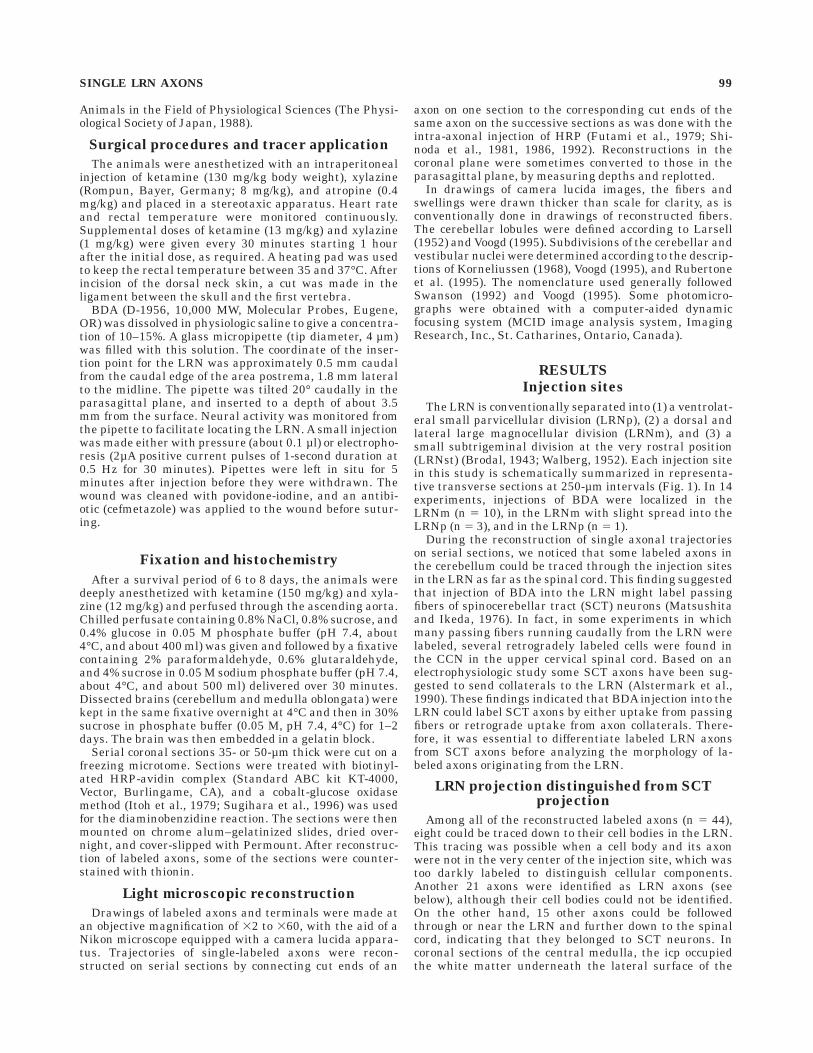
The LRN is conventionally separated into (1) a ventrolat-eral small parvicellular division (LRNp), (2) a dorsal andlateral large magnocellular division (LRNm), and (3) asmall subtrigeminal division at the very rostral position(LRNst) (Brodal, 1943; Walberg, 1952). Each injection sitein this study is schematically summarized in representa-tive transverse sections at 250-µm intervals (Fig. 1). In 14experiments, injections of BDA were localized in theLRNm (n 5 10), in the LRNm with slight spread into theLRNp (n 5 3), and in the LRNp (n 5 1).
During the reconstruction of single axonal trajectorieson serial sections, we noticed that some labeled axons inthe cerebellum could be traced through the injection sitesin the LRN as far as the spinal cord. This finding suggestedthat injection of BDA into the LRN might label passingfibers of spinocerebellar tract (SCT) neurons (Matsushitaand Ikeda, 1976). In fact, in some experiments in whichmany passing fibers running caudally from the LRN werelabeled, several retrogradely labeled cells were found inthe CCN in the upper cervical spinal cord. Based on anelectrophysiologic study some SCT axons have been sug-gested to send collaterals to the LRN (Alstermark et al.,1990). These findings indicated that BDA injection into theLRN could label SCT axons by either uptake from passingfibers or retrograde uptake from axon collaterals. There-fore, it was essential to differentiate labeled LRN axonsfrom SCT axons before analyzing the morphology of la-beled axons originating from the LRN.
LRN projection distinguished from SCTprojection
Among all of the reconstructed labeled axons (n 5 44),eight could be traced down to their cell bodies in the LRN.This tracing was possible when a cell body and its axonwere not in the very center of the injection site, which wastoo darkly labeled to distinguish cellular components.Another 21 axons were identified as LRN axons (seebelow), although their cell bodies could not be identified.On the other hand, 15 other axons could be followedthrough or near the LRN and further down to the spinalcord, indicating that they belonged to SCT neurons. Incoronal sections of the central medulla, the icp occupiedthe white matter underneath the lateral surface of the
SINGLE LRN AXONS 99
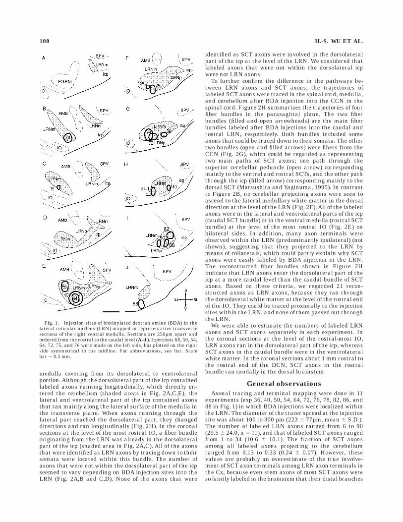
medulla covering from its dorsolateral to ventrolateralportion. Although the dorsolateral part of the icp containedlabeled axons running longitudinally, which directly en-tered the cerebellum (shaded areas in Fig. 2A,C,E), thelateral and ventrolateral part of the icp contained axonsthat ran mainly along the lateral surface of the medulla inthe transverse plane. When axons running through thelateral part reached the dorsolateral part, they changeddirections and ran longitudinally (Fig. 2H). In the coronalsections at the level of the most rostral IO, a fiber bundleoriginating from the LRN was already in the dorsolateralpart of the icp (shaded area in Fig. 2A,C). All of the axonsthat were identified as LRN axons by tracing down to theirsomata were located within this bundle. The number ofaxons that were not within the dorsolateral part of the icpseemed to vary depending on BDA injection sites into theLRN (Fig. 2A,B and C,D). None of the axons that were
identified as SCT axons were involved in the dorsolateralpart of the icp at the level of the LRN. We considered thatlabeled axons that were not within the dorsolateral icpwere not LRN axons.
To further confirm the difference in the pathways be-tween LRN axons and SCT axons, the trajectories oflabeled SCT axons were traced in the spinal cord, medulla,and cerebellum after BDA injection into the CCN in thespinal cord. Figure 2H summarizes the trajectories of fourfiber bundles in the parasagittal plane. The two fiberbundles (filled and open arrowheads) are the main fiberbundles labeled after BDA injections into the caudal androstral LRN, respectively. Both bundles included someaxons that could be traced down to their somata. The othertwo bundles (open and filled arrows) were fibers from theCCN (Fig. 2G), which could be regarded as representingtwo main paths of SCT axons; one path through thesuperior cerebellar peduncle (open arrow) correspondingmainly to the ventral and rostral SCTs, and the other paththrough the icp (filled arrow) corresponding mainly to thedorsal SCT (Matsushita and Yaginuma, 1995). In contrastto Figure 2B, no cerebellar projecting axons were seen toascend to the lateral medullary white matter in the dorsaldirection at the level of the LRN (Fig. 2F). All of the labeledaxons were in the lateral and ventrolateral parts of the icp(caudal SCT bundle) or in the ventral medulla (rostral SCTbundle) at the level of the most rostral IO (Fig. 2E) onbilateral sides. In addition, many axon terminals wereobserved within the LRN (predominantly ipsilateral) (notshown), suggesting that they projected to the LRN bymeans of collaterals, which could partly explain why SCTaxons were easily labeled by BDA injection in the LRN.The reconstructed fiber bundles shown in Figure 2Hindicate that LRN axons enter the dorsolateral part of theicp at a more caudal level than the caudal bundle of SCTaxons. Based on these criteria, we regarded 21 recon-structed axons as LRN axons, because they ran throughthe dorsolateral white matter at the level of the rostral endof the IO. They could be traced proximally to the injectionsites within the LRN, and none of them passed out throughthe LRN.
We were able to estimate the numbers of labeled LRNaxons and SCT axons separately in each experiment. Inthe coronal sections at the level of the rostral-most IO,LRN axons ran in the dorsolateral part of the icp, whereasSCT axons in the caudal bundle were in the ventrolateralwhite matter. In the coronal sections about 1 mm rostral tothe rostral end of the DCN, SCT axons in the rostralbundle ran caudally in the dorsal brainstem.
General observations
Axonal tracing and terminal mapping were done in 11experiments (exp 36, 40, 50, 54, 64, 72, 76, 78, 82, 86, and88 in Fig. 1) in which BDA injections were localized withinthe LRN. The diameter of the tracer spread at the injectionsite was about 100 to 300 µm (223 6 77µm, mean 6 S.D.).The number of labeled LRN axons ranged from 6 to 90(29.5 6 24.0, n 5 11), and that of labeled SCT axons rangedfrom 1 to 34 (10.6 6 10.1). The fraction of SCT axonsamong all labeled axons projecting to the cerebellumranged from 0.13 to 0.33 (0.24 6 0.07). However, thesevalues are probably an overestimate of the true involve-ment of SCT axon terminals among LRN axon terminals inthe Cx, because even stem axons of most SCT axons wereso faintly labeled in the brainstem that their distal branches
Fig. 1. Injection sites of biotinylated dextran amine (BDA) in thelateral reticular nucleus (LRN) mapped in representative transversesections of the right ventral medulla. Sections are 250µm apart andordered from the rostral to the caudal level (A–J). Injections 68, 50, 54,64, 72, 75, and 76 were made on the left side, but plotted on the rightside symmetrical to the midline. For abbreviations, see list. Scalebar 5 0.5 mm.
100 H.-S. WU ET AL.
Fig. 2. Separation of lateral reticular nucleus (LRN) axons andspinocerebellar tract (SCT) axons in the medullary white matter.A,B: Camera lucida drawings in a transverse plane showing axons atthe level of the rostral IO (A) labeled by an injection of biotinylateddextran amine (BDA) into the caudal LRN at the level of thecaudal-most IO (B). Filled arrowheads indicate labeled LRN axons.C,D: Drawings of labeled axons at the level of the rostral IO (C) afterinjection of BDA into the central LRN at the level of the caudal IO (D).Some SCT axons (filled and open arrows) were also labeled in theventrolateral white matter of the medulla. Filled arrowhead indicateslabeled LRN axons. E–G: Camera lucida drawings of SCT axons at thelevel of the rostral and caudal poles of the IO (E,F), which were labeledby an injection of BDA into the central cervical nucleus (CCN) at C4(G). An open arrow shows BDA-labeled fibers that do not enter the icp
but run more rostrally at the medulla, whereas a filled arrow showsBDA-labeled CCN fibers running through the icp. H: Reconstructedtrajectories of bundles of axons from injection sites in the caudal LRN(filled arrowhead) and rostral LRN (open arrowhead), and of rostraland caudal bundles (open and filled arrows, respectively) of SCT axonslabeled by an injection into the CCN. The two vertical straight linesindicate the level of the rostral pole of the IO and the level 1 mmrostral to the cerebellar nuclei, in which the number of labeled axonsthat belonged to each bundle of SCT and LRN axons could be countedseparately. Shaded areas in A, C and E indicate a dorsolateral portionof the inferior cerebellar peduncle in which fibers ran longitudinallytoward the entrance to the cerebellum. Shaded areas in B, D, and Gindicate injection sites of BDA. For abbreviations, see list. Scalebars 5 1 mm.
in the cerebellar white matter became untraceable. There-fore, despite the contamination by labeled SCT axons inthe cerebellar peduncles, contamination by the terminalsof these SCT axons among LRN axon terminals may bemuch less than is indicated by these numbers.
All of the labeled fibers in the Cx bore rosette-typeterminals characteristic of mossy fibers (Ramon y Cajal etal., 1911). No climbing fibers or axons of other typesbearing only small non–rosette-type swellings were ob-served. Therefore, virtually all of the LRN axons project-ing to the Cx appeared to take the form of a mossy fiber. Weextensively analyzed the data from four experiments (exp40, 76, 82, and 88) in which the fraction of labeled SCTaxons was relatively small (23%, 23%, 13%, 25%, respec-tively) on terminal distributions in the Cx, DCN, and VN.Abundant labeled fibers of very small diameters bearingsmall swellings were observed in the DCN and VN as inthe Cx. They were presumably collateral terminations ofLRN axons, as shown by single axon tracings in latersections.
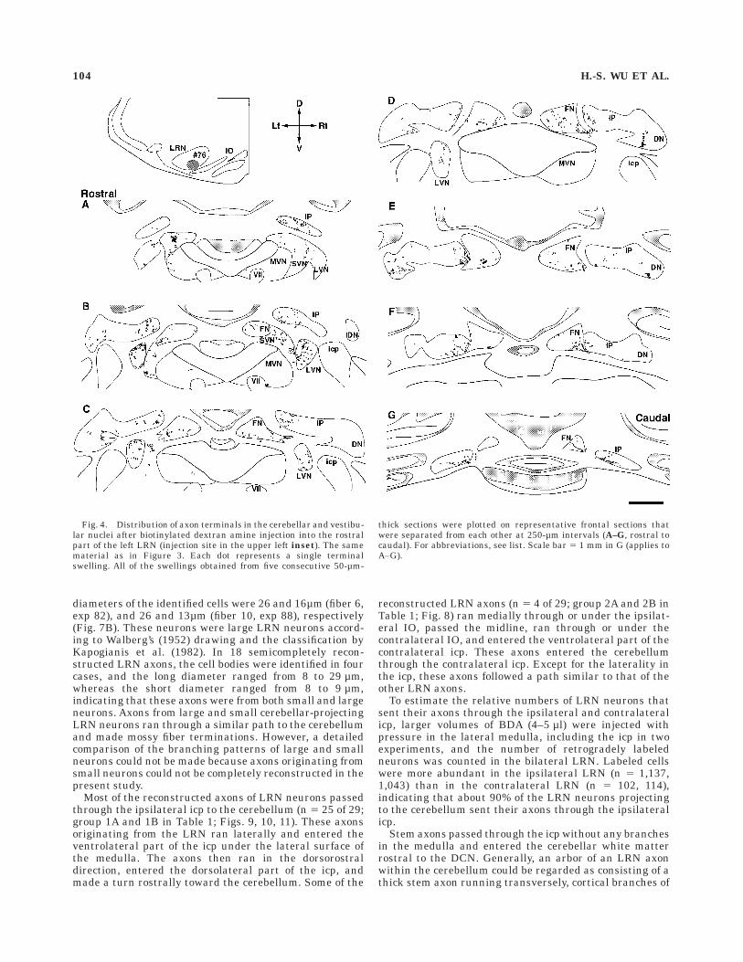
Observations from serial coronal sections showed somedifferences in the distribution of labeled terminals in theCx, DCN, and VN in these four experiments. However,these four cases had many common characteristic featuresof the distribution pattern of axon terminals of the LRNprojection in the cerebellum and were representative of allof the 14 experiments. Figure 3 shows a typical example ofthe distribution of axon terminals in the Cx after BDAinjection into the rostral part of the LRNm (exp 76).Rosettes were densely distributed in the Cx in the bilateralvermis and intermediate areas and hemispheres of lobulesII, III, IV, and V (most dense in IV and V). A less-densedistribution was seen in the bilateral vermis of lobules VI,VII, VIII, and IX, and in hemispheres of simple andansiform lobules, and copula pyramidis. A small number ofrosettes were seen in the left paramedian lobule, and norosettes were seen in the flocculus, paraflocculus, or nodu-lus. There were slightly more rosettes on the ipsilateralside (n 5 3,836) than on the contralateral side (n 5 2,722),and the ratio of ipsilateral terminals to contralateralterminals in the Cx was 1.41:1. Because BDA was injectedinto the left LRN, this finding indicated slight ipsilateralpredominance. In this distribution (Fig. 3), multiple zonesof clusters of rosettes were obvious in the vermis of lobulesIII, V, and VI, and weak zones seemed to be present inother areas such as lobules IV and VII. In lobules III, V,and VI, three zones with a dense distribution of rosetteswere seen, and the width of each was about 0.5 mm. One ofthe zones in the vermis was located at the midline, and theother lateral zones were located about 0.6 mm (lobule III)or 1.2 mm (lobule V, VI) from the midline bilaterally.Swellings in the DCN and VN in the same experimentwere distributed mainly in the fastigial (FN), anteriorinterposed (IPa), posterior interposed (IPp) and dentate(DN) nuclei, and the lateral vestibular nuclei (LVN) bilat-erally (Fig. 4). Dense distribution was seen in the ventro-medial part of the IPp, the medial part of the IPa, theventrolateral part of the FN and the dorsal LVN bilater-ally.
In one experiment (exp 82 in Fig. 1), axon terminalswere distributed almost exclusively ipsilaterally in the Cx(Fig. 5) and mainly ipsilaterally with some contralateraldistribution in the DCN (Fig. 6). This injection site was inthe dorsolateral part of the caudal LRNm. Rosettes weredistributed in lobules II, III, IV, and V in the anterior lobe,
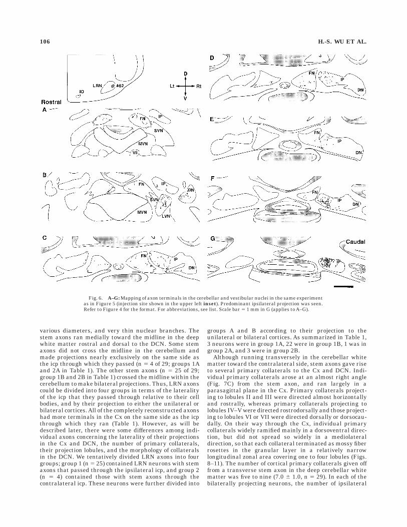
and in lobules VI, VII, VII, and IX, and in the simple,ansiform, and paramedian lobules and copula pyramidisonly ipsilaterally in the posterior lobe. A small number ofrosettes were seen in lobule I and the ansiform lobule. Norosettes were seen in the flocculus, paraflocculus or nodu-lus. Dense distributions were seen in the junction of thelateral-most vermis and the intermediate area of lobulesIV and V, in the lateral-most hemisphere of lobule II, III,IV, and V, in the junction of the lateral vermis andintermediate area in lobule VI-simple lobule, lobule VII-paramedian lobule, and lobule VIII-copula pyramidis, andin the hemispheric portion in the paramedian lobule andcopula pyramidis (most dense in lobule V). Areas of densedistribution seemed to be arranged in a multiple zonalpattern in the anterior lobe and in the paramedian lobuleand copula pyramidis. In the anterior lobe, a zone about0.5 mm wide could be recognized in the right vermis about1 mm lateral from the midline. The second zone was about1 mm or more wide and was located in the lateral-mostvermis through the intermediate area. The third zone wasabout 1 mm wide and was located in the lateral-mosthemisphere. In the paramedian lobule and copula pyrami-dis, four zones about 0.5 mm wide seemed to be located insemiparasagittal planes separated by about 1 mm fromthe junction between the vermis and intermediate area tothe lateral-most hemisphere. In the DCN and VN of thisexperiment, the distribution of labeled terminals waspredominantly ipsilateral as in the Cx (Fig. 6). A densedistribution of swellings was seen in the medial part of theipsilateral IP, and a small number of swellings were seenin bilateral FNs and ipsilateral DN. However, swellingswere almost negligible in the contralateral DN and thecontralateral lateral IP. In the VN, a dense distribution ofswellings was seen in the dorsal part of the ipsilateral LVNbut no terminals were seen in the contralateral VN.
In the other two cases of mapping in which the BDAinjection was located in the caudolateral LRNm (exp 40)and caudoventral LRNm (exp 88), the distribution oflabeled terminals was similar to that with injection intothe rostral LRNm (exp 76 plotted in Figs. 3, 4). Inexperiments in which detailed mappings were not made (7of 11 experiments), the distribution of terminals in the Cx,DCN, and VN was roughly similar to the case shown inFigures 3 and 4. In summary, LRN axons originating fromthe LRNm generally showed a bilateral projection withipsilateral predominance in the anterior lobe and someareas, including the paramedian lobule and copula pyrami-dis, in the posterior lobe with a multiple longitudinal zonalpattern. However, axons originating from the dorsolateralpart of the caudal LRN showed a virtually exclusiveipsilateral projection.
General morphology of single reconstructedLRN axons
The entire trajectories of single LRN axons could becompletely reconstructed from the injection site to termina-tions of every branch in 11 identified LRN axons. Inaddition, nearly complete reconstruction, except for distalportions of some branches in the cerebellum, was possiblein 18 identified LRN axons. These axons (n 5 29) wereused for a detailed morphologic analysis, and other par-tially reconstructed axons (n 5 69) were used to supple-ment the data.
Among the 11 completely reconstructed axons, the cellbodies were identified in two cases. The long and short
102 H.-S. WU ET AL.
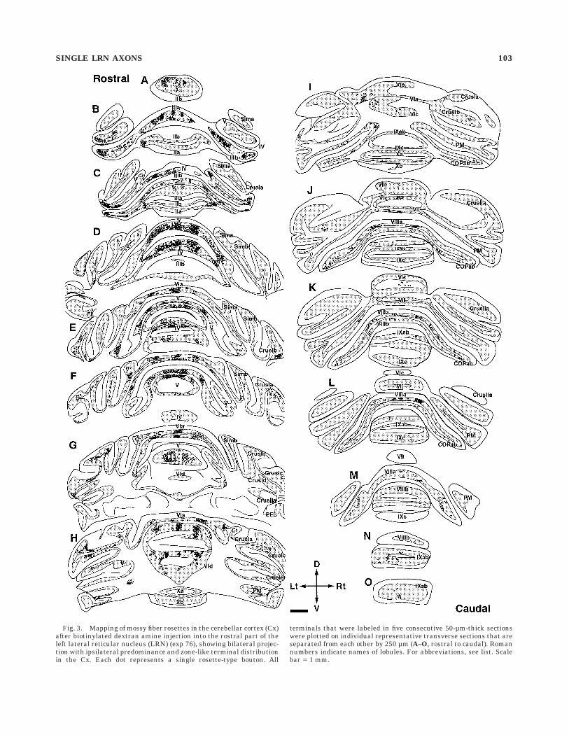
Fig. 3. Mapping of mossy fiber rosettes in the cerebellar cortex (Cx)after biotinylated dextran amine injection into the rostral part of theleft lateral reticular nucleus (LRN) (exp 76), showing bilateral projec-tion with ipsilateral predominance and zone-like terminal distributionin the Cx. Each dot represents a single rosette-type bouton. All
terminals that were labeled in five consecutive 50-µm-thick sectionswere plotted on individual representative transverse sections that areseparated from each other by 250 µm (A–O, rostral to caudal). Romannumbers indicate names of lobules. For abbreviations, see list. Scalebar 5 1 mm.
SINGLE LRN AXONS 103
diameters of the identified cells were 26 and 16µm (fiber 6,exp 82), and 26 and 13µm (fiber 10, exp 88), respectively(Fig. 7B). These neurons were large LRN neurons accord-ing to Walberg’s (1952) drawing and the classification byKapogianis et al. (1982). In 18 semicompletely recon-structed LRN axons, the cell bodies were identified in fourcases, and the long diameter ranged from 8 to 29 µm,whereas the short diameter ranged from 8 to 9 µm,indicating that these axons were from both small and largeneurons. Axons from large and small cerebellar-projectingLRN neurons ran through a similar path to the cerebellumand made mossy fiber terminations. However, a detailedcomparison of the branching patterns of large and smallneurons could not be made because axons originating fromsmall neurons could not be completely reconstructed in thepresent study.
Most of the reconstructed axons of LRN neurons passedthrough the ipsilateral icp to the cerebellum (n 5 25 of 29;group 1A and 1B in Table 1; Figs. 9, 10, 11). These axonsoriginating from the LRN ran laterally and entered theventrolateral part of the icp under the lateral surface ofthe medulla. The axons then ran in the dorsorostraldirection, entered the dorsolateral part of the icp, andmade a turn rostrally toward the cerebellum. Some of the
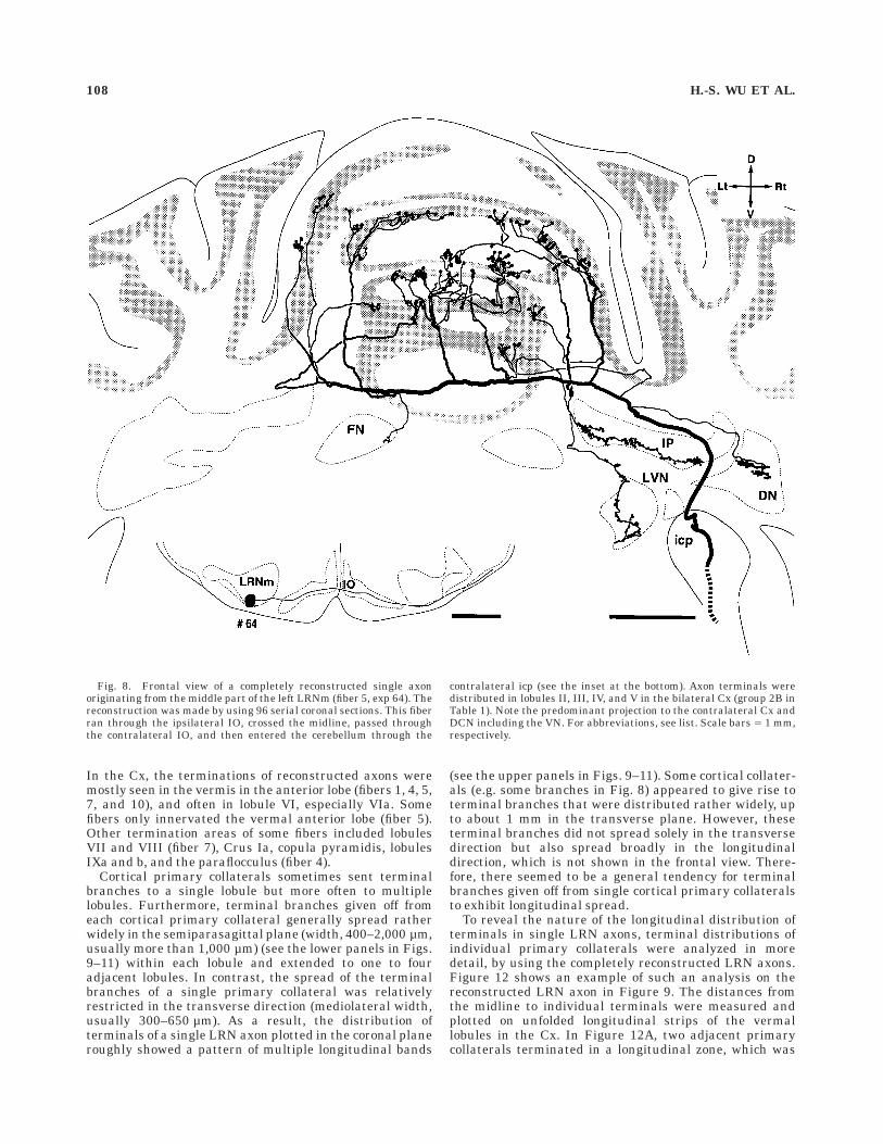
reconstructed LRN axons (n 5 4 of 29; group 2A and 2B inTable 1; Fig. 8) ran medially through or under the ipsilat-eral IO, passed the midline, ran through or under thecontralateral IO, and entered the ventrolateral part of thecontralateral icp. These axons entered the cerebellumthrough the contralateral icp. Except for the laterality inthe icp, these axons followed a path similar to that of theother LRN axons.
To estimate the relative numbers of LRN neurons thatsent their axons through the ipsilateral and contralateralicp, larger volumes of BDA (4–5 µl) were injected withpressure in the lateral medulla, including the icp in twoexperiments, and the number of retrogradely labeledneurons was counted in the bilateral LRN. Labeled cellswere more abundant in the ipsilateral LRN (n 5 1,137,1,043) than in the contralateral LRN (n 5 102, 114),indicating that about 90% of the LRN neurons projectingto the cerebellum sent their axons through the ipsilateralicp.
Stem axons passed through the icp without any branchesin the medulla and entered the cerebellar white matterrostral to the DCN. Generally, an arbor of an LRN axonwithin the cerebellum could be regarded as consisting of athick stem axon running transversely, cortical branches of
Fig. 4. Distribution of axon terminals in the cerebellar and vestibu-lar nuclei after biotinylated dextran amine injection into the rostralpart of the left LRN (injection site in the upper left inset). The samematerial as in Figure 3. Each dot represents a single terminalswelling. All of the swellings obtained from five consecutive 50-µm-
thick sections were plotted on representative frontal sections thatwere separated from each other at 250-µm intervals (A–G, rostral tocaudal). For abbreviations, see list. Scale bar 5 1 mm in G (applies toA–G).
104 H.-S. WU ET AL.
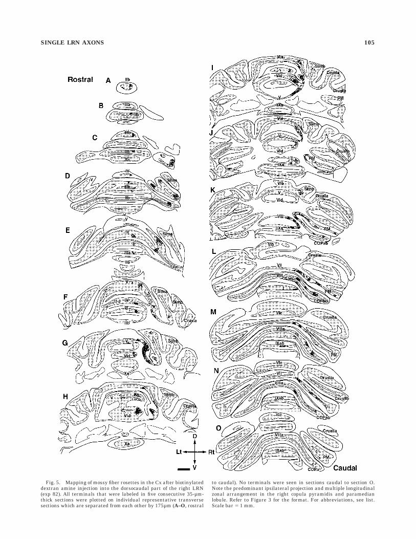
Fig. 5. Mapping of mossy fiber rosettes in the Cx after biotinylateddextran amine injection into the dorsocaudal part of the right LRN(exp 82). All terminals that were labeled in five consecutive 35-µm-thick sections were plotted on individual representative transversesections which are separated from each other by 175µm (A–O, rostral
to caudal). No terminals were seen in sections caudal to section O.Note the predominant ipsilateral projection and multiple longitudinalzonal arrangement in the right copula pyramidis and paramedianlobule. Refer to Figure 3 for the format. For abbreviations, see list.Scale bar 5 1 mm.
SINGLE LRN AXONS 105
various diameters, and very thin nuclear branches. Thestem axons ran medially toward the midline in the deepwhite matter rostral and dorsal to the DCN. Some stemaxons did not cross the midline in the cerebellum andmade projections nearly exclusively on the same side asthe icp through which they passed (n 5 4 of 29; groups 1Aand 2A in Table 1). The other stem axons (n 5 25 of 29;group 1B and 2B in Table 1) crossed the midline within thecerebellum to make bilateral projections. Thus, LRN axonscould be divided into four groups in terms of the lateralityof the icp that they passed through relative to their cellbodies, and by their projection to either the unilateral orbilateral cortices. All of the completely reconstructed axonshad more terminals in the Cx on the same side as the icpthrough which they ran (Table 1). However, as will bedescribed later, there were some differences among indi-vidual axons concerning the laterality of their projectionsin the Cx and DCN, the number of primary collaterals,their projection lobules, and the morphology of collateralsin the DCN. We tentatively divided LRN axons into fourgroups; group 1 (n 5 25) contained LRN neurons with stemaxons that passed through the ipsilateral icp, and group 2(n 5 4) contained those with stem axons through thecontralateral icp. These neurons were further divided into
groups A and B according to their projection to theunilateral or bilateral cortices. As summarized in Table 1,3 neurons were in group 1A, 22 were in group 1B, 1 was ingroup 2A, and 3 were in group 2B.
Although running transversely in the cerebellar whitematter toward the contralateral side, stem axons gave riseto several primary collaterals to the Cx and DCN. Indi-vidual primary collaterals arose at an almost right angle(Fig. 7C) from the stem axon, and ran largely in aparasagittal plane in the Cx. Primary collaterals project-ing to lobules II and III were directed almost horizontallyand rostrally, whereas primary collaterals projecting tolobules IV–V were directed rostrodorsally and those project-ing to lobules VI or VII were directed dorsally or dorsocau-dally. On their way through the Cx, individual primarycollaterals widely ramified mainly in a dorsoventral direc-tion, but did not spread so widely in a mediolateraldirection, so that each collateral terminated as mossy fiberrosettes in the granular layer in a relatively narrowlongitudinal zonal area covering one to four lobules (Figs.8–11). The number of cortical primary collaterals given offfrom a transverse stem axon in the deep cerebellar whitematter was five to nine (7.0 6 1.0, n 5 29). In each of thebilaterally projecting neurons, the number of ipsilateral
Fig. 6. A–G: Mapping of axon terminals in the cerebellar and vestibular nuclei in the same experimentas in Figure 5 (injection site shown in the upper left inset). Predominant ipsilateral projection was seen.Refer to Figure 4 for the format. For abbreviations, see list. Scale bar 5 1 mm in G (applies to A–G).
106 H.-S. WU ET AL.
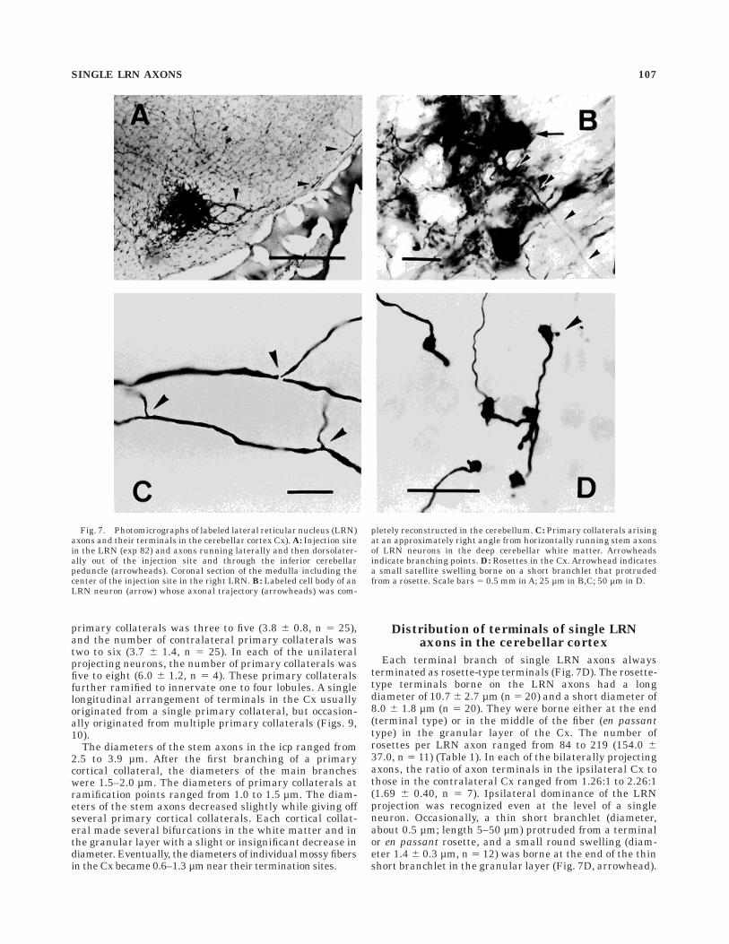
primary collaterals was three to five (3.8 6 0.8, n 5 25),and the number of contralateral primary collaterals wastwo to six (3.7 6 1.4, n 5 25). In each of the unilateralprojecting neurons, the number of primary collaterals wasfive to eight (6.0 6 1.2, n 5 4). These primary collateralsfurther ramified to innervate one to four lobules. A singlelongitudinal arrangement of terminals in the Cx usuallyoriginated from a single primary collateral, but occasion-ally originated from multiple primary collaterals (Figs. 9,10).
The diameters of the stem axons in the icp ranged from2.5 to 3.9 µm. After the first branching of a primarycortical collateral, the diameters of the main brancheswere 1.5–2.0 µm. The diameters of primary collaterals atramification points ranged from 1.0 to 1.5 µm. The diam-eters of the stem axons decreased slightly while giving offseveral primary cortical collaterals. Each cortical collat-eral made several bifurcations in the white matter and inthe granular layer with a slight or insignificant decrease indiameter. Eventually, the diameters of individual mossy fibersin the Cx became 0.6–1.3 µm near their termination sites.
Distribution of terminals of single LRNaxons in the cerebellar cortex
Each terminal branch of single LRN axons alwaysterminated as rosette-type terminals (Fig. 7D). The rosette-type terminals borne on the LRN axons had a longdiameter of 10.7 6 2.7 µm (n 5 20) and a short diameter of8.0 6 1.8 µm (n 5 20). They were borne either at the end(terminal type) or in the middle of the fiber (en passanttype) in the granular layer of the Cx. The number ofrosettes per LRN axon ranged from 84 to 219 (154.0 637.0, n 5 11) (Table 1). In each of the bilaterally projectingaxons, the ratio of axon terminals in the ipsilateral Cx tothose in the contralateral Cx ranged from 1.26:1 to 2.26:1(1.69 6 0.40, n 5 7). Ipsilateral dominance of the LRNprojection was recognized even at the level of a singleneuron. Occasionally, a thin short branchlet (diameter,about 0.5 µm; length 5–50 µm) protruded from a terminalor en passant rosette, and a small round swelling (diam-eter 1.4 6 0.3 µm, n 5 12) was borne at the end of the thinshort branchlet in the granular layer (Fig. 7D, arrowhead).
Fig. 7. Photomicrographs of labeled lateral reticular nucleus (LRN)axons and their terminals in the cerebellar cortex Cx). A: Injection sitein the LRN (exp 82) and axons running laterally and then dorsolater-ally out of the injection site and through the inferior cerebellarpeduncle (arrowheads). Coronal section of the medulla including thecenter of the injection site in the right LRN. B: Labeled cell body of anLRN neuron (arrow) whose axonal trajectory (arrowheads) was com-
pletely reconstructed in the cerebellum. C: Primary collaterals arisingat an approximately right angle from horizontally running stem axonsof LRN neurons in the deep cerebellar white matter. Arrowheadsindicate branching points. D: Rosettes in the Cx. Arrowhead indicatesa small satellite swelling borne on a short branchlet that protrudedfrom a rosette. Scale bars 5 0.5 mm in A; 25 µm in B,C; 50 µm in D.
SINGLE LRN AXONS 107
In the Cx, the terminations of reconstructed axons weremostly seen in the vermis in the anterior lobe (fibers 1, 4, 5,7, and 10), and often in lobule VI, especially VIa. Somefibers only innervated the vermal anterior lobe (fiber 5).Other termination areas of some fibers included lobulesVII and VIII (fiber 7), Crus Ia, copula pyramidis, lobulesIXa and b, and the paraflocculus (fiber 4).
Cortical primary collaterals sometimes sent terminalbranches to a single lobule but more often to multiplelobules. Furthermore, terminal branches given off fromeach cortical primary collateral generally spread ratherwidely in the semiparasagittal plane (width, 400–2,000 µm,usually more than 1,000 µm) (see the lower panels in Figs.9–11) within each lobule and extended to one to fouradjacent lobules. In contrast, the spread of the terminalbranches of a single primary collateral was relativelyrestricted in the transverse direction (mediolateral width,usually 300–650 µm). As a result, the distribution ofterminals of a single LRN axon plotted in the coronal planeroughly showed a pattern of multiple longitudinal bands
(see the upper panels in Figs. 9–11). Some cortical collater-als (e.g. some branches in Fig. 8) appeared to give rise toterminal branches that were distributed rather widely, upto about 1 mm in the transverse plane. However, theseterminal branches did not spread solely in the transversedirection but also spread broadly in the longitudinaldirection, which is not shown in the frontal view. There-fore, there seemed to be a general tendency for terminalbranches given off from single cortical primary collateralsto exhibit longitudinal spread.
To reveal the nature of the longitudinal distribution ofterminals in single LRN axons, terminal distributions ofindividual primary collaterals were analyzed in moredetail, by using the completely reconstructed LRN axons.Figure 12 shows an example of such an analysis on thereconstructed LRN axon in Figure 9. The distances fromthe midline to individual terminals were measured andplotted on unfolded longitudinal strips of the vermallobules in the Cx. In Figure 12A, two adjacent primarycollaterals terminated in a longitudinal zone, which was
Fig. 8. Frontal view of a completely reconstructed single axonoriginating from the middle part of the left LRNm (fiber 5, exp 64). Thereconstruction was made by using 96 serial coronal sections. This fiberran through the ipsilateral IO, crossed the midline, passed throughthe contralateral IO, and then entered the cerebellum through the
contralateral icp (see the inset at the bottom). Axon terminals weredistributed in lobules II, III, IV, and V in the bilateral Cx (group 2B inTable 1). Note the predominant projection to the contralateral Cx andDCN including the VN. For abbreviations, see list. Scale bars 5 1 mm,respectively.
108 H.-S. WU ET AL.
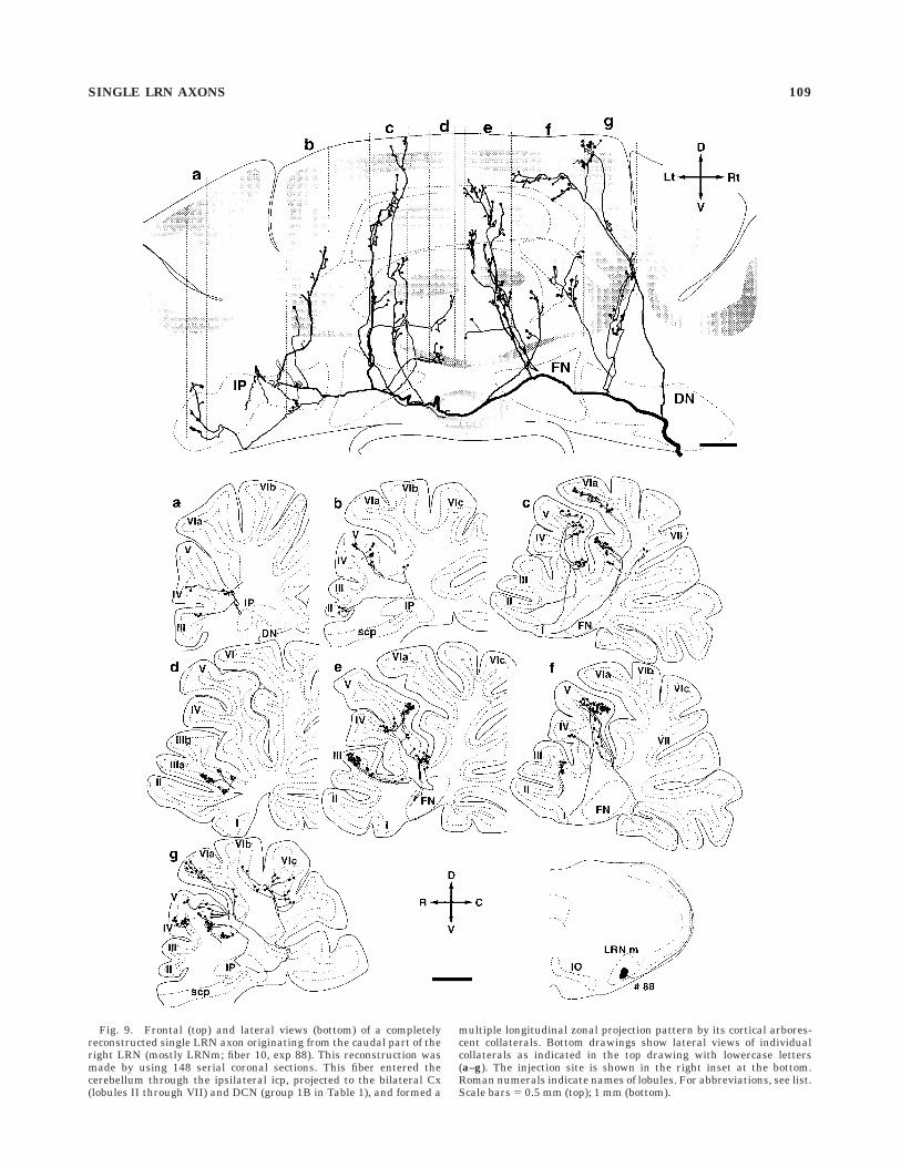
Fig. 9. Frontal (top) and lateral views (bottom) of a completelyreconstructed single LRN axon originating from the caudal part of theright LRN (mostly LRNm; fiber 10, exp 88). This reconstruction wasmade by using 148 serial coronal sections. This fiber entered thecerebellum through the ipsilateral icp, projected to the bilateral Cx(lobules II through VII) and DCN (group 1B in Table 1), and formed a
multiple longitudinal zonal projection pattern by its cortical arbores-cent collaterals. Bottom drawings show lateral views of individualcollaterals as indicated in the top drawing with lowercase letters(a–g). The injection site is shown in the right inset at the bottom.Roman numerals indicate names of lobules. For abbreviations, see list.Scale bars 5 0.5 mm (top); 1 mm (bottom).
SINGLE LRN AXONS 109
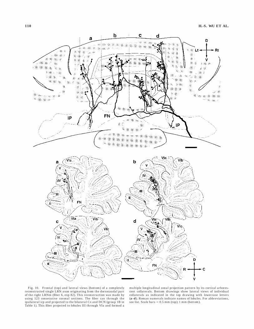
Fig. 10. Frontal (top) and lateral views (bottom) of a completelyreconstructed single LRN axon originating from the dorsocaudal partof the right LRNm (fiber 6, exp 82). This reconstruction was made byusing 123 consecutive coronal sections. The fiber ran through theipsilateral icp and projected to the bilateral Cx and DCN (group 1B inTable 1). This fiber projected to lobules III through VIa and formed a
multiple longitudinal zonal projection pattern by its cortical arbores-cent collaterals. Bottom drawings show lateral views of individualcollaterals as indicated in the top drawing with lowercase letters(a–d). Roman numerals indicate names of lobules. For abbreviations,see list. Scale bars 5 0.5 mm (top); 1 mm (bottom).
110 H.-S. WU ET AL.
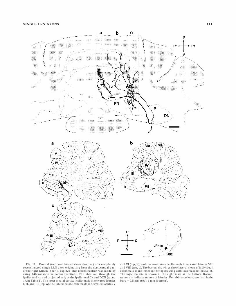
Fig. 11. Frontal (top) and lateral views (bottom) of a completelyreconstructed single LRN axon originating from the dorsocaudal partof the right LRNm (fiber 7, exp 82). This reconstruction was made byusing 146 consecutive coronal sections. The fiber ran through theipsilateral icp and projected only to the ipsilateral Cx and DCN (group1A in Table 1). The most medial cortical collaterals innervated lobulesI, II, and III (top, a), the intermediate collaterals innervated lobules V
and VI (top, b), and the most lateral collaterals innervated lobules VIIand VIII (top, c). The bottom drawings show lateral views of individualcollaterals as indicated in the top drawing with lowercase letters (a–c).The injection site is shown in the right inset at the bottom. Romannumerals indicate names of lobules. For abbreviations, see list. Scalebars 5 0.5 mm (top); 1 mm (bottom).
SINGLE LRN AXONS 111
labeled as zone c in Figure 9. In this zone, terminals thatoriginated from one primary collateral were distributed inlobule V, and those from the other primary collateral weredistributed in lobule VI where two clusters of terminalswere slightly separated in lobule VIa and VId. The longitu-dinal spread of these terminals extended over 6.5 mm,whereas the mediolateral spread of each cluster of termi-nals was localized between 230 µm and 400 µm. Even as awhole, these terminals were well aligned in a singlelongitudinal strip less than 400 µm wide in the transverseplane. Similarly, Figure 12B shows the distribution ofterminals of another primary collateral of the same axon(labeled as zone e in Fig. 9) on the unfolded corticalparasagittal strip. This collateral bifurcated into two mainbranches, of which one branch terminated in lobule III,and the other branch terminated in lobules IV and V.Although terminals were distributed widely along a para-sagittal longitudinal strip (11.6 mm), the mediolateralspread of the terminals was restricted within 600 µm as awhole. As shown in this example, the general feature of thecortical distribution of terminals of a single LRN axoncould be summarized as follows. Terminals belonging toone or sometimes two primary collaterals and spreading ina few lobules made a longitudinal zone in the parasagittalplane that was usually less than 500 µm wide in thetransverse plane, and several such longitudinal zones ofterminals were arranged in parallel in the mediolateraldirection.
Morphology and distribution of collateralsterminating in the DCN and VN
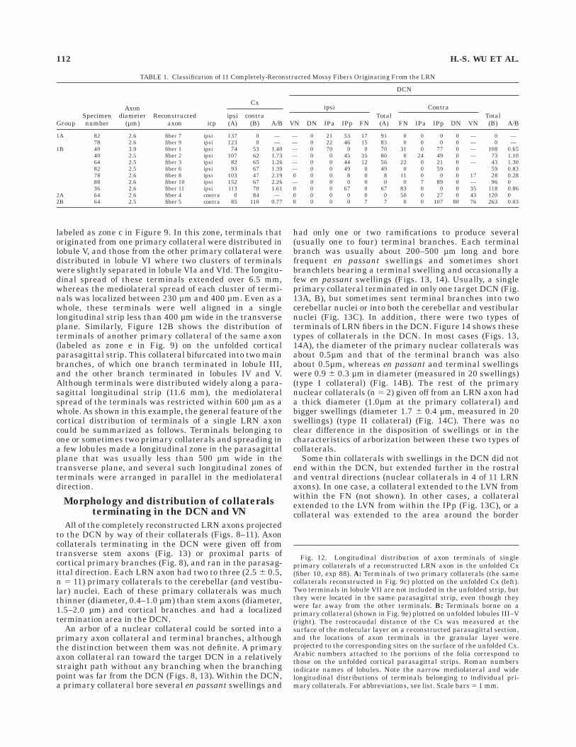
All of the completely reconstructed LRN axons projectedto the DCN by way of their collaterals (Figs. 8–11). Axoncollaterals terminating in the DCN were given off fromtransverse stem axons (Fig. 13) or proximal parts ofcortical primary branches (Fig. 8), and ran in the parasag-ittal direction. Each LRN axon had two to three (2.5 6 0.5,n 5 11) primary collaterals to the cerebellar (and vestibu-lar) nuclei. Each of these primary collaterals was muchthinner (diameter, 0.4–1.0 µm) than stem axons (diameter,1.5–2.0 µm) and cortical branches and had a localizedtermination area in the DCN.
An arbor of a nuclear collateral could be sorted into aprimary axon collateral and terminal branches, althoughthe distinction between them was not definite. A primaryaxon collateral ran toward the target DCN in a relativelystraight path without any branching when the branchingpoint was far from the DCN (Figs. 8, 13). Within the DCN,a primary collateral bore several en passant swellings and
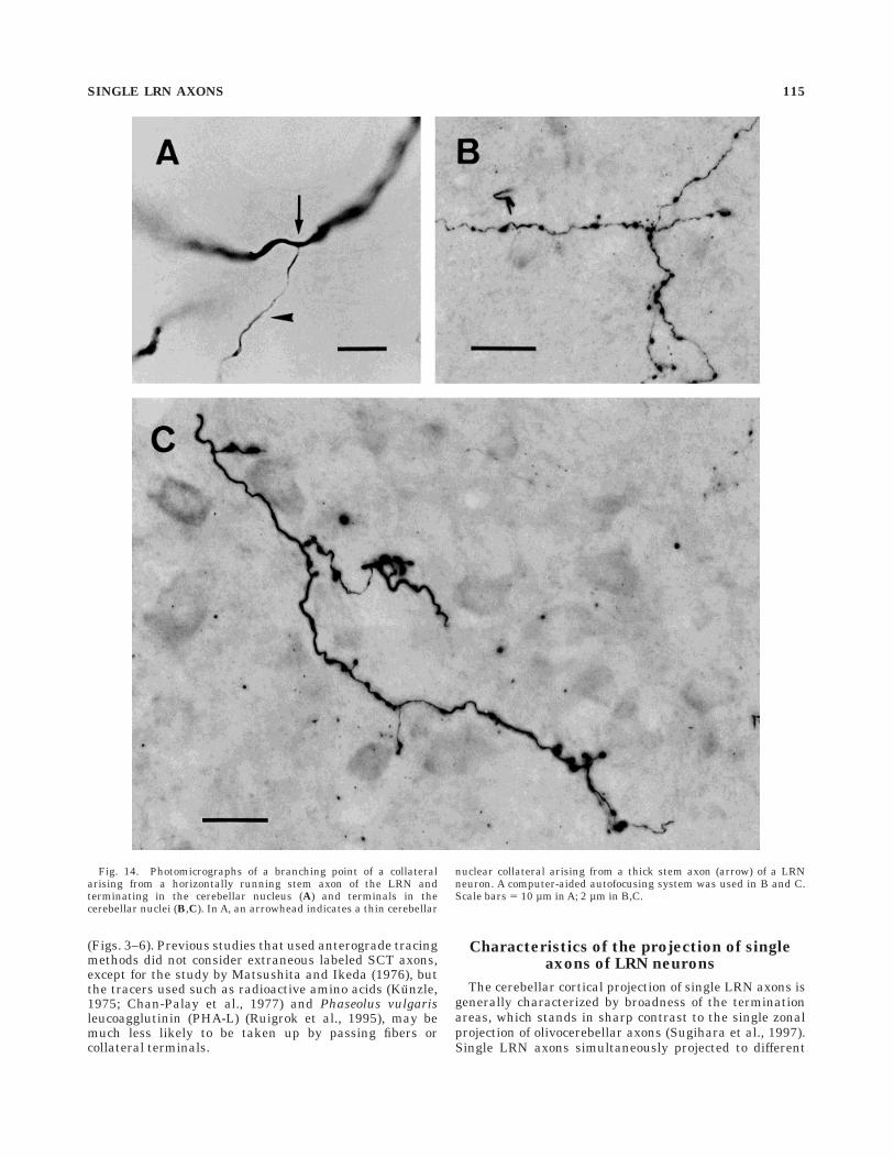
had only one or two ramifications to produce several(usually one to four) terminal branches. Each terminalbranch was usually about 200–500 µm long and borefrequent en passant swellings and sometimes shortbranchlets bearing a terminal swelling and occasionally afew en passant swellings (Figs. 13, 14). Usually, a singleprimary collateral terminated in only one target DCN (Fig.13A, B), but sometimes sent terminal branches into twocerebellar nuclei or into both the cerebellar and vestibularnuclei (Fig. 13C). In addition, there were two types ofterminals of LRN fibers in the DCN. Figure 14 shows thesetypes of collaterals in the DCN. In most cases (Figs. 13,14A), the diameter of the primary nuclear collaterals wasabout 0.5µm and that of the terminal branch was alsoabout 0.5µm, whereas en passant and terminal swellingswere 0.9 6 0.3 µm in diameter (measured in 20 swellings)(type I collateral) (Fig. 14B). The rest of the primarynuclear collaterals (n 5 2) given off from an LRN axon hada thick diameter (1.0µm at the primary collateral) andbigger swellings (diameter 1.7 6 0.4 µm, measured in 20swellings) (type II collateral) (Fig. 14C). There was noclear difference in the disposition of swellings or in thecharacteristics of arborization between these two types ofcollaterals.
Some thin collaterals with swellings in the DCN did notend within the DCN, but extended further in the rostraland ventral directions (nuclear collaterals in 4 of 11 LRNaxons). In one case, a collateral extended to the LVN fromwithin the FN (not shown). In other cases, a collateralextended to the LVN from within the IPp (Fig. 13C), or acollateral was extended to the area around the border
TABLE 1. Classification of 11 Completely-Reconstructed Mossy Fibers Originating From the LRN
GroupSpecimennumber
Axondiameter
(µm)Reconstructed
axon icp
Cx
DCN
ipsiTotal(A)
ContraTotal(B) A/B
ipsi(A)
contra(B) A/B VN DN IPa IPp FN FN IPa IPp DN VN
1A 82 2.6 fiber 7 ipsi 137 0 — — 0 21 53 17 91 0 0 0 0 — 0 —78 2.6 fiber 9 ipsi 123 0 — — 0 22 46 15 83 0 0 0 0 — 0 —
1B 40 3.9 fiber 1 ipsi 74 53 1.40 — 0 70 0 0 70 31 0 77 0 — 108 0.6540 2.5 fiber 2 ipsi 107 62 1.73 — 0 0 45 35 80 0 24 49 0 — 73 1.1064 2.5 fiber 3 ipsi 82 65 1.26 — 0 0 44 12 56 22 0 21 0 — 43 1.3082 2.5 fiber 6 ipsi 93 67 1.39 — 0 0 49 0 49 0 0 59 0 59 0.8378 2.6 fiber 8 ipsi 103 47 2.19 0 0 0 8 0 8 11 0 0 0 17 28 0.2888 2.6 fiber 10 ipsi 152 67 2.26 — 0 0 0 0 0 0 7 89 0 — 96 036 2.6 fiber 11 ipsi 113 70 1.61 0 0 0 67 0 67 83 0 0 0 35 118 0.86
2A 64 2.6 fiber 4 contra 0 84 — 0 0 0 0 0 0 50 0 27 0 43 120 02B 64 2.5 fiber 5 contra 85 110 0.77 0 0 0 0 7 7 0 0 107 80 76 263 0.03
Fig. 12. Longitudinal distribution of axon terminals of singleprimary collaterals of a reconstructed LRN axon in the unfolded Cx(fiber 10, exp 88). A: Terminals of two primary collaterals (the samecollaterals reconstructed in Fig. 9c) plotted on the unfolded Cx (left).Two terminals in lobule VII are not included in the unfolded strip, butthey were located in the same parasagittal strip, even though theywere far away from the other terminals. B: Terminals borne on aprimary collateral (shown in Fig. 9e) plotted on unfolded lobules III–V(right). The rostrocaudal distance of the Cx was measured at thesurface of the molecular layer on a reconstructed parasagittal section,and the locations of axon terminals in the granular layer wereprojected to the corresponding sites on the surface of the unfolded Cx.Arabic numbers attached to the portions of the folia correspond tothose on the unfolded cortical parasagittal strips. Roman numbersindicate names of lobules. Note the narrow mediolateral and widelongitudinal distributions of terminals belonging to individual pri-mary collaterals. For abbreviations, see list. Scale bars 5 1 mm.
112 H.-S. WU ET AL.
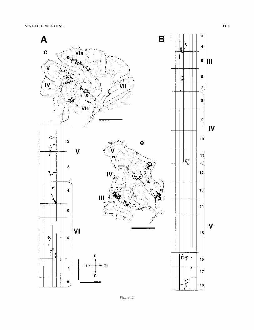
Figure 12
SINGLE LRN AXONS 113
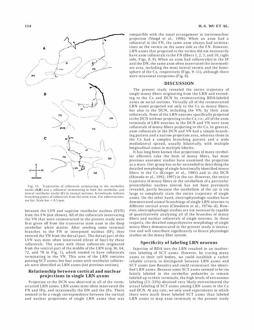
between the LVN and superior vestibular nucleus (SVN)from the FN (not shown). All of the collaterals innervatingthe VN that were reconstructed in the present study werefirst given off from the transverse stem axon in the deepcerebellar white matter. After sending some terminalbranches to the FN or interposed nucleus (IP), theyentered the VN from the dorsal part. The dorsal part of theLVN was most often innervated (three of four) by thesecollaterals. The axons with these collaterals originatedfrom the ventral part of the middle of the LRN (exp 36, 64,72, and 78 in Fig. 1), which tended to have collateralsterminating in the VN. This area of the LRN containspassing SCT axons, but four axons with vestibular collater-als were identified as LRN axons (see previous section).
Relationship between cortical and nuclearprojections in single LRN axons
Projection to the DCN was observed in all of the recon-structed LRN axons. LRN axons most often innervated theFN and IPp, and occasionally the DN and the IPa. Thereseemed to be a rough correspondence between the corticaland nuclear projections of single LRN axons that was
compatible with the zonal arrangement in corticonuclearprojection (Voogd et al., 1996). When an axon had acollateral in the FN, the same axon always had termina-tions on the vermis on the same side as the FN. However,LRN axons that projected to the vermis did not necessarilyhave axon collaterals to the FN (fibers 1, 2, 5, and 10, rightside, Figs. 8, 9). When an axon had collateral(s) in the IPand the DN, the same axon often innervated the intermedi-ate area, including the most lateral vermis and the hemi-sphere of the Cx, respectively (Figs. 9–11), although therewere occasional exceptions (Fig. 8).
DISCUSSION
The present study revealed the entire trajectory ofsingle mossy fibers originating from the LRN and extend-ing to the Cx and DCN by reconstructing BDA-labeledaxons on serial sections. Virtually all of the reconstructedLRN axons projected not only to the Cx as mossy fibers,but also to the DCN, including the VN, by their axoncollaterals. None of the LRN neurons specifically projectedto the DCN without projecting to the Cx; i.e., all of the axonterminals of LRN neurons in the DCN and VN were axoncollaterals of mossy fibers projecting to the Cx. In general,axon collaterals in the DCN and VN had a simple branch-ing pattern and a narrow projection area, whereas those inthe Cx had a complex branching pattern and a widemediolateral spread, usually bilaterally, with multiplelongitudinal zones in multiple lobules.
It has long been known that projections of many cerebel-lar afferents take the form of mossy fibers, but mostprevious anatomic studies have examined the projectionas a mass. Our group has so far succeeded in describing thedetailed morphology of single functionally identified mossyfibers in the Cx (Krieger et al., 1985) and in the DCN(Shinoda et al., 1992, 1997) in the cat. However, the entiretrajectory of mossy fibers in the cerebellum of a particularprecerebellar nucleus neuron has not been previouslyrevealed, partly because the cerebellum of the cat is toolarge to completely stain the entire trajectory of singleaxons. On the other hand, electrophysiologic studies havedemonstrated axonal branchings of single LRN neurons todifferent cortical areas (Clendenin et al., 1974a–d). How-ever, electrophysiologic studies are not necessarily capableof quantitatively analyzing all of the branches of mossyfibers and nuclear collaterals of single neurons. In theserespects, the detailed comprehensive morphology of singlemossy fibers demonstrated in the present study is innova-tive and will contribute significantly to future physiologicstudies on the mossy fiber system.
Specificity of labeling LRN neurons
Injection of BDA into the LRN resulted in an inadver-tent labeling of SCT axons. However, by tracing manyaxons to their cell bodies, we could establish a ratherreliable criteria to distinguish between LRN axons andSCT axons (see Results) and could reconstruct the identi-fied LRN axons. Because some SCT axons seemed to be toofaintly labeled in the cerebellar peduncles to remainlabeled up to their terminals, the high levels of extraneouslabeling (13–33%) obtained very likely overestimated theactual labeling of SCT axons among LRN axons in the Cxand DCN. At any rate, we only used experiments in whichthere were much fewer labeled SCT axons than labeledLRN axons to map axon terminals in the present study
Fig. 13. Trajectories of collaterals terminating in the cerebellarnuclei (A,B) and a collateral terminating in both the cerebellar andlateral vestibular nuclei (C) in coronal sections. Arrowheads indicatebranching points of collaterals from the stem axon. For abbreviations,see list. Scale bar 5 0.5 mm.
114 H.-S. WU ET AL.
(Figs. 3–6). Previous studies that used anterograde tracingmethods did not consider extraneous labeled SCT axons,except for the study by Matsushita and Ikeda (1976), butthe tracers used such as radioactive amino acids (Kunzle,1975; Chan-Palay et al., 1977) and Phaseolus vulgarisleucoagglutinin (PHA-L) (Ruigrok et al., 1995), may bemuch less likely to be taken up by passing fibers orcollateral terminals.
Characteristics of the projection of singleaxons of LRN neurons
The cerebellar cortical projection of single LRN axons isgenerally characterized by broadness of the terminationareas, which stands in sharp contrast to the single zonalprojection of olivocerebellar axons (Sugihara et al., 1997).Single LRN axons simultaneously projected to different
Fig. 14. Photomicrographs of a branching point of a collateralarising from a horizontally running stem axon of the LRN andterminating in the cerebellar nucleus (A) and terminals in thecerebellar nuclei (B,C). In A, an arrowhead indicates a thin cerebellar
nuclear collateral arising from a thick stem axon (arrow) of a LRNneuron. A computer-aided autofocusing system was used in B and C.Scale bars 5 10 µm in A; 2 µm in B,C.
SINGLE LRN AXONS 115
areas in mediolateral locations in several lobules, oftenbilaterally, which suggests that there is significant overlapof the termination areas of single axons originating fromwithin injection sites. Similar broadness of cortical inner-vation has been suggested in axons of other mossy fibersystems such as the cerebellar projection from the pontinenucleus (Shinoda et al., 1992).
Virtually all of the LRN axons that were reconstructedin the present study had collaterals terminating in theDCN, and also in the VN in some cases. This is the firstmossy fiber system in which virtually all of the cerebellarcortical projection neurons are shown to have axon collat-erals to the DCN. However, a high incidence of nuclearprojection by means of axon collaterals cannot be general-ized to all mossy fiber systems, because many mossy fibersoriginating from the pontine nucleus lack nuclear collater-als (Shinoda et al., 1992). Virtually all the entire cerebellarnuclear projection from the LRN was by means of thincollaterals given off from stem axons or primary corticalcollaterals. Previous anterograde or retrograde studies didnot identify whether nuclear projections of LRN neuronsare by means of collaterals of mossy fibers or if there arespecific nuclear-projecting neurons. Because most of thereconstructed LRN axons originated from the LRNm, wecannot discuss in detail the topographical relationship inLRN-nuclear projection as elaborated in a retrogradelabeling study (Parenti et al., 1996). However, it is veryclear that the topographical relationship in LRN-nuclearprojection was much weaker and vaguer than that in theolivonuclear projection (Van der Want et al., 1989; Sugi-hara et al., 1996), in which neurons in a subdivision of theIO project to a relatively localized area in one of thecerebellar nuclei. This is partly because single LRN axonshave multiple nuclear collaterals projecting to more thanone cerebellar nuclei, and multiple cortical collaterals thatspread rather widely in the transverse plane, whereassingle IO neurons have multiple collaterals that spread ina very narrow longitudinal zone in the Cx (Sugihara et al.,1997).
Among our 29 completely reconstructed LRN axons, fouraxons had collaterals terminating in the VN. The dorsalportion of the LVN was most often innervated by LRNaxons, which was consistent with the results of massPHA-L labeling (Ruigrok et al., 1995), and primary collat-erals innervating the VN always innervated the DCN.These collaterals extended ventrally from within the cer-ebellum, as suggested by a previous mass labeling study(Ruigrok et al., 1995). The three cerebellar nuclei and thedorsal LVN, to which LRN axons project, receive Purkinjeinhibition, but the ventral LVN does not (Eccles et al.,1967). These findings may reflect a close relationshipbetween the vestibular and cerebellar nuclei with regardto both ontogeny and phylogeny.
Area-dependent or neuron-dependentdifferences in LRN projection
The results of single axon reconstructions (Figs. 8–11)showed that the cerebellar projection from the LRNm wasgenerally bilateral, predominantly in the vermis of theanterior lobe (especially lobules IV and V), lobule VI, andthe paramedian lobule in the hemisphere, and less inlobules VII and VIII in the intermediate area. This resultis roughly consistent with the results of previous anatomicstudies, which showed that the LRNm mainly projects to
lobules IV and V, whereas the LRNp and the cells locatedat the border between the LRNm and LRNp project tolobules II and III (Hrycyshyn et al., 1982; Ruigrok andCella, 1995). A clear area-dependent difference in LRN-Cxprojection was seen only in the dorsolateral part of thecaudal LRNm (exp 82), although the injections extendedfrom the rostromedial LRNm to the caudal LRNm. In thisexperiment, terminals were almost exclusively localizedipsilaterally, whereas the cerebellar innervation was bilat-eral in all of the other experiments. This finding seemed tobe consistent with the results that the dorsolateral part ofthe caudal LRNm receives input from an ipsilateral distalforelimb and projects exclusively to the ipsilateral Cx(Clendenin et al., 1974a,d), and that no clear topographicalsubdivisions have been reported in projections from differ-ent areas within the LRNm, except for its dorsolateral part(Clendenin et al., 1974a; Dietrichs and Walberg, 1979;Hrycyshyn et al., 1982). However, some topographicalprojection has been shown among the three subdivisions ofthe LRN, i.e., LRNm, LRNp, and LRNst (Brodal, 1943;Ruigrok and Cella, 1995).
Abundant terminals were observed in the FN andmedial portions of the IPa and IPp. This result is partlyconsistent with the retrograde labeling study by Parenti etal. (1996). They showed that the DN received a projectionfrom the dorsomedial part of the rostral LRN, into whichwe could not inject BDA, and distinguished the intermedio-ventral and caudal LRN and dorsolateral part of themiddle LRN, with the former projecting to the FN and thelatter to the IP. The present analysis of single axonmorphology has revealed the existence of multiple axoncollaterals of single LRN neurons projecting to more thanone cerebellar or vestibular nucleus. This is the first mossyfiber system in which single axons have been shown toinnervate multiple cerebellar nuclei.
Multiple longitudinal zonal projection ofsingle LRN neurons in the Cx
The injection of large volumes of BDA into the LRNrevealed multiple longitudinal zones of mossy fiber termi-nals in the Cx. This finding is consistent with the resultsobtained in the rat and the cat by Kunzle (1975) andChan-Palay et al. (1977), by using an autoradiographicmethod, and by Ruigrok and Cella (1995) by using PHA-L.One of the most important findings in this study is theexistence of multiple zonal projection of single LRN axonsin the Cx. This projection is in a sharp contrast with theprojection of the IO, which also shows a multiple longitudi-nal zonal pattern, when labeled by the mass injection of ananterograde tracer (Chan-Palay et al., 1977; Van der Wantet al., 1989). However, at the level of single axons, thecortical projection is generally localized within a singlelongitudinal zone in the climbing fiber system (Sugihara etal., 1997), whereas it is multizonal in the LRN mossy fibersystem. The present multiple zonal projection of a singlemossy fiber may not necessarily contradict the patchy orfractured representation of receptive fields of the facialarea (Shambes et al., 1978), because terminals of singleaxons seemed to be arranged into clusters within eachlongitudinal zone (Fig. 12). The important point of theterminal distribution pattern of a single LRN axon is thatterminals in different lobules originating from single oradjacent primary collaterals, even though they are sepa-rated widely, are localized in a parasagittal longitudinalzone that is restricted to less than 500 µm wide in the
116 H.-S. WU ET AL.
mediolateral direction. This organization is very similar tothe organization of a single olivocerebellar axon in the Cx.Therefore, this longitudinal zonal organization should be abasic and common organization for both the climbing andmossy fiber systems.
One of the important remaining questions is how thelongitudinal arrangement of LRN projection is related tozonal components A, B, C1, C2, C3, D0, D1, and D2, whichwere identified with olivocerebellar and corticonuclearprojections (Groenewegen and Voogd, 1977; Buisseret-Delmas and Angaut, 1993; Voogd, 1995). We attempted invain to correlate these zones with the present zones ofLRN terminals by combining acetylcholine esterase stain-ing and the present staining, because the zones visualizedby acetylcholine esterase staining appeared mainly in theposterior vermis. The relationship between the multiplezones of LRN projection and zebrin-defined zones has beenbriefly addressed by Ruigrok and Cella (1995), and thezones defined by the different methods seem to correspondwell with each other. Thus, the functional significance ofcerebellar compartmentalization, which has so far beenstudied from the perspective of labeling molecular marker-specific Purkinje cells and labeling olivocerebellar andcorticonuclear projections, could be also considered fromthe viewpoint of mossy fiber projections.
Functional significance of axonal branchingpatterns of single LRN axons
Excitatory mossy fiber inputs to the Cx are relayed bygranule cells and parallel fibers to reach Purkinje cells.Although a parallel fiber spreads in the transverse direc-tion, the most effective input from the parallel fiber isgiven to Purkinje cells just above the given granule cellthrough synapses formed by the parallel fiber (Eccles etal., 1967). Therefore, the zonal projection of a single LRNaxon may be functionally significant in conveying anexcitatory input to Purkinje cells in that specific zone.
Individual LRN axons projected to multiple lobules inboth the hemisphere and the vermis, and even oftenbilaterally. Therefore, cerebellar cortical areas innervatedby a single mossy fiber from the LRN may not necessarilybe related to each other in terms of their functional roles.Thus, an important question is what is the role of LRNaxons that presumably send specific information to differ-ent cortical areas (Clendenin et al., 1974b). Concerning thenuclear projection, it is obvious that the cortical andnuclear projections of any single reconstructed LRN axondid not exactly follow the so-called corticonuclear topo-graphical relationship, because the cortical projection gen-erally spread more widely in the transverse direction thanthe nuclear projection (Figs. 8–11, 13). Thus, this situationdoes not exactly correspond to the classic microcomplexscheme of Ito (1984), in which the nuclear inputs by amossy fiber collateral and by a Purkinje cell that receivesthe same mossy fiber input through granule cells convergeonto the same target nuclear output neuron. The nuclearcollaterals of mossy fibers are important in activatingnuclear neurons as a source for the excitation of theirtarget neurons (Shinoda et al., 1992, 1997). To understandthe functional interactions at nuclear efferent neuronsbetween Purkinje cell inputs in widely distributed corticalareas innervated by a single mossy fiber and nuclearinputs by a collateral of the same mossy fiber, we needmore detailed information about how single Purkinje cellsspread in the DCN, how Purkinje cells in different cortical
areas send their information to the DCN, and how thatinformation is integrated as an output in the DCN. Further-more, it would also be useful for understanding thefunctional mechanism of mossy fiber systems to clarify thesimilarities and the differences in mossy fibers in othersystems, such as spinocerebellar and cuneocerebellar sys-tems.
ACKNOWLEDGMENTS
We thank Mr. M. Takada for his photographic assistanceand Dr. K. Miura of for his support of the computer-aideddynamic focusing system. Y.S. received support from theCore Research for Evolutional Science and Technology ofthe Japan Science and Technology Corporation. I.S. andY.S. received grants-in-aid for scientific research from theMinistry of Education, Science, and Culture of Japan.H.-S.W. received a research grant from the SasakawaHealth Science Foundation and a scholarship from theIwaki Scholarship Foundation.
LITERATURE CITED
Alstermark B, Isa T, Tantisira B. 1990. Projection from excitatory C3-C4propriospinal neurons to spinocerebellar and spinoreticular neurons inthe C6-Th1 segments of the cat. Neurosci Res 8:124–130.
Andersson G, Oscarsson O. 1978. Climbing fiber microzones in cerebellarvermis and their projection to different groups of cells in the lateralvestibular nucleus. Exp Brain Res 32:564–579.
Brodal A. 1943. The cerebellar connections of the nucleus reticularislateralis (nucleus funiculi lateralis) in rabbit and cat experimentalinvestigations. Acta Psychiatr Scand 18:171–233.
Brodal A. 1981. Neurological anatomy, 3rd ed, New York: Oxford UniversityPress.
Bruckmoser P, Hepp M-C, Wiesendanger M. 1970. Cortical influence on thesingle neurons of the lateral reticular nucleus of the cat. Exp Neurol26:239–252.
Buisseret-Delmas C, Angaut P. 1993. The cerebellar olivocorticonuclearconnections in the rat. Prog Neurobiol 40:63–87.
Chan-Palay V, Palay SL, Brown JT, Van Itallie C. 1977. Sagittal organiza-tion of olivocerebellar and reticulocerebellar projections: autoradio-graphic studies with 35S-Methionine. Exp Brain Res 30:561–576.
Clendenin M, Ekerot C-F, Oscarsson O, Rosen I. 1974a. The lateralreticular nucleus in the cat I Mossy fibre distribution in cerebellarcortex. Exp Brain Res 21:473–486.
Clendenin M, Ekerot C-F, Oscarsson O, Rosen I. 1974b. The lateralreticular nucleus in the cat. II. Organization of component activatedfrom bilateral ventral flexor reflex tract (bVFRT). Exp Brain Res21:487–500.
Clendenin M, Ekerot C-F, Oscarsson O. 1974c. The lateral reticular nucleusin the cat. III. Organization of component activated from ipsilateralforelimb tract. Exp Brain Res 21:501–513.
Clendenin M, Ekerot C-F, Oscarsson O, Rosen I. 1974d. Functionalorganization of two spinocerebellar paths relayed through the lateralreticular nucleus in the cat. Brain Res 69:140–143.
Dietrichs E, Walberg F. 1979. The cerebellar projection from the lateralreticular nucleus as studied with retrograde transport of horseradishperoxidase. Anat Embryol 155:273–290.
Eccles JC, Ito M, Szentagothai J. 1967. The cerebellum as a neuronalmachine. Berlin-Heidelberg-New York: Springer-Verlag. p 227–261.
Ekerot C-F, Oscarsson O. 1975. Inhibitory spinal paths to the lateralreticular nucleus. Brain Res 99:157–161.
Futami T, Shinoda Y, Yokota J. 1979. Spinal axon collaterals of corticospinalneurons identified by intracellular injection of horseradish peroxidase.Brain Res 164:279–284.
Gerrits NM, Voogd J. 1987. The projection of the nucleus reticularistegmenti pontis and adjacent regions of the pontine nuclei to the centralcerebellar nuclei in the cat. J Comp Neurol 258:52–62.
Grant G. 1962. Spinal course and somatotopically localized termination ofthe spinocerebellar tract. An experimental study in the cat. Acta PhysiolScand 56(Suppl)193:1–45.
SINGLE LRN AXONS 117
Groenewegen HJ, Voogd J. 1977. The parasagittal zonation within theolivocerebellar projection. I. Climbing fiber distribution in the vermis ofthe cat cerebellum. J Comp Neurol 174:417–488.
Hrycyshyn AW, Flumerfelt BA, Anderson WA. 1982. A horseradish peroxi-dase study of the projections from the lateral reticular nucleus to thecerebellum in the rat. Anat Embryol 165:1–18.
Ito M. 1984. The Cerebellum and Neural Control. New York: Raven Press.Ito M, Orlov I, Yamamoto M. 1982. Topographical representation of
vestibulo-ocular reflex in rabbit cerebellar flocculus. Neuroscience7:1657–1664.
Itoh K, Konishi A, Nomura S, Mizuno N, Nakamura Y, Sugimoto T. 1979.Application of coupled oxidation reaction to electron microscopic demon-stration of horseradish peroxidase: cobalt-glucose oxidase method.Brain Res 175:341–346.
Kapogianis EM, Flumerfelt BA, Hrycyshyn AW. 1982. Cytoarchitecture andcytology of the lateral reticular nucleus in the rat. Anat Embryol164:229–242.
Korneliussen HK. 1968. On the morphology and subdivision of the cerebel-lar nuclei of the rat. J Hirnforsch 10:109–122.
Krieger C, Shinoda Y, Smith AM. 1985. Labelling of cerebellar mossy fiberafferents with intra-axonal horseradish. Exp Brain Res 59:414–417.
Kunzle H. 1973. The topographic organization of spinal afferents to thelateral reticular nucleus of the cat. J Comp Neurol 149:103–116.
Kunzle H. 1975. Autoradiographic tracing of the cerebellar projections fromthe lateral reticular nucleus in the cat. Exp Brain Res 22:255–266.
Larsell O. 1952. The morphogenesis and adult pattern of the lobules andfissures of the cerebellum of the white rat. J Comp Neurol 97:281–356.
Matsushita M, Ikeda M. 1976. Projections from the lateral reticularnucleus to the cerebellar cortex and nuclei in the cat. Exp Brain Res24:403–421.
Matsushita M, Yaginuma H. 1995. Projections from the central cervicalnucleus to the cerebellar nuclei in the rat, studied by anterogradeaxonal tracing. J Comp Neurol 353:234–246.
Mihailoff GA. 1993. Cerebellar nuclear projections from the basilar pontinenuclei and nucleus reticularis tegmenti pontis as demonstrated withPHA-L tracing in the rat. J Comp Neurol 330:130–146.
Oscarsson O. 1976. Spatial distribution of climbing and mossy fibre inputsinto the cerebellar cortex. In: Creutzfeldt O, editor. Experimental brainresearch supplement 1: afferent and intrinsic organization of laminatedstructures in the brain. Berlin: Springer Verlag. p 36–42.
Oscarsson O, Rosen I. 1966. Response characteristics of reticulocerebellarneurones activated from spinal afferents. Exp Brain Res 1:320–328.
Palay SL, Chan-Palay V. 1974. Cerebellar cortex. Cytology and organiza-tion. Berlin: Springer-Verlag.
Parenti R, Cicirata F, Panto MR, Serapide MF. 1996. The projection of thelateral reticular nucleus to the deep cerebellar nuclei. An experimentalanalysis in the rat. Eur J Neurosci 8:2157–2167.
Ramon y Cajal S. 1911. Histologie du Systeme Nerveux de l’Homme et desVertebres, Vol II. Paris: Maloine.
Rubertone LA, Mehler WR, Voogd J. 1995. The vestibular nuclear complex.In: Paxinos G, editor. The rat nervous system, 2nd ed. Sydney:Academic Press. p 773–796.
Ruigrok TJH, Cella F. 1995. Precerebellar nuclei and red nucleus. In:Paxinos G, editor. The rat nervous system, 2nd ed. Sydney: AcademicPress. p 277–308.
Ruigrok TJH, Cella F, Voogd J. 1995. Connections of the lateral reticularnucleus to the lateral vestibular nucleus in the rat. An anterogradetracing study with Phaseolus vulgaris leucoagglutinin. Eur J Neurosci7:1410–1413.
Sato Y, Kawasaki T, Ikarashi K. 1983. Afferent projections from thebrainstem to the floccular three zones in cats. II. Mossy fiber projec-tions. Brain Res 272:37–48.
Shambes GM, Beermann DH, Welker W. 1978. Multiple tactile areas incerebellar cortex: another patchy cutaneous projection to granule cellcolumns in rats. Brain Res 157:123–128.
Shinoda Y, Yokota J, Futami T. 1981. Divergent projection of individualcorticospinal axons to motoneurons of multiple muscles in the monkey.Neurosci Lett 23:7–12.
Shinoda Y, Ohgaki T, Futami T. 1986. The morphology of single lateralvestibulospinal tract axons in the lower cervical spinal cord of the cat. JComp Neurol 249:226–241.
Shinoda Y, Sugiuchi Y, Futami T, Izawa R. 1992. Axon collaterals of mossyfibers from the pontine nucleus in the cerebellar dentate nucleus. JNeurophysiol 67:547–560.
Shinoda Y, Sugiuchi Y, Futami T. 1993. Organization of excitatory inputsfrom the cerebral cortex to the cerebellar dentate nucleus. Can J NeurolSci 20(Suppl 3):S19–S28.
Shinoda Y, Izawa Y, Sugiuchi Y, Futami T. 1997. Functional significance ofexcitatory projections from the precerebellar nuclei to interpositus anddentate nucleus neurons for mediating motor, premotor and parietalcortical inputs. In: deZeeuw CI, Strata P, Voogd J, editors. Thecerebellum: from structure to control. Progress in Brain Research, vol114. Amsterdam: Elsevier. p 193–207.
Snider RS. 1950. Recent contributions to the anatomy and physiology of thecerebellum. Arch Neurol Psychiatry 64:196–219.
Sugihara I, Wu H, Shinoda Y. 1996. Morphology of axon collaterals of singleclimbing fibers in the deep cerebellar nuclei of the rat. Neurosci Lett217:33–36.
Sugihara I, Wu H-S, Shinoda Y. 1997. Projection of climbing fibersoriginating from single olivocerebellar neurons in the rat cerebellum.Soc Neurosci Abstr 23:1830.
Swanson LW. 1992. Brain maps: structure of the rat brain. Amsterdam:Elsevier.
Van der Want JJL, Wiklund L, Guegan M, Ruigrok T, Voogd J. 1989.Anterograde tracing of the rat olivocerebellar system with Phaseolusvulgaris leucoagglutinin (PHA-L). Demonstration of climbing fibercollateral innervation of the cerebellar nuclei. J Comp Neurol 288:1–18.
Voogd J. 1964. The cerebellum of the cat. Structure and fibre connexions.Assen: Van Gorcum & Comp NV.
Voogd J. 1995. Cerebellum. In: Paxinos G, editor. The rat nervous system,2nd ed. Sydney: Academic Press. p 309–350.
Voogd J, Jaarsma D, Marani E. 1996. The cerebellum, chemoarchitectureand anatomy. In: Swanson LW, Bjorklund A, Hokfelt T, editors. Inte-grated systems of the CNS, Part III. Cerebellum, basal ganglia,olfactory system. Handbook of chemical neuroanatomy, Vol 12. Amster-dam: Elsevier. p 1–369.
Walberg F. 1952. The lateral reticular nucleus of the medulla oblongata inmammals. J Comp Neurol 96:283–343.
118 H.-S. WU ET AL.