Embed Size (px)
Citation preview
Progressive vascular smooth muscle cell defectsin a mouse model of Hutchinson–Gilfordprogeria syndromeRenee Varga*, Maria Eriksson†, Michael R. Erdos*, Michelle Olive*, Ingrid Harten‡§, Frank Kolodgie¶, Brian C. Capell*,Jun Cheng�, Dina Faddah*, Stacie Perkins*, Hedwig Avallone¶, Hong San*, Xuan Qu*, Santhi Ganesh*,Leslie B. Gordon*,**, Renu Virmani¶, Thomas N. Wight‡§, Elizabeth G. Nabel*††, and Francis S. Collins*‡‡
*Genome Technology Branch and �Genetic Disease Research Branch, National Human Genome Research Institute, National Institutes of Health, 50 SouthDrive, Bethesda, MD 20892; †Department of Medical Nutrition, Karolinska Institutet, Novum, Halsovagen 7, Hiss E, Plan 6, 141 57 Huddinge, Sweden;‡Hope Heart Program, Benaroya Research Institute at Virginia Mason, Seattle, WA 98101-2795; §Department of Pathology, University of WashingtonSchool of Medicine, Seattle, WA 98195; ¶CVPath, Inc., 19 Firstfield Road, Gaithersburg, MD 20878; **Department of Pediatrics, Brown Medical School,Providence, RI 02912; and ††National Heart, Lung, and Blood Institute, National Institutes of Health, 31 Center Drive, Bethesda, MD 20892
Contributed by Francis S. Collins, January 2, 2006
Children with Hutchinson–Gilford progeria syndrome (HGPS) suf-fer from dramatic acceleration of some symptoms associated withnormal aging, most notably cardiovascular disease that eventuallyleads to death from myocardial infarction and�or stroke usually intheir second decade of life. For the vast majority of cases, a de novopoint mutation in the lamin A (LMNA) gene is the cause of HGPS.This missense mutation creates a cryptic splice donor site thatproduces a mutant lamin A protein, termed ‘‘progerin,’’ whichcarries a 50-aa deletion near its C terminus. We have created amouse model for progeria by generating transgenics carrying ahuman bacterial artificial chromosome that harbors the commonHGPS mutation. These mice develop progressive loss of vascularsmooth muscle cells in the medial layer of large arteries, in apattern very similar to that seen in children with HGPS. This mousemodel should prove valuable for testing experimental therapies forthis devastating disorder and for exploring cardiovascular diseasein general.
lamin A � atherosclerosis � laminopathy
Children with Hutchinson–Gilford progeria syndrome (HGPS)appear normal at birth, but begin to display features of the
disease postnatally (see Progeria Research Foundation’s medicaland research database at www.progeriaresearch.org). Progressivesigns and symptoms include growth retardation, loss of hair, and s.c.fat, sclerodermatous skin, and bone abnormalities including cranio-facial disproportion, mandibular, and clavicular hypoplasia, andosteoporosis (1). The most serious aspect of the disease, however,and the cause of death in �90% of cases, is rapid, progressivearterial occlusive disease, with death from myocardial infarction orstroke occurring at an average age of 13 years (range, 8–21 years).
Although autopsy data on progeria patients is very limited,consistent pathologic findings have been reported in the arterialsystem (2–5). Specifically, postmortem studies have identified pro-found loss of vascular smooth muscle cells (VSMC) in the mediallayer of large arteries, such as the aorta and carotid arteries, withreplacement by collagen and extracellular matrix. Superimposed onthese medial changes have been generalized features of atheroscle-rosis with focal areas of calcification. The interlaminar spaces havebeen reported to contain thin and disorganized collagen fibrils aswell as cell debris or matrix vesicles. Cholesterol clefts are lacking,and children with progeria have normal lipid profiles.
HGPS is caused in nearly all classic cases by a de novo mutation,1824C3T (also denoted as G608G; i.e., the encoded amino acid forcodon 608 remains glycine), in exon 11 of the lamin A�C gene onchromosome 1 (6, 7). Some atypical cases of progeria and Werner’ssyndrome have also been traced to other mutations in the LMNAgene (6, 8–12). There are at least eight other diseases in additionto HGPS caused by LMNA mutations, collectively known as the
‘‘laminopathies’’ (13). These include Dunnigan-type familial partiallipodystrophy, mandibuloacral dysplasia, Charcot–Marie–Toothdisorder-type 2, dilated cardiomyopathy-type 1A, and Emery–Dreifuss muscular dystrophy. Currently, over 180 LMNA mutationshave been found in connection with these various laminopathies.
The three normal protein products of the LMNA gene, lamin A,lamin C, and lamin A�10, are all components of the nuclear lamina(14). The nuclear lamina is located just under the inner nuclearmembrane and plays significant roles in nuclear shape, DNAreplication and transcription, cell division, and chromatin organi-zation. Lamin A is posttranscriptionally modified. The initial pre-cursor, prelamin A, undergoes a series of processing steps thatinclude farnesylation of the cysteine in the C-terminal CAAXsequence, cleavage of the terminal AAX sequence with addition ofa methoxy group to the terminal cysteine, and subsequent cleavageof the terminal 15 aa by the protease ZMPSTE24. The HGPSmutation in exon 11 creates a novel splice donor site 150-bpupstream of the true exon 11 splice donor site. The protein that isencoded by using the cryptic splice site, which we call progerin, lacks50 aa near the C terminus. Within these 50 aa is the ZMPSTE24recognition site. Progerin is thus unable to be cleaved, resulting ina permanently farnesylated form of lamin A (6, 15). We hypothe-sized that the retention of the farnesyl group forces progerin toremain embedded in the nuclear membrane and form multimericcomplexes with mature wild-type lamin A and other proteins,creating a mislocalized multiprotein complex that alters nuclearstructure and function. In support of this hypothesis, mutations inZMPSTE24 cause a severe form of mandibuloacral dysplasia, whichis phenotypically similar to HGPS (16).
Several useful mutant Lmna mouse models have been createdand studied (17–19). Functional knockout of the Lmna gene in miceproduces a cardiac and skeletal myopathic phenotype similar tohuman Emery–Dreifuss muscular dystrophy (17). Mounkes et al.(18) also reported a knock-in of the L530P mutation, associated inhumans with autosomal dominant Emery–Dreifuss muscular dys-trophy. Heterozygous LmnaL530P/wt mice are indistinguishable fromwild-type (WT) mice, but homozygous mice have a phenotyperesembling HGPS, with severe growth retardation within 4–5 daysand death within 4–5 weeks. Features that these mice share withchildren with HGPS include micrognathia and abnormal dentition,
Conflict of interest statement: No conflicts declared.
Abbreviations: HGPS, Hutchinson–Gilford progeria syndrome; VSMC, vascular smoothmuscle cells; WT, wild type; BAC, bacterial artificial chromosome; PG, proteoglycan; NP,sodium nitroprusside.
‡‡To whom correspondence should be addressed at: National Human Genome ResearchInstitute, National Institutes of Health, Building 31, Room 4B09, 31 Center Drive,MSC2152, Bethesda, MD 20892-2152. E-mail: [email protected].
© 2006 by The National Academy of Sciences of the USA
3250–3255 � PNAS � February 28, 2006 � vol. 103 � no. 9 www.pnas.org�cgi�doi�10.1073�pnas.0600012103
absence of the s.c. fat layer, sclerodermal-like increases in collagen,decreased bone density, malformation of the scapulae, and hypo-gonadism. Nevertheless, these mice have no obvious defects in theaorta or large vessels (18), and recent studies have documented thatthe knock-in allele actually causes a more complex splicing anom-aly. Yang et al. (19) have recently reported a knock-in model whosemutant Lmna allele codes exclusively for progerin, without anynormal lamin A or C. Heterozygotes exhibit growth retardation andbone disease, but detailed phenotypic characterization of thevascular system has not yet been reported for these animals.
We created a transgenic mouse that carries the G608G mutatedhuman LMNA on a 164-kb bacterial artificial chromosome (BAC).Although this mouse lacks the external phenotype seen in humanprogeria, it does demonstrate the progressive vascular abnormali-ties that closely resemble the most lethal aspect of the humanphenotype.
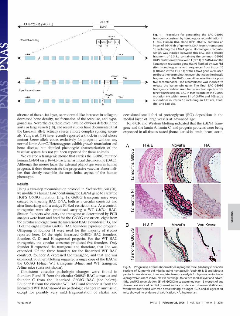
ResultsUsing a two-step recombination protocol in Escherichia coli (20),we modified a human BAC containing the LMNA gene to carry theHGPS G608G mutation (Fig. 1). G608G transgenic mice werecreated by injecting BAC DNA, both as a circular construct andafter linearizing with a unique PI-SceI restriction site. As a control,transgenics were also produced carrying a WT LMNA BAC.Sixteen founders who carry the transgene as determined by PCRanalysis were born and bred for the G608G constructs, eight fromthe circular and eight from the linearized BAC. Founders F, G, andH of the eight circular G608G BAC founders expressed progerin.Offspring of founder H were used for the majority of studiesreported here. Of the eight linearized G608G BAC founders,founders C, D, and H expressed progerin. For the WT BACtransgenics, the circular construct produced five founders. Onlyfounder B expressed the transgene, and therefore, that line wasexpanded. Of the three founders for the linearized WT BACconstruct, founder A expressed the transgene, and that line wasexpanded. Southern blotting suggested a single copy of the BAC inthe G608G H-line, WT transgenic B-line, and WT transgenicA-line mice (data not shown).
Consistent vascular pathologic changes were found infounders F and H from the circular G608G BAC construct andfounder C from the linearized G608G BAC (see below).Founder B from the circular WT BAC and founder A from thelinearized WT BAC showed no pathologic changes in any tissue,except for possibly very mild fragmentation of elastin and
occasional small foci of proteoglycan (PG) deposition in themedial layer of large vessels at advanced age.
RT-PCR and Western blotting indicated that the LMNA trans-gene and the lamin A, lamin C, and progerin proteins were beingexpressed in all tissues tested (bone, ear, skin, brain, heart, aorta,
Fig. 2. Progressive arterial abnormalities in progeria mice. (A) Analysis of aorticsections of 12-month-old mice by using hematoxylin�eosin (H & E) and Movat’spentachrome stain and immunohistochemistry analysis for hyaluronan indicatesa progressive loss of VSMC, elastin breakage, thickened medial layer and adven-titia, and PG accumulation. (B) All G608G mice examined over 16 months of ageshowed evidence of carotid (shown) and aortic (data not shown) calcification,which was confirmed with Von Kossa staining. Younger HGPS and all ages of NTmice showed no evidence of calcification. HA, hyaluronan.
Fig. 1. Procedure for generating the BAC G608Gtransgenic construct by homologous recombination inE. coli. Human BAC clone RP11-702H12 contains aninsert of 164.4 kb of genomic DNA from chromosome1q including the LMNA gene. Homologous recombi-nation was induced between this BAC and a shuttlefragment of 2.3 kb containing the common G608GHGPS mutation within exon 11 (Ex 11) of LMNA and thekanamycin resistance gene (Kanr) flanked by two FRTsites. Homology arms with sequences from intron 10(I-10) and intron 11 (I-11) of the LMNA gene were usedto direct the recombination event between the shuttlefragment and the BAC clone. After selection for posi-tive recombinants, Flpe recombinase was induced torelease the kanamycin gene. The final BAC G608Gtransgenic construct used for pronuclear injection dif-fers from the original BAC in that it contains the G608Gmutation (*) within exon 11 of LMNA and 109 extranucleotides in intron 10 including an FRT site, EcoRIsite, and SacI site.
Varga et al. PNAS � February 28, 2006 � vol. 103 � no. 9 � 3251
GEN
ETIC
S
carotid artery, iliac artery, skeletal muscle, liver, kidney, spleen,testis, and ovary) for the G608G H-line, G608G founder C-line, andWT transgenic A-line and B-line mice (see Fig. 6, which is publishedas supporting information on the PNAS web site, and data notshown). No consistent differences in mortality, size, hair loss, teeth,or skin quality were seen between the G608G H-line mice and theWT transgenic and nontransgenic mice �20 months of age.
Tissues were surveyed at autopsy and by microscopic analysis. Noconsistent pathology was found in the external ear, skin, brain,testis, ovary, skeletal muscle, bone, liver, spleen, kidney, or heart forthe G608G H-line mice. Sections of aorta, carotid artery, and iliacartery were stained with hematoxylin�eosin and Movat’s penta-chrome stains. In the G608G mice, the most dramatic finding wasthe progressive loss of VSMC, elastic fiber breakage, thickening ofthe adventitia and medial layer, accumulation of PGs, and collagendeposition. This phenomenon was first observed in 5-month-old
mice and became severe by 12 months. Arterial calcification wasobserved in older mice with severe VSMC loss and extracellularmatrix deposition. No inflammation was present, and the VSMCthat remained appeared hypertrophied. Although all large arteriesscreened were abnormal, the descending aorta (Fig. 2A) and carotidartery were the most severe. Affinity histochemistry confirmed anaccumulation of hyaluronan with age, which was absent in controls(Fig. 2A). Von Kossa staining confirmed the progressive calcifica-tion found in vessels from older G608G mice; such calcium depositswere absent in age matched controls (Fig. 2B).
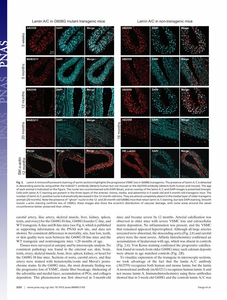
To visualize expression of the transgene in microscopic sections,we took advantage of the fact that the lamin A�C antibody(Ab2559) recognizes both human and mouse lamin, but the laminA monoclonal antibody (mAb3211) recognizes human lamin A andnot mouse lamin A. Immunohistochemistry using these antibodiesshowed that in 5-week-old G608G and the controls lamin A�C was
Fig. 3. Lamin A immunofluorescent staining of aortic sections highlights the progressive VSMC loss in G608G transgenics. The presence of lamin A�C is detectedin descending aorta by using either the mAb3211 antibody (detects human but not mouse) or the Ab2559 antibody (detects both human and mouse). The ageof each animal is indicated on the figure. The nuclei are counterstained with DAPI (blue), and an overlay of the lamin A�C and DAPI images is presented (merge).Cells with lamin A�C staining are present in the three layers of the arteries: intima, media, and adventitia in 5-week-old and 5-month-old transgenic mice. Thenumber of lamin A�C-positive nuclei is dramatically decreased in the 12-month-old mice. They are almost completely absent in the medial layer of older transgenicanimals (20 months). Note the presence of ‘‘ghost’’ nuclei in the 12- and 20-month-old G608G mice that retain lamin A�C staining, but lack DAPI staining. Smoothmuscle �-actin staining confirms loss of VSMCs; these images also show the eccentric distribution of vascular damage, with some areas around the vesselcircumference better preserved than others.
3252 � www.pnas.org�cgi�doi�10.1073�pnas.0600012103 Varga et al.
present in the endothelial cells, in the medial VSMC, and in theadventitia. Over the next 6–12 months, progressive loss of VSMCwas observed in G608G transgenics in an eccentric pattern aroundthe vessel circumference. As the mice reached a more advanced age(20 months), complete depletion of the medial VSMC occurred inmany regions (Fig. 3). Similar observations were made in thecarotid artery (see Fig. 7, which is published as supporting infor-mation on the PNAS web site). We also noticed the presence inolder animals of protein debris nuclear ‘‘ghosts’’ that are positive forlamin A but lack DNA (Fig. 3 and see Fig. 8, which is published assupporting information on the PNAS web site). Smooth muscle�-actin staining confirmed the eccentric VSMC loss in the mediallayer of large arteries, whereas the endothelial cells remainedessentially intact (Fig. 3 and see Fig. 7).
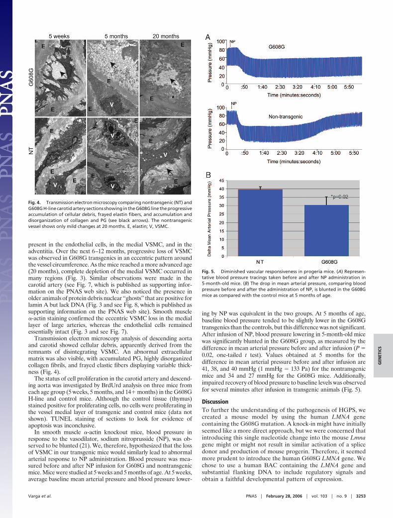
Transmission electron microscopy analysis of descending aortaand carotid showed cellular debris, apparently derived from theremnants of disintegrating VSMC. An abnormal extracellularmatrix was also visible, with accumulated PG, highly disorganizedcollagen fibrils, and frayed elastic fibers displaying variable thick-ness (Fig. 4).
The status of cell proliferation in the carotid artery and descend-ing aorta was investigated by BrdUrd analysis on three mice fromeach age group (5 weeks, 5 months, and 14� months) in the G608GH-line and control mice. Although the control tissue (thymus)stained positive for proliferating cells, no cells were proliferating inthe vessel medial layer of transgenic and control mice (data notshown). TUNEL staining of sections to look for evidence ofapoptosis was inconclusive.
In smooth muscle �-actin knockout mice, blood pressure inresponse to the vasodilator, sodium nitroprusside (NP), was ob-served to be blunted (21). We, therefore, hypothesized that the lossof VSMC in our transgenic mice would similarly lead to abnormalarterial response to NP administration. Blood pressure was mea-sured before and after NP infusion for G608G and nontransgenicmice. Mice were studied at 5 weeks and 5 months of age. At 5 weeks,average baseline mean arterial pressure and blood pressure lower-
ing by NP was equivalent in the two groups. At 5 months of age,baseline blood pressure tended to be slightly lower in the G608Gtransgenics than the controls, but this difference was not significant.After infusion of NP, blood pressure lowering in 5-month-old micewas significantly blunted in the G608G group, as measured by thedifference in mean arterial pressure before and after infusion (P �0.02, one-tailed t test). Values obtained at 5 months for thedifference in mean arterial pressure before and after infusion are41, 38, and 40 mmHg (1 mmHg � 133 Pa) for the nontransgenicmice and 34 and 27 mmHg for the G608G mice. Additionally,impaired recovery of blood pressure to baseline levels was observedfor several minutes after infusion in transgenic animals (Fig. 5).
DiscussionTo further the understanding of the pathogenesis of HGPS, wecreated a mouse model by using the human LMNA genecontaining the G608G mutation. A knock-in might have initiallyseemed like a more direct approach, but we were concerned thatintroducing this single nucleotide change into the mouse Lmnagene might or might not result in similar activation of a splicedonor and production of mouse progerin. Therefore, it seemedmore prudent to introduce the human G608G LMNA gene. Wechose to use a human BAC containing the LMNA gene andsubstantial f lanking DNA to include regulatory signals andobtain a faithful developmental pattern of expression.
Fig. 4. Transmission electron microscopy comparing nontransgenic (NT) andG608G H-line carotid artery sections showing in the G608G line the progressiveaccumulation of cellular debris, frayed elastin fibers, and accumulation anddisorganization of collagen and PG (see black arrows). The nontransgenicvessel shows only mild changes at 20 months. E, elastin; V, VSMC.
Fig. 5. Diminished vascular responsiveness in progeria mice. (A) Represen-tative blood pressure tracings taken before and after NP administration in5-month-old mice. (B) The drop in mean arterial pressure, comparing bloodpressure before and after the administration of NP, is blunted in the G608Gmice as compared with the control mice at 5 months of age.
Varga et al. PNAS � February 28, 2006 � vol. 103 � no. 9 � 3253
GEN
ETIC
S
There were risks associated with this strategy. Creating an extracopy of LMNA could cause a phenotype of its own. Regulation ofthe human BAC transgene might be different in the murineenvironment. Furthermore, the 164-kb BAC carries other genes(UBQLN4, MAPBPIP, and RAB25) in addition to LMNA, whichcould also affect the phenotype. All of these concerns wereaddressed by creating control animals carrying a WT human LMNABAC. The phenotype of the WT transgenics was essentially normal,except for possibly very mild medial arterial changes in animals ofadvanced age, although this was difficult to distinguish from non-transgenic controls.
G608G transgenic mice were found to have a progressive anddramatic defect of the large arteries, consisting of progressivemedial VSMC loss and replacement with PG and collagen. Inaddition, the arteries underwent vascular remodeling in response tothe VSMC loss, with calcification and adventitial thickening. Wewere unable to determine whether VSMC were undergoing apo-ptosis or replicating to replace their loss because cell turnover wastoo slow to detect with BrdUrd or TUNEL staining. These arterialabnormalities were reflected functionally by an altered in vivoresponse to the vasodilator NP. G608G mice demonstrated ablunted initial response to NP, consistent with impaired vascularrelaxation and attenuated blood pressure recovery after infusion.
Although G608G transgenic mice lack pathologic features ofHGPS outside of the vascular system, the pathologic changes inthe medial layer of large vessels show pronounced similarity to thehuman condition (2–5). Both human progeria patients and theG608G transgenic mice have severe VSMC loss, accumulation ofacellular material, and calcification of the vessel wall. The patternin human progeria also includes some degree of intimal prolifera-tion, which is also a form of vascular remodeling, probably inresponse to the progressive loss of medial VSMC (2–5). Weobserved minimal intimal thickening in the transgenic mice; how-ever, it is possible that intimal thickening would occur in responseto vascular injury or other cardiovascular insults.
LMNA and progerin are expressed in a variety of tissues in thesetransgenic animals. Why, then, is the mouse phenotype essentiallylimited to VSMC in large arteries? Based on the report of Lam-merding et al. (22) that lamin A�C-deficient mouse embryo fibro-blasts are more sensitive to mechanical strain, we propose thatprogerin-expressing VSMC in the aorta and proximal arterial treeare in particularly vulnerable locations to experience mechanicalstress. Thus, the shear forces from blood forcefully pounding againstarterial walls, particularly in the aortic arch and at bifurcations,progressively devitalizes VSMC that have been rendered unusuallyfragile by the presence of progerin, ultimately leading to an almostacellular vessel wall. Perhaps, other tissues would show effects if themice lived longer.
This model will potentially be useful to further the understandingof the underlying cause of the vascular pathology in progeria and tostudy potential therapies. For example, we and others recentlyreported that the use of farnesyltransferase inhibitors can improvethe abnormal nuclear structure of human progeria fibroblasts invitro by correcting the blebbing that is a hallmark of the disorder (15,23–25). Additionally, bone marrow transplantation from a com-patible normal donor might provide stem cells that could repopu-late the depleted arterial medial layer in HGPS. A similar strategyhas been reported to improve the clinical status of children withosteogenesis imperfecta (26). Both drug therapy and transplantapproaches to progeria can now be tested with this mouse model.
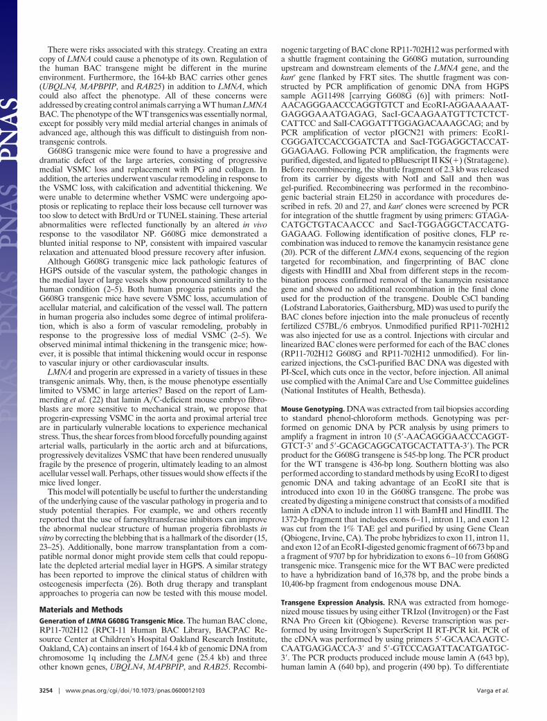
Materials and MethodsGeneration of LMNA G608G Transgenic Mice. The human BAC clone,RP11-702H12 (RPCI-11 Human BAC Library, BACPAC Re-source Center at Children’s Hospital Oakland Research Institute,Oakland, CA) contains an insert of 164.4 kb of genomic DNA fromchromosome 1q including the LMNA gene (25.4 kb) and threeother known genes, UBQLN4, MAPBPIP, and RAB25. Recombi-
nogenic targeting of BAC clone RP11-702H12 was performed witha shuttle fragment containing the G608G mutation, surroundingupstream and downstream elements of the LMNA gene, and thekanr gene flanked by FRT sites. The shuttle fragment was con-structed by PCR amplification of genomic DNA from HGPSsample AG11498 [carrying G608G (6)] with primers: NotI-AACAGGGAACCCAGGTGTCT and EcoRI-AGGAAAAAT-GAGGGAAATGAGAG, SacI-GCAAGAATGTTCTCTCT-CATTCC and SalI-CAGGATTTGGAGACAAAGCAG; and byPCR amplification of vector pIGCN21 with primers: EcoR1-CGGGATCCACCGGATCTA and SacI-TGGAGGCTACCAT-GGAGAAG. Following PCR amplification, the fragments werepurified, digested, and ligated to pBluescript II KS(�) (Stratagene).Before recombineering, the shuttle fragment of 2.3 kb was releasedfrom its carrier by digests with NotI and SalI and then wasgel-purified. Recombineering was performed in the recombino-genic bacterial strain EL250 in accordance with procedures de-scribed in refs. 20 and 27, and kanr clones were screened by PCRfor integration of the shuttle fragment by using primers: GTAGA-CATGCTGTACAACCC and SacI-TGGAGGCTACCATG-GAGAAG. Following identification of positive clones, FLP re-combination was induced to remove the kanamycin resistance gene(20). PCR of the different LMNA exons, sequencing of the regiontargeted for recombination, and fingerprinting of BAC clonedigests with HindIII and XbaI from different steps in the recom-bination process confirmed removal of the kanamycin resistancegene and showed no additional recombination in the final cloneused for the production of the transgene. Double CsCl banding(Lofstrand Laboratories, Gaithersburg, MD) was used to purify theBAC clones before injection into the male pronucleus of recentlyfertilized C57BL�6 embryos. Unmodified purified RP11-702H12was also injected for use as a control. Injections with circular andlinearized BAC clones were performed for each of the BAC clones(RP11-702H12 G608G and RP11-702H12 unmodified). For lin-earized injections, the CsCl-purified BAC DNA was digested withPI-SceI, which cuts once in the vector, before injection. All animaluse complied with the Animal Care and Use Committee guidelines(National Institutes of Health, Bethesda).
Mouse Genotyping. DNA was extracted from tail biopsies accordingto standard phenol-chloroform methods. Genotyping was per-formed on genomic DNA by PCR analysis by using primers toamplify a fragment in intron 10 (5�-AACAGGGAACCCAGGT-GTCT-3� and 5�-GCAGCAGGCATGCACTATTA-3�). The PCRproduct for the G608G transgene is 545-bp long. The PCR productfor the WT transgene is 436-bp long. Southern blotting was alsoperformed according to standard methods by using EcoRI to digestgenomic DNA and taking advantage of an EcoRI site that isintroduced into exon 10 in the G608G transgene. The probe wascreated by digesting a minigene construct that consists of a modifiedlamin A cDNA to include intron 11 with BamHI and HindIII. The1372-bp fragment that includes exons 6–11, intron 11, and exon 12was cut from the 1% TAE gel and purified by using Gene Clean(Qbiogene, Irvine, CA). The probe hybridizes to exon 11, intron 11,and exon 12 of an EcoRI-digested genomic fragment of 6673 bp anda fragment of 9707 bp for hybridization to exons 6–10 from G608Gtransgenic mice. Transgenic mice for the WT BAC were predictedto have a hybridization band of 16,378 bp, and the probe binds a10,406-bp fragment from endogenous mouse DNA.
Transgene Expression Analysis. RNA was extracted from homoge-nized mouse tissues by using either TRIzol (Invitrogen) or the FastRNA Pro Green kit (Qbiogene). Reverse transcription was per-formed by using Invitrogen’s SuperScript II RT-PCR kit. PCR ofthe cDNA was performed by using primers 5�-GCAACAAGTC-CAATGAGGACCA-3� and 5�-GTCCCAGATTACATGATGC-3�. The PCR products produced include mouse lamin A (643 bp),human lamin A (640 bp), and progerin (490 bp). To differentiate
3254 � www.pnas.org�cgi�doi�10.1073�pnas.0600012103 Varga et al.
between endogenous mouse and transgenic human lamin A, BstUIwas used to digest RT-PCR products, resulting in two bands forhuman (386 and 254 bp) and one band for mouse (643 bp). Proteinextracted by using a radioimmunoprecipitation assay was run on 8%Tris�glycine gels (Invitrogen) and probed with mAb3211 lamin Aantibody (Chemicon International) and goat anti-mouse horserad-ish peroxidase secondary antibody (Kirkegaard & Perry Labora-tories). Visualization of the bands was possible by using theECL-plus kit (Amersham Pharmacia Biosciences) and then devel-oping the images on film (Kodak).
Histochemistry. Tissues fixed in 2% paraformaldehyde were dehy-drated with graded alcohols and embedded in paraffin. Cross-sections (4-�m thick) were cut with a rotary microtome andmounted on charged slides (Superfrost, Columbia Diagnostics,Springfield, VA) and then stained with hematoxylin�eosin andMovat’s pentachrome stain (28, 29). Images were captured by usingan Olympus BX51 Microscope equipped with an Olympus DP11digital camera. Images were managed by using Microsoft DIGITALIMAGE PRO 10 and Adobe PHOTOSHOP 7 for WINDOWS.
Immunohistochemistry. Staining for lamins was performed in PBSby using the rabbit polyclonal anti-lamin A diluted 1:10 (Ab2559,Abcam, Inc., Cambridge, MA) on transgenic or nontransgenictissues or the mouse monoclonal anti-lamin A�C nondiluted(mAb3211, Chemicon) on transgenic tissues with goat anti-rabbit orgoat anti-mouse Alexa Fluor 594-conjugated secondary antibodies(Molecular Probes). Antigen retrieval was performed before theincubation of the lamin antibodies and consisted of a 2-minincubation in EDTA (0.37 g�liter) in a pressure cooker. A mouse-to-mouse blocking kit (ScyTek Laboratories, Logan, Utah) wasused with mouse anti-lamin A�C. Staining for smooth muscle�-actin was performed by using a Cy3-conjugated antibody diluted1:100 (Sigma–Aldrich). Slides were mounted in DAPI-containingmedium (Vector Laboratories) Fluorescence emission images wereobtained with a confocal microscope system (Zeiss LMS 510) andcollected with �20 or �40 oil lenses (Zeiss).
PGs were detected in paraffin sections of vessels, after digestionwith 0.2 units�ml chondroitin ABC lyase (Seikagaku America,Rockville, MD) for 1 h at 37°C, by using rabbit anti-mousemonoclonal antibodies (a gift from Larry Fisher, National Instituteof Dental and Craniofacial Research, National Institutes of Health,Bethesda) against versican (LF99), biglycan (LF106), and decorin(LF113) and by using a rat anti-mouse monoclonal antibody againstperlecan (a gift from Koji Kimata, Nagoya University, Chikusa,Nagoya, Japan). Biotinylated secondary antibodies were used (Vec-tor Laboratories) and visualized by using Vector Red reagent(Vector Laboratories). Hyaluronan localization was carried out
with a commonly used biotinylated probe consisting of a mixture ofcartilage, PG, and link protein (provided by T.N.W.).
For BrdUrd analysis, the mice were given 100 mg�kg BrdUrd(Sigma) via i.p. injection 18 and 1 h before death. Sections of aorta,carotid, and thymus were prepared as discussed in ref. 29. Quan-tification of proliferating cells was performed by three blindedobservers counting the number of BrdUrd-positive medial VSMC.Systemic distribution of BrdUrd was confirmed by intense stainingof proliferating thymus cells in all animals receiving the agent.
Transmission Electron Microscopy. Samples were prepared andviewed by using a JEM 1200EX II transmission electron microscope(JEOL) as described in ref. 30. Images were acquired and saved onKodak 4489 electron microscope film with a ‘‘below the viewingscreen’’ film transfer system and scanned at 600 dots per inch byusing an Epson Expression 1680 flat bed scanner for digitization.Ruthenium Red was used in some samples to gain better visual-ization of PGs.
Blood Pressure Analysis. Blood pressure was measured invasively byusing a microtip pressure transducer catheter connected to anelectrostatic chart recorder (Millar Instruments, Houston). Ani-mals were anesthetized with 1–3% isoflurane, and the right externalcarotid artery was cannulated. Once initial baseline blood pressuremeasurements were completed, the mouse was infused with saline(30 �l over 2 min), and blood pressure was again measured. Sodiumnitroprusside (1.5 mg�kg of body weight, 30 �l of total volume over2 min) was then infused, and blood pressure measurements weretaken until a return to baseline or a maximum of 20 min. For5-week-old mice, three G608G and three nontransgenic mice werestudied. For 5-month-old mice, two G608G and three nontrans-genics were included. Because we postulated that the G608Ganimals would have a blunted response to NP, we used a one-tailedtype-2 t test to assess significance, and this was performed by usingMicrosoft EXCEL.
We thank Darryl Leja for assistance in preparing the figures,Drs. David Bodine and Shelley Hoogstraten-Miller for mouse expertise,Dr. Christian A. Combs and Daniela Malide [Light Microscopy CoreFacility, National Heart, Lung and Blood Institute, National Institutes ofHeath (NIH)] for assistance regarding microscopy-related experiments,and Shih Queen Lee-Lin for DNA fingerprinting of BAC constructs.This research was supported in part by the Intramural Research Programof the National Human Genome Research Institute and the NationalHeart, Lung, and Blood Institute (NIH) and by NIH Grant HL-18645 (toT.N.W.). M.E. was supported by grants from the Tore Nilsson Founda-tion, the Åke Wiberg Foundation, the Hagelen Foundation, the Loo andHans Osterman Foundation, the Torsten and Ragnar Soderberg Foun-dation, the Jeansson Foundation, the Swedish Research Council, and theSwedish Foundation for Strategic Research.
1. DeBusk, F. L. (1972) J. Pediatr. (Berlin) 80, 697–724.2. Baker, P. B., Baba, N. & Boesel, C. P. (1981) Arch. Pathol. Lab. Med. 105, 384–386.3. Stehbens, W. E., Wakefield, S. J., Gilbert-Barness, E., Olson, R. E. & Ackerman, J. (1999)
Cardiovasc. Pathol. 8, 29–39.4. Stehbens, W. E., Delahunt, B., Shozawa, T. & Gilbert-Barness, E. (2001) Cardiovasc. Pathol.
10, 133–136.5. Ackerman, J. & Gilbert-Barness, E. (2002) Pediatr. Pathol. Mol. Med. 21, 1–13.6. Eriksson, M., Brown, W. T., Gordon, L. B., Glynn, M. W., Singer, J., Scott, L., Erdos, M. R.,
Robbins, C. M., Moses, T. Y., Berglund, P., et al. (2003) Nature 423, 293–298.7. De Sandre-Giovannoli, A., Bernard, R., Cau, P., Navarro, C., Amiel, J., Boccaccio, I.,
Lyonnet, S., Stewart, C. L., Munnich, A., Le Merrer, M., et al. (2003) Science 300, 2055.8. Bonne, G. & Levy, N. (2003) Lancet 362, 1585–1586; author reply, 1586.9. Csoka, A. B., Cao, H., Sammak, P. J., Constantinescu, D., Schatten, G. P. & Hegele, R. A.
(2004) J. Med. Genet. 41, 304–308.10. Fukuchi, K., Katsuya, T., Sugimoto, K., Kuremura, M., Kim, H. D., Li, L. & Ogihara, T.
(2004) J. Med. Genet. 41, e67.11. Plasilova, M., Chattopadhyay, C., Pal, P., Schaub, N. A., Buechner, S. A., Mueller, H., Miny,
P., Ghosh, A. & Heinimann, K. (2004) J. Med. Genet. 41, 609–614.12. Huang, S., Kennedy, B. K. & Oshima, J. (2005) Novartis Found. Symp. 264, 197–202;
discussion 202-207, 227–230.13. Gruenbaum, Y., Margalit, A., Goldman, R. D., Shumaker, D. K. & Wilson, K. L. (2005) Nat.
Rev. Mol. Cell Biol. 6, 21–31.14. Goldman, R. D., Gruenbaum, Y., Moir, R. D., Shumaker, D. K. & Spann, T. P. (2002) Genes
Dev. 16, 533–547.
15. Capell, B. C., Erdos, M. R., Madigan, J. P., Fiordalisi, J. J., Varga, R., Conneely, K. N., Gordon,L. B., Der, C. J., Cox, A. D. & Collins, F. S. (2005) Proc. Natl. Acad. Sci. USA 102, 12879–12884.
16. Agarwal, A. K., Fryns, J. P., Auchus, R. J. & Garg, A. (2003) Hum. Mol. Genet. 12, 1995–2001.17. Sullivan, T., Escalante-Alcalde, D., Bhatt, H., Anver, M., Bhat, N., Nagashima, K., Stewart,
C. L. & Burke, B. (1999) J. Cell Biol. 147, 913–920.18. Mounkes, L. C., Kozlov, S., Hernandez, L., Sullivan, T. & Stewart, C. L. (2003) Nature 423,
298–301.19. Yang, S. H., Bergo, M. O., Toth, J. I., Qiao, X., Hu, Y., Sandoval, S., Meta, M., Bendale,
P., Gelb, M. H., Young, S. G., et al. (2005) Proc. Natl. Acad. Sci. USA 102, 10291–10296.20. Lee, E. C., Yu, D., Martinez de Velasco, J., Tessarollo, L., Swing, D. A., Court, D. L.,
Jenkins, N. A. & Copeland, N. G. (2001) Genomics 73, 56–65.21. Schildmeyer, L. A., Braun, R., Taffet, G., Debiasi, M., Burns, A. E., Bradley, A. & Schwartz,
R. J. (2000) FASEB J. 14, 2213–2220.22. Lammerding, J., Schulze, P. C., Takahashi, T., Kozlov, S., Sullivan, T., Kamm, R. D., Stewart,
C. L. & Lee, R. T. (2004) J. Clin. Invest. 113, 370–378.23. Glynn, M. W. & Glover, T. W. (2005) Hum. Mol. Genet. 14, 2959–2969.24. Young, S. G., Fong, L. G. & Michaelis, S. (2005) J. Lipid Res. 46, 2531–2558.25. Toth, J. I., Yang, S. H., Qiao, X., Beigneux, A. P., Gelb, M. H., Moulson, C. L., Miner, J. H.,
Young, S. G. & Fong, L. G. (2005) Proc. Natl. Acad. Sci. USA 102, 12873–12878.26. Horwitz, E. M., Gordon, P. L., Koo, W. K., Marx, J. C., Neel, M. D., McNall, R. Y., Muul,
L. & Hofmann, T. (2002) Proc. Natl. Acad. Sci. USA 99, 8932–8937.27. Liu, P., Jenkins, N. A. & Copeland, N. G. (2003) Genome Res. 13, 476–484.28. Schmidt, R. and. Wirtala, J. (1996) J. Histotechnol. 19, 325–327.29. Farb,A.,Weber,D.K.,Kolodgie,F.D.,Burke,A.P.&Virmani,R. (2002) Circulation105,2974–2980.30. Lara, S. L., Evanko, S. P. & Wight, T. N. (2001) Methods Mol. Biol. 171, 271–290.
Varga et al. PNAS � February 28, 2006 � vol. 103 � no. 9 � 3255
GEN
ETIC
S