Embed Size (px)
Citation preview

--
PROGRESSIVE MUSCULAR DYSTROPHY AS AN ENDOCRINE DISEASE
WALTER TIMME, M.D. Assistant Physician to the Neurological Institute, New York City; Consulting
Neurologist, New Rochelle and Volunteer Hospitals; Visiting Neurologist, Ran dati's Island Institutions
NEW YORK
In the present paper, the author desires to report a somewhat atypical form of progressive muscular dystrophy, rather resembling Erb's infantile type, of extremely benign and �low progress. In the cases examined it has occurred as a hereditary affection now in the fourth generation. The number of individuals disabled thus far has been fourteen. Of the seven living members with the disease, all but two have been examined by Roentgen ray, and of these five, four show distinct changes in the pineal gland, producing shadows in the roentgenogram. The evidence presented by the cases reported in the present paper, togethet· with that of previous communications in this field, creates a strong probability of the existence of a close relationship between progressive muscular dystrophy and disease of the pineal gland.
In order to understand the evolution that has now placed this disease among the endocrinopathies, a rapid survey of the development of the syndrome, together with the most important contributions thereto in the last few decades, is almost indispensable and is herewith included. The cases studied have been placed at my disposal by Dr. Pearce Bailey, of the Neurological Institute, and for his valuable suggestions and actual assistance, I desire to express my appreciation and my thanks.
Historical.1-The first observations on muscular atrophies are found in Boerhaeve's Aphorisms (1709), in which Van Swieten mentions the involvement of the deltoid and that of the intrinsic hand muscles. Later, Charles Bell described syndromes including progressive muscular atrophy. But it was reserved for Aran and Duchenne in 1850 to give the first good description of progressive muscular atrophy. Following them, the French physicians called this type of disease "atrophic musculaire progressive type Duchenne-Aran." Shortly thereafter, Cruveilhier, on the basis of a necropsy, stated that the cause of the disease lay in abnormality of the spinal gray; and ever since then the discussion
1. Miiller, Albert: Zur Lehre der Dystrophia Muscularis progressiva, Dissert., Kiel, 1902, Surgeon General's Library.

2
has waxed hot as to whether the lesion is in the spinal cord or in the muscles themselves. A third theory explained it as a trophoneurosis (Remak, Schneevogt, Jaccoud), with disease of the sympathetic system as a basis. This idea was soon given up. As histolog1c technic improved and positive findings in the spinal cord increased, the supporters of the myelopathic character of the disease seemed to carry the day ( Luys, Hayem, J affroy and especially Charcot). Charcot, as a result of many investigations, declared the large multipolar ganglion cells of the anterior horns to be the trophic centers of voluntary muscles and that in their allerations the cause of atrophic muscle disease was to be found. Duchenne himself, although originally arrayed on the side of the myopathic nature of the trouble, came around to Charcot's view. So likewise Erb. In spite of the fact that most neuropathologists agreed with Charcot, Friedreich opposed him. In his monograph2 of 1873 the latter defended the myopathic nature of the trouble in many cases. Thus, he stated that as a result of successive extreme exertions in the same muscle groups, especially in a constitutional diathesis weakening the muscular system's resistance, the atrophies occurred. He described cases following shortly after typhus, typhoid, measles, acute rheumatism, cholera and even after the puerperium, and thereupon declared the condition to be one of "polymyositis chronica progressiva." The changes in the anterior horn cells he considered secondary to the muscle deterioration. But the majority of neurologists stood behind Charcot. Then in 1878 Lichtheim presented a case with high-grade atrophy of great extent in which no cord changes were seen at all, nor were any evident in the peripheral nerves. Therefore, he came to the necessary conclusion that changes in the anterior horn cells are not a sine qua non of the condition, and further, that Charcot's theory could not be the true one. Charcot's preeminence, however, still carried the day. Finally, more recent investigators, after carefully separating all conditions that resembled the progressive muscular atrophies, such as poliomyelitis, chronic multiple neuritis, atrophies with joint lesions, syringomyelia, and so on, from them, two distinct types remained; those that did show spinal cord changes, and those that did not. In 1876 Leyden proposed to separate hereditary forms, on account of their etiology and the localization of the atrophies (pelvic girdle), from the Duchenne-Aran type, and at the same time he showed their resemblance to the hypertrophic type. Lastly, Erb, in 1882, on the basis of a vast variety of cases personally examined by him, declared that there were two distinct types of progressive muscuiar atrophy, different in their syndrome, localization, evolution, and in their objective behavior, as well as in their anatomic foundation; a spinal
2. Fried reich, N.: Ueber progressive muskelatrophie; iiber wahre und false he �Iuskelhypertrophie, Berlin, 1873.

'lot
3
atrophic form and a muscular dystrophic form. To this differentiation even the Charcot School assented. But even here it did not take long to discover that there were transitional forms, and in each group of cases intrinsic differences. These depend on the different muscle groups first affected, and on the degree and nature of the muscle changes. Thus have arisen the many confusing types of the disease, and not, as at first thought, distinct diseases. One great work which we ought to be thankful to Erb for having done for us is that he grouped all these muscular types in one large family, which he called dystrophia muscularis progressiva.3 In this family he included his juvenile muskelatrophie, pseudohypertrophie infantile form and the hereditary forms. The spinal types he placed under the name "Amyotrophia spinalis progressiva, Duchenne-Aran." Soon thereafter Landouzy and Dejerine ( 1885) described two cases of "atrophie de l'enfance"• in one of which they obtained a necropsy. To their great surprise, they found no changes in the spinal cord, and described the case as a new one, differing entirely from the Duchenne-Aran kind. They called it "myopathie, type facio-scapulo-humeral," and a second type with "integrite de la face, type scapulo-humeral." Undoubtedly these types are nothing else than the Erb juvenile form, which at times involves the face and again does not.
In the course of time, all observers, Charcot and Marie included, agreed to accept this classification·of Erb's:
1. Dystrophia muscularis progress iva (infantum). a. Hypertrophic form
(1) With pseudohypertrophy (2) With true hypertrophy
b. Atrophic form (1) With primary facial involvement (Duchenne's infantile form) (2) Without facial involvement. Simple atrophic form.
2. Dystrophia muscularis progressiva juvenum et adultorum. (juvenile form)
It is characteristic of the·familial, hereditary forms to exhibit conditions at times of one of these groups, at others, of another group; so that in several generations of the same family individuals will be found to conform with almost any one of the types outlined above. It is so with the family herein studied.
TYPES OF THE DISEASE
Various authors have described atypical forms of muscular dystrophy, that is, they were atypical either in their course or their chron-
3. Erb, W.: Ueber die juvenile form der progressiven Muskclatrophie, Deutsch. Arch. f. klin. Med., 1884, 34, 467.
4. Landouzy, L., and Dejerine, J.: De Ia myopathic atrophique progressive, Rev. de meEd .. 1885. 5, 81.

4
icity, or else in the groups of muscles affected or in the intensity of such impairment; in the presence or absence of contractures; in the slow or rapid involvement of the entire skeletal structure, together with the muscular, making of the patients helpless cripples, and ending in death in a comparatively short time. Some of these important types it may be well here to describe and classify, so that their important characteristics may serve to differentiate them at the same time from the group of cases which I propose to submit further on in this paper. It will also be noticed that as we progress the recent authors show more and more marked a tendency to include symptoms referable to the sympathetic nervous system, including the endocrine glands.
Barsickow's group is late in appearance, slowly progressive and not fatai. It was the first group of great importance, studied by Barsickow• in 1871, and consisted of twenty-four cases occurring in two families from one ascendent through five generations. This series resembles most closely the series herewith to be presented, and yet differs from it in noteworthy particulars. It is presented in extenso for its importance. A composite picture of Barsickow's cases showed that the members of the widely scattered families in which they arose were as a rule in good health and lived to be old. The greatgrandfather had only a stiffness in gait and carriage, and yet twenty-three of his descendants (five out of seven of his immediate children) showed muscular disease, not only in function, but also in muscle volume. The members seem to have been attacked without rule, seemingly showing some predisposition in heredity. The children remained healthy if the parents were unaffected, and there were an equal number of males and females attacked. The cause of the original incidence, Barsickow states, was probably lead poi�oning, for this original grandfather was a typesetter and had had many attacks of lead colic. In some cases chlorosis, cholera and varioloid acted as preceding agents conducing to the development of the trouble. The onset in seventeen of the cases was at the following ages: five between the ages of 10 and 20, se\'en between 20 and 30, and five later in life.
It was ushered in by almost simultaneous affection of both an upper and lower limb, or both upper and lower limbs. The serratus anticus was among the first muscles to cause trouble. The most prominent symptom was the disturbed muscular function; in the mildest cases being stiffness and rapid exhaustion, going to absolute paralysis of single muscles, or entire groups in severe cases. The involvement was symmetrical. In most of the cases the diseased muscles gradually disappeared or atrophied, and hypertrophy was very rare. Frequently there was a lordosis with winged scapulae, with protrusion of the
5. Barsickow, Hermann: Zwei Familien mit Lipomatosis muscularis progressiva, Dissert., Halle. 1872, Surgeon General's Library, Washington.

5
shoulders or flattening of the chest. Fibrillary twitching was rare, only once in fact. There was in the skin occasionally loss of pigment or abnormal coloring. Of the eleven who died while under Barsickow's observation, one was over 80 years of age, four over 70 years, three over 60, one 58 and two 33. So that the disease apparently did little to cut short their lives.
Taking up Barsickow's individual cases, those that showed symptoms seemingly not accounted for in the usual syndrome of dystrophia musculorum progressiva, we see in his Case 1 the complaint of rheumatoid pains in many parts of the body, dirty brown pigmented skin and patches of vitiligo. Case 19 was that of a woman of 40 in whom the fingers and toes easily fell asleep and got cold, white and waxy in the winter. Case 7 was that of a woman in whom the disease began at the menopause.
The reason I mention the above symptoms, which in the light of our present knowledge are referable to a disturbed autonomic nervous system with inclusion of the endocrine glands, is that the theory oi Remak, Schneevogt, Jaccoud, and Dumenil relative to the dependence of the affection on such disturbance of the vegetative system has now been shown to have some foundation.
The microscopic examination of the muscle fibers in Barsickow's cases showed changes in the muscle fibrils, some of which were much thickened, while between the primitive muscle bundles were many fat cells and connective tissue. The skin spots appeared in the neighborhood of recently affected muscles. In three cases with necropsy there was no abnormality of the nervous system demonstrable, and yet the author inclined to the idea that the disease is of a "vasomotor-trophicnerve character." The Barsickow cases undoubtedly belong to the Erb juvenile and adult types.
In Friedreich's group of cases the disease was early in appearance, rapidly progressive, and fatal. Friedreich6 published the cases of four brothers whose mother was unaffected by the disease, but two of whose maternal uncles died of it in their 15th and 16th years. The brothers cited died at 5, 6, 12 and 16 years of age. Intellectually they were all well advanced. The affection began in the weakness of the lumbar muscles, with constantly increasing difficulty in arising from a sitting posture. Muscular atrophy began in the muscles on each side of the spine at the same time. There then arose a difficulty in raising the arms to the shoulders on both sides. One of the victims died of suffocation from an asthmatic bronchial attack. In this series of cases the rapidity of the course to a fatal termination is the characteristic. The necropsy showed, among other atrophies, that the pectorals were
6. Fried reich, N.: Progressive Muskelatrophic. Berlin. 1873.

6
reduced to thin grayish-red, skin-like lamellae, through which the ribs showed.
In Gowers' group the disease was early in appearance, rapid in progress, and was fatal in adolescence. Gowers' in a clinical lecture in 1879 reported four brothers, aged 9, 10, 5 and 3 years, all affected, whose parents were both healthy, but whose maternal uncle died at 15 of a wasting muscular disease. These patients had a little difficulty in putting their feet down on the floor on account of a tense, shortened Achilles tendon. The extensors of the knee and hip were weak; the flexors of the hip were feeble. The latissimus dorsi and the lower part of the pectorals were gone. There were no sensory changes, the kneejerks were absent and there was a marked lordosis. The upper limbs were weak, although they moved freely. Gowers reports that out of 220 cases that he collected from the literature, 190 of the patients were male and thirty were female. While he mentions Barsickow's cases, yet he excludes them from his statistics, because they were all of the adult type. Some generalizations from these 220 cases are drawn by Gowers: ( 1) the disease almost never is heard of on the side of the father; (2) the age of onset is an etiologic factor of great importance and occurs in the worst cases before the 6th year, the more severe the case, the earlier it begins; ( 3) occasionally the disease follows physical injury; ( 4) shortening and permanent contractions of certain muscles lead to distortions in !he positions of the joints; especially contraction of the calf muscles, leading to inability to place the heels on the floor; ( 5) the patients lose the power of standing at 10 or 12 years of age, and death comes on between 14 and 18 years; ( 6) of diagnostic importance is not the actual muscular enlargement, but the distribution of the muscular disease, especially the wasting of the lower pectorals and the latissimus dorsi; (7) the later the appearance of the disease, the more slowly it advances; the older the patient, the better the prognosis.
The commentary on these cases is that some of the conclusions, especially the last, are quite contrary to our type of the disease to be described hereinafter.
Erb's8 types are of the accepted classification of progressive muscular dystrophy. In one of the publications of Erb3 he gives a general description of the juvenile type of the disease, saying that it begins always before the 20th year and usually with atrophy of the upper arms and shoulder girdle muscles and is often combined with hypertrophy. There are no reactions of degeneration and no muscular fibril-
7. Gowers, W. R.: Pseudohypertrophic Paralysis, Clinical Lecture, London, 1879.
8. Erb, \V.: Dystrophia musculorum progressiva, Deutsch. Ztschr. f. )r erYenh., 1891, t, 173.

7
lary twitching. The following muscles are almost constantly affected; pectorals (except the clavicular portion), cucullaris, latissimus dorsi, biceps and brachialis anticus and the long supinator. The glutei are weak and calves large. The disease lasts from 20 to 40 years with periods of quiescence. He states that this juvenile form is identical with Friedreich's hereditary progressive muscular atrophy. These latter forms belong in the same group with pseudohypertrophy.
In one publication,8 Erb gives the results of the examination of the available pathologic material of all these forms and says in summarizing that all the noticeable changes are in the muscle fiber chiefly, from an increase in volume to absolute disappearance of the muscle. There is an increase in the muscle nuclei and these are both normally and abnormally placed. There are clefts in the long axis of the muscle between the muscle fibers and an increase of connective tissue is found with a later deposit of fat cells and a thickening of the vessel walls. As a result of the identical pathologic findings in all forms of this elusive muscle disease, he proposes the classification of all these forms as types under the generic term of dystrophia musculorum progressiva. This classification has already been given earlier in this paper.
Among other types the next cases of importance in the development of the semeiology of the disease were reported by Prager.9 Here the patients, two in number, were children of first cousins, who themselves were free of the disease. The importance of the citation of one of these cases is in the fact that while the patient, a woman of 48, had always had a waddling gait and a difficulty in going upstairs, yet after her first puerperium, which lasted three months, the difficulty in walking was so intensified that she needed support. Her muscular system gradually underwent the usual atrophies in the pectorals, latissimi, trapezius, rhomboids, biceps, deltoids, triceps and supinator longus; but these atrophies were disguised by masses of fat in the arms and legs. To these physical signs, however, were appended the statements: ( 1) the patient was easily excited and then developed a tremor; (2) there was difficulty in deglutition for fifteen years; ( 3) dyspnea in cold weather and ( 4) increase in stools. Further on is the remark that the patient had a "struma" on the neck and complained of urticaria. That is, she was a hyperthyroid subject. We shall find as we go along in the analysis of the symptoms as presented by the author frequent indications of disturbance in the endocrine system. In all probability, had such indications been then understood, there would have resulted a far wider range of symptoms in the muscular dystrophies; for what to the older observers probably appeared negligible in this regard, now assumes great importance. We shall see how more and more often
9. Prager, M.: Dystrophia musculorum progressiva, Dissert., Erlangen, 1891.

8
in later years investigators have described conditions present in progressive muscular dystrophy, apart from the actual muscle changes, that are seemingly part of the disease process; which conditions have received but scant reference in the earlier works, presumably because they seemed so utterly adventitious. So Hahn10 states that skeletal anomalies are often seen and the idea is now that these are part of the condition of progressive muscular dystrophy; and he cites Fried reich that bone atrophy is not secondarily due to disease of the bone through immobility of the joint, but to nervous and trophic influence. Eulenberg11 had a case which was combined with acromegaly. Hahn's conclusion is that because so many cases show bony changes, there may be some connection between the two.
Bregman12 cites cases in which together with a "facies myopathique" there was difficulty in looking up and in closing the eyes; inequality of the pupils and of the palpebral fissures. This is interesting from the fact, to be shown from our own cases, of the frequent involvement of the pineal gland; in which condition just such eye muscle difficulties arise (Bailey and Jelliffe18). Bregman's cases further showed internal glandular disturbances as follows : In Case 1 the patient could not close the eyes properly and had nyctalopia; also there was intense sweating of the extremities with cyanotic hands and a pulse of 100. In Case 2 there was marked prognathism, the upper teeth standing prominently forward; the skin was mottled like marble. In Case 4 the hands and feet were livid; there was large skeletal development of the hands and feet in contrast to the rest of the skeleton, and marked protrusion of the upper jaw. In Case 5 there was extremely large body growth. In these cases there is manifestly disturbance of both pituitary and thyroid glands.
Cestan and Lejonne14 publish two cases with contractures and state that it 1� banal to say that contracture� accc..mpany all forms of progressive muscular dystrophy, but that ordinarily contractures are slight and rarely sufficiently marked to alter the general attitude of the myopathic patient. Schultze16 presents two cases, both with necropsy, of brother and sister; in the former there was a thinning of the long bones, the humerus being thinner than the middle finger of a normal hand, and
10. Hahn, F.: Ueber d. Auftreten v. Contracture bei Dystrophic, Deutsch. Ztschr. f. Nervenh., 1901, 20, 137.
11. Eulenberg: Deutsch. med. Wchnschr., 1896, 22, 458. 12. Bregman, L. E.: Ein Casuistischer Beitrag z. progressive Muskelatrophie,
Deutsch. Ztschr. f. Nervenh., 1899, 14, Cases 3 and 4. 13. Bailey, Pearce, and Jelliffe, S. E.: Tumors of the Pineal Body, THE
ARCHIVES INT. MED .. 1911, 8, 851. 14. Cestan and Lejonne: Une myopathic familiale avec retractions, Nouvelle
Iconograph, d. I. Salpetriere, 1902, 15, 38. 15. Schultze, Fr.: Ueber Combination v. famil. progress. Pseudohypertr. d.
muskeln m. Knochenatrophie, Deutsch. Ztschr. f. Nervenh., 1898-1899, 14.

9
the medullary cavity very small. The sister had a stiffness in all the joints of the body, with atrophy of the bones. Schultze states that he had found only two other instances in the literature in which was found bone atrophy with muscular dystrophy, one a case of Friedreich's and the other Le Gendre's.16 In all these cases the atrophy was a concentric one of the long bones with no diminution in the length. In Le Gendre's case there was also an undue hypertrophy of the genitals, with an enlarged prostate gland in a youth aged 20; while Friedreich's case showed infantilism in the sexual organs, voice and facial expression. The necropsies in Schultze's cases showed no apparent changes in the spinal cord, not even in the ganglion cells, and he therefore concludes that we must look elsewhere than in the nervous system for the cause of this "riddle-like disease," and advises us that we cannot neglect the theory of predisposition, with accidental factors superimposed, such as overstrain, trauma, underfeeding, infection and intoxication.
0. With11 published a familial type of the disease affecting three boys in one family and sparing the four girls. What interests us in these cases is the involvement of the tonsils, which were hypertrophied, a chronic angina, a hypertrophied lower jaw, with difficulty in mastication and deglutition.
An article of extreme importance on account of the cure of the patient, by Marina/8 describes a case of a girl of 8% years of age, who had all the signs of a beginning dystrophy with hypertrophic deltoids and gastrocnemii and so weak in the shoulder girdle group that she could barely lift her arms horizontally. He lost sight of her for five years and then she appeared again entirely well and cured. The author corresponded personally with Erb in this matter, questioning him as to whether a patient with muscular dystrophy had ever in Erb's experience been cured, or whether he had ever seen "formes frustes" of this disease. Erb replied that he had never seen a single case of "formes frustes"-they had all been progressive-but that he had seen one single case of cure in a young English girl. Erb and Marina agreed that the cure in both of these cases was to be credited to the normal evolution of the patient; presumably to the fact that following the beginning of menstruation, normal development of the internal secretion succeeded, a most pregnant idea in the light of our present knowledge of the interrelation of the ovaries with the other endocrine glands. A fitting companion piece to the above is the case of Levi and Rothschild19
16. Le Gendre: Gaz. med. de Paris, 1860, 15, 365. 17. With, Otto: Eine familiare atypische Form d. Dystroph. muse. prog.,
Dissertation, Freiburg, 1906, Surgeon General's Library. 18. Marina, Alessandro: Gibt es Formes Frustes d. muscularen Dystrophy?
Deutsch. med. Wchnschr., 1908, 34, 1087. 19. Levi and Rothschild: Rev. neurol., 1907, 15, 613.
10
of a myopathic atrophy which showed considerable improvement on pituitary extract.
Henri Claude20 in 1908 described a case of familial progressive muscular dystrophy of rather different character from the usual in that the patient had asymmetrical atrophies, more marked on the right side. Besides the muscular condition, the bones on the right side were less developed than on the left side, as shown by roentgenogram, and there were vasomotor changes particularly evident in the right hand, with perspiration and cyanosis. In cold weather the patient could not use this hand on account of the disability thereby occasioned. The temperature of the right hand was constantly lower than the left and the sphygmomanometric pressure was also less on the right side, Claude drew the conclusion from these findings that the whole disease picture does not embrace merely the muscular difficulty, but includes as well the central or peripheral nervous systems.
Jendrassik21 after citing the cases of Marina and Erb, above mentioned, in which a seemingly spontaneous cure had been accomplished in muscular dystrophy coincident with adolescence, presents two somewhat similar cases, of which the one was a true progressive dystrophy. This patient was a girl who, just before puberty set in, had had the dystrophic process cease, and then after menstruation had been established became entirely cured, even to the return of the knee reflexes. There had been a precocious body growth between the lOth and 11th years, in which the breasts had participated, growing to a mature size,
Von Werdt22 describes a case with necropsy, which arose in a woman at the advanced age of 46 and was not familial. It came on after a first pregnancy complicated with phlebitis. She became immediately bed-ridden, and remained so until her death at 63 years. The postmortem examination showed, besides the usual muscle condition, changes in the thyroid, which had developed a large colloid struma with but a little functionating tissue, a small tumor in the spleen, softened suprarenals, small pancreas, somewhat atrophic ovaries and small fatty deposits in the liver. This case is interesting and suggestive as having developed after a disturbance of the ovaries due to a late first pregnancy.
Boveri23 described several members of a family afflicted with muscle dystrophy in which some of the muscles had been entirely replaced by fibrous tissue or fatty tissue, and thought it remarkable
20. Claude, Henri: Dystroph. muscul. progressiv. familial, L'encephal, 1908, Part 2, p. 512.
21. J endrassik, Ernst: Gibt es heilbare Faile von Distrophie? Deutsch. med. Wchnschr., 1909, 35, 830.
22. Von Werdt, Felix: Ein Fall v. Dystroph. muscular. progressiva m. Sektionsbefund, Ztschr. f. Pathol., 1908-1909, 2, 577.
23. Boveri, P.: Nevrite hypertroph. familiale, Semaine med .. 1910, 30, 145.

1 1
that all of the patients had also exophthalmic goiter. Collins and Climenko,2• among their numerous cases, make rr.ention of the general growth anomalies in several, apart from the general muscular dystrophy. Thus, one patient had a general adenitis with abnormal teeth, especially the incisors, and undescended testicles. Another one had spongy gums, poor and irregularly placed teeth, a high arched palate, long uvula with large tonsils; at the age of 14 this patient was undersized and had as yet no signs of puberty.
Other authors report the presence of anomalies in the skull with asymmetry. Seegard in 1905, on the basis of twenty-one cases, disputed the hereditary factor in the etiology and claimed the entire process to be a metabolic one.
M'Crudden and Sargent,25 in a careful laboratory study of a case of progressive muscular dystrophy, showed that a condition of hypoglycemia probably underlay the great muscular fatigability. On account of the close relationship between hypoglycemia, muscular asthenia and deficiency of the suprarenals and hypophysis, epinephrin and pituitary extract were administered with resulting improvement in health, strength and weight.
DISCUSSION OF PATHOGENESIS
\Ve see that the more recent investigators have severally and individually been approaching a position regarding progressive muscular dystrophy whose supports are disturbances in the endocrine glands, and have begun to consider the actual muscular disturbances merely as incidents in a widespread affection. And indeed, when we consider the converse of the proposition, namely, that known endocrine glandular disturbances, especially those of the pineal gland, have produced in the muscular, bony, and vasomotor systems conditions very similar to those found in the muscular dystrophies (although not all at one time in any one patient), we must accept some causal relationship between the two. It may be here not amiss to cite, among many observations, some of those that bear directly on this latter proposition. First and foremost is the contribution of Bailey and J elliffe13 on tumors of the pineal gland. In their own case, one of the prominent symptoms, and one for which the patient originally sought relief, was difficulty in walking. The several examinations disclosed a hypotonic condition of the muscles of the legs. Furthermore, the patient had grown fat rapidly. In some of the cases appearing in the literature of pineal gland tumors, cited by Bailey and J elliffe, symptoms and signs
24. Collins and Climenko: Clinical Study of Fifty Cases of Muscular Dystrophy, Post-Graduate, 25th Anniversary Number, 1908.
25. M'Crudden and Sargent: Hypoglycemia and Progressive Muscular Dystrophy, THE ARCHIVES INT. MEn., 1916, 17. 465.

12
are disclosed that are interesting to us now in the light of the findings in progressive muscular dystrophy. We quote some of these. Massot's26 patient complained that his leg could not support him. Feilchenfeld's�n patient showed great weakness of the lower extremities with much loss of motor power. One of Nothnagel's28 patients had a very marked disturbance in gait of a waddling character and the general bodily musculature was weak, and although the man was large and seemingly strongly built, he had much fat deposited throughout.
Fig. i.-Attitude at rest, standing, showing the contractions at the elbow and phalangeal joints; the disparity between fairly normal calves and atrophied thighs; also the leaning forward of the head and the retraction of the chest.
26. Massot, M.: Note sur un Cas de tumeur cerebrate avec polyuria, Lyon rued., 1872, 10. 373.
27. Feilchenfeld, L.: Ein Fall v. Tumor cerebri, Neural. Centralbl., 1885, 4, 409.
28. Nothnagel, H.: Geschwulst d. Vierhiigel, Hydrocephalus abflissen von cerebralfliissigkeit durch die Nase, \i\fien. rued. Bl., 1888, 11, 162.

13
Of interest is Zenner's29 patient, who developed marked contractures in both arms. Von Hoesslin30 describes a patient with pineal tumor, who, although he had no paralysis, did develop extreme weakness of the lower extremities so that they could no longer support him. HempeP1 observed a patient who developed general muscular weakness, and later became bedridden on account of contractures in the extremities, so that he was almost unable to move hand or foot. He lay in bed all drawn together. Joukovsky82 describes an infant of 6
days with pineal cyst who had contractured arms. Marburg's33 patient
Fig. 2.-Manner of arising from the floor; climbing up on thighs.
29. Zenner: A Case of Tumor of the Pineal Gland, Alienist and Neurol., 1892, 13, 470.
30. Von Hoesslin, R.: Tumor d. Epiphysis Cerebri, Miinchen. med. Wchnschr., 1896, 43. 292.
31. Hempel, K.: Ein Beitrag zur Patholog. d. Glandula Pinealis, Inaug. Dissert., Leipzig, 1901.
32. Joukovsky, V.: Hydrocephalie et tumor d. I. glande pineale, Rev. mens. d. mal. de l'enf., 1901, 19. 197.
33. Marburg, 0.: Zur Kenntniss d. Histologie d. Zirbeldiirse, Arb. a. d. Neurol. Institut, 1909, 17. 217.

14
developed much fat and showed weakness in one upper extremity and diminished motor power in both lower extremities. Hart34 published a case in which among the prominent symptoms was increasing muscular fatigability.
Raymond and Claude35 reported a case most apropos of a glioma of the pineal gland in a boy of 10. He began to grow fat, and grew weak rapidly. Then the left leg became feeble, so that he could scarcely raise it from the ground. He then developed much muscular weakness and finally contractures in the neck and arms arose. Here
Fig. 3.-The isolated muscle bundle that still remains of the left pectoralis, the rest of the muscle being gone; note the funnel-like neck sunken into the aperture made by the clavicles, and the atrophic arm muscle.
was a syndrome, exclusive of the other signs of the tumor, strongly suggestive of progressive muscular dystrophy.
Finally, Howell's88 patient developed extreme weakness in one arm and leg and in the muscles of the back so that he showed a tendency to fall backward. His legs gave way when he tried to walk.
34. Hart, C.: Ein Fall v. Angiosarcoma der Glandula pinealis, Bert. klin. Wchnschr., 1909, 16. 2298.
35. Raymond and Claude: Les tumeurs de Ia glande pineale, Bull. de I' Acad. de med., Paris, 1910, 63, 265.
36. Howell: Tumors of the Pineal Body, Proc. Roy. Soc. Med., 1910, 3, 77.

15
Taking up at this point the manifestations of disease of the pituitary gland, we find it almost banal to refer to the changes in bony structure caused in this way.37 Especially in hypopituitarism do we see, combined with fatty development and weak musculature, underdeYeloped bony tissue38 with rarefaction. Insufficiency of the suprarenals produces great fatigability in the muscular system ;39 thyroid insufficiency, through its effects on metabolism, produces occasionally changes in joints leading to their immobility and to contractures in the muscles and tendons controlling them.
Fig. 4.-Contractions in the tendons of the arm, the picture giving the extreme limit of extension of the arms and hands possible. Kote also the pouting of the lips with a suggestion of the "facies myopathique.''
By combinations of these disturbances many of the symptoms exhibited in the cases of progressive muscular dystrophy might be produced. It is highly suggestive that the variations in this most elusive disease, which are enlarged on by almost every investigator, have given rise to almost as many types or forms of the disease as
37. Cushing, H.: The Pituitary Body and Its Disorders, 1912. Timme, \V.: Pituitary Disease, New York Med. Jour., 1915, 102, 801.
38. Timme, W.: A Case of Polyglandular Insufficiency with Akromikria, Boston Med. and Surg. Jour., 1915, 172, 828; Conference.; of New York Neurological Institute.
39. Cannon, W.: Bodily Changes in Pai'n, Fear, Hunger and Rage, D. Appleton & Co., 1915.

16
there are investigators. And this is not to be wondered at if the syndrome depends on as many variables as are represented by the endocrine glands.
The findings at necropsy in the various types of progressive muscular dystrophy do not contradict an internal glandular basis for the disease. Kollarits40 quotes Blocq and Marinesco,61 Flandre,42 and Pennato43 to show that a relatively negative result, in so far as the nervous system is concerned, was obtained by the more recent investigators. This is to say, in by far the greater number of cases that went to necropsy, no important changes were found in the cerebrospinal system, although in a small number there were minor changes in the anterior horn cells of an atrophic nature. These minor changes are
Fig. 5.-Extreme extent of flexion of trunk on thighs that is possible, with legs extended fully, this deficiency being due to the short tendons of the thigh and leg.
not always the same in the different cases and :ue not proportionate to the clinical disturbances. "For this reason (quoting Kollarits) Erb stated that the dystrophy is probably dependent on functional disturbances of trophic centers not demonstrable anatomically." The actual muscle changes found histologically agree in the main with one another in the different investigations (Spiller ... Miinzer,46 Fulda/6
40. Kollarits, Jeno: Beitrag zur Kenntniss d. anat. Grundlage d. :\fuskela-trophie, Budapest Clinic, Division 2, Deutsch. Arch. f. klin. Med., 1901, 70, 157.
41. Blocq and Marinesco: Arch. d. Keurol.. 1893, 25, 189. 42. Flandre: These de Paris, 1893. 43. Pennato: Clin. med. ita!., 1898, 37, 31. 44. Spiller, W. G.: New York Med. Rec .. 1898. 54, 9. 45. Miinzer: Ztschr. f. klin. Med .. 1893. 22, 564. 46. Fulda: Deutsch. Arch. f. l<iin. Med., 1894-1895, 54, 525.

1 7
Kollarits•o). The muscles show a large amount of fatty tissue between the muscle fibers, and a few of the muscle fibers with rounded ends. The width of the muscle fiber varies from the normal and the muscle nuclei are increased in number and are grouped together. There is splitting and vacuolization of the muscle fibers ahd entire disappearance of certain muscles. At times the entire muscle seems to be a gray, fatty mass (SchaedelH). The bony changes are at times atrophic in character and at times hypertrophic (Eulenberg11).
Vve can certainly say that postmortem evidence points to a metabolic disturbance. But metabolism in the body is so closely interwoven with the activities of the internal glandular system and the vegetative nervous system that they can hardly be considered apart.
Fig. 6.-Roentgenogram of the forearm and hand, l'howing the thinness of both ulna and radius and of the phalanges (Patient 24, Fig. 11).
AUTHOR'S TYPE OF FAMILIAL PROGRESSIVE MUSCULAR DYSTROPHY
It has been my good fortune to observe several members of a large family during the past year who were affected with a type of progressive muscular dystrophy rather resembling the classic Erb's juvenile form, but differing from it in some particulars. The family tree is herewith given (Fig. 1 1 ) . The onset and progress of the disease are graphically described by one of those affiicteJ, a highly intelligent graduate student of one of our foremost universities, and I cannot do better than to give his own description of this insidious affection:
The muscular disease which afflicts our family is similar in all the cases I have investigated. In my own case the trouble appeared at the age of 3, and was noticed by my peculiar method of arising from the floor. The same difficulty was observed in my brother and my sister during early childhood. A general lack of development of the muscles throughout the body marks our disease;

IX
and there is an accompanying tightening of some of the tenduns which limits movement. In the case of my brother the heels are raised from the floor as much as three inches when he stands erect ; and my sister is compelled to walk on her toes and the front of her foot so that a special shoe is needed. ·with the advance of years the difficulty in arising becomes more pronounced. I find it much harder to get up from the floor now than it was seven or eight years ago. My sister, father, uncle and aunt, who have the trouble, cannot arise if they fall down, and they cannot get up from a chair of ordinary height. My sister has put on much weight lately, resembling in this my grandfather, who also had the disease. The latter could not roll over in bed, arise, or walk alone. The
Fig. 7.-Arrow points toward the shadow produced by the pineal gland (Patient 14, Fig. 11).
difficulty in climbing stairs, and in general, in all movement becomes more pronounced with advancing years. The contractions with the drawing-up feature become worse during the growth from childhood to maturity. There is some reason to believe that this contraction continues slowly through the periods of maturity and especially of senescence. The general health of the affected individuals is very good. I cannot remember of illness, or of the report of illness in the case of my father or my uncle ; the family is long lived and intellectually sound. There is no bodily pain connected with the disease. There is only a great physical limitation with its accompanying embarrassment. Tradition has it that the original case of the disease arose at the age of 12 following an injury in the field.

19
Of the fourteen members of the family through three generations who were affected, six have died, at the ages of 88, 73, 71, 69, 64, and 53. Of those living, three are 69, 66, and 64 years old, wh:le the rest are of the present gc11eration in the teens or twenties. That is, the span of life seems rather to be above the average. In this regard, the affection differs from most of those heretofore cited. Gowers,5 out of a study of 220 cases that he collected, declared that the earlier the disease begins the more severe it is, its usual onset being before 6. Out of thirty of his own patients, twenty-four died between 10 and
Fig. B.-Roentgenogram of skull of Patient 26 (Fig. 11), a youth 21 years of age, showing shadow in the pineal region.
20; and the oldest Jived to be only 40. He also gave the predominant male to female ratio of 190 to 30. In our cases, nine were male and five female. Barsickow,6 in his study of twenty-four cases, while giving an age at death closely approximating ours, yet declared the · onset in all of seventeen known cases to be after the lOth year and mostly between the 20th and 30th years, truly ar: adult type. So that our cases differ from these two types in the combination of an early onset and a long, rather benign course.
20
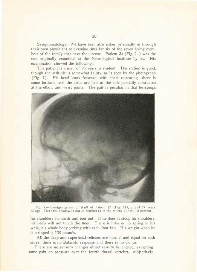
Symptomatology : \N e have been able either personally or through their own physicians to examine thus far six of the seven living mem· bers of the family that have the disease. Patient 24 (Fig. 1 1 ) was the one originally examined at the Neurological Institute by us. His examination showed the following:
The patient is a man of 23 years, a student. The station is good, though the attitude is somewhat faulty, as is seen by the photograph (Fig. 1 ) . His head leans forward, with chest retreating ; there is some lordosis, and the arms are held at the side partially contracted at the elbow and wrist joints. The gait is peculiar in that he stoops
Fig. 9.-Roentgenogram of skull of patient 27 ( l?ig. 11) , a girl 19 years of age. Here the shadow is not so distinct as in the others, but still is present.
his shoulders forwards and toes out. If he doesn't stoop his shoulders, his heels will not touch the floor. There is little or no spring in his walk, his whole body jerking with each foot fall. His weight when he is stripped is 108 pounds.
All the deep and superficial reflexes are normal and equal on both sides; there is no Babinski response and there is no clonus.
There are no sensory changes objectively to be elicited, excepting some pain on pressure over the fourth dorsal vertebra ; subjectively

21
he complains of no pain whatever excepting at times "rheumatic" pains in the joints. This soreness in the joints all the affected members of the family experience. Those unaffected with the dystrophy do not have the joint pains.
There are no signs of involvement of any of the cranial nerves, with exception possibly of those controlling the eye muscles. Every dystrophic member of the family gives a history of eye trouble and
Fig. 10.-Roentgenogram of skull of patient shown in Figures from 1 to 5, inclusive. The shadows in the pineal region are marked (Patient 24, Fig. 11).
must wear glasses. These ocular muscle disturbances are important as referring possibly to the pineal gland in its relation to the anterior corpora quadrigemina. His father, also affected with dystrophy, awoke suddenly one morning, blind, and never entirely recovered his VISion. There is no difficulty in swallowing or in speech, and the tongue is protruded in the middle line.

22
The only external signs of an involvement of the internal glandular system is in the spacing of the incisors, and in the prognathism of the upper jaw. \Ve shall later have occasion to point out the extreme probability of the dependence of the entire syndrome, the bony atrophy, fatty deposits in the muscles, etc., on such a disturbance of the endocrine system.
There is no arteriosclerosis; the pulse rate varies from 68 to 72, increasing on slight muscular effort to 90. The blood pressure, systolic, varies from 125 to 145 ; diastolic, from 75 to 100.
The patient is highly intelligent, a postgraduate student at an important university, fitting himself for the philosophical degree. His auditory memory is good, while his visual memory is very poor. He is fairly frequently depressed, seclusive and shut-in and has a reaction of embarrassment in company. He feels his disability keenly.
1>
r1. .3
r,.. � • • l ' ,. ,, I fl..
p.J 6 • 6 13 2.</ z.r
f-i � •
H '-? �t 1f Jo
"R.o<1-l!£SS111l: M"''"'-'ll.
}it.J IEI> IT'Al\Y l>YST)IO"l'HY. CI!Al!T.
Jt )� ' J
Fig. H.-Heredity chart of the family in which the dystrophy cases occurred, those in black being the individuals affected.
Urine and blood are quite normal in all particulars. The 'Wassermann reaction in the serum is negative.
It is in the muscular system that the chief characteristics of the condition lie. The first striking feature is the almost entire absence of the pectorals of both sides, with the exception of the clavicular strands and a few strands of the pectoralis minor (Fig. 3). Both deltoids are very atrophic, and the intrinsic neck muscles are so much so that the neck seems to fit like a funnel into the aperture made by the clavicles and sternum instead of being attached thereto. The trapezius, the serratus magnus and the rhomboids are only partially atrophied. The biceps and triceps are much atrophied; the long supinators less so, while the muscles of the forearm are only slightly affected. In the leg the quadriceps and the adductors are smaller than normal

23
and quite inefficient, so that walking upstairs or getting up from a squatting position is a task. It is accomplished by climbing up on the thighs by means of the arms (Fig. 2 ) . Of the hypertrophies that are present, the infraspinatus is moderately enlarged, as are the calf muscles. The facial muscles show a slight atrophy, with everted and pouting lips, although there is no difficulty in whistling. The electrical excitability is somewhat increased to faradism in all the muscles, while the galvanic response is unaltered in degree and quality, that is, there is no reaction of degeneration, no myoclonus, or myoidema; there is no asynergy and no incoordination. There are no actual paralyses, but simply weaknesses. The patient can hold his arms out from his body at the level of the shoulder for one and one-half minutes without fatigue. There then ensues a tremor of the entire limb, and he must let the arms fall. The various measurements of the circumference of
the limbs are herewith given in millimeters :
Right Left • {Upper 230 225
Upper arm. . . . . . Middle 190 190 Elbow 193 190
Forearm . . . . . . . . { Largest Smallest
{Upper Thigh . . . . . . . . . . Middle
Knee
L S Calf ower leg· · · · · · · 1 Thinnest
200 125
465 420 320
335 173 '
202 127
465 415 330
325 175
There are contractions in the tendons of the arm muscles and of the calf muscles on both sides-contractions, and not contractures. That is to say, by assuming such positions as will give little tension to the muscles of the particular member examined, the patient recognizes no difficulty in fully extending that limb. Thus, if he relaxes the calf muscles by stooping forward in walking, his heels touch the ground ; but if he stands fully erect with shoulders thrown back, then his heels do not touch the ground on account of the short Achilles tendons. The photograph showing the arm outstretched to its fullest extent gives an idea of the shortening of the arm flexor tendons, in the resulting flexion of the fingers and wrist (Fig. 4). When the, wrist is fully flexed, the fingers can be extended; when 1the fingers are fully extended and kept in that position, the wrist cannot be extended at th� same time. It is these contractions that give the patient his greatest difficulty, for they impede the free use of his limbs and are progressively, although slowly, increasing (Fig. 5).
The Roentgen ray shows that the bones of the arm and forearm are very small and thin, though not shorter than the normal (Fig. 6).

24
The skull roentgenogram shows a normal sella turcica, but there are several shadows in the region of the pineal gland (Fig. 10). These shadows are three in number, and although discrete, range closely together.
Three other members of this immediate family, Patient 14 (Fig. 1 1 ) , father of this man, and Patients 26 and 27, brother and sister of the patient, give similar physical signs and symptoms to those exhibited by this patient, with some minor differences. Thus, his sister, 19 years of age, is quite adipose, and has more marked tendon contractions than the others. She constantly walks on her toes as a result of the Achilles tendon shortening (pes equinus) , and must have special shoes to accommodate this malposition of the foot to her gait. The brother, aged 21, is in many respects the counterpart of the patient, only not quite so far advanced. The father, aged 65, cannot get up from a sitting position without help. There is the same difficulty in all of these members of the family in going upstairs, almost an impossibility to lift themselves from step to step. But aU of them are active mentally. The father still attends to his duties as a minister. The one point that they all have in common, and this we wish to emphasize most strongly, is that the roentgenogram in each instance shows shadows in the region of the pineal gland. In the sister's case these shadows are very faint, but still present (Figs. 7, 8 and 9).
One member, No. 32, of a collateral branch of this family, but still descended from the common ancestor ::--Jo. I , is affected by a type of the disease which is sufficiently distinct from that of the others to merit individual description. He is a schoolboy of 15, living in a small Pennsylvania town. His difficulty seemingly followed an attack of chickenpox four years ago. He has no difficulty in squatting down or in arising from the floor, thereby differing from every other member with the disease. Within six months of the onset of the disease, his joints became stiff and the seat of calcareous deposits. Various tendon contractions followed, notably the Achilles and the flexors of the fingers. Muscular atrophies, especially of the deltoids, pectorals, trapezius, biceps, and sternocleidomastoids succeeded. The pectorals are very deficient, the sternocleido muscles also, almost like a leadpencil in thickness. The entire bodily musculature seems to be deficient in volume and gives the appearance of a big bony frame with insufficient covering. The myotatic irritability is increased in the biceps and deltoids. The bony framework is large, the middle finger measuring 105 mm. from the metacarpophalangeal joint to the tip. Both ankles are deformed, with a marked protrusion of the tarsals inward and a pes planus. The foot cannot be flexed dorsally on account of a short Achilles tendon, or ventrally on account of a short tibialis anticus. This gives the patient a rather queer shuffiing gait. There are cal-

' 25
careous nodules on many of the phalanges, especially at the terminal joints: On account of atrophic supinators, supination can be carried out only to a very limited extent by the biceps assisting. This syndrome is rather different from that of the other members, in that it began at the comparatively late age of 11 and that it was not accompanied by difficulty in arising or in going upstairs and in its marked disposition to affect the bony skeleton. The Roentgen ray also in this case did not show the pineal gland shadows, but did show a large sella turcica. As the boy is but 15, it is within the realm of probability that within a few years the shadows will appear.
As the members of the various branches of the family are widely scattered, indeed from coast to coast, I have not personally been able as yet to examine all those affected with the disease. The roentgenograms were made by different roentgenologists in various parts of the country, including Dr. Leix of Los . \ngeles, Dr. Bowen of Philadelphia and Dr. Evans of �ew York.
To recapitulate, then, we have a somewhat atypical form of progressive muscular dystrophy, rather resembling Erb's infantile type, of extremely benign and slow progress. It occurs as a hereditary affection, now in the fourth generation, which has disabled fourteen individuals. Of the seven living members with the disease, all but two haYe been examined thus far by Roentgen ray, �nd of these five, four show distinct changes in the pineal gland producing shadows in the roentgenogram. The fifth, a youth of 15 years, whose affliction is rather of different type than the others, in that the bony growth is abnormal, shows an enlarged sella turcica, but no shadows in the pineal as yet. \Veighing all the evidence that is advanced by previous investigators to show derangement of the internal glandular balance in cases of progressive muscular dystrophy, and giving due significance especially to the changes produced by tumors and diseases of the pineal gland on the various tissues of the body, changes that resemble in character, if not entirely in degree or disposition those present in progressive muscular dystrophy, we must admit the extreme probability o[ a causal relationship betw·een the two. If to this probability we actually adduce the evidence shown by cases of progressive muscular dystrophy of changes in the pineal gland demonstrated by roentgenology, the probability approaches pretty closely to proof that dist•Jrbances of the pineal gland play an important role in the pathogeny of progressive muscular dystrophy.
In criticism of this deduction, there may be advanced the statement that frequently otherwise normal individuals show shadows in the pineal gland. In answer thereto, Schiiller of Vienna, who was the first to demonstrate the identity of the shadows with deposits in the pineal gland, states that in older persons who already have reached

26
the stage of calcification of the arteries, the shadows are but eYidence of such calcified bodies in the pineal gland. But here we have three of the four patients under 24 years of age. The author personally examined about 150 roentgenograms of the skull in the laboratory of Dr. Lewis G. Cole of Kew York, and found only about 2 per cent. with pineal shadow.
To ask why changes in the ovaries or thyroid or pituitary gland, all of which changes have had their part in the production of symptoms in many of the cases of progressive muscular dystrophy herein cited, should be laid at the door of a diseased pineal gland, brings up the whole question of the interdependence of the endocrine glands. Suffice it to say that the pineal gland is supposed to cease its function at puberty. If earlier or later, then the other glands partially compensate, producing their own particular symptoms in the endeavor to right the wrong. Occasionally this wrong is righted and a cure of the condition produced by the peryerted pineal gland results. Thus have come about the several cures reported by Erb, Marina, Levi and Rothschild, and Jendrassik. In each of these cases a rapidly developing gonadal system incidental to puberty produced the cure.
A final report of all the members of this family will be presented by me within the coming year, together with a short disquisition on the therapy of the condition based on the foregoing theory of the paUlogenesis of progressive muscular dystrophy, namely, that the disease must be classed as an endocrinop?.thy.

Re,hrinled from the .·lrrhi<·es of h1/ernal Medicint ]aumrry, 1916, I 'o/. XIX, pp. i9-104
:\\1 �RICA X �lEOICM. ASSOCIATIO"
Fn·& Iluxouo A:<D Tu!RT\'·I'IvE XoRTII D&ARBOR:< STREET
CHIC.-\ GO