Embed Size (px)
Citation preview
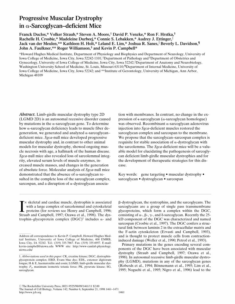
The Rockefeller University Press, 0021-9525/98/09/1461/11 $2.00The Journal of Cell Biology, Volume 142, Number 6, September 21, 1998 1461–1471http://www.jcb.org 1461
Progressive Muscular Dystrophy
in
a
-Sarcoglycan–deficient Mice
Franck Duclos,* Volker Straub,* Steven A. Moore,
‡
David P. Venzke,* Ron F. Hrstka,
§
Rachelle H. Crosbie,* Madeleine Durbeej,* Connie S. Lebakken,* Audrey J. Ettinger,
i
Jack van der Meulen,** Kathleen H. Holt,* Leland E. Lim,* Joshua R. Sanes,
i
Beverly L. Davidson,
¶
John A. Faulkner,** Roger Williamson,
§
and Kevin P. Campbell*
*Howard Hughes Medical Institute, Department of Physiology and Biophysics and Department of Neurology, University of Iowa College of Medicine, Iowa City, Iowa 52242-1101;
‡
Department of Pathology and
§
Department of Obstetrics and
Gynecology, University of Iowa College of Medicine, Iowa City, Iowa 52242;
i
Department of Anatomy and Neurobiology,
Washington University School of Medicine, St. Louis, Missouri 63110;
¶
Department of Internal Medicine, University ofIowa College of Medicine, Iowa City, Iowa 52242; and **Institute of Gerontology, University of Michigan, Ann Arbor,Michigan 48109
Abstract.
Limb-girdle muscular dystrophy type 2D (LGMD 2D) is an autosomal recessive disorder caused
by mutations in the
a
-sarcoglycan gene. To determine how
a
-sarcoglycan deficiency leads to muscle fiber de-generation, we generated and analyzed
a
-sarcoglycan–deficient mice.
Sgca-
null mice developed progressive muscular dystrophy and, in contrast to other animal models for muscular dystrophy, showed ongoing mus-cle necrosis with age, a hallmark of the human disease.
Sgca-
null mice also revealed loss of sarcolemmal integ-rity, elevated serum levels of muscle enzymes, in-creased muscle masses, and changes in the generation of absolute force. Molecular analysis of
Sgca-
null mice demonstrated that the absence of
a
-sarcoglycan re-sulted in the complete loss of the sarcoglycan complex, sarcospan, and a disruption of
a
-dystroglycan associa-
tion with membranes. In contrast, no change in the ex-pression of
e
-sarcoglycan (
a
-sarcoglycan homologue) was observed. Recombinant
a
-sarcoglycan adenovirus injection into
Sgca
-deficient muscles restored the sarcoglycan complex and sarcospan to the membrane. We propose that the sarcoglycan–sarcospan complex is requisite for stable association of
a
-dystroglycan with the sarcolemma. The
Sgca
-deficient mice will be a valu-able model for elucidating the pathogenesis of sarcogly-can deficient limb-girdle muscular dystrophies and for the development of therapeutic strategies for this dis-ease.
Key words: gene targeting • muscular dystrophy • sarcoglycan • dystroglycan • sarcospan
I
N
skeletal and cardiac muscle, dystrophin is associatedwith a large complex of sarcolemmal and cytoskeletalproteins (for reviews see Henry and Campbell, 1996;
Straub and Campbell, 1997; Ozawa et al., 1998). The dys-trophin–glycoprotein complex (DGC)
1
includes
a
- and
b
-dystroglycan, the syntrophins, and the sarcoglycans. Thesarcoglycans are a group of single pass transmembraneglycoproteins, which form a complex within the DGC,consisting of
a
-,
b
-,
g
-, and
d
-sarcoglycan. Recently the 25-kD component of the DGC was characterized and namedsarcospan (Crosbie et al., 1997). The DGC confers a struc-tural link between laminin 2 in the extracellular matrix andthe F-actin cytoskeleton (Ervasti and Campbell, 1993),and is thought to protect muscle cells from contraction-induced damage (Weller et al., 1990; Petrof et al., 1993).
Primary mutations in the genes encoding several com-ponents of the DGC have been associated with musculardystrophy (Straub and Campbell, 1997; Ozawa et al.,1998). In autosomal recessive limb-girdle muscular dystro-phy (LGMD), mutations in any of the sarcoglycan genes(Roberds et al., 1994; Bönnemann et al., 1995; Lim et al.,1995; Noguchi et al., 1995; Nigro et al., 1996) lead to the
Address all correspondence to Kevin P. Campbell, Howard Hughes Med-ical Institute, University of Iowa College of Medicine, 400 EMRB,Iowa City, IA 52242. Tel.: (319) 335-7867. Fax: (319) 335-6957. E-mail:[email protected] WWW site: http://www-camlab.physiology.uiowa.edu/
1.
Abbreviations used in this paper
: CK, creatine kinase; DGC, dystrophin-glycoprotein complex; EBD, Evans blue dye; EDL, extensor digitorumlongus; H & E, haematoxilin and eosin; LGMD, limb-girdle muscular dys-trophy;
P
o
, maximum isometric tetanic force; PK, pyruvate kinase; SG,sarcoglycan.
The Journal of Cell Biology, Volume 142, 1998 1462
concomitant loss or reduction of all four sarcoglycans (
a
,
b
,
g
, and
d
) from the sarcolemma. Thus far, the BIO 14.6hamster has served as an animal model for sarcoglycan-deficient muscular dystrophy (Roberds et al., 1993
b
). Thehamster has a genomic deletion in the
d
-sarcoglycan gene(Nigro et al., 1997; Sakamoto et al., 1997) that results in re-duced sarcoglycan levels in striated muscles (Nigro et al.,1997; Sakamoto et al., 1997). However, mutations in thehuman
d
-sarcoglycan gene (LGMD 2F) seem to be veryrare compared with the prevalence of
a
-sarcoglycan genemutations (LGMD 2D) (Duggan et al., 1997). Further-more, the BIO14.6 hamster reveals a comparatively mildskeletal muscle pathology and develops a hypertrophiccardiomyopathy (Homburger, 1979) generally not seen inpatients with primary sarcoglycan deficiency. Generationof a phenotypically more accurate model of LGMD istherefore critical for developing effective therapeuticstrategies as well as for elucidating the pathogenesis of thedisease.
In the present study, we have developed
Sgca
-null micein order to analyze the biological role of the sarcoglycansin the pathophysiology of LGMD. The null mutant repre-sents the first engineered animal model for autosomal re-cessive muscular dystrophy with a primary sarcoglycangene defect. The mice developed histopathological fea-tures of muscular dystrophy shortly after birth and showedongoing fiber degeneration until nine months of age. Bio-chemical analysis revealed loss of the entire sarcoglycancomplex along with a complete loss of sarcospan. Our dataindicate that sarcospan is an integral component of thesarcoglycan complex. In addition to sarcoglycan/sarcospandeficiency we observed a reduction of all other DGC com-ponents. The disruption of the DGC in
Sgca
-null mutantmice resulted in increased masses of both extensor digi-torum longus (EDL) and soleus muscles and a decrease inspecific force developed by the EDL. We found no alter-ation in the expression level of
e
-sarcoglycan, the recentlyidentified homologue of
a
-sarcoglycan (Ettinger et al.,1997; McNally et al., 1998). Intramuscular injection of a re-combinant
a
-sarcoglycan adenovirus in homozygous mu-tants restored expression of the DGC at the sarcolemma,demonstrating the feasibility of gene transfer for sarco-glycan-deficient LGMD. Our results suggest that the ab-sence of the sarcoglycan–sarcospan complex due to a nullmutation in the
Sgca
gene causes dissociation of the DGCand contributes to progressive muscle degeneration inLGMD 2D.
Materials and Methods
Isolation of
a
-Sarcoglycan and Vector Construction
The
a
-sarcoglycan gene was isolated from a
l
FIXII 129/sv genomic li-brary by homology screening using a radiolabeled human
a
-sarcoglycancDNA. A 16.2-kb NotI fragment was characterized by restriction map-ping and limited sequencing (GenBank/EMBL/DDBJ accession numberAF064081). Two StyI fragments of 0.16 and 0.9 kb carrying exon 1 and aportion of 5
9
and 3
9
contiguous intron were coligated into the XbaI site ofpPNT. Orientation of the two inserted fragments was checked by PCRand sequencing. A 9-kb fragment containing a portion of intron 3, andexon 4–exon 9 was amplified using a high-fidelity PCR reaction (Takara,Shiga, Japan) with intron 3 forward primer from: CCCCTCGAGCCGT-TCCTCAGACTTTTTATTC and exon 9 reverse primer from: AAT-GCGGCCGCTCTCCTGTACGAACAT. The PCR fragment was restric-
tion digested by NotI and XhoI and inserted into a XhoI-NotI cutplasmid. Correct targeting replaced exon 2 (containing part of the signalsequence), exon 3,571 bp of intron 1, and 65 bp of intron 3 with a neomy-cin resistance gene in opposite transcriptional orientation (see Fig. 1).
Generation of Sgca-deficient Mice
The NotI linearized construct was introduced into 2
3
10
7
R
1
ES cells byelectroporation (240 V, 500
m
F; Bio-Rad Gene Pulser; Hercules, CA).The ES cells were maintained on feeder layers and passaged clonally. Tar-geting fidelity was determined by Southern analysis. Cells from three cor-rectly targeted clones were microinjected into C57BL/6J blastocysts andtransferred into pseudopregnant recipients. After germ-line transmission,genotypes were determined by PCR on DNA from tail biopsies (see Fig.1). The following primers and PCR conditions were used: (
a
) INT1 in in-tron 1: CAGGGCTGGGAGCTGGGTTCTG; (
b
) EX2 in intron 3 (de-leted in the null allele): CCCAGGGCCTTGATGCCT; and (
c
) NEOTR:GCTATCAGGACATAGCGTTGGCTA: first denaturation at 94
8
C for5 min, followed by 30 cycles of 1 min at 94
8
C, 1 min at 64
8
C, 2 min 30 s at72
8
C, and 7 min last extension at 72
8
C. All three primers were used in thesame PCR reaction. Wild-type and null alleles corresponded to PCR frag-ments of 1,061 and 618 bp, respectively.
Northern Blot Analysis
Total RNA from control, heterozygous, and homozygous-null mutantskeletal and cardiac tissues was extracted using RNAzol (Tel-Test,Friendswood, TX) according to manufacturer specifications. 30
m
g of totalRNA was run on a 1.25% agarose gel containing 5% formaldehyde andtransferred to Hybond N membrane (Amersham Corp., ArlingtonHeights, IL). RNA was cross-linked to the membrane using a StratageneUV cross-linker (La Jolla, CA). Membranes were then prehybridized andhybridized using standard methods. Washes were carried out at 65
8
C in1
3
SSC/1% SDS initially, then 0.1
3
SSC/0.1% SDS. Blots were ex-posed for autoradiography.
Evans Blue Dye Injection and Microscopic Evaluation
Evans blue dye (EBD) (Sigma Chemical Co., St. Louis, MO) was dis-solved in PBS (10 mg/ml) and sterilized by passage through membrane fil-ters with a pore size of 0.2
m
m. Mice were injected intravenously with 0.25
m
l per 10 g of body weight of the dye solution through the tail vein. Ani-mals were killed 6 h after injection by cervical dislocation. During the timeperiod between injection and cervical dislocation, animals were kept instandard laboratory cages. All mice were skinned and inspected for dyeuptake in the skeletal muscles, indicated by blue coloration. Muscle sec-tions for microscopic Evans blue detection were incubated in ice-cold ace-tone at
2
20
8
C for 10 min, and after a rinse with PBS, sections weremounted with Vectashield mounting medium (Vector Laboratories, Inc.,Burlingame, CA). Sections were observed under a Zeiss Axioplan fluores-cence microscope (Carl Zeiss, Inc., Thornwood, NY) or a MRC-600 laserscanning confocal microscope (Bio-Rad Laboratories, Hercules, CA).
Serum Levels of Muscle Enzymes
Activities of muscle specific pyruvate kinase (PK) isozyme found in theblood serum were measured as previously documented (Edwards andWatts, 1981). Quantitative, kinetic determination of creatine kinase activ-ity in serum of control and
Sgca
-deficient mice was measured using cre-atine kinase (CK) reagent (Sigma Chemical Co.) according to the manu-facturer’s instructions. Blood was collected from the retroorbital sinus of2–18-wk-old mice and the serum was stored at
2
80
8
C before measure-ments.
Antibodies
Monoclonal antibodies IIH6 against
a
-dystroglycan (Ervasti and Camp-bell, 1991) and 8D5 against
b
-dystroglycan (Lim et al., 1995) were previ-ously characterized. mAbs 20A6 against
a
-sarcoglycan, 5B1 against
b
-sarcoglycan, and 21B5 against
g
-sarcoglycan were generated in collabo-ration with L.V.B. Anderson (Newcastle General Hospital, Newcastleupon Tyne, UK). We used a mAb against caveolin-3 (Transduction Labo-ratories, Lexington, KY). Rabbit polyclonal antibodies against
a
-sarco-glycan (Roberds et al., 1993
a
), dystrophin, and utrophin (Ohlendieck etal., 1991
a
), neuronal nitric oxide synthase (Crosbie et al., 1998), the
a
1
subunit of the dihyrdopyridine receptor (Ohlendieck et al., 1991
b
), and

Duclos et al.
a
-Sarcoglycan–deficient Muscular Dystrophy
1463
the laminin
a
2-chain (Allamand et al., 1997) were described previously.Two affinity-purified rabbit antibodies (rabbit 208 and 215) were pro-duced against a full-length COOH-terminal fusion protein of
g
-sarcogly-can, and against an NH
2
-terminal peptide (MMPQEQYTHHRSTMP-GAA) of
d
-sarcoglycan, respectively. An affinity-purified goat antibody(goat 26) was produced against a NH
2
-terminal fusion protein of
b
-sar-coglycan containing amino acids 1–65. Polyclonal antibodies against
a
-dystroglycan fusion protein D were affinity-purified from goat 20(Ibraghimov-Beskrovnaya et al., 1992). An affinity-purified rabbit anti-body (rabbit 235) was produced against a COOH-terminal fusion proteinof sarcospan (CFVMWKHRYQVFYVGVGLRSLMASDGQLPKA). Twopolyclonal antibodies against
e
-sarcoglycan were used. One was previ-ously characterized (Ettinger et al., 1997) and the other (rabbit 232) wasgenerated against a COOH-terminal peptide of
e
-sarcoglycan (PHQT-QIPQQQTTGKWYP).
Immunofluorescence Analysis
For immunofluorescence analysis, 7-
m
m transverse cryosections were pre-pared from control and
Sgca
-null mutant skeletal and cardiac muscle. Allprocedures were performed at room temperature. Sections were blockedwith 5% BSA in PBS for 1 h and then incubated with the primary antibod-ies for 90 min. After washing with 1% BSA/PBS, sections were incubatedwith Cy3-conjugated secondary antibodies (1:250) for 1 h and thenwashed with 1% BSA/PBS. After a rinse with PBS, sections weremounted with Vectashield mounting medium (Vector Laboratories, Inc.)and observed under a Zeiss Axioplan fluorescence microscope (Carl ZeissInc.).
Immunoblot Analysis of Membrane Preparations
KCl-washed membranes from skeletal and cardiac muscle were preparedas described previously (Ohlendieck et al., 1991
b
) with the addition of twoprotease inhibitors, calpeptin and calpain inhibitor I (Calbiochem-Nova-biochem Corp., San Diego, CA). Both inhibitors were used in the buffersat a concentration of 2 nM. Membranes were resolved by SDS-PAGE(Laemmli, 1970) on 3–15% linear gradient gels and transferred to nitro-cellulose membranes (Towbin et al., 1979). Immunoblot staining was per-formed as previously described (Ohlendieck et al., 1991
b
).
Contractile Properties
For the measurement of contractile properties of the EDL or soleus mus-cles of control or
Sgca
-deficient mice in vitro, mice were anesthetized withsodium pentobarbital (30–50 mg/kg for control or
Sgca
-deficient mice, re-spectively) (Nembutal; Abbott Laboratories, Chicago, IL). Contractileproperties were measured on 28 muscles, seven EDL and five soleus mus-cles from heterozygous littermates of
Sgca
-deficient mice and 10 EDL andsix soleus muscles from eight
Sgca
-null mutant mice. Muscles were iso-lated and removed carefully from the anesthetized mice and immersed inan oxygenated (95% O
2
and 5% CO
2
) bath containing a buffered mam-malian Ringer’s solution, pH 7.4, which included curare. The solution wasmaintained at 25
8
C. The tendons were tied securely to a servomotor and aforce transducer. Muscles were stimulated directly by the current flow be-tween two large platinum electrodes (Brooks and Faulkner, 1988). Thevoltage of the stimulator was set to provide maximum twitch force andthe muscle length was set at optimum length for force development. Withthe muscle at optimum length, the frequency of stimulation was increaseduntil force plateaued at maximum isometric tetanic force (
P
o
). After themeasurement of Po, the length of the muscle was set at 90% of the fiberlength and with the muscle passive the muscle was stretched to 110% of fi-ber length at 1 fiber length(s) and then returned at the same velocity to90% of fiber length. The peak force at the end of the stretch was used as ameasure of the resistance to stretch of the passive muscle. The muscle wasremoved from the bath, blotted, and weighed to obtain the muscle mass.Based on the direct measurements of muscle mass, muscle length, fiberlength, and Po, total fiber cross-sectional area and specific Po were calcu-lated. The total fiber cross-sectional area (mm2) was calculated by divid-ing the muscle mass (mg) by the fiber length (mm) and then by 1.06 (g/cm2) to correct for the muscle density (Brooks and Faulkner, 1988). Theforce (kN) was divided by the total fiber cross-sectional area (m2) to ob-tain an estimate of the specific force (kN/m2) of the EDL and soleus mus-cles. Each data set was analyzed by a two-way analysis of variance(ANOVA) in a general linear model algorithm appropriate for unequalsample sizes (Statistical Analysis System, Gary, NC). In circumstanceswhere the overall F-ratio for the ANOVA was significant, the differences
between individual group means were determined by post hoc pairwiset-comparisons of least square means with appropriate correction of thesignificance level to account for multiple comparisons. Significant differ-ences between data on control and Sgca-deficient mice are indicated inFig. 6 by asterisks. Significance was set a priori at P , 0.05.
Recombinant Adenovirus InjectionsThe human a-sarcoglycan cDNA sequence was subcloned into thepAdRSVpA adenovirus vector through standard methods of homologousrecombination with Ad5 backbone dl309 by the University of Iowa GeneTransfer Vector Core. Lysates from the infected cells were collected andtested for the expression of a-sarcoglycan using a polyclonal antibody.Recombinant viruses were plaque purified three times, amplified, andthen concentrated using established methods (Graham and Eb, 1973;Davidson et al., 1994). Recombinant adenovirus injections were per-formed as previously described (Holt et al., 1998). For 2-d-old Sgca-nullmutant mice, the adenovirus was injected directly through the skin intothe hamstring. 3-wk-old mice were anesthetized by intraperitoneal injec-tion of sodium pentobarbital at a calculated dose of 50 mg/kg. A humand-sarcoglycan adenovirus (Holt et al., 1998) was used as control.
Results
Generation of Sgca-null Mutant Mice
To design a targeting vector to generate Sgca-null mice,we cloned the murine homologue of the human SGCAgene from a mouse genomic library. Murine and humana-sarcoglycan are highly related at the amino acid leveland their expression pattern at the mRNA level is similar(Roberds et al., 1994; Liu et al., 1997). The structural orga-nization of the gene into 10 exons is shared by both species(GenBank/EMBL/DDBJ accession number AF064081).
Targeted inactivation of one of the Sgca alleles was ac-complished by replacement of exons 2 and 3 and flankingintronic sequences with the neomycin resistance gene (Fig.1 a). The targeting construct was designed to create a mu-tant allele of Sgca representative of certain human muta-tions. One-third of a-sarcoglycan mutations characterizedto date affect exons 2 and 3 of the SGCA gene (Piccolo etal., 1995; Carrie et al., 1997). A total of 1,023 colonies sur-viving G418 and gancyclovir selection were analyzed bySouthern-blotting for the presence of homologous recom-bination (Fig. 1 b). Two clones yielded chimeras producinggerm-line transmission. Transmission of the mutant allelefollowed normal Mendelian segregation ratios for an auto-somal recessive gene in mice derived from both clones.Homozygous mutant and heterozygous newborn pups ap-peared healthy, showing no gross developmental abnor-malities compared with control littermates.
To determine if the targeting approach produced a nullallele, we evaluated tissues from homozygous mutants andheterozygous mice, and compared them to wild-type mice.Northern blot analysis using a probe against the full-lengthcoding sequence revealed the absence of a-sarcoglycantranscript in Sgca-deficient skeletal and heart tissue fromthe two independently derived lines (Fig. 1 d and data notshown). In contrast to control and heterozygous animals,no a-sarcoglycan expression was detected in the homozy-gous mutant mice (Fig. 1 e). Reverse transcription (RT)-PCR revealed the presence of a minor transcript in skele-tal muscle RNA resulting from the use of cryptic splicesites in the neomycin cassette in homozygous mutants andheterozygous mice. Sequencing of the RT-PCR product

The Journal of Cell Biology, Volume 142, 1998 1464
revealed that this mutant transcript encoded exon 1 and astretch of 516 bp from the inverted neo cassette splicedwith exon 4 of the Sgca gene (data not shown). Unexpect-edly, this aberrant splicing event inserted 172 amino acidsfrom the noncoding strand of the neo cassette, maintainingthe frame with exon 4. Translation of the altered transcriptwould produce a protein lacking the 91 amino acids en-coded by exons 2 and 3, including part of the signal se-quence. Using a COOH-terminal peptide antibody andmAb 20A6 against a-sarcoglycan, this mutant proteincould not be detected in Sgca-deficient skeletal and car-diac tissues by immunoblot or immunofluorescence analy-sis (Fig. 1 e and see Figs. 4 and 5).
Sgca-null Mutant Mice Display a ProgressiveMuscular Dystrophy
Sgca-null mutant mice did not show any overt signs of amyopathy, and were in this respect similar to dystrophindeficient mdx mice. To examine the progression of themuscular dystrophy in these mutant mice, haematoxilinand eosin (H & E)-stained frozen sections of the sural tri-ceps and the diaphragm muscles were evaluated betweenthe ages of 8 d and 9 mo. Pathology characteristic of mus-cular dystrophy was observed in every Sgca-deficientmouse but never in control animals. The earliest changesconsisted of widely scattered clusters of necrotic myocytesor regenerating myocytes with internally placed nuclei(Fig. 2). These clusters increased in both number and sizeas the mice increased in age (Fig. 2). Based on the evalua-tion of 200–1,100 myocytes per muscle, the number ofnonregenerating myocytes with internally placed nuclei
also increased with age. At 8 d, between 1 and 2.5% of thesural triceps and diaphragm myocytes already showed cen-tral nuclei, respectively. These numbers continuously in-creased and at 8–16 wk of age more than 70% and as highas 99% of the Sgca-deficient myocytes contained centralnuclei (Fig. 2). In wild-type mice on the other hand, thenumbers of centrally placed nuclei never exceeded 1%.
In addition to necrosis, regeneration, and central nucle-ation, a broad spectrum of other dystrophic changes wasalso noted in Sgca-deficient muscle (Fig. 2). The mostprominent of these included atrophy, hypertrophy, fibersplitting, and endomysial fibrosis. In some Sgca-deficientmice 8 wk of age or older, dystrophic calcification wasnoted in association with myocyte necrosis (Fig. 2). Fattyinfiltration was present in some of the muscles from 16-wk-old mice. A qualitative comparison of fiber type distri-bution assessed with ATPase staining as well as stainingcharacteristics with NADH and Gomori trichrome stainssuggested no substantial additional differences betweennull mutant and wild type mice at any age (data notshown). The homozygotes from both correctly targetedcell lines demonstrated an identical dystrophic phenotype.
Sarcolemmal Integrity ina-Sarcoglycan–deficient Muscle
To test whether the mutation of the a-sarcoglycan geneleads to damage of the plasma membrane, we intrave-nously injected Sgca-deficient mice with Evans blue dye(EBD), a normally membrane impermeant molecule. Thisdye penetrates into the cytoplasm of fibers with compro-mised sarcolemmal integrity (Matsuda et al., 1995; Straub
Figure 1. Generation of Sgca-null mutantmice. (a) Restriction map of the wild-typeSgca locus (Sgca1), the targeting con-struct, and the targeted locus. A region of902 bp including exons 2 and 3 (E2 andE3) was deleted and replaced by a phos-phoglycerate kinase-neomycin cassette(NEOR). (b) Southern blot analysis. Usingthe probe shown (black box), the targetedlocus contains an EcoR1 fragment of 8.8kb, whereas the intact allele shows a 5.5-kb band: clone 594 is correctly targeted.(c) Genotyping by PCR. Primer sites areshown in a; using primers INT1 and EX2the wild-type allele (1/1) corresponds toa 1,061-bp fragment; using primers INT1and NEOTR, the null allele is 618 bp. (d)Northern blotting. An a-sarcoglycan cDNAprobe reveals the correct sized transcriptin wild type (1/1) and heterozygotes(1/2) from 30 mg total RNA extractedfrom skeletal muscle; homozygous mu-tant tissue shows no a-sarcoglycan tran-script. (e) Western blot analysis. Using anaffinity-purified polyclonal antibody againsta COOH-terminal peptide of a-sarcogly-can, membrane-enriched preparations ofskeletal muscle reveal the protein in (1/1),and (1/2), but not in (2/2).
Duclos et al. a-Sarcoglycan–deficient Muscular Dystrophy 1465
et al., 1997). No obvious uptake of the blue tracer intoskeletal muscles of heterozygous and control mice was de-tected by macroscopic inspection. In contrast, EBD up-take was consistently observed in skeletal muscle fibers of4–20-wk-old homozygous-null mutants. The extent ofEBD accumulation varied among muscles. Areas of bluestaining appeared mainly within the proximal limb mus-cles and the muscles of the pelvic and the shoulder girdle.Skeletal muscles which macroscopically showed dye up-take always revealed red EBD autofluorescence by fluo-rescence microscopy analysis (Fig. 3 a). Most EBD-posi-tive fibers showed intense staining, whereas the signal wasfaint in others. Interestingly, EBD-positive fibers were dis-tributed in clusters throughout the different muscles (Fig.3 a). Fibers that had taken up the tracer and were assumedto have pathologic plasma membrane permeability oftenshowed characteristic features of degeneration and necro-sis by H & E staining. In contrast, dye uptake was notreadily visible in cardiac muscle by macroscopic inspec-tion, and microscopic analysis revealed no EBD in cardi-omyocytes of control or diseased animals.
We also evaluated membrane damage in Sgca-deficientmice by determining the release of muscle enzymes intothe circulating blood. Therefore we measured muscle-spe-cific serum PK (Fig. 3 b) activity and serum CK activity. In7–10-wk-old wild-type and heterozygous mice we foundnormal serum levels of PK activity. Age-matched homozy-
gous mice, on the other hand, exhibited high serum levelsof PK activity similar to that of mdx mice, indicating thatmembrane damage occurred to comparable extents inSgca-null mutants as in mdx mice (Fig. 3 b). Similarly, inolder animals (up to 18 wk of age) we found no differencesin PK activity between Sgca-null mice and age-matchedmdx mice. The serum CK activity was z10 times higher inSgca-deficient mice compared with control animals (datanot shown).
Loss of Sarcoglycan and Sarcospan Expression inSgca-null Mutant Mice
Immunofluorescence analysis was performed for eachcomponent of the sarcoglycan complex. In Sgca-deficientmice, a-sarcoglycan protein was absent from the sarco-lemma of skeletal and cardiac muscle fibers. In addition,there was a concomitant drastic reduction of b-, g-, andd-sarcoglycan (Fig. 4, a and b). Other components of theDGC were also examined by immunofluorescence micros-copy. The laminin-a2 chain and b-dystroglycan werepresent at comparable levels with control muscle (Fig. 4, aand b). We observed a slight reduction in the a-dystrogly-can staining in Sgca-deficient muscle compared with con-trol muscle. However, the sarcolemmal staining for dystro-phin was consistently patchy and reduced in Sgca-nullmutant skeletal muscle, although dystrophin staining in
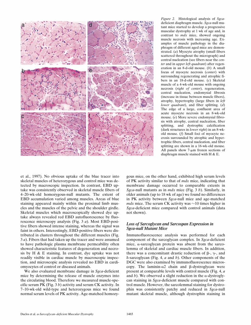
Figure 2. Histological analysis of Sgca-deficient diaphragm muscle. Sgca-null mu-tant mice started to develop a progressivemuscular dystrophy at 1 wk of age and, incontrast to mdx mice, showed ongoingmuscle necrosis with increasing age. Ex-amples of muscle pathology in the dia-phragm of different aged mice are demon-strated. (a) Myocyte atrophy (small fibersscattered throughout the micrograph) andcentral nucleation (see fibers near the cen-ter and in upper left quadrant) after regen-eration in an 8-d-old mouse. (b) A smallfocus of myocyte necrosis (center) withsurrounding regenerating and atrophic fi-bers in an 18-d-old mouse. (c) Skeletalmuscle of a 4-wk-old mouse with ongoingnecrosis (right of center), regeneration,central nucleation, endomysial fibrosis(increase in tissue between muscle fibers),atrophy, hypertrophy (large fibers in leftlower quadrant), and fiber splitting. (d)The edge of a large, confluent area ofacute myocyte necrosis in an 8-wk-oldmouse. (e) More severe endomysial fibro-sis with atrophy, central nucleation, fibersplitting, and dystrophic calcification(dark structures in lower right) in an 8-wk-old mouse. (f) Small foci of myocyte ne-crosis surrounded by atrophic and hyper-trophic fibers, central nucleation, and fibersplitting are shown in a 16-wk-old mouse.All panels show 7-mm frozen sections ofdiaphragm muscle stained with H & E.

The Journal of Cell Biology, Volume 142, 1998 1466
heart appeared similar to control (Fig. 4, a and b). We alsoanalyzed expression of the newly identified e-sarcoglycan(Ettinger et al., 1997) and the 25-kD DGC componentsarcospan (Crosbie et al., 1997). In contrast to theother sarcoglycans, skeletal and cardiac muscle stainingfor e-sarcoglycan in Sgca-deficient mice was comparableto control levels. Interestingly, sarcospan was absent alongwith the sarcoglycans at the sarcolemma of homozygous-null mice (Fig. 4, a and b), indicating that sarcospan maybe an integral component of the sarcoglycan complex.
Dissociation of the DGC in Sgca-null Mutant Mice
To further examine the expression of DGC components,immunoblot analysis was performed on isolated mem-brane preparations from control and homozygous mutantskeletal muscle. We observed that Sgca-deficient musclepreparations were more susceptible to proteolytic degra-dation. Use of calpain inhibitor I and calpeptin reduced
degradation in the membrane preparations from Sgca-nullmice. The ryanodine receptor and dystrophin, for exam-ple, were almost completely degraded in skeletal musclemembrane preparation without the use of calpain inhibi-tor I and calpeptin. Coomassie blue staining (data notshown) and staining for caveolin-3, the a1 subunit of thedihydropyridine receptor, and the ryanodine receptor(data not shown) showed that equivalent levels of mem-brane protein were present in control and homozygousmutant preparations (Fig. 5). a-Sarcoglycan was not de-tected in skeletal and cardiac membrane preparationsfrom Sgca-null mice (refer to Fig. 1 e, Fig. 5, and data notshown). Heterozygotes expressed control levels of a-sar-coglycan (Fig. 1 e). b-, g-, and d-sarcoglycan were greatlyreduced in a-sarcoglycan–deficient skeletal muscle mem-branes compared with control muscle (Fig. 5). Dystrophinwas slightly reduced in accordance with the patchy stain-ing observed by immunofluorescence. Together with thedystrophin reduction we also found reduced levels of thefree radical-producing enzyme neuronal nitric oxide syn-thase, which is anchored to the sarcolemma by dystrophin(Brenman et al., 1995). Utrophin, the autosomal homo-logue of dystrophin, was found at higher levels in mem-brane-enriched preparations from homozygous mutantmice compared with control mice. This observation couldbe related to the large number of regenerating fibers inSgca-deficient muscle, which would be expected to expresshigher levels of utrophin (Helliwell et al., 1992). In addi-tion, a- and b-dystroglycan were reduced in membranepreparations of dystrophic mice compared with controlmice (Fig. 5), whereas immunofluorescence showed onlylittle dystroglycan reduction at the sarcolemma (Fig. 4). Inthe supernatant from Sgca-deficient membrane prepara-tions, a-dystroglycan was enriched and fully glycosylatedbut was not tightly associated with membranes (data notshown). Thus, a-dystroglycan is synthesized correctly butis not stably anchored to the sarcolemma in the absence ofthe sarcoglycan complex. In addition, Western blot analysisconfirmed the immunofluorescence analysis in showing thatdystrophin was no longer tightly held at the skeletal plasmamembrane in the absence of the sarcoglycan complex.
Abnormal Contractile Properties ofSgca-deficient Muscles
The body masses of 8-wk-old Sgca-null mutant mice (34 61 g) were not significantly different from their heterozy-gous littermates (31 6 1 g) (Fig. 6). For the EDL musclesof the Sgca-deficient mice compared with heterozygouslittermates, the masses were 40% greater, whereas the ab-solute forces were not significantly different. As a con-sequence, the specific Po of the EDL muscles of Sgca-defi-cient mice was 31% lower than that of the control mice.The resistance to stretch of the passive muscle in homozy-gous null mutants was 75% greater than the values forcontrol animals. In contrast to the changes observed in theEDL muscles, the soleus muscles in the Sgca-null mutantmice responded in a completely different manner. Com-pared with soleus muscles in heterozygous littermates,those in the Sgca-null mutant mice had a 62% greatermass, a 39% greater absolute Po, and no significant differ-ence in either specific Po or resistance to stretch.
Figure 3. Evaluation of sarcolemma permeability. (a) Heterozy-gous (1/2) and homozygous-null (2/2) mice were intravenouslyinjected with EBD. The panels show dye uptake into muscle fi-bers of the femoral quadriceps and diaphragm muscles 6 h afterinjection. Dye accumulation was only detected in skeletal musclefrom Sgca-null mutants. Activity of muscle-specific PK in 7–10-wk-old wild-type (1/1), heterozygotes (1/2), homozygotes (2/2),and mdx mice (b). Measurement of PK released from the musclefiber into the circulating blood showed similar high levels of PKactivity in (2/2) and mdx mice compared with (1/2) and control(1/1). Error bars indicate the standard deviation where n equalsthe number of mice in each set. Bar, 50 mm.

Duclos et al. a-Sarcoglycan–deficient Muscular Dystrophy 1467
Restoration of the Sarcoglycan–Sarcospan Complexby Gene Transfer
We used an adenovirus construct encoding human a-sar-coglycan to test the ability of exogenously provided a-sar-coglycan cDNA to restore the sarcoglycan–sarcospancomplex in Sgca-deficient skeletal muscle. To circumvent
a possible immune response against the neoantigen or ad-enovirus itself, the a-sarcoglycan adenovirus was injectedinto the hamstring muscles of 2-d-old Sgca-deficient pups.We found high levels of a-sarcoglycan expression at thesarcolemma (Fig. 7) over a time interval between 5 d and 2mo after injection. In addition, a-sarcoglycan–positive fi-
Figure 4. Immunofluores-cence analysis of sarcolemmaproteins in Sgca-deficientskeletal and cardiac muscle.Skeletal (a) and cardiac (b)muscle cryosections fromwild-type (1/1) and Sgca-null (2/2) mice were stainedwith antibodies against dys-trophin (DYS), a-dystrogly-can (a-DG), b-dystroglycan(b-DG), laminin a2-chain,(lama2), a-, b-, g-, d-, ande-sarcoglycan (SG), and sar-cospan (SPN). The sarco-glycans and sarcospan weredrastically reduced in theSgca-deficient muscle whereasthe a-sarcoglycan homologuee-sarcoglycan was present incomparable amount to con-trol. Dystrophin staining isreduced in Sgca-null mutantmice from the sarcolemma ofskeletal muscle, whereas it ismaintained at equal levels tocontrol in cardiomyocytes.Bar, 50 mm.

The Journal of Cell Biology, Volume 142, 1998 1468
bers revealed expression of the entire sarcoglycan com-plex, sarcospan and dystrophin by immunofluorescencemicroscopy (Fig. 7). Immunostaining of the sarcoglycanswas found in up to 70% of fibers. Interestingly, central nu-cleation was reduced in Sgca-null mutant muscle after in-jection of the a-sarcoglycan adenovirus (data not shown).
An adenovirus construct encoding human d-sarcoglycanwas used as a control for the gene transfer studies. Ina-sarcoglycan–deficient muscles injected with the d-sar-coglycan adenovirus, no reconstitution of the complex wasdetected.
DiscussionProgressive muscle weakness initially in pelvic and shoul-der girdle muscles is a hallmark of patients with a primarysarcoglycan gene defect. Mutations in the a-sarcoglycangene have been well documented and are associated withthe deficiency of the entire sarcoglycan complex from thesarcolemma (Roberds et al., 1994; Hayashi et al., 1995;Passos-Bueno et al., 1995; Piccolo et al., 1995; Duggan etal., 1996). To ascertain how the sarcoglycan defect causesmuscular dystrophy, we evaluated structural and func-tional characteristics of skeletal and cardiac muscle inSgca-null mutant mice.
In contrast to the other sarcoglycans, expression ofa-sarcoglycan is specifically restricted to striated muscle
fibers (Roberds et al., 1993a; Ettinger et al., 1997; Eymardet al., 1997; Liu et al., 1997). During development, the ex-pression of a-sarcoglycan is coincident with primary myo-genesis (Yuan et al., 1990; Liu et al., 1997). In particular,these studies indicated that at 17 d of gestation in rabbit,a-sarcoglycan was already present in all myotubes, whereit was confined to the cell periphery (Jorgensen et al.,1990; Yuan et al., 1990). Similarly, it has been reportedthat a-sarcoglycan in mice was not detected before the on-set of myogenesis, whereas it was expressed at the sarco-lemma of newly formed fibers at day E14 (Liu et al., 1997).
Targeted disruption of the a-sarcoglycan gene in ho-mozygous mutants resulted in the absence of normal tran-script and protein. Analysis of skeletal muscle histologydemonstrated that Sgca-deficient mice presented with aprogressive muscular dystrophy similar to human sarco-glycan deficient LGMD. The histological changes appearedas early as 1 wk of age and extended to most skeletal mus-cles as evidenced by EBD incorporation and elevatedserum PK levels. The presence of extensive central nucle-ation, connective tissue proliferation, increased variabilityof muscle fiber diameter, and necrotic fibers was docu-mented during the entire course of the disease studied todate. One striking observation was the persistence of de-generation and regeneration with extensive areas of ne-crosis in a-sarcoglycan–deficient muscle. The early onsetand the severity and persistence of the pathology distin-guished the Sgca-null mutant mice from mdx mice, inwhich these features decline after a regeneration peak at3–4 wk of age (McArdle et al., 1995). Dystrophic pathol-ogy in the Sgca-null mutant mice is more similar to whathas been documented in mdx/utrn2/2 double-knockoutmice (Deconinck et al., 1997; Grady et al., 1997). The com-plete absence of sarcospan in addition to the loss of thesarcoglycan complex may be one reason for the severity ofthe disease. The complete lack of sarcospan expression inSgca-null mice suggests that sarcospan is an integral com-
Figure 5. Immunoblot analysis of skeletal muscle membranes.Skeletal muscle microsomes from control (1/1) and Sgca-defi-cient (2/2) mice were analyzed by 3–15% SDS-PAGE and im-munoblotting using antibodies against several DGC components.In particular we used antibodies against the sarcoglycans (a-, b-,g-, d-, and e-SG), dystroglycans (a- and b-DG), and dystrophin(DYS). In addition we stained blots with antibodies against neu-ronal nitric oxide synthase (NOS), which has been shown to beassociated with dystrophin, and the dystrophin homologue utro-phin (UTR). To demonstrate equal loading of protein samples weused the a1 subunit of the dihydropyridine receptor (a1-DHPR)and caveolin-3 (CAV-3) as positive markers.
Figure 6. Abnormal contractile properties of Sgca-deficientmuscle. The data on EDL and soleus muscles of the Sgca-nullmutant mice are represented as a percentage of the values formuscles of control mice. The diagram shows bar graphs for thedata on body mass, muscle mass, absolute maximum isometric te-tanic force, specific force, and peak force during resistance tostretch of passive muscles. Asterisk, significant differences be-tween the data obtained in Sgca-deficient and control mice. Alldata are presented as the mean 6 one SEM.

Duclos et al. a-Sarcoglycan–deficient Muscular Dystrophy 1469
ponent of the sarcoglycan complex. Correct assembly andproper transportation of the sarcoglycan–sarcospan com-plex to the plasma membrane seems to be prevented bythe null mutation in the Sgca-gene. Investigation of a-sarco-glycan–deficient hearts revealed no gross morphologicalchanges in animals up to 8 mo of age, which is in contrastto what has been observed for the sarcoglycan-deficientBIO 14.6 hamster, which starts to develop hypertrophiccardiomyopathy between 30 and 60 d of age (Homburger,1979). It will be of interest to observe if cardiac pathologymanifests as mice age.
Immunoblot analysis of membrane-enriched prepara-tions demonstrated that a-dystroglycan binding to the sar-colemma was greatly destabilized by loss of the sarcogly-can–sarcospan complex. Our findings provide new clueson the molecular interactions between the components ofthe DGC. On the one hand, reduction of dystrophin re-moved a major F-actin binding site from the sarcolemma,whereas on the other hand, unbound a-dystroglycan elimi-nated the major sarcolemmal laminin-binding site. Freea-dystroglycan could be detected in the supernatant ofmembrane preparations confirming that it is synthesized,as observed by immunofluorescence, and glycosylated.This result suggests that the sarcoglycan–sarcospan com-plex anchors a-dystroglycan to the sarcolemma. Further-more, a direct or mediated interaction of the sarcoglycan–sarcospan complex with dystrophin is implicated by ourfindings. The interaction of dystrophin with b-dystrogly-can does not seem to be sufficient enough to maintain asolid anchorage of dystrophin to the sarcolemma. The de-crease of dystrophin is in accordance with reduced levelsof neuronal nitric oxide synthase, which has been reportedto bind to dystrophin (Brenman et al., 1995). Reduced dys-trophin expression has also been reported in patients withsarcoglycan-deficient LGMD (Vainzof et al., 1996).
During membrane preparations, we found that isolatedmuscle membranes of Sgca-null mice were more degradedthan control membranes. The degradation process couldbe prevented by the use of calcium-activated protease in-hibitors, calpeptin, and calpain inhibitor I. Our results im-
plicate that compared with control muscle, sarcoglycan-deficient muscle tissue contains an increased amount ofproteases, which may be activated during the membranepreparation. Several studies provided indirect evidenceconsistent with a role for calpains in DMD and mdx pa-thology (Kumamoto et al., 1995; Spencer et al., 1995).
The structural destabilization of the sarcolemma by lossof the entire sarcoglycan complex and sarcospan resultedin dramatically different adaptations in the EDL and so-leus muscles. Both muscles showed significant hypertro-phy in response to the a-sarcoglycan deficiency. Hypertro-phy of skeletal muscles has also been reported in the limbmuscles of mdx mice (Faulkner et al., 1997), but not of themagnitude observed here. The hypertrophy was function-ally less effective in the EDL muscles of the Sgca-null mu-tant mice in which the absolute force was unchanged andspecific force decreased compared with control EDL mus-cles. In contrast, the greater hypertrophy in the weight-bearing soleus muscles produced a considerable gain inabsolute force and a value for specific force equivalent tothat of soleus muscles in heterozygous litter mates. Themechanisms underlying these major adaptive responses tothe a-sarcoglycan deficiency will require further investiga-tion.
In mdx mice, pathology is thought to be attenuated be-cause dystrophin is partially replaced by its autosomal ho-mologue utrophin (Deconinck et al., 1997; Grady et al.,1997). To test whether lack of a-sarcoglycan was compen-sated by a protein homologue, we assessed the expressionof e-sarcoglycan in Sgca-null mutant animals. e-sarcogly-can shares 44% identity with a-sarcoglycan at the aminoacids level (Ettinger et al., 1997), but is only weakly ex-pressed at the sarcolemma (Ettinger et al., 1997). In theSgca-deficient mice, e-sarcoglycan appeared to be nor-mally expressed at the sarcolemma and in the capillaries ofskeletal and cardiac tissue. Thus, e-SG does not seem to bean additional member of the known tetrameric complex ofa-, b-, g-, and d-sarcoglycan in skeletal muscle. Neverthe-less, the presence of e-SG may suggest that it could be partof a distinct complex at the sarcolemma.
Figure 7. Restoration of sarcoglycan com-plex after adenovirus injection. Recombi-nant a-sarcoglycan adenovirus mediatesrestoration of DGC components to thesarcolemma. 2-d-old Sgca-null mutantpups were injected in their hamstring mus-cle with a recombinant a-sarcoglycan ade-novirus containing the human a-sarco-glycan coding sequence under the controlof a viral RSV promoter. Serial transversecryosections of injected muscle after 3 wk,were stained with antibodies against a-sar-coglycan (a), b-sarcoglycan (b), g-sarco-glycan (c), and d-sarcoglycan (d), dystro-phin (e), and sarcospan (f). Bar, 50 mm.

The Journal of Cell Biology, Volume 142, 1998 1470
Intramuscular injection of recombinant adenovirus wasperformed to test the potential of a-sarcoglycan to restorethe sarcoglycan–sarcospan complex in the Sgca-deficientmice. Our results demonstrated that in fibers which harboredthe a-sarcoglycan recombinant adenovirus, complete as-sembly and restoration of the sarcoglycan–sarcospan com-plex to the sarcolemma has occurred. Of particular note,high efficiency gene transfer was achieved when the a-sarco-glycan adenovirus was injected at an early stage of life,preceding the onset of muscle damage and establishmentof immunity. These experiments have broad implicationsfor the development of gene therapy for LGMDs with aprimary sarcoglycan deficiency. They confirm the feasibil-ity of these procedures which are facilitated by the smallsize of sarcoglycan coding sequences compared with dys-trophin. They also suggest that a genetic interventionshould be performed as early as possible to circumventboth the extension of the dystrophic process and the im-mune response. The Sgca-deficient mice will be a valuablemodel for the development of therapeutic strategies forsarcoglycan deficient LGMD.
Overall, our results suggest that the absence of thesarcoglycans and sarcospan due to a null mutation in theSgca gene causes dissociation of the DGC and contributesto progressive muscle degeneration in LGMD 2D. Wepropose that the sarcoglycan–sarcospan complex is requi-site for the stable association of a-dystroglycan with thesarcolemma.
We thank J.C. Lee, R.D. Anderson, B. Squires, and D. Hunt (all fourfrom University of Iowa, Iowa City, IA) for expert technical assistance.We thank H. Yamada (University of Iowa) for providing helpful data ona-dystroglycan. All DNA sequencing was carried out at the University ofIowa DNA core facility (NIH DK25295). Data on contractile propertieswere obtained with the help of the Mechanotransduction Core of theNathan Shock Center (University of Michigan, Ann Arbor, MI) P30-AG13283.
V. Straub was supported by the Deutsche Forschungsgemeinschaft (Str498/1-1). R.H. Crosbie is supported by the Robert G. Sampson postdoc-toral research fellowship from the Muscular Dystrophy Association. M.Durbeej was supported by The Swedish Foundation for International Co-operation in Research and Higher Education (STINT). C.S. Lebakkenwas supported by the Iowa Cardiovascular Interdisciplinary Research Fel-lowship (HL07121). K.H. Holt was supported by the University of IowaDiabetes and Endocrinology Research Center (DK07018). A.J. Ettingerand J.R. Sanes were supported by the National Institutes of Health(R01NS19195). This work was also supported by the Muscular DystrophyAssociation. K.P. Campbell is an investigator of the Howard HughesMedical Institute.
Received for publication 1 July 1998 and in revised form 20 August 1998.
References
Allamand, V., Y. Sunada, M.A. Salih, V. Straub, C.O. Ozo, M.H. Al-Turaiki,M. Akbar, T. Kolo, H. Colognato, X. Zhang, L.M. Sorokin, P.D. Yurchenco,K. Tryggvason, and K.P. Campbell. 1997. Mild congenital muscular dystro-phy in two patients with an internally deleted laminin alpha2-chain. Hum.Mol. Genet. 6:747–752.
Bönnemann, C.G., R. Modi, S. Noguchi, Y. Mizuno, M. Yoshida, E. Gussoni,E.M. McNally, D.J. Duggan, C. Angelini, et al. 1995. Beta-sarcoglycan(A3b) mutations cause autosomal recessive muscular dystrophy with loss ofthe sarcoglycan complex. Nat. Genet. 11:266–273.
Brenman, J.E., D.S. Chao, H. Xia, K. Aldape, and D.S. Bredt. 1995. Nitric ox-ide synthase complexed with dystrophin and absent from skeletal musclesarcolemma in Duchenne muscular dystrophy. Cell. 82:743–752.
Brooks, S.V., and J.A. Faulkner 1988. Contractile properties of skeletal mus-cles from young, adult and aged mice. J. Physiol. (Lond.). 404:71–82.
Carrie, A., F. Piccolo, F. Leturcq, C. de Toma, K. Azibi, C. Beldjord, J.M. Val-
lat, L. Merlini, T. Voit, C. Sewry, J.A. Urtizberea, N. Romero, F.M. Tome,M. Fardeau, Y. Sunada, K.P. Campbell, J.C. Kaplan, and M. Jeanpierre.1997. Mutational diversity and hot spots in the alpha-sarcoglycan gene in au-tosomal recessive muscular dystrophy (LGMD2D). J. Med. Genet. 34:470–475.
Crosbie, R.H., J. Heighway, D.P. Venzke, J.C. Lee, and K.P. Campbell. 1997.Sarcospan, the 25-kDa transmembrane component of the dystrophin-glyco-protein complex. J. Biol. Chem. 272:31221–31224.
Crosbie, R.H., V. Straub, H.Y. Yun, J.C. Lee, J.A. Rafael, J.S. Chamberlain,V.L. Dawson, T.M. Dawson, and K.P. Campbell. 1998. mdx muscle pathol-ogy is independent of nNOS perturbation. Hum. Mol. Genet. 7:823–829.
Davidson, B.L., S.E. Doran, D.S. Shewach, J.M. Latta, J.W. Hartman, and B.J.Roessler. 1994. Expression of Escherichia coli beta-galactosidase and ratHPRT in the CNS of Macaca mulatta following adenoviral mediated genetransfer. Exp. Neurol. 125:258–267.
Deconinck, A.E., J.A. Rafael, J.A. Skinner, S.C. Brown, A.C. Potter, L. Metz-inger, D.J. Watt, J.G. Dickson, J.M. Tinsley, and K.E. Davies. 1997. Utro-phin-dystrophin-deficient mice as a model for Duchenne muscular dystro-phy. Cell. 90:717–727.
Duggan, D.J., M. Fanin, E. Pegoraro, C. Angelini, and E.P. Hoffman. 1996. al-pha-Sarcoglycan (adhalin) deficiency: complete deficiency patients are 5%of childhood-onset dystrophin-normal muscular dystrophy and most partialdeficiency patients do not have gene mutations. J. Neurol. Sci. 140:30–39.
Duggan, D.J., J.R. Gorospe, M. Fanin, E.P. Hoffman, and C. Angelini. 1997.Mutations in the sarcoglycan genes in patients with myopathy. N. Engl. J.Med. 336:618–624.
Edwards, R.J., and D.C. Watts. 1981. Specific spectrophotometric assay for theM isoenzymes of pyruvate kinase in plasma samples containing mixtures ofthe muscle (M) and liver (L) isoenzymes. Clin. Chem. 27:906–909.
Ervasti, J.M., and K.P. Campbell. 1991. Membrane organization of the dystro-phin-glycoprotein complex. Cell. 66:1121–1131.
Ervasti, J.M., and K.P. Campbell. 1993. A role for the dystrophin–glycoproteincomplex as a transmembrane linker between laminin and actin. J. Cell Biol.122:809–823.
Ettinger, A.J., G. Feng, and J.R. Sanes. 1997. Epsilon-sarcoglycan, a broadlyexpressed homologue of the gene mutated in limb-girdle muscular dystrophy2d. J. Biol. Chem. 272:32534–32538.
Eymard, B., N.B. Romero, F. Leturcq, F. Piccolo, A. Carrie, M. Jeanpierre, H.Collin, N. Deburgrave, K. Azibi, M. Chaouch, L. Merlini, C. Themar-Noel, I.Penisson, M. Mayer, O. Tanguy, K.P. Campbell, J.C. Kaplan, F.M. Tome,and M. Fardeau. 1997. Primary adhalinopathy (alpha-sarcoglycanopathy):clinical, pathologic, and genetic correlation in 20 patients with autosomal re-cessive muscular dystrophy. Neurology. 48:1227–1234.
Faulkner, J.A., S.V. Brooks, R.G. Dennis, and G.S. Lynch. 1997. The functionalstatus of dystrophic muscles and functional recovery by skeletal muscles fol-lowing myoblast transfer. Basic Appl. Myol. 7:257-264.
Grady, R.M., H. Teng, M.C. Nichol, J.C. Cunningham, R.S. Wilkinson, and J.R.Sanes. 1997. Skeletal and cardiac myopathies in mice lacking utrophin anddystrophin: a model for Duchenne muscular dystrophy. Cell. 90:729–738.
Graham, F.L., and A.J. v.d. Eb. 1973. Transformation of rat cells by DNA ofhuman adenovirus 5. Virology. 54:536–539.
Hayashi, Y.K., Y. Mizuno, M. Yoshida, I. Nonaka, E. Ozawa, and K. Arahata.1995. The frequency of patients with 50-kd dystrophin-associated glycopro-tein (50DAG or adhalin) deficiency in a muscular dystrophy patient popula-tion in Japan: immunocytochemical analysis of 50DAG, 43DAG, dystro-phin, and utrophin. Neurology. 45:551–554.
Helliwell, T.R., N.T. Man, G.E. Morris, and K.E. Davies. 1992. The dystrophin-related protein, utrophin, is expressed on the sarcolemma of regeneratinghuman skeletal muscle fibres in dystrophies and inflammatory myopathies.Neuromuscul. Disord. 2:177–184.
Henry, M.D., and K.P. Campbell. 1996. Dystroglycan: an extracellular matrixreceptor linked to the cytoskeleton. Curr. Opin. Cell Biol. 8:625–631.
Holt, K.H., L.E. Lim, V. Straub, D.P. Venzke, F. Duclos, R.D. Anderson, B.L.Davidson, and K.P. Campbell. 1998. Functional rescue of the sarcoglycancomplex in the BIO 14.6 hamster using d-sarcoglycan gene transfer. Mol.Cell. 1:841–848.
Homburger, F. 1979. Myopathy of hamster dystrophy: history and morphologicaspects. Ann. NY. Acad. Sci. 317:1–17.
Ibraghimov-Beskrovnaya, O., J.M. Ervasti, C.J. Leveille, C.A. Slaughter, S.W.Sernett, and K.P. Campbell. 1992. Primary structure of dystrophin-associ-ated glycoproteins linking dystrophin to the extracellular matrix. Nature.355:696–702.
Jorgensen, A.O., W. Arnold, A.C.-Y. Shen, S. Yuan, M. Gaver, and K.P.Campbell. 1990 Identification of novel proteins unique to either transversetubules (TS28) or the sarcolemma (SL50) in rabbit skeletal muscle. J. CellBiol. 110:1173–1185.
Kumamoto, T., H. Ueyama, S. Watanabe, K. Yoshioka, T. Miike, D.E. Goll, M.Ando, and T. Tsuda. 1995. Immunohistochemical study of calpain and its en-dogenous inhibitor in the skeletal muscle of muscular dystrophy. Acta Neu-ropathol. (Berl.). 89:399–403.
Laemmli, U.K. 1970. Cleavage of structural proteins during the assembly of thehead of bacteriophage T4. Nature. 227:680–685.
Lim, L.E., F. Duclos, O. Broux, N. Bourg, Y. Sunada, V. Allamand, J. Meyer, I.Richard, C. Moomaw, C. Slaughter, et al. 1995. Beta-sarcoglycan: character-ization and role in limb-girdle muscular dystrophy linked to 4q12. Nat.

Duclos et al. a-Sarcoglycan–deficient Muscular Dystrophy 1471
Genet. 11:257–265.Liu, L., P.H. Vachon, W. Kuang, H. Xu, U.M. Wewer, P. Kylsten, and E. Eng-
vall. 1997. Mouse adhalin: primary structure and expression during latestages of muscle differentiation in vitro. Biochem. Biophys. Res. Commun.235:227–235.
Matsuda, R., A. Nishikawa, and H. Tanaka. 1995. Visualization of dystrophicmuscle fibers in mdx mouse by vital staining with Evans blue: evidence ofapoptosis in dystrophin-deficient muscle. J. Biochem. 118:959–964.
McArdle, A., R.H. Edwards, and M.J. Jackson. 1995. How does dystrophin de-ficiency lead to muscle degeneration? Evidence from the mdx mouse. Neu-romuscul. Disord. 5:445–456.
McNally, E.M., C.T. Ly, and L.M. Kunkel. 1998. Human epsilon-sarcoglycan ishighly related to alpha-sarcoglycan (adhalin), the limb girdle muscular dys-trophy 2D gene. FEBS (Fed. Eur. Biochem. Soc.) Lett. 422:27–32.
Nigro, V., E. de Sa Moreira, G. Piluso, M. Vainzof, A. Belsito, L. Politano,A.A. Puca, M.R. Passos-Bueno, and M. Zatz. 1996. Autosomal recessivelimb-girdle muscular dystrophy, LGMD2F, is caused by a mutation in thedelta-sarcoglycan gene. Nat. Genet. 14:195–198.
Nigro, V., Y. Okazaki, A. Belsito, G. Piluso, Y. Matsuda, L. Politano, G. Nigro,C. Ventura, C. Abbondanza, A.M. Molinari, et al. 1997. Identification of theSyrian hamster cardiomyopathy gene. Hum. Mol. Genet. 6:601–607.
Noguchi, S., E.M. McNally, K. Ben Othmane, Y. Hagiwara, Y. Mizuno, M.Yoshida, H. Yamamoto, C.G. Bönnemann, E. Gussoni, P.H. Denton, et al.1995. Mutations in the dystrophin-associated protein gamma-sarcoglycan inchromosome 13 muscular dystrophy. Science. 270:819–822.
Ohlendieck, K., J.M. Ervasti, K. Matsumura, S.D. Kahl, C.J. Leveille, and K.P.Campbell. 1991a. Dystrophin-related protein is localized to neuromuscularjunctions of adult skeletal muscle. Neuron. 7:499–508.
Ohlendieck, K., J.M. Ervasti, J.B. Snook, and K.P. Campbell. 1991b. Dystro-phin-glycoprotein complex is highly enriched in isolated skeletal muscle sar-colemma. J. Cell Biol. 112:135–148.
Ozawa, E., S. Noguchi, Y. Mizuno, Y. Hagiwara, and M. Yoshida. 1998. Fromdystrophinopathy to sarcoglycanopathy: evolution of a concept of musculardystrophy. Muscle Nerve. 21:421–438.
Passos-Bueno, M.R., R. Bashir, E.S. Moreira, M. Vainzof, S.K. Marie, Vasquez,L, P. Iughetti, E. Bakker, S. Keers, A. Stephenson, et al. 1995. Confirmationof the 2p locus for the mild autosomal recessive limb-girdle muscular dystro-phy gene (LGMD2B) in three families allows refinement of the candidateregion. Genomics. 27:191–195.
Petrof, B.J., J.B. Shrager, H.H. Stedman, A.M. Kelly, and H.L. Sweeney. 1993.Dystrophin protects the sarcolemma from stresses developed during musclecontraction. Proc. Natl. Acad. Sci. USA. 90:3710–3714.
Piccolo, F., S.L. Roberds, M. Jeanpierre, F. Leturcq, K. Azibi, C. Beldjord, A.Carrie, D. Recan, M. Chaouch, A. Reghis, et al. 1995. Primary adhalinopa-thy: a common cause of autosomal recessive muscular dystrophy of variableseverity. Nat. Genet. 10:243–245.
Roberds, S.L., R.D. Anderson, O. Ibraghimov-Beskrovnaya, and K.P. Camp-bell. 1993a. Primary structure and muscle-specific expression of the 50-kDadystrophin-associated glycoprotein (adhalin). J. Biol. Chem. 268:23739–23742.
Roberds, S.L., J.M. Ervasti, R.D. Anderson, K. Ohlendieck, S.D. Kahl, D. Zo-loto, and K.P. Campbell. 1993b. Disruption of the dystrophin-glycoproteincomplex in the cardiomyopathic hamster. J. Biol. Chem. 268:11496–11499.
Roberds, S.L., F. Leturcq, V. Allamand, F. Piccolo, M. Jeanpierre, R.D. Ander-son, L.E. Lim, J.C. Lee, F.M. Tome, N.B. Romero, et al. 1994. Missense mu-tations in the adhalin gene linked to autosomal recessive muscular dystro-phy. Cell. 78:625–633.
Sakamoto, A., K. Ono, M. Abe, G. Jasmin, T. Eki, Y. Murakami, T. Masaki, T.Toyo-oka, and F. Hanaoka. 1997. Both hypertrophic and dilated cardiomyo-pathies are caused by mutation of the same gene, delta-sarcoglycan, in ham-ster: an animal model of disrupted dystrophin-associated glycoprotein com-plex. Proc. Natl. Acad. Sci. USA. 94:13873–13878.
Spencer, M.J., D.E. Croall, and J.G. Tidball. 1995. Calpains are activated in ne-crotic fibers from mdx dystrophic mice. J. Biol. Chem. 270:10909–10914.
Straub, V., and K.P. Campbell. 1997. Muscular dystrophies and the dystrophin-glycoprotein complex. Curr. Opin. Neurol. 10:168–175.
Straub, V., J.A. Rafael, J.S. Chamberlain, and K.P. Campbell. 1997. Animalmodels for muscular dystrophy show different patterns of darcolemmal Dis-ruption. J. Cell Biol. 139:375–385.
Towbin, H., T. Staehelin, and J. Gordon. 1979. Electrophoretic transfer of pro-teins from polyacrylamide gels to nitrocellulose sheets: procedure and someapplications. Proc. Natl. Acad. Sci. USA. 76:4350–4354.
Vainzof, M., M.R. Passos-Bueno, M. Canovas, E.S. Moreira, R.C. Pavanello,S.K. Marie, L.V. Anderson, C.G. Bönnemann, E.M. McNally, V. Nigro,L.M. Kunkel, and M. Zatz. 1996. The sarcoglycan complex in the six autoso-mal recessive limb-girdle muscular dystrophies. Hum. Mol. Genet. 5:1963–1969.
Weller, B., G. Karpati, and S. Carpenter. 1990. Dystrophin-deficient mdx mus-cle fibers are preferentially vulnerable to necrosis induced by experimentallengthening contractions. J. Neurol. Sci. 100:9–13.
Yuan, S., W. Arnold, and A.O. Jorgensen. 1990. Biogenesis of transverse tu-bules: immunocytochemical localization of a transverse tubular protein(TS28) and a sarcolemmal protein (SL50) in rabbit skeletal muscle develop-ing in situ. J. Cell Biol. 110:1187–1198.