Embed Size (px)
Citation preview

biomolecules
Review
Progress in the Development of Chitosan-BasedBiomaterials for Tissue Engineering andRegenerative Medicine
Bolat Sultankulov 1, Dmitriy Berillo 2,3, Karina Sultankulova 4, Tursonjan Tokay 5 andArman Saparov 6,*
1 Department of Chemical Engineering, School of Engineering, Nazarbayev University,Nur-Sultan 010000, Kazakhstan
2 Water Technology Center (WATEC) Department of Bioscience - Microbiology, Aarhus University,Aarhus 8000, Denmark
3 Department of Biotechnology, Al-Farabi Kazakh National University, Almaty 050040, Kazakhstan4 Karaganda Medical University, Karaganda 100000, Kazakhstan5 School of Science and Technology, Nazarbayev University, Nur-Sultan 010000, Kazakhstan6 School of Medicine, Nazarbayev University, Nur-Sultan 010000, Kazakhstan* Correspondence: [email protected]; Tel.: +7-7172-706140
Received: 2 August 2019; Accepted: 23 August 2019; Published: 10 September 2019�����������������
Abstract: Over the last few decades, chitosan has become a good candidate for tissue engineeringapplications. Derived from chitin, chitosan is a unique natural polysaccharide with outstandingproperties in line with excellent biodegradability, biocompatibility, and antimicrobial activity. Due tothe presence of free amine groups in its backbone chain, chitosan could be further chemically modifiedto possess additional functional properties useful for the development of different biomaterials inregenerative medicine. In the current review, we will highlight the progress made in the developmentof chitosan-containing bioscaffolds, such as gels, sponges, films, and fibers, and their possibleapplications in tissue repair and regeneration, as well as the use of chitosan as a component for drugdelivery applications.
Keywords: chitosan; biomaterials; tissue engineering; regenerative medicine; bone; cartilage
1. Introduction
Development of biomaterials is an active research field with the purpose of designing scaffoldsfor the regeneration of tissues and organs damaged by disease or injuries. Defining and designingappropriate material for tissue engineering is a critical step in tissue engineering and regenerativemedicine [1]. In the past few decades, significant attention has been given to natural polymers becauseof their biocompatibility and structural similarity to the extracellular matrix components. Abundantavailability and unique biological activity of each natural polymer makes them a matching candidatefor the development of novel natural or/and semi-synthetic materials closely resembling the naturalstructure and functionality of tissues required for successful regeneration. Starch, collagen, alginate,cellulose, hyaluronic acid, chitin, and chitosan (CS), are attractive natural polymers suitable for tissueregeneration. CS is a linear natural carbohydrate biopolymer derived from chitin with a structuralsimilarity to glycosaminoglycans of the extracellular matrix (ECM) implicated in cell–cell adhesion [2].The hydrophilic structure of CS promotes cell adhesion, proliferation, and differentiation of differenttypes of cells and the polycationic nature of CS at a mildly acidic condition allows immobilization ofnegatively charged enzymes, proteins, and DNA for gene delivery [3,4]. CS for tissue engineering and
Biomolecules 2019, 9, 470; doi:10.3390/biom9090470 www.mdpi.com/journal/biomolecules

Biomolecules 2019, 9, 470 2 of 16
regenerative medicine could be designed in various forms, such as hydrogels, sponges, fibers, sheets,films, and other structures [5].
2. Structure and Physico-Chemical Properties
Chitin is the second most abundant natural polymer [6] and consists of 2-acetamido-2-deoxy-β-d-glucose through a β (1→4) linkage and is extracted from the shells of marine crustaceans, insects,or fungi. Chitin is insoluble in water and most organic solvents, and therefore its use in biomaterialsfabrication is limited. CS is a linear polysaccharide derived from partial deacetylation of chitin, as shownin Figure 1. It is a copolymer of randomly located (1→4)-2-acetamido-2-deoxy-β-d-glucan (N-acetyld-glucosamine) and (1→4)-2-amino-2-deoxy-β-d-glucan (d-glucosamine) units. The number of aminogroups as a ratio between d-glucosamine to the sum of d-glucosamine and N-acetyl d-glucosamineis indicated as a deacetylation degree (DD) and should be at least 60% for CS. The deacetylationof chitin is conducted by chemical hydrolysis (alkaline conditions) [7] or by enzymatic hydrolysis(chitin deacetylase) [8]. CS is soluble in dilute organic acids such as acetic acid [9], as well as dilutedhydrochloric acid, and further modification of CS is accessible due to the availability of aminogroups [6]. The fungal source of CS is preferred at the industrial scale because of its narrower molecularmass distribution, all-year-round availability, more controlled and scalable production, and lessimmunogenicity in comparison to a seafood source, which could cause allergies and limit biomedicalapplication [7].
Biomolecules 2019, 9, x FOR PEER REVIEW 2 of 16
2. Structure and Physico-Chemical Properties
Chitin is the second most abundant natural polymer [6] and consists of 2-acetamido-2-deoxy-β-D-glucose through a β (1→4) linkage and is extracted from the shells of marine crustaceans, insects, or fungi. Chitin is insoluble in water and most organic solvents, and therefore its use in biomaterials fabrication is limited. CS is a linear polysaccharide derived from partial deacetylation of chitin, as shown in Figure 1. It is a copolymer of randomly located (1→4)-2-acetamido-2-deoxy-β-D-glucan (N-acetyl D-glucosamine) and (1→4)-2-amino-2-deoxy-β-D-glucan (D-glucosamine) units. The number of amino groups as a ratio between D-glucosamine to the sum of D-glucosamine and N-acetyl D-glucosamine is indicated as a deacetylation degree (DD) and should be at least 60% for CS. The deacetylation of chitin is conducted by chemical hydrolysis (alkaline conditions) [7] or by enzymatic hydrolysis (chitin deacetylase) [8]. CS is soluble in dilute organic acids such as acetic acid [9], as well as diluted hydrochloric acid, and further modification of CS is accessible due to the availability of amino groups [6]. The fungal source of CS is preferred at the industrial scale because of its narrower molecular mass distribution, all-year-round availability, more controlled and scalable production, and less immunogenicity in comparison to a seafood source, which could cause allergies and limit biomedical application [7].
Figure 1. Chitin and chitosan structure.
The physical properties of CS depend on several factors, such as the molecular weight, DD, and purity of the product [10]. CS solubility is pH dependent [11] and it is soluble in diluted acids achieved by protonation of the amino groups of the D-glucosamine residues [12]. Availability of protonated amino groups enables CS to form complexes with metal ions [13,14], natural or synthetic anionic (poly(acrylic acid)) polymers [15], lipids, proteins, and DNA. CS-based scaffolds can be chemically cross-linked by glutaraldehyde, oxidized dextran or other oxidized carbohydrates, 1,1,3,3-tetramethoxypropan, and genipin [15–17]. It is important to note that CS is a unique semi-natural positively charged polysaccharide at acidic conditions [18]. This property is used to develop CS-based polyelectrolytes for the preparation of films via a layer-by-layer deposition technique [15]. The amino groups of CS could react with aldehyde groups through reductive amination [9]. Hydroxyl groups along a CS chain enables etherification and esterification [19]. In addition, CS possesses important properties, such as high biocompatibility, biodegradability, antibacterial activity, non-antigenicity, and high adsorption properties that make CS a good candidate for tissue engineering and other biomedical applications [8].
3. Chitosan in Tissue Engineering and Regenerative Medicine
3.1. Chitosan for Wound Healing
Figure 1. Chitin and chitosan structure.
The physical properties of CS depend on several factors, such as the molecular weight,DD, and purity of the product [10]. CS solubility is pH dependent [11] and it is soluble in dilutedacids achieved by protonation of the amino groups of the d-glucosamine residues [12]. Availabilityof protonated amino groups enables CS to form complexes with metal ions [13,14], natural orsynthetic anionic (poly(acrylic acid)) polymers [15], lipids, proteins, and DNA. CS-based scaffoldscan be chemically cross-linked by glutaraldehyde, oxidized dextran or other oxidized carbohydrates,1,1,3,3-tetramethoxypropan, and genipin [15–17]. It is important to note that CS is a unique semi-naturalpositively charged polysaccharide at acidic conditions [18]. This property is used to develop CS-basedpolyelectrolytes for the preparation of films via a layer-by-layer deposition technique [15]. The aminogroups of CS could react with aldehyde groups through reductive amination [9]. Hydroxyl groupsalong a CS chain enables etherification and esterification [19]. In addition, CS possesses importantproperties, such as high biocompatibility, biodegradability, antibacterial activity, non-antigenicity, andhigh adsorption properties that make CS a good candidate for tissue engineering and other biomedicalapplications [8].

Biomolecules 2019, 9, 470 3 of 16
3. Chitosan in Tissue Engineering and Regenerative Medicine
3.1. Chitosan for Wound Healing
Skin regeneration is a complex process that consists of four overlapping phases—hemostasis,inflammation, proliferation, and tissue remodeling [20]. In other words, skin regeneration is a dynamicprocess involving blood elements, extracellular components, soluble factors, and cells [21]. Therefore,the treatment of skin lesions requires dressing that not only ensures physical protection of the woundbut also enhances the healing, provides antimicrobial protection, and reduces scar formation [22].
CS has very strong hemostatic activity which is not dependent on host coagulation pathway [23]but depends on CS’s molecular weight and DD [24,25]. The number of amine groups has a direct effecton blood coagulation, where moderate DD (68.36%) causes the formation of a mesh-like structurewithin CS, thus facilitating interaction with blood components, whereas higher DD results in strongerhydrogen bonds within CS causing the formation of a crystalline structure with limited ability to interactwith red blood cells [24–27]. Higher molecular weight could further increase the procoagulation effectdue to higher interaction between polyelectrolytes [28,29]. There are several CS containing hemostaticproducts available on the market and approved by the Food and Drug Administration of the UnitedStates (FDA), such as Celox®, HemCon®, Axiostat®, Chitoflex®, and Chitoseal® [30].
In addition to the hemostatic effect of CS, it was shown that CS affects all stages of healingin various ways. It was shown that CS induces migration of neutrophils [31], and neutrophil-likeHL60 cells secrete IL-8, a potent neutrophil chemokine, in response to CS in direct correlationwith the level of N-acetylation [32]. CS has an immunomodulatory effect which is important forthe wound healing process and depends on DD [33]. It was shown that micro- and nano-sizedCS particles induce inflammasome formation by macrophages [33–36]. In contrast, macro-sizedCS scaffolds inhibit the release of IL-1β and thus the formation of inflammasomes in mouse andhuman macrophages in vitro [37], making the use of macro-sized CS scaffolds rational when excessiveinflammation is present. Moreover, CS also affects the expression of growth factors by increasingTGF-β1 expression in the early post-injury phase [38] and decreasing it in later stages by bindingto anionic growth factors [39]. High DD CS stimulates proliferation of dermal fibroblasts allowingfibrous tissue formation and re-epithelialization [40,41]. The polyelectrolyte complex-based cryogel ofCS-gelatin-oxidized dextran (Ox.D) and different CS-oxidized dextran compositions showed elasticmodulus in the range 2.7–14.3 + 0.4 kPa. The proliferation rate for cell culture of fibroblasts onCS-Ox.D-gelatin (1:1:1) increased significantly compared to the other CS compositions with Ox.Ddue to internal porosity of pore walls [15,16]. CS containing scaffolds for wound healing could bemade as 2D (films and fibers) and 3D (gels and sponges) with the properties required for woundmanagement [42]. The antimicrobial effect of CS could be enhanced by the addition of antimicrobialagents. In a recent study, a complex CS-cordycepin hydrogel with increased antimicrobial activity wasdeveloped without the addition of any cross-linking agents via a freeze-drying method where negativelycharged cordycepin adhered to positively charged CS chains [43]. In another study, textile polyethyleneterephthalate composed of layer-by-layer coated CS was loaded with chlorhexidine and the mechanicalstability of the composite was increased by thermal post-treatment which also increased the durationof chlorhexidine release up to 7 weeks [44]. CS alone or in complex with other natural polymers isalso used as a part of asymmetric membranes, usually in an underlying layer that is in contact withthe damaged skin [45]. Addition of nanoparticles (NPs) into hydrogels is another strategy used inbiomaterial preparation [13]. Shah and colleagues developed triple-component nanocomposite filmthat contained CS-silver-sericin and was loaded with moxifloxacin. The obtained films possess not onlyhigh antimicrobial activity against methicillin-resistant Staphylococcus aureus (MRSA) strains (clinicalisolates) but also support wound healing in a rat model, similar to commercial wound dressings [46].Most of the CS composite films containing collagen have intrinsic properties to induce healing, but thedrawback is an allergic reaction to non-human collagen and therefore other safe substitutes are indemand. For example, human keratin-CS membrane with improved mechanical properties produced

Biomolecules 2019, 9, 470 4 of 16
by the UV-crosslinking method shows potential as a wound dressing [47]. CS-chondroitin sulfate-basedpolyelectrolyte complex shows an efficient antimicrobial effect and cytocompatibility suitable forwound healing applications [48]. Furthermore, positively charged CS containing biomaterials could beloaded with growth factors and cytokines to improve their performance in the wound healing process.In a recent study, CS NPs prepared through ionotropic gelation with tripolyphosphate [49] wereloaded with granulocyte-macrophage colony-stimulating factor (GM-CSF) as a part of a nanocrystallinecellulose–hyaluronic acid composite prepared by a freeze-drying method [50]. Loading efficiency ofGM-CSF was as high as 97.4 ± 1.68% with sustained release of ~100% over 48 h and in vivo experimentshave shown that composites loaded with encapsulated GM-CSF in CS NPs induce greater woundclosure compared to the composite alone [50]. Polycaprolactone nanofibers loaded with CS NPscontaining GM-CSF also showed accelerated wound closure [51]. Modification of CS with peptidesalso promotes wound closure, for example, CS hydrogels made from Ser-Ile-Lys-Val-Ala-Val-chitosanmacromers [52] when applied in vivo induces collagen expression, angiogenesis, expression of TGF-β1,and inhibits the expression of TNF-α, IL-1β, and IL-6 mRNA in a mouse skin wound model [53].CS could be further modified to increase affinity for the growth factors. For example, developedheparin-like polysaccharide (2-N, 6-O-sulfated CS) has a high affinity to the vascular endothelialgrowth factor in comparison to heparin due its higher sulfonation degree [54,55].
3.2. Bone and Cartilage Regeneration
During the development of biomaterials for bone and cartilage regeneration, it is necessary to notonly create a scaffold that is biocompatible and biodegradable, but also contains suitable mechanicalproperties with interconnected pores [15] that supports the differentiation status of cells, as well asthe differentiation of stem cells into osteocytes and chondrocytes [56]. It is sometimes not possible toprepare a biomaterial with these desired properties using only one polymer. Therefore, compositeor hybrid materials are created where a supportive scaffold could be added to comply with thenecessary mechanical properties [57]. CS is used to create biomaterials for the regeneration of hardtissues such as bone and cartilage. In a hydrated state, CS scaffolds lack mechanical stability andtherefore require extra modifications [58]. CS induces apatite deposition [59–61] and this phenomenonof the polymer has been used to enhance biomineralization of composite materials because CS favorscalcium/phosphate ion accumulation and enhances the biomineralization potential of poly(ethyleneglycol) diacrylate/CS-based hydrogel [52].
3.2.1. Bone
CS mechanical properties are usually increased by the addition of hydroxyapatite due to itsbiological similarity to bone inorganic component [62]. In addition to hydroxyapatite, other composites,such as nano-zirconia/CS, nano-calcium zirconate/CS, and strontium-modified CS/montmorillonitecomposites with comparable mechanical properties were designed [63,64]. It was shown thatMC3T3-E1 pre-osteoblastic cells when cultured on a CS-graft-polycaprolactone copolymer surface,in comparison to a tissue culture-treated polystyrene surface, show significantly higher alkalinephosphatase activity, deposition of calcium, and ECM synthesis [65]. For example, the addition ofhydroxyapatite or bioglass to the matrix led to a compressive strength increase compared to CS alone.The polycationic nature of CS provides the possibility of designing polyelectrolyte complexes withpolyanionic polymers to improve the mechanical properties of composite scaffolds [15,66]. In one study,CS/chondroitin/nano-bioglass-based polyelectrolyte composite material was developed with improvedbioactivity, such as accumulation of apatite and increased expression of type-1 collagen by MG63osteoblast-like cells in vitro and with osteointegration of the scaffold in vivo [67]. CS possesses activebiomineralization properties and these could be further increased by introducing other polymers suchas fucoidan [17,68] and bioglass [69].
Freeze-dried CS/gelatin scaffolds crosslinked with either glutaraldehyde or genipin support boneregeneration in vivo in mice inducing ECM production with minimal inflammatory reactions [70].

Biomolecules 2019, 9, 470 5 of 16
Thermosensitive hydrogel based on CS and beta-glycerophosphate was developed, however,it presented some biocompatibility issues due to an increased amount of substances required forgelation at body temperature. Recently, it was shown that the addition of TEMPO-oxidized cellulosenanofiber induced faster gelation and increased porosity with improved biocompatibility in vitroand in vivo in comparison to CS [71]. CS could be layered on top of metal (e.g., titanium) implantsto increase osteointegration [72,73]. Composite materials based on polypyrrole/CS was synthesizedthrough in situ electrochemical polymerization in oxalic acid medium and coated on 316L SS implantsshowing biocompatibility and protection against corrosion [74]. Recently, CS has been utilized in 3Dprinting for various tissue engineering applications [75]. CS-hydroxyapatite hydrogels were producedby a thermal cross-linking reaction using glycerol phosphate disodium salt and successfully printed onan extruder-based bioprinter. As a result, cells seeded on the printed scaffold increased osteogenicmarkers expression in comparison to 3D printed alginate and alginate-hydroxyapatite scaffolds [76].
3.2.2. Cartilage
Regeneration of cartilage damaged by injury, disease (osteoarthritis), and degeneration as a resultof aging is an important task in modern orthopedics. The approaches used to regenerate cartilage aremicrofracture, mosaicplasty, autologous chondrocyte, and biomaterial implantation [77]. An importantlimitation is the absence of blood vessels in the cartilage tissue, thus, the task of creating a biomaterialcapable of stimulating the regeneration of cartilage under avascular conditions is the main goal oftissue engineering [78].
Designed biomaterials created for cartilage regeneration should be able to support cell proliferationand differentiation. Therefore, the use of cells and 3D scaffold together is a practical approach in tissueengineering [79,80]. The microstructural architecture, physicochemical, and biochemical properties ofthe scaffold should be able to provide a temporary template for cells and support ECM synthesis requiredfor the formation of cartilage tissue [81]. This means that scaffolds, in addition to their biocompatibilityand biodegradability, should be porous with interconnected pores [79]. Three-dimensional scaffolds,such as hydrogels, fibrous materials, and foams/sponges, are common scaffolds used in cartilageregeneration research [81]. Usually, scaffolds include cells (differentiated chondrocytes and stem cells)and bioactive molecules (peptides, growth factors, and cytokines). Hydrogels could offer high watercontent and support chondrogenesis potential, implantation without open surgery, and in situ scaffoldformation. The low mechanical properties of hydrogels (E ≈ 200 kPa) [77] can be overcome with theuse of solid supporters which improve the mechanical stability of the hydrogel [82].
CS as a natural material with a structural similarity to sulfated glycosaminoglycansprovides a compatible microenvironment for chondrocyte proliferation, ECM synthesis,and chondrogenesis [78,80,83–85]. It was also demonstrated that chondrocytes cultured in CS-alginatebeads reduce the expression of inflammatory cytokines (IL-6 and IL-8) and increase cartilage matrixcomponents (hyaluronan and aggrecan) synthesis in vitro, in comparison to alginate beads alone [86].CS derivative carboxymethyl-CS in a dose-dependent manner reduced the inflammatory profile ofprimary rat chondrocytes by reducing iNOS expression and upregulating the anti-inflammatorycytokine IL-10 in vitro [87]. In another study, the addition of hyaluronic acid-CS NPs to a pelletco-culture of the human infrapatellar fat pad (IPFP)-derived mesenchymal stem cells (MSCs) withosteoarthritic chondrocytes increased chondrogenic differentiation [88]. Human IPFP-MSCs seeded on3D-printed CS scaffolds in chondrogenic media containing TGF-β3 and BMP-6 attach, proliferate, anddifferentiate into chondrocyte-like cells modulating the formation of cartilaginous tissue in vitro [89].
CS also interacts with collagen via electrostatic interactions between abundant amino groupsand sulfo groups [90], and freeze-dried type 2 collagen-CS hybrid scaffold possesses improvedstiffness in comparison to single component scaffolds, with a good porous structure resemblingcartilage [91]. Moreover, type II collagen-CS scaffolds were also combined in the bi-layered scaffoldwith poly(lactic-co-glycolic acid) (PLGA) to further increase the mechanical and functional propertiesof biocomposites for cartilage regeneration [92]. CS-silk fibroin blends have also shown potential

Biomolecules 2019, 9, 470 6 of 16
in cartilage regeneration [93,94]. One study found that bovine chondrocytes seeded on CS fibersmade by an electrospinning method with a diameter of 300 nm have a 2-fold higher ratio of collagenII/collagen I in comparison to cells cultured on sponge-like scaffolds [95]. It is also important to notethat a new type of supermacroporous scaffold made by a cryogelation method (cryogel) is gainingattention [13–16]. Supermacroporous (85–100 µm pore diameter) CS-agarose-gelatin scaffolds madeby a cryogelation method (cryogel) possess good mechanical properties with an affable compressionmodulus of approximately 44 kPa of 5% cryogel at 15% deformation [96]. In vivo experiments for therepair of subchondral cartilage defects in female New Zealand white rabbits using CS-agarose-gelatincryogels have shown the formation of hyaline cartilage without any hypertrophy markers by thefourth week post-implantation [97]. It is important to note that CS films induce human bone marrowMSCs to differentiate into chondrocyte-like spheroids in vitro via mTOR/S6K activation [98]. The mainadvantages of CS for skin, bone, and cartilage regeneration are highlighted in Figure 2.
Biomolecules 2019, 9, x FOR PEER REVIEW 6 of 16
fibers made by an electrospinning method with a diameter of 300 nm have a 2-fold higher ratio of collagen II/collagen I in comparison to cells cultured on sponge-like scaffolds [95]. It is also important to note that a new type of supermacroporous scaffold made by a cryogelation method (cryogel) is gaining attention [13–16]. Supermacroporous (85–100 µm pore diameter) CS-agarose-gelatin scaffolds made by a cryogelation method (cryogel) possess good mechanical properties with an affable compression modulus of approximately 44 kPa of 5% cryogel at 15% deformation [96]. In vivo experiments for the repair of subchondral cartilage defects in female New Zealand white rabbits using CS-agarose-gelatin cryogels have shown the formation of hyaline cartilage without any hypertrophy markers by the fourth week post-implantation [97]. It is important to note that CS films induce human bone marrow MSCs to differentiate into chondrocyte-like spheroids in vitro via mTOR/S6K activation [98]. The main advantages of CS for skin, bone, and cartilage regeneration are highlighted in Figure 2.
Figure 2. Main properties of chitosan (CS) used for skin, bone, and cartilage regeneration.
3.3. Chitosan for Drug Delivery
As a natural component, CS presents itself as an interesting substance for drug delivery applications. It is biodegradable and susceptible to degradation by lysozyme produced by mucosal tissue [99] and chitinase produced by intestinal flora [100]. CS solubility increases under acidic conditions which is useful for oral delivery of the drug. However, low solubility under physiological pH possesses some limitations. Due to its mucoadhesive nature [101], CS has been used as a vehicle to deliver drugs to nasal [102], ocular [103], buccal [104], and pulmonary tissues [105]. For drug delivery purposes, CS is used in the form of nano/microparticles which is synthesized by emulsion, coacervation/precipitation, ionic gelation, reverse micellar methods, etc. [9]. The problem of solubility of CS under physiological conditions, which is required for efficient delivery of drugs, is usually solved by chemical modification of CS and includes quaternization, alkylation, acetylation, carboxymethylation, CS/polyol salt combinations, synthesis of N-trimethyl CS, generation of sugar-bearing CS, conjugation with polyethylene oxide, generation of glycol-CS, etc. [9,106]. For the encapsulation of hydrophobic substances, amphiphilic CS derivatives were synthesized [107]. CS moiety is modified with a long chain alkyl group with hydrophobic function, and the addition of hydrophilic groups, such as succinyl, to the amino group enables CS derivative to form micelles in
Figure 2. Main properties of chitosan (CS) used for skin, bone, and cartilage regeneration.
3.3. Chitosan for Drug Delivery
As a natural component, CS presents itself as an interesting substance for drug delivery applications.It is biodegradable and susceptible to degradation by lysozyme produced by mucosal tissue [99]and chitinase produced by intestinal flora [100]. CS solubility increases under acidic conditionswhich is useful for oral delivery of the drug. However, low solubility under physiological pHpossesses some limitations. Due to its mucoadhesive nature [101], CS has been used as a vehicleto deliver drugs to nasal [102], ocular [103], buccal [104], and pulmonary tissues [105]. For drugdelivery purposes, CS is used in the form of nano/microparticles which is synthesized by emulsion,coacervation/precipitation, ionic gelation, reverse micellar methods, etc. [9]. The problem of solubility ofCS under physiological conditions, which is required for efficient delivery of drugs, is usually solved bychemical modification of CS and includes quaternization, alkylation, acetylation, carboxymethylation,CS/polyol salt combinations, synthesis of N-trimethyl CS, generation of sugar-bearing CS, conjugationwith polyethylene oxide, generation of glycol-CS, etc. [9,106]. For the encapsulation of hydrophobicsubstances, amphiphilic CS derivatives were synthesized [107]. CS moiety is modified with a longchain alkyl group with hydrophobic function, and the addition of hydrophilic groups, such as succinyl,to the amino group enables CS derivative to form micelles in aqueous media [108]. Micelle-formingN-succinyl-N′-octyl CS (SOC), N-octyl-N-trimethyl CS, and N-octyl-O-sulfate have been studied to
Biomolecules 2019, 9, 470 7 of 16
deliver doxorubicin, hydroxycamptothecin (10-HCPT), and paclitaxel for tumor-targeted therapy withincreased encapsulation [107].
CS NPs produced by an emulsion method is also used for the delivery of proteins and peptides [109].Its high loading efficiency and sustained release of proteins in CS particles have been reported. However,it includes sequential cross-linking with tripolyphosphate, glutaraldehyde, and genipin, which couldaffect the biological activity of loaded proteins [110]. The emulsion method’s limitation could beprevented with the use of coacervation/precipitation, ionic gelation, polyelectrolyte formation, spraydrying, and supercritical fluid drying methods [111]. CS microspheres loaded with recombinanthuman interleukin-2 have been prepared by a coacervation/precipitation method without the use ofcross-linking agents [112]. The polycationic nature of CS is used to prepare polyelectrolyte complexeswhich spontaneously form upon mixing. For example, heparin is widely used with CS polyelectrolytecomplex due to its ability to bind growth factors and cytokines [113–117]. In a recent study, CS-heparinNPs were used for the delivery of siRNA against vascular endothelial growth factor in human retinalepithelial cells (ARPE-19) with a 2-fold higher transfection efficiency in comparison to carrying plasmidDNA alone [118]. In addition to gene delivery, CS could be modified to deliver growth factors andcytokines [119]. The addition of a sulfate group to CS mimics heparin and heparan sulfate and retainsits intrinsic antimicrobial properties [120]. Sulfated CS is able to bind fibroblast growth factor-2 [121]and bone morphogenetic protein-2 [122] and protects them from proteolytic cleavage [123]. Moreover,it was shown that sulfated CS binds to the proteins better than heparin [124].
CS as a non-viral gene delivery system has also been explored [9,125]. CS was used as a non-viraldelivery system for plasmid transfection in 1995 [126]. The polycationic nature of CS interacts not onlywith negatively charged nucleic acid molecules forming a polyelectrolyte complex [127,128], but alsowith negatively charged cellular membranes, which results in increased uptake efficiency [129].Nowadays, CS is used to deliver siRNA [130] and miRNA [131,132]. A widely used methodfor the preparation of CS for gene delivery is ionic gelation [133,134] and coacervation [135,136].Recent application of CS and its derivatives for drug delivery is summarized in Table 1.
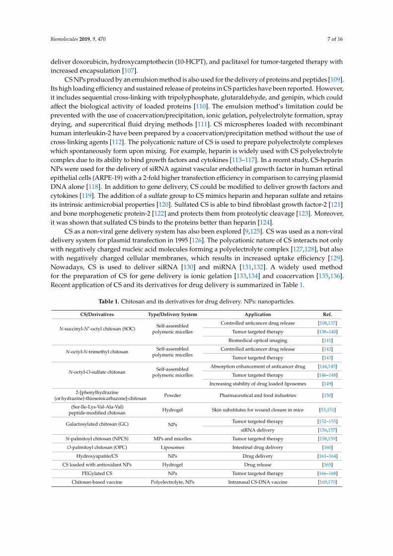
Table 1. Chitosan and its derivatives for drug delivery. NPs: nanoparticles.
CS/Derivatives Type/Delivery System Application Ref.
N-succinyl-N′-octyl chitosan (SOC) Self-assembledpolymeric micelles
Controlled anticancer drug release [108,137]
Tumor targeted therapy [138–140]
Biomedical optical imaging [141]
N-octyl-N-trimethyl chitosan Self-assembledpolymeric micelles
Controlled anticancer drug release [142]
Tumor targeted therapy [143]
N-octyl-O-sulfate chitosan Self-assembledpolymeric micelles
Absorption enhancement of anticancer drug [144,145]
Tumor targeted therapy [146–148]
Increasing stability of drug loaded liposomes [149]
2-[phenylhydrazine(or hydrazine)-thiosemicarbazone]-chitosan Powder Pharmaceutical and food industries [150]
(Ser-Ile-Lys-Val-Ala-Val)peptide-modified chitosan Hydrogel Skin substitutes for wound closure in mice [53,151]
Galactosylated chitosan (GC) NPsTumor targeted therapy [152–155]
siRNA delivery [156,157]
N-palmitoyl chitosan (NPCS) MPs and micelles Tumor targeted therapy [158,159]
O-palmitoyl chitosan (OPC) Liposomes Intestinal drug delivery [160]
Hydroxyapatite/CS NPs Drug delivery [161–164]
CS loaded with antioxidant NPs Hydrogel Drug release [165]
PEGylated CS NPs Tumor targeted therapy [166–168]
Chitosan-based vaccine Polyelectrolyte, NPs Intranasal CS-DNA vaccine [169,170]

Biomolecules 2019, 9, 470 8 of 16
4. Conclusions
CS, as a natural polymer, is actively used in tissue engineering and regenerative medicine asa biomaterial alone, as well as in combination with other polymers. In addition to its suitable mechanicalphysico-chemical properties, CS has a natural ability to stimulate tissue regeneration. Active researchis underway in improving CS-containing scaffolds for wound healing, bone, and cartilage regeneration.In addition to this, CS-containing polymers are being actively studied for the delivery of drugs fortargeted tumor therapy and nucleic acid delivery in genetic engineering applications. Further researchon the preparation of CS-containing scaffolds via 3D printing and cryogelation methods will facilitatethe application of CS in biomedicine. CS, as a part of any material, could introduce valuable propertiessuch as antimicrobial activity, mucoadhesiveness, and biocompatibility, which are in demand forbiomedical use. We believe that further research on CS and the search for new variations in its usewith other polymers will reveal even greater prospects and properties of this unique polymer inbiomedical applications.
Funding: A.S. is supported by a grant from the Ministry of Education and Science of the Republic of Kazakhstan.
Conflicts of Interest: The authors declare no conflict of interest.
References
1. Oryan, A.; Alidadi, S.; Moshiri, A.; Maffulli, N. Bone regenerative medicine: Classic options, novel strategies,and future directions. J. Orthop. Surg. Res. 2014, 9, 18. [CrossRef] [PubMed]
2. Rodríguez-Vázquez, M.; Vega-Ruiz, B.; Ramos-Zúñiga, R.; Saldaña-Koppel, D.A.; Quiñones-Olvera, L.F.Chitosan and Its Potential Use as a Scaffold for Tissue Engineering in Regenerative Medicine. BioMed Res. Int.2015, 2015, 1–15. [CrossRef] [PubMed]
3. Hoven, V.P.; Tangpasuthadol, V.; Angkitpaiboon, Y.; Vallapa, N.; Kiatkamjornwong, S. Surface-chargedchitosan: Preparation and protein adsorption. Carbohydr. Polym. 2007, 68, 44–53. [CrossRef]
4. Saranya, N.; Moorthi, A.; Saravanan, S.; Devi, M.P.; Selvamurugan, N. Chitosan and its derivatives forgene delivery. Int. J. Biol. Macromol. 2011, 48, 234–238. [CrossRef] [PubMed]
5. Venkatesan, J.; Anil, S.; Kim, S.K.; Shim, M.S. Chitosan as a vehicle for growth factor delivery: Variouspreparations and their applications in bone tissue regeneration. Int. J. Biol. Macromol. 2017, 104, 1383–1397.[CrossRef] [PubMed]
6. Tharanathan, R.N.; Kittur, F.S. Chitin—The Undisputed Biomolecule of Great Potential. Crit. Rev. FoodSci. Nutr. 2003, 43, 61–87. [CrossRef] [PubMed]
7. Ghormade, V.; Pathan, E.K.; Deshpande, M.V. Can fungi compete with marine sources for chitosan production?Int. J. Biol. Macromol. 2017, 104, 1415–1421. [CrossRef] [PubMed]
8. Grifoll-Romero, L.; Pascual, S.; Aragunde, H.; Biarnés, X.; Planas, A. Chitin Deacetylases: Structures,Specificities, and Biotech Applications. Polymers 2018, 10, 352. [CrossRef]
9. Ahsan, S.M.; Thomas, M.; Reddy, K.K.; Sooraparaju, S.G.; Asthana, A.; Bhatnagar, I. Chitosan as biomaterialin drug delivery and tissue engineering. Int. J. Biol. Macromol. 2018, 110, 97–109. [CrossRef] [PubMed]
10. Balau, L.; Lisa, G.; Popa, M.; Tura, V.; Melnig, V. Physico-chemical properties of Chitosan films. Open Chem.2004, 2, 638–647. [CrossRef]
11. Fan, M.; Hu, Q.; Shen, K. Preparation and structure of chitosan soluble in wide pH range. Carbohydr. Polym.2009, 78, 66–71. [CrossRef]
12. Roberts, G.A.F. Solubility and Solution Behaviour of Chitin and Chitosan. In Chitin Chemistry; MacmillanEducation: London, UK, 1992; pp. 274–329.
13. Berillo, D.; Mattiasson, B.; Kirsebom, H. Cryogelation of chitosan using noble-metal ions: In situ formationof nanoparticles. Biomacromolecules 2014, 15, 2246–2255. [CrossRef]
14. Berillo, D.; Cundy, A. 3D-macroporous chitosan-based scaffolds with in situ formed Pd and Pt nanoparticlesfor nitrophenol reduction. Carbohydr. Polym. 2018, 192, 166–175. [CrossRef] [PubMed]
15. Berillo, D.; Elowsson, L.; Kirsebom, H. Oxidized Dextran as Crosslinker for Chitosan Cryogel Scaffoldsand Formation of Polyelectrolyte Complexes between Chitosan and Gelatin. Macromol. Biosci. 2012, 12,1090–1099. [CrossRef] [PubMed]

Biomolecules 2019, 9, 470 9 of 16
16. Akilbekova, D.; Shaimerdenova, M.; Adilov, S.; Berillo, D. Biocompatible scaffolds based on natural polymersfor regenerative medicine. Int. J. Biol. Macromol. 2018, 114, 324–333. [CrossRef] [PubMed]
17. Lu, H.-T.; Lu, T.-W.; Chen, C.-H.; Lu, K.-Y.; Mi, F.-L. Development of nanocomposite scaffolds based onbiomineralization of N,O-carboxymethyl chitosan/fucoidan conjugates for bone tissue engineering. Int. J. Biol.Macromol. 2018, 120, 2335–2345. [CrossRef] [PubMed]
18. Nilsen-Nygaard, J.; Strand, S.; Vårum, K.; Draget, K.; Nordgård, C.; Nilsen-Nygaard, J.; Strand, S.P.;Vårum, K.M.; Draget, K.I.; Nordgård, C.T. Chitosan: Gels and Interfacial Properties. Polymers 2015, 7, 552–579.[CrossRef]
19. Badwan, A.; Rashid, I.; Omari, M.; Darras, F. Chitin and Chitosan as Direct Compression Excipients inPharmaceutical Applications. Mar. Drugs 2015, 13, 1519–1547. [CrossRef]
20. Rousselle, P.; Montmasson, M.; Garnier, C. Extracellular matrix contribution to skin wound re-epithelialization.Matrix Biol. 2019, 75, 12–26. [CrossRef]
21. Clark, R. Wound repair: Lessons for tissue engineering. In Principles of Tissue Engineering; Academic Press:San Diego, CA, USA, 1997.
22. Abdelrahman, T.; Newton, H. Wound dressings: Principles and practice. Surgery 2011, 29, 491–495. [CrossRef]23. Khan, M.A.; Mujahid, M. A review on recent advances in chitosan based composite for hemostatic dressings.
Int. J. Biol. Macromol. 2019, 124, 138–147. [CrossRef] [PubMed]24. Hu, Z.; Lu, S.; Cheng, Y.; Kong, S.; Li, S.; Li, C.; Yang, L. Investigation of the Effects of Molecular Parameters
on the Hemostatic Properties of Chitosan. Molecules 2018, 23, 3147. [CrossRef] [PubMed]25. Yang, J.; Tian, F.; Wang, Z.; Wang, Q.; Zeng, Y.-J.; Chen, S.-Q. Effect of chitosan molecular weight and
deacetylation degree on hemostasis. J. Biomed. Mater. Res. Part B Appl. Biomater. 2008, 84, 131–137. [CrossRef][PubMed]
26. Guo, X.; Sun, T.; Zhong, R.; Ma, L.; You, C.; Tian, M.; Li, H.; Wang, C. Effects of Chitosan Oligosaccharides onHuman Blood Components. Front. Pharmacol. 2018, 9, 1412. [CrossRef] [PubMed]
27. Zhou, X.; Zhang, X.; Zhou, J.; Li, L. An investigation of chitosan and its derivatives on red blood cellagglutination. RSC Adv. 2017, 7, 12247–12254. [CrossRef]
28. Klokkevold, P.R.; Fukayama, H.; Sung, E.C.; Bertolami, C.N. The effect of chitosan (poly-N-acetyl glucosamine)on lingual hemostasis in heparinized rabbits. J. Oral Maxillofac. Surg. 1999, 57, 49–52. [CrossRef]
29. Hattori, H.; Ishihara, M. Changes in blood aggregation with differences in molecular weight and degree ofdeacetylation of chitosan. Biomed. Mater. 2015, 10, 015014. [CrossRef]
30. Hu, Z.; Zhang, D.-Y.; Lu, S.-T.; Li, P.-W.; Li, S.-D. Chitosan-Based Composite Materials for ProspectiveHemostatic Applications. Mar. Drugs 2018, 16, 273. [CrossRef]
31. Ueno, H.; Yamada, H.; Tanaka, I.; Kaba, N.; Matsuura, M.; Okumura, M.; Kadosawa, T.; Fujinaga, T.Accelerating effects of chitosan for healing at early phase of experimental open wound in dogs. Biomaterials1999, 20, 1407–1414. [CrossRef]
32. Park, C.J.; Gabrielson, N.P.; Pack, D.W.; Jamison, R.D.; Wagoner Johnson, A.J. The effect of chitosan on themigration of neutrophil-like HL60 cells, mediated by IL-8. Biomaterials 2009, 30, 436–444. [CrossRef]
33. Bueter, C.L.; Lee, C.K.; Rathinam, V.A.K.; Healy, G.J.; Taron, C.H.; Specht, C.A.; Levitz, S.M. Chitosan butNot Chitin Activates the Inflammasome by a Mechanism Dependent upon Phagocytosis. J. Biol. Chem. 2011,286, 35447–35455. [CrossRef] [PubMed]
34. Bueter, C.L.; Lee, C.K.; Wang, J.P.; Ostroff, G.R.; Specht, C.A.; Levitz, S.M. Spectrum and Mechanisms ofInflammasome Activation by Chitosan. J. Immunol. 2014, 192, 5943–5951. [CrossRef] [PubMed]
35. Gudmundsdottir, S.; Lieder, R.; Sigurjonsson, O.E.; Petersen, P.H. Chitosan leads to downregulation of YKL-40and inflammasome activation in human macrophages. J. Biomed. Mater. Res. Part A 2015, 103, 2778–2785.[CrossRef] [PubMed]
36. Fong, D.; Grégoire-Gélinas, P.; Cheng, A.P.; Mezheritsky, T.; Lavertu, M.; Sato, S.; Hoemann, C.D. Lysosomalrupture induced by structurally distinct chitosans either promotes a type 1 IFN response or activates theinflammasome in macrophages. Biomaterials 2017, 129, 127–138. [CrossRef] [PubMed]
37. Vasconcelos, D.P.; de Torre-Minguela, C.; Gomez, A.I.; Águas, A.P.; Barbosa, M.A.; Pelegrín, P.; Barbosa, J.N.3D chitosan scaffolds impair NLRP3 inflammasome response in macrophages. Acta Biomater. 2019, 91,123–134. [CrossRef]
Biomolecules 2019, 9, 470 10 of 16
38. Baxter, R.M.; Dai, T.; Kimball, J.; Wang, E.; Hamblin, M.R.; Wiesmann, W.P.; McCarthy, S.J.; Baker, S.M.Chitosan dressing promotes healing in third degree burns in mice: Gene expression analysis shows biphasiceffects for rapid tissue regeneration and decreased fibrotic signaling. J. Biomed. Mater. Res. A 2013, 101,340–348. [CrossRef] [PubMed]
39. Tsai, C.-W.; Chiang, I.-N.; Wang, J.-H.; Young, T.-H. Chitosan delaying human fibroblast senescence throughdownregulation of TGF-β signaling pathway. Artif. Cells Nanomed. Biotechnol. 2017, 46, 1–12. [CrossRef][PubMed]
40. Howling, G.I.; Dettmar, P.W.; Goddard, P.A.; Hampson, F.C.; Dornish, M.; Wood, E.J. The effect of chitinand chitosan on the proliferation of human skin fibroblasts and keratinocytes in vitro. Biomaterials 2001, 22,2959–2966. [CrossRef]
41. Hamilton, V.; Yuan, Y.; Rigney, D.A.; Puckett, A.D.; Ong, J.L.; Yang, Y.; Elder, S.H.; Bumgardner, J.D.Characterization of chitosan films and effects on fibroblast cell attachment and proliferation. J. Mater. Sci.Mater. Med. 2006, 17, 1373–1381. [CrossRef]
42. Croisier, F.; Jérôme, C. Chitosan-based biomaterials for tissue engineering. Eur. Polym. J. 2013, 49, 780–792.[CrossRef]
43. Song, R.; Zheng, J.; Liu, Y.; Tan, Y.; Yang, Z.; Song, X.; Yang, S.; Fan, R.; Zhang, Y.; Wang, Y. A naturalcordycepin/chitosan complex hydrogel with outstanding self-healable and wound healing properties.Int. J. Biol. Macromol. 2019, 134, 91–99. [CrossRef] [PubMed]
44. Aubert-Viard, F.; Mogrovejo-Valdivia, A.; Tabary, N.; Maton, M.; Chai, F.; Neut, C.; Martel, B.; Blanchemain, N.Evaluation of antibacterial textile covered by layer-by-layer coating and loaded with chlorhexidine forwound dressing application. Mater. Sci. Eng. C 2019, 100, 554–563. [CrossRef] [PubMed]
45. Alves, P.; Santos, M.; Mendes, S.; Miguel, P.S.; de Sá, D.K.; Cabral, S.D.C.; Correia, J.I.; Ferreira, P.; Alves, P.;Santos, M.; et al. Photocrosslinkable Nanofibrous Asymmetric Membrane Designed for Wound Dressing.Polymers 2019, 11, 653. [CrossRef] [PubMed]
46. Shah, A.; Ali Buabeid, M.; Arafa, E.-S.A.; Hussain, I.; Li, L.; Murtaza, G. The wound healing and antibacterialpotential of triple-component nanocomposite (chitosan-silver-sericin) films loaded with moxifloxacin.Int. J. Pharm. 2019, 564, 22–38. [CrossRef] [PubMed]
47. Lin, C.-W.; Chen, Y.-K.; Lu, M.; Lou, K.-L.; Yu, J. Photo-Crosslinked Keratin/Chitosan Membranes as PotentialWound Dressing Materials. Polymers 2018, 10, 987. [CrossRef] [PubMed]
48. Sharma, S.; Swetha, K.L.; Roy, A. Chitosan-Chondroitin sulfate based polyelectrolyte complex for effectivemanagement of chronic wounds. Int. J. Biol. Macromol. 2019, 132, 97–108. [CrossRef] [PubMed]
49. Koukaras, E.N.; Papadimitriou, S.A.; Bikiaris, D.N.; Froudakis, G.E. Insight on the Formation of ChitosanNanoparticles through Ionotropic Gelation with Tripolyphosphate. Mol. Pharm. 2012, 9, 2856–2862. [CrossRef][PubMed]
50. Karimi Dehkordi, N.; Minaiyan, M.; Talebi, A.; Akbari, V.; Taheri, A. Nanocrystalline cellulose–hyaluronic acidcomposite enriched with GM-CSF loaded chitosan nanoparticles for enhanced wound healing. Biomed. Mater.2019, 14, 035003. [CrossRef]
51. Tanha, S.; Rafiee-Tehrani, M.; Abdollahi, M.; Vakilian, S.; Esmaili, Z.; Naraghi, Z.S.; Seyedjafari, E.; Javar, H.A.G-CSF loaded nanofiber/nanoparticle composite coated with collagen promotes wound healing in vivo.J. Biomed. Mater. Res. Part A 2017, 105, 2830–2842. [CrossRef]
52. Chen, X.; Zhang, M.; Chen, S.; Wang, X.; Tian, Z.; Chen, Y.; Xu, P.; Zhang, L.; Zhang, L.; Zhang, L.Peptide-Modified Chitosan Hydrogels Accelerate Skin Wound Healing by Promoting Fibroblast Proliferation,Migration, and Secretion. Cell Transplant. 2017, 26, 1331–1340. [CrossRef]
53. Chen, X.; Fu, W.; Cao, X.; Jiang, H.; Che, X.; Xu, X.; Ma, B.; Zhang, J. Peptide SIKVAV-modified chitosanhydrogels promote skin wound healing by accelerating angiogenesis and regulating cytokine secretion.Am. J. Transl. Res. 2018, 10, 4258–4268. [PubMed]
54. Yu, Y.; Chen, R.; Sun, Y.; Pan, Y.; Tang, W.; Zhang, S.; Cao, L.; Yuan, Y.; Wang, J.; Liu, C. Manipulation ofVEGF-induced angiogenesis by 2-N, 6-O-sulfated chitosan. Acta Biomater. 2018, 71, 510–521. [CrossRef][PubMed]
55. Wang, C.; Yu, Y.; Chen, H.; Zhang, S.; Wang, J.; Liu, C. Construction of cytokine reservoirs based on sulfatedchitosan hydrogels for the capturing of VEGF in situ. J. Mater. Chem. B 2019, 7, 1882–1892. [CrossRef]
56. Titorencu, I.; Albu, M.; Nemecz, M.; Jinga, V. Natural Polymer-Cell Bioconstructs for Bone Tissue Engineering.Curr. Stem Cell Res. Ther. 2016, 12, 165–174. [CrossRef] [PubMed]

Biomolecules 2019, 9, 470 11 of 16
57. Saravanan, S.; Leena, R.S.; Selvamurugan, N. Chitosan based biocomposite scaffolds for bone tissueengineering. Int. J. Biol. Macromol. 2016, 93, 1354–1365. [CrossRef] [PubMed]
58. Deepthi, S.; Venkatesan, J.; Kim, S.-K.; Bumgardner, J.D.; Jayakumar, R. An overview of chitin or chitosan/nanoceramic composite scaffolds for bone tissue engineering. Int. J. Biol. Macromol. 2016, 93, 1338–1353. [CrossRef][PubMed]
59. He, L.-H.; Yao, L.; Xue, R.; Sun, J.; Song, R. In-situ mineralization of chitosan/calcium phosphate compositeand the effect of solvent on the structure. Front. Mater. Sci. 2011, 5, 282–292. [CrossRef]
60. Tuzlakoglu, K.; Reis, R.L. Formation of bone-like apatite layer on chitosan fiber mesh scaffolds by a biomimeticspraying process. J. Mater. Sci. Mater. Med. 2007, 18, 1279–1286. [CrossRef]
61. Leonor, I.B.; Baran, E.T.; Kawashita, M.; Reis, R.L.; Kokubo, T.; Nakamura, T. Growth of a bonelike apatite onchitosan microparticles after a calcium silicate treatment. Acta Biomater. 2008, 4, 1349–1359. [CrossRef]
62. Elkholy, S.; Yahia, S.; Awad, M.; Elmessiery, M. In vivo evaluation of β-CS/n-HA with different physicalproperties as a new bone graft material. Clin. Implant Dent. Relat. Res. 2018, 20, 416–423. [CrossRef]
63. Gaihre, B.; Jayasuriya, A.C. Comparative investigation of porous nano-hydroxyapaptite/chitosan,nano-zirconia/chitosan and novel nano-calcium zirconate/chitosan composite scaffolds for their potentialapplications in bone regeneration. Mater. Sci. Eng. C 2018, 91, 330–339. [CrossRef] [PubMed]
64. Koç Demir, A.; Elçin, A.E.; Elçin, Y.M. Strontium-modified chitosan/montmorillonite composites as bonetissue engineering scaffold. Mater. Sci. Eng. C 2018, 89, 8–14. [CrossRef] [PubMed]
65. Georgopoulou, A.; Kaliva, M.; Vamvakaki, M.; Chatzinikolaidou, M. Osteogenic Potential of Pre-OsteoblasticCells on a Chitosan-graft-Polycaprolactone Copolymer. Materials 2018, 11, 490. [CrossRef] [PubMed]
66. Habibovic, P.; Barralet, J.E. Bioinorganics and biomaterials: Bone repair. Acta Biomater. 2011, 7, 3013–3026.[CrossRef] [PubMed]
67. Singh, B.N.; Veeresh, V.; Mallick, S.P.; Jain, Y.; Sinha, S.; Rastogi, A.; Srivastava, P. Design and evaluation ofchitosan/chondroitin sulfate/nano-bioglass based composite scaffold for bone tissue engineering. Int. J. Biol.Macromol. 2019, 133, 817–830. [CrossRef] [PubMed]
68. Lu, H.-T.; Lu, T.-W.; Chen, C.-H.; Mi, F.-L. Development of genipin-crosslinked and fucoidan-adsorbednano-hydroxyapatite/hydroxypropyl chitosan composite scaffolds for bone tissue engineering. Int. J. Biol.Macromol. 2019, 128, 973–984. [CrossRef] [PubMed]
69. Keller, L.; Regiel-Futyra, A.; Gimeno, M.; Eap, S.; Mendoza, G.; Andreu, V.; Wagner, Q.; Kyzioł, A.;Sebastian, V.; Stochel, G.; et al. Chitosan-based nanocomposites for the repair of bone defects. Nanomed.Nanotechnol. Biol. Med. 2017, 13, 2231–2240. [CrossRef]
70. Georgopoulou, A.; Papadogiannis, F.; Batsali, A.; Marakis, J.; Alpantaki, K.; Eliopoulos, A.G.; Pontikoglou, C.;Chatzinikolaidou, M. Chitosan/gelatin scaffolds support bone regeneration. J. Mater. Sci. Mater. Med. 2018,29, 59. [CrossRef]
71. Nguyen, T.H.M.; Abueva, C.; Van Ho, H.; Lee, S.-Y.; Lee, B.-T. In vitro and in vivo acute response towardsinjectable thermosensitive chitosan/TEMPO-oxidized cellulose nanofiber hydrogel. Carbohydr. Polym. 2018,180, 246–255. [CrossRef]
72. Zhang, L.; Wu, K.; Song, W.; Xu, H.; An, R.; Zhao, L.; Liu, B.; Zhang, Y. Chitosan/siCkip-1 biofunctionalizedtitanium implant for improved osseointegration in the osteoporotic condition. Sci. Rep. 2015, 5, 10860.[CrossRef]
73. Dwivedi, P.; Narvi, S.S.; Tewari, R.P. Application of polymer nanocomposites in the nanomedicine landscape:Envisaging strategies to combat implant associated infections. J. Appl. Biomater. Funct. Mater. 2013, 11,129–142. [CrossRef]
74. Kumar, A.M.; Suresh, B.; Das, S.; Obot, I.B.; Adesina, A.Y.; Ramakrishna, S. Promising bio-composites ofpolypyrrole and chitosan: Surface protective and in vitro biocompatibility performance on 316L SS implants.Carbohydr. Polym. 2017, 173, 121–130. [CrossRef] [PubMed]
75. Liu, J.; Sun, L.; Xu, W.; Wang, Q.; Yu, S.; Sun, J. Current advances and future perspectives of 3D printingnatural-derived biopolymers. Carbohydr. Polym. 2019, 207, 297–316. [CrossRef] [PubMed]
76. Demirtas, T.T.; Irmak, G.; Gümüsderelioglu, M. A bioprintable form of chitosan hydrogel for bone tissueengineering. Biofabrication 2017, 9, 035003. [CrossRef] [PubMed]
77. Liao, I.-C.; Moutos, F.T.; Estes, B.T.; Zhao, X.; Guilak, F. Composite Three-Dimensional Woven Scaffolds withInterpenetrating Network Hydrogels to Create Functional Synthetic Articular Cartilage. Adv. Funct. Mater.2013, 23, 5833–5839. [CrossRef] [PubMed]

Biomolecules 2019, 9, 470 12 of 16
78. Nettles, D.L.; Elder, S.H.; Gilbert, J.A. Potential Use of Chitosan as a Cell Scaffold Material for CartilageTissue Engineering. Tissue Eng. 2002, 8, 1009–1016. [CrossRef]
79. Freedman, B.R.; Mooney, D.J. Biomaterials to Mimic and Heal Connective Tissues. Adv. Mater. 2019, 31,1806695. [CrossRef]
80. Jin, R.; Moreira Teixeira, L.S.; Dijkstra, P.J.; Karperien, M.; van Blitterswijk, C.A.; Zhong, Z.Y.; Feijen, J.Injectable chitosan-based hydrogels for cartilage tissue engineering. Biomaterials 2009, 30, 2544–2551.[CrossRef]
81. Liu, M.; Zeng, X.; Ma, C.; Yi, H.; Ali, Z.; Mou, X.; Li, S.; Deng, Y.; He, N. Injectable hydrogels for cartilage andbone tissue engineering. Bone Res. 2017, 5, 17014. [CrossRef]
82. Huang, H.; Zhang, X.; Hu, X.; Dai, L.; Zhu, J.; Man, Z.; Chen, H.; Zhou, C.; Ao, Y. Directing chondrogenicdifferentiation of mesenchymal stem cells with a solid-supported chitosan thermogel for cartilage tissueengineering. Biomed. Mater. 2014, 9, 035008. [CrossRef]
83. Kuo, C.-Y.; Chen, C.-H.; Hsiao, C.-Y.; Chen, J.-P. Incorporation of chitosan in biomimeticgelatin/chondroitin-6-sulfate/hyaluronan cryogel for cartilage tissue engineering. Carbohydr. Polym. 2015,117, 722–730. [CrossRef] [PubMed]
84. VandeVord, P.J.; Matthew, H.W.T.; DeSilva, S.P.; Mayton, L.; Wu, B.; Wooley, P.H. Evaluation of thebiocompatibility of a chitosan scaffold in mice. J. Biomed. Mater. Res. 2002, 59, 585–590. [CrossRef] [PubMed]
85. Choi, B.; Kim, S.; Lin, B.; Wu, B.M.; Lee, M. Cartilaginous Extracellular Matrix-Modified Chitosan Hydrogelsfor Cartilage Tissue Engineering. ACS Appl. Mater. Interfaces 2014, 6, 20110–20121. [CrossRef] [PubMed]
86. Oprenyeszk, F.; Sanchez, C.; Dubuc, J.-E.; Maquet, V.; Henrist, C.; Compère, P.; Henrotin, Y. Chitosan enrichedthree-dimensional matrix reduces inflammatory and catabolic mediators production by human chondrocytes.PLoS ONE 2015, 10, e0128362. [CrossRef] [PubMed]
87. Kong, Y.; Zhang, Y.; Zhao, X.; Wang, G.; Liu, Q. Carboxymethyl-chitosan attenuates inducible nitric oxidesynthase and promotes interleukin-10 production in rat chondrocytes. Exp. Ther. Med. 2017, 14, 5641–5646.[CrossRef]
88. Huang, S.; Song, X.; Li, T.; Xiao, J.; Chen, Y.; Gong, X.; Zeng, W.; Yang, L.; Chen, C. Pellet cocultureof osteoarthritic chondrocytes and infrapatellar fat pad-derived mesenchymal stem cells with chitosan/
hyaluronic acid nanoparticles promotes chondrogenic differentiation. Stem Cell Res. Ther. 2017, 8, 264.[CrossRef]
89. Ye, K.; Felimban, R.; Traianedes, K.; Moulton, S.E.; Wallace, G.G.; Chung, J.; Quigley, A.; Choong, P.F.M.;Myers, D.E. Chondrogenesis of infrapatellar fat pad derived adipose stem cells in 3D printed chitosan scaffold.PLoS ONE 2014, 9, e99410. [CrossRef]
90. Sionkowska, A.; Wisniewski, M.; Skopinska, J.; Kennedy, C.J.; Wess, T.J. Molecular interactions in collagenand chitosan blends. Biomaterials 2004, 25, 795–801. [CrossRef]
91. Haaparanta, A.-M.; Järvinen, E.; Cengiz, I.F.; Ellä, V.; Kokkonen, H.T.; Kiviranta, I.; Kellomäki, M. Preparationand characterization of collagen/PLA, chitosan/PLA, and collagen/chitosan/PLA hybrid scaffolds for cartilagetissue engineering. J. Mater. Sci. Mater. Med. 2014, 25, 1129–1136. [CrossRef]
92. Su, J.-Y.; Chen, S.-H.; Chen, Y.-P.; Chen, W.-C. Evaluation of Magnetic Nanoparticle-Labeled ChondrocytesCultivated on a Type II Collagen-Chitosan/Poly(Lactic-co-Glycolic) Acid Biphasic Scaffold. Int. J. Mol. Sci.2017, 18, 87. [CrossRef]
93. Bhardwaj, N.; Nguyen, Q.T.; Chen, A.C.; Kaplan, D.L.; Sah, R.L.; Kundu, S.C. Potential of 3-D tissueconstructs engineered from bovine chondrocytes/silk fibroin-chitosan for in vitro cartilage tissue engineering.Biomaterials 2011, 32, 5773–5781. [CrossRef] [PubMed]
94. Liu, J.; Fang, Q.; Yu, X.; Wan, Y.; Xiao, B. Chitosan-Based Nanofibrous Membrane Unit with GradientCompositional and Structural Features for Mimicking Calcified Layer in Osteochondral Matrix. Int. J.Mol. Sci. 2018, 19, 2330. [CrossRef] [PubMed]
95. Noriega, S.E.; Hasanova, G.I.; Schneider, M.J.; Larsen, G.F.; Subramanian, A. Effect of fiber diameter on thespreading, proliferation and differentiation of chondrocytes on electrospun chitosan matrices. Cells TissuesOrgans 2012, 195, 207–221. [CrossRef] [PubMed]
96. Bhat, S.; Tripathi, A.; Kumar, A. Supermacroprous chitosan–agarose–gelatin cryogels: In vitro characterizationand in vivo assessment for cartilage tissue engineering. J. R. Soc. Interface 2011, 8, 540–554. [CrossRef][PubMed]

Biomolecules 2019, 9, 470 13 of 16
97. Gupta, A.; Bhat, S.; Jagdale, P.R.; Chaudhari, B.P.; Lidgren, L.; Gupta, K.C.; Kumar, A. Evaluation ofthree-dimensional chitosan-agarose-gelatin cryogel scaffold for the repair of subchondral cartilage defects:An in vivo study in a rabbit model. Tissue Eng. Part A 2014, 20, 3101–3111. [CrossRef] [PubMed]
98. Lu, T.-J.; Chiu, F.-Y.; Chiu, H.-Y.; Chang, M.-C.; Hung, S.-C. Chondrogenic Differentiation of MesenchymalStem Cells in Three-Dimensional Chitosan Film Culture. Cell Transplant. 2017, 26, 417–427. [CrossRef][PubMed]
99. Vårum, K.M.; Holme, H.K.; Izume, M.; Stokke, B.T.; Smidsrød, O. Determination of enzymatic hydrolysisspecificity of partially N-acetylated chitosans. Biochim. Biophys. Acta 1996, 1291, 5–15. [CrossRef]
100. Rathore, A.S.; Gupta, R.D. Chitinases from Bacteria to Human: Properties, Applications, and FuturePerspectives. Enzyme Res. 2015, 2015, 791907. [CrossRef]
101. Sogias, I.A.; Williams, A.C.; Khutoryanskiy, V.V. Why is Chitosan Mucoadhesive? Biomacromolecules 2008, 9,1837–1842. [CrossRef]
102. Casettari, L.; Illum, L. Chitosan in nasal delivery systems for therapeutic drugs. J. Control. Release 2014, 190,189–200. [CrossRef]
103. Irimia, T.; Dinu-Pîrvu, C.-E.; Ghica, M.V.; Lupuleasa, D.; Muntean, D.-L.; Udeanu, D.I.; Popa, L.Chitosan-Based in Situ Gels for Ocular Delivery of Therapeutics: A State-of-the-Art Review. Mar. Drugs2018, 16, 373. [CrossRef] [PubMed]
104. Batista, P.; Castro, P.; Madureira, A.R.; Sarmento, B.; Pintado, M. Development and Characterization ofChitosan Microparticles-in-Films for Buccal Delivery of Bioactive Peptides. Pharmaceuticals 2019, 12, 32.[CrossRef] [PubMed]
105. Ortiz, M.; Jornada, D.S.; Pohlmann, A.R.; Guterres, S.S. Development of Novel Chitosan Microcapsules forPulmonary Delivery of Dapsone: Characterization, Aerosol Performance, and In Vivo Toxicity Evaluation.AAPS PharmSciTech 2015, 16, 1033–1040. [CrossRef] [PubMed]
106. Baranwal, A.; Kumar, A.; Priyadharshini, A.; Oggu, G.S.; Bhatnagar, I.; Srivastava, A.; Chandra, P. Chitosan:An undisputed bio-fabrication material for tissue engineering and bio-sensing applications. Int. J. Biol.Macromol. 2018, 110, 110–123. [CrossRef] [PubMed]
107. Ahmed, S.; Ali, A.; Sheikh, J. A review on chitosan centred scaffolds and their applications in tissueengineering. Int. J. Biol. Macromol. 2018, 116, 849–862. [CrossRef] [PubMed]
108. Xu, X.; Li, L.; Zhou, J.; Lu, S.; Yang, J.; Yin, X.; Ren, J. Preparation and characterization of N-succinyl-N′-octylchitosan micelles as doxorubicin carriers for effective anti-tumor activity. Colloids Surf. B Biointerfaces 2007,55, 222–228.
109. Mohammed, M.A.; Syeda, J.T.M.; Wasan, K.M.; Wasan, E.K. An Overview of Chitosan Nanoparticles and ItsApplication in Non-Parenteral Drug Delivery. Pharmaceutics 2017, 9, 53. [CrossRef]
110. Chen, K.-Y.; Zeng, S.-Y. Fabrication of Quaternized Chitosan Nanoparticles Using Tripolyphosphate/GenipinDual Cross-Linkers as a Protein Delivery System. Polymers 2018, 10, 1226. [CrossRef]
111. Divya, K.; Jisha, M.S. Chitosan nanoparticles preparation and applications. Environ. Chem. Lett. 2018, 16,101–112. [CrossRef]
112. Özbas-Turan, S.; Akbuga, J.; Aral, C. Controlled Release of Interleukin-2 from Chitosan Microspheres.J. Pharm. Sci. 2002, 91, 1245–1251. [CrossRef]
113. Samorezov, J.E.; Alsberg, E. Spatial regulation of controlled bioactive factor delivery for bone tissueengineering. Adv. Drug Deliv. Rev. 2015, 84, 45–67. [CrossRef] [PubMed]
114. Lin, C.; Romero, R.; Sorokina, L.V.; Ballinger, K.R.; Place, L.W.; Kipper, M.J.; Khetani, S.R. A polyelectrolytemultilayer platform for investigating growth factor delivery modes in human liver cultures. J. Biomed.Mater. Res. A 2018, 106, 971–984. [CrossRef] [PubMed]
115. Tan, Q.; Tang, H.; Hu, J.; Hu, Y.; Zhou, X.; Tao, Y.; Wu, Z. Controlled release of chitosan/heparinnanoparticle-delivered VEGF enhances regeneration of decellularized tissue-engineered scaffolds. Int.J. Nanomed. 2011, 6, 929–942. [CrossRef]
116. Liang, Y.; Kiick, K.L. Heparin-functionalized polymeric biomaterials in tissue engineering and drug deliveryapplications. Acta Biomater. 2014, 10, 1588–1600. [CrossRef] [PubMed]
117. Mansurov, N.; Chen, W.C.W.; Awada, H.; Huard, J.; Wang, Y.; Saparov, A. A controlled release systemfor simultaneous delivery of three human perivascular stem cell-derived factors for tissue repair andregeneration. J. Tissue Eng. Regen. Med. 2018, 12, e1164–e1172. [CrossRef] [PubMed]

Biomolecules 2019, 9, 470 14 of 16
118. Pilipenko, I.; Korzhikov-Vlakh, V.; Sharoyko, V.; Zhang, N.; Schäfer-Korting, M.; Rühl, E.; Zoschke, C.;Tennikova, T. pH-Sensitive Chitosan–Heparin Nanoparticles for Effective Delivery of Genetic Drugs intoEpithelial Cells. Pharmaceutics 2019, 11, 317. [CrossRef] [PubMed]
119. Zeng, K.; Groth, T.; Zhang, K. Recent Advances in Artificially Sulfated Polysaccharides for Applications inCell Growth and Differentiation, Drug Delivery, and Tissue Engineering. ChemBioChem 2019, 20, 737–746.[CrossRef]
120. Zhong, Z.; Li, P.; Xing, R.; Liu, S. Antimicrobial activity of hydroxylbenzenesulfonailides derivatives ofchitosan, chitosan sulfates and carboxymethyl chitosan. Int. J. Biol. Macromol. 2009, 45, 163–168. [CrossRef][PubMed]
121. Ho, Y.-C.; Wu, S.-J.; Mi, F.-L.; Chiu, Y.-L.; Yu, S.-H.; Panda, N.; Sung, H.-W. Thiol-Modified Chitosan SulfateNanoparticles for Protection and Release of Basic Fibroblast Growth Factor. Bioconj. Chem. 2010, 21, 28–38.[CrossRef]
122. Peschel, D.; Zhang, K.; Fischer, S.; Groth, T. Modulation of osteogenic activity of BMP-2 by cellulose andchitosan derivatives. Acta Biomater. 2012, 8, 183–193. [CrossRef]
123. Weltrowski, A.; da Silva Almeida, M.-L.; Peschel, D.; Zhang, K.; Fischer, S.; Groth, T. Mitogenic Activity ofSulfated Chitosan and Cellulose Derivatives is Related to Protection of FGF-2 from Proteolytic Cleavage.Macromol. Biosci. 2012, 12, 740–750. [CrossRef] [PubMed]
124. Farrugia, B.L.; Mi, Y.; Kim, H.N.; Whitelock, J.M.; Baker, S.M.; Wiesmann, W.P.; Li, Z.; Maitz, P.; Lord, M.S.Chitosan-Based Heparan Sulfate Mimetics Promote Epidermal Formation in a Human OrganotypicSkin Model. Adv. Funct. Mater. 2018, 28, 1802818. [CrossRef]
125. Santos-Carballal, B.; Fernández Fernández, E.; Goycoolea, F.; Santos-Carballal, B.; Fernández Fernández, E.;Goycoolea, F.M. Chitosan in Non-Viral Gene Delivery: Role of Structure, Characterization Methods,and Insights in Cancer and Rare Diseases Therapies. Polymers 2018, 10, 444. [CrossRef] [PubMed]
126. Mumper, R.; Wang, J.; Claspell, J.; Rolland, A. Novel polymeric condensing carriers for gene delivery.Proc. Control. Release Soc. 1995, 22, 178–179.
127. Jayakumar, R.; Chennazhi, K.P.; Muzzarelli, R.A.A.; Tamura, H.; Nair, S.V.; Selvamurugan, N. Chitosanconjugated DNA nanoparticles in gene therapy. Carbohydr. Polym. 2010, 79, 1–8. [CrossRef]
128. MacLaughlin, F.C.; Mumper, R.J.; Wang, J.; Tagliaferri, J.M.; Gill, I.; Hinchcliffe, M.; Rolland, A.P. Chitosanand depolymerized chitosan oligomers as condensing carriers for in vivo plasmid delivery. J. Control. Release1998, 56, 259–272. [CrossRef]
129. Venkatesh, S.; Smith, T.J. Chitosan-membrane interactions and their probable role in chitosan-mediatedtransfection. Biotechnol. Appl. Biochem. 1998, 27, 265–267.
130. Vauthier, C.; Zandanel, C.; Ramon, A.L. Chitosan-based nanoparticles for in vivo delivery of interferingagents including siRNA. Curr. Opin. Colloid Interface Sci. 2013, 18, 406–418. [CrossRef]
131. Cryan, S.-A.; McKiernan, P.; Cunningham, C.M.; Greene, C. Targeting miRNA-based medicines to cysticfibrosis airway epithelial cells using nanotechnology. Int. J. Nanomed. 2013, 8, 3907. [CrossRef]
132. Ban, E.; Kwon, T.-H.; Kim, A. Delivery of therapeutic miRNA using polymer-based formulation. Drug Deliv.Transl. Res. 2019. [CrossRef]
133. Csaba, N.; Köping-Höggård, M.; Alonso, M.J. Ionically crosslinked chitosan/tripolyphosphate nanoparticlesfor oligonucleotide and plasmid DNA delivery. Int. J. Pharm. 2009, 382, 205–214. [CrossRef] [PubMed]
134. Csaba, N.; Köping-Höggård, M.; Fernandez-Megia, E.; Novoa-Carballal, R.; Riguera, R.; Alonso, M.J. Ionicallycrosslinked chitosan nanoparticles as gene delivery systems: Effect of PEGylation degree on in vitro andin vivo gene transfer. J. Biomed. Nanotechnol. 2009, 5, 162–171. [CrossRef] [PubMed]
135. Mao, H.Q.; Roy, K.; Troung-Le, V.L.; Janes, K.A.; Lin, K.Y.; Wang, Y.; August, J.T.; Leong, K.W. Chitosan-DNAnanoparticles as gene carriers: Synthesis, characterization and transfection efficiency. J. Control. Release 2001,70, 399–421. [CrossRef]
136. Corsi, K.; Chellat, F.; Yahia, L.; Fernandes, J.C. Mesenchymal stem cells, MG63 and HEK293 transfectionusing chitosan-DNA nanoparticles. Biomaterials 2003, 24, 1255–1264. [CrossRef]
137. Zhu, H.; Cao, J.; Cui, S.; Qian, Z.; Gu, Y. Enhanced Tumor Targeting and Antitumor Efficacy viaHydroxycamptothecin-Encapsulated Folate-Modified N-Succinyl-N′-Octyl Chitosan Micelles. J. Pharm. Sci.2013, 102, 1318–1332. [CrossRef] [PubMed]
138. Gu, Y.; Li, S.; Feng, S.; Ding, L.; Liu, Y.; Zhu, Q.; Qian, Z. Nanomedicine engulfed by macrophages fortargeted tumor therapy. Int. J. Nanomed. 2016, 11, 4107–4124. [CrossRef] [PubMed]

Biomolecules 2019, 9, 470 15 of 16
139. Tu, Z.; Ma, Y.; Akers, W.; Achilefu, S.; Gu, Y. Therapeutic effect of the treatment for colorectal cancer withadenoviral vectors mediated estrogen receptor β gene therapy combined with thermotherapy. J. Cancer Res.Clin. Oncol. 2014, 140, 623–632. [CrossRef]
140. Woraphatphadung, T.; Sajomsang, W.; Rojanarata, T.; Ngawhirunpat, T.; Tonglairoum, P.; Opanasopit, P.Development of Chitosan-Based pH-Sensitive Polymeric Micelles Containing Curcumin for Colon-TargetedDrug Delivery. AAPS PharmSciTech 2018, 19, 991–1000. [CrossRef]
141. Deng, D.; Qu, L.; Zhang, J.; Ma, Y.; Gu, Y. Quaternary Zn–Ag–In–Se Quantum Dots for Biomedical OpticalImaging of RGD-Modified Micelles. ACS Appl. Mater. Interfaces 2013, 5, 10858–10865. [CrossRef]
142. Zhang, C.; Ding, Y.; Yu, L.; Ping, Q. Polymeric micelle systems of hydroxycamptothecin based on amphiphilicN-alkyl-N-trimethyl chitosan derivatives. Colloids Surf. B Biointerfaces 2007, 55, 192–199. [CrossRef]
143. Zhang, F.; Fei, J.; Sun, M.; Ping, Q. Heparin modification enhances the delivery and tumor targetingof paclitaxel-loaded N-octyl-N -trimethyl chitosan micelles. Int. J. Pharm. 2016, 511, 390–402. [CrossRef][PubMed]
144. Mo, R.; Jin, X.; Li, N.; Ju, C.; Sun, M.; Zhang, C.; Ping, Q. The mechanism of enhancement on oral absorptionof paclitaxel by N-octyl-O-sulfate chitosan micelles. Biomaterials 2011, 32, 4609–4620. [CrossRef] [PubMed]
145. Mo, R.; Xiao, Y.; Sun, M.; Zhang, C.; Ping, Q. Enhancing effect of N-octyl-O-sulfate chitosan on etoposideabsorption. Int. J. Pharm. 2011, 409, 38–45. [CrossRef] [PubMed]
146. Jin, X.; Mo, R.; Ding, Y.; Zheng, W.; Zhang, C. Paclitaxel-Loaded N-Octyl-O-sulfate Chitosan Micelles forSuperior Cancer Therapeutic Efficacy and Overcoming Drug Resistance. Mol. Pharm. 2014, 11, 145–157.[CrossRef] [PubMed]
147. Ju, C.; Sun, J.; Zi, P.; Jin, X.; Zhang, C. Thermosensitive Micelles–Hydrogel Hybrid System Based on Poloxamer407 for Localized Delivery of Paclitaxel. J. Pharm. Sci. 2013, 102, 2707–2717. [CrossRef]
148. Qu, G.; Yao, Z.; Zhang, C.; Wu, X.; Ping, Q. PEG conjugated N-octyl-O-sulfate chitosan micelles for deliveryof paclitaxel: In vitro characterization and in vivo evaluation. Eur. J. Pharm. Sci. 2009, 37, 98–105. [CrossRef][PubMed]
149. Qu, G.; Wu, X.; Yin, L.; Zhang, C. N-octyl-O-sulfate chitosan-modified liposomes for delivery of docetaxel:Preparation, characterization, and pharmacokinetics. Biomed. Pharmacother. 2012, 66, 46–51. [CrossRef]
150. Zhong, Z.; Zhong, Z.; Xing, R.; Li, P.; Mo, G. The preparation and antioxidant activity of 2-[phenylhydrazine(or hydrazine)-thiosemicarbazone]-chitosan. Int. J. Biol. Macromol. 2010, 47, 93–97. [CrossRef]
151. Chen, X.; Cao, X.; Jiang, H.; Che, X.; Xu, X.; Ma, B.; Zhang, J.; Huang, T. SIKVAV-Modified Chitosan Hydrogelas a Skin Substitutes for Wound Closure in Mice. Molecules 2018, 23, 2611. [CrossRef]
152. Liu, W.; Wang, F.; Zhu, Y.; Li, X.; Liu, X.; Pang, J.; Pan, W. Galactosylated Chitosan-Functionalized MesoporousSilica Nanoparticle Loading by Calcium Leucovorin for Colon Cancer Cell-Targeted Drug Delivery. Molecules2018, 23, 3082. [CrossRef]
153. Ning, Q.; Liu, Y.-F.; Ye, P.-J.; Gao, P.; Li, Z.-P.; Tang, S.-Y.; He, D.-X.; Tang, S.-S.; Wei, H.; Yu, C.-Y. Deliveryof Liver-Specific miRNA-122 Using a Targeted Macromolecular Prodrug toward Synergistic Therapy forHepatocellular Carcinoma. ACS Appl. Mater. Interfaces 2019, 11, 10578–10588. [CrossRef] [PubMed]
154. Liu, W.; Zhu, Y.; Wang, F.; Li, X.; Liu, X.; Pang, J.; Pan, W. Galactosylated chitosan-functionalized mesoporoussilica nanoparticles for efficient colon cancer cell-targeted drug delivery. R. Soc. Open Sci. 2018, 5, 181027.[CrossRef] [PubMed]
155. Yen, H.; Young, Y.; Tsai, T.; Cheng, K.; Chen, X.; Chen, Y.; Chen, C.; Young, J.; Hong, P. Positively chargedgold nanoparticles capped with folate quaternary chitosan: Synthesis, cytotoxicity, and uptake by cancercells. Carbohydr. Polym. 2018, 183, 140–150. [CrossRef] [PubMed]
156. Huang, Y.; Guo, J.; Gui, S. Orally targeted galactosylated chitosan poly(lactic-co-glycolic acid) nanoparticlesloaded with TNF-a siRNA provide a novel strategy for the experimental treatment of ulcerative colitis.Eur. J. Pharm. Sci. 2018, 125, 232–243. [CrossRef]
157. Xiao, B.; Chen, Q.; Zhang, Z.; Wang, L.; Kang, Y.; Denning, T.; Merlin, D. TNFα gene silencing mediatedby orally targeted nanoparticles combined with interleukin-22 for synergistic combination therapy ofulcerative colitis. J. Control. Release 2018, 287, 235–246. [CrossRef] [PubMed]
158. Bai, M.-Y.; Tang, S.-L.; Chuang, M.-H.; Wang, T.-Y.; Hong, P. Evaluation of Chitosan Derivative MicroparticlesEncapsulating Superparamagnetic Iron Oxide and Doxorubicin as a pH-Sensitive Delivery Carrier in HepaticCarcinoma Treatment: An in vitro Comparison Study. Front. Pharmacol. 2018, 9, 1025. [CrossRef] [PubMed]

Biomolecules 2019, 9, 470 16 of 16
159. Bin, J.; Xiao, Y.; Lin, Z.T.; Deng, Y.L.; Chen, Y.; Le, D.E.; Bin, J.; Li, M.; Liao, Y.; Liu, Y.; et al. High molecularweight chitosan derivative polymeric micelles encapsulating superparamagnetic iron oxide for tumor-targetedmagnetic resonance imaging. Int. J. Nanomed. 2015, 10, 1155. [CrossRef] [PubMed]
160. Zariwala, M.G.; Bendre, H.; Markiv, A.; Farnaud, S.; Renshaw, D.; Taylor, K.M.; Somavarapu, S.Hydrophobically modified chitosan nanoliposomes for intestinal drug delivery. Int. J. Nanomed. 2018, 13,5837–5848. [CrossRef]
161. Chiesa, E.; Dorati, R.; Conti, B.; Modena, T.; Cova, E.; Meloni, F.; Genta, I. Hyaluronic Acid-DecoratedChitosan Nanoparticles for CD44-Targeted Delivery of Everolimus. Int. J. Mol. Sci. 2018, 19, 2310. [CrossRef]
162. Song, Q.; Jia, J.; Niu, X.; Zheng, C.; Zhao, H.; Sun, L.; Zhang, H.; Wang, L.; Zhang, Z.; Zhang, Y. An oral drugdelivery system with programmed drug release and imaging properties for orthotopic colon cancer therapy.Nanoscale 2019. [CrossRef]
163. Feiz, M.S.; Meshkini, A. Targeted delivery of adenosine 5′-triphosphate using chitosan-coated mesoporoushydroxyapatite: A theranostic pH-sensitive nanoplatform with enhanced anti-cancer effect. Int. J. Biol.Macromol. 2019, 129, 1090–1102. [CrossRef] [PubMed]
164. Kim, G.H.; Won, J.E.; Byeon, Y.; Kim, M.G.; Wi, T.I.; Lee, J.M.; Park, Y.-Y.; Lee, J.-W.; Kang, T.H.; Jung, I.D.; et al.Selective delivery of PLXDC1 small interfering RNA to endothelial cells for anti-angiogenesis tumor therapyusing CD44-targeted chitosan nanoparticles for epithelial ovarian cancer. Drug Deliv. 2018, 25, 1394–1402.[CrossRef] [PubMed]
165. Iglesias, N.; Galbis, E.; Díaz-Blanco, M.J.; Lucas, R.; Benito, E.; de-Paz, M.-V. Nanostructured Chitosan-BasedBiomaterials for Sustained and Colon-Specific Resveratrol Release. Int. J. Mol. Sci. 2019, 20, 398. [CrossRef][PubMed]
166. Chen, J.; Yang, X.; Huang, L.; Lai, H.; Gan, C.; Luo, X. Development of dual-drug-loaded stealth nanocarriersfor targeted and synergistic anti-lung cancer efficacy. Drug Deliv. 2018, 25, 1932–1942. [CrossRef] [PubMed]
167. Xie, P.; Du, P.; Li, J.; Liu, P. Stimuli-responsive hybrid cluster bombs of PEGylated chitosan encapsulatedDOX-loaded superparamagnetic nanoparticles enabling tumor-specific disassembly for on-demand drugdelivery and enhanced MR imaging. Carbohydr. Polym. 2019, 205, 377–384. [CrossRef] [PubMed]
168. Wang, K.; Zhuang, J.; Liu, Y.; Xu, M.; Zhuang, J.; Chen, Z.; Wei, Y.; Zhang, Y. PEGylated chitosan nanoparticleswith embedded bismuth sulfide for dual-wavelength fluorescent imaging and photothermal therapy.Carbohydr. Polym. 2018, 184, 445–452. [CrossRef] [PubMed]
169. Zhao, H.; Yang, J.; Qian, Q.; Wu, M.; Li, M.; Xu, W. Mesenteric CD103+DCs Initiate Switched CoxsackievirusB3 VP1-Specific IgA Response to Intranasal Chitosan-DNA Vaccine Through Secreting BAFF/IL-6 andPromoting Th17/Tfh Differentiation. Front. Immunol. 2018, 9, 2986. [CrossRef] [PubMed]
170. Sinani, G.; Sessevmez, M.; Gök, M.K.; Özgümüs, S.; Alpar, H.O.; Cevher, E. Modified chitosan-basednanoadjuvants enhance immunogenicity of protein antigens after mucosal vaccination. Int. J. Pharm. 2019,569, 118592. [CrossRef]
© 2019 by the authors. Licensee MDPI, Basel, Switzerland. This article is an open accessarticle distributed under the terms and conditions of the Creative Commons Attribution(CC BY) license (http://creativecommons.org/licenses/by/4.0/).


![Journal of Biomaterials Applications ‘Green’ biocompatible ... Biomater Appl-20… · PVA/ chitosan/nano-ZnO composite nanofibrous membranes Antibacterial and antifungal [16]](https://img.pdfslide.us/doc/110x75/605be37fd9239d416832e8c2/journal-of-biomaterials-applications-agreena-biocompatible-biomater-appl-20.jpg)