Embed Size (px)
Citation preview
2019, 2 (2): 74-83|74
Progress in Chemical and Biochemical Research
Journal homepage: www.pcbiochemres.com
* Corresponding author: Cletus Anes Ukwubile Tel: +2348036985667, E-mail: [email protected] DOI: 10.33945/SAMI/pcbr.2019.92077
Acute and subchronic toxicity profiles of Melastomastrum capitatum (Vahl) Fern. (Melastomataceae) root aqueous extract in Swiss albino mice
Cletus Anes Ukwubile a*, Emmanuel Oise Ikpefanb, Mathias Simon Bingaric, Livinus Tam d
a Department of Pharmacognosy and Drug Development, Faculty of Pharmaceutical Sciences, Ahmadu Bello
University, Zaria, Nigeria. b Department of Pharmacognosy and Traditional Medicine, Faculty of Pharmacy, Delta State University,
Abraka, Nigeria. c Department of Biological Sciences, Faculty of Science, Taraba State University, Jalingo, Nigeria. d Department of Hematology, Laboratory Sciences Division, Sancta Maria Clinic Integrated Laboratory,
fhi360/USAID Affiliate, Bali, Nigeria.
Progress in Chemical and Biochemical Research 2019, 2(2), 74-83
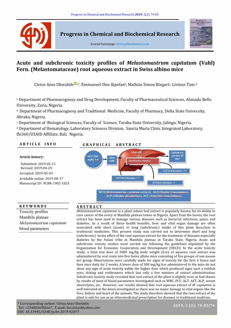
G R A P H I C A L A B S T R A C T
A R T I C L E I N F O
Article history:
Submitted: 2019-02-11 Revised: 2019-04-29
Accepted: 2019-05-03
Available online: 2019-08-17
Manuscript ID: PCBR-1902-1025
A B S T R A C T Melastomastrum capitatum is a plant whose leaf extract is popularly known for its ability to cure cancer of the ovary in Mambila plateau towns in Nigeria. Apart from the leaves, the root extract has been used to manage various diseases such as bacterial infections, pains, and diabetes. As a result of these health benefits, liver and vital organ damage are often associated with short (acute) or long (subchronic) intake of this plant decoction in traditional medicines. This present study was carried out to determine short and long (subchronic) terms effect of the root aqueous extract for the treatment of diseases especially diabetes by the Fulani tribe in Mambila plateau in Taraba State, Nigeria. Acute and subchronic toxicity studies were carried out following the guidelines stipulated by the Organization for Economic Cooperation and Development (OECD). In the acute toxicity study, a limit test dose of 2000 mg/kg body weight (b.w) of aqueous root extract was administered by oral route into five Swiss albino mice consisting of five groups of one mouse per group. Observations were carefully made for signs of toxicity for the first 4 hours and then once daily for 2 weeks. A lower dose of 300 mg/kg b.w administered to the mice do not show any sign of acute toxicity unlike the higher dose which produced signs such a reddish eyes, itching and restlessness which last only a few minutes of extract administration. Subchronic toxicity study revealed that root extract of the plant is slightly toxic as had shown by results of most of blood parameters investigated such as WBC, PCV, ALT, AST, ALP, serum electrolytes, etc. However, our results showed that root aqueous extract of M. capitatum is well tolerated at the doses investigated as there was no major damage to vital organs like the liver, kidney and heart of the animals. The study therefore showed that the root extract of the plant is safe for use as an ethnomedicinal prescription for diseases in traditional medicine.
K E Y W O R D S
Toxicity profiles
Mambila plateau
Melastomastrum capitatum
blood parameters
Prog. Chem. Biochem. Res. ARTICLE
2019, 2 (2): 74-83 | 75
1. Introduction
Traditional medicine is the sum total of the knowledge,
skills, and practices based on the theories, beliefs of
peoples, and experiences indigenous to different cultures
used in the maintenance of health, prevention of diseases
and improvement of physical and mental illness. In
practice, traditional medicine refers to the following
components: acupuncture, Ayurveda, Unani, traditional
birth attendant’s medicine, mental healer’s medicine,
herbal medicine, and various forms of indigenous
medicine. Complementary or alternative medicine, on the
other hand, refers to a broad set of healthcare practices
that are not part of a country’s own tradition and are not
integrated into the popular healthcare system. Traditional
medicine has maintained its popularity in all regions of
the developing world, and its use is rapidly spreading in
industrialized countries. The number of medicinal plants
has been estimated to be on the order of 40,000 to 70,000
[1], which means that almost 25% of all plant species have
some sort of medicinal use somewhere in the world.
Thus, medicinal plants are used in crude or pure form in
the preparation of drugs in different systems. In countries
like India, China and others with well-established
traditional systems of herbal medicine, plant-based
formulations occupy an important place in health
management [2-4]. The therapeutic use of herbs is as old
as human civilization and has evolved along with it. The
vast majority of people on this planet still rely on their
indigenous system of medicine and use herbal drugs. The
Indian and Chinese systems of medicine are well
established with written records going back around 3000
years. Medicinal plant drug discovery continues to
provide new and important leads against various
pharmacological targets including cancer, malaria,
cardiovascular diseases, and neurological disorders.
Interest in herbal drugs and natural medicine is
undergoing a renaissance at the present time. The use of
medicinal plants as remedies for diseases is extensive,
increasing and complex. A recent study showed that 20–
33% of the UK population use herbal medicines alone or
in combination with conventional medicines for disease
treatments. This usage is particularly frequent amongst
those who are over-the-counter medicines-users. In
general outlook, there is not a wide understanding of the
usefulness of herbal medicines to many people in the
developed countries. Despite this, healthcare
professionals and students commonly use herbal products
for managing and treating various ailments. In the United
States, approximately 38% of adults and approximately
12% of children are using herbal medicines or herbal
products [5]. For the US alone, recent study has shown
that 38 million adults in the US (18.9% of the population)
used herbal medicines or supplements [6]. Data for other
regions are even more limited, but the usage of herbal
medicines is widespread in countries like India, Indonesia,
Australia, and China, to name just a few.
The use of herbal medicines in Africa especially, Nigeria is
becoming rampant due to their affordability, accessibility
and easy to use as well as cultural beliefs. Traditional
medicine practice in Nigeria is not fully regulated because
of high proliferation many herbalists that are not licensed.
Because of this, the administration of herbal medicines by
traditional medicine practitioners in most developing
countries is without accurate or proper drug doses. This
has resulted in many side effects as a result of short
(acute) or long (chronic) term usage of herbal drugs [7].
In most continental European countries, such
phytomedicines are licensed medicinal products and are
used under medical supervision to avoid any side effect or
adverse reaction of herbal drugs. However, the
widespread use of herbal medicines by the general public
raises several important issues bordering on the precise
dosage of the drugs to be administered to patients. Some
of these issues also relate to how individuals, whether
consumers or healthcare professionals, perceive and use
these preparations; other concerns relating to the quality,
safety, and efficacy of the herbal medicines themselves [8-
10].
Among medicinal plants used in traditional and
complementary (folkloric) medicines in Nigeria is
Melastomastrum capitatum. It belongs to the family
Melastomataceae a taxon of dicotyledonous flowering
plants commonly found in the tropics [11]. It is an annual
or a perennial herb, shrub or small tree with simple
opposite leaves having characteristic variation pattern
and multi-colored when matured. It can grow up to 1∙25
m high in wetlands and stream-banks especially in
Mambila plateau Taraba State Nigeria [12], Guinea, Mali,
Uganda, and Angola. In Nigeria, it is locally called ‘’Belkon''
by the Fulani Village tribe of Mambila plateau Nigeria
where the leaf decoction is used to manage and treat
ovarian cancer by traditional medicine practitioners. A
large part of the leaf has sweet to sour taste. The leaf-sap
diluted into a little water is used in Nguroje Village of
Mambila plateau as a sedative. Its leaf extract can reduce
cholesterol, pains, and as antidiabetic agent [13], and
purifies blood vessels. Preliminary phytochemical
screening of the leaf methanol extract revealed the
Cletus Anes Ukwubile et.al Prog. Chem. Biochem. Res.
76|2019, 2 (2): 74-83
presence of various types of glycosides and alkaloids
including carbohydrates [12, 14]. The short (acute) and
long (subchronic) term administration of root aqueous
extract in disease management without proper recourse
to dose limit may present some problems if not
investigated. In this present study, we determined the
acute and subchronic toxic profiles of root aqueous
extract of M. capitatum in Swiss albino mice.
2. Materials and Experimental Methods
2.1. Chemicals and Apparatus
Some of the solvents used are methanol, hematoxylin &
eosin stains, 10 % formalin, dissecting kits, liver function,
and lipid profiles kits, Mindray BC-2800 auto hematology
analyzer (Guangzhou Medsinglong medical equipment Co.,
Ltd, China), Roche Reflotron plus autoanalyzer, scanning
biological microscope, etc.
2.2. Experimental Animals
Thirty-five Swiss albino mice of opposite sexes weighing
15-20.5 g were obtained from the animal houses of the
Departments of Pharmacology, University of Jos, Nigeria.
The animals were housed in stainless steel cages, and
supplied with clean drinking water, and fed with food (ad
libitum) with standard animal feeds (Royal Feeds, Nigeria
Ltd) in an environment of ambient temperature (26 0C)
and lighting period of 12 h per day, with relative
humidity of 60 %. The animals were allowed to
acclimatize to the laboratory condition where the study
took place for 1 week. The experimental procedures for
this study were approved by the research ethics
committee of the above-mentioned universities, and their
guidelines were strictly followed.
2.3. Collection and Identification of Plant
Roots of Melastomastrum capitatum were collected from
Mambila plateau Sarduana Local Government Area,
Taraba State, Nigeria, in the evening hour corresponding
with the time the traditional medicine practitioners
collect the plant for medicinal purposes. It was
authenticated at the herbarium of Department of Botany,
Ahmadu Bello University and Zaria by a taxonomist Mr.
Namadi Sunusi with a voucher specimen number
ABU2761 deposited at the herbarium for future reference.
2.4. Preparation of Plant Material
Roots of M. capitatum were air-dried under shade for 3
weeks and were pulverized into a fine powder using an
electronic blender (Model 5000 MH, Japan). The powder
was then sieved using sieve number 20 mesh to obtain the
fine powder, and remove any unwanted debris, It was
then weighed using an electronic scale balance to obtain
the initial weight of the powdered leaves. The powdered
plant material was stored in a clean and dried polythene
bag protected from direct sunlight using aluminium foil
sheet and kept in the refrigerator (37 0C) for further use.
2.5. Preparation of Aqueous Root Extract
Exactly 300 g of the powdered roots were weighed and
defatted with 800 mL petroleum ether by cold maceration
for 24 h to remove fat, latex and non-polar compounds of
high molecular weights. The defatted plant residues were
then macerated in 1000 mL water distilled for 92 h to
obtain aqueous root extract (MCRE). The collected extract
was filtered through Whatman № 1 filter paper. Finally,
the filtrate was concentrated in vacuo using rotary
evaporator to obtain the gel-like dark extract. The extract
was weighed and the final weight was noted. The extract
was then stored in a clean neatly labelled sample bottle
protected with aluminium foil and kept in a refrigerator
for further use.
2.6. Acute Toxicity Study of Melastomastrum capitatum
Leaf Methanol Extract
Acute toxicity study was carried out in accordance with
the Organization for Economic Cooperation and
Development (OECD) guidelines paragraph 453. Five
Wistar albino rats of opposite sexes were weighed and
fasted overnight. A test dose of M. capitatum leaf methanol
extract was calculated in terms of the animal's body
weights. A limit test dose of 2000 mg/kg body weight was
administered to all the rats via oral gavages. After this,
the animals were regularly observed for behavioral
changes and general toxicity signs such as restless,
hypoactive, itching and reddish eyes, as well as death
especially within the first 4 h of administration and for 24
h. After these, observations were continued daily for a
total of 14 days with free access to water and food [15,
16].
2.7. Determination of Body Weights of Mice After 28 Days of
Extract Administration
The initial body weights of the mice were measured on an
electronic scale balance. Subsequently, the body weight of
each mouse was measured three days in a week for
changes in body weights for 28 days.
Prog. Chem. Biochem. Res. ARTICLE
2019, 2 (2): 74-83 | 77
2.8. Subchronic Toxicity Study of Melastomastrum
capitatum Aqueous Root Extract
Thirty Swiss albino mice of opposite sexes were weighed
and divided into five groups of six each. The animals were
kept in separate cages and labelled accordingly. Animals
in Group I served as the normal control group and were
administered distilled water throughout the study period
while mice in groups II, III, IV, and V were orally
administered doses of 250, 500, 1000 and 2000 mg/kg
body weight M. capitatum root extract (MCRE)
respectively, once daily for 28 days. The body weights of
all the mice were recorded weekly throughout the
experimental period. After 28 days of treatment, the mice
were fasted for 8 hours and gently anesthetized using
chloroform. Thereafter, the blood samples were
immediately collected by cardiac puncturing for
hematological and biochemical parameters [17].
2.9. Determination of Organ Weights of Mice After 28 Days After 28 days, the mice were sacrificed under chloroform
anaesthesia, and the weights of some vital organs such as
the heart, lungs, liver, kidneys, testes, and ovaries were
determined by dissecting carefully each organ from the
sacrificed rats into 10 % (v/v) formalin in a clean petri
dish. Water was drained from the organs using cotton
wool and thereafter weighed on a micro scale balance.
Three replicate measurements were taken and the
average was taken as the weight of the organ.
2.10. Determination of Hematological Parameters Hematological parameters were determined using
Mindray BC-2800 auto hematology analyzer (Guangzhou
medsinglong medical equipment Co., Ltd, China) at a
temperature of 37 0C and normal atmospheric pressure
[18].
2.11. Determination of Serum Biochemical Parameters
and Electrolytes of Mice
Biochemical parameters such as liver function and kidney
function as well as serum electrolytes were determined
using Reflotron® Plus (Roche, Germany) auto-analyzer
with various kits [19].
2.12. Histopathological Examination of Vital Organs of Mice
after Oral Administration
Histopathological examination of these organs liver,
kidney, heart, lung, pancreas, testes, and ovary of the rat
from each group was carried out by fixing the tissues in 10
% formalin solution. The organs were then partially
dehydrated, and a thin section of the organs was on clean
slides. The tissue of the organs then stained with
hematoxylin and eosin stain and viewed with an Olympus
microscope under 40x magnification [20, 21].
2.13. Statistical analysis
Data obtained were subjected to one-way analysis of
variance (one-way ANOVA). Data were shown as the
means of three individual experiments represented as the
mean ± SD. Values of p < 0.05 were considered statistically
significant.
3. Results
3.1. Acute Toxicity Study In acute toxicity study, there was no death; however,
slight signs of toxicity were observed in the mice
administered with a limit dose of 2000 mg/kg of MRCE
after 14 days. Clinically, the mice do not show any sign of
toxicity at 300 mg/kg b.w within the observed period.
They were active, and their eyes were reddish with slight
body itching in the first 4 h after the high dose was given.
3.2. Body Weights of Mice After 28 Days of Root Extract
Administration The body weights of the mice slightly increased in Groups
I-V at doses 250, 500, 1000 and 2000 mg/kg of MCRE
orally from day one to day 28 of the study. However, these
increases were not significantly different (p < 0.05) from
the normal control group that received distilled water
within the same period (Table 1).
3.3. Organ Weights of Mice after 28 Days
There was an increase in relative weights of the vital
organs after 28 days of MCRE administration in the
treatment groups orally except the liver and the kidneys.
Despite these increase at doses of 250, 500 and 1000
mg/kg of MCRE. These increases were not significantly
different (p < 0.05) from the negative control group (Table
2).
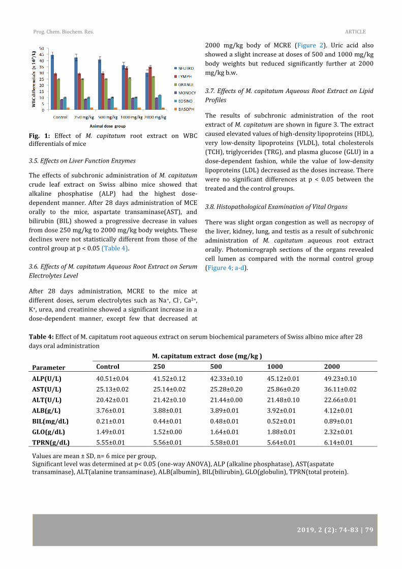
3.4. Effects on Hematological Parameters
The results of hematological parameters obtained showed
that most of the parameters increase in a dose-dependent
fashion in all the groups (Table 3; Figure 1). White blood
cell differentials decrease in doses dependent fashion, but
the WBC differentials, do not show a significant increase
at doses 250 to 2000 mg/kg.
Cletus Anes Ukwubile et.al Prog. Chem. Biochem. Res.
78|2019, 2 (2): 74-83
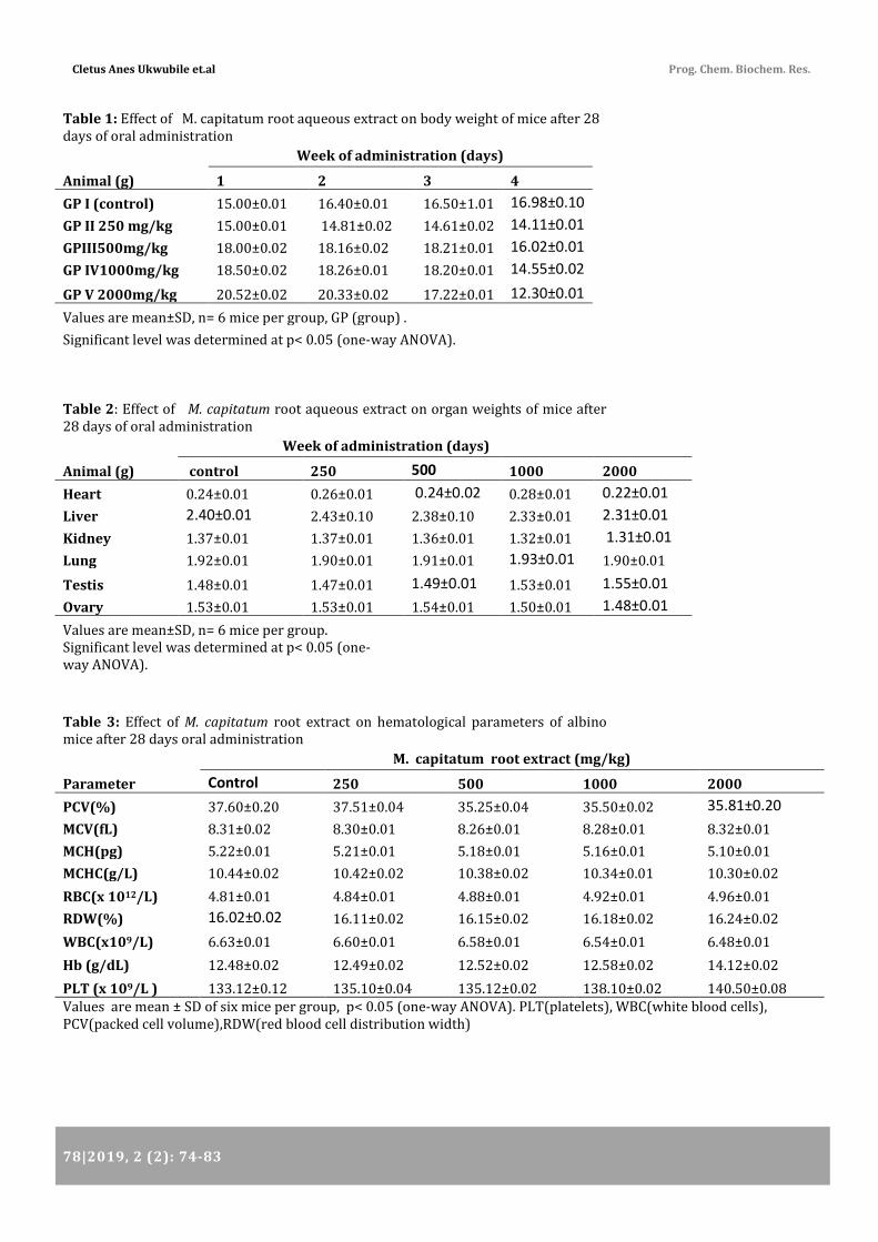
Table 1: Effect of M. capitatum root aqueous extract on body weight of mice after 28 days of oral administration
Week of administration (days)
Animal (g) 1 2 3 4
GP I (control) 15.00±0.01 16.40±0.01 16.50±1.01 16.98±0.10
GP II 250 mg/kg 15.00±0.01 14.81±0.02 14.61±0.02 14.11±0.01
GPIII500mg/kg 18.00±0.02 18.16±0.02 18.21±0.01 16.02±0.01
GP IV1000mg/kg 18.50±0.02 18.26±0.01 18.20±0.01 14.55±0.02
GP V 2000mg/kg 20.52±0.02 20.33±0.02 17.22±0.01 12.30±0.01
Values are mean±SD, n= 6 mice per group, GP (group) .
Significant level was determined at p< 0.05 (one-way ANOVA).
Table 2: Effect of M. capitatum root aqueous extract on organ weights of mice after 28 days of oral administration
Week of administration (days)
Animal (g) control 250 500 1000 2000
Heart 0.24±0.01 0.26±0.01 0.24±0.02 0.28±0.01 0.22±0.01
Liver 2.40±0.01 2.43±0.10 2.38±0.10 2.33±0.01 2.31±0.01
Kidney 1.37±0.01 1.37±0.01 1.36±0.01 1.32±0.01 1.31±0.01
Lung 1.92±0.01 1.90±0.01 1.91±0.01 1.93±0.01 1.90±0.01
Testis 1.48±0.01 1.47±0.01 1.49±0.01 1.53±0.01 1.55±0.01
Ovary 1.53±0.01 1.53±0.01 1.54±0.01 1.50±0.01 1.48±0.01
Values are mean±SD, n= 6 mice per group.
Significant level was determined at p< 0.05 (one-way ANOVA).
Table 3: Effect of M. capitatum root extract on hematological parameters of albino mice after 28 days oral administration
M. capitatum root extract (mg/kg)
Parameter Control 250 500 1000 2000
PCV(%) 37.60±0.20 37.51±0.04 35.25±0.04 35.50±0.02 35.81±0.20
MCV(fL) 8.31±0.02 8.30±0.01 8.26±0.01 8.28±0.01 8.32±0.01
MCH(pg) 5.22±0.01 5.21±0.01 5.18±0.01 5.16±0.01 5.10±0.01
MCHC(g/L) 10.44±0.02 10.42±0.02 10.38±0.02 10.34±0.01 10.30±0.02
RBC(x 1012/L) 4.81±0.01 4.84±0.01 4.88±0.01 4.92±0.01 4.96±0.01
RDW(%) 16.02±0.02 16.11±0.02 16.15±0.02 16.18±0.02 16.24±0.02
WBC(x109/L) 6.63±0.01 6.60±0.01 6.58±0.01 6.54±0.01 6.48±0.01
Hb (g/dL) 12.48±0.02 12.49±0.02 12.52±0.02 12.58±0.02 14.12±0.02
PLT (x 109/L ) 133.12±0.12 135.10±0.04 135.12±0.02 138.10±0.02 140.50±0.08 Values are mean ± SD of six mice per group, p< 0.05 (one-way ANOVA). PLT(platelets), WBC(white blood cells), PCV(packed cell volume),RDW(red blood cell distribution width)
Prog. Chem. Biochem. Res. ARTICLE
2019, 2 (2): 74-83 | 79
Fig. 1: Effect of M. capitatum root extract on WBC differentials of mice
3.5. Effects on Liver Function Enzymes
The effects of subchronic administration of M. capitatum
crude leaf extract on Swiss albino mice showed that
alkaline phosphatise (ALP) had the highest dose-
dependent manner. After 28 days administration of MCE
orally to the mice, aspartate transaminase(AST), and
bilirubin (BIL) showed a progressive decrease in values
from dose 250 mg/kg to 2000 mg/kg body weights. These
declines were not statistically different from those of the
control group at p < 0.05 (Table 4).
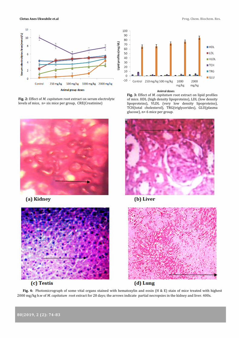
3.6. Effects of M. capitatum Aqueous Root Extract on Serum
Electrolytes Level
After 28 days administration, MCRE to the mice at
different doses, serum electrolytes such as Na+, Cl-, Ca2+,
K+, urea, and creatinine showed a significant increase in a
dose-dependent manner, except few that decreased at
2000 mg/kg body of MCRE (Figure 2). Uric acid also
showed a slight increase at doses of 500 and 1000 mg/kg
body weights but reduced significantly further at 2000
mg/kg b.w.
3.7. Effects of M. capitatum Aqueous Root Extract on Lipid
Profiles
The results of subchronic administration of the root
extract of M. capitatum are shown in figure 3. The extract
caused elevated values of high-density lipoproteins (HDL),
very low-density lipoproteins (VLDL), total cholesterols
(TCH), triglycerides (TRG), and plasma glucose (GLU) in a
dose-dependent fashion, while the value of low-density
lipoproteins (LDL) decreased as the doses increase. There
were no significant differences at p < 0.05 between the
treated and the control groups.
3.8. Histopathological Examination of Vital Organs
There was slight organ congestion as well as necropsy of
the liver, kidney, lung, and testis as a result of subchronic
administration of M. capitatum aqueous root extract
orally. Photomicrograph sections of the organs revealed
cell lumen as compared with the normal control group
(Figure 4; a-d).
Table 4: Effect of M. capitatum root aqueous extract on serum biochemical parameters of Swiss albino mice after 28
days oral administration
M. capitatum extract dose (mg/kg )
Parameter Control 250 500 1000 2000
ALP(U/L) 40.51±0.04 41.52±0.12 42.33±0.10 45.12±0.01 49.23±0.10
AST(U/L) 25.13±0.02 25.14±0.02 25.28±0.20 25.86±0.20 36.11±0.02
ALT(U/L) 20.42±0.01 21.42±0.10 21.44±0.00 21.48±0.10 22.66±0.01
ALB(g/L) 3.76±0.01 3.88±0.01 3.89±0.01 3.92±0.01 4.12±0.01
BIL(mg/dL) 0.21±0.01 0.44±0.01 0.48±0.01 0.52±0.01 0.89±0.01
GLO(g/dL) 1.49±0.01 1.52±0.00 1.64±0.01 1.88±0.01 2.32±0.01
TPRN(g/dL) 5.55±0.01 5.56±0.01 5.58±0.01 5.64±0.01 6.14±0.01
Values are mean ± SD, n= 6 mice per group, Significant level was determined at p< 0.05 (one-way ANOVA), ALP (alkaline phosphatase), AST(aspatate transaminase), ALT(alanine transaminase), ALB(albumin), BIL(bilirubin), GLO(globulin), TPRN(total protein).
Cletus Anes Ukwubile et.al Prog. Chem. Biochem. Res.
80|2019, 2 (2): 74-83
Fig. 4: Photomicrograph of some vital organs stained with hematoxylin and eosin (H & E) stain of mice treated with highest
2000 mg/kg b.w of M. capitatum root extract for 28 days; the arrows indicate partial necropsies in the kidney and liver. 400x.
Fig. 2: Effect of M. capitatum root extract on serum electrolyte levels of mice, n= six mice per group, CRE(Creatinine)
Fig. 3: Effect of M. capitatum root extract on lipid profiles of mice. HDL (high density lipoproteins), LDL (low density lipoproteins), VLDL (very low density lipoproteins), TCH(total cholesterol), TRG(triglycerides), GLU(plasma glucose), n= 6 mice per group.
Prog. Chem. Biochem. Res. ARTICLE
2019, 2 (2): 74-83 | 81
4. Discussion
Recently, it has been estimated that about 80 % of the
population of low-income countries are fully dependent
upon herbal medicines for their primary healthcare.
Higher medicinal plants are the major source of drug in
traditional medicine [22].
The World Health Organization (WHO) also estimated that
80 to 90 % of the world’s population relies mainly on local
herbal practitioners [23]. Medicinal plants are used as
tinctures, poultices, powders and teas followed by
formulations, and lastly as pure compounds. The extracts
from these plants have been used by humans since time
immemorial for treating many ailments. These plants have
also provided very important drugs such as analgesics
(like morphine), antitussives, antihypertensives,
cardiotonic, antineoplastics, and antimalarials. Even
though African traditional medicine is the oldest and
probably the most diverse of all healthcare systems yet,
detailed documentation on the use of medicinal plants in
Africa is lacking till date. Aside, with rapid urbanization in
most places, traditional oral knowledge is dwindling fast.
The side effects resulting from the use of herbal drugs
could be very fatal if not checked early, and this aspect of
the toxicological study has not been given the needed
attention.
Animals have been used as models for centuries to
predict what chemicals and environmental factors would
do to humans. The earliest uses of experimental animals
are lost in prehistory, and much of what is recorded in
early history about toxicology testing indicates that
humans were the models of choice. This work consisted of
dosing test animals with known quantities of agents
(poisons or drugs) and included the careful recording of
the resulting clinical signs and gross necropsy
observations. The use of animals as predictors of potential
ill effects has grown since that time. Animal experiments
also have served rather successfully as identifiers of
potential hazards and toxicity in humans for conventional
drugs with many intended uses [24]. This is the main
reason why mice were used in this study. Behavioural
changes such as body itching, reddish eyes, and weight
loss are salient indices for determining the effects of
short-term use of herbal drugs on animals [25]. In this
present study, in the acute study, there were no
observable signs of deaths or mortality at 300 mg/kg b.w
of M. capitatum aqueous root extract. But at a higher dose
of 2000 mg/kg b.w MCRE, there was reddish eyes, itching
and restless in the mice which only lasted for a few
minutes, as seen in Table 1.
Subchronic toxicity study revealed significant weight
increase from week one to week four among the
treatment groups (p <0.05; one-way ANOVA). These
increases were significantly different from the control
group, which suggest that the extract must have supplied
the animals with other nutrients for their metabolism. The
animals did not show much increase in the relative
weights of organs in the doses administered, but there
was no loss of weights from the lowest dose of 250 mg/kg
b.w to the highest dose of 2000 mg/kg b.w. (Table 2) The
result further suggests that the extract did not induce any
form of infection in the rats at these doses. It has been
reported that many herbal drugs or medicinal plants
when used in the treatment of diseases usually induced
infections in the animals due to unhygienic ways of
preparation and storage [26].
In hematology and clinical pathology, damage to the
cardiovascular systems is usually indicated by the values
of the hematological parameters. From our study, all the
hematological parameters increased in a dose-dependent
fashion (Table 3). The monocytes and basophils do not
show further increase at 1000 and 2000 mg/kg b.w. doses
in the rats which suggest that high doses of the extract do
not have a significant effect on these WBC differentials
(Figure 1). However, these values were within the
acceptable range, and any significant decrease in them
could suggest a sign of anomalies in the body, hence the
28 days subchronic administration of this extract further
suggests that M. capitatum methanol leaf extract does not
possess any deleterious toxicological effects on the rats at
the doses investigated.
Our study also revealed that at the doses of 250, 500,
1000, and 2000 mg/kg b.w. serum phosphate ions and
uric acid decreased progressively. Serum sodium ion
(Na+) had the highest value compared to other serum
electrolytes, followed by serum chloride ion (Cl-) (Figure
2). It is, therefore, justified on why the sweet-salty taste of
the plant root is chewed by the Fulani herder's
communities in Nigeria as a remedy for stomach ache.
The liver is the largest organ in the body of an animal,
which is involved in the metabolism of toxic substances
and drugs. To know the condition of the liver, liver
enzyme biomarkers are important parameters to use in
the medical diagnosis of liver related ailments. It has been
reported that elevated levels of alanine transaminase
(ALT), alanine phosphatase (ALP) and aspartate
transaminase (AST) biomarker enzymes in the liver cells
is an indication of a diseased liver condition [27]. On the
other hand, ALT is a better biomarker enzyme that is
specific to liver injuries, because AST is indicative of
Cletus Anes Ukwubile et.al Prog. Chem. Biochem. Res.
82|2019, 2 (2): 74-83
disease condition of other body organs such as the
muscles and heart. In addition, an elevated ALP value is an
indication that the bile duct is not normal or there is
blockage of the bile duct due to the tumor. From the
results in Table 4, ALT, AST, and ALP values do not
increase beyond their normal recommended range for
mice. Bilirubin is a product obtained from the breakdown
of hemoglobin, and it is often linked to liver diseases such
as jaundice and nonmetabolic red blood cells. From our
study, the progressive decrease in the value of bilirubin at
doses of 250, 500, 1000, and 2000 mg/kg b.w. showed
that MCRE does not present any injuries to the liver or the
heart (Figure; 4a-d).
High levels of lipid a condition known as hyperlipidemia is
the major cause of atherosclerosis in humans. In
cardiovascular pharmacology, less attention has been paid
to hyperlipidemia, unlike hypolipidemia. Our study had
revealed that oral administration of M. capitatum root
extract (MCRE) on mice for 28 days reduces low density
lipoproteins (LDL) at increased doses while other lipid
profiles namely: high-density lipoproteins(HDL), very
low-density lipoproteins (VLDL), total cholesterol(TCH),
triglycerides(TRG), and plasma glucose (GLU) showed
slight dose-dependent values (Figure 3). These values
were not different when compared to the untreated
normal control group at p < 0.05 (one-way ANOVA). The
low value of TCH obtained from this study further
supported the previous findings by Ukwubile et al. (2016)
when they showed that MCRE can reduce total cholesterol
level in albino mice [28, 29]. The presence of cells within
the lumen of vital organs has been reported to cause
blockage of blood vessels thereby, obstructing the normal
flow of blood [30]. In this present study, histopathological
examination of some of the organs namely: kidney, liver,
heart, and testis (Fig.4a-d) do not show the presence of
cells in the lumen, necrosis, liver congestion and
thickening of cells, which further affirmed safety of the
plant extract in the treatment of certain diseases in
traditional medicine in Nigeria.
Finally, we conclude that Melastomastrum capitatum
aqueous root extract is considered safe at the doses
investigated in the animals. Nevertheless, it is safer to use
the extract at moderate doses because, high dose (2000
mg/kg b.w) of the root extract is slightly toxic to the mice,
and this effect can be seen in humans when used in
traditional medicine for the treatment of diseases.
5. Conclusion In these present findings from our study, it is concluded
that aqueous root extract of Melastomastrum capitatum is
not toxic at lower doses and well tolerated at higher
doses. This is because there were no serious clinical
symptoms in the animals after oral subchronic study.
There were no mortalities in all the groups, and organ
necropsies as well as dense liver congestion. The initial
results showed the potentials of exploring therapeutic and
pharmaceutical products of interest from the root extract
which has reduced or no adverse effects. It is, therefore,
recommended that further study be to carry out in to
determine the toxicity effect of M. capitatum root extract
animal embryos, as well as gestation cycles of animals for
comparing results. Finally, the use of M. capitatum root
extract as an oral medication in traditional medicines for
disease treatment should be done at doses which do not
elicit any abnormality in its host.
6. Declaration
6.1 Conflict of Interest
We have no conflict of interest.
6.2 Funding
No available source of funding from external bodies
except by the authors.
6.3 Author's Contribution
This work was designed by Cletus Anes Ukwubile,
Emmanuel Oise Ikpefan, and Mathias Simon Bingari.
Proper laboratory research and the first manuscript draft
was done by Cletus Anes Ukwubile while Livinus Tam,
Emmanuel Oise Ikpefan and Mathis Simon Bingari are
collaborators.
Acknowledgment
The authors are grateful to the management and staff of Sancta Maria Clinic/Integrated Laboratory Bali Nigeria for allowing us access to some of the equipment used in this study.
Prog. Chem. Biochem. Res. ARTICLE
2018, 2(2), 74-83 | 83
References
[1] J.R. Tabuti, K.A. Lye and S.S. Dhillion, Traditional herbal
drugs of Bulamogi, Uganda: plants, use and
administration. J Ethnopharmacol, 88 (2003) 19-44. [2] A. Gupta and H. Chitme, Herbal medicine for health. The
eastern pharmacist, 43 (2000) 41-5. [3] S.N. Yoganarasiman, Medicinal plants of India-Tamilnadu.
Vol. 2. (2000).
[4] Z.-G. Wang and J. Ren, Current status and future direction
of Chinese herbal medicine. Trends in
Pharmacological Sciences, 23 (2002) 347-348. [5] A. Sofowora, Medicinal plants and traditional medicine in
Africa. (1996): Karthala.
[6] U. Epundu, E. Adinma, B. Ogbonna and O. Epundu,
Medical tourism, public health and economic
development in Nigeria: Issues and Prospects. Asian
Journal of Medicine and Health, 7 (2017) 1-10. [7] H.M. Burkill, The useful plants of west tropical Africa.
Volume 2: Families EI. (1994): Royal Botanic
Gardens.
[8] OECD, Draft Guidance Document on the Design and
Conduct of Chronic Toxicity and Carcinogenicity
Studies. . OECD Series on Testing and Assessment
Vol. 116. (2009), Paris.
[9] A. Hutchings, Zulu medicinal plants: An inventory. (1996):
University of Natal press.
[10] J. Nebedum, K. Ajeigbe, E. Nwobodo, C. Uba, O.
Adesanya, O. Fadare and D. Ofusori, Comparative
study of the ethanolic extracts of four Nigerian plants
against some pathogenic microorganisms. Res J Med
Plant, 3 (2009) 23-28. [11] H. Fluck, Medicinal Plants and Their uses. Vol. . (2013),
New York: W. Fulsome and Comp. Ltd, .
[12] C.A. Ukwubile, I.E. Oise and A.I. Bruno, Application of
Melastomastrum capitatum Fern.(Melastomataceae)
loaded-exosome as analgesic drug carrier in acetic
acid-induced Swiss albino mice. Molecular Biology
Research and Innovations, 1 (2016) 19-23. [13] J.D. Bancroft and M. Gamble, Theory and practice of
histological techniques. (2008): Elsevier health
sciences.
[14] A. Babaei, M. Aminikhah and A.R. Taheri, A Multi-
Walled Carbon Nano-Tube and Nickel Hydroxide
Nano-Particle Composite-Modified Glassy Carbon
Electrode as a New Sensor for the Sensitive
Simultaneous Determination of Ascorbic Acid,
Dopamine and Uric Acid. Sensor letters, 11 (2013)
413-422. [15] T.N. OECD, 423: Acute Oral toxicity–Acute Toxic Class
Method. OECD Guidelines for the Testing of
Chemicals, Section, 4 (2001)
[16] Y. Saidu, F. Nwachukwu, L. Bilbis, U. Faruk and A.
Abbas, Toxicity studies of the crude aqueous root
extract of albizzia chevalieri harms in albino rats.
Nigerian Journal of Basic and Applied Sciences, 18 (2010)
[17] G. Mbaka, O. Adeyemi and A. Oremosu, Acute and sub-
chronic toxicity studies of the ethanol extract of the
leaves of Sphenocentrum jollyanum
(Menispermaceae). Agriculture and Biology Journal
of North America, 1 (2010) 265-272. [18] O. Olorunnisola, G. Bradley and A. Afolayan, Acute and
sub-chronic toxicity studies of methanolic extract of
Tulbaghia violacea rhizomes in Wistar rats. African
Journal of Biotechnology, 11 (2012) 14934-14940. [19] V. Fuster, J. Eric and E. Nabel, Pathno-biology of
asymptomatic arthrosclerosis leading to symptomatic
artherothrombosis. J. Am. Cardiol, 46 (2005) 937-
941. [20] W.M. Kluwe, Renal function tests as indicators of kidney
injury in subacute toxicity studies. Toxicology and
applied pharmacology, 57 (1981) 414-424. [21] S. Sasser, M. Varghese, A. Kellermann and J. Lormand,
Prehospital trauma care systems. 2005. Geneva:
World Health Organization,
[22] T.S. Ballard, P. Mallikarjunan, K. Zhou and S. O’Keefe,
Microwave-assisted extraction of phenolic
antioxidant compounds from peanut skins. Food
Chemistry, 120 (2010) 1185-1192. [23] J.T. Mukinda and J.A. Syce, Acute and chronic toxicity of
the aqueous extract of Artemisia afra in rodents.
Journal of ethnopharmacology, 112 (2007) 138-
144. [24] S. Tiwari, Plants: A rich source of herbal medicine.
Journal of natural products, 1 (2008) 27-35. [25] B. Thapa and A. Walia, Liver function tests and their
interpretation. The Indian Journal of Pediatrics, 74 (2007) 663-671.
[26] J. Ozer, M. Ratner, M. Shaw, W. Bailey and S.
Schomaker, The current state of serum biomarkers of
hepatotoxicity. Toxicology, 245 (2008) 194-205. [27] P.W. Wilson, R.D. Abbott, R.J. Garrison and W.P.
Castelli, Estimation of very-low-density lipoprotein
cholesterol from data on triglyceride concentration in
plasma. Clinical chemistry, 27 (1981) 2008-2010. [28] K. Kamari and A. Taheri, Preparation and evaluation of
magnetic core–shell mesoporous molecularly
imprinted polymers for selective adsorption of
amitriptyline in biological samples. Journal of the
Taiwan Institute of Chemical Engineers, 86 (2018)
230-239. [29] D. Chokshi, Subchronic oral toxicity of a standardized
white kidney bean (Phaseolus vulgaris) extract in rats.
Food and chemical toxicology, 45 (2007) 32-40. [30] M.I. Ezeja, A.O. Anaga and I.U. Asuzu, Acute and sub-
chronic toxicity profile of methanol leaf extract of
Gouania longipetala in rats. Journal of
ethnopharmacology, 151 (2014) 1155-1164.