Embed Size (px)
Citation preview

Chapter 7
Programmed Cell Death as a Response to High Light,UV and Drought Stress in Plants
Weronika Wituszyńska and Stanisław Karpiński
Additional information is available at the end of the chapter
http://dx.doi.org/10.5772/53127
1. Introduction
Because of their sessile nature, plants are unable to avoid fluctuating environment condi‐tions like high light, ultraviolet radiation, drought, salt stress, heat, cold or flooding. Uponcertain threshold of these changes, plant cells can no longer maintain proper metabolic proc‐esses and programmed cell death (PCD) is induced.
PCD is an essential cell suicide process in animals, yeast and plants. In multicellular organ‐isms, it plays an important role in the cell homeostasis maintenance, tissue specialization,removing of damaged or infected cells and acclimatory response. In contrast to necroticdeath, which proceeds via swelling, lysis and leakage of cell content, PCD is a highly regu‐lated and organized process. This controlled disassembly of cell involves the condensation,shrinkage and fragmentation of both cytoplasm and nucleus and DNA laddering. Further‐more, while PCD can occur during development and is regulated by complex mechanisms,necrosis does not require the activity of proteases nor nucleases and is not associated withsignal transduction pathways [1].
There are two main categories of PCD in plants: developmentally- and environmentally-in‐duced PCD. The first one is a genetically encoded process which plays a crucial role in thedevelopment of some tissues and organs. It is involved in tracheary elements formulationduring xylem differentiation. Tracheary elements are long cells that transport water andmineral salts, and serve as a structural support in vascular plants. Their formation occursafter secondary wall synthesis and begins with a collapse of the central vacuole and releaseof lytic enzymes, followed by degradation of cellular content [2]. Another example of devel‐opmentally-induced PCD is a formation of unisexual flowers in monoecious species (e.g.maize), bearing generative organs of both sexes on the same plant. Sex determination inthese species involves the developmental arrest of one of the organ primordia - either the
© 2013 Wituszyńska and Karpiński; licensee InTech. This is an open access article distributed under the termsof the Creative Commons Attribution License (http://creativecommons.org/licenses/by/3.0), which permitsunrestricted use, distribution, and reproduction in any medium, provided the original work is properly cited.

female or male within a bisexual floral meristem [3]. The production of complex leaf shapesalso frequently employs PCD. Such remodeling of leaf blades occurs in Monstera obliqua,Monstera deliciosa or lace plant [4]. These species tend to induce death pathway in somepatches of cells and thus form distinctive perforations within the leaf [5]. PCD is also engag‐ed in such processes as dying of aleurone cells in seeds of monocots, root cap shedding oranther dehiscence. Senescence, which is the final stage of vegetative and generative develop‐ment, preceding plant organs death, also involves PCD. In deciduous trees, senescence is ex‐hibited in the changes of leaves color developing during autumn. It enables the activeturnover of cellular material and its use in other organs. For example, nutrients, such as ni‐trogen, recycled from leaves are used for the synthesis of proteins that will be stored instems and will support growth in the following vegetative season. Moreover, PCD duringsenescence helps to block spreading of diseases to still vital parts of the plant [6].
Developmental PCD is induced by internal factors and occurs at defined time and in partic‐ular plant tissue. On the contrary, environmentally-induced PCD is triggered by differentstimuli ranging from pathogen infection to environmental factors [7]. During infection ofplant leaves by pathogens, a specific gene-for-gene (avr–R) interaction triggers defense re‐sponses. Upon such plant-microbe interaction, cell death takes a form of so-called hypersen‐sitive response (HR) and includes a burst of reactive oxygen species (ROS). HR leads to theformation of a lesion which is clearly delimited from surrounding healthy cells and thusprevents the spread of pathogen throughout the plant tissue. Certain mutations in manyplant species have been demonstrated to cause spontaneous, HR-resembling lesions, whichsuggests that this type of cell death is under a genetic control. Such lesion mimic mutantsare divided into two groups: related to the initiation of PCD (inappropriate induction ofPCD and formation of localized lesion spots) or propagation (inability to stop PCD once ithas been initiated). Both these groups of mutants are currently widely investigated sincethey can provide insight into the general mechanism of PCD in plants [8–12]. The existenceof these two classes suggests that genetically distinct processes underlie the lesion forma‐tion: the initiation of cell death and its spread to surrounding cells as well as the existence ofcommunication signals between dying and healthy cells in determining the lesion size.
In natural habitats, plants are constantly exposed to a variety of environmental stresses thatcan lead to the disturbance in cellular homeostasis and consequently limit crop yield. Pro‐grammed cell death is a fundamental cellular process associated with the defense responsesto abiotic stimuli such as excessive irradiation, ozone, ultraviolet radiation, heat, cold,drought or flooding. One of the example factor triggering PCD is hypoxia, a condition inwhich plant is deprived of oxygen supply. In response to waterlogging and lower O2 con‐centration in the ground, cortex of the root can form aerating tissue called aerenchyma [13].The internal air spaces are generated through PCD and facilitate gas diffusion from aerialorgans to waterlogged roots [14]. Although it is unfavorable for biomass production, the se‐lective death of cells and tissues under abiotic stresses eventually provides survival advan‐tages for the whole organism. At the organismal level, PCD helps to maintain tissue andorgan homeostasis, enables developmental adaptation and nutrient resorption from dyingcells thus increases the probability of survival. It also leads to the signals transduction from
Abiotic Stress - Plant Responses and Applications in Agriculture208

cells undergoing PCD to healthy, not-affected cells and triggers stress tolerance and acclima‐tion to adverse conditions [6,15].
2. Hallmarks and the regulation of programmed cell death
While the cascade of events and molecules regulating PCD have been already well described inanimal cells, mechanisms underlying plant PCD remain still inexplicable. Therefore, numer‐ous studies in plants rely on the comparison of PCD mechanisms to animals. Apoptosis (well-studied form of animal PCD) features in cell shrinkage, chromatin condensation, cleavage ofDNA (called DNA laddering) and nuclear fragmentation. The mechanism of PCD depends ona family of cysteine proteases called caspases that cleave their target proteins after aspartic acidresidues. Caspases are synthesized in the cell as inactive precursors (procaspases). Once acti‐vated, caspases cleave and activate other procaspases which results in a self-amplifying cas‐cade. They can also cleave other proteins such as nuclear lamins or proteins that hold DNA-degrading enzymes in inactive form, releasing DNases to cut DNA. The destructive proteasecascade is irreversible, therefore caspase activity needs to be tightly controlled. Procaspase ac‐tivation is induced by the release of electron carrier protein - cytochrome c from mitochondriato the cytosol. The family of Bcl-2 proteins regulates the activation of programmed cell death.Some members of this family (e.g. Bcl-2) block the release of cytochrome c, inhibiting apopto‐sis. Others (e.g. Bax and Bak) act as PCD inducers, promoting cytochrome c leakage. IAP (in‐hibitor of apoptosis) proteins are another family involved in apoptosis regulation as they bindto some procaspases, preventing their activation or to caspases, inhibiting their activity. Pro‐teins that block IAPs are released together with cytochrome c which increases the efficiency ofcell death process [16].
Many hallmarks of plant PCD seem to be similar to animals such as cytoplasm shrinkage,chromatin condensation and DNA cleavage, mitochondrial swelling, disruption of organ‐elles and plasma membrane collapse [17]. The major difference in executing PCD betweenanimals and plants lies in the process of removing the cell content after its death. While inanimal cells, removal action is undertaken by other cells to avoid the activation of inflamma‐tory response, in plants there is a leakage of the cell content into the apoplast and remainsare not engulfed by surrounding cells [10]. Moreover, plants exhibit some distinctive fea‐tures of PCD that result from the presence of chloroplasts and the significance of vacuoles[18,19]. Plant vacuoles represent important storage organelles that are the repository of hy‐drolytic enzymes such as proteases, lipases and nucleases. Vacuoles are therefore postulatedto play a role in the turnover of organelles and cytoplasm during autophagy as a part ofclean-up system for dying cells. The component of this system is a caspase-like protease -the vacuolar processing enzyme (VPE) which plays a crucial role in such PCD pathways assenescence, lateral root formation and hypersensitive response. Upon receiving pro-apoptot‐ic signals, VPE activates hydrolases that execute the degradation of vacuolar membrane re‐sulting in the release of hydrolytic enzymes and subsequent degradation of cell content [19].
Programmed Cell Death as a Response to High Light, UV and Drought Stress in Plantshttp://dx.doi.org/10.5772/53127
209

Chloroplasts are strongly suggested to be key players during cell death responses as theyconstitute an important source of defense signaling molecules such as ROS, reactive nitro‐gen species (RNS) and defense hormones like salicylic acid (SA) and jasmonic acid (JA). Theoxidative burst is one of the earliest and most common plant response to abiotic and bioticstimuli [20]. The application of chloroplast-targeted, ROS-generating herbicides such asmethyl viologen (paraquat) induces cell death with the typical apoptotic traits [21].
Some of key proteins controlling animal cell death such as the Bcl-2 family and caspaseshave been proven to be not conserved in plants. It suggests that plants have developed someunique mechanisms of PCD [15]. Although orthologs of caspases have not been found inplants based on the sequence similarity, several studies using caspase-specific peptide inhib‐itors suggested the presence of caspase-like proteases (metacaspases) [22]. These caspase in‐hibitors have been demonstrated to prevent chemically-, UV- or HR-induced PCD [23–25]indicating that caspase-like proteins are indeed involved in the regulation of PCD in plants.Metacaspases (MC) differ from animal caspases in their substrate specificity as they cleaveproteins after arginine or a lysine residues. Nine predicted metacaspase-encoding geneshave been found in Arabidopsis thaliana and divided into two classes, depending on the pres‐ence (type I) or absence (type II) of the N-terminal zinc-finger domain that has the homologyto the LESION SIMULATING DISEASE 1 (LSD1) protein (see later) [26,27]. This domain isknown to participate in protein–protein interactions and could indicate that oligomerizationis important for MCs type I activation. The catalytic activities of AtMC4, AtMC5 and AtMC8have been found to be Ca2+-dependent while AtMC9 is active under mildly acidic condi‐tions. Thus, alterations in cellular Ca2+ concentration and pH, that are common during vari‐ous stresses, may help to control MCs activation. The sequence of AtMC4 has also revealedpotential self-cleavage sites that may facilitate additional regulation of protease activity toachieve sensitive control of PCD [28]. Additionally, metacaspase ATMC4 (AtMCP2) hasbeen proven to play a positive regulatory role in biotic and abiotic stress-induced PCD [29].AtMC1 and AtMC2, belonging to type I metacaspases, have opposite roles in the cell deathcontrol. There is a genetic evidence that AtMC2 acts as a negative regulator of AtMC1-in‐duced PCD. Therefore, it is hypothesized that proteins belonging to MC family execute ei‐ther anti- or pro-apoptotic functions and compete with each other in making the cell life-death decisions [30].
Although no orthologues of Bcl-2 family genes (Bcl-2 or Bax) have been found in plants,there are some studies demonstrating that the expression of these genes in plants can regu‐late programmed cell death pathway [31,32]. Transgenic plants overexpressing animal antia‐poptosis genes such as Bcl-2 have been proven to exhibit enhanced tolerance to both bioticand abiotic stress conditions [33,34]. Moreover, the homologue for animal Bax Inhibitor (BI)has been identified in Arabidopsis [35] and proven to inhibit cell death in plants expressingmammalian Bax [36]. Arabidopsis BI-1 (AtBI-1) has been reported to localize in the endoplas‐mic reticulum (ER) and to contain predicted transmembrane α-helices in the sequence, thatare conserved in two other AtBI-1-related proteins: BI-2 and BI-3. These proteins are hy‐pothesized to function in a similar fashion to the Bcl-2 family - as regulators of pro-death orsurvival pathways [10]. In plants, mRNA level of AtBI-1 increases during leaf senescence
Abiotic Stress - Plant Responses and Applications in Agriculture210
and under different abiotic stresses. The loss of function in AtBI1 results in the mutant hy‐persensitivity to environmental stimuli, whereas its overexpression in retarded PCD [37].
Numerous signals are constantly integrated by the cell to decide whether to enter or not thecell death pathway. Different plant hormones are involved in the regulation of cell death un‐der unfavorable conditions. One of the most important is SA which is intensively producedin cells after pathogen infection or various abiotic stresses [38]. Many lesion mimic mutantshave constitutively elevated levels of SA [39]. At high concentrations, SA functions as a celldeath inducer in cooperation with other signals. It can be also transported beyond the site ofsynthesis, acting as a signaling molecule and mediating systemic acquired resistance (SAR) -a whole-plant resistance response that prepares plant for another infection [40]. The exis‐tence of SA-dependent generation of ROS and the feedback control of SA synthesis by ROShave been also demonstrated [41]. SA and ROS have been proposed to work in a potentia‐tion feedback loop which acts to amplify signals leading to cell death. Another cell deathsignaling molecule - nitric oxide (NO) has been also demonstrated to regulate key steps inSA biosynthesis during pathogen infection [42]. Additionally, NO has been proven to coop‐erate with ROS and SA in inducing cell death [43]. Other phytohormones regulating celldeath under stress conditions are JA, gibberellic acid (GA), abscisic acid (ABA) and ethylene(ET). The latter is involved in the regulation of PCD during different developmental proc‐esses and responses to environmental stimuli. ET has been proven to participate in the for‐mation of aerenchyma in roots under hypoxia [14]. Antisense inactivation of the ETbiosynthetic enzyme - ACC oxidase delays leaf senescence and cell death in tomato [44].Ethylene is also required for the continuation of ROS accumulation - external supply of ETduring cell death increases ROS production and causes accelerated spreading of cell death[45]. JA is a plant signaling molecule best known for its role in the wound response but it isalso produced during wide range of biotic and abiotic stresses. It is involved in the inhibi‐tion of ROS- and ET-dependent lesion propagation [46]. JA derivatives such as methyl jasm‐onate (MeJA) are also engaged in the regulation of plant immune responses [47]. Uponexposure to stress, MeJA is produced and causes the activation of PCD through the induc‐tion of ROS generation, alterations in mitochondrial dynamics and photosynthetic collapse[48]. Another phytohormone - GA has been proven to promote cell death in cooperationwith ROS, whereas ABA delays GA-induced PCD. Such counteracting role of these hor‐mones relates to their influence on the ROS-scavenging enzymes expression [49]. ABA hasbeen also shown to delay ET- and GA-induced cell death in rice epidermal cells [50]. Allthese interactions between phytohormones and ROS indicate the complexity of PCD regula‐tion. Overmyer and colleagues [39] suggested the following series of events during oxida‐tive stress-triggered PCD. At the site of lesion initiation, the action of ROS is amplified.Increased ROS accumulation together with SA induces the cell death. During the initialprocess, JA signaling is hindered by SA and ET. Meanwhile, the burst of ET from the initialsite disperses to surrounding cells, amplifies ROS production that promotes the lesionspread. This is the signal to induce competitive reactions to PCD. Cell death results in theproduction of JA which acts as a negative regulator of the oxidative cell death cycle. JA,through the suppression of SA biosynthesis/signaling and the attenuation of ET sensitivity,inhibits the lesion propagation.
Programmed Cell Death as a Response to High Light, UV and Drought Stress in Plantshttp://dx.doi.org/10.5772/53127
211
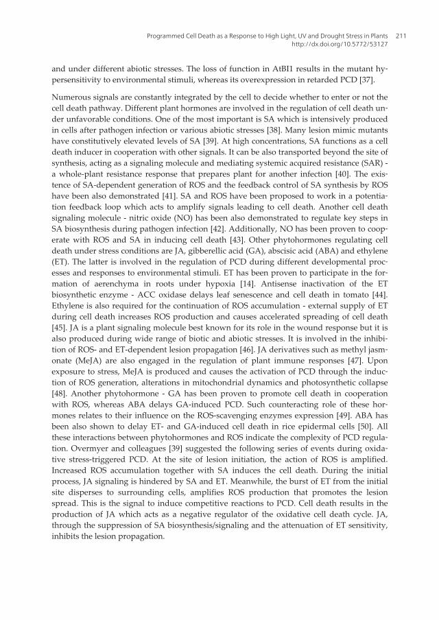
During early events of hypersensitive response, ion fluxes are induced. Ca2+ influx causedby external hydrogen peroxide application has been demonstrated to be sufficient in trigger‐ing HR in soybean cells [51]. Moreover, several cell death signaling proteins in plants exhibita function associated with lipids. Two Arabidopsis mutants, eds1 and pad4 have been provento be defective in HR signaling. EDS1 (ENHANCED DISEASE SUSCEPTIBILITY 1) andPAD4 (PHYTOALEXIN DEFICIENT 4) genes encode proteins with triacylglycerol lipasefunction [52,53] which provides a genetic evidence that phospholipid signaling is involvedin the induction of PCD. The level of phosphatidic acid (PA), produced by the phospholi‐pase D (PLD) increases during defense response [54]. One of PLD isoforms in Arabidopsishas been shown to impair ROS-mediated PCD in response to biotic and abiotic stimuli [55],indicating the role of PA as a negative signal of cell death propagation. It is also hypothe‐sized that the perturbation in sphingosine transport may cause cell death in plants since themutation in ACD11 gene encoding a sphingosine-transport protein, results in a lesion-mimicphenotype that is dependent on EDS1, PAD4, SA and light [56]. EDS1 and PAD4 are exten‐sively studied regulators of PCD. They constitute a regulatory hub that transduces redoxsignals in response to biotic and abiotic stresses. Both EDS1 and PAD4 are also importantactivators of SA signaling and mediate antagonism between JA and ET pathways during de‐fense responses [57]. Furthermore, they are responsible for the biotic and abiotic stress-in‐duced PCD in the LESION SIMULATING DISEASE 1 (LSD1) mutant [58,59]. The lsd1mutant fails to limit the spread of PCD under long photoperiod or after the infection withavirulent pathogens. It is one of the best characterized mutants in terms of programmed celldeath. The lsd1 mutant exhibits a runaway cell death (RCD) phenotype (Figure 1B) manifest‐ed in the inability to restrict the progression of cell death once it has been initiated [9,11],which provides a genetic evidence for LSD1 as a PCD repressor.
Figure 1. Runaway cell death (RCD) phenotype in the lsd1 mutant. 3-week-old Arabidopsis thaliana rosettes grown inlong photoperiod (>11 h); A – wild type; B – lsd1 mutant. Arrows indicate leaves undergoing runaway cell death.
The lsd1/cao double mutant which has reduced photosystem II (PSII) antenna size and thusreduced light absorption capacity due to the cao mutation in chloroplast signal recognitionparticle (cpSRP43), exhibits higher non-photochemical quenching (see later) and inhibitedRCD after excess light exposure in comparison to the lsd1 mutant. Therefore, the RCD in thelsd1 mutant has been linked to the amount of light energy absorbed in excess by the PSIIlight harvesting complex [11]. It has been shown that ET and ROS production in the lsd1
Abiotic Stress - Plant Responses and Applications in Agriculture212

mutant plants is elevated after plastoquinone reduction. The RCD in the lsd1 mutant plantshas been also proven to be inhibited by the mutation in EIN2, which encodes an ethylenereceptor. Additionally, the artificial impeding of foliar gas exchange in lsd1 has been shownto induce RCD, while high CO2 level has prevented cell death in this mutant. Importantly,lsd1 phenotype depends on EDS1 and PAD4, since in double mutants eds1/lsd1 and pad4/lsd1PCD is inhibited [11,14,60]. The formation of ROS by plasma-membrane-bound NADPH ox‐idase has been proposed to play a major role in RCD in the lsd1 mutant during the shift fromshort to long photoperiod, since the inhibition of this enzyme diminishes the formation oflesions [61]. All these results suggest that LSD1 acts as a ROS rheostat and is necessary foracclimation to conditions that promote oxidative stress. While LSD1 has been proven to neg‐atively regulate the cell death, a highly similar protein - LOL1 (LSD1 like 1) is suggested tobe a positive PCD-regulator [62]. It has been proposed that LSD1 and LOL1 might functionin an antagonistic fashion to regulate the cell death propagation. Both LSD1 and LOL1 areputative transcription factors (TF) or scaffold proteins since they possess zinc-finger do‐mains responsible for DNA/protein binding. Such Zn-finger motif of the C2C2 class hasbeen found in plants, algae and protozoa, but not in animals. Apart from LSD1 and LOL1,only five other Arabidopsis proteins contain one or more LSD1-like Zn-finger domains: LOL2,LOL6 and already mentioned metacaspases: AtMC1, AtMC2 and AtMC3 [12]. The secondand third Zn-finger domains of LSD1 are responsible for interacting with metacaspaseAtMC1, which is a positive regulator of PCD. The atmc1 mutation is able to suppress celldeath in lsd1. Furthermore, the interaction of LSD1 with AtbZIP10 transcription factor pre‐vents its translocation to the nucleus. AtbZIP10 has been proven to be a positive mediator ofRCD observed in the lsd1 mutant [63].
3. Reactive oxygen species in plants
The signaling during PCD proceeds mainly through the regulation of reactive oxygen spe‐cies [60,64,65]. ROS are produced continuously as by-products of various pathways local‐ized in chloroplasts, mitochondria and peroxisomes. They can occur as free radicals:superoxide radical (O2
•−), hydroxyl radical (•OH), perhydroxyl radical (HO2•), alkoxy radical
(RO•) or in non-radical forms: singlet oxygen (1O2) and hydrogen peroxide (H2O2). Mostabiotic stresses evoke the overproduction of ROS in plant tissues. Because of their high reac‐tivity, ROS can cause damage of proteins, lipids, carbohydrates and nucleic acids, ultimatelyleading to cell death (Figure 2).
The single reduction of O2 results in the formation of O2•−. From O2
•− other more reactiveROS like •OH or HO2
• can be formed. The Haber-Weiss reaction generates hydroxyl radicalfrom hydrogen peroxide and superoxide. In this reaction O2
•− donates an electron to Fe3+, re‐ducing ferric ion to ferrous:
Fe3+ + O2•−→ Fe2+ + O2
Programmed Cell Death as a Response to High Light, UV and Drought Stress in Plantshttp://dx.doi.org/10.5772/53127
213

The second step of •OH formation is the Fenton reaction in which reduced form of iron(Fe2+) transfers electrons to H2O2:
Fe2+ + H2O2 → Fe3+ + OH− + •OH
O2•− can be also protonated to form HO2
•. Furthermore, O2•− can react with another free radi‐
cal species as NO• to generate peroxynitrite (OONO−) [66]. Another form of ROS – singletoxygen is the first excited electron state of O2 that originates when an electron is elevated toa higher energy orbital (Figure 3).
Figure 2. ROS production and programmed cell death induced by abiotic stress (according to Gill and Tuteja,2010) [66].
Abiotic Stress - Plant Responses and Applications in Agriculture214
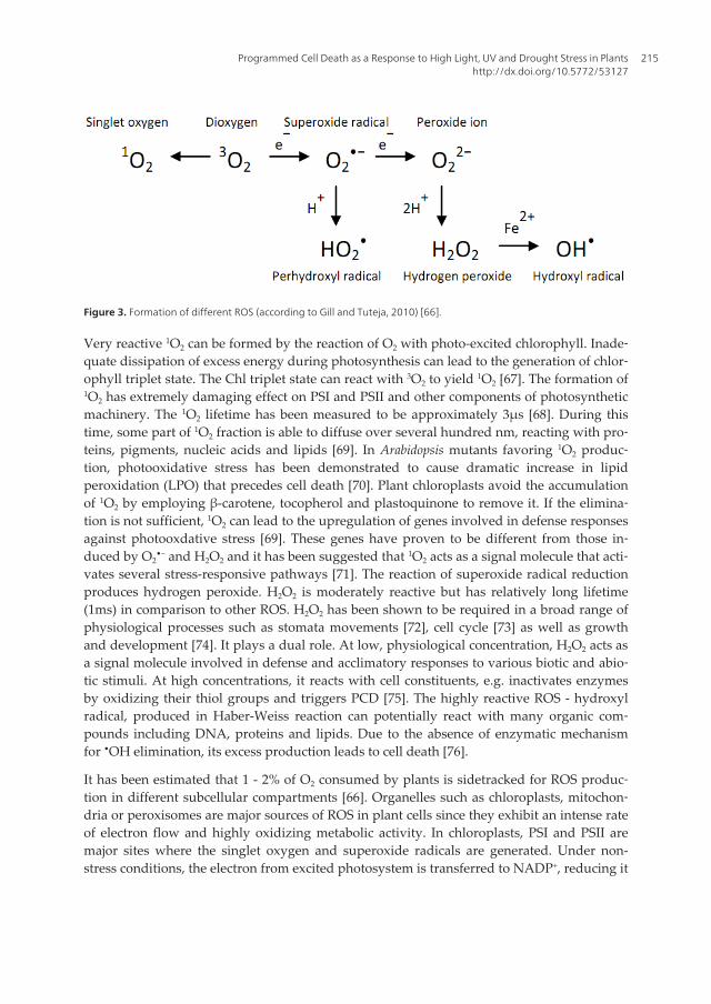
Figure 3. Formation of different ROS (according to Gill and Tuteja, 2010) [66].
Very reactive 1O2 can be formed by the reaction of O2 with photo-excited chlorophyll. Inade‐quate dissipation of excess energy during photosynthesis can lead to the generation of chlor‐ophyll triplet state. The Chl triplet state can react with 3O2 to yield 1O2 [67]. The formation of1O2 has extremely damaging effect on PSI and PSII and other components of photosyntheticmachinery. The 1O2 lifetime has been measured to be approximately 3µs [68]. During thistime, some part of 1O2 fraction is able to diffuse over several hundred nm, reacting with pro‐teins, pigments, nucleic acids and lipids [69]. In Arabidopsis mutants favoring 1O2 produc‐tion, photooxidative stress has been demonstrated to cause dramatic increase in lipidperoxidation (LPO) that precedes cell death [70]. Plant chloroplasts avoid the accumulationof 1O2 by employing β-carotene, tocopherol and plastoquinone to remove it. If the elimina‐tion is not sufficient, 1O2 can lead to the upregulation of genes involved in defense responsesagainst photooxdative stress [69]. These genes have proven to be different from those in‐duced by O2
•− and H2O2 and it has been suggested that 1O2 acts as a signal molecule that acti‐vates several stress-responsive pathways [71]. The reaction of superoxide radical reductionproduces hydrogen peroxide. H2O2 is moderately reactive but has relatively long lifetime(1ms) in comparison to other ROS. H2O2 has been shown to be required in a broad range ofphysiological processes such as stomata movements [72], cell cycle [73] as well as growthand development [74]. It plays a dual role. At low, physiological concentration, H2O2 acts asa signal molecule involved in defense and acclimatory responses to various biotic and abio‐tic stimuli. At high concentrations, it reacts with cell constituents, e.g. inactivates enzymesby oxidizing their thiol groups and triggers PCD [75]. The highly reactive ROS - hydroxylradical, produced in Haber-Weiss reaction can potentially react with many organic com‐pounds including DNA, proteins and lipids. Due to the absence of enzymatic mechanismfor •OH elimination, its excess production leads to cell death [76].
It has been estimated that 1 - 2% of O2 consumed by plants is sidetracked for ROS produc‐tion in different subcellular compartments [66]. Organelles such as chloroplasts, mitochon‐dria or peroxisomes are major sources of ROS in plant cells since they exhibit an intense rateof electron flow and highly oxidizing metabolic activity. In chloroplasts, PSI and PSII aremajor sites where the singlet oxygen and superoxide radicals are generated. Under non-stress conditions, the electron from excited photosystem is transferred to NADP+, reducing it
Programmed Cell Death as a Response to High Light, UV and Drought Stress in Plantshttp://dx.doi.org/10.5772/53127
215

to NADPH. However, under various abiotic stresses, the electron transport chain (ETC)tends to be overloaded and a part of the electron flow is diverted from ferredoxin to O2, re‐ducing it to O2
•−. The photoreduction of O2 at PSI proceeds via Mehler reaction and produ‐ces O2
•−, which is disproportionated to H2O2 and O2 with the use of superoxide dismutase.H2O2 is rapidly detoxified to H2O by the ascorbate peroxidase pathway (Figure 4A). Becauseof the electron flow from water in PSII to water in PSI that occurs in this process, it has beentermed the water–water cycle [77]. This cycle does not only scavenge O2
•− and H2O2, but alsogenerates a pH gradient across thylakoid membranes which enhances non-radiative dissipa‐tion of light energy by non-photochemical quenching (see later). Therefore, the water–watercycle is considered to function as a dissipatory mechanism of the excess energy [77,78].
H2O2 is also produced during a process that proceeds concurrently to the photosynthesis –photorespiration. During photosynthetic carbon assimilation, ribulose-1,5-bisphosphate car‐boxylase/oxygenase enzyme (Rubisco) uses CO2 to carboxylate ribulose-1,5-bisphosphate(RuBP). CO2 uptake results in the formation of two molecules of 3-phosphoglycerate (3-PGA) that are utilized for biosynthetic reactions and the recycling of the RuBP acceptor mol‐ecule. However, Rubisco can also use O2 to oxygenate RuBP, forming one molecule of 3-PGA and one molecule of 2-phosphoglycolate (2-PG). The latter cannot be used forbiosynthetic reactions and is considered as an inhibitor of the chloroplast function. Photores‐piration functions to convert 2-PG back to 3-PGA and thus to recover carbon. It constitutes aseries of reactions taking place in chloroplasts, peroxisomes, and mitochondria. 2-PG is de‐phosphorylated to glycolate in the chloroplast and transported to the peroxisome where it isoxidized to glyoxylate. O2 is the electron donor in this reaction, which results in H2O2 gener‐ation. Glyoxylate is transaminated to glycine which is transported to the mitochondrion,where two molecules of glycine are converted to serine and the remaining carbon and nitro‐gen are released as CO2 and NH3, respectively. The amine group is used to form a new gly‐cine from glyoxylate and the resulting hydroxypyruvate is reduced to glycerate. Finally,glycerate is phosphorylated in the chloroplast to form 3-PGA, which can be fed back to theCalvin cycle [79,80].
The production of ROS is also an unavoidable consequence of the aerobic respiration. It oc‐curs under normal respiratory conditions but can be enhanced in response to biotic andabiotic stress. ROS produced in mitochondria are regarded to be essential in PCD regulation[81]. In mitochondria, O2
•− is mainly produced in complex I, ubiquinone and complex III ofETC [82]. This O2
•− can be further converted into highly toxic •OH which may penetratemembranes and leave the mitochondrion [83]. Hydroxyl radical can also initiate the peroxi‐dation of mitochondrial membrane polyunsaturated fatty acids (PUFA) that leads to the for‐mation of cytotoxic lipid aldehydes, alkenals and hydroxyalkenals, such asmalonyldialdehyde (MDA). Lipid peroxidation products may cause cellular damage by re‐acting with other lipids, proteins and nucleic acids. The mitochondrial ETC produces signifi‐cant amount of ROS but the mitochondrial enzyme - alternative oxidase (AOX) can preventROS overproduction [84]. Some studies, performed on tobacco plants, have demonstratedthat the lack of AOX induces PCD while the AOX overexpression decreases the lesion sizeduring HR [85,86].
Abiotic Stress - Plant Responses and Applications in Agriculture216

Another source of ROS - peroxisomes are small, spherical organelles with an oxidative typeof metabolism. There are two sites of O2
•− generation in peroxisomes: in the organelle ma‐trix, where xanthine oxidase (XOD) catalyzes the oxidation of xanthine and hypoxanthine touric acid and in the membrane, by components of peroxisomal ETC. The main metabolicprocesses responsible for H2O2 generation in peroxisomes are photorespiratory glycolate ox‐idase reaction, fatty acid β-oxidation, enzymatic reaction of flavin oxidases and dispropor‐tionation of superoxide radicals [87].
ROS are also generated in the apoplast by NADPH oxidases residing in the plasma mem‐brane and generating superoxide radicals. The extracellular O2
•− is quickly mutated intoH2O2 or converted to •OH. The latter initiates a series of reactions that cause a plasma mem‐brane damage, finally leading to cell death. Two Arabidopsis respiratory burst oxidase genes,RBOHD and RBOHF, that encode NADPH oxidases have been proven to be responsible forROS production during the HR. Enzymes such as cell wall peroxidases, germin-like oxalateoxidases and amine oxidases have been proposed as a source of hydrogen peroxide in theapoplast. The alkalization of apoplast upon elicitor recognition precedes the production ofH2O2 by pH-dependent cell wall peroxidases [88].
The peroxidation of lipids is considered as one of the most damaging processes occurring inthe cell. The damage of membrane is often considered as a parameter determining the levelof cell destruction under various stresses. Upon ROS overproduction, polyunsaturated pre‐cursors undergo lipid peroxidation, forming small hydrocarbon fragments such as ketonesor aldehydes. LPO in both cellular and organellar membranes affects proper cellular func‐tions and aggravates oxidative stress by the production of lipid-derived radicals [89]. Thisprocess often affects PUFA, since they contain multiple double bonds in between which liemethylene (-CH2-) groups with reactive hydrogens. Hydroxyl or perhydroxyl radicals com‐bining with a hydrogen atom produce water and a fatty acid radical. The fatty acid radicalsare unstable and react rapidly with molecular oxygen, creating a peroxyl-fatty acid radical(ROO•). Once initiated, ROO• can further propagate the peroxidation chain reaction by ab‐stracting a hydrogen atom from PUFA side chains. The resulting lipid hydroperoxide easilydecomposes into several reactive species including: lipid alkoxyl radicals, MDA, alkanesand lipid epoxides. Thus, LPO generates multiple peroxide molecules and results in themembrane fluidity decrease, its leakiness to substances that do not normally cross it, thedamage of membrane proteins and ion channels. It has been found that such PUFAs as lino‐leic and linolenic acids are particularly susceptible to ROS attack [90]. Increased level of LPOhas been demonstrated in many abiotic stress studies, for instance under salt stress in Oryzasativa [91].
Apart from lipid peroxidation, the accumulation of ROS leads to protein oxidation. Onlyfew types of these covalent modifications are reversible, most of them are irreversible [92]. Awidely used marker for protein oxidation is their carbonylation level. The oxidation of ami‐no acids such as arginine, histidine, lysine, proline, threonine and tryptophan causes the for‐mation of free carbonyl groups, that may inhibit or alter the protein activity and increase thesusceptibility towards proteolytic attack [90]. Proteins with sulfur-containing amino acidsand thiol groups are often the target for ROS. Cysteine and methionine are especially reac‐
Programmed Cell Death as a Response to High Light, UV and Drought Stress in Plantshttp://dx.doi.org/10.5772/53127
217

tive with 1O2 and •OH. Activated oxygen radical can abstract the hydrogen atom from cys‐teine residue to form a thiyl radical that cross-links to a second thiyl radical and leads to theformation of disulphide bridges. Oxygen can also be added onto the methionine residue toform a methionine sulphoxide. The best characterized response to the oxidation of peptideresidues is the induction of proteases that break down the oxidized proteins [93].
DNA damage, triggered by ROS is particularly dangerous for the cell since it causes replica‐tion errors and genomic instability. From all ROS, •OH has the most damaging effect toDNA as it can modify all components of the nucleic acid molecule: purines, pyrimidines andthe deoxyribose backbone [94]. The major types of DNA damage resulted from oxidativestress are the formation of dimers between adjacent pyrimidines, cross-links, base deletion,strand breaks and base modifications such as alkylation and oxidation. To counteract theDNA damage, plant cells evolved mechanisms for the DNA repair in both nucleus and mi‐tochondria. These include the direct inversion of modifications or the replacement of thewhole nucleotide [95].
To protect themselves against toxic oxygen intermediates, plant cells possess a vast antioxi‐dant system. Stress-induced ROS accumulation is counteracted by both enzymatic and non-enzymatic antioxidants. Enzymatic ROS scavengers include superoxide dismutases (SOD),catalases (CAT), ascorbate peroxidases (APX), glutathione reductases (GR), monodehy‐droascorbate reductases (MDHAR), dehydroascorbate reductases (DHAR), glutathione per‐oxidases (GPX) and glutathione-S- transferases (GST). Low-molecular, non-enzymaticantioxidants include ascorbic acid (AsA), glutathione (GSH), proline, α-tocopherol, carote‐noids and flavonoids [73].
Metalloenzyme SOD is the most effective enzymatic antioxidant which is ubiquitous in allsubcellular compartments. SODs remove O2
•− by catalyzing its dismutation (Figure 4A):
O2•− + O2
•− + 2H+ → 2H2O2 + O2
This reaction eliminates O2•− and hence decreases the risk of •OH formation. SODs are classi‐
fied into three types, depending on their metal cofactor: copper/zinc (Cu/Zn-SOD), manga‐nese (Mn-SOD) and iron (Fe-SOD). Different types of SODs are located in different cellularcompartments [96]. Arabidopsis thaliana genome encodes three Fe-SOD (FSD1, FSD2 andFSD3), three Cu/Zn-SOD (CSD1, CSD2 and CSD3) and one Mn-SOD (MSD1) [97]. Mn-SODhas been found in mitochondria and peroxisomes of plant cells [98]. Cu/Zn-SOD isoenzymeshave been found in the cytosol and in chloroplasts of higher plants. Fe-SODs are usually as‐sociated with chloroplasts [99]. The upregulation of SODs during biotic or abiotic stress-trig‐gered oxidative stress has a critical role in the overcoming of adverse conditions and in theplant survival. Many reports indicate that the overexpression of different SODs leads to thegeneration of abiotic stress-tolerant plants, e.g. Mn-SOD overexpressing Arabidopsis hasshown increased salt tolerance [100] and Cu/Zn-SOD overexpressing transgenic tobacco hasdemonstrated multiple stress tolerance [101]. Interestingly, FSD2 and FSD3 play also an es‐sential role in the chloroplast development, protecting chloroplast nucleoids from ROS[102].
Abiotic Stress - Plant Responses and Applications in Agriculture218

Figure 4. Different pathways for ROS scavenging in plants: A - the water–water cycle (Mehler reaction); B - the ascor‐bate–glutathione cycle; C – the glutathione peroxidase cycle. Superoxide dismutase (SOD) acts as the first line of de‐fense converting O2
•− into H2O2, then ascorbate peroxidases (APX), glutathione peroxidases (GPX) and catalases (CAT –not shown) eliminate H2O2. In contrast to CAT, both APX and GPX require ascorbate (AsA) or glutathione (GSH) regen‐erating cycles that use electrons from the photosynthesis (A) or NAD(P)H (B, C) as reducing power. ROS are indicatedin red, ROS-scavenging enzymes in green and low-molecular antioxidants in blue. Abbreviations: DHA - dehydroascor‐bate; DHAR - DHA reductase; Fd - ferredoxin; GR - glutathione reductase; GSSG – oxidized glutathione; MDA - mono‐dehydroascorbate; MDAR - MDA reductase; PSI - photosystem I; tAPX - thylakoid-bound APX (according to Mittler etal., 2004) [73].
Catalases are tetrameric enzymes containing heme with the potential to dismutate H2O2 intoH2O and O2.
2H2O2 → 2H2O + O2
CAT1 and CAT2 are localized in peroxisomes and cytosol, whereas CAT3 is targeted to mi‐tochondria. Increased CAT activity has been reported in various abiotic stress studies in dif‐ferent species, e.g. under drought stress in wheat [103]. Moreover, a vast number of researchindicate that CAT overexpression leads to the abiotic stress tolerance, e.g. wheat catalase ex‐pressed in transgenic rice has been demonstrated to improve the tolerance against low tem‐peratures [104].
Another group of antioxidising enzymes - ascorbate peroxidases are involved in H2O2 scav‐enging in water-water and glutathione-ascorbate cycles and use ascorbic acid as the electrondonor (Figure 4A and B). The reaction catalysed by APXs is the transfer of electrons fromascorbate to hydrogen peroxide, producing dehydroascorbate and water
H2O2 + C6H8O6 → 2H2O + C6H6O6
In Arabidopsis thaliana, the presence of eight APX isoenzymes has been confirmed: solublecytosolic (APX1, APX2, APX6), bounded to the microsome membrane (APX3, APX4, APX5),chloroplast stromal (sAPX) and thylakoid (tAPX). Higher expression levels of APXs havebeen demonstrated during different stress conditions and their overexpression has been pro‐
Programmed Cell Death as a Response to High Light, UV and Drought Stress in Plantshttp://dx.doi.org/10.5772/53127
219

ven to enhance the plant resistance, e.g. tobacco plants with higher chloroplast APX expres‐sion are more tolerant to the salt stress and water deficit [101].
Glutathione reductases are oxidoreductases participating in the glutathione-ascorbate cycle(Figure 4B). They play an essential role in the defense against ROS by sustaining reduced sta‐tus of glutathione (GSH), a tripeptide molecule involved in many regulatory and antioxida‐tive processes in plants. They are localized predominantly in chloroplasts, but small amountshave been also found in mitochondria and cytosol [105]. GRs catalyze the NADPH-dependentreduction of the oxidized form of glutathione (GSSG) (Figure 4B and C) thus are important formaintaining the GSH pool. Increased GR activity has been demonstrated in various abioticstress studies, e.g. in drought stressed rice seedlings [106]. Transgenic plants with lower GR ac‐tivity have shown enhanced sensitivity to oxidative stress while these with higher GR havebeen proved to be abiotic stress tolerant. Elevated chloroplastic GR activity has been demon‐strated to decrease chilling-induced photoinhibition in transgenic cotton [107].
Monodehydroascorbate reductase is an enzymatic component of the glutathione-ascorbate cy‐cle (Figure 4B). MDHARs are present in chloroplasts, mitochondria, peroxisomes and cytosol,where they participate in H2O2 scavanging [108]. They exhibit high specificity for monodehy‐droasorbate as the electron acceptor and use NADH as the electron donor (Figure 4B):
NADH + C6H7O6 → NAD+ + C6H8O6
Overexpression of MDHAR in the transgenic tobacco has been demonstrated to increase thetolerance against ozone, salt and osmotic stress [109].
Dehydroascorbate reductases function in the regeneration of ascorbic acid from the oxidizedform (Figure 4B) and therefore regulate cellular AsA redox state. DHAR overexpression hasbeen demonstrated to enhance salt tolerance in Arabidopsis [110] as well as drought andozone stress resistance in tobacco [111].
Glutathione S-transferases are a large and diverse group of enzymes with 54 members re‐ported in Arabidopsis [112]. They catalyze the conjugation of electrophilic substrates withglutathione. Plant GSTs are known to participate in herbicides detoxification, hormone ho‐meostasis maintenance, sequestration of anthocyanin and regulation of PCD in response tobiotic and abiotic stimuli. They are mostly located in the cytoplasm but chloroplastic, nucle‐ar and apoplastic isoforms have also been reported [113].
Glutathione peroxidases are another group of isoenzymes that use GSH to reduce H2O2, or‐ganic and lipid hydroperoxides (Figure 4C). A family of seven related proteins (AtGPX1-AtGPX7) residing in cytosol, chloroplasts, mitochondria and endoplasmic reticulum hasbeen identified in Arabidopsis [114]. Overexpression of GPX in transgenic tobacco has beendemonstrated to confer tolerance towards chilling and salt stress [115].
Apart from enzymes participating in redox homeostasis maintenance, plants possess a vastnumber of non-enzymatic compounds acting as antioxidants. Ascorbic acid (vitamin C) ispresent in all plant tissues but especially high levels occur in photosynthetically active or‐
Abiotic Stress - Plant Responses and Applications in Agriculture220
gans, meristems and some fruits. Mitochondria play a central role in the metabolism of AsAin plants. Ascorbic acid reacts with oxidants such as O2
•− and •OH, transfering a single elec‐tron and forming its own radical ion in the following reaction:
RO• + C6H7O6− → ROH + C6H6O6
•−
The oxidized form of ascorbate - dehydroascorbate (DHA) is relatively unreactive and donot cause cellular damage. However, it has a short lifetime and needs to be regenerated backinto AsA. Ascorbic acid antioxidative capacity provides a protection to membranes by directscavenging ROS and by regenerating α-tocopherol from tocopheroxyl radical. In chloro‐plasts, AsA acts also as a violaxantin de-epoxidase cofactor, sustaining dissipation of excessexcitation energy [116].
Another powerful antioxidant – glutathione is a tripeptide (γ-glu-cys-gly). In plant tissues itoccurs in a reduced form (GSH) and plays a central role in several physiological processes,including detoxification of xenobiotics, signal transduction, conjugation of different metabo‐lites, differentiation, senescence and cell death regulation [117]. By serving as an electron do‐nor, GSH is converted into oxidized form - two glutathione molecules linked by a disulfidebond (glutathione disulfide, GSSG). Once oxidized, glutathione can be reduced by gluta‐thione reductases that use NADPH as an electron donor (Figure 4C). The GSH/GSSG ratio isoften used as a measure of cellular redox state. GSH is necessary to maintain reduced stateof cell, counteracting inhibitory effects of ROS. It plays a key role in the antioxidative de‐fense system by regenerating other antioxidants like AsA via the glutathione-ascorbate cycle(Figure 4B). GSH is particularly important in chloroplasts since it helps to protect the photo‐synthetic apparatus from oxidative damage [118].
Proline is considered as another important antioxidant and potential inhibitor of PCD. It hasbeen well established that it acts as an osmoprotectant and protein-stabilizing agent. How‐ever, it has been also proven to be the O2
•− and •OH scavenger and inhibitor of LPO [119].Increased concentration of proline has been correlated with enhanced tolerance to variousabiotic stresses, e.g. transgenic tobacco cells with silenced proline dehydrogenase, accumu‐lating more proline than wild-type cells, have shown improved osmotolerance [120]. Over-expression of proline biosynthetic pathway genes has been also found to increase thedrought stress tolerance in transgenic soybean [121].
Out of four tocopherol isomers (α, β, γ, δ) found in plants, α–tocopherol (vitamin E) has thehighest antioxidative activity because of the presence of three methyl groups [122]. α-toco‐pherol, a lipid soluble antioxidant molecule is considered as a potential scavenger of ROSand lipid radicals in membranes. It has been shown to prevent the chain propagation step inthe lipid autooxidation reaction [123]. It has been demonstrated that oxidative stress acti‐vates the expression of tocopherols synthesis pathway genes. Higher tocopherol level has al‐so been reported during water stress [124].
Another group of plant compounds with antioxidant abilities are lipid soluble carotenoids.They play various functions in the plant metabolism such as absorption of light at wave‐
Programmed Cell Death as a Response to High Light, UV and Drought Stress in Plantshttp://dx.doi.org/10.5772/53127
221
length between 400 and 550 nm (light-harvesting role), assembly and stabilization of lightharvesting complex proteins (structural role), and protection of the photosynthetic appara‐tus from free radicals (antioxidant role) [66].
Apart from the antioxidant function, flavonoids are responsible for flowers, fruits and seedspigmentation, protection against UV, defense against pathogens and signal transductionduring stress. Mutant plants, defficient in chalcone synthase and chalcone isomerase that areunable to accumulate flavonoids have been demonstrated to be more sensitive to UV light[125]. Many genes encoding flavonoid biosynthesis components are induced under stressconditions. Considerable increase in flavonoid level has been demonstrated in response toabiotic stresses such as wounding, drought and nutrient deprivation [126].
Under steady state conditions, ROS are eliminated by antioxidative mechanisms describedabove (78). Different abiotic and biotic stresses such as drought, high salinity, heavy metals,high light, UV radiation, high/low temperature or pathogen attack may disturb the balancebetween the ROS production and scavenging. The equilibrium between ROS productionand scavenging influences their mode of action as protective, signaling or damaging factors.The increase in cellular ROS level can cause significant damage to cell structures, cell deathand in consequence loss in crop production [127]. The vast role of ROS in the response toenvironmental conditions and cell-death signaling are well documented [65,128]. There areresults suggesting that H2O2 antagonizes the 1O2-mediated signaling and that the cross-talkbetween signaling pathways, transferred by different ROS, may contribute to the overall re‐sponse of plant exposed to adverse environmental conditions [129]. Moreover, ROS interactwith several other signaling pathways including NO and hormones like SA, JA and ET.Such interactions and the ROS/hormonal balance determine whether the cell will stay aliveor enter the PCD pathway [14,39,60]. Finally, the role of ROS as messenger molecules cannotbe underestimated, since it has been demonstrated that they trigger the transduction ofstress signals and systemic acclimation to adverse environmental conditions [130,131].
4. High and excess light stress
Light is an essential factor in the regulation of plant growth, development and stress re‐sponses but it is also responsible for the production of reactive oxygen species leading toPCD. The cell death phenotype of many lesion mimic mutants of Arabidopsis thaliana and Zeamays is dependent on light [132–134]. Plant cells have been equipped with sophisticatedlight-perception mechanisms and signaling pathways that are very important for the plantdefense. Three families of photoreceptors collecting different light qualities exist in plantcells: phytochromes (PHY), cryptochromes (CRY) and phototropins (PHOT). They localizein the plasma membrane, cytoplasm or nucleus. While photoreceptors play mainly a regula‐tory role, providing information about diurnal and seasonal light-quality changes, the light-quantity sensing system is located in chloroplasts. The absorption of photons byphotosynthetic apparatus is possible owing to chlorophylls located in light-harvesting com‐plexes (LHCs) of photosystem II (PSII) and photosystem I (PSI) in the thylakoid membrane
Abiotic Stress - Plant Responses and Applications in Agriculture222
of chloroplasts. PSII is enriched in chlorophyll b molecules which results in a maximum ab‐sorption at the orange/red light spectrum (650–680 nm), whereas, PSI is enriched in chloro‐phyll a molecules and absorbs in the far red (700nm). The reaction centers of PSII and PSIare coupled by a chain of electron carriers. A spectral imbalance of light may result in anunequal excitation of two photosystems, leading to increased or decreased ROS production[130]. Therefore, the distribution of light-absorbing antenna complexes between PSII and PSIis under control and can be regulated through a short-term adaptation (e.g. state transition)or long-term acclimation processes. State transition is a reversible phosphorylation of themain LHCII protein and its migration between PSII and PSI [135,136]. Thylakoid-associatedkinase 1 (TAK1) is essential for this process since it is responsible for the phosphorylation ofthylakoid proteins [137]. In contrast, long-term responses employ modifications of the pho‐tosynthetic complexes structure through the adjustment of LHCII and PSII size or PSI/PSIIratio [138,139]. Both short-and long-term processes are triggered by the perception of imbal‐anced photosystem excitation via redox signals that come from the photosynthetic electrontransport (PET) chain, especially from one of electron carriers - plastoquinone (PQ)[130,140].
In natural environment, plants are often exposed to high light (HL) intensities that lead tothe absorption of more light energy than can be used for carbon dioxide fixation [77]. Theamount of absorbed light energy that is excessive and cannot be used for photosynthetic me‐tabolism is termed excess excitation energy (EEE) [77,130]. In response to EEE, there is animmediate increase in the electron transport rate and in consequence redox changes of PETcomponents. Alterations in the redox status of PET, especially the reduction of PQ poolleads to the expression deregulation of nuclear and chloroplastic genes that encode photo‐synthesis components such as LHC proteins [141–143] and antioxidants like APX [144,145].The response to EEE involves not only the alteration in photosynthetic flux but it is also ac‐companied by changes in the water status and temperature of the leaf, and in consequence itis associated with elevated ABA levels, changes in the redox state of glutathione pool andincreased activity of heat shock transcription factors [146,147]. If the accumulation of ROSexceeds the ability of removing them by antioxidant systems, it may cause a photooxidativedamage of the photosynthetic apparatus which may lead to cell death, manifested bybleaching, chlorosis or bronzing of leaves [148]. Therefore, the avoidance of EEE, its dissipa‐tion and HL tolerance are fundamental for the plant survival. EEE-mediated PCD can beconsidered as a beneficial process, as it triggers signal transduction to systemic cells andtheir acclimation to high light [130,131].
Avoidance strategies include such processes as: movements of chloroplasts, decrease in thenumber of photosynthetic reaction centers, curling of leaves and increase in the thickness ofcuticular wax [149]. During HL treatment, chloroplasts have been demonstrated to move tothe anticlinal wall (Figure 5) and this response has been proven to be mediated by blue/UVAreceptors [150].
Programmed Cell Death as a Response to High Light, UV and Drought Stress in Plantshttp://dx.doi.org/10.5772/53127
223
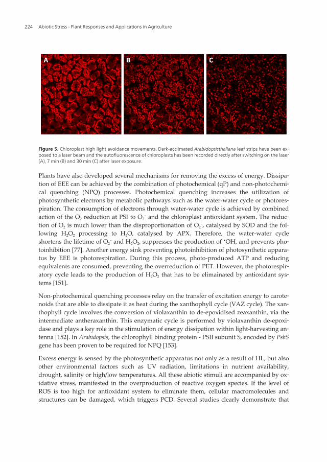
Figure 5. Chloroplast high light avoidance movements. Dark-acclimated Arabidopsisthaliana leaf strips have been ex‐posed to a laser beam and the autofluorescence of chloroplasts has been recorded directly after switching on the laser(A), 7 min (B) and 30 min (C) after laser exposure.
Plants have also developed several mechanisms for removing the excess of energy. Dissipa‐tion of EEE can be achieved by the combination of photochemical (qP) and non-photochemi‐cal quenching (NPQ) processes. Photochemical quenching increases the utilization ofphotosynthetic electrons by metabolic pathways such as the water-water cycle or photores‐piration. The consumption of electrons through water-water cycle is achieved by combinedaction of the O2 reduction at PSI to O2
− and the chloroplast antioxidant system. The reduc‐tion of O2 is much lower than the disproportionation of O2
−, catalysed by SOD and the fol‐lowing H2O2 processing to H2O, catalysed by APX. Therefore, the water-water cycleshortens the lifetime of O2
− and H2O2, suppresses the production of •OH, and prevents pho‐toinhibition [77]. Another energy sink preventing photoinhibition of photosynthetic appara‐tus by EEE is photorespiration. During this process, photo-produced ATP and reducingequivalents are consumed, preventing the overreduction of PET. However, the photorespir‐atory cycle leads to the production of H2O2 that has to be elimainated by antioxidant sys‐tems [151].
Non-photochemical quenching processes relay on the transfer of excitation energy to carote‐noids that are able to dissipate it as heat during the xanthophyll cycle (VAZ cycle). The xan‐thophyll cycle involves the conversion of violaxanthin to de-epoxidised zeaxanthin, via theintermediate antheraxanthin. This enzymatic cycle is performed by violaxanthin de-epoxi‐dase and plays a key role in the stimulation of energy dissipation within light-harvesting an‐tenna [152]. In Arabidopsis, the chlorophyll binding protein - PSII subunit S, encoded by PsbSgene has been proven to be required for NPQ [153].
Excess energy is sensed by the photosynthetic apparatus not only as a result of HL, but alsoother environmental factors such as UV radiation, limitations in nutrient availability,drought, salinity or high/low temperatures. All these abiotic stimuli are accompanied by ox‐idative stress, manifested in the overproduction of reactive oxygen species. If the level ofROS is too high for antioxidant system to eliminate them, cellular macromolecules andstructures can be damaged, which triggers PCD. Several studies clearly demonstrate that
Abiotic Stress - Plant Responses and Applications in Agriculture224

programmed cell death is affected by light. The spread of wound-induced PCD in maize tis‐sue has been shown to be transmitted by chloroplast-produced ROS [134]. It has also beenfound that the HR-mediated cell death is accelerated by the loss of chloroplast function[154]. Moreover, a study using light- and dark-grown plant cell culture has proven that theyrespond differently to PCD-inducing stimuli, resulting in various levels of DNA fragmenta‐tion and cell-content condensation [65]. Direct induction of programmed cell death by expo‐sure of Arabidopsis rosettes to excess light (EL) (2000 µmol of photons m¯² s¯¹) has also beendemonstrated [130].
Although ROS produced during the progress of PCD are damaging for the cell, they are alsoneeded as messenger molecules preparing other cells for a struggle with stress conditions.H2O2 is thought to freely diffuse across biological membranes, thus it has been proposed todirectly influence the function of extra-chloroplastic signaling components. The possibilitythat H2O2 acts as an intracellular messenger molecule has been suggested since it triggeressystemic response to EL [130]. When low-light-grown Arabidopsis rosettes have been partial‐ly exposed to EL, unexposed leaves have become acclimated to EEE and to photooxidativestress. This phenomenon, termed systemic acquired acclimation (SAA) is attributed to chlor‐oplasts, as it is associated with specific changes in redox status of the photosynthetic elec‐tron carrier chain. Such redox changes lead to the alternation in transcription profiles,triggering SAA. By the use of photosynthesis inhibitors: 3,4-dichlorophenyl-1,1-dimethylur‐ea (DCMU), which blocks the reduction of PQ and 2,5-dibromo-3-methyl-6-isopropyl-p-ben‐zoquinone (DBMIB), that blocks PQ oxidation, it has been demonstrated that the PQ poolredox status is predominantly responsible for SAA [60,130]. DCMU has been also proven toinhibit effects of EEE, such as higher NPQ, production of ET, H2O2 and decrease in stomatalconductance. On the other hand, DBMIB has been shown to trigger the production of ET,H2O2 and stomatal closure even under low light. These results indicate that the redox statusof PQ pool and NPQ play a critical role in the initiation of response to EEE, in ET and H2O2
signaling and consequently in the PCD regulation [11,60,130,155,156]. Activation of the SA-dependent pathway in response to EEE has been also observed after an initial induction ofROS/ET signaling [60]. Other hormones such as JA and ABA, synthesized at least partiallywithin the chloroplast, also participate in response to EEE [149]. A large number of tran‐scripts encoding different antioxidant defense enzymes is induced in local and systemicleaves after EL treatment. Apart from APX1 and APX2 [130], higher expression level hasbeen proven for GPX2, GSTs, APX3 and CSD1 [60,157]. Furthermore, a recent study has sug‐gested that local and systemic responses to EL are associated with changes in the plasmamembrane electrical potential (photoelectrophysiological signaling - PEPS). During the ELincident, PEPS has been shown to be initiated by quantum redox changes in PSII, trans‐duced by bundle sheath cells and its systemic propagation has been proven to be dependenton APX2 function. Therefore, PEPS is suggested as a new component of signaling cascadesthat regulate light acclimatory responses. Furthermore, it has been proposed that leaves areable to memorize different EL episodes and use this information for improving their accli‐mation and survival under prolonged periods of unfavorable light conditions [131].
Programmed Cell Death as a Response to High Light, UV and Drought Stress in Plantshttp://dx.doi.org/10.5772/53127
225

In the last decade a significant progress has been made in improving light-induced oxidativestress tolerance in plants. Various components of antioxidative system involved in ROSscavenging have been up- or down-regulated to develop transgenic lines with altered anti‐oxidants levels. Overexpression of enzymes involved in AsA biosynthesis has been shownto confer oxidative stress tolerance in tomato plants [158]. Increased AsA content has beenalso demonstrated to enhance high light stress tolerance in Arabidopsis [159]. Higher concen‐tration of GSH has proven to protect potato plants against oxidative damage [160]. More‐over, reduced level of light-mediated cellular damage has been observed in transgenictobacco plants overexpressing chloroplast-localized Cu/Zn-SOD (161) and thylakoid mem‐brane-bound APX [162]. MDHAR overexpression in Arabidopsis has been demonstrated toenhance the tolerance towards photooxidative stresses [163]. Moreover, some studies havereported that combined expression of antioxidant enzymes in transgenic plants acts synerg‐istically on stress tolerance, e.g. simultaneous overexpression of Cu/Zn-SOD and APX in to‐bacco chloroplasts enhances the resistance to the photooxidative stress in comparison totheir single overexpression [164].
5. UV radiation stress
Being exposed to sunlight, plants need to deal with the damaging effect of ultraviolet (UV)radiation which reduces the genome stability, impeding their growth and productivity.These effects result from damage to cell components including not only nucleic acids, butalso proteins and membrane lipids. Upon UV exposure, strongly mutagenic cross-linkedforms of DNA can be produced [165]. In order to minimize effects of UV radiation, plantsaccumulate UV-absorbing secondary metabolites, perform the monomerization of UV-in‐duced pyrimidine dimers (DNA repair) and neutralize generated ROS [166,167].
UV radiation consists of UV-C (below 280 nm), UV-B (280-320 nm) and UV-A (320-390). Al‐though UV-C is not physiologically relevant to plants since it is efficiently blocked by thestratosphere, the UV-C-triggered cell damage is comparable to induced with UV-B radia‐tion, which reaches Earth's surface [168]. Therefore, UV-C radiation has been widely used tostudy DNA damage and repair mechanisms upon UV stress [169].
UV has been demonstrated to trigger apoptosis in animals [170] and apoptosis-like changesin Arabidopsis, including DNA laddering, changes in nucleus morphology (crescent shape)and its fragmentation [168]. It has been also proven to induce oxidative burst in plant cells[171], considered as the main cause of cell death, which aims at the limitation of damagespreading. Light is necessary for UV-C-triggered cell death and caspase-like proteases par‐ticipate in this process since caspase-inhibitors are able to block the onset of DNA fragmen‐tation [25,172]. Recent study performed on Arabidopsis protoplasts has shown that duringthe early stage of UV stress, a burst of ROS in chloroplasts and adjacent mitochondria is de‐tected. Mitochondria dysfunction has been also observed, manifested by changes in theirdistribution, mobility and the loss in mitochondrial transmembrane potential. Moreover, thepre-incubation with antioxidant molecule - ascorbic acid or inhibitor of photosynthetic elec‐
Abiotic Stress - Plant Responses and Applications in Agriculture226
tron transport - DCMU decreases the ROS production and retards PCD. These results provethat mitochondria and ROS act as mediators in the UV-C-induced cell death [173] and thatAsA can be considered as an important antioxidant during this process [174], which is con‐sistent with what has been reported in various types of PCD. It has been also shown thatArabidopsis proteins AtDAD1 and AtDAD2 (defender against apoptotic death), localized inthe endoplasmic reticulum membrane can suppress DNA fragmentation, indicating an in‐volvement of the ER in UV-C-triggered PCD pathway (25). The microarray approach hasidentified numerous genes responsible for ROS scavenging, signaling, transcription regula‐tion and involved in DNA replication or conformation changes that have been deregulatedafter exposure to UV-C radiation [175]. Metacaspase-8 (AtMC8) has been proven to bestrongly up-regulated by UV-C. Overexpression of AtMC8 in Arabidopsis has resulted inmore severe cell death, while knocking-out AtMC8 has reduced the UV-C-triggered PCD,which suggests that metacaspase-8 is a part of PCD pathway activated by UV radiation[176]. The activation of PCD program upon UV helps plants in eliminating damaged cells tocontrol cell quality and quantity after the trauma.
6. Drought stress - soil water deficit
Drought is one of the most unfavorable environmental factors that affects growth and devel‐opment of plants and consequently limits plant productivity. Plants have developed specificacclimation and adaptation mechanisms to survive the soil water deficit. In response todrought, plants can exhibit either escape (ability to complete the life cycle before severestress) or resistance mechanisms. Resistance mechanisms include drought avoidance anddrought tolerance. The latter depends on the cell turgor maintenance by accumulating os‐molytes and soluble sugars [177]. There are several examples of molecules that help to main‐tain an osmotic balance under dehydration conditions: sugars, polyols and proline [178].Proline is accumulated in the cytoplasm and chloroplast stroma while other solutes (sugars,organic acids, potassium) are cumulated in the vacuole. When the cellular water content de‐creases, they stabilize cellular structures through hydrophilic interactions and hydrogenbonding [179]. A similar role is fulfilled by late embryogenesis abundant (LEA) proteins - afamily of unstructured proteins. LEA proteins accumulate in response to dehydration andABA treatment. Because of their high hydrophilicity and solubility in water, it has been pro‐posed that they play a role in protecting cytoplasmic structures during dehydration [180].The avoidance mechanism is possible by the maintenance of high water potential in planttissue despite soil water deficit. It can be achieved by: improved water uptake under stress,the ability to hold water as well as by the reduction of its loss through smaller leaf area andlesser stomatal and cuticular conductance. One of the first acclimation responses to droughtis the decrease in leaf growth, which helps to maintain the cell turgor and reduces the tran‐spiration area. In Arabidopsis, the size of leaf is regulated by both cell division and cell ex‐pansion. Under drought stress, Arabidopsis leaves have been demonstrated to compensatefor the low rate of expansion by the extension of expansion duration [181]. The cell expan‐sion is a process of cell wall loosening performed by enzymatic and non-enzymatic compo‐
Programmed Cell Death as a Response to High Light, UV and Drought Stress in Plantshttp://dx.doi.org/10.5772/53127
227

nents, from which expansins are key proteins engaged in this process. The onset of cellexpansion involves pumping of protons into the cell wall, which makes the surroundingmore acidic. As a result, expansins become activated. They loosen connections between cel‐lulose microfibrils, which leads to the cell wall relaxation, water uptake and consequentlythe cell expansion [182]. It has been demonstrated that the mild osmotic stress causes the in‐duction of expansin genes [183].
Photosynthesis is one of major processes affected by water deficit since stomata closurecauses reduced CO2 diffusion to the chloroplast. As a result of the inhibition of photosynthe‐sis and the predominance of photorespiration, ROS are generated [184]. It has been demon‐strated that in drought-stressed plants, the ABA-controlled stomata closure is mediated byH2O2 [185]. Under severe drought stress, some antioxidant enzymes have been shown to behighly induced [186]. However, studies on many drought-stressed crop species showed aninconsistency in their expression since in some cases they have been induced, but in otherrepressed, suggesting that different ROS balance may be required during different responsephases [187].
During water deficit, ROS are responsible for the induction of leaf senescence, which is exe‐cuted through the programmed cell death and plays an important role in the plant survival.It contributes to the nutrients remobilisation during stress and allows the rest of plant tobenefit from them and stay alive. Drought-induced PCD enables also the abscission of someleaves and thus the avoidance of further water loss through the transpiration. It occurs grad‐ually and is manifested by specific biochemical and molecular changes such as chromatincondensation, thylakoid swelling, lipid peroxidation, degradation of chlorophyll (leaf yel‐lowing) and proteins. Apart from ROS, cytokinins and ABA have been shown to be in‐volved in the regulation of water-deficit-triggered senescence [123]. Recent studies haveshown that the water deficit triggers PCD not only in green tissues but also in plant roottips. Apical meristem cells of primary roots undergoing PCD, demonstrate increased size ofvacuole, degradation of organelles and the collapse of plasma membrane [188].
Early events in the perception of drought stress signals include the activation of transcrip‐tion factors belonging to such classes as DREB/CBF (e.g. DREB1a, DREB2a), ABF (e.g. ABF2,ABF4), MYB (e.g. MYB2), MYC, NAC and WRKY. Many of them possess stress responsivecis-regulatory elements in their promoter sequences like abscisic acid-responsive elements(ABRE) and drought-responsive elements (DRE) [189–191]. The plasma membrane-associat‐ed NTL4 (NAC transcription factor) after drought or ABA treatment has been shown to beproteolytically activated and transported to the nucleus where it induces expression ofNADPH oxidase involved in ROS generation [192]. Moreover, the dehydration stimulatesexpression of BAX inhibitor-1 (AtBI-1). The atbi1 mutant has been shown to display moresevere cell death, indicating that ER-located AtBI-1 modulates the water-deficit-inducedPCD [188]. Drought stress has been also proven to regulate the expression and activity ofaquaporins - a family of channel proteins that facilitate the transport of water along trans‐membrane water potential gradients [193].
Upon soil water deficit, the accumulation of ABA and the induction of ABA-associated sig‐naling genes occur. ABA induces various second messengers such as cytosolic Ca2+, ROS
Abiotic Stress - Plant Responses and Applications in Agriculture228

and NO in guard cells. These signals evoke ion efflux through plasma membrane ion chan‐nels, resulting in the reduction of guard cell turgor pressure and stomata closure to reducewater loss through the transpiration [194]. Mutants with the perturbation of ABA synthesisor signaling display drought hypersensitivity, manifested in significant growth reductionwhich suggests that ABA is needed for the proper response to drought [177]. In Arabidopsis,stomata closure has been shown to be regulated by ABI1 and ABI3 (ABA-insensitive 1 and 3)belonging to a group of genes identified through mutant screens and being associated withABA-mediated metabolic responses to stress. Under drought, ABI1 transcription is up-regu‐lated while ABI3 is usually down-regulated. Recently, ABI3 has been hypothesized to be es‐sential for the successful drought recovery [195]. The cell-surface ABA receptors have notbeen recognized yet. However, a recent study has proposed the flowering time control pro‐tein A (FCA) and the chloroplastic magnesium protoporphyrin-IX chelatase H subunit(CHLH) as candidates for ABA receptors, both of which have been shown to bind ABA invitro [196]. Two genes have been found to play a crucial role in the prevention of stomataopening - GPA1 (G PROTEIN ALPHA SUBUNIT 1) and PLDα1 (PHOSPHOLIPASE D AL‐PHA 1) [197]. PLD-produced phosphatidic acid has been also shown to play an importantrole in the plant response to drought stress [198].
Similarly to ABA, JA also triggers stomata closure and such response is conserved amongvarious plant species [199]. At the early stage of moderate drought, plants accumulate highconcentrations of ABA and induce ABA-responsive genes. At this stage, no significant dif‐ferences in JA-responsive genes are observed. At later stage of drought stress, ABA level re‐turns to normal, while JA synthesis and JA signaling genes are significantly down-regulated. This suggests the negative correlation between ABA and JA pathways [177]. Thehigh concentration of JA is probably undesirable during drought stress, as it inhibits the cellexpansion and results in stunted growth [200]. Therefore, plants down-regulate JA synthesisand signaling pathways to minimize the inhibitory effect of JA on growth, establishing anew hormone homeostasis.
Downstream of early stress perception events, signaling molecules are activated. Such sec‐ondary messengers include Ca2+ ions and ROS. They induce further genes that are needed toestablish a new cellular homeostasis leading to drought resistance and tolerance [180]. Re‐cent studies have strongly proven that drought response progresses through mitogen-acti‐vated protein kinase (MAPK) pathways [201]. In yeast and animals, MAPK-regulatedpathways take part in the production of osmolytes and antioxidants. These MAPK path‐ways are activated by receptors/sensors such as protein tyrosine kinases, G-protein–coupledreceptors and histidine kinases. Among these, G-protein–associated receptors have beenproposed to serve as one kind of membrane-bounded receptors for ABA. A family of histi‐dine kinases (HK) have been also identified in plants [199]. An Arabidopsis AtHK1 has beensuggested to be involved in the osmotic stress signal transduction [202]. Other members ofthis family, ATHB7 and ATHB12 have been proposed to maintain the reduction of plantgrowth under drought, which is an acclimation response to survive prolonged droughtstress [177].
Programmed Cell Death as a Response to High Light, UV and Drought Stress in Plantshttp://dx.doi.org/10.5772/53127
229

The elucidation of mechanisms controlling drought stress responses has enabled to engineerplants by the expression of specific stress-related genes. Although it was believed that themodulation of osmoregulatory genes would be the best strategy, attempts failed to result inany significant drought-stress tolerance improvement [203]. However, constitutive expres‐sion of some LEA proteins has conferred tolerance to soil water deficit in transgenic rice[204] and wheat [205]. Moreover, tomato plants overexpressing Arabidopsis CBF1 (DREB1B)have exhibited improved drought tolerance [206]. Similar results have been obtained fortransgenic Arabidopsis and rice plants overexpressing stress-responsive NAC genes [207].
7. Conclusions
Plants have evolved various strategies to acclimate to different environmental stresses. Themost fundamental strategy is the development of high plasticity of plant tissues. It has beendemonstrated that programmed cell death plays an important role in this plasticity and sub‐sequent adaptation to unfavorable conditions. There is a growing evidence that PCD is acrucial process in both morphogenetic changes execution and the following adaptation. Al‐though each decade brings a vast number of research, our understanding of plant PCD andits underlying mechanisms is still in its early stage. Further insight into details of the PCDmolecular machinery in plants is important, since it is an attractive target for improvingstress tolerance and plant yield under adverse conditions. Essentially, it could lead to thegeneration of pathogen-resistant and stress-tolerant crops as well as fruit varieties with anextended shelf life.
Acknowledgments
This work was supported by the Welcome/2008/1 Program operated within the frameworkof the Foundation for Polish Science, co-financed by the European Regional DevelopmentFund.
Abbreviations
ABA - abscisic acid; AOX - alternative oxidase; APX - ascorbate peroxidase; AsA - ascorbicacid; BI - Bax-Inhibitor; CAT – catalase; Chl – chlorophyll; DBMIB - 2,5-dibromo-3-methyl-6-isopropyl-p-benzoquinone; DCMU - 3,4-dichlorophenyl-1,1-dimethylurea; DHA – dehy‐droascorbate; DHAR - dehydroascorbate reductase; EEE - excess excitation energy; EL -excess light; ER - endoplasmic reticulum; ET – ethylene; ETC - electron transport chain; FAD- flavin adenine dinucleotide; GA - gibberellic acid; GPX - glutathione peroxidase; GR - glu‐tathione reductase; GSH - reduced glutathione; GSSG – oxidized glutathione; GST - gluta‐thione-S- transferase; HL - high light; IAP - inhibitor of apoptosis; JA - jasmonic acid; HR -
Abiotic Stress - Plant Responses and Applications in Agriculture230

hypersensitive response; LEA - late embryogenesis abundant; LHC - light-harvestingcomplex; LPO - lipid peroxidation; LSD1 - LESION SIMULATING DISEASE 1; MC – meta‐caspase; MDA – malonyldialdehyde; MDHAR - monodehydroascorbate reductase; MeJA -methyl-jasmonic acid; MAPK - mitogen-activated protein kinase; NADPH - nicotinamideadenine dinucleotide phosphate; NO - nitric oxide; NPQ - non-photochemical quenching;PA - phosphatidic acid; PCD - programmed cell death; PEPS - photoelectrophysiological sig‐naling; PET - photosynthetic electron transport; PG – phosphoglycolate; PGA – phosphogly‐cerate; PLD - phospholipase D; PSI and PSII - photosystem I and II; PQ – plastoquinone;PUFA - polyunsaturated fatty acids; qP - photochemical quenching; RCD - runaway celldeath; RNS - reactive nitrogen species; ROS - reactive oxygen species; Rubisco - ribulose-1,5-bisphosphate carboxylase/oxygenase; RuBP - ribulose-1,5-bisphosphate; SA - salicylic acid;SAA - systemic acquired acclimation; SAR - systemic acquired resistance; SOD - superoxidedismutase; TF - transcription factor; UV - ultraviolet radiation; VPE - vacuolar processingenzyme; XOD - xanthine oxidase.
Author details
Weronika Wituszyńska and Stanisław Karpiński
*Address all correspondence to: [email protected]
Department of Plant Genetics, Breeding and Biotechnology, Warsaw University of Life Sci‐ences, Warsaw, Poland
References
[1] Pennell RI, Lamb C. Programmed Cell Death in Plants. Plant Cell. 1997;9(7):1157–68.
[2] Fukuda H. Programmed cell death of tracheary elements as a paradigm in plants.Plant Mol. Biol. 2000;44(3):245–53.
[3] Dellaporta SL, Calderon-Urrea A. Sex determination in flowering plants. Plant Cell.1993;5(10):1241–51.
[4] Gunawardena AHLAN, Greenwood JS, Dengler NG. Programmed cell death remod‐els lace plant leaf shape during development. Plant Cell. 2004;16(1):60–73.
[5] Gunawardena AHLAN. Programmed cell death and tissue remodelling in plants. J.Exp. Bot. 2008;59(3):445–51.
[6] Gan S, Amasino RM. Making Sense of Senescence (Molecular Genetic Regulation andManipulation of Leaf Senescence). Plant Physiol. 1997;113(2):313–9.
[7] Williams B, Dickman M. Plant programmed cell death: can’t live with it; can’t livewithout it. Mol. Plant Pathol. 2008;9(4):531–44.
Programmed Cell Death as a Response to High Light, UV and Drought Stress in Plantshttp://dx.doi.org/10.5772/53127
231
[8] Dietrich RA, Delaney TP, Uknes SJ, Ward ER, Ryals JA, Dangl JL. Arabidopsis mu‐tants simulating disease resistance response. Cell. 1994;77(4):565–77.
[9] Dangl JL, Dietrich RA, Richberg MH. Death Don’t Have No Mercy: Cell Death Pro‐grams in Plant-Microbe Interactions. Plant Cell. 1996;8(10):1793–807.
[10] Lam E. Controlled cell death, plant survival and development. Nat. Rev. Mol. CellBiol. 2004;5(4):305–15.
[11] Mateo A, Mühlenbock P, Rustérucci C, Chang CC-C, Miszalski Z, Karpinska B, et al.LESION SIMULATING DISEASE 1 is required for acclimation to conditions that pro‐mote excess excitation energy. Plant Physiol. 2004;136(1):2818–30.
[12] Coll NS, Epple P, Dangl JL. Programmed cell death in the plant immune system. CellDeath Differ. 2011;18(8):1247–56.
[13] Evans DE. Aerenchyma formation. New Phytologist. 2004;161(1):35–49.
[14] Mühlenbock P, Plaszczyca M, Plaszczyca M, Mellerowicz E, Karpinski S. Lysigenousaerenchyma formation in Arabidopsis is controlled by LESION SIMULATING DIS‐EASE1. Plant Cell. 2007;19(11):3819–30.
[15] Swidzinski JA, Sweetlove LJ, Leaver CJ. A custom microarray analysis of gene ex‐pression during programmed cell death in Arabidopsis thaliana. Plant J. 2002;30(4):431–46.
[16] Muñoz-Pinedo C. Signaling pathways that regulate life and cell death: evolution ofapoptosis in the context of self-defense. Adv. Exp. Med. Biol. 2012;738:124–43.
[17] Mittler R, Simon L, Lam E. Pathogen-induced programmed cell death in tobacco. J.Cell. Sci. 1997;110:1333–44.
[18] Samuilov VD, Lagunova EM, Kiselevsky DB, Dzyubinskaya EV, Makarova YV, Gu‐sev MV. Participation of chloroplasts in plant apoptosis. Biosci. Rep. 2003;23(2-3):103–17.
[19] Hatsugai N, Kuroyanagi M, Nishimura M, Hara-Nishimura I. A cellular suicidestrategy of plants: vacuole-mediated cell death. Apoptosis. 2006;11(6):905–11.
[20] Torres MA, Dangl JL. Functions of the respiratory burst oxidase in biotic interactions,abiotic stress and development. Curr. Opin. Plant Biol. 2005;8(4):397–403.
[21] Chen S, Dickman MB. Bcl-2 family members localize to tobacco chloroplasts and in‐hibit programmed cell death induced by chloroplast-targeted herbicides. J. Exp. Bot.2004;55(408):2617–23.
[22] Lam E, Pozo O del. Caspase-like protease involvement in the control of plant celldeath. Plant Molecular Biology. 2000;44(3):417–28.
[23] del Pozo O, Lam E. Caspases and programmed cell death in the hypersensitive re‐sponse of plants to pathogens. Curr. Biol. 1998;8(20):1129–32.
Abiotic Stress - Plant Responses and Applications in Agriculture232

[24] De Jong AJ, Hoeberichts FA, Yakimova ET, Maximova E, Woltering EJ. Chemical-in‐duced apoptotic cell death in tomato cells: involvement of caspase-like proteases.Planta. 2000;211(5):656–62.
[25] Danon A, Rotari VI, Gordon A, Mailhac N, Gallois P. Ultraviolet-C overexposure in‐duces programmed cell death in Arabidopsis, which is mediated by caspase-like ac‐tivities and which can be suppressed by caspase inhibitors, p35 and Defender againstApoptotic Death. J. Biol. Chem. 2004;279(1):779–87.
[26] Vercammen D, Cotte B van de, Jaeger GD, Eeckhout D, Casteels P, Vandepoele K, etal. Type II Metacaspases Atmc4 and Atmc9 of Arabidopsis thaliana Cleave Sub‐strates after Arginine and Lysine. J. Biol. Chem. 2004;279(44):45329–36.
[27] Watanabe N, Lam E. Two Arabidopsis metacaspases AtMCP1b and AtMCP2b are ar‐ginine/lysine-specific cysteine proteases and activate apoptosis-like cell death inyeast. J. Biol. Chem. 2005;280(15):14691–9.
[28] Zhang Y, Lam E. Sheathing the swords of death: post-translational modulation ofplant metacaspases. Plant Signal Behav. 2011;6(12):2051–6.
[29] Watanabe N, Lam E. Arabidopsis metacaspase 2d is a positive mediator of cell deathinduced during biotic and abiotic stresses. Plant J. 2011;66(6):969–82.
[30] Coll NS, Vercammen D, Smidler A, Clover C, Van Breusegem F, Dangl JL, et al. Ara‐bidopsis type I metacaspases control cell death. Science. 2010;330(6009):1393–7.
[31] Lacomme C, Cruz SS. Bax-induced cell death in tobacco is similar to the hypersensi‐tive response. PNAS. 1999;96(14):7956–61.
[32] Mitsuhara I, Malik KA, Miura M, Ohashi Y. Animal cell-death suppressors Bcl-x(L)and Ced-9 inhibit cell death in tobacco plants. Curr. Biol. 1999;9(14):775–8.
[33] Lincoln JE, Richael C, Overduin B, Smith K, Bostock R, Gilchrist DG. Expression ofthe antiapoptotic baculovirus p35 gene in tomato blocks programmed cell death andprovides broad-spectrum resistance to disease. PNAS. 2002;99(23):15217–21.
[34] Li W, Dickman MB. Abiotic stress induces apoptotic-like features in tobacco that isinhibited by expression of human Bcl-2. Biotechnol. Lett. 2004;26(2):87–95.
[35] Sanchez P, de Torres Zabala M, Grant M. AtBI-1, a plant homologue of Bax inhibi‐tor-1, suppresses Bax-induced cell death in yeast and is rapidly upregulated duringwounding and pathogen challenge. Plant J. 2000;21(4):393–9.
[36] Kawai-Yamada M, Jin L, Yoshinaga K, Hirata A, Uchimiya H. Mammalian Bax-in‐duced plant cell death can be down-regulated by overexpression of Arabidopsis BaxInhibitor-1 (AtBI-1). PNAS. 2001;98(21):12295–300.
[37] Watanabe N, Lam E. Arabidopsis Bax inhibitor-1 functions as an attenuator of bioticand abiotic types of cell death. Plant J. 2006;45(6):884–94.
Programmed Cell Death as a Response to High Light, UV and Drought Stress in Plantshttp://dx.doi.org/10.5772/53127
233

[38] Janda T, Horváth E, Szalai G, PáLdi E. Role of Salicylic Acid in the Induction of Abio‐tic Stress Tolerance. In: Hayat S, Ahmad A, editors. Salicylic Acid: A Plant Hormone.Springer Netherlands; 2007 p. 91–150
[39] Overmyer K, Brosché M, Kangasjärvi J. Reactive oxygen species and hormonal con‐trol of cell death. Trends Plant Sci. 2003;8(7):335–42.
[40] Alvarez ME. Salicylic acid in the machinery of hypersensitive cell death and diseaseresistance. Plant Mol. Biol. 2000;44(3):429–42.
[41] Leon J, Lawton MA, Raskin I. Hydrogen Peroxide Stimulates Salicylic Acid Biosyn‐thesis in Tobacco. Plant Physiol. 1995;108(4):1673–8. [
[42] Draper J. Salicylate, superoxide synthesis and cell suicide in plant defence. Trends inPlant Science. 1997;2(5):162–5.
[43] Delledonne M, Zeier J, Marocco A, Lamb C. Signal interactions between nitric oxideand reactive oxygen intermediates in the plant hypersensitive disease resistance re‐sponse. PNAS. 2001;98(23):13454–9.
[44] John I, Drake R, Farrell A, Cooper W, Lee P, Horton P, et al. Delayed leaf senescencein ethylene-deficient ACC-oxidase antisense tomato plants: molecular and physiolog‐ical analysis. The Plant Journal. 1995;7(3):483–90.
[45] Overmyer K, Tuominen H, Kettunen R, Betz C, Langebartels C, Sandermann H Jr, etal. Ozone-sensitive arabidopsis rcd1 mutant reveals opposite roles for ethylene andjasmonate signaling pathways in regulating superoxide-dependent cell death. PlantCell. 2000;12(10):1849–62.
[46] Turner JG, Ellis C, Devoto A. The jasmonate signal pathway. Plant Cell. 2002;14Suppl:S153–164.
[47] Seo HS, Song JT, Cheong J-J, Lee Y-H, Lee Y-W, Hwang I, et al. Jasmonic acid carbox‐yl methyltransferase: A key enzyme for jasmonate-regulated plant responses. PNAS.2001;98(8):4788–93.
[48] Zhang L, Xing D. Methyl jasmonate induces production of reactive oxygen speciesand alterations in mitochondrial dynamics that precede photosynthetic dysfunctionand subsequent cell death. Plant Cell Physiol. 2008;49(7):1092–111.
[49] Fath A, Bethke PC, Jones RL. Enzymes That Scavenge Reactive Oxygen Species AreDown-Regulated Prior to Gibberellic Acid-Induced Programmed Cell Death in Bar‐ley Aleurone. Plant Physiol. 2001;126(1):156–66.
[50] Steffens B, Sauter M. Epidermal Cell Death in Rice Is Regulated by Ethylene, Gibber‐ellin, and Abscisic Acid. Plant Physiol. 2005;139(2):713–21.
[51] Levine A, Pennell RI, Alvarez ME, Palmer R, Lamb C. Calcium-mediated apoptosisin a plant hypersensitive disease resistance response. Curr. Biol. 1996;6(4):427–37.
Abiotic Stress - Plant Responses and Applications in Agriculture234
[52] Falk A, Feys BJ, Frost LN, Jones JDG, Daniels MJ, Parker JE. EDS1, an essential com‐ponent of R gene-mediated disease resistance in Arabidopsis has homology to eu‐karyotic lipases. Proc Natl Acad Sci U S A. 1999;96(6):3292–7.
[53] Jirage D, Tootle TL, Reuber TL, Frost LN, Feys BJ, Parker JE, et al. Arabidopsis thali‐ana PAD4 encodes a lipase-like gene that is important for salicylic acid signaling.Proc. Natl. Acad. Sci. U.S.A. 1999;96(23):13583–8.
[54] Meijer HJG, Munnik T. Phospholipid-Based Signaling in Plants. Annual Review ofPlant Biology. 2003;54(1):265–306.
[55] Zhang W, Wang C, Qin C, Wood T, Olafsdottir G, Welti R, et al. The Oleate-Stimulat‐ed Phospholipase D, PLDδ, and Phosphatidic Acid Decrease H2O2-Induced CellDeath in Arabidopsis. Plant Cell. 2003;15(10):2285–95.
[56] Brodersen P, Petersen M, Pike HM, Olszak B, Skov S, Ødum N, et al. Knockout ofArabidopsis ACCELERATED-CELL-DEATH11 encoding a sphingosine transfer pro‐tein causes activation of programmed cell death and defense. Genes Dev. 2002;16(4):490–502.
[57] Wiermer M, Feys BJ, Parker JE. Plant immunity: the EDS1 regulatory node. Curr.Opin. Plant Biol. 2005;8(4):383–9.
[58] Rustérucci C, Aviv DH, Holt BF, Dangl JL, Parker JE. The Disease Resistance Signal‐ing Components EDS1 and PAD4 Are Essential Regulators of the Cell Death Path‐way Controlled by LSD1 in Arabidopsis. Plant Cell. 2001;13(10):2211–24.
[59] Mateo A, Funck D, Mühlenbock P, Kular B, Mullineaux PM, Karpinski S. Controlledlevels of salicylic acid are required for optimal photosynthesis and redox homeosta‐sis. J. Exp. Bot. 2006;57(8):1795–807.
[60] Mühlenbock P, Szechynska-Hebda M, Plaszczyca M, Baudo M, Mateo A, MullineauxPM, et al. Chloroplast signaling and LESION SIMULATING DISEASE1 regulatecrosstalk between light acclimation and immunity in Arabidopsis. Plant Cell.2008;20(9):2339–56.
[61] Jabs T, Dietrich RA, Dangl JL. Initiation of runaway cell death in an Arabidopsis mu‐tant by extracellular superoxide. Science. 1996;273(5283):1853–6.
[62] Epple P, Mack AA, Morris VRF, Dangl JL. Antagonistic control of oxidative stress-induced cell death in Arabidopsis by two related, plant-specific zinc finger proteins.Proc. Natl. Acad. Sci. U.S.A. 2003;100(11):6831–6.
[63] Kaminaka H, Näke C, Epple P, Dittgen J, Schütze K, Chaban C, et al. bZIP10-LSD1antagonism modulates basal defense and cell death in Arabidopsis following infec‐tion. EMBO J. 2006;25(18):4400–11.
[64] Karpinski S, Gabrys H, Mateo A, Karpinska B, Mullineaux PM. Light perception inplant disease defence signalling. Curr. Opin. Plant Biol. 2003;6(4):390–6.
Programmed Cell Death as a Response to High Light, UV and Drought Stress in Plantshttp://dx.doi.org/10.5772/53127
235

[65] Doyle SM, Diamond M, McCabe PF. Chloroplast and reactive oxygen species in‐volvement in apoptotic-like programmed cell death in Arabidopsis suspension cul‐tures. J. Exp. Bot. 2010;61(2):473–82.
[66] Gill SS, Tuteja N. Reactive oxygen species and antioxidant machinery in abiotic stresstolerance in crop plants. Plant Physiol. Biochem. 2010;48(12):909–30.
[67] Buchert F, Forreiter C. Singlet oxygen inhibits ATPase and proton translocation activ‐ity of the thylakoid ATP synthase CF1CFo. FEBS Lett. 2010;584(1):147–52.
[68] Hatz S, Lambert JDC, Ogilby PR. Measuring the lifetime of singlet oxygen in a singlecell: addressing the issue of cell viability. Photochem. Photobiol. Sci. 2007;6(10):1106–16.
[69] Krieger-Liszkay A, Fufezan C, Trebst A. Singlet oxygen production in photosystem IIand related protection mechanism. Photosyn. Res. 2008;98(1-3):551–64.
[70] Triantaphylidès C, Krischke M, Hoeberichts FA, Ksas B, Gresser G, Havaux M, et al.Singlet oxygen is the major reactive oxygen species involved in photooxidative dam‐age to plants. Plant Physiol. 2008;148(2):960–8.
[71] op den Camp RGL, Przybyla D, Ochsenbein C, Laloi C, Kim C, Danon A, et al. Rapidinduction of distinct stress responses after the release of singlet oxygen in Arabidop‐sis. Plant Cell. 2003;15(10):2320–32.
[72] Bright J, Desikan R, Hancock JT, Weir IS, Neill SJ. ABA-induced NO generation andstomatal closure in Arabidopsis are dependent on H2O2 synthesis. Plant J.2006;45(1):113–22.
[73] Mittler R, Vanderauwera S, Gollery M, Van Breusegem F. Reactive oxygen gene net‐work of plants. Trends Plant Sci. 2004;9(10):490–8.
[74] Foreman J, Demidchik V, Bothwell JHF, Mylona P, Miedema H, Torres MA, et al. Re‐active oxygen species produced by NADPH oxidase regulate plant cell growth. Na‐ture. 2003;422(6930):442–6.
[75] Quan L-J, Zhang B, Shi W-W, Li H-Y. Hydrogen peroxide in plants: a versatile mole‐cule of the reactive oxygen species network. J Integr Plant Biol. 2008;50(1):2–18.
[76] Vranová E, Atichartpongkul S, Villarroel R, Van Montagu M, Inzé D, Van Camp W.Comprehensive analysis of gene expression in Nicotiana tabacum leaves acclimatedto oxidative stress. Proc Natl Acad Sci U S A. 2002;99(16):10870–5.
[77] Asada K. THE WATER-WATER CYCLE IN CHLOROPLASTS: Scavenging of ActiveOxygens and Dissipation of Excess Photons. Annu. Rev. Plant Physiol. Plant Mol. Bi‐ol. 1999;50:601–39.
[78] Foyer CH, Noctor G. Redox homeostasis and antioxidant signaling: a metabolic inter‐face between stress perception and physiological responses. Plant Cell. 2005;17(7):1866–75.
Abiotic Stress - Plant Responses and Applications in Agriculture236

[79] Maurino VG, Peterhansel C. Photorespiration: current status and approaches formetabolic engineering. Curr. Opin. Plant Biol. 2010;13(3):249–56.
[80] Peterhansel C, Maurino VG. Photorespiration Redesigned. Plant Physiol. 2011;155(1):49–55.
[81] Lam E, Kato N, Lawton M. Programmed cell death, mitochondria and the plant hy‐persensitive response. Nature. 2001;411(6839):848–53.
[82] Navrot N, Rouhier N, Gelhaye E, Jacquot J-P. Reactive oxygen species generationand antioxidant systems in plant mitochondria. Physiologia Plantarum. 2007;129(1):185–95.
[83] Rhoads DM, Umbach AL, Subbaiah CC, Siedow JN. Mitochondrial Reactive OxygenSpecies. Contribution to Oxidative Stress and Interorganellar Signaling. Plant Physi‐ol. 2006;141(2):357–66.
[84] Maxwell DP, Wang Y, McIntosh L. The alternative oxidase lowers mitochondrial re‐active oxygen production in plant cells. PNAS. 1999;96(14):8271–6.
[85] Ordog SH, Higgins VJ, Vanlerberghe GC. Mitochondrial Alternative Oxidase Is Nota Critical Component of Plant Viral Resistance But May Play a Role in the Hypersen‐sitive Response. Plant Physiol. 2002;129(4):1858–65.
[86] Robson CA, Vanlerberghe GC. Transgenic Plant Cells Lacking Mitochondrial Alter‐native Oxidase Have Increased Susceptibility to Mitochondria-Dependent and -Inde‐pendent Pathways of Programmed Cell Death. Plant Physiol. 2002;129(4):1908–20.
[87] del Río LA, Corpas FJ, Sandalio LM, Palma JM, Gómez M, Barroso JB. Reactive oxy‐gen species, antioxidant systems and nitric oxide in peroxisomes. J. Exp. Bot.2002;53(372):1255–72.
[88] Bolwell GP, Wojtaszek P. Mechanisms for the generation of reactive oxygen speciesin plant defence – a broad perspective. Physiological and Molecular Plant Pathology.1997;51(6):347–66.
[89] Montillet J-L, Chamnongpol S, Rustérucci C, Dat J, van de Cotte B, Agnel J-P, et al.Fatty acid hydroperoxides and H2O2 in the execution of hypersensitive cell death intobacco leaves. Plant Physiol. 2005;138(3):1516–26.
[90] Møller IM, Jensen PE, Hansson A. Oxidative modifications to cellular components inplants. Annu Rev Plant Biol. 2007;58:459–81.
[91] Khan M, Panda S. Alterations in root lipid peroxidation and antioxidative responsesin two rice cultivars under NaCl-salinity stress. Acta Physiologiae Plantarum.2008;30(1):81–9.
[92] Ghezzi P, Bonetto V. Redox proteomics: identification of oxidatively modified pro‐teins. Proteomics. 2003;3(7):1145–53.
Programmed Cell Death as a Response to High Light, UV and Drought Stress in Plantshttp://dx.doi.org/10.5772/53127
237
[93] Sadanandom A, Poghosyan Z, Fairbairn DJ, Murphy DJ. Differential Regulation ofPlastidial and Cytosolic Isoforms of Peptide Methionine Sulfoxide Reductase in Ara‐bidopsis. Plant Physiol. 2000;123(1):255–64.
[94] Cooke MS, Evans MD, Dizdaroglu M, Lunec J. Oxidative DNA damage: mecha‐nisms, mutation, and disease. FASEB J. 2003;17(10):1195–214.
[95] Tuteja N, Singh MB, Misra MK, Bhalla PL, Tuteja R. Molecular mechanisms of DNAdamage and repair: progress in plants. Crit. Rev. Biochem. Mol. Biol. 2001;36(4):337–97.
[96] Mittler R. Oxidative stress, antioxidants and stress tolerance. Trends Plant Sci.2002;7(9):405–10.
[97] Kliebenstein DJ, Monde R-A, Last RL. Superoxide Dismutase in Arabidopsis: AnEclectic Enzyme Family with Disparate Regulation and Protein Localization. PlantPhysiol. 1998;118(2):637–50.
[98] del Río LA, Sandalio LM, Altomare DA, Zilinskas BA. Mitochondrial and peroxiso‐mal manganese superoxide dismutase: differential expression during leaf senes‐cence. J. Exp. Bot. 2003;54(384):923–33.
[99] Alscher RG, Erturk N, Heath LS. Role of superoxide dismutases (SODs) in control‐ling oxidative stress in plants. J. Exp. Bot. 2002;53(372):1331–41.
[100] Wang Y, Ying Y, Chen J, Wang X. Transgenic Arabidopsis overexpressing Mn-SODenhanced salt-tolerance. Plant Science. 2004;167(4):671–7.
[101] Hamid Badawi G, Yamauchi Y, Shimada E, Sasaki R, Kawano N, Tanaka K, et al. En‐hanced tolerance to salt stress and water deficit by overexpressing superoxide dis‐mutase in tobacco (Nicotiana tabacum) chloroplasts. Plant Science. 2004;166(4):919–28.
[102] Myouga F, Hosoda C, Umezawa T, Iizumi H, Kuromori T, Motohashi R, et al. A het‐erocomplex of iron superoxide dismutases defends chloroplast nucleoids against oxi‐dative stress and is essential for chloroplast development in Arabidopsis. Plant Cell.2008;20(11):3148–62.
[103] Simova-Stoilova L, Vaseva I, Grigorova B, Demirevska K, Feller U. Proteolytic activi‐ty and cysteine protease expression in wheat leaves under severe soil drought andrecovery. Plant Physiol. Biochem. 2010;48(2-3):200–6.
[104] Matsumura T, Tabayashi N, Kamagata Y, Souma C, Saruyama H. Wheat catalase ex‐pressed in transgenic rice can improve tolerance against low temperature stress.Physiologia Plantarum. 2002;116(3):317–27.
[105] Edwards EA, Rawsthorne S, Mullineaux PM. Subcellular distribution of multipleforms of glutathione reductase in leaves of pea (Pisum sativum L.). Planta.1990;180(2):278–84.
Abiotic Stress - Plant Responses and Applications in Agriculture238

[106] Sharma P, Dubey RS. Modulation of nitrate reductase activity in rice seedlings underaluminium toxicity and water stress: role of osmolytes as enzyme protectant. J. PlantPhysiol. 2005;162(8):854–64.
[107] Kornyeyev D, Logan BA, Payton PR, Allen RD, Holaday AS. Elevated chloroplasticglutathione reductase activities decrease chilling-induced photoinhibition by increas‐ing rates of photochemistry, but not thermal energy dissipation, in transgenic cotton.Functional Plant Biol. 2003;30(1):101–10.
[108] Leterrier M, Corpas FJ, Barroso JB, Sandalio LM, del Río LA. Peroxisomal Monode‐hydroascorbate Reductase. Genomic Clone Characterization and Functional Analysisunder Environmental Stress Conditions. Plant Physiol. 2005;138(4):2111–23.
[109] Eltayeb AE, Kawano N, Badawi GH, Kaminaka H, Sanekata T, Shibahara T, et al.Overexpression of monodehydroascorbate reductase in transgenic tobacco confersenhanced tolerance to ozone, salt and polyethylene glycol stresses. Planta.2007;225(5):1255–64.
[110] Ushimaru T, Nakagawa T, Fujioka Y, Daicho K, Naito M, Yamauchi Y, et al. Trans‐genic Arabidopsis plants expressing the rice dehydroascorbate reductase gene are re‐sistant to salt stress. J. Plant Physiol. 2006;163(11):1179–84.
[111] Eltayeb AE, Kawano N, Badawi GH, Kaminaka H, Sanekata T, Morishima I, et al. En‐hanced tolerance to ozone and drought stresses in transgenic tobacco overexpressingdehydroascorbate reductase in cytosol. Physiologia Plantarum. 2006;127(1):57–65.
[112] Dixon DP, Davis BG, Edwards R. Functional divergence in the glutathione transfer‐ase superfamily in plants. Identification of two classes with putative functions in re‐dox homeostasis in Arabidopsis thaliana. J. Biol. Chem. 2002;277(34):30859–69.
[113] Dixon DP, Skipsey M, Edwards R. Roles for glutathione transferases in plant secon‐dary metabolism. Phytochemistry. 2010;71(4):338–50.
[114] Millar AH, Mittova V, Kiddle G, Heazlewood JL, Bartoli CG, Theodoulou FL, et al.Control of ascorbate synthesis by respiration and its implications for stress respons‐es. Plant Physiol. 2003;133(2):443–7.
[115] Yoshimura K, Miyao K, Gaber A, Takeda T, Kanaboshi H, Miyasaka H, et al. En‐hancement of stress tolerance in transgenic tobacco plants overexpressing Chlamy‐domonas glutathione peroxidase in chloroplasts or cytosol. Plant J. 2004;37(1):21–33.
[116] Smirnoff N, Wheeler GL. Ascorbic acid in plants: biosynthesis and function. Crit.Rev. Biochem. Mol. Biol. 2000;35(4):291–314.
[117] Xiang C, Werner BL, Christensen EM, Oliver DJ. The biological functions of gluta‐thione revisited in arabidopsis transgenic plants with altered glutathione levels.Plant Physiol. 2001;126(2):564–74.
[118] Meyer AJ. The integration of glutathione homeostasis and redox signaling. J. PlantPhysiol. 2008;165(13):1390–403.
Programmed Cell Death as a Response to High Light, UV and Drought Stress in Plantshttp://dx.doi.org/10.5772/53127
239

[119] Ashraf M, Foolad MR. Roles of glycine betaine and proline in improving plant abio‐tic stress resistance. Environmental and Experimental Botany. 2007;59(2):206–16.
[120] Tateishi Y, Nakagawa T, Esaka M. Osmotolerance and growth stimulation of trans‐genic tobacco cells accumulating free proline by silencing proline dehydrogenase ex‐pression with double-stranded RNA interference technique. Physiologia Plantarum.2005;125(2):224–34.
[121] Simon-Sarkadi L, Kocsy G, Várhegyi Á, Galiba G, De Ronde J. Stress-inducedchanges in the free amino acid composition in transgenic soybean plants having in‐creased proline content. Biologia Plantarum. 2006;50(4):793–6.
[122] Kamal-Eldin A, Appelqvist LA. The chemistry and antioxidant properties of toco‐pherols and tocotrienols. Lipids. 1996;31(7):671–701.
[123] Munné-Bosch S, Alegre L. Die and let live: leaf senescence contributes to plant sur‐vival under drought stress. Functional Plant Biol. 2004;31(3):203–16.
[124] Gang W, Zhen-Kuan W, Yong-Xiang W, Li-Ye C, Hong-Bo S. The mutual responsesof higher plants to environment: physiological and microbiological aspects. ColloidsSurf B Biointerfaces. 2007;59(2):113–9.
[125] Filkowski J, Kovalchuk O, Kovalchuk I. Genome stability of vtc1, tt4, and tt5 Arabi‐dopsis thaliana mutants impaired in protection against oxidative stress. Plant J.2004;38(1):60–9.
[126] Winkel-Shirley B. Biosynthesis of flavonoids and effects of stress. Curr. Opin. PlantBiol. 2002;5(3):218–23.
[127] Apel K, Hirt H. REACTIVE OXYGEN SPECIES: Metabolism, Oxidative Stress, andSignal Transduction. Annual Review of Plant Biology. 2004;55(1):373–99.
[128] Dat JF, Pellinen R, Beeckman T, Van De Cotte B, Langebartels C, Kangasjärvi J, et al.Changes in hydrogen peroxide homeostasis trigger an active cell death process in to‐bacco. Plant J. 2003;33(4):621–32.
[129] Laloi C, Stachowiak M, Pers-Kamczyc E, Warzych E, Murgia I, Apel K. Cross-talk be‐tween singlet oxygen- and hydrogen peroxide-dependent signaling of stress respons‐es in Arabidopsis thaliana. Proc. Natl. Acad. Sci. U.S.A. 2007;104(2):672–7.
[130] Karpinski S, Reynolds H, Karpinska B, Wingsle G, Creissen G, Mullineaux P. System‐ic Signaling and Acclimation in Response to Excess Excitation Energy in Arabidopsis.Science. 1999;284(5414):654–7.
[131] Szechyńska-Hebda M, Kruk J, Górecka M, Karpińska B, Karpiński S. Evidence forLight Wavelength-Specific Photoelectrophysiological Signaling and Memory of Ex‐cess Light Episodes in Arabidopsis. Plant Cell. 2010;22(7):2201–18.
[132] Jabs T, Dietrich RA, Dangl JL. Initiation of Runaway Cell Death in an ArabidopsisMutant by Extracellular Superoxide. Science. 1996;273(5283):1853–6.
Abiotic Stress - Plant Responses and Applications in Agriculture240

[133] Mach JM, Castillo AR, Hoogstraten R, Greenberg JT. The Arabidopsis-acceleratedcell death gene ACD2 encodes red chlorophyll catabolite reductase and suppressesthe spread of disease symptoms. PNAS. 2001;98(2):771–6.
[134] Gray J, Janick-Buckner D, Buckner B, Close PS, Johal GS. Light-dependent death ofmaize lls1 cells is mediated by mature chloroplasts. Plant Physiol. 2002;130(4):1894–907.
[135] Külheim C, Ågren J, Jansson S. Rapid Regulation of Light Harvesting and Plant Fit‐ness in the Field. Science. 2002;297(5578):91–3.
[136] Allen JF. Botany. State transitions--a question of balance. Science.2003;299(5612):1530–2.
[137] Snyders S, Kohorn BD. Disruption of thylakoid-associated kinase 1 leads to alterationof light harvesting in Arabidopsis. J. Biol. Chem. 2001;276(34):32169–76.
[138] Murchie EH, Horton P. Contrasting patterns of photosynthetic acclimation to thelight environment are dependent on the differential expression of the responses to al‐tered irradiance and spectral quality. Plant, Cell & Environment. 1998;21(2):139–48.
[139] Dietzel L, Pfannschmidt T. Photosynthetic acclimation to light gradients in plantstands comes out of shade. Plant Signal Behav. 2008;3(12):1116–8.
[140] Mullineaux P, Ball L, Escobar C, Karpinska B, Creissen G, Karpinski S. Are diversesignalling pathways integrated in the regulation of arabidopsis antioxidant defencegene expression in response to excess excitation energy? Philos. Trans. R. Soc. Lond.,B, Biol. Sci. 2000;355(1402):1531–40.
[141] Bailey S, Walters RG, Jansson S, Horton P. Acclimation of Arabidopsis thaliana to thelight environment: the existence of separate low light and high light responses. Plan‐ta. 2001;213(5):794–801.
[142] Ballottari M, Dall’Osto L, Morosinotto T, Bassi R. Contrasting behavior of higherplant photosystem I and II antenna systems during acclimation. J. Biol. Chem.2007;282(12):8947–58.
[143] Mishra Y, Johansson Jankanpaa H, Kiss AZ, Funk C, Schroder WP, Jansson S. Arabi‐dopsis plants grown in the field and climate chambers significantly differ in leafmorphology and photosystem components. BMC Plant Biology. 2012;12(1):6.
[144] Karpinski S, Escobar C, Karpinska B, Creissen G, Mullineaux PM. Photosyntheticelectron transport regulates the expression of cytosolic ascorbate peroxidase genes inArabidopsis during excess light stress. Plant Cell. 1997;9(4):627–40.
[145] Pfannschmidt T, Nilsson A, Allen JF. Photosynthetic control of chloroplast gene ex‐pression. Nature. 1999;397(6720):625–8.
[146] Fryer MJ, Ball L, Oxborough K, Karpinski S, Mullineaux PM, Baker NR. Control ofAscorbate Peroxidase 2 expression by hydrogen peroxide and leaf water status dur‐
Programmed Cell Death as a Response to High Light, UV and Drought Stress in Plantshttp://dx.doi.org/10.5772/53127
241
ing excess light stress reveals a functional organisation of Arabidopsis leaves. Plant J.2003;33(4):691–705.
[147] Ball L, Accotto G-P, Bechtold U, Creissen G, Funck D, Jimenez A, et al. Evidence for adirect link between glutathione biosynthesis and stress defense gene expression inArabidopsis. Plant Cell. 2004;16(9):2448–62.
[148] Külheim C, Ågren J, Jansson S. Rapid Regulation of Light Harvesting and Plant Fit‐ness in the Field. Science. 2002;297(5578):91–3.
[149] Mullineaux P, Karpinski S. Signal transduction in response to excess light: gettingout of the chloroplast. Curr. Opin. Plant Biol. 2002;5(1):43–8.
[150] Tlalka, Runquist, Fricker. Light perception and the role of the xanthophyll cycle inblue-light-dependent chloroplast movements in lemna trisulca L. Plant J. 1999;20(4):447–59.
[151] Wingler A, Lea PJ, Quick WP, Leegood RC. Photorespiration: metabolic pathwaysand their role in stress protection. Philos Trans R Soc Lond B Biol Sci. 2000;355(1402):1517–29.
[152] Demmig-Adams B, Adams WW 3rd. Photoprotection in an ecological context: the re‐markable complexity of thermal energy dissipation. New Phytol. 2006;172(1):11–21.
[153] Li X-P, Muller-Moule P, Gilmore AM, Niyogi KK. PsbS-dependent enhancement offeedback de-excitation protects photosystem II from photoinhibition. Proc. Natl.Acad. Sci. U.S.A. 2002;99(23):15222–7.
[154] Seo S, Okamoto M, Iwai T, Iwano M, Fukui K, Isogai A, et al. Reduced levels of chlor‐oplast FtsH protein in tobacco mosaic virus-infected tobacco leaves accelerate the hy‐persensitive reaction. Plant Cell. 2000;12(6):917–32.
[155] Slesak I, Karpinska B, Surówka E, Miszalski Z, Karpinski S. Redox changes in thechloroplast and hydrogen peroxide are essential for regulation of C(3)-CAM transi‐tion and photooxidative stress responses in the facultative CAM plant Mesembryan‐themum crystallinum L. Plant Cell Physiol. 2003;44(6):573–81.
[156] Rossel JB, Wilson PB, Hussain D, Woo NS, Gordon MJ, Mewett OP, et al. Systemicand intracellular responses to photooxidative stress in Arabidopsis. Plant Cell.2007;19(12):4091–110.
[157] Karpinski S, Escobar C, Karpinska B, Creissen G, Mullineaux PM. Photosyntheticelectron transport regulates the expression of cytosolic ascorbate peroxidase genes inArabidopsis during excess light stress. Plant Cell. 1997;9(4):627–40.
[158] Zhang C, Liu J, Zhang Y, Cai X, Gong P, Zhang J, et al. Overexpression of SlGMEsleads to ascorbate accumulation with enhanced oxidative stress, cold, and salt toler‐ance in tomato. Plant Cell Rep. 2011;30(3):389–98.
Abiotic Stress - Plant Responses and Applications in Agriculture242
[159] Wang Z, Xiao Y, Chen W, Tang K, Zhang L. Increased vitamin C content accompa‐nied by an enhanced recycling pathway confers oxidative stress tolerance in Arabi‐dopsis. J Integr Plant Biol. 2010;52(4):400–9.
[160] Eltayeb AE, Yamamoto S, Habora MEE, Matsukubo Y, Aono M, Tsujimoto H, et al.Greater protection against oxidative damages imposed by various environmentalstresses in transgenic potato with higher level of reduced glutathione. Breeding Sci‐ence. 2010;60(2):101–9.
[161] Gupta AS, Heinen JL, Holaday AS, Burke JJ, Allen RD. Increased resistance to oxida‐tive stress in transgenic plants that overexpress chloroplastic Cu/Zn superoxide dis‐mutase. Proc Natl Acad Sci U S A. 1993;90(4):1629–33.
[162] Yabuta Y, Motoki T, Yoshimura K, Takeda T, Ishikawa T, Shigeoka S. Thylakoidmembrane-bound ascorbate peroxidase is a limiting factor of antioxidative systemsunder photo-oxidative stress. Plant J. 2002;32(6):915–25.
[163] Li F, Wu Q-Y, Sun Y-L, Wang L-Y, Yang X-H, Meng Q-W. Overexpression of chloro‐plastic monodehydroascorbate reductase enhanced tolerance to temperature andmethyl viologen-mediated oxidative stresses. Physiol Plant. 2010;139(4):421–34.
[164] Tang L, Kwon S-Y, Kim S-H, Kim J-S, Choi JS, Cho KY, et al. Enhanced tolerance oftransgenic potato plants expressing both superoxide dismutase and ascorbate peroxi‐dase in chloroplasts against oxidative stress and high temperature. Plant Cell Rep.2006;25(12):1380–6.
[165] Britt. Molecular genetics of DNA repair in higher plants. Trends Plant Sci. 1999;4(1):20–5.
[166] Molinier J, Oakeley EJ, Niederhauser O, Kovalchuk I, Hohn B. Dynamic response ofplant genome to ultraviolet radiation and other genotoxic stresses. Mutation Re‐search/Fundamental and Molecular Mechanisms of Mutagenesis. 2005;571(1–2):235–47.
[167] Kunz BA, Cahill DM, Mohr PG, Osmond MJ, Vonarx EJ. Plant Responses to UV Ra‐diation and Links to Pathogen Resistance. A Survey of Cell Biology. Academic Press;2006. p. 1–40.
[168] Danon A, Gallois P. UV-C radiation induces apoptotic-like changes in Arabidopsisthaliana. FEBS Lett. 1998;437(1-2):131–6.
[169] Sinha RP, Häder DP. UV-induced DNA damage and repair: a review. Photochem.Photobiol. Sci. 2002;1(4):225–36.
[170] Kulms D, Schwarz T. Molecular mechanisms of UV-induced apoptosis. Photoderma‐tol Photoimmunol Photomed. 2000;16(5):195–201.
[171] Babu TS, Akhtar TA, Lampi MA, Tripuranthakam S, Dixon DG, Greenberg BM. Simi‐lar Stress Responses are Elicited by Copper and Ultraviolet Radiation in the Aquatic
Programmed Cell Death as a Response to High Light, UV and Drought Stress in Plantshttp://dx.doi.org/10.5772/53127
243
Plant Lemna gibba: Implication of Reactive Oxygen Species as Common Signals.Plant Cell Physiol. 2003;44(12):1320–9.
[172] Zhang L, Xu Q, Xing D, Gao C, Xiong H. Real-time detection of caspase-3-like pro‐tease activation in vivo using fluorescence resonance energy transfer during plantprogrammed cell death induced by ultraviolet C overexposure. Plant Physiol.2009;150(4):1773–83.
[173] Gao C, Xing D, Li L, Zhang L. Implication of reactive oxygen species and mitochon‐drial dysfunction in the early stages of plant programmed cell death induced by ul‐traviolet-C overexposure. Planta. 2008;227(4):755–67.
[174] Gao Q, Zhang L. Ultraviolet-B-induced oxidative stress and antioxidant defense sys‐tem responses in ascorbate-deficient vtc1 mutants of Arabidopsis thaliana. J. PlantPhysiol. 2008;165(2):138–48.
[175] Molinier J, Oakeley EJ, Niederhauser O, Kovalchuk I, Hohn B. Dynamic response ofplant genome to ultraviolet radiation and other genotoxic stresses. Mutat. Res.2005;571(1-2):235–47.
[176] He R, Drury GE, Rotari VI, Gordon A, Willer M, Farzaneh T, et al. Metacaspase-8modulates programmed cell death induced by ultraviolet light and H2O2 in Arabi‐dopsis. J. Biol. Chem. 2008;283(2):774–83.
[177] Harb A, Krishnan A, Ambavaram MMR, Pereira A. Molecular and physiologicalanalysis of drought stress in Arabidopsis reveals early responses leading to acclima‐tion in plant growth. Plant Physiol. 2010;154(3):1254–71.
[178] Yoshiba Y, Kiyosue T, Nakashima K, Yamaguchi-Shinozaki K, Shinozaki K. Regula‐tion of Levels of Proline as an Osmolyte in Plants under Water Stress. Plant CellPhysiol. 1997;38(10):1095–102.
[179] Verslues PE, Sharma S. Proline Metabolism and Its Implications for Plant-Environ‐ment Interaction. Arabidopsis Book. 2010
[180] Ramanjulu S, Bartels D. Drought- and desiccation-induced modulation of gene ex‐pression in plants. Plant Cell Environ. 2002;25(2):141–51.
[181] Aguirrezabal L, Bouchier-Combaud S, Radziejwoski A, Dauzat M, Cookson SJ, Gra‐nier C. Plasticity to soil water deficit in Arabidopsis thaliana: dissection of leaf devel‐opment into underlying growth dynamic and cellular variables reveals invisiblephenotypes. Plant Cell Environ. 2006;29(12):2216–27.
[182] Cosgrove DJ. Growth of the plant cell wall. Nat. Rev. Mol. Cell Biol. 2005;6(11):850–61.
[183] Skirycz A, De Bodt S, Obata T, De Clercq I, Claeys H, De Rycke R, et al. Developmen‐tal stage specificity and the role of mitochondrial metabolism in the response of Ara‐bidopsis leaves to prolonged mild osmotic stress. Plant Physiol. 2010;152(1):226–44.
Abiotic Stress - Plant Responses and Applications in Agriculture244
[184] Noctor G, Veljovic‐Jovanovic S, Driscoll S, Novitskaya L, Foyer CH. Drought andOxidative Load in the Leaves of C3 Plants: a Predominant Role for Photorespiration?Ann Bot. 2002;89(7):841–50.
[185] Pei ZM, Murata Y, Benning G, Thomine S, Klüsener B, Allen GJ, et al. Calcium chan‐nels activated by hydrogen peroxide mediate abscisic acid signalling in guard cells.Nature. 2000;406(6797):731–4.
[186] Dat J, Vandenabeele S, Vranová E, Van Montagu M, Inzé D, Van Breusegem F. Dualaction of the active oxygen species during plant stress responses. Cell. Mol. Life Sci.2000;57(5):779–95.
[187] Cruz de Carvalho MH. Drought stress and reactive oxygen species. Plant Signal Be‐hav. 2008;3(3):156–65.
[188] Duan Y, Zhang W, Li B, Wang Y, Li K, Sodmergen, et al. An endoplasmic reticulumresponse pathway mediates programmed cell death of root tip induced by waterstress in Arabidopsis. New Phytol. 2010;186(3):681–95.
[189] Bartels D, Sunkar R. Drought and Salt Tolerance in Plants. Critical Reviews in PlantSciences. 2005
[190] Fujita Y, Fujita M, Shinozaki K, Yamaguchi-Shinozaki K. ABA-mediated transcrip‐tional regulation in response to osmotic stress in plants. J. Plant Res. 2011;124(4):509–25.
[191] Rushton DL, Tripathi P, Rabara RC, Lin J, Ringler P, Boken AK, et al. WRKY tran‐scription factors: key components in abscisic acid signalling. Plant Biotechnol. J.2012;10(1):2–11.
[192] Lee S, Park C-M. Regulation of reactive oxygen species generation under droughtconditions in Arabidopsis. Plant signaling & behavior. 2012
[193] Johansson I, Karlsson M, Johanson U, Larsson C, Kjellbom P. The role of aquaporinsin cellular and whole plant water balance. Biochim. Biophys. Acta. 2000;1465(1-2):324–42.
[194] Skirycz A, Inzé D. More from less: plant growth under limited water. Curr. Opin. Bi‐otechnol. 2010;21(2):197–203.
[195] Pinheiro C, Chaves MM. Photosynthesis and drought: can we make metabolic con‐nections from available data? J. Exp. Bot. 2011;62(3):869–82.
[196] Wasilewska A, Vlad F, Sirichandra C, Redko Y, Jammes F, Valon C, et al. An updateon abscisic acid signaling in plants and more. Mol Plant. 2008;1(2):198–217.
[197] Zhao Z, Stanley BA, Zhang W, Assmann SM. ABA-regulated G protein signaling inArabidopsis guard cells: a proteomic perspective. J. Proteome Res. 2010;9(4):1637–47.
[198] Li M, Hong Y, Wang X. Phospholipase D- and phosphatidic acid-mediated signalingin plants. Biochim. Biophys. Acta. 2009;1791(9):927–35.
Programmed Cell Death as a Response to High Light, UV and Drought Stress in Plantshttp://dx.doi.org/10.5772/53127
245
[199] Xiong L, Schumaker KS, Zhu J-K. Cell signaling during cold, drought, and salt stress.Plant Cell. 2002;14 Suppl:S165–183.
[200] Zhang Y, Turner JG. Wound-Induced Endogenous Jasmonates Stunt Plant Growthby Inhibiting Mitosis. PLoS ONE. 2008;3(11):e3699.
[201] Mishra NS, Tuteja R, Tuteja N. Signaling through MAP kinase networks in plants.Arch. Biochem. Biophys. 2006;452(1):55–68.
[202] Urao T, Yakubov B, Satoh R, Yamaguchi-Shinozaki K, Seki M, Hirayama T, et al. Atransmembrane hybrid-type histidine kinase in Arabidopsis functions as an osmo‐sensor. Plant Cell. 1999;11(9):1743–54.
[203] Bhatnagar-Mathur P, Vadez V, Sharma KK. Transgenic approaches for abiotic stresstolerance in plants: retrospect and prospects. Plant Cell Rep. 2008;27(3):411–24.
[204] Xu D, Duan X, Wang B, Hong B, Ho T, Wu R. Expression of a Late EmbryogenesisAbundant Protein Gene, HVA1, from Barley Confers Tolerance to Water Deficit andSalt Stress in Transgenic Rice. Plant Physiol. 1996;110(1):249–57.
[205] Sivamani, Bahieldin1, Wraith, Al-Niemi, Dyer, Ho, et al. Improved biomass produc‐tivity and water use efficiency under water deficit conditions in transgenic wheatconstitutively expressing the barley HVA1 gene. Plant Sci. 2000;155(1):1–9.
[206] Liu F-X, Tan Z-B, Zhu J-Q, Deng X-J. [Arabidopsis CBF1 in plant tolerance to lowtemperature and drought stresses]. Yi Chuan. 2004;26(3):394–8.
[207] Nakashima K, Takasaki H, Mizoi J, Shinozaki K, Yamaguchi-Shinozaki K. NAC tran‐scription factors in plant abiotic stress responses. Biochim. Biophys. Acta.2012;1819(2):97–103.
Abiotic Stress - Plant Responses and Applications in Agriculture246