Embed Size (px)
Citation preview

17th International Symposium on Regulatory Peptides
January 25-28, 2009
Fess Parker’s Doubletree Resort Santa
Barbara,
California, USA

Acknowledgements
We gratefully acknowledge the following support.
National Institute of Health NIDDK R13 DK083177
Meeting grant.
AGA Foundation Conference Grant.
Gold SponsorOpening Lecture Sponsor
Bachem
Educational Grants
Amylin-LillyNovo Nordisk
Sponsors
Proctor & GambleInvitrogen
Phoenix PharmaceuticalsBristol Myers SquibbFujiFilm Life Sciences
1

Meeting Rooms Plan
SANTA YNEZ - All LecturesSAN RAFAEL - Posters, Exhibits, Breakfast, Breaks and LunchREAGAN ROOM - Reception (Sunday) and Closing Dinner (Tuesday)
2

Dear Colleagues,
On behalf of the International and of the Local Organizing Committees, we are delighted to welcome you to the 17th International Symposium on Regulatory Peptides.
We would like to extend our sincere gratitude to Joseph L. Goldstein who graciously accepted to deliver the Opening Lecture of the Symposium and to Drs Olivier Civelli, Mark von Zastrow, Masamitsu Nakazato, Timothy C. Wang and Martín Martín for accepting to deliver plenary lectures. We congratulate Dr. Herbert Herzog who was selected to present the 8th Viktor Mutt Lecture and Dr. Dai H. Chung who was selected to deliver the 1st Werner Creutzfeldt Lecture.
We also like thank all that accepted to act as Co‐Chairs of the Scientific Sessions and congratulate the young investigators that received Travel Awards. In order to enable all participants to present their excellent research and stimulate interactions and discussions, the posters will be displayed for the duration of the meeting in addition to the official poster session. We hope this will give ample opportunity to view and discuss the posters during the breaks, lunch and breakfast.
We also like to express our appreciation to the Institutions, Foundations and Companies that provided Educational Grants and Sponsorship for the Symposium. We are immensely grateful to James Sinnett‐Smith and Jacqueline Ismen for their input in the organization of the Symposium; without their indefatigable work this meeting could not have taken place.
Lastly, we extend our thanks to the Steering Committee of the International Society of Regulatory Peptides and all participants, the staff of the Fess Parker’s Doubletree resort and the local organizing committee.
Sincerely yours,
Enrique Rozengurt, Chair
Yvette Taché and Joseph Pisegna, Co‐Chairs
3

17 International Symposium on Regulatory Peptides th
Steering Committee
Daniel Fourmy, PhD (Chair) Inserm, Toulouse, France Christoph Beglinger, MD, PhD (Treasurer)
Division of Gastroenterology, University Hospital, Basel, Switzerland
Graham Baldwin, PhD Department of Surgery University of Melbourne, Austin Health Melbourne, Victoria, Australia
Dan Drucker, MD, PhD Mount Sinai Hospital Samuel Lunenfeld Research Institute Toronto, Ontario, Canada
Linda Hilsted, MD, PhD University Department of Clinical Biochemistry, Rigshospitalet Copenhagen, Denmark
Wolfgang E. Schmidt, MD, PhD Department of Gastroenterology and Hepatology, St. Josef‐Hospital, Ruhr‐University Bochum School of Medicine Bochum, Germany
Kentaro Sugano, MD, PhD Department of Internal Medicine, Division of Gastroenterology Jichi Medical University, Tochigi, Japan
Anrea Varro, MD, PhD Physiological Laboratory, School of Biomedical Sciences, University of Liverpool, Liverpool, UK
Yvette Taché, PhD Department of Medicine, UCLA Los Angeles, California, USA
Seiji Shioda, MD, PhD Department of Anatomy I, Showa University, School of Medicine, Tokyo, Japan
Michael Wolfe, MD Section of Gastroenterology, Boston Medical Center, Boston University _ School of Medicine, Boston, MA, USA
Local Organizing Committee, UCLA, Los Angeles, USA
Enrique Rozengurt, DVM, PhD Chair
Joseph Pisegna, MD Co‐Chair
Yvette Taché, PhD Co‐Chair
Joyce Fried Stephen J. Pandol, MD Vay Liang Go, MD Charalabos Pothoulakis, MD Jacqueline Ismen Joseph Reeve, PhD Jonathan Kaunitz, MD Jim Sinnett‐Smith, BS Martín Martín, MD Catia Sternini, MD Emeran Mayer, MD James Waschek , PhD
4
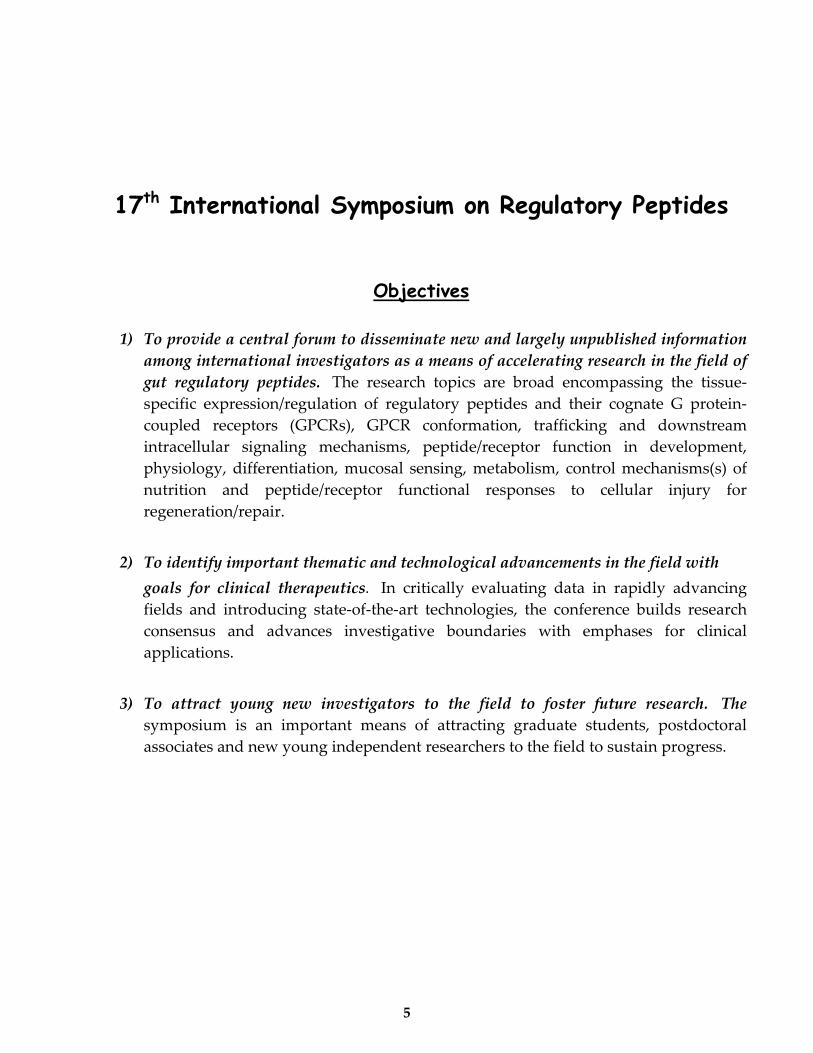
17th International Symposium on Regulatory Peptides
Objectives
1) To provide a central forum to disseminate new and largely unpublished information among international investigators as a means of accelerating research in the field of gut regulatory peptides. The research topics are broad encompassing the tissue‐specific expression/regulation of regulatory peptides and their cognate G protein‐coupled receptors (GPCRs), GPCR conformation, trafficking and downstream intracellular signaling mechanisms, peptide/receptor function in development, physiology, differentiation, mucosal sensing, metabolism, control mechanisms(s) of nutrition and peptide/receptor functional responses to cellular injury for regeneration/repair.
2) To identify important thematic and technological advancements in the field with goals for clinical therapeutics. In critically evaluating data in rapidly advancing fields and introducing state‐of‐the‐art technologies, the conference builds research consensus and advances investigative boundaries with emphases for clinical applications.
3) To attract young new investigators to the field to foster future research. The symposium is an important means of attracting graduate students, postdoctoral associates and new young independent researchers to the field to sustain progress.
5

17th International Symposium on Regulatory Peptides 25-28 January, 2009
Scientific Program ______________________________________________________________________ Sunday, January 25, 2009
2:00‐6:00 PM: Arrival and Registration at the Fess Parkerʹs Doubletree Resort, Santa
Barbara, California.
6:00‐6:10 PM: Opening Remarks from the Local Organizing Committee:
Enrique Rozengurt (Chair), Joseph Pisegna and Yvette Taché (Co‐Chairs).
6:10‐7:00 PM: Opening Lecture:
Novel modifications of gut regulatory peptides
“Biochemical Characterization of the Membrane‐bound Acyltransferase that Octanoylates Ghrelin, an Appetite‐Stimulating Peptide Hormone”
Joseph L. Goldstein, University of Texas Southwestern Medical Center, Dallas, USA.
Sponsored by Bachem.
7:00‐9:30 PM: Opening Reception in the Reagan Room at the Fess Parkerʹs Doubletree
Resort.
___________________________________________________________________
6

Monday, January 26, 2009
7:00‐8:30 AM: Breakfast/Registration
8: 30‐10:00 AM: Session 1: GPCR Structure and Regulation
Session Chairs: Graham S. Baldwin (University of Melbourne, Melbourne, Australia) and Alan Kopin (Tufts University School of Medicine, Boston, MA, USA).
8:30‐9:10 AM: Plenary Lecture: GPCRs and orphan receptors. Olivier Civelli, University of California, Irvine, Irvine, USA.
9:10‐9:25 AM: A novel molecular switch of GPCR activation based on the free fatty acid one receptor Irina G. Tikhonova2, Chi Shing Sum1, Stefano Costanzi2, Marvin C. Gershengorn1
1Clinical Endocrinology Branch and 2Laboratory of Biological Modeling, National Institute of Diabetes and Digestive and Kidney Diseases, National Institutes of Health, Bethesda, Maryland, USA
9:25‐9:40 AM: Development of a bivalent photolabile secretin probe for investigation of the stoichiometry of ligand‐receptor occupation Maoqing Dong, Delia I. Pinon, and Laurence J. Miller. Mayo Clinic, Scottsdale, AZ, USA. 9:40‐ 9:55 AM: Molecular approximations between residues 6 and 12 of glucagon‐like peptide 1 and its receptor demonstrated by photoaffinity labeling. Quan Chen, Delia I. Pinon, Laurence J. Miller and Maoqing Dong Mayo Clinic, Scottsdale, AZ, USA.
9:55‐ 10:05 AM: Structure activity analysis of human urotensin II using a library of 288 peptide variants. Sebastian Bandholtz1 Anja Klussmeier1, Jörg Wichard2, Ronald Kühne2, Bertram Wiedenmann1 and Carsten Grötzinger1 1Charité ‐ Universitätsmedizin Berlin, Department of Hepatology and Gastroenterology, Berlin, Germany; 2Leibnitz‐Institut für Molekulare Pharmakologie (FMP), Drug Design/Molecular Modeling Group, Berlin, Germany.
10:05‐10:20 AM: Membrane‐tethered ligands: novel probes for exploring class B1 G protein‐coupled receptor function. Jean‐Philippe Fortin1, Yuantee Zhu1, Charles Choi2, Martin Beinborn1, Michael N. Nitabach2 and Alan S. Kopin1
1Tufts Medical Center, Tufts University School of Medicine, Boston, MA 2Department of Cellular and Molecular Physiology, Yale School of Medicine, New Haven, CT, USA
10:20‐10:40 AM: Break‐Exhibits
7

10:40 AM‐12:30 PM: Session 2: GPCR Interactions and Trafficking
Session Chairs: Catia Sternini (CURE/University of California, Los Angeles, USA) and Christiane Susini (INSERM U858, Toulouse, France)
10:40‐11:20 AM: Plenary Lecture: Regulatory implications of cellular trafficking of receptors Mark von Zastrow, University of California, San Francisco, USA.
11:20‐10:35 AM: Mechanisms of pharmacological regulation of CCK2 receptor internalization. Rémi Magnan, Chantal Escrieut, Ingrid Langer, Magali Foucaud and Daniel Fourmy. INSERM, Institut de Médecine Moléculaire de Rangueil, Toulouse, France.
11:35‐11:50 AM: Evidence for secretin receptor interaction with receptor activity modifying protein 3 using resonance energy transfer and cellular translocation assays. Kaleeckal G. Harikumar1, Patrick. M. Sexton2, and Laurence J. Miller1. 1Department of Molecular Pharmacology and Experimental Therapeutics, Mayo Clinic, Scottsdale, Arizona, USA. 2Department of Pharmacology, Monash University, Clayton, Victoria, Australia
11:50 AM‐12:05 PM: Filamin A‐sst2 somatostatin receptor complex, a gain‐of‐function interaction that inhibits PI3 Kinase pathway and cell survival. Souad Najib1, N. Saint‐Laurent1, JP Estève1, J Lattig1, Daniel Fourmy1, S. Schulz2, Christiane Susini1. 1INSERM, Institut de Médecine Moléculaire de Rangueil, Hôpital Rangueil, Département Cancer, 31432 Toulouse, France. 2Department of Pharmacology and Toxicology, Universität Würzburg, Würzburg, Germany.
12:05‐12:20 PM: Differential engagement of downstream signaling pathways by calcium and L‐amino acids through the extracellular calcium‐sensing receptor Osvaldo Rey1, Steven Young1, Romeo Papazyan1 and Enrique Rozengurt1. 1CURE: Digestive Diseases Research Center, Division of Digestive Diseases, David Geffen School of Medicine at UCLA, University of California, Los Angeles, CA, USA
12:30‐1:30 PM: Lunch and Exhibits
3:30‐6:00 PM: Session 3: Signal Transduction Mechanisms, Secretion, Cell Proliferation
and Angiogenesis
Session Chairs: B Mark Evers (Galveston, Tx, USA) and Enrique Rozengurt (CURE/ University of California, Los Angeles, CA, USA)
3:30‐3:45 PM: Mitogen‐Activated Protein Kinases Regulate Bombesin‐Stimulated IP3 Receptor Activity and their Association with Bcl‐2 Family Proteins Xiaodong Wen1, Kirk Ives1, and Mark R. Hellmich1
1Departments of Surgery and Neuroscience and Cell Biology, Sealy Center for Cancer Cell Biology, University of Texas Medical Branch, Galveston, Texas, USA.
8
3:45‐4:00 PM: Neuromedin B Stimulates Proliferation of Rat Calvarial Osteoblasts through ERK Signaling Pathway. Hiroki Saito1, Ryuichi Ikeda2, Kazuhiko Inoue1, Atsuro Miyata1 1Department of Pharmacology, 2Department of Clinical Pharmacy and Pharmacology, Graduate School of Medical and Dental Sciences, Kagoshima University, Sakuragaoka, Kagoshima, Japan 4:00‐4:15 PM: Bombesin Receptor Subtype‐3 Mediates Glucose Uptake In Differentiated Human Visceral Adipocytes Via GLUT4 Translocation. Jessica Lam1, Anita Ratnasari1, Dongmei Xiao1, David Coy2, and H. Christian Weber11Boston University School of Medicine, Boston, Massachusetts, USA; 2Tulane School of Medicine, Peptide Research Laboratory, New Orleans, Louisiana, USA
4:15‐4:30 PM: Neurotensin induces IL‐6 secretion in mouse preadipocytes and adipose tissues during TNBS colitis. Hon‐Wai Koon, You Sun Kim1, Hua Xu,, Iordanis Karagiannides, Dezheng Zhao, Paul Dobner2, Charalabos Pothoulakis. Division of Digestive Diseases, David Geffen School of Medicine at UCLA, Los Angeles, CA and 1Department of Internal Medicine, Seoul Paik Hospital, Inje University College of Medicine, Seoul, Korea, 2Department of Molecular Genetics and Microbiology, University of Massachusetts Medical School, Worcester, MA, USA. 4:30‐4:45 PM: Protein Kinase D/Kidins220‐mediated regulation of neurotensin secretion Jing Li, Courtney M. Townsend, Jr., and B. Mark Evers Department of Surgery, The University of Texas Medical Branch, Galveston, TX, USA, 77555‐0536 4:45‐5:00 PM: Break‐Exhibits
5:00‐5:15 PM: Crosstalk between insulin and G protein‐coupled receptor signaling systems in human pancreatic cancer cells. Krisztina Kisfalvi1, James Sinnett‐Smith1, Robert Kui1, Guido Eibl2 and Enrique Rozengurt11,2CURE: Digestive Diseases Research Center; 1Division of Digestive Diseases, Department of Medicine; 2Department of Surgery; David Geffen School of Medicine at University of California, Los Angeles, CA, USA
5:15‐5:30 PM: Identification of Thrombospondin‐1 as a critical paracrine effector of somatostatin receptor sst2 tumor suppressive activity on pancreatic tumor growth and microenvironment Hanane Laklai1, N. Saint‐Laurent1, M. Hadegorn2, A. Bikfalvi2, P. Rochaix3, Corinne Susini1, Christiane Bousquet11INSERM U858, Institut de Médecine Moléculaire de Rangueil, Hôpital Rangueil,Toulouse, France; 2INSERM E0113, Université Bordeaux I, France ; 3Institut Claudius Regaud, Service d’anatomie et cytologie pathologiques, Toulouse, France. 5:30‐5:45 PM: Gastrin precursors regulate the expression of the pro‐angiogenic factor VEGF in colon cancer cells. Catherine Do, Claudine Bertrand, Aline Kowalski‐Chauvel, Cecile Resa, Catherine Seva. INSERM, U.858, Cancer Department, Toulouse, France.
9

5:45‐6:00 PM: A role of CRH family peptides in intestinal angiogenesis. Eunok Im1, Yong Seek Park2, Sang Hoon Rhee1 and Charalabos Pothoulakis11Division of Digestive Diseases, David Geffen School of Medicine, University of California, Los Angeles, CA, USA; 2Kyung Hee University, College of Medicine, Seoul, Korea.
20:00 ‐ 22:30 PM: Poster Session (SAN RAFAEL Room)
Tuesday, January 27, 2009
7:00‐8:30 AM: Breakfast
8:30‐9:20 AM: 8th Viktor Mutt Lecture
Session Chairs: Daniel Fourmy, (INSERM, Toulouse, France) and Jens Rehfeld (Department of Clinical Biochemistry, Rigshospitalet, University Hospital of Copenhagen, Denmark).
The role of NPY in health and disease: Insights from transgenic and KO models
Herbert Herzog, Garvan Institute of Medical Research, Sydney, Australia.
9:20‐10:25 AM: Session 4: Gut‐Brain Peptides and Energy Homeostasis
Session Chairs: M. Michael Wolfe, (Boston, MA, USA) and Yvette Taché (CURE/University of California, Los Angeles, CA, USA).
9:20‐9:40 AM: Phylogenetic Analysis and Examination of Evolutionary Glucose‐Dependent Insulinotropic Polypeptide (GIP) Expression. Michelle C. Musson1, Lisa I. Jepeal1, Patrick D. Mabray2, Scott I. Kavanaugh3, Irina V. Zhdanova2, Stacia A. Sower3, and M. Michael Wolfe11 Section of Gastroenterology, Boston University School of Medicine, Boston, MA, 2Department of Anatomy and Neurobiology, Boston University School of Medicine, Boston, MA 3Department of Biochemistry and Molecular Biology, University of New Hampshire, Durham, NH, USA.
9:40‐9:55 AM: Lipase inhibition acutely increases appetite and attenuates the postprandial concentrations of GLP‐1, CCK and PYY. Mark Ellrichmann1, Mario Kapelle1, Peter R. Ritter1, Jens J. Holst2, Karl‐Heinz Herzig3 Wolfgang E. Schmidt1, Frank Schmitz4, Juris J. Meier11Department of Medicine I, St. Josef‐Hospital, Ruhr‐University Bochum, Germany 2Department of Biomedical Sciences, The Panum‐Institute, University of Copenhagen, Denmark, 3Department of Surgery and A.I. Virtanen Institute, University of Kupio, Finland, 4Department of Medicine II, Clinic Hildesheim, Germany.
10

9:55‐10:10 PM: Effects of oral GLP‐1 on glucose homeostasis and appetite profile following an oGTT in healthy male subjects. Robert E. Steinert 1,2, B. Poller1,3, C. Castelli4, A. R. Huber5, J. Drewe1,3, Christoph Beglinger1,21Clinical Research Center, Department of Biomedicine and 2Division of Gastroenterology; 3Department of Clinical Pharmacology & Toxicology, University Hospital, 4031 Basel, Switzerland; 4Emisphere Technologies, Tarrytown, New York, USA; 5Center of Laboratory Medicine, Kantonsspital Aarau, Switzerland. 10:10‐10:25 AM: CART is a regulator of islet function and a possible incretin hormone. Nils Wierup1, Sörhede‐Winzell M2, Lindqvist A1, Fex M1 Korsgren O3, Kuhar MJ4, Ahren B2, Sundler F1
1Department of Experimental Medical Science, Lund University, Lund, Sweden; 2Department of Clinical Sciences, Lund University, Lund, Sweden; 3Department of Clinical Immunology, University of Uppsala, Sweden; 4Division of Neuroscience, Yerkes National Primate Research Center of Emory University, Atlanta, USA. 10:25‐10:45 AM Break‐Exhibits
10:45 AM‐12:30 PM: Session 5: Ghrelin and Novel Peptides
Session Chairs: Linda Hilsted, (University Department of Clinical Biochemistry, Rigshospitalet, Copenhagen, Denmark) and Joseph Reeve (CURE/University of California, Los Angeles, CA, USA).
10:45‐11:25 AM: Plenary Lecture: Ghrelin and novel peptides in the regulation of energy homeostasis Masamitsu Nakazato, Miyazaki Medical College, Miyazaki. Japan.
11:25‐11:40 AM Role of adenosine in the regulation of gastric somatostatin and ghrelin release from the mouse stomach. Gary K. Yang1, Linda Yip2, Jiang‐Fan Chen3, Bertil B. Fredholm4, Timothy J. Kieffer1, Yin Nam Kwok1. 1Department of Cellular and Physiological Sciences, University of British Columbia, Vancouver, British Columbia, Canada; 2Department of Medicine, Stanford University, Stanford, California, USA; 3Department of Neurology, Boston University School of Medicine, Boston, Massachusetts, USA; 4Department of Physiology and Pharmacology, Karolinska Institutet, Stockholm, Sweden. 11:40‐11:55 AM: Role of taste receptors in the regulation of ghrelin secretion Sara Janssen, B. De Smet, T Peeters, J Tack, Inge Depoortere Centre for Gastroenterological Research, University of Leuven, Leuven, Belgium.
11:55 AM‐12:10 PM: Neuropeptide W (NPW)‐containing neuron network in the hypothalamus Fumiko Takenoya1,2, Haruaki Kageyama1, Yukari Date3, Masamitsu Nakazato4, Seiji Shioda11Department of Anatomy, Showa University School of Medicine, Tokyo, Japan. 2Department of Physical Education, Hoshi University School of Pharmacy and Pharmaceutical Science, Tokyo, Japan. 3Department of Frontier Science Research Center, and 4 Neurology, Respirology, Endocrinology and Metabolism, University of Miyazaki, Miyazaki, Japan.
11

12:10‐12:25 PM: Nesfatin‐1 is a new molecule that mediates food reducing effect via a brain CRF‐dependent pathway Andreas Stengel1, Miriam Goebel1, Lixin Wang1, Jean Rivier2, Peter Kobelt3, Hubert Mönnikes4, and Yvette Taché11Department of Medicine, CURE Digestive Diseases Research Center, Center for Neurobiology of Stress, Digestive Diseases Division UCLA, VA Greater Los Angeles Healthcare System, Los Angeles, CA, USA; 2Peptide Biology Laboratories, Salk Institute, La Jolla, CA, USA; 3Department of Medicine, Division Psychosomatic Medicine and Psychotherapy; Charité – Universitätsmedizin Berlin, Campus Mitte, Berlin, Germany; 4Department of Medicine and Institute of Neurogastroenterology at Martin‐Luther‐Hospital, Berlin, Germany.
12:30‐1:30 PM: Lunch and Exhibits.
3:00‐5:20 PM: Session 6: Regulatory Peptides in Physiology and Disease
Session Chairs: Stephen Pandol (CURE/VAGLAHS/University of Los Angeles, CA, USA) and Juanita Merchant (University of Michigan, MI, USA).
3:00‐3:15 PM: Nesfatin‐1: A Novel Metabolic Hormone in Rodents. Sima Mortazavi1, Akansha Tiwari1 and Suraj Unniappan1
1Laboratory of Integrative Neuroendocrinology, Department of Biology, York University, Toronto, Ontario M3J1P3, Canada. 3:15‐3:30 PM: Identification and characterization of nesfatin‐1 immunoreactivity in endocrine cells of the rat gastric oxyntic mucosa. Miriam Goebel1, Andreas Stengel1, Iskandar Yakubov2, Lixin Wang1, Yvette Taché1, George Sachs2, Nils W.G. Lambrecht2. CURE/Digestive Diseases Research Center, 1Center for Neurobiology of Stress, 2Membrane Biology Laboratory, Digestive Diseases Division, Department of Medicine, UCLA, VA Greater Los Angeles Healthcare System, Los Angeles, CA, USA. 3:30‐3.45 PM The effect of exogenous apelin on the secretion of pancreatic juice in anaesthetized rats Małgorzata Kapica1, Alicja Jankowska2, Hanna Antushevich2, Marta Zabielska1, Iwona Puzio1, Romuald Zabielski3 1Department of Biochemistry and Animal Physiology, Faculty of Veterinary Medicine, University of Life Sciences in Lublin, Lublin, 2The Kielanowski Institute of Animal Physiology and Nutrition, Polish Academy of Science, Jabłonna, 3Department of Physiological Sciences, Faculty of Veterinary Medicine, Warsaw University of Life Sciences, Warsaw, Poland.
3:45‐4:40 PM: Differential responses of the incretin hormones GIP and GLP‐1 to increasing doses of ingested carbohydrate. Stephanie M Yoder, Qing, Y, Kindel, TL, and Tso, P Department of Pathology and Laboratory Medicine, University of Cincinnati, Cincinnati, USA. 4:00‐4:20 PM Break‐Exhibits
12

4:20‐4:35 PM Precursor processing of human defensin‐5 is essential to the physiological functions in vivo and in vitro. Hiroki Tanabe1, Chisato Ishikawa1, Atsuo Maemoto1, Jiro Watari1, Mikihiro Fujiya1, Yutaka Kohgo1
1Division of Gastroenterology and Hematology/Oncology, Department of Internal Medicine, Asahikawa Medical College, Asahikawa Hokkaido, Japan.
4:35‐4:50 PM Protective effect of μ opioid receptor on inflammation induced by intestinal ischemia and reperfusion in mice. Francesca Saccani1‐2, Laura Anselmi1, Ingrid Jaramillo1, Simona Bertoni2, Mariannina Impicciatore2, Elisabetta Barocelli2, Catia Sternini1. 1 CURE: Digestive Research Center, Department of Medicine, Division of Digestive Diseases, David Geffen School of Medicine at University of California, Los Angeles, USA; 2 Department of Pharmacological, Biological and Applied Chemical Sciences, University of Parma, Parma, Italy. 4:50‐5:05 PM Endogenous PACAP protects the brain against focal cerebral ischemia in mice. Tomoya Nakamachi1, Hirokazu Ohtaki1, Sachiko Yofu1, Naoko Nonaka2, William A. Banks3 and Seiji Shioda11Department of Anatomy, Showa University School of Medicine, Tokyo, Japan; 2Department of Oral Anatomy, Showa University School of Dentistry, Tokyo, Japan; 3Veterans Affairs Medical Center‐St. Louis and Saint Louis University School of Medicine, Division of Geriatrics, Department of Internal Medicine, St. Louis, MO, USA.
5:05‐5:20 PM Deficiency in the Intestinal Hormone VIP Results in Gastric Atrophy and Hypochlorhydria as Determined in a Knockout Mouse Model. Yuxin Lu1,2, Alex Cantrell1, 2, Catherine Lee1,2, David S Oh,1,2, Gordon V. Ohning,1,2, Patrizia Germano,1,2, James Wascheck3 and Joseph R Pisegna1, 2.
1Departments of Gastroenterology and Hepatology, Veterans administration GLAHS, Los Angeles, California, USA and 2Departments of Medicine and 3Psychiatry, David Geffen School of Medicine, University of California at Los Angeles, California, USA.
5:20‐5:40 PM Break‐Exhibits
5: 40‐6:30 PM: 1st Werner Creutzfeldt Lecture
Session Chairs: Wolfgang Schmidt (St. Josef‐Hospital, Ruhr‐University of Bochum, Bochum, Germany) and Christoph Beglinger (Clinical Research Centre, University Hospital Basel, Basel, Switzerland).
Oncogenic Functions of Gastrin‐Releasing Peptide in Neuroblastoma Dai H. Chung University of Texas Medical Branch, Galveston, Tx, USA.
7:30 PM ‐ Closing Dinner (Reagan Room)
13

Wednesday, January 28, 2009 7:00‐8:15 AM: Breakfast
8:15‐9:45 AM: Session 7: Stem cells, Regulatory Peptides and Cancer
Session Chairs: Catherine Seva (INSERM, Toulouse, France) and Charalabos Pothoulakis (CURE/Univeristy of California, Los Angeles, CA, USA).
8:15‐8:55 AM: Plenary Lecture: Regulatory peptides and Cancer in the GI tract, Timothy C. Wang, Columbia University, New York, USA.
8:55‐9:15 AM: Helicobacter infection suppresses Shh expression via IL‐1β promoting gastric atrophy Waghray M., Saqui‐Salces M., Zavros Y., Andreas Todisco, Juanita L Merchant. University of Michigan, MI, USA.
9:15‐9:35 AM: Expression of gastrin precursors by CD133 positive‐colon cancer stem cells is crucial for tumour growth. Audrey Ferrand, Mauro S. Sandrin, Arthur Shulkes and Graham S. Baldwin. University of Melbourne Department of Surgery, Austin Health, Heidelberg, Victoria 3084, Australia.
9:35‐9:50 AM: Expression of Gastrin‐Releasing Peptide Receptor (GRPR) in Carcinoma Associated Fibroblasts (CAF) Correlates with Colorectal Cancer Lymph Node (LN) Metastasis. Celia Chao1,2, Heidi Weiss2, Guillermo Gomez1, Courtney M. Townsend, Jr1, Mark R. Hellmich1,2
Division of Surgical Oncology, Department of Surgery1, and The Sealy Center for Cancer Cell Biology2, University of Texas Medical Branch; Galveston. USA
9:50‐10:10 AM: Break‐Exhibits
10:10‐11:40 AM: Session 8: Enteroendocrine Cells in Health and Disease
Session Chairs: Seiji Shioda (Tokyo, Japan) and H. Cristian Weber (Boston, MA, USA)
10:10‐10:50 AM: PlenaryLecture: Enteroendocrine cell development and malabsorption disease. Martín Martín, UCLA, Los Angeles, USA.
10:50‐11:10 AM: Induction of colitis and colorectal tumors in PACAP‐deficient mice Nicole Nemetz1, Catalina Abad2, Greg Lawson1, Hiroko Nobuta2, Seririthanar Chhith2, Lucy Duong2, Gary Tse2 and James A. Waschek2
1Departments of Laboratory Animal Medicine and 2Department of Psychiatry, David Geffen School of Medicine, Semel Institute for Neuroscience and Mental Retardation Research Center, University of California at Los Angeles, USA
14

11:10‐11:30 AM: Chromogranin A as a predictor of progression, regression or stable disease in ileo‐cecal (midgut) carcinoid tumours. Linda Hilsted1, K. Højsgaard Jensen2, T. Mynster3, U. Knigge2Department of Clinical Biochemistry1 and Department of Surgery C2, Rigshospitalet and Department of Surgery K, Bispebjerg Hospital 3, Faculty of Health Sciences, University of Copenhagen, Denmark
11:30‐11:45 AM: Linaclotide, a Novel Peptide Therapeutic Agent in Clinical Development for the Treatment of IBS‐C and Chronic Constipation is Digested in the Small Intestine to Small Peptides and Ultimately to L‐Amino Acids. Marco M. Kessler1, Robert W. Busby1, Wilmin P. Bartolini1, Paul R. Blomquist1, Alexander P. Bryant1, Jenny V. Tobin1, James D. Wakefield1, Caroline B. Kurtz1 and Mark G. Currie11Ironwood Pharmaceuticals, Inc., Cambridge, MA, USA.
11:45 AM: Announcement of next REGPEP Symposium:
Daniel Fourmy (INSERM, Toulouse, France)
12.00 AM: MEETING ENDS
15

17th International Symposium on
Regulatory Peptides
ABSTRACTS
Abstract Number Section
1‐10 GPCR STRUCTURE, INTERACTIONS and TRAFFICKING
11‐22 SIGNAL TRANSDUCTION
23‐27 CHEMICAL and NEURAL SENSING
28‐35 INCRETINS
36‐44 GHRELIN
45‐50 VIP/PACAP
51‐60 NOVEL PEPTIDES
61‐65 INFLAMMATION
66‐78 CANCER
79‐82 METHODOLOGIES
16

GPCR STRUCTURE, INTERACTIONS and
TRAFFICKING 1. Structure activity analysis of human urotensin II using a library of 288 peptide variants Sebastian Bandholtz1 Anja Klussmeier1, Jörg Wichard2, Ronald Kühne2, Bertram Wiedenmann1 and Carsten Grötzinger1 1 Charité - Universitätsmedizin Berlin, Department of Hepatology and Gastroenterology, Augustenburger Platz 1, D-13353 Berlin, Germany; 2 Leibnitz-Institut für Molekulare Pharmakologie (FMP), Drug Design/Molecular Modeling Group, Robert-Rössle-Str. 10, D-13125 Berlin, Germany Urotensin II (U-II) is the most potent vasoconstrictor peptide yet identified, the effects by U-II are species-, tissue- and endothelium-dependent. U-II plays a key role in different physiological processes as well as disease conditions like heart failure and cardiac cell growth, renal dysfunction, diabetes, and mitogenesis in vascular and tumor cells. Key features of these diseases are increased expression and activity of U-II receptor (U-IIR). The purpose of the current study was to analyze the peptide receptor interaction and to establish a map of structure activity relationships for each amino acid residue within the molecule with regard to receptor binding, intracellular signalling and endocytosis. For that, we designed a peptide library of 288 U-II variants that contains substitution variants of each position with one of the other 19 proteinogenic amino acids. In addition, a number of multiple substitutions and species variants were included in the peptide library. Peptide variant activity was measured in different cell-based high-throughput fluorescent assays like calcium imaging, binding and internalization assays. For each assay, concentration response curves were determined and a statistical analysis based on ridge-regression models was performed in order to determine the underlying structure-activity relationship. We determined the role of individual amino acid positions and the side chain characteristics which are important for U-IIR binding, activation and internalization. A model for the interaction of the peptide with its receptor with regard to these functions will be presented.
17
2. Molecular approximations between residues 6 and 12 of glucagon-like peptide 1 and its receptor demonstrated by photoaffinity labeling Quan Chen, Delia I. Pinon, Laurence J. Miller and Maoqing Dong Mayo Clinic, Scottsdale, AZ 85259 The glucagon-like peptide 1 (GLP1) receptor is an important drug target within the Family B G protein-coupled receptors. The natural agonist ligand, GLP1, holds promise for the management of the type 2 diabetes. Recently the crystal structure of the antagonist-bound amino terminus of the GLP1 receptor has been reported. However, how GLP1 binds and activates the intact GLP1 receptor is still unclear. In this study, we explored the spatial approximations of position 6 and 12 of GLP1 with its receptor using photoaffinity labeling. It should be noted that these two positions were within the aminoterminal region of the GLP1(7-36) peptide and were not included in the truncated antagonist that was used in the crystal structure of the GLP1 receptor amino terminus. Two photolabile probes were synthesized by incorporating a benzoyl phenylalanine in positions 6 and 12 of the GLP1(7-36) peptide. Both probes were full agonists (position 6 probe, EC50 = 0.7 ± 0.1 nM; position 12 probe, EC50= 0.05 ± 0.02 nM), stimulating cAMP accumulation in receptor-bearing CHO cells in a concentration-dependent manner. They bound the GLP1 receptor specifically and saturably (position 6 probe, Ki = 11.2 ± 2.1 nM; position 12 probe, Ki = 1.2 ± 0.1 nM). They labeled the receptor efficiently, with this inhibited by GLP1 in a concentration-dependent manner. The photoaffinity labeled receptor migrated at approximate 66 kD and shifted to 42 kD after deglycosylation. The labeled receptor was then purified and submitted to chemical and proteinase cleavages. This identified the juxtamembrane region of the amino-terminal domain of the GLP1 receptor as the region of labeling for the position 12 probe. Using similar peptide mapping approaches, the site of labeling for the position 6 probe was localized to a distinct region that includes the third extracellular loop. Receptor mutants are being generated to further localize the sites of labeling for both probes by further chemical and proteinase cleavages and radiochemical sequencing. These data are consistent with a common ligand binding and activation mechanism for Family B G protein-coupled receptors. As we expand the number of such experimental constraints, we should be able to meaningfully model the agonist-bound GLP1 receptor, adding to our insights into the molecular basis of ligand binding to this important receptor.
3. Development of a bivalent photolabile secretin probe for investigation of the stoichiometry of ligand-receptor occupation Maoqing Dong, Delia I. Pinon, and Laurence J. Miller Mayo Clinic, Scottsdale, AZ 85259 The secretin receptor is a prototypic Family B G protein-coupled receptor (GPCR). This family includes many potentially important drug targets. Like many GPCRs, the secretin receptor forms homodimers. It is notable that these complexes are present constitutively and are stable even in the presence of secretin. Recently, multiple photoaffinity labeling constraints have been used to build a model of the natural agonist ligand-bound secretin receptor (Dong, et al., 2008). However, whether secretin interacts with one or both receptor molecules in the dimeric complex is currently unknown. In this work, we have directly explored this question using photoaffinity labeling with a secretin analogue probe containing dual photolabile benzoylphenylalanine (Bpa) residues as sites of covalent. This probe incorporated a Bpa at the amino-terminal extension, in position -2, and at the carboxyl-terminal region, in position 26, of the 27-amino acid secretin peptide. The incorporation of a Bpa in position -2 was designed to minimize its negative impact on the function. This doubly-reactive probe was demonstrated to be a full agonist, stimulating cAMP accumulation in secretin receptor-bearing CHO cells in a concentration-dependent manner. It labeled the secretin receptor specifically and saturably. The photoaffinity labeled receptor migrated at approximate 70 kD and shifted to 42 kD after deglycosylation. This electrophoretic migration represented labeling of a single protomer of the dimerizing secretin receptor. No radiolabeled receptor dimer was observed except after the positive control of cross-linking with disuccinimidyl suberate. The labeled receptor was purified and submitted to cyanogen bromide cleavage and this identified the first cyanogen bromide fragment at the amino terminus (a region labeled by the single attachment position 26 probe) and the fragment including the sixth transmembrane and third extracellular loop region (a region labeled by the single attachment position -2 probe) of the secretin receptor. Absence of demonstrable receptor dimerization after the establishment of dual sites of covalent attachment supports the presence of these two domains within a single receptor protomer. This is further supported by the demonstration of the covalent adduct of a single probe molecule with the two expected cyanogen bromide fragments of the secretin receptor. In conclusion, these data are consistent with a model of one molecule of secretin occupying one copy of the secretin receptor, even though it is present in a dimeric complex. It will be key to explore other possible approximations between secretin and the second receptor protomer in future studies.
18
4. Membrane-tethered Ligands: Novel Probes For Exploring Class B1 G Protein-Coupled Receptor Function
Jean-Philippe Fortin1, Yuantee Zhu1, Charles Choi2, Martin Beinborn1, Michael N. Nitabach2 and Alan S. Kopin1 1 Tufts Medical Center, Tufts University School of Medicine, Boston, MA 2 Department of Cellular and Molecular Physiology, Yale School of Medicine, New Haven, CT Class B1 (secretin family) G protein-coupled receptors (GPCRs) modulate a wide range of physiological functions including glucose homeostasis, feeding behavior, fat deposition, bone remodeling and vascular contractility. Endogenous peptide ligands for these GPCRs are of intermediate length (27-44 amino acids) and include receptor affinity (C-terminal) as well as receptor activation (N-terminal) domains. We have developed a novel technology in which a peptide ligand tethered to the cell membrane selectively modulates corresponding class B1 GPCR-mediated signaling. The engineered cDNA constructs encode a single protein comprised of (i) a transmembrane domain (TMD) with an intracellular C-terminus, (ii) a poly(asparagine-glycine) linker extending from the TMD into the extracellular space, and (iii) a class B1 receptor ligand positioned at the N-terminus. We demonstrate that membrane-tethered peptides, like corresponding soluble ligands, trigger dose-dependent receptor activation. The broad applicability of this approach is illustrated by experiments utilizing tethered versions of seven mammalian endogenous class B1 GPCR agonists. In parallel, we carried out mutational studies primarily focused on incretin ligands of the glucagon-like peptide-1 receptor. These experiments suggest that tethered ligand activity is conferred, in large part, by the Nterminal domain of the peptide hormone. Follow-up studies revealed that interconversion of tethered agonists and antagonists can be achieved with introduction of selected point mutations. Such complementary receptor modulators provide important new tools for probing receptor structure-function relationships as well as for future studies aimed at dissecting the tissue specific biological role of a GPCR in vivo (e.g. in the brain vs. in the periphery).
5. Constitutive Secretion of Somatostatin (SS) by RIN-14B Cells is Increased by its CCK-1 and CCK-2 Receptors Occupation: is There a Paradox? K. El-Kouhen, J. Morisset. Département de médecine, Univ. Sherbrooke, Qc, Canada. Introduction: Our objectives were to demonstrate the CCKR subtypes in RIN-14B cells, a SS secreting cell line and characterize their implication in SS secretion. Methods: Cells grew in RPMI medium. Western blots (WB) and immunofluorescence (IF) helped detect CCKR subtypes, SS, CCK and progastrin. Caerulein (Cae), JMV180 and pentagastrin (PG) alone or with CCKR antagonists were evaluated on SS secretion over 4h by ELISA. Results: CCKR were identified by WB and IF in RIN along with SS, CCK, progastrin. Cae and PG dose-dependently increased SS release, 3 fold at 1 .M Cae, 2.5-fold at 10 µM PG with occupation of both CCKR subtypes, confirmed by L364,718 and L365,260 inhibition of 1 and 2 CCKR. Occupation of high affinity (HA) CCK-1R by Cae was confirmed on SS release with JMV-180, a HA CCK1-R agonist and absence of SS release inhibition to high Cae conc occupying low affinity (LA) CCK1-R. Stimulated or not, cells release > 60% of their SS content, a sign of constitutive secretion, confirmed by cycloheximide and brefeldin, inhibiting SS synthesis and intracellular trafficking, respectively. Conclusion: Both CCKR subtypes are present in RIN-14B cells and involved in SS secretion through constitutive secretion controlled at level of SS synthesis. SS secretion via CCK1-R occupation mobilizes its HA sites.
19

6. Evidence for Secretin Receptor Interaction with Receptor Activity Modifying Protein 3 Using Resonance Energy Transfer and Cellular Translocation Assays Kaleeckal G. Harikumar1, Patrick. M. Sexton2, and Laurence J. Miller1. 1 Department of Molecular Pharmacology and Experimental Therapeutics, Mayo Clinic, Scottsdale, Arizona 85259, USA.
2 Department of Pharmacology, Monash University, Clayton, Victoria 3800, Australia.
Receptor activity modifying proteins (RAMPs) are single transmembrane proteins that have been shown to associate with various G protein-coupled receptors (GPCR) during receptor trafficking, that can modify receptor function. Biochemical and morphological approaches indicate that RAMPs can form stable complexes with some Family B GPCRs, and thereby influence some aspects of receptor function. However, RAMP interactions with the secretin receptor, a prototypic member of this family, have not been well studied. Here, we have utilized a cellular translocation assay to monitor possible interaction of the secretin receptor with RAMPs in living cells. This assay demonstrated the ability of the untagged secretin receptor to translocate specific YFPtagged RAMPs to the cell surface, as monitored by fluorescence microscopy. This assay was complemented with bioluminescence resonance energy transfer (BRET), fluorescence resonance energy transfer (FRET), and bimolecular fluorescence complementation (BiFC) approaches. The results demonstrated that RAMP3, but not RAMP1 or RAMP2,is able to interact with the secretin receptor. BRET, FRET and BiFC approaches supported these observations. To gain further insights into the molecular regions responsible for these interactions, we have utilized truncated N- and C-terminal secretin receptor constructs in analogous studies. Both truncated receptors were able to translocate RAMP3 to the cell surface. Additionally, N-terminal truncation of RAMP3 had no effect on the ability of the secretin receptor to translocate this molecule to the cell surface. This suggests that the intramembranous regions of the secretin receptor and RAMP3 are likely to interact. Further evaluation of the details of this interaction is in progress.
7. Mechanisms of pharmacological regulation of CCK2 receptor internalization Rémi Magnan, Chantal Escrieut, Ingrid Langer, Magali Foucaud and Daniel Fourmy. INSERM U858, Institut de Médecine Moléculaire de Rangueil, 1 avenue Jean Poulhès, 31432, Toulouse, FRANCE Background: CCK2-receptor (CCK2-R) and others G-protein-coupled receptors undergo desensitization and internalization after ligand stimulation. Once activated, G-protein-coupled receptors are phosphorylated by G-protein-coupled receptor kinases, thus becoming able to bind -arrestins which subsequently recruit several components of the endocytosis machinery. Several studies have shown the crucial involvement of the C-terminus tail of GPCRs in the interaction with - arrestins. This led to the conception of different classes of GPCR with regards of internalization and binding to -arrestins: class A receptors that internalize with a weak interaction with -arrestins and recycle back to the membrane quickly and class B receptors that internalize with a strong and sustained co-localization with -arrestins at the endosomes level thus recycling poorly and slowly to the membrane. Furthermore, it is now thought that the interaction between GPCRs and -arrestins is ligand specific allowing a GPCR to be either class A or B according to the ligand used. The C terminus of the CCK2-receptor has been shown to be involved in CCK2-R internalization, although the mechanisms of its pharmacological regulation remain poorly understood. Aim: To investigate arrestins recruitment after CCK2-R stimulation by different ligands in order to better characterize CCK2-R regulation. Results: Using CCK2-R and -arrestin1-GFP, -arrestin2 -GFP chimera as well as fluorescent CCK and Gastrin, we show that both CCK and Gastrin enable CCK2-R rapid internalization and both -arrestin1/2 translocation to the membrane. Dominant negative -arrestin2 strongly reduces internalization of CCK2-R. Both -arrestin1 and -arrestin2 co-localize with CCK2-R in endosomes over a long time period after CCK or Gastrin stimulation thus classifying CCK-2R as a Class B receptor. Moreover, non peptide ligands of CCK2-R showing partial and inverse agonist with regards of the Inositol Phosphate pathway are not able to evoke neither CCK2-R internalization nor -arrestin2 recruitment but still capable of activating MAPKinase pathway. To further examine - arrestin1/2 recruitment we generated several mutants of CCK2-R at the C-terminus of the receptor, and we report here the relevance of specific clusters of serine/threonine within the C-terminus for the interaction between activated CCK-2R and -arrestin2. Conclusion: Taken together, our results indicate that CCK2-R is a class B receptor with regards of its trafficking and that different ligands with distinct pharmacological profiles show no effect on CCK2-R down-regulation.
20

8. Filamin A-sst2 somatostatin receptor complex, a gain-of-function interaction that inhibits PI3 Kinase pathway and cell survival. S. Najib1, N. Saint-Laurent1, JP Estève1, J Lattig1, D. Fourmy1, S. Schulz2, C. Susini1.
1INSERM U858, Institut de Médecine Moléculaire de Rangueil, Hôpital Rangueil, Département Cancer, 31432 Toulouse, France. 2Department of Pharmacology and Toxicology, Universität Würzburg, Versbacher Str. 9, 97078 Würzburg, Germany. Background: The role of filamins in actin cross-linking and membrane stabilization is well established, but recently their ability to interact with a variety of transmembrane receptors and signalling proteins has led to speculation of additional roles in scaffolding and signal transduction. Somatostatin is a neuropeptide which acts as an endogenous inhibitor of various cellular functions including endocrine and exocrine secretions and cell proliferation. In our previous studies, we have demonstrated that somatostatin, acting through its G protein coupled receptor (GPCR) sst2, has potent proapoptotic and anti-invasive activities on normal and cancer cells and that sst2 behaves as a tumor suppressor gene for pancreatic cancer and is a valid target for pancreatic cancer therapy. However, the mechanisms whereby sst2 exerts its anti-oncogenic effects are not understood entirely. Aim: to investigate whether Filamin A interacts with sst2 and to determine the role of this interaction in signaling and anti-oncogenic effects of sst2. Results: In this work, we have identified Filamin A (FLNa) as a novel sst2 partner. FLNa interacts with sst2 in several cell lines including the human neuroendocrine tumor BON cells and human melanoma tumor cell line A7. In addition, sst2-FLNa interaction is enhanced by treatment with the sst2 agonist RC-160. In FLNa depleted (FLNa-/- or FLNa-si-RNA treated cells) human melanoma M2 cells expressing heterologous sst2 and Bon cells expressing endogenous sst2, somatostatin fails to inhibit proliferation and to induce apoptosis. Molecular modelling and surface plasmon resonance identified the 65IYV67 sequence in the sst2 first intracellular loop as responsible for a direct interaction between sst2 and FLNa. Mutation of this sequence prevented the inhibitory effect of somatostatin on cell survival in vitro, and on tumor progression in vivo. We have recently demonstrated a direct interaction between sst2 and the p85 regulatory subunit of PI3-Kinase. Ligand-activated sst2 inhibited PI3-Kinase activity by disrupting the pre-existing p85-sst2 complex. In the present study, our analysis of the FLNa-sst2 complex revealed an overlap between FLNa and p85 binding sites on sst2 first intracellular loop. We therefore, demonstrated that these proteins compete for binding to sst2 in vivo, and in vitro. Furthermore, in cells lacking FLNa, or expressing a mutated sst2 unable to interact with FLNa, we have shown that the formation of the sst2- FLNa complex is critical and is required for the dissociation of p85-sst2 complex and the inhibition of PI3-Kinase pathway by somatostatin. Conclusion: Our results identify FLNa as a novel sst2 partner, required for the somatostatin dependent inhibition of the PI3K/Akt survival pathway and inhibition of carcinogenesis. 9. The expression and the localization of orexin-1 receptor (OX1R) after traumatic brain injury in mice. Yuko Mihara1, Kenji Dohi1,2, Sachiko Yofu2, Tomoya Nakamachi2, Haruaki Kageyama2, Seiji Shioda2 and Tohru Aruga1 1 Department of Emergency and Critical Care Medicine, 2 Department of Anatomy, Showa University School of Medicine, 1-5-8 Hatanodai, Shinagawa-ku, Tokyo, Japan Background: Orexins are neuropeptides that have various physiological effects including regulation of feeding behavior, neuroendocrine functions and sleep-wake cycles. Recent studies have suggested that the orexin system may also be involved in traumatic brain injury, and changes in sleep patterns, energy homeostasis and neuroendocrine functions often occur in neurological conditions associated with brain injury. However, the expression and the localization of orexin receptors have not been elicited. Aim: To investigate the expression and the cellular localization of orexin-1 receptor (OX1R) and its time-dependent changes in the mouse brain after controlled cortical impact (CCI) injury by using immunohistochemical techniques. Results: OX1R immunoreactivity was first detected at 6 hours after CCI injury in the pericontusional cortex. The intensity of immunoreactivity was increased at 12 hours and peaked at 1 day, then decreased from 4 to 7 days after CCI injury. At 4 and 7 days, OX1R immunoreactivity was present predominantly in injured cortex and thalamus, with a few scattered in the hippocampus. To identify the cellular localization of OX1R, double-immunohistochemical staining with OX1R and some cell markers antibodies at 1 and 7 days after CCI injury was also performed. The OX1R immunopositive cells were obviously co-localized with a neuronal marker, NeuN-immunoreactivity at 7 days after CCI injury, while not at 1day. The OX1R immunoreaction was also expressed in the surface of the immunopositive cells of CD11b, a microgilal marker, at 1 and 7 days. To identify the direct contact of orexin A immunoreactive fibers with OX1R, double-immunohistochemical staining with orexin A and OX1R was performed. Orexin A-immunoreactive fibers were seen in direct contact with OX1R containing neurons in the peri-contusional area. Conclusion: These results suggested that orexin and its receptor might play important roles in traumatic brain injury, and that OX1R might be induced by neurons and also have functional roles associated with microglial reactivity after traumatic brain injury.
21

10. A novel molecular switch of GPCR activation based on the free fatty acid one receptor
Irina G. Tikhonova2, Chi Shing Sum1, Stefano Costanzi2, Marvin C. Gershengorn1 1 Clinical Endocrinology Branch and 2 Laboratory of Biological Modeling, National Institute of Diabetes and Digestive and Kidney Diseases, National Institutes of Health, Bethesda, Maryland 20892, USA Background: Activation of a number of class A G protein-coupled receptors (GPCRs) is thought to involve two molecular switches, a rotamer toggle switch representing by the aromatic residues within the transmembrane domain and an ionic lock between an Arg residue of the helix 3 and a Glu of the helix 6 near the cytoplasmic surface of the receptor; however, the mechanism by which agonist binding changes these molecular interactions is not understood. Importantly, 44% of GPCRs including free fatty acid receptor 1 (FFA1) lack the complement of amino acid residues implicated in either or both of these two switches; the mechanism of activation of these GPCRs is therefore less clear. Aim: To identify novel important interactions responsible for regulating interconversion between inactive and active receptor conformations on the example of a free fatty acid receptor one, previously known as a GPR40, a novel anti-diabetic target. Results: By homology modeling based on the recently published crystal structure of the ß2- adrenergic receptor, we identified two Glu residues (Glu145 and Glu172) in the second extracellular loop of FFA1 that form putative interactions individually with two transmembrane Arg residues [Arg(5.39) and Arg(7.35)]. The followed molecular dynamics simulations showed that binding of agonists to FFA1 leads to the breakage of these Glu-Arg interactions. We hypothesized that the Arg-Glu interactions in the unliganded receptor function as locks that keep the receptor in the inactive state and agonists activate the receptor by weakening or breaking these interactions. To validate the computational studies we conducted the mutagenesis experiments in which agonist-stimulated activities were assessed by measuring linoleatestimulated increases in cytoplasmic free calcium concentration and constitutive receptor activities were assessed using a luciferase-based transcriptional reporter assay. We found that the breakage of these two putative interactions by substituting Ala for Glu145 and Glu172 caused constitutative receptor activation. Two antagonists identified by us previously contain a less electronegative nitro group instead of a carboxyl group which may be unable to release the ionic lock and activate the receptor. Conclusion: Our results reveal a novel molecular switch for receptor activation present on the extracellular surface of FFA1 that is broken by agonist binding. Our results and phylogenetic analysis of human GPCRs provided the hypothesis that similar ionic locks between the transmembrane domains and the extracellular loops may constitute a mechanism common also to other class A GPCRs.
22

SIGNAL TRANSDUCTION 11. Neuromedin B stimulates Proliferation of Rat Calvarial Osteoblasts through ERK Signaling Pathway Hiroki Saito1, Ryuichi Ikeda2, Kazuhiko Inoue1, Atsuro Miyata1 1 Department of Pharmacology, 2 Department of Clinical Pharmacy and Pharmacology, Graduate School of Medical and Dental Sciences, Kagoshima University, Sakuragaoka, Kagoshima 890-8544, Japan
Neuromedin B (NMB) is one of the bombesin related peptide in mammals, and was originally isolated from porcine spinal cords. Rat NMB mRNA is expressed in high amounts in the olfactory region and esophagus. On the other hand, NMB receptor is expressed widely in different brain regions, and peripheral tissues. NMB exhibits various physiological effects on regulations of smooth muscle contraction, feeding, blood glucose, body temperature and so forth. Based on the finding of a fragment of chicken NMB cDNA in EST database from chicken rib cartilage, we identified the gene expressions of NMB and its receptor in chicken calvarial tissue. However, it has not been yet clarified whether NMB is involved in bone function or not. Therefore, we attempted to elucidate the function of NMB in bone morphogenesis. By RT-PCR and sequence analysis, we characterized the mRNA expressions of NMB and NMB receptor (NMB-R) in rat calvarial bone tissue. As compared with other tissues, the expression level of NMB mRNA in the calvarial bone was found as high as that in the brain, whereas the expression level of NMB-R mRNA in the calvarial bone was found lower than that in the brain, but more than that in the kidney, jejunum, or ileum. Furthermore, we confirmed NMB and NMB-R gene expressions in rat primary osteoblast, that is, NMB mRNA was weakly expressed, but NMB-R mRNA was strongly expressed. Then we investigated the effect of NMB on proliferation of rat primary osteoblasts, which were obtained from rat calvaria (wistar-rats of postnatal 1 day). WST-8 assay demonstrated that NMB (10-9 to 10-6 M) significantly induced the proliferation in a dose dependent manner. Western blotting analysis demonstrated that treatment of 10-8M NMB activated ERK1/2 MAPK in the primary osteoblast. Furthermore, pretreatment with U0126, MAPK kinase inhibitor attenuated the NMB-induced cell proliferation. As for the differentiation of the primary osteoblast, however, NMB treatment exhibited no effect as assessed by matrix mineralization using alizarin red S staining, alkaline phosphatase activity, and the expression of osteoblast-specific genes (osteocalcin, osteonectin, alkaline phosphatase, core binding factor alpha 1, Type 1 collagen). On the other hand, in order to investigate the effect of BMP-2 (Bone Morphogenetic Protein-2) and TGF-(Transforming Growth Factor-beta), which are involved in bone formation, on the expression of NMB and NMB-R genes, rat primary osteoblasts were treated with BMP-2 (100ng/ml) or TGF- (5ng/ml) for 2 days. As a result, BMP-2 exhibited no effect on the expression of NMB and NMB-R gene, but TGF-b significantly decreased NMB gene expression. In conclusion, these findings suggest that NMB/NMB-R signaling has potential physiological roles not in the differentiation but in the proliferation of osteoblast.
23

12. Effect of the phosphatases PHLPP1, PHLPP2 and PP2 on AKT Phosphorylation and Cell Death
C. J. Nitsche,1,2 M. Edderkaoui,1 J. Mayerle,2 M. M. Lerch,2 A. S. Gukovskaya1 1 Pancreatic research group VAGHLAS and University of California Los Angeles, CA; 2 Department of Internal Medicine A Ernst Moritz Arndt University Greifswald, Germany
Background & Aims: Akt kinase is a potent prosurvival factor in pancreatic cancer (PaCa). Akt activation is mediated by its phosphorylation. Little is known on the inactivation of Akt by phosphatases. Here we investigated the effects of phosphatases PHLPP1 and PHLPP2 and PP2A on Akt activity, and death of PaCa cells through apoptosis, necrosis and autophagy. Methods: Apoptosis was measured by DNA fragmentation, necrosis as percentage of cells permeable to PI, autophagy by measuring LC3 dots, Akt phosphorylation with western blot. Results: In human pancreatic adenocarcinoma MIA PaCa-2 and PANC-1 cells overexpression of PHLPP1 or 2 reduced the phosphorylation of Akt at Ser473 but had no effect on the phosphorylation at Thr308. PHLPP1 or 2 overexpression increased apoptosis, necrosis and autophagy and also stimulated gemcitabine-induced cell death in Mia PaCa-2 and PANC-1 cells. Differently inactivation of PP2A reduced Akt phosphorylation at Thr308 but not at Ser473 and did not decrease PaCa cell death. Thus, PHLPP1 and 2 rather than PP2A dephosphorylate Akt at Ser473 and mediate resistance of PaCa cells to death. The data also show that phosphorylation at Ser473 rather than at Thr308 is critical for prosurvival effects of Akt. Conclusion: The data indicate PHLPP1 and 2 dephosphorylate Akt in PaCa cells leading to death. Thus, upregulation of PHLPP1 and/or 2 could be considered as a promising strategy to stimulate death of pancreatic cancer cells. 13. Bombesin Receptor Subtype-3 Mediates Glucose Uptake In Differentiated Human Visceral Adipocytes Via GLUT4 Translocation. Jessica Lam1, Anita Ratnasari1, Dongmei Xiao1, David Coy2, and H. Christian Weber1 1 Boston University School of Medicine, Boston, Massachusetts, USA 2 Tulane School of Medicine, Peptide Research Laboratory, New Orleans, Louisiana, USA Background: Adipose tissue is a highly active endocrine organ and critical for proper regulation of energy homeostasis. Insulin-dependent glucose uptake is facilitated through membrane translocation of GLUT4. Disruption of glucose uptake results in insulin resistance, a hallmark of the metabolic syndrome, diabetes mellitus type 2 and obesity. Mice deficient in bombesin receptor subtype-3 (BRS-3) are characterized by impaired glucose metabolism, weight gain, insulin resistance, and GLUT4 translocation was found impaired in fat cells. We hypothesized that the orphan human BRS-3 (hBRS- 3) might regulate glucose uptake in human adipocytes by regulating GLUT4 translocation. Methods: Primary cultured human undifferentiated and differentiated adipocytes from subcutaneous, mesenteric and omental depots were examined for human BRS-3 (hBRS-3) expression by RT-PCR. Glucose uptake was measured in differentiated adipocytes with standard protocols using [3H]2-deoxy-D-glucose. AKT (Ser473) and ERK1/2 phosphorylation was examined by immunoblotting with phosphospecific antibodies and GLUT4 trafficking was detected by immunofluorescence. Glucose uptake and GLUT4 trafficking was assessed subsequent to selective hBRS-3 agonist stimulation (peptide 3513; 100 nM) in the absence and presence of pharmacological inhibitors of PI3 kinase (wortmannin [100 nM], LY294002 [10 M]), MEK1 (PD98059; 20 M) and PKC (GFX; 2 M), respectively. Results: Using RT-PCR, we first demonstrated hBRS-3 expression in preadipocytes and mature adipocytes isolated from all three fat depots. HBRS-3 agonist stimulation of adipocytes caused timedependent AKT and ERK1/2 phosphorylation, whereby AKT phosphorylation was abolished in the presence of PI3 kinase inhibitor wortmannin and MEK1 inhibitor PD98059. In subcutaneous, mesenteric and omental adipocytes, stimulation with insulin (100 nM), the positive control, resulted in two-fold increase of glucose uptake (p<0.05 vs. control). HBRS-3 stimulation with a selective synthetic peptide agonist (100nM) significantly (p<0.05) induced glucose uptake in omental (mean+/-SEM;1.8+/-0.4-fold) and mesenteric adipocytes (2.6+/-0.1-fold) but not in subcutaneous adipocytes (1.0+/- 0.6-fold). GLUT4 translocation from intracellular to membrane compartment was demonstrated in response to insulin (100 nM) and hBRS-3 agonist stimulation, respectively, in differentiated mesenteric and subcutaneous adipocytes. HBRS-3 dependent glucose uptake and GLUT4 membrane translocation was abolished in the presence of inhibitors of PI3K, MEK1 and PKC. Conclusions: hBRS-3 is expressed in human subcutaneous, mesenteric and omental fat depots. However, only hBRS-3 expression in human visceral adipocytes of mesenteric and omental origin facilitated insulin-independent glucose uptake subsequent to ligand activation equipotent to insulin action. HBRS-3 dependent signaling in human visceral fat cells mediated PI3 kinase-, MEK1- and PKC-dependent GLUT4 translocation and glucose uptake thereby communicating with insulin-dependent signal pathways. Thus, human fat depots possess distinct biological characteristics and glucose homeostasis is regulated by hBRS-3 in visceral fat depots. Human BRS-3 agonist might be a useful treatment option for insulin resistant states.
24

14. Smoking-Induced Pancreatic Disorders: A lesson from in-vivo and in-vitro studies with nicotine P Chowdhury, Department of Physiology & Biophysics, College of Medicine, University of Arkansas for Medical Sciences, Little Rock, Arkansas, USA
The pathogenic processes involved in the evolution of human disease and subsequent intervention can be best appreciated on the basis of development of effective animal models. It has been recognized that cigarette smoking contributes to a large extent in the induction of these diseases and nicotine, an addictive and psychoactive component in cigarette smoke may play a role as a major risk factor in the induction of these disease processes. The aim of our research interests is to understand the mechanism of action of nicotine in the pancreas that can be best evaluated in a rodent model as well as in cell culture models. Our ultimate aim is to assess the pathophysiology of pancreatic injury that is induced by exposure to nicotine. Materials and Methods: Adult rats exposed to nicotine by ingestion or inhalation for various durations were sacrificed and examined for histopathological changes of pancreas. Blood levels of nicotine were determined by high performance liquid chromatography. Pancreatic function was determined as fractional amylase release by acinar cells isolated from control and nicotine exposed rats in response to maximal hormonal stimulation with cholecystokinin (CCK-8, 10-10 M). Mechanistic studies employing isolated pancreatic acinar cells and differentiated pancreatic tumor cell line, AR42J cells, were conducted to determine whether the mitogen activated signaling pathways are responsible for cell proliferation by nicotine. To evaluate whether nicotine induced changes may involve oxidative stress in cells, malondialdehyde, an end product of lipid peroxidation, was measured in the cell lysate.To confirm the role of nicotine in this process, hydrogen peroxide, an oxidative biomarker was used for comparison. Results show that animals exposed to nicotine in vivo induced morphological changes (increased vacuolation, cellular swelling, appearance of pyknotic nuclei etc...) in the pancreas in conjunction with compromised pancreatic function (loss of stimulated amylase release). In vitro studies with isolated acinar cells show the activation of multiple receptors mediated calcium regulated pathways by nicotine leading to high levels of intracellular calcium levels and also revealed the activation of mitogen activated protein kinase and oxidative stress signaling pathways. Discussion: These results suggest that effects of nicotine on pancreatic function are mediated by both calcium regulated process and oxidative stress and thus perhaps contributes to cytotoxicity. The data obtained from both in vivo animal and in-vitro cell culture models will be presented and discussed.
15. Crosstalk between insulin receptor and G protein-coupled receptor signaling systems in human pancreatic cancer cells Krisztina Kisfalvi, James Sinnett-Smith, Osvaldo Rey and Enrique Rozengurt Division of Digestive Diseases, Department of Medicine, David Geffen School of Medicine at University of California, Los Angeles, CA 90095, USA. Background: Crosstalk between the insulin receptor and heptahelical G protein-coupled receptor (GPCR) signaling systems plays a key role in the regulation of normal physiological functions as well as in the pathogenesis of a variety of abnormal processes, including malignant transformation. Hyperinsulinaemia, a major characteristic of latent Type-2 diabetes, obesity and metabolic syndrome, increases the risk of cancers, including pancreatic cancer. Using human pancreatic adenocarcinoma cells (PANC-1, MiaPaCa-2, HPAF II and BxPc-3), we recently examined crosstalk between the insulin/IGF receptor and GPCR signaling by focusing on the effect of insulin on the earliest events induced by GPCR agonists in these cells, including phosphatidylinositol 4,5-bisphosphate hydrolysis, Ins(1,4,5)P3 generation, increase in the intracellular Ca2+ concentration and activation of the PKC/PKD axis. Results: Our results show that brief exposure of pancreatic cancer cells to physiological concentrations of insulin (1-10ng/ml) rapidly and strikingly augmented signaling in response to subsequent stimulation with agonists of Gq-coupled receptors, including bombesin and neurotensin. Importantly, insulin-induced potentiation of Gq signaling was prevented by inhibitors of PI 3-kinase or by rapamycin, an specific inhibitor of the mTOR complex 1 (mTORC1). These results indicate that in addition to its established role in the regulation of protein synthesis, the rapamycin-sensitive PI 3-kinase/TORC1/S6K pathway mediates a novel crosstalk between insulin receptor on GPCR signaling systems. Metformin, the most widely used drug in the treatment of Type-2 diabetes, is known to activate AMP kinase, which negatively regulates mTOR. We found that metformin disrupts the crosstalk between insulin/IGF receptor and GPCR signaling in pancreatic cancer cells. Importantly, insulin also enhanced long-term biological responses induced by GPCR agonists in pancreatic cancer cells, including DNA synthesis, and cell proliferation assayed either in adherent (plain dishes) or non-adherent conditions (anchorage-independent growth in PolyHEMA-coated dishes). Conclusion: We hypothesize that potentiation of Gq signaling by insulin through an mTOR-dependent pathway provides a crosstalk mechanism by which insulin enhances the responsiveness of human pancreatic cancer cells to Gq-coupled receptor agonists.
25

16. Potent and selective disruption of protein kinase D functionality by a benzoxoloazepinolone Elizabeth R. Sharlow1,2, Karthik V. Giridhar1, Courtney R. LaValle1, Jun Chen1, Stephanie Leimgruber2, Rebecca Barrett2, Karla Bravo-Altamirano3, Peter Wipf2,3, John S. Lazo1,2 , and Q. Jane Wang1 1 Department of Pharmacology and Chemical Biology, 2 University of Pittsburgh Drug Discovery Institute, and 3 Department of Chemistry, University of Pittsburgh, Pittsburgh, PA15261. Protein kinase D (PKD) is a novel family of serine/threonine kinases targeted by the second messenger diacylglycerol. It has been implicated in many important cellular processes and pathological conditions. However, further analysis of PKD in these processes is severely hampered by the lack of a PKD-specific inhibitor that can be readily applied to cells and in animal models. We now report the discovery of the first potent and selective cell-active small molecule inhibitor for PKD - benzoxoloazepinolone CID755673. This inhibitor was identified from the National Institutes of Health small molecule repository library of 196,173 compounds using a human PKD1 (PKC)-based fluorescence polarization high throughput screening assay. CID755673 suppressed half of the PKD1 enzyme activity at 182 nM and exhibited selective PKD1 inhibition when compared to AKT, PLK1, CAK, CAMKII. and three different PKC isoforms. Moreover, it was not competitive with ATP for enzyme inhibition. In cell-based assays, CID755673 blocked phorbol ester-induced endogenous PKD1 activation in LNCaP cells in a concentration-dependent manner. Functionally, CID755673 inhibited the known biological actions of PKD1 including phorbol ester-induced class IIa histone deacetylase 5 nuclear exclusion, vesicular stomatitis virus glycoprotein transport from the Golgi to the plasma membrane, and the Ilimaquinone-induced Golgi fragmentation. Moreover, CID755673 inhibited prostate cancer cell proliferation, cell migration and invasion. In summary, our findings indicate that CID755673 is a potent and selective PKD1 inhibitor with valuable pharmacological and cell biological potential. 17. Mitogen-activated protein kinases regulate bombesin-stimulated IP3 receptor activity and their association with Bcl-2 family proteins
Xiaodong Wen, Kirk Ives, and Mark R. Hellmich Departments of Surgery and Neuroscience and Cell Biology, Sealy Center for Cancer Cell Biology, University of Texas Medical Branch, Galveston, TX 77555, USA Background: Gastrointestinal peptides promote the pathogenesis of many human malignances in part by activating G protein-coupled receptor linked to the mobilization of intracellular Ca2+ ([Ca2+]i). Peptide-induced increases in [Ca2+]i, in turn, lead to changes in the activities of cellular enzymes, adaptor and structural proteins, and transcription factors that stimulate cancer cell exocytosis, motility, proliferation and inhibit apoptosis. Members of the Bcl-2 family of proteins regulate apoptosis in part by modulating Ca2+ homeostasis. We have previously reported that the basal activation of state of the MEK/ERK mitogen-activated kinase signaling axis, which is up-regulated in many cancers, modulates the efficacy of peptide hormonestimulated inositol (1,4,5)-trisphosphate (IP3)-mediated Ca2+ release from intracellular stores. Specifically, we have shown that increased basal MEK/ERK activity enhances peptide hormonestimulated Ca2+ release, whereas inhibition of MEK/ERK signaling axis inhibits Ca2+ mobilization. The mechanism(s) involved however remain undefined. Aim: To determine the mechanisms by which the MEK/ERK signaling axis regulates peptide hormone stimulated Ca2+ release from IP3-sensitive stores. Results: Pretreating human SIIA gastric cancer cells with 10 µM of either U0126 or PD98059 (inhibitors of MEK/ERK) for 10 min blocked over 80% of the bombesin (BBS)-stimulated increase [Ca2+]i. The MEK/ERK inhibitors did not effect BBS-stimulated generation of IP3, determined by a radiolabeled binding assay, but decreased IP3-induced activation of the IP3 receptor Ca2+ channel in detergent-permeabilized cells loaded with Mag-Fura-2AM. Furthermore, stimulation of the MEK/ERK pathway with EGF (1 ng/ml) for 10 minutes prior to the addition of either BBS (0.1-10 nM) to intact cells loaded with Fura-2AM or IP3 (1-5 µM) to detergent-permeabilized cells increased IP3 receptor Ca2+ conductance when compared to vehicle treated controls. Recently, pro- and anti-apoptotic Bcl-2 family proteins have been shown to regulate Ca2+ homeostasis during apoptosis by effecting IP3 receptor conductance. Since the MEK/ERK pathway and BBS regulate apoptosis, we over expressed either the antiapoptotic Bcl-2 protein or the pro-apoptotic Bax protein in SIIA and assessed the role of the MEK/ERK signaling axis on IP3 receptor association with Bcl-2 family members and Ca2+ conductance. Immunoprecipitation experiments demonstrated pretreatment with U0126 enhanced the binding of Bcl-2 to the IP3 receptor subtype III and decreased the binding Bax. Fluorescence image experiments on either detergent-permeabilized or whole cells revealed that overexpression of Bcl-2 blocked the inhibitory effects of U0126 and PD98059 on both IP3- and BBS-stimulated IP3 receptor channel activity. In contrast, IP3-induced IP3 receptor activation was still sensitive to MEK/ERK inhibition in Bax overexpressing cells. Conclusion: These data demonstrate a novel role for the MEK/ERK signaling axis in regulating IP3 receptor activation and are the first to demonstrate that Bcl-2 and Bax associate with IP3 receptor in a MEK/ERK-dependent manner. Further experimentation is required to assess the implication of these data on BBS regulated apoptosis.
26

18. Sequential PKC-Dependent and PKC-Independent Protein Kinase D Catalytic Activation via Gg-Coupled Receptors: Differential Regulation of Activation Loop Ser “and Ser” Phosphorylation Rodrigo Jacamo, James Sinnett-Smith, Osvaldo Rey, Richard T. Waldron and Enrique Rozengurt Division of Digestive Diseases, Department of Medicine, David Geffen School of Medicine, CURE: Digestive Diseases Research Center University of California, Los Angeles, CA 90095. Background: Protein kinase D (PKD) is a serine/threonine protein kinase rapidly activated by G protein-coupled receptor (GPCR) agonists via a protein kinase C (PKC)-dependent pathway. Recently, PKD has been implicated in the regulation of long-term cellular activities but little is known about the mechanism(s) of sustained PKD activation. Results: Here, we show that cell treatment with the preferential PKC inhibitors GF 109203X or Gö 6983 blocked rapid (1-5 min) PKD activation induced by bombesin stimulation, but this inhibition was greatly diminished at later times of bombesin stimulation (e.g. 45 min). These results imply that GPCR-induced PKD activation is mediated by early PKC-dependent and late PKC-independent mechanisms. Western blot analysis with site-specific antibodies that detect the phosphorylated state of the activation loop residues Ser744 and Ser748 revealed striking PKC-independent phosphorylation of Ser748 as well as Ser744 phosphorylation that remained predominantly but not completely PKC-dependent, at later times of bombesin or vasopressin stimulation (20-90 min). To determine the mechanisms involved, we examined activation loop phosphorylation in a set of PKD mutants, including kinase-deficient, constitutively activated and PKD forms in which the activation loop residues were substituted for alanine. Conclusion: Our results show that PKC-dependent phosphorylation of the activation loop Ser744 and Ser748 is the primary mechanism involved in early phase PKD activation, whereas PKD autophosphorylation on Ser748 is a major mechanism contributing to the late phase of PKD activation occurring in cells stimulated by GPCR agonists. The present studies identify a novel mechanism induced by GPCR activation that leads to late, PKC-independent PKD activation.
19. Endogenous Protein kinase D contributes to Gq-coupled receptor induction of DNA synthesis in Swiss 3T3 cells. James Sinnett-Smith, Rodrigo Jacamo, Robert Kui, Michele Wang, Robert Kui and Enrique Rozengurt Unit of Signal Transduction and Gastrointestinal Cancer, Division of Digestive Diseases, Department of Medicine, David Geffen School of Medicine at UCLA, UCLA-CURE Digestive Diseases Research Center and Molecular Biology Institute, UCLA. Background: Protein kinase D (PKD) and two recently identified serine protein kinases termed PKD2 and PKC� /PKD3, which are similar in overall structure and primary amino acid sequence to PKD, constitute a new protein kinase subfamily separate from the previously identified PKCs. PKD has been implicated in the regulation of multiple biological processes including DNA synthesis and cell proliferation. Indeed, it has previously been shown that PKD overexpression strikingly potentiates the stimulation of DNA synthesis and cell proliferation induced by G protein-coupled receptor agonists, including bombesin/gastrin-releasing peptide (GRP), in Swiss 3T3 cells. One of the mechanisms by which PKD signaling enhances DNA synthesis is by increasing the duration of MEK/ERK/p90RSK signaling in response to the GPCR agonists. However, the precise role of endogenous PKD in cell proliferation and ERK activation in response to GPCR agonists and other stimuli has not been elucidated. Aim: To examine the hypothesis that endogenous PKD plays a key role in mediating stimulation of DNA synthesis in Swiss 3T3 cells in response to bombesin and other GPCR agonists.Results: To determine the role of endogenous PKD in GPCR-induced mitogenesis in Swiss 3T3 cells, we depleted its expression using siRNAs that target specifically PKD. We found that siRNA delivery via the reverse transfection format (Dharmacon) is highly efficient for depleting PKD expression in Swiss 3T3 cells. Reverse transfected siRNAs targeting PKD produced striking knockdown of PKD, as shown by Western blot analysis of cell lysates with an antibody directed against the C-terminal region of PKD. This antibody detects a doublet consisting of PKD (upper band, 110 kDa) and PKD2 (lower band, 105 kDa). The intensity of the PKD2 band was not changed by siRNAs targeting PKD, showing the specificity of the siRNAs used. We then determined the role of PKD in the stimulation of DNA synthesis by the GPCR agonists bombesin, and vasopressin. Stimulation of Swiss 3T3 cells with increasing concentrations of bombesin induced [3H] thymidine incorporation into DNA in a concentration-dependent manner. PKD knockdown prevented the increase in DNA synthesis induced by this agonist in Swiss 3T3 cells. In other experiments, we also found that PKD knockdown strikingly inhibited DNA synthesis induced by vasopressin or the phorbol ester, 12,13 dibutyrate. In constrast DNA synthesis induced by EGF a growth factor that does not activate PKD was not affected by PKD knockdown. PKD knockdown also partially inhibited the activation of ERK1/2 induced by bombesin.Conclusion: Our results show that knockdown of PKD prevents DNA synthesis induced by GPCR stimulation in Swiss 3T3 cells providing support for the hypothesis that PKD plays a key role in mediating GPCR-induced mitogenesis.
27

20. Translocation of Phosphorylated Protein Kinase D (PKD) to the Plasma Membrane and Nucleus of Agonist Stimulated IEC18 and IEC6 Cells Monitored by Quantum Dot Conjugated Secondary Antibodies.
Steven H. Young, James Sinnett-Smith, and Enrique Rozengurt. Unit of Signal Transduction and Gastrointestinal Cancer, Division of Digestive Diseases, Department of Medicine, David Geffen School of Medicine at UCLA, UCLA-CURE Digestive Diseases Research Center and Molecular Biology Institute, UCLA. Background: Protein Kinase D is a serine/threonine protein kinase that can be activated by many physiological stimuli such as growth factors, antigen-receptor engagement, and G protein-coupled receptor agonists. PKD activation has been implicated in signal transduction, membrane tracking, cell migration, and differentiation, but the cellular distribution of activated PKD is not adequately known. We have used the highly fluorescent Quantum Dots to detect endogenous levels of phosphorylated PKD throughout single cells, and have traced the movement of phosphorylated PKD from the plasma membrane to nucleus in stimulated cells. Methods: Quantum Dots (Qdots) produce an extremely bright fluoresecent output while being very resistant to photobleaching. Conjugation of Qdots to secondary antibodies therefore produces a very efficient detection system, capable of detecting molecules at very low density. IEC18 and IEC6 cells growing in cell culture were stimulated with 50 nM angiotensin or 50 nM vasopresin added to the culture media. After periods of times ranging from minutes to several hours, the cells were fixed in 4% paraformaldehyde in phosphate buffered saline (PBS). Fixation was followed by permeablization with 0.4% Triton-X in PBS. Primary antibodies against total PKD (C-20), or phosphorylated PKD (p916) raised in rabbit were applied overnight (18 hrs.), followed by 1 hr exposure to secondary goat anti-rabbit antibodies conjugated to Qdot 655 (Invitrogen). Photomicrographs were taken with a cooled CCD camera (SPOT, Digital Diagnostics) mounted on an epi-fluorescence microscope (Axioskop, Carl Zeiss). Results: In IEC18 and EIC6 cells, stimulation with angiotensin or vasopresin produced a rapid (within 2-3 min) accumulation of phosphorylated PKD at the plasma membrane. This increase in phosphorylation was prevented by pre-treatment with the PKC inhibitor GFI (3.5 μM). In the continued presence of the agonists, this accumulation declined to resting levels within minutes 5 – 60 min. Decline was slowed in cells pre-treated with GF I. In both IEC18 and IEC6 cells, stimulation by vasopresin produces a nuclear accumulation of phosphorylated PKD by 3 hours. Stimulation by 50 nM angiotensin produces similar results. Conclusion: Qdot-conjugated secondary antibodies can be used to form a very sensitive detector of low density molecules. We have shown that endogenous levels of phosphorlated PKD can be traced throughout single cells, and have traced the movement of PKD from the plasma membrane to nucleus in stimulated cells. 21. Quantum Dots conjugated to peptides label the G-Protein coupled receptors bombesin and angiotensin II in many different living cells.
Steven H. Young and Enrique Rozengurt. Unit of Signal Transduction and Gastrointestinal Cancer, Division of Digestive Diseases, Department of Medicine, David Geffen School of Medicine at UCLA, UCLA-CURE Digestive Diseases Research Center and Molecular Biology Institute, UCLA. Background: Quantum Dots (Qdots) produce an extremely bright fluoresecent output while emitting over a very narrow spectral range, and exhibit low bleaching. The availability of several center emission wavelengths (colors) makes the use of Qdots in simulataneously detecting several types of low density components quite attractive. We have made use of these properties to design a system to detect the binding of peptides to (potentially) low density G-Protein Coupled Receptors (GPCRs) in living cells. The long term goal would be to screen a large chemical library for antagonists to GPCRs by simulataneously adding multiple colored peptides (one color per peptide) to cells which express multiple receptors, along with the putative antagonist, and then to oberserve which peptide (color) does not bind the cell. Methods: Qdots emitting at 655 nm (red) and 565 nm (green) and coupled to streptavidin were purchased from Quantum Dot Corporation. Biotinylated bombesin was purchased from Anaspec Inc. To contruct Qdot-peptide, the Qdot-streptavidin was first diluted into PBS with 1 mg/ml BSA to a concentration of 80nM. Biotinylated peptide was diluted in PBS with 1mg/ml BSA to a concentration of 400nM. The two components were then mixed together, and kept at 4oC with constant mixing for 30-45 min. (Qdot:peptide ratio 1:5). Before cell labeling, the mixture was diluted to a final Qdot concentration of 2-4nM. Swiss 3T3 cells , RAT-1 cells, and IEC-18 cells, and HPAF cells were each sub-cultured onto small glass coverslips which could be placed on the stage of a fluorescence microscope.Cells were labled with Qdot-peptide for 10 min at room temperature. Images of Qdot fluorescence were obtained with a cooled CCD camera, and stored on computer disk for later anslysis. An image analysis program (NIH Image) was used to measure the average fluoresence intensity over selected regions of interest over areas covering several (6-10) cells. The intensities of three more regions were averaged together. Results: Qdot-bombesin labeled Swiss 3T3 cells and Rat-1 cells at intensities far greater than unlabeled cells, or cells exposed for 10 min to noncoupled Qdot-strepatavidin. Qdot-angiotensin II labeled IEC-18 and HPAF cells at intensities far greater than unlabeled cells, or cells exposed for 10 min to noncoupled Qdot-strepatavidin. Qdot 655 is brighter, and more photostabile than Cy-3, an organic dye of similar color; Qdot 565 is brighter, and more photostabile than fluorescein, an organic dye of similar color. These properties make Qdot-peptides excellent choices to monitor not only receptor binding, but receptor internalization, which we demonstrate with angiotensin II receptors in EIC-18 cells. Conclusion: Our results demonstrate that Qdot-peptides can label many different living cells with greater intensity and less bleaching than organic dyes, and show that Qdot technology can be adapted to monitor ligand binding to GPCRs. Combined with the narrow and symmetric emission profile of Qdots, this suggests a new multiplex strategy to determine the effect of agonists/antagonists on agonist binding to several GPCRs simultaneously in living cells.
28

22. Focal Adhesion kinase (FAK) plays a fundamental role in early polarizing events in wound healing: elucidating the requirement of FAK serine 843 phosphorylation
J. Adrian Lunn1 and Enrique Rozengurt1
1Unit of Signal Transduction and Gastrointestinal Cancer, Division of Digestive Diseases, David Geffen School of Medicine at University of California, Los Angeles, CA90095, USA Background, significance and Aims: The fundamental mechanisms underlying directed cell migration into a wound, are still being clarified. Early events in wound healing require cells at the wound edge to reorient their cytoskeletons (specifically the centriole-nuclear axis) so as to polarize the centriole towards the wound. This process is a microtubule-dependent process, in part mediated by the small GPTase Cdc42 and polarity proteins Par3 and Par6. Focal adhesion kinase (FAK), a cytoplasmic tyrosine kinase, is thought to regulate cell migration largely through autophosphorylation at FAK tyrosine 397(FAK Tyr-397). G protein-coupled receptor(GPCR) agonists including bombesin and Angiotensin II (AngII) stimulate FAK Tyr-397 phosphorylation. Classically, cells expressing the mutated FAK Y397F do not migrate. GPCR agonist AngII also stimulates FAK Ser-843 phosphorylation. However, a role for FAK, and specifically FAK serine phosphorylation, in early cytoplasmic polarization in cells undergoing directed cell migration unknown. GPCR agonist AngII also stimulates FAK Ser-843 phosphorylation. Aims: To examine role of both FAK Tyr-397 and FAK serine 843(FAK Ser-843) in centriole-nucleus axis re-orientation, an initial early event in directed cell translocation (haptotaxis). Methods and Results: We used rat intestinal crypt derived IEC-18 cells transfected with either non-specific or FAK directed pool of siRNA, as well as murine embryo fibroblasts (MEF) from FAK null animals (FAK -/-), and FAK-/- cells, stably expressing wt FAK, FAK 843 Ser to Ala (FAK843A)(mimicking dephosphorylated Ser), or FAK 843 Ser to Asp (FAK843D) (mimicking phosphorylated Ser). When IEC-18 cells transfected with FAK-specific siRNA vs non-specific siRNA are subjected to a wound assay, migration is severely inhibited. Furthermore the centriole-nucleus axes in FAK siRNA cell shows no re-orientation towards the wound. We then examined the role of FAK in centriole-nucleus axis re-orientation. Confluent monolayers of FAK -/- cells and cell lines stably expressing wt FAK, FAK 397F, FAK843A, and FAK843D were wounded and assayed at 6h using a centriole-nucleus re-orientation assay. The % of cells with centriole-nucleus axes re-oriented toward the wound in FAK 843A mutants were indistinguishable from wt FAK (>95%). While those for FAK -/- cells and FAK 843D expressing mutants were both very low(<20%). Surprisingly however, cells expressing FAK 397F exhibit wild type levels of centriole-nucleus axis re-orientation. Conclusions and Future Directions: These results suggest FAK plays a critical role both early and late events in directed cell translocation. Centriole-nucleus axis re-orientation is regulated through the modulation of FAK Ser-843 phosphorylation rather than through FAK Tyr-397(probably involved in later events). We are currently investigating the role of FAK Ser-843 and FAK Tyr-397 in a direct focal adhesion turnover assay.
29

CHEMICAL and NEURAL SENSING
23. Role of taste receptors in the regulation of ghrelin secretion Janssen S, De Smet B, Peeters T, Tack J, Depoortere I Centre for Gastroenterological Research, University of Leuven, Leuven, Belgium Introduction: Ghrelin is a hunger hormone released by the stomach. Plasma ghrelin levels increase before a meal and decrease after ingestion of nutrients. The mechanisms that regulate ghrelin secretion are unknown. Recently, a family of G protein-coupled taste receptors have been identified in the gastrointestinal tract. It has been suggested that they play an important role in molecular sensing by gastrointestinal cells.
Aim: To investigate if gustducin, the alpha subunit of a trimeric G-protein involved in sweet, bitter and umami taste transduction plays a role in nutrient and glucose-induced ghrelin inhibition. Methods: The co-localization of ghrelin and a-gustducin was investigated by immunohistochemistry. Sections from the mouse stomach were taken at the limiting ridge, the boundary between the fundus and the oxyntic mucosa, and double stained with a rabbit anti-gustducin and goat anti-ghrelin antibody. After washing, sections were incubated with the appropriate fluorochrome labeled secondary antibody. The effect of re-feeding and oral gavage of glucose on plasma ghrelin levels was measured in male wild-type (a-gust+/+) and a-gustducin knockout (a-gust-/-) mice. Mice were fasted for 18h, re-fed with mouse chow for 2h after which blood samples were taken. Another group of mice were gavaged with 150 µl glucose (5g/kg) after an overnight fast. Blood samples were taken at 0, 10, 20, 30 and 40 min after gavage. Plasma total ghrelin levels were determined by radioimmunoassay. Results: Several ghrelin-positive cells were visualized in close proximity to a-gustducin-positive cells. No co-localization was observed. The brush cells containing a-gustducin were spanning the epithelium and reaching the central lumen whereas ghrelin cells were of the ‘closed type’ and did not reach the apical surface of the mucosa. Fasting caused a significant increase of ghrelin in both groups, although the difference was more distinct in the wild-type group (P<0,01). In re-fed mice plasma ghrelin levels were higher in a-gust-/- (1154±180 pg/ml) than in a-gust+/+ (725±81 pg/ml) mice. To specifically investigate the role of sweet taste receptors coupled to a-gustducin in ghrelin secretion, fasted mice were gavaged with a glucose solution. In a-gust+/+ mice plasma ghrelin levels decreased from 3730±240pg/ml (0 min) to 2589±204 pg/ml (10 min) and 2485±350 pg/ml (40 min). In a-gust-/- mice plasma ghrelin levels remained unaffected during glucose infusion: 2125±281 pg/ml (0 min), 2242±280 pg/ml (10 min), 2085±225 pg/ml (40 min). Conclusions: Our findings suggest that taste receptors coupled to a-gustducin may be involved in the suppression of plasma ghrelin levels by nutrients and glucose.
30

24. Expression patterns of taste signalling elements along the human gut Robert E. Steinert1,2, H. Gutmann1,3, J. Drewe1,3, Christoph Beglinger1,2
1 Clinical Research Center, Department of Biomedicine and 2 Division of Gastroenterology, University Hospital Basel, Switzerland; 3 Department of Clinical Pharmacology & Toxicology, University Hospital, Basel, Switzerland Background: The gastrointestinal (GI) tract responds to a large array of signals originating in the lumen, including nutrients. Molecular sensing by GI cells plays a critical role in the control of multiple fundamental functions, including digestion, food intake, and metabolic regulation. The recent identification of chemosensory receptors that perceive chemical components of ingested substances, in open enteroendocrine cells has important implications for understanding molecular sensing in the human GI tract. Several nutrient responsive G-protein coupled receptors (GPCRs) have been identified in the gut, including the sweet taste responsive T1R2/T1R3 heterodimer, the amino acid/umami responsive T1R1/T1R3 as well as GPR120 for unsaturated long-chain free fatty acids (FFAs). A systematic mapping of these receptors along the human gut has not been performed yet. Aim: We aimed to characterize the expression patterns of taste signalling elements (hT1R2, hT1R3, a-gustducin, GPR120) along the human gut using real time polymerase chain reaction (PCR). Methods: Intestinal biopsies were obtained from 10 healthy subjects (5 male, 5 female) undergoing upper and lower GI tract endoscopy within a cancer-screening program. Biopsy specimens were obtained from the duodenum, the ileum and different parts of the colon. Taste signalling elements along the human gut (hT1R2, hT1R3, a-gustducin, GPR120) were quantified from these biopsies using quantitative real-time PCR (TaqMan). Results: The data show that hT1R3, a-gustducin and GPR120 are consistently expressed along the gastrointestinal (GI) tract. Transcript expression was equally distributed in all gut segments for hT1R3, whereas a-gustducin was found to be expressed predominantly in the proximal gut (duodenum, less in the terminal ileum). GPR120 was abundantly expressed in the terminal gut (ascending colon, transverse colon, sigmoidal colon). No transcript expression in any of the gut regions was found for hT1R2. Conclusion: The variation of expression patterns of the individual taste signalling elements suggests specific functions in the different parts of the GI tract. hT1R3 and a-gustducin, abundantly expressed in the upper GI part, might be implicated in early nutrient detection associated with early GI peptide secretion taking place mainly in the duodenum and jejunum. GRP120 found to be expressed mainly in distal segments of the GI tract might be involved in the later phase of GI peptide secretion. 25. Differential Engagement of Downstream Signaling Pathways by Calcium and L-Amino Acids Through the Extracellular Calcium-Sensing Receptor Osvaldo Rey, Steven Young, Romeo Papazyan and Enrique Rozengurt
CURE: Digestive Diseases Research Center, Division of Digestive Diseases, David Geffen School of Medicine at UCLA, Los Angeles, CA, USA
A change in the concentration of intracellular free calcium ([Ca2+]i) is a key signal in the initiation of a wide variety of biological processes including cell proliferation and differentiation, hormone and neurotransmitter secretion, cell motility and apoptosis. The identification and cloning of the G protein-coupled extracellular Ca2+-sensing receptor (CaR) adds a new dimension to Ca2+ signaling by demonstrating that this ion can also serve as an extracellular first messenger. Our studies show that, in addition to its role as a sensor of extracellular Ca2+ ([Ca2+]o), the activity of the CaR is also regulated by aromatic amino acids. Both extracellular Ca2+ and aromatic amino acids stimulate CaR-mediated increases in [Ca2+]i. However, our results show that aromatic amino acid stimulation of the CaR induces repetitive, low frequency, [Ca2+]i spikes that return to the base-line level, a pattern known as transient oscillations. In contrast, [Ca2+]o-elicited CaR activation produces high frequency sinusoidal oscillations upon a raised plateau level of [Ca2+]i. A rapid increase in the synthesis of lipid-derived second messengers with subsequent activation of protein phosphorylation cascades is an important early response to agonist-induced activation of GPCRs. A key reaction in this process is the phospholipase C-mediated hydrolysis of phosphatidylinositol 4,5-bisphosphate to produce two second messengers: Ins (1,4,5)P3 and diacylglycerol (DAG). Ins (1,4,5)P3 bind to its intracellular receptor, a ligand-gated Ca2+ channel located in the endoplasmic reticulum membrane, and triggers the release of Ca2+ from internal stores. DAG promotes the plasma membrane translocation and activation of protein kinase D (PKD), a protein kinase involved in the regulation of multiple biological processes. To further investigate the mechanism mediating the induction of [Ca2+]i oscillations by the CaR, we examined the changes in Ins(1,4,5)P3 and DAG generation in response to the CaR stimulation. Single cells real-time analysis indicate that [Ca2+]o-induced CaR activation leads to periodic synthesis of Ins(1,4,5)P3 and DAG production whereas L-Phe stimulation of the CaR does not induce any detectable changes in the level of these second messengers. Further studies from our laboratory show that the transient [Ca2+]i oscillations produced by the CaR in response to amino acids stimulation is mediated by a novel signaling pathway that involves multiple components including the actin cytoskeleton, the small GTPase Rho, heterotrimeric proteins of the G12 subfamily, filamin-A and TRPC1. Overall, our results indicate that indicated that aromatic amino acids and extracellular Ca2+, which bind to topographically separated sites in the extracellular domain of the CaR, trigger different Ca2+ oscillations via distinct signal transduction pathways. These allosteric characteristics of the CaR regarding agonists binding and signaling outcomes places this receptor in a unique position to contribute to the physiological regulation of the many organs and tissues where it is expressed
31

26. Distinct temporal profiles of vagal afferent excitation by CCK-8 in different branches of the rat subdiaphragmatic vagus nerve indicate differential contributions to vago-vagal reflex control of forestomach and hindstomach motor function. Shiho Matsumoto-Okano, Yvette Taché, and David W. Adelson CURE:DDRC – VAGLAHS and the David Geffen UCLA School of Medicine Vagal afferent neurons innervating the gut express receptors for a wide variety of GI peptide hormones. However, the differential distribution and functional significance of vagal afferent hormone receptor expression, and its influence on vago-vagal reflex control of GI function, are poorly understood. Recently, we determined that fine branches of the ventral gastric vagus nerve (VGV) innervating the rat forestomach contain a distinctly different population of vagal efferent fibers than occur in the larger VGV branch innervating corpus, antrum, and proximal duodenum. Differences in CCK-evoked efferent activity in these branches are consistent with distinct roles of the fore- and hindstomach in gastric reservoir and antral pump functions, respectively. AIM: Correlate CCK-evoked vagal afferent activity in the accessory celiac vagus (ACV), dVGV, and pVGV with efferent activity in the dVGV and pVGV, to examine relative contributions of afferents in each to vago-vagal reflex modulation of hind- and forestomach motor activity, respectively. METHODS: Simultaneous dual-recording of multi-unit efferent and afferent activity in vagal nerve strands and intragastric manometry in urethane-anesthetized rats. Consecutive bolus iv injections of CCK-8 (0-1000 pmol/kg) at 25 min intervals. Data analyzed as recently detailed (Adelson et al J Neurophysiol. 2007;97:3004-14). RESULTS: IGP and vagal efferent and afferent activity were investigated in 19 rats. CCK-8 dose-dependently increased efferent firing in pVGV. The temporal pattern of this excitation paralleled reductions in the tonic component of intragastric pressure (tIGP), which reflects gastric accommodation. In contrast, CCK-8 inhibited efferent firing in dVGV. The timecourse of this inhibition paralleled the inhibition of the phasic component of intragastric pressure (pIGP), which reflects gastric contractile activity. The timecourse of CCK-evoked afferent firing in the dVGV mirrored that of efferent excitation in the pVGV (forestomach branch), but differed from that of efferent inhibition in the dVGV (hindstomach branch). However, the timecourse of afferent excitation in the ACV (intestinal branch) did parallel that of efferent inhibition in the dVGV. CONCLUSIONS: Patterns of CCK-evoked afferent and efferent activity in ACV, dVGV, and pVGV suggest that ACV afferents innervating the intestines are predominantly responsible for vago-vagal reflex inhibition of antral pump function of the hindstomach, while dVGV afferents innervating the hindstomach and proximal duodenum are predominantly responsible for CCK-evoked vago-vagal reflex modulation of forestomach gastric reservoir function. These results emphasize the importance of understanding vago-vagal reflex circuitry in determining the significance of modulatory effects of regulatory peptides on vagal efferent and afferent fibers innervating distinct segments of the GI tract. 27. Selective CRF2 activation stimulates release of colonic nitric oxide and inhibits colonic contractions in vivo in the rat
Tatsuro Tanaka, Pu-Qing Yuan, Guillaume Gourcerol, David Adelson, Yvette Taché , Mulugeta Million, CURE: Digestive Diseases Research Center and Center for Neurobiology of Stress (CNS) Division of Digestive Diseases, David Geffen School of Medicine at University of California, Los Angeles, VAGLAHS, Los Angeles, CA, USA Background: Corticotropin-releasing factor (CRF) related peptides and their receptors (CRF1 and CRF2) play role in the gut secretomotor response to stress (JCl.117:33-40, 2007). CRF-peptides stimulate the colon by activating CRF1 receptors. The role and mechanisms of CRF2 mediated effects on the colon are not known. Aims: Determine the effects of selective CRF2 activation on colonic motor response and nitric oxide production in the rat. Methods: Urethane anesthetized (1.5 g/kg ip) male SD rats were fitted with rod-shaped porphyrinic sensors in the transverse colon submucosal space (~8 cm from the anus) for electrochemical detection of NO and with mini-pressure transducer placed in the lumen of the transverse-colon, through the anus, for colonic contraction measurement. The effect of selective CRF1 and CRF2 agonists, cortagine and urocortine 2 (Ucn2) respectively, on NO production and on area under the curve of colonic contraction (AUC) were studied. CRF2 and neuronal NOS (nNOS) colocalization in the colonic submucosal and myeneteric plexi neurons was determined. Results: The selective CRF2 agonist, Ucn 2, induced NO production in a dose-dependent (1-10 .g/kg, iv) and in a mono-modal pattern with peak release at 702.7±50.9s. Ucn 2 (10 .g/kg, iv) raised colonic NO concentration (basal:19.4±5.6nM to 34.1±8.3nM, p<0.05) and reduced AUC (-30±9.5% from basal activity; p<0.05). L-NAME (10mg/kg, iv) rapidly decreased the Ucn 2-induced NO elevation to basal level whereas S-nitro-N-acetyl-penicillamine (1mg/kg, iv), a NO donor, increased colonic NO level (19.4± 5.6 vs 27.9±9.2nM, p<0.05) with a peak release at 448.2 ± 67.1s. By contrast, a selective CRF1 agonist, cortagine (10.g/kg, iv), did not affect colonic NO level (basal; 22.5±10.2nM, peak; 24.9±13.3 nM) or colonic AUC (91.4±21.0% of basal activity). The selective CRF2 receptor antagonist, astressin2-B (50µg/kg, iv) prevented Ucn 2 (10µg/kg)-induced colonic NO elevation (control; 109.8±3.2%, astressin2B; 19.3±4.1% of the peak response to Ucn 2 alone, p<0.01) and the inhibition of colonic AUC (control;-26.2±18.0%, astressin2- B;+18.3±8.3% from baseline; p<0.05). CRF2 is colocalized with nNOS in the neuronal cell body and fibers of colonic submucosal and myenteric plexi. Conclusion: Simultaneous detection of colonic NO production and contraction in vivo revealed that selective CRF2 activation, stimulates NO production and inhibits transverse colonic contractility in rats. Co-localization of CRF2 and nNOS in the colonic enteric neurons suggests that these neurons could be a peripheral site of action for Ucn 2. The link between
CRF2 signaling and NO generation in the rat colon may have relevance in the coping response of the colon to stress.
32
INCRETINS 28. Lipase inhibition acutely increases appetite and attenuates the postprandial concentrations of GLP-1, CCK and PYY Mark Ellrichmann1, Mario Kapelle1, Peter R. Ritter1, Jens J. Holst2, Karl-Heinz Herzig3 Wolfgang E. Schmidt1, Frank Schmitz4, Juris J. Meier1 1: Department of Medicine I, St. Josef-Hospital, Ruhr-University Bochum, Germany ;2: Department of Biomedical Sciences, The Panum-Institute, University of Copenhagen, Denmark ;3: Department of Surgery and A.I. Virtanen Institute, University of Kupio, Finland; 4: Department of Medicine II, Clinic Hildesheim, Germany Background: Intestinal lipase inhibition using tetrahydrolipstatin (orlistat) has been widely used in the pharmacotherapy of morbid obesity. However, the effects of orlistat on the secretion of appetite regulating gastrointestinal (GI) hormones and appetite sensations are still debated. We addressed whether orlistat alters gastric motility and gallbladder emptying and influences the secretion of GLP-1, CCK, PYY and ghrelin as well as postprandial appetite sensations. Methods: 25 healthy human volunteers were examined with a solid-liquid test meal following the oral administration of orlistat or placebo in a randomised fashion. Gastric emptying, gallbladder volume and the plasma levels of CCK, PYY, GLP-1 and ghrelin were determined and appetite sensations were measured using visual analogue scales. Results: Gastric emptying was significantly accelerated by orlistat administration (p < 0.0001). At the same time, gallbladder emptying was reduced from 79 ± 2 % in the placebo experiments to 49 ± 3 % after orlistat treatment (p < 0.0001). There was a significant reduction in postprandial plasma levels of CCK (by ~53%), PYY (by ~40%), and GLP-1 (by ~20%; p < 0.001 for all three peptides), whereas ghrelin levels were completely unaffected by orlistat treatment (p = 0.18). Satiety and fullness were lowered by orlistat (p < 0.0001), whereas appetite and prospective food consumption increased (p < 0.0001). The changes in CCK and PYY levels and in the mean hunger ratings after orlistat treatment were closely related to the inhibition of gallbladder motility. Conclusions: Intestinal lipase inhibition accelerates gastric empyting and reduces the postprandial secretion of the anorexigenic hormones GLP-1, PYY and CCK. These alterations in GI hormone levels are likely to explain the increased appetite sensations after orlistat treatment. The changes in GI hormone levels after orlistat treatment may be partly caused by reduced gallbladder emptying. These studies underline the importance of gastrointestinal hormones in the control of energy homoeostasis.
33

29. Effects of oral GLP-1 on glucose homeostasis and appetite profile following an oGTT in healthy male subjects Robert E. Steinert1,2, B. Poller1,3, C. Castelli4, A. R. Huber5, J. Drewe1,3, Christoph Beglinger1,2 1 Clinical Research Center, Department of Biomedicine and 2 Division of Gastroenterology; 3 Department of Clinical Pharmacology & Toxicology, University Hospital, 4031 Basel, Switzerland; and 4 Emisphere Technologies, Tarrytown, New York, USA, 5 Center of Laboratory Medicine, Kantonsspital Aarau, Switzerland Introduction: Glucagon like peptide-1 (GLP-1) is a peptide secreted from intestinal L-cells; plasma levels of GLP-1 rise after a meal. Peripheral administration of GLP- 1(7-36) inhibits food intake. After GLP-1 is released into the splachnic and portal circulation, it is (i) subject to rapid breakdown by circulating dipeptidyl peptidase IV (DPP-IV) and (ii) submitted to a high liver extraction. Highest concentrations of GLP-1 will therefore be found in the splanchnic blood rather than in the systemic circulation with important consequences for regulatory functions in the control of appetite and food intake. An oral enteric delivery system would most likely follow the physiological path of endogenous secretion. Sodium N- [8-(2-hydroxybenzoyl) amino] caprylate (SNAC) is a delivery agent that facilitates the transport of macromolecules across biological membranes such as the gastrointestinal tract. We have recently shown that satiety peptides such as GLP-1 can be orally delivered safely and effectively in humans (Beglinger et al., Clin Pharmacol Ther. 2008 Mar 26). Aim and Methods: The aim of the study was to further investigate the pharmacokinetic and pharmacodynamic effects of a single dose (2mg) of oral GLP-1 mixed with SNAC administered 15 minutes prior to an oral glucose tolerance test (oGTT) in 16 healthy male subjects. The study was designed as a randomized, double-blind, placebo-controlled cross-over trial. Hormones were measured with specific assay systems. Results (means ± SEM): Oral GLP-1 was readily absorbed from the gastrointestinal tract, leading to concentrations ten fold higher than endogenous hormone levels achieved after an oGTT [AUC (pmol min/L) GLP-1/SNAC = 1230.6 ± 121.4, Placebo/SNAC = 535.7 ± 41.3 (p < 0.001); Cmax (pmol/L) GLP-1/SNAC = 46.7 ± 6.1, Placebo/SNAC = 4.0 ± 0.5 (p < 0.001)]. Oral GLP-1 significantly stimulated insulin release [AUC (µU min/mL) GLP-1/SNAC = 377.1 ± 101.1; Placebo/SNAC = 164.4 ± 17.5 (p = 0.047), Cmax (µU/mL) GLP-1/SNAC = 38.6 ± 8.7; Placebo/SNAC = 13.1 ± 1.2 (p = 0.007)] with marked effects on glucose levels. The peak of glucose levels after the oGTT was delayed with oral GLP-1 suggesting an effect on gastric emptying. Furthermore, oral GLP-1 suppressed glucagon in the fasting state [AUC (pg min/mL) GLP-1/SNAC = 549.5 ± 43.6, Placebo/SNAC = 661.7 ± 59.0 (p = 0.067); Cmin (pg/mL) GLP-1/SNAC = 35.8 ± 2.9, Placebo/SNAC = 41.5 ± 3.9 (p = 0.024)]. The appetite profile showed increased fullness ratings between 20 - 40 min after peptide administration. Conclusion: Oral administration of GLP-1 induced a rapid increase in plasma drug concentrations. The pharmacodynamic profile revealed that the peptide fully retained its biological activity with a strict glucose dependent insulinotropic action and further effects on glucagon secretion, gastric emptying and the appetite profile. 30. The differential responses of the incretin hormones GIP and GLP-1 to increasing doses of ingested carbohydrate Yoder, SM, Qing, Y, Kindel, TL, and Tso, P Department of Pathology and Laboratory Medicine, University of Cincinnati, Cincinnati, OH 45237, USA Background: Following the ingestion of nutrients, the incretin hormones glucose-dependent insulinotropic polypeptide (GIP) and glucagon-like peptide-1 (GLP-1) are secreted from the enteroendocrine cells; it has been documented that the incretin response is altered in type 2 diabetic and obese individuals. To gain a better understanding of the etiology and thus treatment of the defects in GIP and GLP-1 secretion, it is imperative to first understand the mechanisms regulating the release of the incretin hormones in a normal physiologic state. Previous studies have shown that oral ingestion of carbohydrate stimulates secretion of both incretins; however, it is unclear if there is a dose-dependent response between the amount of carbohydrate ingested and the secretion of the hormones in vivo. Hypothesis: We hypothesized that a direct dose-dependent relationship exists between the amount of carbohydrate infused and the secretion of GIP and GLP-1. Aim & Method: To test this hypothesis, we used our newly established lymph fistula rat model. The major mesenteric lymphatic duct of male Sprague-Dawley rats was cannulated under isoflurane anesthesia. A duodenal infusion tube was also installed. Each animal received a single, isovolumetric bolus (3 ml) of saline or dextrin plus saline (0.275, 0.55, 1.1, 2.2, 4.4 kcal carbohydrate) via the duodenal feeding tube. Lymph was continuously collected for 3 h and analyzed for GIP and GLP-1 content by ELISA. Results: In response to increasing infused carbohydrate calories, the cumulative secretion of GLP-1 increased dose-dependently over the 3 h time period. In contrast, the total 3 h GIP secretion responded incrementally rather than dose-dependently to an increasing carbohydrate load. Cumulative GIP secretion induced by the four smallest doses (0.275 to 1.1 kcal carbohydrate) comprised the first increment (2 fold increase over the saline control). The largest dose (4.4 kcal carbohydrate) mounted a sustained GIP response, which was substantially larger (3 fold greater) than that elicited by the lower four doses; accordingly, the second increment consisted of the GIP effect produced by the largest carbohydrate dose. Conclusions: Our data suggest that the mechanisms underlying carbohydrate-induced GIP and GLP-1 secretion differ. Furthermore, the GLP-1-secreting L cells appear to be more sensitive than the GIP-secreting K cells to caloric changes within the intestinal lumen.
34

31. CART is a regulator of islet function and a possible incretin hormone Wierup N1, Sörhede-Winzell M2, Lindqvist A1, Fex M1 Korsgren O3, Kuhar MJ4, Ahren B2, Sundler F1 1 Department of Experimental Medical Science, Lund University, Lund, Sweden; 2 Department of Clinical Sciences, Lund University, Lund, Sweden; 3 Department of Clinical Immunology, University of Uppsala, Sweden. 4 Division of Neuroscience, Yerkes National Primate Research Center of Emory University, Atlanta, USA. Background: CART is an anorexigenic peptide expressed in the central, peripheral, and enteric nervous systems. CART has recently been found to be constituent of islet endocrine cells and nerve fibers innervating the islets in several laboratory animals. CART null mutant mice exhibit impaired glucose-stimulated insulin secretion in vivo and in vitro, together with impaired glucose elimination and reduced expression of GLUT-2 and PDX-1. Furthermore, a mutation in the human CART gene cosegregates with obesity and type 2 diabetes. CART 55-102 regulates islet hormone secretion from isolated rat islets and is upregulated in the beta cells of type 2 diabetic rodents. Aim: We examined the effect of CART 55-102 on insulin secretion in vivo and in vitro in mice. Furthermore, CART expression was examined in human pancreas from type 2 diabetic patients and control subjects, and regulation of beta cell CART was studied in vivo in rats, in isolated human and rat islets, as well as in clonal beta cells. In addition, CART expression was studied in human and rodent GI-tract. Results: Peripherally administered CART lowered plasma glucose and increased glucose stimulated insulin secretion after an IVGTT in mice. CART increased glucose stimulated insulin secretion from isolated mouse islets stimulated with several different secretagogues. CART peptide and mRNA was massively upregulated in the beta cells of rats made type-2 diabetic with daily injections of dexamethasone; this was prevented by daily insulin treatment. CART was found to be regulated by both glucose and glucocorticoids in rat and human islets, as well as in INS-1 (832/13) cells. CART mRNA and protein expression was evident in human islet cells and nerve fibers innervating the islets. CART was localised to both alpha and beta cells. The number of CART-expressing alpha cells and beta cells was 2.5-fold higher in type 2 diabetic patients, as compared to matched control subjects. The CART containing fibers were mainly intrinsic, VIP-containing fibers, known to regulate islet hormone release. Furthermore we found that CART is a constituent of the gastrin producing G-cells in the gastric antrum as well as of several endocrine cells, predominantly EC-cells, in the upper small intestine, paving the way for CART as a novel incretin hormone. Conclusion: We conclude that 1) CART is a regulator of insulin secretion also at the whole-body level. 2) CART expression in -cells is regulated by glucose. 3) CART is expressed in human islets and in endocrine cells in the GI-tract. 4) Islet CART is upregulated in type 2 diabetic subjects. CART may therefore play important roles in glucose homeostasis and in the pathophysiology of type 2 diabetes. 32. Glucose-Dependent Insulinotropic Polypeptide (GIP) Stimulates Pancreatic Cancer Cell Growth. Daniel Prabakaran, Joseph D. Feuerstein, Jennifer Ku, Priti Bijpuria, and M. Michael Wolfe. Section of Gastroenterology, Boston University Medical Center, Boston, MA. Background: Several epidemiologic studies have reported that rates of pancreatic cancer (PC) and other malignancies are increased in obese individuals. The ever-increasing prevalence of obesity emphasizes the importance of conducting a detailed characterization of the relationship between obesity and cancer, as well as the mechanisms that may be involved. Glucosedependent insulinotropic polypeptide (GIP) is a 42-amino acid peptide released from intestinal K-cells in response to the ingestion of carbohydrates and lipids. In addition to its primary role as a mediator of the enteroinsular axis, GIP appears to function as an efficiency hormone by promoting nutrient storage and may play a critical role in the pathogenesis of obesity. Aim: To assess the possibility that enhanced GIP expression, which occurs in obesity, might contribute to the pathogenesis of obesity-related carcinogenesis. Methods: Total RNA was extracted from different PC cell lines, and cDNA was synthesized and amplified by PCR using GIP receptor (GIPR)-specific primers. Cellular protein was extracted and assessed by Western analysis for the expression of GIPR, and cell proliferation was measured by MTS assay. MIA PaCa-2 cells, a human PC cell line, were incubated in the presence of GIP, and activation of Akt and ERK kinases was determined using phospho-specific antibodies. Results: Both GIPR mRNA and protein were detected in MIA PaCa-2 cells and several other PC cell lines by RT-PCR and Western analysis. The inclusion of GIP in the incubation medium significantly increased cell proliferation in a concentration- and time-dependent manner; the growth of MIA PaCa-2 cells was enhanced by 25.5.2.7%, 36.4.5.5%, 67.4.11.4% at 72 h with 0.1 nM , 10 nM and 100 nM GIP, respectively. We next investigated specific signaling components that might mediate these trophic properties. GIP (0.1-100 nM) induced the activation of Akt and ERK in a concentration-dependent manner. GIP also activated the phosphorylation of p70S6K, a component of mTOR pathway. GIP-stimulated p70S6K activation was abolished in the presence of rapamycin, a known mTOR inhibitor. Finally, preliminary studies suggested that GIPstimulated PC cell growth may be mediated in part through intracellular pathways involving -catenin and cyclooxygenase-2. Summary and Conclusion: GIPR transcripts and protein were detected in several PC cell lines, and the growth of MIA PaCa-2cells was enhanced by the inclusion of GIP in the incubation medium. Our preliminary results of signaling studies demonstrate that the trophic properties of GIP appear to be mediated in part by the activation of Akt and ERK pathways. These studies indicate that the presence of receptors to this important efficiency hormone in pancreatic cancer may enable ligand binding and, in so doing, stimulate cell proliferation. The overexpression of GIP, which occurs in obesity, might thereby be contributing to the enhanced rate of carcinogenesis observed in obesity.
35

33. Phylogenetic Analysis and Examination of Evolutionary Glucose-Dependent Insulinotropic Polypeptide (GIP) Expression. Michelle C. Musson1, Lisa I. Jepeal1, Patrick D. Mabray2, Scott I. Kavanaugh3, Irina V. Zhdanova2, Stacia A. Sower3, and M. Michael Wolfe1 1 Section of Gastroenterology, Boston University School of Medicine, Boston, MA 2 Department. of Anatomy and Neurobiology, Boston University School of Medicine, Boston, MA 3 Department of Biochemistry and Molecular Biology, University of New Hampshire, Durham, NH Background: As an incretin hormone, GIP, along with insulin, ensures the efficient storage of ingested nutrients. Although GIP expression has been characterized in mammals, little is known regarding its expression in early vertebrates. A determination of GIP phylogeny, and in particular its relationship to insulin, would greatly improve our understanding of the precise role of GIP in promoting nutrient efficiency and potentially in the pathogenesis of obesity. Aim: To characterize GIP gene expression and function in early vertebrates. Methods: Expression patterns of GIP and insulin were analyzed by RT-PCR and Northern analysis in the sea lamprey (SL), which lacks a distinct pancreas, and in zebrafish (ZF) and frogs, which both possess an extraintestinal pancreas. Total RNA was isolated from adult SL, ZF, and frog internal organs and from ZF larvae, and transcript size and expression of GIP and insulin were determined. ZF GIP cDNA was cloned into a mammalian expression vector and transfected into GH3 cells. The ability of the secreted protein to activate the rat GIP receptor (GIPR) and the zebrafish-predicted glucagon receptor (glucR) was assessed using specific bioassays. Results: Although SL genomic analysis predicted a potential GIP-like gene, GIP transcripts were not identified, and insulin expression was confined to the caudal pancreatic bud of the intestinal tract. Northern analysis identified ZF GIP (~0.63 kb) and insulin (~0.6 kb) transcripts, and RT-PCR of adult ZF detected GIP expression in both the intestine and pancreas. ZF larvae first expressed GIP at 3 days post-fertilization, while insulin was expressed from 24 h to 8 days post-fertilization. Transient transfection studies demonstrated that putative ZF GIP cDNA, when expressed in GH3 cells, was capable of activating both the rat GIPR and ZF glucR. Sequence analysis showed that ZF and mammalian GIP share 36% amino acid homology. While GIP in the frog was detected in both the intestine and pancreas, the expression ratio more closely resembled mammalian biosynthesis. Summary and Conclusion: GIP is expressed in both the intestine and pancreas of adult ZF and frogs. In ZF larvae, GIP expression begins shortly after completion of pancreatic development, and shortly before independent feeding commences. Furthermore, ZF GIP activates rat GIPR, and ZF glucR is a potential in vivo target of GIP. The presence of insulin-secreting cells in the SL intestine appears to obviate the need for GIP and an enteroinsular axis. In contrast, the ZF may represent an evolutionary transition, in which GIP is expressed but does not yet function as an incretin. These studies are consistent with the hypothesis that GIP may have evolved as an incretin hormone, functioning in concert with insulin to ensure nutrient efficiency, which, while offering a distinct evolutionary survival advantage, might also contribute to the development of obesity. 34. Metabolic consequences of a 50% partial pancreatectomy in humans Bjoern A.Menge1, Henning Schrader1, Thomas G.K. Breuer1, Waldemar Uhl2, Wolfgang E. Schmidt1, Juris J. Meier1. 1 Department of Medicine I, St. Josef-Hospital, Ruhr-University Bochum, Germany 2 Department of Surgery, St. Josef-Hospital, Ruhr-University Bochum, Germany Background: Partial pancreatectomies are frequently performed in patients with pancreatic tumours or chronic pancreatitis (CP), but little is known about the metabolic impact of such intervention. We examined the effects of a ~50% partial pancreatectomy on glucose homoeostasis and insulin secretion. Methods: 14 Patients with CP, 10 patients with pancreatic carcinoma, and 13 patients with benign pancreatic tumours or extra-pancreatic masses (control group) underwent 240 min oral glucose tolerance tests before and after pancreatic tail resection (n = 12), duodenopancreatectomy (n = 19), or duodenum-preserving pancreatic head resection (n = 6). Results: Partial pancreatectomy led to a reduction in post-challenge insulin excursions by 49% in CP patients, 52 % in carcinoma patients and 55 % in controls (p < 0.05). Nevertheless, postchallenge glucose concentrations were even transiently ameliorated after surgery (p < 0.001). In control subjects, pancreatic head resection caused a transient reduction of post-challenge glycaemia, whereas pancreatic tail resection increased both fasting and post-challenge glycaemia (p < 0.05). Insulin sensitivity was highest in CP patients before surgery (p < 0.01), but remained unchanged by the partial pancreatectomy. High pre-operative body weight and elevated fasting glucose levels were associated with a poor glycaemic control after surgery. Conclusions: Insulin secretion is diminished after pancreatic head and tail resection, but postchallenge glucose concentrations can even be ameliorated after pancreatic head resection. These data highlight the unequal impact of different surgical procedures on glucose control and suggest that obesity and high pre-operative glucose levels should be considered as risk factors for the development of hyperglycaemia after pancreatic surgery.
36

35. Impaired glucose-induced glucagon suppression after 50% partial pancreatectomy in humans
Bjoern A. Menge1, Henning Schrader1, Thomas G. K. Breuer1, Peter R. Ritter1, Waldemar Uhl2, Jens J. Holst3, Wolfgang E. Schmidt1, Juris J. Meier1
1: Department of Medicine I, St. Josef-Hospital, Ruhr-University Bochum, Germany 2: Department of Surgery, St. Josef-Hospital, Ruhr-University Bochum, Germany 3: Department of Biomedical Sciences, The Panum-Institute, University of Copenhagen, Denmark Introduction: The glucose-induced decline in glucagon levels is often lost in patients with type 2 diabetes. It is unclear, whether this is due to an independent defect in alpha-cell function or secondary to the impairment in insulin secretion. We examined, whether a 50% partial pancreatectomy in humans would also impair post-challenge glucagon concentrations, and if so, whether this could be attributed to the reduction in insulin levels. Patients and methods: 36 patients were studied before and after an ~50% partial pancreatectomy with a 240 min oral glucose challenge, and the plasma concentrations of glucose, insulin, C-peptide, and glucagon were determined. Results: Fasting and post-challenge insulin and C-peptide levels were significantly lower after partial pancreatectomy (p < 0.0001), whereas fasting glucagon concentrations were not altered by the intervention (p = 0.11). Oral glucose ingestion elicited a decline in glucagon concentrations before surgery (p < 0.0001), but this was lost after partial pancreatectomy (p < 0.01 vs. pre OP values). The loss of glucose-induced glucagon suppression was found after both pancreatic head (p < 0.001) and tail (p < 0.05) resection. The glucose-induced changes in glucagon levels were closely correlated to the respective increments in insulin and C-peptide concentrations (p < 0.01). Conclusions: The glucose-induced suppression in glucagon levels is lost after a 50% partial pancreatectomy in humans, most likely as a consequence reduced insulin levels. This suggest that impaired alpha-cell function in patients with type 2 diabetes may also be secondary to reduced beta-cell mass. Alterations in glucagon regulation should be considered as a potential side effects of partial pancreatectomies.
37

GHRELIN 36. Ghrelin and novel peptides in the regulation of energy homeostasis.
Masamitsu Nakazato.
Division of Neurology, Respirology, Endocrinology and Metabolism, Department of Internal Medicine, University of Miyazaki,
Kiyotake, Miyazaki 889-1692, JAPAN
Energy balance is controlled by the complicated and minute interactions of substances to stimulate or suppress food intake and energy expenditure. The molecular mechanisms of energy balance are coming to light by the recent robust progresses in the molecular biology and neuroscience. Hypothalamus, the center of energy homeostasis, receives information related to satiety and fast from the body and other brain regions, integrate them, and mediate interactions with efferent pathways. The gastrointestinal tract produces a large array of peptides to regulate feeding. Ghrelin, an endogenous peptide ligand for the growth hormone secretagogue receptor, is produced mainly by stomach cells and stimulates hunger centers in the hypothalamus which controls food intake and body mass. Ghrelin secretion is stimulated upon energy deficits and induces food intake. The plasma ghrelin concentration is upregulated before meal and decreases after feeding. At present ghrelin is the only peptide to transmit hunger information from the periphery to the brain. We have clarified that the vagal afferent is the major pathway conveying signals of gut hormones to the brain. Clinical trials to investigate therapeutic potentials of ghrelin have started for patients with anorexia nervosa, cancer, chronic heart failure, chronic obstructive pulmonary disease, or chronic respiratory infections. Ghrelin suppresses proinflammatory cytokines expression in leukocytes and this anti-inflammatory activity has a potency to inhibit the development of cachexia induced by cancer progression. The effects of ghrelin on energy balance have implications in its potential use as a therapeutic target associated with cachexic and/or inflammatory diseases.
Technological advancement in mass spectrometry, along with genomes being sequenced, has made it possible to study a whole set of endogenously processed peptides. We have identified two novel amidated peptides, designated neuroendocrine regulatory peptide (NERP)-1 and NERP-2 (J Biol Chem 2007), secreted from human medullary thyroid carcinoma TT cells. NERPs function to regulate vasopressin secretion from the hypothalamus and posterior pituitary, suggesting that NERPs are novel modulators in body fluid homeostasis. NERPs are also produced in the gut. I will introduce our findings of novel bioactive peptides involved in the regulation of energy homeostasis.
38

37. Role of adenosine in the regulation of gastric somatostatin and ghrelin release from the mouse stomach. Gary K. Yang1, Linda Yip2, Jiang-Fan Chen3, Bertil B. Fredholm4, Timothy J. Kieffer1, Yin Nam Kwok1. 1Department of Cellular and Physiological Sciences, University of British Columbia, Vancouver, British Columbia, V6T 1Z3, Canada; 2Department of Medicine, Stanford University, Stanford, California, 94305, USA 3Department of Neurology, Boston University School of Medicine, Boston, Massachusetts, 02118, USA 4Department of Physiology and Pharmacology, Karolinska Institutet, SE17177 Stockholm, Sweden Background: The stomach releases various regulatory peptides that are involved in the integrated responses of acid secretion, appetite, energy expenditure, and glucose and lipid metabolism. Two of these key peptides are somatostatin and ghrelin. Gastric somatostatin has important inhibitory functions on the release of various endocrine and exocrine peptides as well as gastric acid from the stomach; whereas the orexigenic hormone ghrelin is important in regulating appetite, energy expenditure and nutrient metabolism. Although the functions of somatostatin and ghrelin have been well documented, the mechanisms involved in the control of their release from the stomach are less well understood. One of the possible mechanisms involves the adenosine signaling pathway. Adenosine is an endogenous purine nucleoside that modulates various physiological functions via activation of the adenosine A1, A2A, A2B and A3 receptors. These receptor subtypes have been cloned and identified in a variety of organs and tissues in various species including humans. The abundance of adenosine receptors in the stomach suggests that adenosine may have an integral role in regulating the release of various gastric peptides. Aim: To examine the role of adenosine in the regulation of gastric somatostatin and ghrelin release from an isolated vascularly perfused mouse stomach using pharmacological tools and selective adenosine receptor knockout mice. Results: Adenosine and selective adenosine receptor analogs exerted similar actions on the release of both gastric somatostatin and ghrelin. Perfusion of 10 µM adenosine stimulated the release of both peptides in the perfused mouse stomach. However, in the presence of the adenosine A2A receptor antagonist, ZM 241385 (1.0 µM), adenosine inhibited the release of somatostatin and ghrelin. Perfusion of the A2A selective agonist (CGS 21680) stimulated the release of these gastric peptides, while the A1 selective agonist (CCPA) inhibited their release. Administration of A3 selective agonists did not alter the release of either gastric peptide. In adenosine A1 receptor knockout mice, administration of 10 µM adenosine resulted in a significant increase in somatostatin and ghrelin release and this effect was abolished by ZM 241385 (1.0 µm). Conversely, in adenosine A2A receptor knockout mice, administration of 10 µM adenosine resulted in a significant inhibition in somatostatin and ghrelin release and this effect was abolished by the A1 selective antagonist, DPCPX (1.0 µM). Conclusion: Our results suggest that adenosine has dual actions on regulating the release of both gastric somatostatin and ghrelin from the mouse stomach: stimulatory via activation of A2A receptors and inhibitory via activation of A1 receptors. The local extracellular level of adenosine is changed by cellular metabolism and various pathophysiological states, thus understanding adenosine signaling in the stomach may provide insight into the alteration of release of gastric peptides associated with various diseases. 38. Intestinal mucosa autophagy dependence from exogenous leptin and ghrelin in milk formula fed piglets Monika Slupecka1, Jaroslaw Wolinski1, Malgorzata Gajewska2, Stefan G. Pierzynowski3. 1The Kielanowski Institute of Animal Physiology and Nutrition, Polish Academy of Science, Jablonna, Poland, 2 Department of Physiological Sciences, Faculty of Veterinary Medicine, Warsaw University of Life Science, Warsaw, Poland, 3 Department of Cell and Organism Biology, Lund University, Hlegonavägen 3b, Sweden. Background: Autophagy is the process maintaining balance in turnover of enterocyte and thus preserving normal gut growth and development. That pathway play essential role during starvation but also in cellular differentiation and cell death. The aim of the present study was to investigate the role of exogenous leptin and ghrelin administrated intragastrically on the autophagy of a small intestinal epithelial cells of neonatal piglets. Results: We examined 3 groups of newborn piglets (n=18). Twenty-four hours after delivery all piglets were transported to the laboratory where they were rearing with milk formula via “artificial sow system” for the next 6 days. Additionally every 8h animals received intragastrically leptin in dose 10µg/kg body weight (BW) (n=6, HG) and ghrelin in dose 15 µg/kg BW (n=6, HL) or saline (n=6, C). At day 7 all animal were sacrificed by CO2. Segments of mid jejunum and mucosal scrapings were examined for LC3 expression and LC3 level by immunohistochemistry (ICH) and Western blotting. LC3 associates with the autophagosome membranes and is used as a sensitive marker for distinguishing autophagy in mammalian cells. ICH demonstrated LC3 positive cells located in epithelial cells on the entire length of crypt- villi in all examined intestinal segments. Moreover, Western blot analysis confirmed LC3 activity in all groups and showed that both ghrelin and leptin administration decrease LC3 level comparing to control group (24%, 43% respectively). Conclusions: Our results indicate that feeding hormone free milk formula trigger autophagy in the intestinal epithelial cells. It seems that enteral supplementation of exogenous leptin or ghrelin may in part reverse this process
39
39. Bolus injection of ghrelin immediately before a liquid meal does not worsen glucose metabolism in type 2 diabetic patients. Tomomi Shiiya, Hiroaki Ueno, Kazutaka Shiomi and Masamitsu Nakazato. Division of Endocrinology and Metabolism Department of Internal Medicine University of Miyazaki Faculty of Medicine, 5200 Kihara, Kiyotake, Miyazaki 889-1692, Japan
Background: Ghrelin is an acylated 28-amino-acid peptide and mainly secreted from X or A-like cells in the stomach mucosa. Ghrelin stimulates food intake, growth hormone (GH) secretion, and gastric motility in many species, including humans. It has been reported that in normal healthy subjects bolus ghrelin injction (1~ 5 .g/kg) decreased insulin levels and significantly increased glucose levels. Meanwhile, ghrelin regulates cell proliferation and increases muscle and bone mass. Thus ghrelin has potential as a new therapy for diabetes. However, it is still unknown whether a single intravenous injection of ghrelin has an impact on type 2 diabetic patients’ eating behavior and glucose metabolism. Aim: To clarify the effects of a single bolus injection of 0.5 .g/kg of ghrelin on appetite, hormone levels, and glucose concentration in type 2 diabetic patients without complications. Subjects and Methods: At 0800 h after overnight fasting, 7 type 2 diabetic patients without complications (6 men and 1 woman; mean age: 56.4 years; age range: 45 to 71 years; and body mass index: 24.0 ..1.7) were intravenously administered human synthetic ghrelin or saline immediately before a liquid meal (400kcal/ 400ml, 62.5 % carbohydrate, 17.5 % protein, and 20 % fat). Plasma glucose, insulin, GH levels, and appetite (using visual analogue scale) were measured before a liquid meal and at 15, 30, 45, 60, 90, 120, and 180 min after a liquid meal. Results: Plasma glucose concentration increased from approximately 125 to 250 mg/dl at 60.120 min after a liquid meal. Serum GH concentration increased from approximately 0.4 to 13 ng/ml at 15 and 30 min after ghrelin administration, while did not increase after saline administration. Insulin concentration increased from 5 to 45 .IU/ml at 45.120 min after a liquid meal. There were no significant differences in both plasma glucose and serum insulin concentrations between ghrelin and saline administration. Plasma ghrelin levels increased from approximately 10.7 to 402.5 fmol/ml at 15 min after bolus injection of ghrelin. Analysis of appetite using visual analogue scale confirmed no difference between saline and ghrelin administration during the test. Conclusion: These results suggest that bolus injection of ghrelin (0.5 .g/kg) immediately before a liquid meal does not worsen glucose metabolism in type 2 diabetic patients. Ghrelin is expected to be a novel therapeutic target for the treatment of type 2 diabetes mellitus. 40. Intra gastric administration of exogenous ghrelin effect on a remodelling of the small intestinal mucosa in piglets fed with milk formula Monika Slupecka1, JaroslawWolinski1, Stefan G. Pierzynowski2 1 The Kielanowski Institute of Animal Physiology and Nutrition, Polish Academy of Science, 05-110 Jablonna, Poland 2 Department of Cell and Organism Biology, Lund University, Helgonvägen 3b, Sweden Background: Ghrelin, the endogenous ligand for the growth hormone secretagogue receptor (GHR-S), is mainly secreted from stomach but substantial amounts of ghrelin were found also in the small intestine mucosa. Ghrelin is known to affect on feeding behaviour and regulate cells metabolic activity. We previously demonstrated ghrelin presence in sow’s colostrum and milk. The present study aimed to investigate effect of ghrelin on the remodelling of a small intestine mucosa in neonatal piglets. Results: We examined 3 groups of newborn piglets (n=18). After delivery all piglets were kept with their dam for the first 24 hours and then transported to the laboratory where they were rearing with milk formula via “artificial sow system”. For 6 days animals received intragastrically ghrelin in dose 7.5 µg/kg body weight (BW) (n=6, LG), 15 µg/kg BW (n=6, HG) or saline (n=6, C) every 8h. At day 7 all animal were sacrificed by CO2. The specimens from duodenum, jejunum (middle part) and ileum were taken and prepared for immnohistochemistry. Intestines were examined for a expression of Ki67 (marker of cell proliferation) and apoptosis via TUNEL staining. Further, we measured intestinal mucosa and mucosal villus and crypts. Ghrelin administration in high dose (HG treatment significantly enhanced Ki67 labelling (p=0.003) and increased crypt depth (p=0.0001) in all examined parts of small intestine. Comparing to control group (C) ghrelin treatment (LG, HG) decreased apoptosis in small intestinal epithelial cells (p=0.003). Interestingly ghrelin treatment decreased mucosa thickness and villus length in mid jejunum and ileum. In conclusion, ghrelin administrated enterally influences on remodelling of the small intestine mucosa. Conclusion: Our results suggest that ghrelin obtained with colostrum and milk play important role in gastrointestinal development in early postnatal period.
40

41. Exogenous obestatin has no effect on the gastrointestinal tract development in suckling rats. Jaroslaw Wolinski1, Monika Slupecka1, Piotr Zakrzewski1, Stefan G. Pierzynowski2, Ichiro Kato3, Atsukazu Kuwahara4, Bjorn Wesrtöm2. 1Department of Gastrointestinal Physiology, The Kielanowski Institute of Animal Physiology and Nutrition, Polish Academy of Sciences, Jablonna, Poland, 2Department of Cell and Organism Biology, Lund University, Lund, Sweden, 3Department of Bioorganic Chemistry, Faculty of Pharmaceutical Sciences, Hokuriku University, 3 Ho, Kanagawa-machi Kanazawa, Japan, 4Laboratory of Physiology, University of Shizuoka, Shizuoka, Japan. Background: Obestatin is a novel 23 amino acid peptide derived from the preproghrelin polypeptide and produced in the rat stomach. Obestatin has proven to be involved in the regulation of energy balance, manifesting inhibitory effects on feeding, causing reductions in food intake, body weight and jejunal contraction in rodents. For these reasons it was considered an antagonist of ghrelin. Additionally many of the biological activities originally attributed to obestatin, in particular the actions on feeding and gut motility, have not been confirmed in successive experiments. The present study investigated effect of exogenous obestatin on GI tract development in suckling rats. The study was performed on rats under a controlled environment. Each dam with her litter was housed individually and with free access to tap water and a pelleted breeding chow dispensed from a lid feed hopper. The birth date of the pups was designated as day 0. At age 10 d, the pups were weighed and divided into two treatment groups in a split-litter mode. Five pups per litter (two litters) were orogastrically fed twice per day (every 12 hours) a single dose of obestatin 0.1ug/g b. wt. (rat obestatin, Yanaihara Institute, Japan) dissolved in BSA using a Teflon feeding tube in a volume of 0.1ml/g b. wt. After six day’s treatment the pups were anaesthetized by a subcutaneous injection of a mixture of azaperone and ketamine. Rats were euthanized by bleeding following heart puncture. The entire pancreas was then dissected out and weighed. The small intestine (SI) was removed and measured. The stomach was dissected, opened and rinsed in ice-cold saline before being weighed. The stomach content was kept on ice for pH measurements. Finally, the spleen, liver and thymus were dissected out and weighed. Intestinal samples (25%, 50%, 75%) were taken and prepared for histology analysis. Results: Orogastric feeding of obestatin had no effect on the body weight and body weight gain of the treated rats as compared with control. After obestatin administration, little or no effects were seen on the gut organ weights. This dose had no effect on the pH of the stomach contents and structure. Moreover, obestatin had no effect on the SI structure (mucosa thickness, villi length, crypt depth and muscularis thickness). In conclusion, orogastric feeding of obestatin had no effect on the GI tract development in suckling rats. 42. The effect of intra gastric (IG) and intra duodenal (ID) ghrelin administration on stomach mucosa in newborn piglets Jaroslaw Wolinski 1, Monika Slupecka 1, Ichiro Kato2, Atsukazu Kuwahara3, Stefan. G. Pierzynowski 4 1The Kielanowski Institute of Animal Physiology and Nutrition, Polish Academy of Science, 05-110 Jablonna, Poland, 2Department of Cell and Organism Biology, Lund University, Lund, Sweden, 3Department of Bioorganic Chemistry, Faculty of Pharmaceutical Sciences, Hokuriku University, 3 Ho, Kanagawa-machi Kanazawa, Japan, 4Department of Cell and Organism Biology, Lund University, Helgonvägen 3b, Sweden Background: We previously demonstrated that colostrums and milk rearing decrease thickness of stomach mucosa comparing with milk formula feeding. Short mucosa layer is connected with low activity of gastric acid secretion that is important for preserving biological active substances from colostrum and milk. Ghrelin, naturally presented in colostrums and milk, is an endogenous ligand for growth hormone secretagogue receptor (GHS-R) secreted from A- like cells in gastric fundus with multiple biological action in gastrointestinal track. The aim of this study was to investigated the effect of exogenous ghrelin, in different ways of administration (IG vs. ID), on stomach mucosa in newborn piglets rearing with milk formula. Results: We examined 3 groups of newborn piglets (n= 22). After delivery all piglets were kept with their dam for the first 24 hours and then transported to the laboratory where they were rearing with milk formula via “artificial sow system”. For 6 days animals received ghrelin in dose 7.5µg/kg of body weight (BW) (n=6, LG) IG, 15 µg/kg BW (n=6, HG) IG and ID (n=4, HGK) or saline (n=6, C) IG every 8h. At day 7 all animal were sacrificed by CO2 and segment from stomach antrum were taken for histology and immunohistochemistry. We examined thickness of stomach mucosa and Ki67 expression in stomach epithelial cells. Both treatments of IG administrated ghrelin decrease stomach mucosa thickness together with Ki67 labelling comparing to pure formula feeding (group C). Interestingly, comparing to C, LG, HG treatments ghrelin administrated ID increased thickness of stomach mucosa. The expression of Ki67 in HGK was significantly higher than in IG ghrelin treatment (LG, HG). Conclusion: Collected data indicate that ghrelin administration in both ways influences on stomach mucosa but only administrated intragastrically may decrease gastric acid secretion via reduction stomach mucosa thickness and act similar to colostrums and milk. That suggest that colostrum/milk ghrelin may be important factor for maintain intact intestinal absorption of bioactive peptides.
41

43. Oral administration of omega-3 polyunsaturated acids improves cachectic status in patients with chronic respiratory disease through the elevation of plasma ghrelin levels. Hiroaki Ueno, Tomomi Shiiya, Tsuyoshi Kodama, Nobuhiro Matsumoto Jun-ichi Ashitani and Masamitsu Nakazato Neurology, Respirology, Endocrinology and Metabolism, Miyazaki Medical College, University of Miyazaki, Miyazaki, 889-1692, Japan Background: For cachectic patients with chronic respiratory disease, conventional enteral nutrition formula is an optional treatment to maintain energy balance. The molecular mechanism of conventional enteral nutrition formula for the control of appetite and weight remains unknown. Ghrelin is a 28-amino-acids with octanoic acid modification peptide mainly secreted from the stomach mucosa. Ghrelin stimulates food intake, growth hormone secretion, and gastric motility. Plasma total ghrelin levels are negatively correlated with body mass index (BMI). Aim: To examine the hypothesis that enteral nutrition formula rich in omega-3 polyunsaturated acids (Racol®) improves the nutritional status of cachectic patients with chronic respiratory disease through the elevation of plasma ghrelin concentration. Results: We measured plasma ghrelin and des-acyl ghrelin levels in 11 cachectic patients with chronic respiratory disease (71.1 ± 2.0 years old, 7 male and 4 female, BMI: 16.9 ± 0.2) and 20 BMI matched healthy, young and thin controls (28.9 ± 1.5 years old, one male and 19 female, BMI: 17.4 ± 0.2) after overnight fasting. Although plasma des-acyl ghrelin and total ghrelin levels in the cachectic patients were similar to BMI matched healthy young subjects, fasting plasma ghrelin levels in the patients were significantly lower than the healthy subjects (19.0 ± 3.4 and 8.0 ± 1.3 fmol/ml, respectively, P = 0.025). We examined changes in 24-h plasma ghrelin and des-acyl ghrelin profiles before and after administration of Racol®. When 400 kcal Racol®, containing 62.5% carbohydrate, 17.5% protein, 20% fat, and 1.4g octanoic acid, was administered orally in the morning, plasma ghrelin levels significantly increased from before dinner to next early morning. Two-week treatment with 400 kcal Racol®/day was effective in increasing weight, appetite and plasma ghrelin levels (8.0 ± 1.3 to 13.1 ± 1.5 fmol/ml, P = 0.0002) as well as in improving the nutrition status in the patients. Scoring by Edmonton Symptom Assessment System showed the effectiveness of Racol® administration on anxiety, drowsiness, and sense of well-being in patients with chronic respiratory disease. Conclusion: Low plasma ghrelin concentrations in cachectic patients with chronic respiratory disease might be a cause of cachexia. Racol® improved nutritional status and psychosomatic parameters, and increased plasma ghrelin concentrations in cachectic patients with chronic respiratory disease. Thus, elevation of plasma ghrelin levels by oral administration of enteral nutrition formula rich in omega-3 polyunsaturated acids is useful to improve cachexia in patients with chronic respiratory disease. 44. Is obestatin a physiological opponent of ghrelin? Results from exocrine pancreas studies in rats. Malgorzata Kapica1, Alicja Jankowska2, Iwona Puzio1, Hanna Antushevich2, Ikuo Kato3, Atsukazu Kuwahara4, Romuald Zabielski5 1Department of Biochemistry and Animal Physiology, Faculty of Veterinary Medicine, University of Life Sciences in Lublin, 2The Kielanowski Institute of Animal Physiology and Nutrition, Polish Academy of Sciences, Jablonna, Poland, 3Department of Bioorganic Chemistry, Faculty of Pharmaceutical Sciences, Hokuriku University, Kanagawa-machi, Kanazawa, 4Laboratory of Physiology, University of Shizuoka, Shizuoka, Japan, 5Department of Physiological Sciences, Faculty of Veterinary Medicine, Warsaw University of Life Sciences, Warsaw, Poland Background: Ghrelin (28 aa), endogenous ligand for growth hormone secretagogue receptor, and obestatin (23 aa), ligand of an undefined yet receptor, are derived by a prohormone convertase 1/3 from the same ghrelin/obestatin preprohormone (GHRL) precursor. The major site of GHRL synthesis and posttranslational processing are the endocrine (A-like) cells in the gastric mucosa. Following release from the stomach, ghrelin and obestatin are detected in the circulation suggesting some endocrine function, and obestatin is often claimed to be a physiological opponent of ghrelin. That was not the case in the endocrine pancreas, since ghrelin and obestatin had almost identical stimulatory effect on glucagon, and inhibitory effect on PP and somatostatin release from mouse and rat islets. However, obestatin inhibited insulin secretion more effectively than ghrelin. We have shown previously that circulating ghrelin may reduce the secretion of pancreatic juice protein in anaesthetized rat. Aim: Our study aimed to compare the effects of exogenous ghrelin and obestatin on the exocrine rat pancreas as well as the controlling mechanisms involved. Results: Anesthetized male Wistar rats (200±15 g body weight) were prepared for collection of pancreatic-biliary juice (PBJ) and hormone intravenous (IV) and intraduodenal (ID) administrations. Pentaghrelin IV or ID boluses (1.2, 12, 50 nmol/kg body weight, b. wt., Human, Rat, 1-5, H-Gly-Ser-Ser(n-Octanoyl)-Phe-Leu-NH2, Peptides International, Inc., USA), and obestatin IV or ID boluses (30, 100 and 300 nmol/kg b. wt., Yanaihara Institute, Japan) were given every 30 min and PBJ was collected for volume, total protein and enzyme activity. Ghrelin and obestatin injections were also done following subdiaphragmatic vagotomy, capsaicin deafferentation and pharmacological block of mucosal CCK1 receptor with tarazepide. Dispersed acinar cells were obtained from rat pancreata by collagenase digestion, CCK-8-stimulated (10-8 M), and incubated with pentaghrelin (10-9–10-6 M) or obestatin (10-9–10-6 M) in vitro. Pentaghrelin boluses reduced the volume of pancreatic-biliary juice, protein and trypsin outputs in a dose-dependent manner. IV and ID obestatin did not affect PBJ volume, and significantly increased protein, trypsin, amylase and lipase outputs. Vagotomy, capsaicin and tarazepide pretreatment abolished effects of ghrelin as well as obestatin indicating that major effects were driven through a neurohormonal pathway involving duodenal CCK1 receptor and vagal afferent neurons. In contrast to the effects in vivo, obestatin (10-9–10-7 M) and to a lesser degree pentaghrelin decreased amylase release from acinar cells in vitro. Conclusion: Though obestatin did not oppose the interaction between the pancreatic acini and ghrelin, the indirect vagally-mediated mechanisms were contradictory in anaesthetized rats.
42

VIP/PACAP
45. Endogenous PACAP protects the brain against focal cerebral ischemia in mice. Tomoya Nakamachi1, Hirokazu Ohtaki1, Sachiko Yofu1, Naoko Nonaka2, William A. Banks3 and Seiji Shioda1 1 Department of 1st Anatomy, Showa University School of Medicine, Tokyo, Japan 2 Department of Oral Anatomy, Showa University School of Dentistry, Tokyo, Japan 3 Veterans Affairs Medical Center-St. Louis and Saint Louis University School of Medicine, Division of Geriatrics, Department of Internal Medicine, St. Louis, MO, USA Pituitary adenylate cyclase-activating polypeptide (PACAP) is a neuropeptide which was originally isolated from the ovine hypothalamus and exists in two a-amidated forms with 27 (PACAP27) or 38 (PACAP38) amino acid residues. PACAP containing cells are mainly distributed in the hypothalamus and the fibers were widely spread in the brain. Numerous in vitro and in vivo evidences have revealed on the neurotrophic and neuroprotective effects of PACAP, such as induction of neuroprogenitor cell growth, stimulation of neurite outgrowth and differentiation of cortical neurons as well as neuroprotection against various adverse stresses. However, few report focused on the effect of endogenous PACAP for neuroprotection. Therefore, we first clarify the physiological role of endogenous PACAP on neuronal cell death, using PACAP deficient mice. When the animas were subjected to focal ischemia by permanent middle cerebral artery occlusion (pMCAO), heterozygous and PACAP null (PACAP+/-, PACAP-/-) mice showed severer neurological deficient at 2 hours after ischemia, and higher mortality rate and larger infarct size at 24 hours after ischemia rather than wild-type (PACAP+/+) animals. To confirm these effects, PACAP38 (1 pmol) or vehicle were intracerebroventricular (icv) administered in PACAP+/+ and +/- mice immediately after pMCAO. The infarct volume, neurological score and mortality all improved in the PACAP38-treated groups. To investigate the pathway underlying cell death, levels of cytochrome c (an apoptotic marker, released from the mitochondria to cytoplasm) and bcl-2 (a mitochondrial anti-apoptotic marker) before and after pMCAO were determined by immunoblotting following cell fractionation. Cytochrome c and bcl-2 signals were localized mostly in the mitochondrial fraction 0h after pMCAO. However, 6h later, the cytochrome c levels in the cytoplasmic fraction of PACAP+/- and -/- mice were higher than those in PACAP+/+ mice. Conversely, bcl-2 levels in PACAP+/- and -/- mice decreased. These effects were abolished by administration of PACAP38 (1pmol, icv) immediately after pMCAO. These results suggest that endogenous PACAP plays as an endogenous neuroprotectant linked with bcl-2 signal during ischemic brain injury. We next used oligonucleotide antisenses against peptide transport system-6 (PTS-6) which has PACAP27 efflux (transport from brain to blood) potency. Intravenously injection of the antisense increased PACAP27 brain uptake by 4 fold, but not PACAP38 and random sequence antisense. The oligonucleotide antisenses infusion reduced infarction volume after ischemia. On the other hand, the antisense did not affect the cerebral blood flow in the ipsilateral and contralateral hemispheres during ischemia. These results suggest that regulations of BBB transport system of PACAP would be a target for new drug design.
43

46. Interaction of Sonic hedeghog signaling with PACAP in cerebellar granule cell progenitors
Pawel Niewiadomski, Annie Zhujiang, and James A. Waschek Neuropsychiatric Institute, David Geffen School of Medicine at UCLA, Los Angeles, CA Granule cells of the cerebellum arise from a secondary germinal zone, the external granule layer (EGL), where granule cell progenitors (GCPs) proliferate in response to stimulation with Sonic hedgehog (Shh). Deregulation of Shh signaling in the cerebellum results in medulloblastoma (MB) in both mice and humans. Pituitary adenylate cyclaseactivating polypeptide (PACAP), a ubiquitous and multifunctional polypeptide, is produced by the Purkinje cells of the cerebellum and by GCPs during development and binds to specific PAC1 receptors on GCPs. It has been shown that 1) PACAP signaling prevents apoptosis of GCPs exposed to mutiple insults, 2) it is capable of counteracting the proliferative effect of Shh on these cells and 3) its deletion in ptc1+/- mice significantly increases MB incidence. In the current study we present evidence suggesting that PACAP antagonizes Shh signaling in GCPs through activation of protein kinase A. Our results also imply that PKA activation is regulated by Shh- and PACAP-activated signaling pathways independently of each other. We also show through microarray analysis of cultured GCPs that PACAP and Shh activate distinct sets of genes in these cells and that virtually all Shh-induced changes in gene expression can be efficiently inhibited by 10nM PACAP.
47. Induction of colitis and colorectal tumors in PACAP-deficient mice Nicole Nemetz1, Catalina Abad2, Greg Lawson1, Hiroko Nobuta2, Seririthanar Chhith2, Lucy Duong2, Gary Tse2 and James A. Waschek 2 1 Departments of Laboratory Animal Medicine and 2 Department of Psychiatry, David Geffen School of Medicine, Semel Institute for Neuroscience and Mental Retardation Research Center, University of California at Los Angeles, USA The neuropeptide pituitary adenylyl cyclase activating peptide (PACAP) is expressed in central, sensory, autonomic, and enteric neurons. PACAP classically acts as a neurotransmitter/neuromodulator, but is also known to regulate immune function. In this regard, in vivo PACAP administration has been shown to greatly reduce the symptoms and inflammation in mouse models of diverse human inflammatory diseases such as Crohn’s Disease, multiple sclerosis, rheumatoid arthritis, and septic shock. To determine if endogenous sources of PACAP play a protective role in inflammatory bowel disease (IBD), PACAP-deficient mice were given three cycles of dextran sulfate sodium (DSS) in drinking, an established mouse model for ulcerative colitis. Compared to wild type (WT) controls, these mice had significantly greater clinical symptoms of colitis and greater localized inflammation in the colon. Moreover, in the two month trial period, more than half of the PACAP-deficient mice, but no WT mice developed aggressive colorectal tumors. The latter is consistent with published data indicating that WT mice given DSS only rarely develop invasive colorectal tumors, and only after 6-8 months. The results demonstrate a new mouse model which rapidly develops inflammation associated colorectal cancer in the absence of a carcinogen.
44

48. Deficiency in the Gastrointestinal Hormone VIP Results in Gastric Atrophy and Hypochlorhydria as Determined in a Knockout Mouse Model Yuxin Lu,1,2, Alex Cantrell1,2, Catherine Le1,2, David S Oh, MD1,2, Gordon V Ohning, MD1,2, Patrizia Germano, MD1,2, James Waschek, PhD3 and Joseph R Pisegna, MD1,2. 1Departments of Gastroenterology and Hepatology, Veterans Administration GLAHS, Los Angeles, California 90073, USA and 2Departments of Medicine and 3Psychiatry, David Geffen School of Medicine, University of California at Los Angeles, California 90095,USA Purpose: The presence of vasoactive intestinal peptide (VIP) in the gastrointestinal tract is well documented, but its effects on gastric acid secretion are not fully understood. Intracerebral or intravenous injection of VIP caused a decrease in gastric acid secretion in anesthetized rats. This inhibitory effect of VIP was also observed in the isolated, luminally perfused mouse stomach, via somatostatin release. However, VIP was demonstrated to stimulate acid secretion in isolated rat gastric glands in vitro. To clarify the role of VIP in the regulation of gastric acid output, a VIP deficient mouse model was developed and studied. Methods: Establishment of the VIP deficient mice was achieved by disrupted gene targeting of the VIP/ PHI gene and replacement with a neomycin resistance cassette in inverse direction. The resulting chimeric mice (129/Sv x C57BL/6) were bred with C57BL/6 females and VIP -/- selected by genotyping. Gastric acid measurements were performed in urethane anesthetized mice using the pylorus ligation model. Acid output (AO) was calculated from the titratable acidity following sham-, or gastrin-, or histamine-infusions in both VIP deficient mice and wild type littermates. Paraffin sections of mouse stomachs were H&E stained for morphological and immunohistochemical studies. Results: During the 2 hour observation period, the average basal gastric AO in urethane-anesthetized VIP deficient mice was 0.17 ± 0.04 umol (10min) - 1, whereas for the wild type littermates the average basal AO was 0.22 ± 0.04 umol (10min) -1 (n=4 for each group). This translated to an approximately 1.3 fold lower basal stimulation in the VIP deficient mice, compared to their wild type littermates, a result that was statistically significant (p=0.0015). The gastrin and histamine stimulatory pathways in gastric acid secretion in the VIP deficient mice were intact. Histological study showed that the average length of oxyntic glands in the VIP deficient mice was 0.32±0.06 mm, less than that 0.40±0.09 mm in the wild type littermates (n=4 for each group). Conclusion: The results reported here indicate that VIP plays an important role in the development of the oxyntic gland and regulates gastric acid secretion. Deletion mutation of the VIP gene results in gastric atrophy and hypochlorhydria. The result reported here is the first observation of gastric physiology in VIP deficient mice. Measurements of the vagal outflow and the expression levels of gastric marker genes, and immunohistochemical studies on gastric D cells, ECL cells, and G cells in the VIP deficient mice will likely further our nderstanding of the role of VIP in gastric acid secretion. 49. Involvement of pituitary adenylate cyclase-activating polypeptide (PACAP) in diabetic neuropathy of streptozotocin(STZ) treated mice Keigo Ikeda, Takao Shimizu, Makiko Yokoi, Kazuhiko Inoue, Atsuro Miyata Department of Pharmacology, Kagoshima University, Graduate School of Medical and Dental Sciences, Sakuragaoka Kagoshima 890-8520, Japan Pituitary adenylate cyclase-activating polypeptide (PACAP) was first isolated from ovine hypothalamus and is a neuropeptide that shares 68% homology with vasoactive intestinal peptide (VIP).PACAP have high affinity for a family of three receptors: PAC1 receptor, which binds PACAP with higher affinity than VIP, and VPAC1 receptor and VPAC2 receptor, both of which bind PACAP and VIP with equally high affinity. The highest concentration of PACAP is found in hypothalamus and significant concentration of PACAP is also found in many other regions of brain. These findings suggest that PACAP has a crucial role in central nervous systems and there is many evidences that PACAP functions as a hypothalamic hormone, neurotrophic factor, neurotransmitter or neuromodulator. Further it was reported that very low concentration of PACAP potentiates glucose-induced insulin secretion from islets in pancreas and that PACAP increases in pancreas of streptozotocin (STZ)-induced diabetic mice, suggesting that PACAP might be associated regulation of insulin secretion. Regarding pain transmission (nociception), we reported that the intrathecal (i.t.) administration of PACAP exhibits nociceptive effect. However, we also reported that the intracerebroventricular (i.c.v.) injection of PACAP in mice reduced the hot plate responses, the tail pinch responses and the formalin-induced responses, suggesting that i.c.v. injection of PACAP exhibits analgesic effect. On the other hand, it was reported that PACAP gene knockout mouse did not exhibit inflammatory pain induced by intraplantar injection of carrageenan and neuropathic pain induced by L5 spinal nerve transaction, suggesting that PACAP might be associated with the allodynia (Tamaki, 2004). These findings prompted us to evaluate the involvement of PACAP in neuropathy associated with diabetic mellitus using STZ-induced diabetic mice. STZ-induced diabetic mice (8-9 weeks of age; blood glucose above 300 mg / dl) were used for experiments 4 weeks after the injection. In the assessment of allodynia by von Frey test, the maximum decrease in the pain threshold was observed at 4 weeks after STZ injection, as in comparison to control mice. We also assessed the gene expression of PACAP and PAC1 in the brain and spinal cord by RT-PCR. The expression level of PACAP mRNA was not changed between control and diabetic mice in both tissues. Meanwhile, that of PAC1 mRNA was significantly increased in the brain of diabetic mice, although that was not changed in the spinal cord. The i.t. injection of PACAP elicited mechanical allodynia in control mice and reduced further significantly the withdrawal threshold in von Frey test in diabetic mice, suggesting that PACAP-PAC1 signaling might be involved in diabetic neuropathy. Now, we are examining whether i.t. injection of PAC1-selective antagonist remedy the diabetic neuropathy.
45

50. PACAP regulates in embryonic spinal cord patterning and counteracts hedgehog dependent motor neuron production from cultured embryonic stem cells Megumi Hirose1, Pawel Niewiadomski1, Gary Tse1, Vincent Lelievre1,2, Akop Seksenyan1, Gloria C. Chi1, Hongmei Dong1, Ellen M. Carpenter1, James A. Waschek1 1Department of Psychiatry and Biobehavioral Sciences, University of California Los Angeles, Los Angeles CA 90095 USA 2Institute of Cellular and Integrative Neurosciences (INCI), UMR7168/LC2 CNRS-Université Louis Pasteur, Strasbourg, France Background: PACAP (pituitary adenylyl cyclase activating peptide) is a neuropeptide signaling molecule involved in enumerative neurophysiological processes. PACAP and the high-affinity PACAP-specific receptor PAC1 are widely expressed in the developing mouse neural tube. Dorsoventral patterning of neural fate is thought to occur in part as a result of opposing gradients of the morphogens sonic hedgehog (Shh) and bone morphogenetic proteins. It has also been previously shown hedgehog action is antagonized by cAMP-dependent protein kinase A (PKA), although the mechanism of PKA activation is unknown. In previous studies, we found that PACAP increases cAMP levels in cultures of embryonic hindbrain neural progenitors, decreases proliferation, and inhibits gene expression of gli-1, a target gene of Shh. Therefore it has been suggested that PACAP acts in the neural tube during pattering to control cell proliferation and gene expression. Aim: To determine the role of PACAP in vivo, we performed immunohistochemistry, mitotic labeling, and real-time RT-PCR on wild type and PACAP knockout mice at early stages of neural tube development. Moreover, to investigate PACAP’s action on motor neuron generation induced by Shh, we used HB9::GFP transgenic mouse-derived embryonic stem (ES) cell culture system in which phenotypically-differentiated motor neurons are labeled with GFP. Results: We found that the number of mitotic cells was increased, the area occupied by Pax6-positive cells was expanded, and that gene expression for the motor neuron marker islet2 was increased in the embryonic spinal cord of PACAP knockout compared to wild type mice. PACAP inhibited the hedgehog pathway induction of GFP expression in HB9::GFP transgenic mouse-derived embryonic stem (ES) cell cultures, indicating an inhibition of motor neuron production. Real-time RT-PCR analyses showed that PACAP inhibited the induction of islet2 gene expression, confirming the antagonistic effect of PACAP on hedgehog pathway-induced motor neuron production. Conclusion: These results suggest that PACAP acts in vivo to inhibit neuroblast proliferation and to oppose the patterning action of Shh on motor neuron production in the embryonic mouse neural tube.
46

Novel Peptides 51. Identification and characterization of nesfatin-1 immunoreactivity in endocrine cells of the rat gastric oxyntic mucosa Miriam Goebel1, Andreas Stengel1, Iskandar Yakubov2, Lixin Wang1, Yvette Taché1, George Sachs2, Nils W.G. Lambrecht2 CURE/Digestive Diseases Research Center, 1 Center for Neurobiology of Stress, 2 Membrane Biology Laboratory, Digestive Diseases Division, Department of Medicine, UCLA, VA Greater Los Angeles Healthcare System, Los Angeles, 90073, CA, USA
Introduction and Objectives: Hypothalamic nesfatin-1, derived from its precursor nucleobindin2 (NUCB2), inhibits nocturnal food intake as well as body weight gain in rats. Nesfatin-1 is able to cross the blood brain barrier, suggesting a peripheral source of nesfatin-1. Many food intake regulatory neuropeptides, which are centrally active, are produced in the periphery, especially in the gastrointestinal tract. Methods: Therefore, we investigated the gene expression of NUCB2 and protein content in several peripheral organs as well as distribution of nesfatin-1 immunoreactivity in the stomach. Results: Microarray mRNA expression profiles in purified small endocrine cells of the gastric mucosa substantiated by RT-qPCR showed significantly higher NUCB2 mRNA expression compared to brain and heart. Western blot confirmed the expression of NUCB2 protein and its transport into a secretory soluble fraction of gastric mucosal endocrine cell homogenates. Immunohistochemical co-labeling for nesfatin-1 and ghrelin, histidine decarboxylase (HDC) or somatostatin revealed two subtypes of nesfatin-1 positive endocrine cells. Cells in the midportion of the glands co-expressed ghrelin, whereas few cells in the glandular base co-expressed somatostatin or HDC. High resolution 3-dimensional volume imaging revealed two separate populations of intracytoplasmic vesicles in these cells, one containing nesfatin-1 and the other ghrelin immunoreactivity. Microarray rat genome expression data of NUCB2 in small gastric endocrine cells confirmed by real time RT-qPCR showed significant down-regulation of NUCB2 after 24 h fasting. Conclusions: NUCB2 mRNA expression and protein content are present in a specific subset of gastric endocrine cells, most of which co-express ghrelin. NUCB2 gene expression is significantly controlled by metabolic state suggesting a regulatory role of peripheral nesfatin-1 in energy homeostasis.
47

52. Nesfatin-1 is a new molecule that mediates food reducing effect via a brain CRF-dependent pathway (1) Andreas Stengel, (1) Miriam Goebel, (1) Lixin Wang, (2) Jean Rivier, (3) Peter Kobelt, (4) Hubert Mönnikes, and (1) Yvette Taché (1) Department of Medicine, CURE Digestive Diseases Research Center, Center for Neurobiology of Stress, Digestive Diseases Division UCLA, VA Greater Los Angeles Healthcare System, Los Angeles, CA, USA (2) Peptide Biology Laboratories, Salk Institute, La Jolla, CA, USA (3) Department of Medicine, Division Psychosomatic Medicine and Psychotherapy; Charité – Universitätsmedizin Berlin, Campus Mitte; Berlin, Germany (4) Department of Medicine and Institute of Neurogastroenterology at Martin-Luther-Hospital, Berlin, Germany
Background & Aims: Nesfatin-1 is a polypeptide recently discovered in the rat hypothalamus and shown in one study to reduce food intake. We investigated the central action of nesfatin-1 on food intake and gastric emptying and the role of corticotrophin releasing factor (CRF)-receptor pathways.
Methods: Food intake was measured after intracerebroventricular (ICV), intracisternal (IC) or intraperitoneal (IP) injection of nesfatin-1. Gastric emptying (GE) was investigated in fasted rats. Detection of Fos and nesfatin-1 was performed by immunohistochemistry.
Results: Nesfatin-1 (0.05 µg/rat, ICV) decreased hourly food intake in the dark phase by 87% during the 2-3h post injection and the cumulative food intake by 45% during the 3-6h period while having no effect during the first 2-h compared to vehicle. The 24-h post injection body weight was also significantly reduced. The CRF1/CRF2-receptor antagonist astressin-B or the selective CRF2 antagonist astressin2-B (ICV) abolished nesfatin-1 inhibitory action, whereas an astressin analog, devoid of CRF-binding affinity, had no effect. Nesfatin-1 (0.5 µg/rat, IC) decreased cumulative food intake during the first hour and from 3-h to 6-h, whereas 2 µg/rat IP had no effect. Nesfatin-1 (0.05 or 0.5 µg/rat, ICV) dose dependently reduced GE by 26% and 43% respectively and astressin2-B did not modify this effect. Nesfatin-1 ICV did not induce Fos expression but restraint stress activated nesfatin-ir neurons in hypothalamus and brainstem.
Conclusions: Nesfatin-1 acts in the brain to induce a delayed inhibition of the dark phase food intake through activation of brain CRF2-receptor and restraint activates nesfatin-1 positive neurons in the hypothalamus and brainstem suggesting a role in stress-related alterations of food intake.
53. Nesfatin-1: A Novel Metabolic Hormone in Rodents Sima Mortazavi, Akansha Tiwari and Suraj Unniappan Laboratory of Integrative Neuroendocrinology, Department of Biology, York University, Toronto, Ontario M3J1P3, Canada Background: Neuroendocrine regulation of energy balance in vertebrates is achieved by the coordinated action of a myriad of peptidyl endocrine factors. A large number of endocrine factors constitute the multiple redundant pathways involved in the complex regulation of energy homeostasis. Nesfatin-1 is a novel appetite inhibitory peptide encoded in nucleobindin 2 (prepronesfatin). Prohormone convertases cleave prepronesfatin to produce nesfatin-1. The sole publication available to date on the anorectic role of nesfatin-1 reports an inhibition of food intake and a reduction in body weight and fat mass after acute administration of nesfatin-1. However, the role of nesfatin-1 on appetite regulation remains poorly understood. Aims: The main objectives of our studies were to (i) determine the effects of chronic long-term and short-term administration of synthetic nesfatin-1 on food intake and body weight of rats, and (ii) to localize nesfatin-1 in the gastrointestinal tract. Results: In agreement with the studies described in the original publication on nesfatin-1, we found that continuous infusion of nesfatin-1 using subcutaneously implanted osmotic minipumps causes a significant reduction in the food intake of Fischer 344 rats during the dark phase (p<0.05). However, continuous infusion of nesfatin-1 for seven days did not cause any change in body weight at the end of the seven day study period. A significant reduction in serum nesfatin-1 levels was observed in twenty-four hour fasted Fischer 344 rats when compared to ad libitum fed controls (p<0.05). It was also found that plasma nesfatin-1 levels were significantly higher at 10 PM (three hours after the commencement of dark phase), when compared to nesfatin-1 levels at 6 PM (p<0.05). Collectively, these results provide evidence for a short-term anorexigenic role for nesfatin-1 in rats. We found prepsonesfatin mRNA and protein expression in the stomach and intestine of Fischer 344 rats and C57BL/6 mice. A significant reduction in prepronesfatin mRNA expression in the gut of Fischer 344 rats fasted for twenty-four hours compared to ad libitum fed controls (p<0.05). Our immunohistochemical studies using an antibody targeted to the rat prepronesfatin indicate wide presence of nesfatin immunopositive cells in the stomach and intestine of Fischer 344 rats. Presence of nesfatin in the gastrointestinal tract suggests a meal related appetite regulatory role for this novel peptide. Conclusions: Our results indicate that chronic administration of nesfatin-1 causes a transient reduction in food intake of rats. However, long-term, continuous administration was found ineffective in reducing the body weight of treated animals. The expression of nesfatin-1 mRNA and protein in the gastrointestinal tract and changes in its expression and release in response to a meal suggest that nesfatin is a meal responsive appetite regulatory factor.
48

54. Neuropeptide W (NPW)-containing neuron network in the hypothalamus Fumiko Takenoya1,2, Haruaki kageyama1, Yukari Date3, Masamitsu Nakazato4, Seiji Shioda1 1 Department of Anatomy, Showa University School of Medicine, Tokyo, 142-8555, Japan. 2 Department of Physical Education, Hoshi University School of Pharmacy and Pharmaceutical Science, Tokyo, 142-8501, Japan.3 Department of Frontier Science Research Center, and 4 Neurology, Respirology, Endocrinology and Metabolism, University of Miyazaki, Miyazaki 889- 1692, Japan Background: Neuropeptide W (NPW) is a novel brain-gut peptide which was recently isolated from the porcine hypothalamus, it is shown to be an endogenous ligand for GPR7-8 of the orphan G protein-coupled receptors (GPCRs). GPR7 is abundantly expressed in the brain regions that include feeding behavior. Recently reported that intracerebroventricular (icv) infusion of NPW increases food intake in the light phase. On the other hand, in the dark phase, it reduces food intake. NPW are distributed in the rat brain especially abundant in the hypothalamus and antral G cells of rat, mouse, and human stomach. Aim: Morphological studies of NPW have been performed but the distribution of NPW-positive cell bodies in the hypothalamus is not yet identified and it requires to be studied in detail. To address this question, we examined at first the effect of colchicine treatment on the expression of NPW-like immunoreactivity (LI) in the rat hypothalamus. Then, we used the real-time PCR analysis to identify the genre expression of NPW mRNA in rat brain and we further studied the distribution of NPW-LI by use of immunohistochemistry both at light and electron microscopic level. Finally, we examined to identify the neuron network between NPW-positive neurons and other feeding regulating neurons such as orexin-, MCH-, NPY-containing neurons in the hypothalamus. Results: The expression of NPW mRNA was demonstrated in the hypothalamic paraventricular nucleus (PVN), arcuate nucleus (ARC), ventromedial nucleus (VMH) and lateral hypothalamus (LH). NPW- LI was dramatically enhanced when the animals were treated with colchicine. At the light microscopic observation, NPW-positive cell bodies were found in the preoptic area (POA), PVN, ARC, VMH, LH, periaqueductal gray (PAG), lateral parabrachial nucleus (LPB), prepontine nucleus etc. NPW-positive axon terminals were shown in the POA, bed nucleus of the stria terminals, amygdala, PVN, ARC, VMH, LH, PAG. In addition, at the electron microscopic level, NPW-positive cell bodies and dendritic processes were often received inputs from other unknown neurons in the ARC, PVN, VMH and amygdala. Moreover, NPW-positive axon terminals were often found to make synapses with orexin-, MCH- and NPY-containing neurons in the hypothalamus. Conclusion: These findings strongly suggest that NPW-containing neurons interact with NPY-, orexin- and/or MCH-containing neurons in the hypothalamus and NPW plays neuromodulatory function in feeding behavior in harmony with other feeding-regulating peptides in brain. 55. Inhibition of food intake by gastrin releasing peptide-29 (GRP-29), the major molecular form of GRP in rats Ayman I. Sayegh1, Martha C. Washington1, Nakeisha Thomas1 and Joseph R. Reeve Jr.2. 1 Gastroenterology Laboratory, Department of Biomedical Sciences, College of Veterinary Medicine, Tuskegee University, Tuskegee, AL 36088. 2 David Geffen UCLA School of Medicine, CURE: Digestive Diseases Research Center, Research (151) Bldg. 115, VA GLAHS, 11303 Wilshire Blvd., Los Angeles, CA 90073. Background: Gastrin releasing peptide (GRP) shares a strong amino acid sequence homology with bombesin (Bn), a biologically potent peptide isolated from the skin of the European frog Bombina bombina. In most mammals GRP exists in two molecular forms GRP-10 and GRP-29. The main source of peripheral GRP is the myenteric plexus of the stomach, which is part of the enteric nervous system of the gastrointestinal tract, and other regions of the gut, brain and spinal cord comprise minor sources of GRP. Physiologically, GRP stimulates gastrin secretion, pancreatic secretion, smooth muscle contraction and reduces food intake. Aims: To determine the forms of GRP in rat and (2) to test the effect of novel GRP form on the intake of a 10% sucrose test meal. Results: Two forms of GRP were identified in rat intestinal extracts during gel-permeation chromatography. The two forms were purified by reverse phase HPLC then characterized by microsequence and mass spectral analysis. Both types of analysis agreed that the two forms were GRP-29 and GRP-10. (2) GRP-29 (1, 3, 6, 12, 30 and 50ug/kg) or saline control were injected intraperitoneally (i.p) to adult, fully-habituated, male, Sprague Dawley rats following overnight food-deprivation, and the intake was recorded every 5 min for a total of 60-min. All doses of GRP-29 reduced total food intake more than saline. The magnitude of this reduction over the 60 min test period was 50=30=12=6=3>1ug/kg. However, the magnitude of reduction in the first 15-20min, the period which denotes the end of the first meal, was 50>30>12=6=3>1ug/kg. Conclusions: There are two forms of GRP in the rat, GRP-29 and GRP-10. (2) GRP-29 reduced food intake as previously shown by other forms of GRP. Since GRP is released from the myenteric neurons of the stomach, the role of these neurons in the reduction of food intake by GRP should be evaluated. We found that Bn increased Fos-like immunoreactivity, a marker for neuronal activation, in central areas that control food intake in the dorsal vagal complex of the hindbrain such as area postrema, nucleus tractus solitarius and dorsal motor nucleus of the vagus suggesting that GRP-29 might also activate these central areas that control food intake.
49

56. Investigation of bile secretion affected by prostaglandin F2α and phentolamin in rat liver
Mozheitova Olexandra Arnoldivna1, Nurischenko Natalia Eugenivna1
1Taras Shechenko National University, Kyiv Background: Bile secretion has been reported to be regulated by circulating hormones and by autonomic liver nerves. There is a hypothesis that effects of Prostaglandinum on a liver are mediated by sympathetic neurones, adrenoreceptors are thus involved. Thus, research of the influence of Prostaglandinum on changes of cholesecretion and bile components level under condition of blockade of sympathetic nervous system became the purpose of our work. Aim: To investigate the bile secretion changes by prostaglandin F2α in vivo. Results: The research involves acute physiological experiment with bile duct cannulation and the thin-layer chromatography for biochemical analysis of bile gathered. The obtained data undergo statistical analysis by STATISTICA 6.0 (StatSoft, USA). Prostaglandin F2α and a blocker of sympathetic nervous system - phentolamin (2mg/kg of mass of a body) were entered into a portal vein. At prostaglandin F2α introduction in a dose 1μg/100g masses of a body against blockade of adrenoreceptors the augmentation of cholepoiesis level in comparison with introduction of prostaglandin against intact sympathetic nervous system was observed. The maintenance in bile of cholic acids was enlarged: cholic - on 36 % and deoxycholic on 56 %. Also rising of level of aethers of cholesterol was observed. Administration of 1mg prostaglandin F2α into the portal vein over 10 minutes after leading of phentolamin (2mg) reduced bile flow and bile acids secretion. Conclusion: Our results shown that infusion of prostaglandin F2α into the portal vein and phentolamin intraperitoneal introduction results in a reduction of bile flow and bile acid secretion. 57. The effect of exogenous apelin on the secretion of pancreatic juice in anaesthetized rats Malgorzata Kapica1, Alicja Jankowska2, Hanna Antushevich2, Marta Zabielska1, Iwona Puzio1, Romuald Zabielski3 1Department of Biochemistry and Animal Physiology, Faculty of Veterinary Medicine, University of Life Sciences in Lublin, Lublin, 2 The Kielanowski Institute of Animal Physiology and Nutrition, Polish Academy of Science, 05-110 Jablonna, 3 Department of Physiological Sciences, Faculty of Veterinary Medicine, Warsaw University of Life Sciences, 02-776 Warsaw, Poland.
Background: Apelin was isolated from bovine stomach in 1998 by Tatemoto and co-workers as an endogenous ligand for the G protein-coupled orphan receptor, APJ. Immunohistochemistry studies have been revealed apelin peptide stored in gastric epithelial cells in vesicle-like structures adjacent to the lumen of the gastric glands. There are several molecular forms of apelin in the tissues, consisting of 36, 17, 16 and 13 amino acids produced from the same 77 amino acid pre-prohormone. Apelin is considered important gastric peptide with a potential physiological role in the gastrointestinal tract. Expression of apelin mRNA has not been found in the pancreas, but apelin receptors (APJ) are present on pancreatic β-cells. Apelin has been shown to inhibit insulin response to glucose and stimulate CCK release but no data regarding control of the exocrine pancreas are available. Aim: Our study aimed to determine the effect of exogenous apelin on the secretion of pancreatic juice. Results: Anesthetized male Wistar rats (200±15 g body weight, BW) were prepared for collection of pancreatic-biliary juice (PBJ) and intravenous and intraduodenal administration of apelin. Apelin-13 boluses (10, 20 and 50 nmol/kg BW) were given every 30 min, and PBJ was collected for volume, total protein and trypsin activity analyses. Apelin was also administered following block of duodenal mucosal CCK1 receptors with tarazepide, vagotomy, and capsaicin pretreatment. In the second step, a direct effect of apelin on pancreatic acinar cells was has been studied. Dispersed acinar cells were obtained from rat pancreata by collagenase digestion, and incubated with apelin (10-9–10-6 M) and/or CCK-8 (10-8 M). Intravenous apelin boluses decreased PBJ volume, protein and trypsin outputs in a dose-dependent manner. Both pharmacological blockades and vagotomy could abolish apelin effects. Earlier studies demonstrated significant inhibition of insulin secretion in mice by apelin-36. It seems therefore probable that circulating apelin may control the secretion of pancreatic juice by interfering the insulo-acinar axis. In contrast, intraduodenal apelin significantly stimulated PBJ secretion. A neuro-hormonal mechanism involving CCK and vagal nerves is a proposed candidate since intraduodenal apelin can stimulate the release of endogenous CCK. Moreover, in our rats the stimulatory effect of intraduodenal apelin was abolished by pharmacological blockade of duodenal mucosal CCK1 receptor, capsaicinization of vagal afferent fibers or vagotomy. The release of amylase from non-stimulated and CCK-8- stimulated acinar cells was increased, but only with 10-6 M apelin. That suggests that though apelin receptor may exist on the pancreatic acini, the observed in vivo effects might be mediated rather through some indirect pathways. Conclusion: Intraduodenal as well as circulating apelin may control the secretion of pancreatic juice through distinct mechanisms in anaesthetized rats. The direct, apparently pharmacological, effect needs further clarification.
50

58. Bombesin-like peptides change the secreted bile volume and the output of trihydroxycholates in rat bile Olesia Moroz1, Taras Lyaschenko1, Stanislav Veselsky2 and Natalia Nuryschenko2 1 Human Physiology Department of Bilogical faculty of National Taras Shevchenko University of Kyiv, Ukraine 2 Peter Bogach Institute of Physiology of Bilogical faculty of National Taras Shevchenko University of Kyiv, Ukraine Background: Bombesin-related peptides are wide spread regulators of gut functions. The majority of bombesin secretory effects are known to be mediated by BB2 (GRP) receptor activation. But the mechanisms of bombesin-like peptides influence on choleresis are not fully decribed yet. It is reveled that bombesin and related peptides stimulate water and bicarbonate flow into bile and also its secretion from hepatocytes. Nevertheless the influence of bombesin-like peptides on the amount and proportion of different bile acids that is crucial for digestive properties of bile is not investigated. Aim: to determine the regulatory effect of bombesin (BN) and BN-like peptide gastrin-releasing peptide (GRP) on bile flow and trihydroxy bile acids secretion in rat in vivo. Results: BN (1 μg per 100 g b.w.) caused the elevation of secreted bile volume by 46% (Р<0,05) in the 1st sample, by 18% (Р<0,05) in the 4th sample and by 23% (Р<0,05) in the last half-hour probe. Administration of GRP (2 μg per 100 g b.w.) resulted in significant increase of bile flow rate in all gathered samples (Р<0,01). Bombesin-like peptides appeared to stimulate free and conjugated trihydroxycholates biliary secretion. We showed that BN enhanced the output of cholic acid (CA) in the first three probes by 42% (Р<0,05), 20% (Р<0,05) and 27% (Р<0,05) respectively. The amount of the secreted CA rose by 28-38% (Р<0,05) in every sample in comparison with control data after GRP infusion. The output of taurocholic acid (TCA) did not change significantly after BN infusion but GRP administration caused the increase of TCA secretion by 82-89% in the first four probes and in the last sample the debit of TCA was two times higher than in control (Р<0,001). The content of glycocholic acid (GCA) was elevated in every sample by 47-69% (Р<0,01) after BN application and rose by 69-82% (Р<0,001) after GRP infusion. Conclusion: Obtained data show that BN and GRP enhance the secreted bile volume and intensify the trihydroxy bile acids secretion. Also these peptides stimulate the conjugation of bile acids with taurine and glycine and BN mostly affects binding with glycine and GRP with taurine. Such an effect of examined peptides improves the physical chemical properties of bile, namely the lipid-solubilizing capacity. The dominant role of BB2 (GRP) receptors in realization of the regulatory effect of bombesin and GRP can be suggested basing on the fact that GRP reveals the most binding capacity to the BB2 (GRP) receptors and that in our experiment the action of GRP was more intensive tnan BN effect. 59. Linaclotide, a Novel Peptide Therapeutic Agent in Clinical Development for the Treatment of IBS-C and Chronic Constipation is Digested in the Small Intestine to Small Peptides and Ultimately to L-Amino Acids. Marco M. Kessler1, Robert W. Busby1, Wilmin P. Bartolini1, Paul R. Blomquist1, Alexander P. Bryant1, Jenny V. Tobin1, James D. Wakefield1, Caroline B. Kurtz1 and Mark G. Currie1
1Ironwood Pharmaceuticals, Inc., Cambridge, MA 02141, USA Background: Linaclotide is a novel, first-in-class peptide therapeutic agent currently in clinical development for the treatment of constipation predominant irritable bowel syndrome (IBS-C) and chronic constipation (CC). This 14 amino acid peptide contains three disulfide bonds and is a potent agonist of the intestinal receptor guanylate cyclase C (GC-C). Linaclotide is locally acting, has low oral bioavailability in animals, and no detectable systemic exposure in humans at therapeutic doses. Previous studies have shown that linaclotide is rapidly metabolized in rat small intestine to form the active metabolite, MM-419447, followed by reduction and proteolysis of both active peptides. Aim: To characterize the proteolytic degradation of linaclotide and MM-419447 by identifying peptide fragments that result from linaclotide digestion in the rat small intestine and subsequent degradation to naturally occurring amino acids. Methods: Loops were surgically ligated in the small intestine and injected with Krebs- Ringer bicarbonate buffer. After 30 min, the loops were excised and the luminal contents removed and frozen. Linaclotide was incubated in this intestinal fluid, proteins were precipitated with trichloroacetic acid, and peptides were extracted and analyzed by LCTOF/MS. Metabolic peptide intermediates were identified and selected intermediates were synthesized and tested for pharmacological activity (stimulation of cyclic GMP production) in intestinal human T84 cells. Amino acids resulting from complete peptide digestion were measured by LC-MS/MS following derivatization with 6-aminoquinolyl- N-hydroxysuccinimidyl carbamate and analyzed by Ultra Performance Liquid Chromatography (UPLC). Results: The metabolism of linaclotide in rat small intestinal loop fluid can be slowed down to trap intermediates that include peptide fragments from 3 to 13 amino acids long; however, these metabolic peptide intermediates are themselves broken down to amino acids under prolonged incubation conditions. Some of the reduced peptide fragments were synthesized and showed no ability to elicit cyclic GMP accumulation in T84 cells. Linaclotide, MM-419447, and a C-terminal fragment of linaclotide are digested in intestinal loop fluid to completion with quantitative recovery of most amino acids. Conclusion: These studies support a linaclotide metabolism/digestion pathway in rats that involves formation of the active metabolite MM-419447, reduction of the disulfide bonds in both active peptides, and subsequent proteolysis. This leads to inactive peptide fragments which are ultimately digested to L-amino acids.
51
60. CART is a regulator of islet function and a possible incretin hormone Wierup N1, Sörhede-Winzell M2, Lindqvist A1, Fex M1 Korsgren O3, Kuhar MJ4, Ahren B2, Sundler F1 1 Department of Experimental Medical Science, Lund University, Lund, Sweden. 2 Department of Clinical Sciences, Lund University, Lund, Sweden. 3 Department of Clinical Immunology, University of Uppsala, Sweden. 4 Division of Neuroscience, Yerkes National Primate Research Center of Emory University, Atlanta, USA. Background: CART is an anorexigenic peptide expressed in the central, peripheral, and enteric nervous systems. CART has recently been found to be constituent of islet endocrine cells and nerve fibers innervating the islets in several laboratory animals. CART null mutant mice exhibit impaired glucose-stimulated insulin secretion in vivo and in vitro, together with impaired glucose elimination and reduced expression of GLUT-2 and PDX-1. Furthermore, a mutation in the human CART gene cosegregates with obesity and type 2 diabetes. CART 55-102 regulates islet hormone secretion from isolated rat islets and is upregulated in the beta cells of type 2 diabetic rodents. Aim: We examined the effect of CART 55-102 on insulin secretion in vivo and in vitro in mice. Furthermore, CART expression was examined in human pancreas from type 2 diabetic patients and control subjects, and regulation of beta cell CART was studied in vivo in rats, in isolated human and rat islets, as well as in clonal beta cells. In addition, CART expression was studied in human and rodent GI-tract. Results: Peripherally administered CART lowered plasma glucose and increased glucose stimulated insulin secretion after an IVGTT in mice. CART increased glucose stimulated insulin secretion from isolated mouse islets stimulated with several different secretagogues. CART peptide and mRNA was massively upregulated in the beta cells of rats made type-2 diabetic with daily injections of dexamethasone; this was prevented by daily insulin treatment. CART was found to be regulated by both glucose and glucocorticoids in rat and human islets, as well as in INS-1 (832/13) cells. CART mRNA and protein expression was evident in human islet cells and nerve fibers innervating the islets. CART was localised to both alpha and beta cells. The number of CART-expressing alpha cells and beta cells was 2.5-fold higher in type 2 diabetic patients, as compared to matched control subjects. The CART containing fibers were mainly intrinsic, VIP-containing fibers, known to regulate islet hormone release. Furthermore we found that CART is a constituent of the gastrin producing G-cells in the gastric antrum as well as of several endocrine cells, predominantly EC-cells, in the upper small intestine, paving the way for CART as a novel incretin hormone. Conclusion: We conclude that 1) CART is a regulator of insulin secretion also at the whole-body level. 2) CART expression in -cells is regulated by glucose. 3) CART is expressed in human islets and in endocrine cells in the GI-tract. 4) Islet CART is upregulated in type 2 diabetic subjects. CART may therefore play important roles in glucose homeostasis and in the pathophysiology of type 2 diabetes.
52

INFLAMMATION 61. Anti-inflammatory effects of cathelicidin during colonic inflammation. Hon-Wai Koon*, Dezheng Zhao#, Hua Xu# and Charalabos Pothoulakis* *Division of Digestive Diseases, David Geffen School of Medicine at UCLA, Los Angeles, CA 90095 and #Gastrointestinal Neuropeptide Center, Division of Gastroenterology, Beth Israel Deaconess Medical Center, Harvard Medical School, Boston, MA 02215. Background and Aims: Cathelicidin is a family of endogenous anti-microbial peptides. This family constitutes a part of the innate immune system that protects the host from bacterial, fungal and antiviral infections. Each mammalian species has one or more forms of cathelicidins, for example, LL-37 in humans and CRAMP in mice. However, it is not clear whether cathelicidin participates in the pathophysiology of colonic inflammation. Here we determined whether cathelicidin is expressed in colon during colitis and examined cathelicidin protects the colonic mucosa from inflammation by acting on epithelial cells. Methods: Human colon cDNA, mice with TNBS-colitis and non-transformed NCM460 colonocytes were used. Results: Real-time RT-PCR of human colon tissue cDNAs showed higher expression of LL-37 in colon from inflammatory bowel disease than in normal colon. Western blot analyses showed that mice colon express higher level of CRAMP on 5 days of 5% DSS-colitis than in normal colon. Exposure to proinflammatory mediators such as Substance P (10 nM), toxin A (10 nM) and proinflammatory cytokine cocktail (IFN- TNF and IL-1, each at a concentrations of 10 ng/ml) induced LL-37 secretion from NCM460 colonocytes within 8 hours. We next examined whether cathelicidin can reduced inflammation in vitro and in vivo. Incubation with LL-37 dose-dependently (0.1 M – 2 M) reduced PMA (1 M) induced secretion and COX-2 expression from NCM460 colonocytes with 6 hours, suggesting profound anti-inflammatory effects. Interestingly, co-incubation of LL-37 with PMA significantly reduced PMA induced Jnk-phosphorylation that mediates PMA induced COX-2 pathway. Blockade of putative cathelicidin receptor by Formyl Peptide Receptor-Like 1 (FPRL1) antagonist (WRW) significantly reduced the anti-inflammatory effects of LL-37 in colonocytes, suggesting involvement of FPRL1 in LL-37 signaling pathway. Conclusions: These are the first results indicating that cathelicidin participates in the antiinflammatory role via FPRL1 pathway during colitis.
53

62. NT induces IL-6 secretion in mouse preadipocytes and adipose tissues during TNBS colitis. Hon-Wai Koon, You Sun Kim1, Hua Xu, Iordanis Karagiannides, Dezheng Zhao, Paul Dobner2, Charalabos Pothoulakis. Division of Digestive Diseases, David Geffen School of Medicine at UCLA, Los Angeles, CA 90095 and 1 Department of Internal Medicine, Seoul Paik Hospital, Inje University College of Medicine, Seoul, Korea, 2 Department of Molecular Genetics and Microbiology, University of Massachusetts Medical School, Worcester, MA 01655.
Background and Aims: Neurotensin (NT) mediates acute intestinal inflammation via its high affinity receptor NTR1. We recently found that colonic inflammation associated with trinitrobenzene sulfonic acid (TNBS) in mice is also accompanied by profound inflammatory changes in the proximal mesenteric depots, including increased expression of IL-6. In fat tissues, preadipocytes are major sources of IL-6, while IL-6 levels are increased in the blood and colonic mucosa of Crohn’s disease (CD) patients. Here we determined whether: a) NT and NTR1 are expressed in the mesenteric depots, including mouse preadipocytes and whether their expression is altered during TNBS-induced colitis b) NT plays a functional role in these responses, and c) NT can stimulate IL-6 release from mouse preadipocytes. Methods: We compared NT deficient and control mice to study colonic and adipose tissue inflammatory responses during TNBS-induced colitis and 3T3-L1 preadipocytes to study the effect of NT and NTR1 receptor in IL-6 expression. Results: TNBS administration caused inflammation in colon and adipose tissues that were accompanied by elevated NT and NTR1 mRNA expression. Compared to wild type, homozygous NT knockout (KO) mice were protected from TNBS induced colonic inflammation with reduced colonic tissue damage, and diminished macrophage infiltration of the mesenteric adipose and colonic tissues. Colonic and mesenteric fat tissue of NT KO mice had also reduced phospho-p65 expression (immunohistochemistry) and IL-6 levels (ELISA) compared to wild type mice. We found that mouse 3T3-L1 preadipocytes express NTR1 mRNA with increased NTR1 expression upon exposure to a proinflammatory cytokine cocktail (TNF, IL-1, IFN). NT also stimulated IL-6 secretion and induced PKC, IB.and NF-κB p65 subunit phosphorylation in 3T3-L1 preadipocytes. Pharmacologic inhibition of PKCδ but not PKCθ, PKCε and Ca2+-dependent PKCs, significantly reduced NT-induced IL-6 secretion and IL-6 promoter activity. Inhibition of PKC. with rottlerin or RNA silencing inhibited NT-induced PKC. dependent NF-κB phosphorylation and IL-6 expression and promoter activity. In modified Boyden chambers, NT exposure of 3T3-L1 preadipocytes stimulated macrophage (Raw264.7 cells) migration towards preadipocytes that was inhibited by an antibody against IL-6. Conclusions: This is the first report to directly demonstrate an important role for NT in adipose tissue inflammation and the participation of adipose tissues in intestinal inflammatory responses via NT-NTR1 related pathway in vitro and in vivo.
63. Precursor processing of human defensin-5 is essential to the physiological functions in vivo and in vitro
Hiroki Tanabe, Chisato Ishikawa, Atsuo Maemoto Jiro Watari, Mikihiro Fujiya, Yutaka Kohgo Division of Gastroenterology and Hematology/Oncology, Department of Internal Medicine, Asahikawa Medical College, Asahikawa Hokkaido, 078-8510, Japan
Background: Intestinal epithelial cells are the barriers against bacteria, making tight surface and producing antimicrobial molecules. Human defensin (HD)-5 is one of the major antimicrobial peptide secreted by Paneth cells in the human small intestine. The physiological functions of HD-5 were investigated using human material and transgenic mouse as well as protein chemistry. HD-5 are produced and stored as propeptide in Paneth cell granule, secreted by the stimulation of cholinergic reagent or bacterial antigen. The activation process by trypsin occurs in the intestinal lumen to produce mature HD-5. Aim: We evaluated the difference between proHD-5 and mature HD-5 in bactericidal activity and induction of chemokine secretion in vitro. Result: Recombinant defensin peptides were produced in E.coli expression system. Mature HD-5 showed the bactericidal activities against all bacterial strains determined in this study. Though, proHD-5, without enzymatic cleavage, possessed less antimicrobial ability against Salmonella typhimurium and Escherichia coli but not against Staphylococcus aureus. The mature HD-5 also induced colonic cancer cells secretion of interleukin-8 in protein and mRNA levels. To assess the physiological activity in vivo, the dextran sulfate sodium-induced mouse colitis was conducted. The expression of endogenous mouse defensins, determined with acid urea-PAGE, was not changed in the small intestine of DSS colitis mice. Additional injection of HD-5 improved the mortality (P<0.005). Conclusion: This study clarified the physiological roles in activation process of human defensin and the therapeutic possibility of antimicrobial peptides in clinical application to the inflammatory bowel diseases. HD-5 was considered one of the regulatory peptides in human small intestine.
54

64. Protective effect of µ opioid receptor on inflammation induced by intestinal ischemia and reperfusion in mice. Francesca Saccani1-2, Laura Anselmi1, Ingrid Jaramillo1, Simona Bertoni2, Mariannina Impicciatore2, Elisabetta Barocelli2, Catia Sternini1. 1 Department of Medicine, Division of Digestive Diseases, David Geffen School of Medicine at University of California, Los Angeles, CA90095, USA. 2 Department of Pharmacological, Biological and Applied Chemical Sciences, University of Parma, via Usberti 27, 43100 Parma, Italy. Background: In the gastrointestinal (GI) tract, the µ opioid receptor (µOR) is expressed in enteric neurons and lymphocytes. µORs regulate several GI functions, including motility and secretion. µOR has been shown to possess anti-inflammatory properties in an experimental model of colitis. Aim: The aim of this study was to test the hypothesis that activated µORs have a protective role in the acute inflammatory response induced by intestinal ischemia and reperfusion in mice. Methods: Intestinal ischemia was induced in female C57BL/6 mice by occlusion of the superior mesenteric artery for 45 min and was followed by 5 hr reperfusion (I/R). Sham operated (SO) animals served as controls. I/R and SO mice received one of the following treatment: (1) the µOR selective agonist, [D-Ala2,N-Me-Phe4,Gly5-ol]-enkephalin (DAMGO) (0.01 mg kg-1), (2) DAMGO and the selective µOR antagonist [H-D-Phe-Cys-Tyr-D-Trp-Arg-Thr-Pen-Thr-NH2] (CTAP) (0.1 mg kg-1) or (3) CTAP alone before ischemia by subcutaneous injection. The degree of intestinal inflammation was evaluated using a myeloperoxidase activity assay (MPO) in intestinal tissue. In addition, the levels of TNF-mRNA. a cytokine that plays a prominent role in inflammation, were measured by quantitative RT-PCR in extracts of intestinal tissue. Results: Intestinal ischemia and reperfusion induced a marked and significant increase in MPO activity compared to SO (I/R saline = 0.85±0.13 mU/mg tissue, SO saline = 0.14±0.03 mU/mg, p<0.01). There was a pronounced reduction of the MPO levels in I/R mice treated with DAMGO compared to saline (I/R DAMGO = 0.29±0.07 mU/mg, p<0.01). The µOR antagonist, CTAP reversed DAMGO effect on MPO in I/R mice (I/R CTAP+DAMGO = 1.62±0.65 mU/mg, p<0.01 vs. DAMGO), indicating that this was a receptor-mediated effect. Furthermore, CTAP alone induced an increase of MPO activity compared to saline I/R group (I/R CTAP = 1.38±0.34 mU/mg), which was not significantly different. None of these effects were observed in SO mice. TNF- mRNA expression was higher in I/R animals compared to the SO animals (I/R saline = 3.84±0.75 RQ_fold; SO saline = 2.21±0.48). DAMGO induce a 30% reduction of TNF- mRNA expression in I/R mice (I/R DAMGO = 2.73±0.45), but not in SO animals. The DAMGO-induced reduction of TNF- mRNA expression was abolished by the µOR antagonist, CTAP (I/R CTAP+DAMGO = 5.22±1.30). Conclusion: These data provide evidence that exogenous activation of µORs protects from the development of acute intestinal inflammation following mesenteric ischemia and reperfusion injury perhaps through a mechanism involving the regulation of cytokine expression. This suggests that peripheral µOR agonists might be useful for treating the inflammatory injury induced by intestinal ischemia and reperfusion. 65. Profiling Protease Activation with Novel Activity-Based Probes in Experimental Acute Pancreatitis. Victoria Lyo1, Silvia Amadesi1, Eileen Grady1, Matthew Bogyo2, Nigel W. Bunnett1, and Kimberly S. Kirkwood1. 1 University of California, San Francisco, Department of Surgery, San Francisco, CA 94143 USA 2 Stanford University, Department of Pathology, Stanford, CA 94305 USA Premature activation of trypsinogen is an early event in pathogenesis of acute pancreatitis. Trypsins and mast cell tryptase activate Protease Activated Receptor 2, which sensitize transient receptor potential (TRP) channels on pancreatic nociceptive neurons producing inflammation and pain. Endogenous protease inhibitors limit the availability of measurable, biologically active trypsin II. Trypsin IV, which degrades these endogenous inhibitors, may remain active and thereby play an important role in pancreatitis pain. We hypothesize that serine protease activity contributes to the onset and progression of pancreatitis pain. We compared results using novel activity-based probes (ABPs), which covalently bind to active proteases, to selectively quantify pancreatic trypsin-like activity with those using a BZiPAR fluorogenic trypsin assay in cerulein-induced pancreatitis. Samples reacted with Biotin-labeled diphenylphosphonate APBs were run on SDS-PAGE and analyzed with Western blot. Bio-PK had the greatest sensitivity for bovine trypsin and inhibitorresistant forms of trypsin, rat p23 and human trypsin IV. Bio-QK and Bio-NK specifically recognized trypsin and trypsin IV. As expected, none of the trypsin probes bound trypsinogen, factor Xa, VIIa, or thrombin. Injection of cerulein in mice (50 µg/kg, IP hourly, 6-12 hr) increased serum amylase, pancreatic myeloperoxidase activity, pancreatic histology severity score and resulted in an 18-fold increase in c-fos expressing neurons in laminae I/II of the spinal cord (T8-10) and an increase percentage of time spent in a hunched position (82% min vs. 15%) compared to controls, indicating activation of nociceptive spinal neurons and behavioral pain. Cerulein-treated mice exhibited an approximately 4-fold increase in pancreatic serine protease activity measured by ABPs compared to controls. The Bio-PK-DPP probe detected as little as 4 ng of trypsin within a 30.g pancreatic homogenate. These results were supported by a significant increase fluorogenic trypsin-like activity assay. Pretreatment of homogenate with either soybean trypsin inhibitor or the synthetic trypsin inhibitor benzamidine attenuated flurogenic trypsin activity indicating specificity to trypsin-like serine proteases. We conclude that increased trypsin activity is associated with pain in pancreatitis. ABPs represent a useful tool for measuring the activities of specific proteases which can be applied to the in vivo setting. Therefore, profiling protease activity with ABPs may serve as a novel non-invasive marker of disease severity in acute, and potentially, chronic pancreatitis.
55

CANCER 66. An advanced metastatic serotonin producing tumour with poor prognosis that responded to alpha interferon illustrates the sensitivity of Neurokinin A assay to monitor stable disease. Ardill Joy1,2 Armstrong E1 Eatock M1,3 McCance DR,1 Johnston BT1. 1.Regional Regulatory Peptide Laboratory and Neuroendocrine Tumour Group, Royal Victoria Hospital, Belfast BU12 6BA, UK. 2 Department of Medicine, Queen’s University, Belfast BT12 6BJ, UK. 3 Belfast Cancer Centre. Belfast City Hospital, Belfast BT9 7AB UK Tachykinins are the most common of all neuropeptides and are found throughout the animal kingdom. Three tachykinins are found in human, substance P, neurokinin A (NKA) and neurokinin B.
We have developed an assay for NKA and in a retrospective study of 150 patients with serotonin producing tumours of the ileum and colon have shown that raised NKA is an independent indicator of poor prognosis and furthermore the most recent NKA is the most accurate prognostic indicator. A prospective study is showing that when raised circulating NKA is addressed successfully survival is significantly improved. To illustrate the high sensitivity of NKA to successful treatment we present the case report of a patient with advanced metastatic serotonin producing carcinoid.
A 52 year old lady previously diagnosed with irritable bowel syndrome presented again at a GI clinic with weight loss (17lbs over 6 months) abdominal cramps, cyclical diarrhoea which persisted for 3-5 days and occasional facial flushing. Markers for neuroendocrine tumours were measured. Urinary 5HIAA was 637umol/24h (RR 10-47), urinary 5HT 12.05 (RR 0.30-1.30) pancreastatin (CgA) 1050ng/L (RR 0-50) (NKA) 350ng/L (RR 0-20). Radiology showed extensive hepatic metastases with extensive lymphadenopathy. No abnormal findings were identified in the ileum or colon suggestive of a primary lesion. The surgical team considered the hepatic disease to be inoperable. Palliative somatostatin analogue therapy (SST) was commenced but symptoms increased and tumours markers rose dramatically over the following 3 months despite several incremental increases in treatment.
Alpha interferon was commenced concomitant with SST. Within 2 months, symptoms eased and by 6 months symptoms had abated. The patient has been reviewed monthly when haematology has been monitored and tumour markers measured. Over the following year tumour markers gradually fell. Urinary 5HIAA settled around the upper limit of normal and 5HT returned within the reference range after 18 months. CgA reduced but remains around ten-fold normal (pancreastatin and DAKO assay). NKA gradually reduced and has remained 40-70ng/l. After 6 years liver disease was assessed at 80% of that at diagnosis.
Alpha interferon is a drug with unpleasant side effects. This patient has reduced the dose from time to time and the drug has been withdrawn for a short period on a few occasions. Circulating NKA has been extremely sensitive to this. Survival in now almost 7 years (initial prognosis was <2years).
Conclusion:- Alpha interferon, concomitant with SST, offers an effective treatment for some patients with advanced metastatic serotonin producing tumours. This may be long-term. Dosage of Interferon may be titrated to maintain a balance between disease control and side-effects. NKA is a prognostic indicator for serotonin producing tumours. It is highly sensitive to changes in tumour activity and is the best available marker for monitoring treatment.
56

67. Substance P mediates development of colon tumors in DSS-exposed Apc/Min mice and cultured human colon cancer cells. Hon-Wai Koon#, Dezheng Zhao*, Hua Xu* and Charalabos Pothoulakis# *Gastrointestinal Neuropeptide Center, Division of Gastroenterology, Beth Israel Deaconess Medical Center, Harvard Medical School, Boston, MA 02215, and #Division of Digestive Diseases, David Geffen School of Medicine at UCLA, Los Angeles, CA 90095. Background and Aims: Substance P (SP) via the neurokinin-1 receptor (NK-1R) mediates intestinal inflammation and mucosal healing in vitro and in vivo. Previous reports indicate that NK-1R is involved in glioblastoma, and breast and pancreatic carcinomas. However, very little evidence implicates SP-NK-1R interactions in the pathophysiology of colon cancer. Here we determined whether NK-1R is expressed in human colon tumors and examined whether SP modulates colitis-associated colon tumor growth. Methods: Human colon tumor lysates, cultured human colon cancer cell lines (SW403), colon adenocarcinoma HT-29 and SW620 cells stably transfected with NK-1R, and Apc/Min mice were used. Results: Real-time RT-PCR of human colon tissue cDNAs showed higher expression of NK-1R in colon tumors than in normal colon. Western blot analyses showed that 3 of 5 different human colon tumor lysates express NK-1R while all matching adjacent normal colon tissues were negative for NK-1R. In NK-1R expressing human colonic cancer cells(transfected and non-transfected) SP induced ERK1/2 phosphorylation (Western blots), stimulated cell proliferation (MTS assay) via an ERK-1/2 dependent pathway, and inhibited cell apoptosis in response to Fas ligand (FasL). In modified Boyden chamber assays, SP augmented cell migration and invasiveness of SW620- NK-1R cells, indicating that SP may promote colon cancer development and metastasis. We next examined whether SP promotes colon cancer development in vivo. Colitis was induced in colon cancer – prone Apc/Min mice (n= 8 per group) by adding 5% dextran sulfate (DSS) in their drinking water for 5 days and then switched to drinking water for 8 week. Apc/Min mice were injected with the NK-1R antagonist CJ-12255 5 mg/kg i.p. or vehicle every 2 d during the last two weeks of the experiment, then sacrificed and colon tumors were evaluated. ~80% of DSS-exposed mice in the vehicle-injected group showed an average of 4 colon tumors per mouse with tumor lyates showing elevated Akt and ERK1/2 phosphorylation. CJ-12225 treatment significantly reduced tumor onset, number and size, and inhibited Akt and ERK1/2 phosphorylation. DSS colitis (one week, 2% DSS) also promoted colon tumor formation after treatment with the carcinogen azoxymethane in wild-type mice. However, homozygous NK-1R knockout mice receiving the same treatment showed significantly less tumor onset, number and size. Conclusions: These are the first results indicating that SP and NK-1R participate in the development of colitis-associated colon cancer. 68. Inhibitory Effects of Glucagon-like peptide 1 on the Growth Rate of Pancreatic Cancer Stem Cells in Vitro H Hui1,2, G Tang3, KY Wang1, SJ Pandol1,2, VL.Go1 1 UCLA Center of Excellence in Pancreatic Disease, David Geffen School of Medicine at UCLA; 2 Department of Medicine, Veterans Affairs Greater Los Angeles Healthcare System; 3 Division of Medical Genetics, Cedar-Sinai Medical Center, Los Angeles CA.
Background and aim. Pancreatic cancer is the fourth most common cancer death in the United States. The presence of pancreatic cancer stem cell is believed to be responsible for the resistance to chemotherapy and radiation therapy. Glucagon-like peptide 1 (GLP-1), an intestine L-cell produced incretin hormone, is able to induce the differentiation of pancreatic ductal carcinoma cells and stem cells (Tissue Eng. 2006 12(8):2105-16; Diabetes. 2001;50(4):785-96). Thus, we hypothesize that GLP-1 may have the therapeutic potential in pancreatic cancer treatment by altering the proliferation and differentiation of pancreatic cancer stem cell. The aim of this study was to assess the effect of GLP-1 on the growth rate of pancreatic cancer stem cell in vitro. Methods and Results. Pancreatic cancer stem cells (with antigens of CD44 CD24 ESA ) were isolated and sorted from pancreatic cancer cell line, Panc-1, with Fluorescence-Activated Cell Sorting (FACS). RT-PCR demonstrated the expression of GLP-1 receptor in all CD44 CD24 ESA positive cells. These cells were treated with GLP-1 (10 nm) up to 120 hours, Exendin 4 (GLP-1 agonist, 10 nm) was used as a positive control, while Exendin 9 (an antagonist of GLP-1, 100 nm) was used as an inhibitor. Cell viability was determined with alive/dead viability assays kit, and all cells treated with peptides were calcein-AM positive and ethidium homodimer negative. Cell proliferation was measured by MTT assay, cell cycle analysis and Hthymidine incorporation. MTT assay revealed a 3 cell proliferation inhibition of 21.73% at 48 hours and 33.61% at 72 hours by GLP-1 treatment, compared to the control groups (N=4 , p<0.05). The mean reduction of cell growth rate in S phase with GLP-1 treatment was about 26.59% at 48 h and 38.93% at 72 h (N=3, P< 0·001 and P< 0·01 respectively), compared with the untreated cells. The DNA synthesis of the pancreatic stem cells was correspondingly inhibited 54.25 % (N=2, P < 0.001) at 72 hours after GLP-1 treatment. Exendin 4 showed a similar effect as GLP-1, while Exendin 9 abolished the inhibitory effect of GLP-1 on the proliferation of pancreatic cancer stem cells. Conclusion. These results suggested a growth inhibitory effect of GLP-1 peptide on pancreatic cancer stem cell in vitro. Additional experiments are underway to characterize the differentiation induction effect of GLP-1 on pancreatic cancer stem cells isolated from human pancreatic cancer tissue.
57

69. A role of CRH family peptides in intestinal angiogenesis Eunok Im1, Yong Seek Park2, Sang Hoon Rhee1 and Charalabos Pothoulakis1 1 Division of Digestive Diseases, David Geffen School of Medicine, UCLA, Los Angeles, CA 90095, USA; 2 Kyung Hee University, College of Medicine, Seoul 130-701, Korea
Background: Angiogenesis, the formation of new blood vessels from an existing one, is an indispensible pathological component of nflammatory bowel diseases. Our published reports indicated that corticotropin releasing hormone (CRH) family peptides [CRH, Urocortin (Ucn) I, II and III] mediated intestinal inflammation through activation of two G-protein coupled receptors, CRH receptor (CRHR) 1 and 2. Furthermore, CRHR2 is recently identified as a suppressor of vascularization by showing that CRHR2 deficient mice become hypervascularized postnatally. However, no studies have suggested that the CRH family peptides are involved in intestinal angiogenesis. Aim: To examine whether the CRH family peptides regulate intestinal angiogenesis and further elucidate their molecular mechanism of action. Results: Primary cultures of human intestinal endothelial cells (HIMECs) spontaneously developed vessels when plated on the MatrigelTM. Activation of CRHR1 by adding CRH greatly enhanced tube formation than vehicle-treated control. On the contrary, Ucn III, a specific ligand of CRHR2 inhibited tube formation. Moreover, reducing CRHR1 or CRHR2 levels by siRNA or pharmacological inhibitors (antalarmin or astressin 2B, respectively) abolished CRH or Ucn III-mediated tube responses. Given that CRHRs regulate tube response and G protein coupled-receptor can activate PI3K pathway, we considered the possibility that CRHRs modulate PI3K activity to control in vitro angiogenesis. In CRH-stimulated HIMECs, phosphoAkt as an output of PI3K activity was increased concentration-dependently. However, Ucn III stimulation decreased it. Additionally, the expression of cell cycle inhibitor p21 was declined after CRH stimulation whereas Ucn III increased it. These results suggest that CRH increases endothelial cell survival and proliferation and this enhances vessel formation and stabilization, whereas Ucn III prevents vessels from growing and/or being stabilized. Furthermore, epithelial cells are known to produce pro-angiogenic factors and our previous report showed that increased CRHR expression in human colitis mucosa (mostly epithelial cells). To this end, we further investigated whether intestinal epithelial cells produce angiogenic molecules upon CRHR activation. When CRHR1 was overexpressed in NCM460 colonic epithelial cells, VEGF production was increased than the parental cells, but decreased in CRHR2-overexpressing cells. Moreover, stimulation of colonocytes with CRH increased granulocyte-colony stimulating factor (G-CSF) and matrix metalloproteinase (MMP)-1 expression. Conclusion: Activation of CRHR1 increased angiogenesis of intestinal endothelial cells, whereas activation of CRHR2 suppressed it. Additionally, overexpression and/or activation of CRHRs in colonic epithelial cells modulated pro-angiogenic cytokine productions including VEGF and G-CSF. Our results indicate that CRH family peptides regulate intestinal angiogenesis at the level of both endothelial and epithelial cells. 70. Expression of gastrin precursors by CD133 positive-colon cancer stem cells is crucial for tumour growth. Audrey Ferrand, Mauro S. Sandrin, Arthur Shulkes and Graham S. Baldwin. University of Melbourne Department of Surgery, Austin Health, Heidelberg, Victoria 3084, Australia. Background: The development of colorectal carcinomas (CRC) is determined by a small subpopulation of stem cells, which are able to proliferate extensively, to initiate tumour growth after xeno-transplantation in immuno-deficient mice, and to differentiate into the heterogeneous groups of cells that form the tumours. These CRC stem cells are resistant to current anti-tumour therapies, and are thus likely to be responsible for tumour recurrence after therapy. Indeed the identification of CRC stem cells is a prerequisite for the development of an effective treatment that will totally eradicate the tumour. Considerable effort has been expended on the identification of markers for CRC stem cells, and the surface glycoproteins CD133 and CD44 have been proposed as candidates. Precursors of the hormone gastrin, such as progastrin and glycine-extended gastrin (G-gly), have been detected in colorectal polyps and tumours, and in the blood of patients with colorectal carcinoma, while their expression is lower in healthy subjects. Although gastrin precursors have been shown to stimulate the growth of normal colorectal mucosa and of CRC cell lines, the expression of gastrin precursors in CRC stem cells has not previously been reported.
Aim: To investigate whether or not progastrin and G-gly are expressed by CD133- and CD44- positive stem cells in human CRC tissues and in the human CRC cell line DLD-1.
Results: Cultures of the human CRC cell line DLD-1 contained colonies with three distinct morphologies. One class of colony retained a stem cell subpopulation, and was characterised by high expression of CD133 and CD44. The great majority of the cells expressing CD133 and CD44 also expressed gastrin precursors in both DLD-1 cells and human CRC specimens. The CD133- positive cells also displayed enhanced activation of the signalling molecules JAK2, STAT3, ERK1/2 and Akt, all of which are known to mediate the induction of proliferation and/or survival by gastrin precursors. Moreover, downregulation of the gastrin gene by stable transfection of DLD- 1 cells with an antisense gastrin construct reduced CD133 and CD44 expression and abolished tumour development as xenografts in SCID mice. Conclusion: We conclude that gastrin precursors may provide a target for therapies directed against the CRC stem cells that are responsible for tumour development and recurrence.
58
71. Gastrin precursors regulate the expression of the pro-angiogenic factor VEGF in colon cancer cells Catherine Do, Claudine Bertrand, Aline Kowalski-Chauvel, Cecile Resa, Catherine Seva. INSERM, U.858, I2MR, Cancer Department, Toulouse, France. Immature forms of gastrin, Glycine-extended gastrin (G-gly) and progastrin, have been implicated in the development of colon cancers. High concentrations of these gastrin precursors have been observed in colon tumors and in blood of patients with colorectal cancer. In addition, numerous groups have demonstrated that G-gly and progastrin act as growth factors for non-transformed cells and colon cancer cells. These non mature forms also stimulate cell migration and possess anti-apoptotic effects. Recently, it has been reported that G-gly enhances the angiogenic potential of endothelial cells, suggesting a role of this peptide in tumoral angiogenesis. In the present study we found that gastrin precursors induce the expression of a very potent pro-angiogenic factor, VEGF. By RT/QPCR we showed that treatment with exogenous G-gly increases VEGF mRNA level in two different colon cancer cell lines. This increase was accompanied by an induction at the protein level. This effect is likely not mediated by HIF-1, a transcription factor known to regulate VEGF expression. Indeed we did not observed in response to G-gly the induction or stabilization of this transcription factor in the two cell lines. However we showed in these models the activation of the PI3Kinase/AKT pathway by exogenous G-gly. Treatment with the PI3Kinase inhibitor, LY, blocked G-gly-stimulated VEGF expression and secretion. In contrast an inhibitor of the ERKs pathway had no effect. Gastrin precursors are also known to play an important role as autocrine growth factors in colon cancer cells. Therefore we used small interfering RNA against the gastrin gene to analyze the impact of the gastrins autocrine loop on VEGF expression. In colon cancer cells transfected with specific siRNA we observed a very important decrease in gastrins mRNA expression accompanied by a significant inhibition in AKT phosphorylation as well as cell proliferation. In these conditions we also observed a decrease in VEGF expression. Finally, we analysed the expression of VEGF in a transgenic mice model with colonic hyperplasia that over-express the glycine-extended form of gastrin (MTI/G-Gly mice). By immunofluorescence microscopy on colonic tissue sections, we detected a significant overexpression of VEGF in MTI/G-Gly mice as compared to wild type controls. In these mice we previously showed an over-activation of the PI3Kinase/AKT pathway. Together, our data show a regulation of the expression of the pro-angiogenic factor VEGF by the gastrin precursor G-gly that involves the PI3Kinase/AKT pathway. They suggest that gastrin precursors might play a role in tumor angiogenesis at two levels: (i) by acting directly on endothelial cells proliferation as previously reported, but also (ii) by stimulating the production of pro-angiogenic factors by tumor cells.
72. N-terminal analogs of gastrin suppress growth of colonic cancer cells
Jeffrey Copps, Shawn Ahmed, Richard F. Murphy and Sándor Lovas. Creighton University School of Medicine, Department of Biomedical Sciences, 2500 California Plaza, Omaha, NE 68178 Gastrin-17-Gly (G17-Gly) has been shown to bind to non-CCK nanomolar and micromolar affinity sites on DLD-1 and HT-29 human colonic carcinoma cells and to stimulate cellular proliferation. However, in previous studies, we showed that C-terminal truncation of the gastrin- 17 (G17) to the G17 analog G17(1-12) and then to G17(1-6)-NH2 did not remove the ability to bind to DLD-1 cells or to activate proliferation. This implies that residues and/or structural motifs required for bioactivity at these receptors rest in the N-terminal region of G17. In this work, radioligand binding studies conducted with further C-terminally truncated analogs revealed that sequences as short as G17(1-4) still bind to a single receptor with micromolar affinity. Additionally, cell proliferation assays showed that, as in the case of HT-29 cells, G17(1-12) stimulates proliferation of DLD-1 cells, but sequences shorter than G17(1-6)-NH2, including nonamidated G17(1-6), were incapable of stimulating proliferation. These observations indicate that the tetrapeptide pGlu-Gly-Pro-Trp is the minimum N-terminal sequence for binding to the probable growth-promoting site on DLD-1 cells. Since analogs shorter than G17(1-6) are able to bind the receptor, these peptides may be of use for developing selective antagonists.
59

73. Expression of Gastrin-Releasing Peptide Receptor (GRPR) in Carcinoma Associated Fibroblasts (CAF) Correlates with Colorectal Cancer Lymph Node (LN) Metastasis Celia Chao1,2, Heidi Weiss2, Guillermo Gomez1, Courtney M. Townsend, Jr1, Mark R. Hellmich1,2 Division of Surgical Oncology, Department of Surgery1, and The Sealy Center for Cancer Cell Biology2, University of Texas Medical Branch; Galveston, TX 77555, USA Background: Colorectal cancer remains a common malignancy in the United States and the second most frequent cause of cancer-related death. It is recognized that tumor initiation, progression, and metastasis rely on reciprocal interactions and paracrine signaling occur between the epithelial and mesenchymal compartments of neoplastic tissue. Cells in the stromal microenvironment (fibroblasts, immunocytes, and endothelial cells) facilitate tumor progression and “co-evolve” with the epithelial cancer cells. Previously, we have shown that in 95% of freshly harvested human colorectal cancers examined, functional GRPR is expressed by carcinoma associated fibroblasts (CAF), rather than the malignant epithelial cells. Additionally, in contrast to the epithelial cancer cells, the adjacent stromal cells are characterized generally by greater overall genomic stability. We reasoned that genomic profiling of the more genetically stable CAFs will generate a molecular fingerprint with higher predictive value for LN metastasis across the patient population. Aim: Since both prognosis and treatment for colorectal cancer are dependent on whether or not the cancer has spread to LN, we wanted to correlate the CAF gene expression patterns, functional GRPR expression, along with LN status in CAFs derived from colorectal cancers. Results: Freshly resected colorectal tumors were collected under an IRB-approved protocol, macrodissected by a surgical pathologist, minced and enzymatically digested. Cells were plated and fibroblasts were selected using trypsinization. Monocultures of CAFs were confirmed by immunocytoochemistry for vimentin and smooth muscle a-actin as fibroblast markers, and epithelial contamination was excluded using pan-cytokeratin staining. Total RNA was extracted (RNAqueous, Ambion) and gene expression analysis using Affymetrix Hu133 2.0+ gene chips were performed on CAFs obtained from 5 patients in triplicate (15 chips). Three CAF cultures were derived from LN negative tumors and expressed functional GRPR, by bombesin-stimulated intracellular calcium imaging, at relatively low levels: 4%, 22% and 27%. Two CAF cultures were derived from LN positive cancers and expressed relatively higher levels of GRPR: 47 and 94%. By applying two-way hierarchical clustering algorithms, we compared the gene expression profiles of CAFs obtained from LN negative tumors versus LN positive cancers. Successful discrimination of LN negative CAFs versus LN positive CAFs generated a phylogenetic tree that represented the histopathologic relationship between colorectal cancer and its LN status, a powerful prognostic predictor of survival. Statistical analysis using both student’s t-test (p<0.05) and Benjamani-Hochberg multiple test correction identified 251 unique genes, including hepatocyte growth factor, plateletderived growth factor, and spondin-2, a Wnt receptor ligand. Conclusion: Significant differences in global gene expression patterns were identified from LN negative versus LN positive colorectal cancer CAF, correlating with low and high functional GRPR expression levels, respectively. Future studies with additional patient samples are necessary to confirm and independently validate the prognostic biomarkers from CAFs identified in this study. 74. Identification of Cholecystokinin-B/Gastrin receptor on various cancer cell lines and effect of the quinazolinone derivatives ongrowth of pancreatic cancer cell line. Joyita Chowdhury1, Daman Saluja1, Madhu Chopra1 1Dr. B. R. Ambedkar Center for Biomedical Research, University of Delhi, Delhi-110007, INDIA. Background: Cholecystokinin, a type of GPCR, is a vital gastrointestinal hormone and one of the most abundant neurotransmitter peptides found in the brain. Two types of CCK receptors- A and B have been distinguished on the basis of affinity to sulfated and non sulfated CCK analogs. CCK-A receptor is responsible for gallbladder contraction, pancreatic exocrine secretion, gastrointestinal motility, satiety and glucose homeostasis. CCK-B receptor is involved in gastric acid secretion, anxiety regulation. The trophic action of Gastrin/ CCK have been documented in gastric, pancreatic and colon cancer cells supporting the potential role for this regulatory peptide in the growth of these malignancies. Cell surface receptors overexpressed in tumor tissues could act as targets for anticancer drugs attached to receptor ligands. Aim: The aim of our study was to check the presence of CCK-B receptor on various cell lines and characterize quinazolinone derivatives as a non peptidic antagonist by MTT assay. Results: Expression of CCK-B receptor was checked on twelve cancer cell lines such as ACHN, A-498, HCT-15, IMR-32, MDAMB-468 through RT-PCR and Western. A longer isoform of the receptor i.e, CCK-CR was detected through RT-PCR and it was confirmed through western and sequence analysis. MiaPaCa-2, AR-42J and Jurkat were used as positive control for the expression of CCK-B/Gastrin receptor. Our lab has already developed new quinazolinone antagonists against CCK-B/Gastrin receptor. Toxicity of the synthesized derivatives were assessed by MTT assay in serum free media on MiaPaCa-2 cell line. It showed a maximum growth stimulating effect at pentagastrin concentration of 10-9 mol/ml. The stimulatory effect of pentagastrin was blocked by the antagonists. The trophic effect of pentagastrin was blocked by CCK-B/Gastrin receptor antagonists. Conclusion: The study revealed the presence of CCK-B/Gastrin receptor on the cell lines chosen which can be used as targets for the synthesized antagonists screened on positive control.
60

75. Chromogranin A as a predictor of progression, regression or stable disease in ileo-cecal (midgut) carcinoid tumours L. Hilsted1, K. Højsgaard Jensen2, T. Mynster3, U. Knigge2 Department of Clinical Biochemistry (1) and Department of Surgery C (2), Rigshospitalet and Department of Surgery K, Bispebjerg Hospital (3), Faculty of Health Sciences, University of Copenhagen, Denmark
Chromogranin A (CgA) is considered the best general marker in plasma for neuroendocrine tumours including intestinal carcinoid tumours. We investigated the diagnostic accuracy of plasma CgA as a predictor for progression, regression or stable disease compared to changes in imaging and surgical resection and angiographic liver embolization in 100 patients with ileo-coecal carcinoid tumours immunohistochemically positive for CgA, synaptophysin and serotonin. Thirtytwo patients had a localized solitary, but often advanced tumour, 25 had regional or distant lymph node metastases and 43 had additional liver metastases. Each patient was evaluated by events. An event was recorded when a previous computerized tomography scan (CT), ultrasonography (US) or octreoscintigraphy (SRS) was followed by another respective imaging or when surgery/embolization was performed. More than 50% change in tumour burden was defined as regression or progression, while less than 50% change was defined as stable disease. There were 384 events, of these 145 with progression, 178 with stable disease and 61 with regression. Based on ROC curves a cut-off value of 25% change was selected as the discriminatory value for testing the power of neutral (within ±25% change), increase and decrease in plasma CgA, using a radioimmunoassay specific for the hCgA(340-348) sequence. In the 145 events showing progression by imaging and surgery/embolization plasma CgA increased in 77% (sensitivity), was stable in 19% and decreased in only 3%. The specificity was 80%, and the positive predictive and negative predictive value was 70% and 85%, respectively. In the 178 events showing stable disease by imaging and surgery/embolization plasma CgA was stable in 65% (sensitivity), increased in 24% and decreased in 11%. The specificity was 78%, and the positive predictive and negative predictive values were 72%. In the 61 events showing regression by imaging and surgery/embolization plasma CgA decreased in 61% (sensitivity), increased in 9% and was stable in 30%. The specificity was 92%, and the positive predictive and negative predictive value was 60% and 93%, respectively. Almost identical values were obtained when plasma CgA was compared with either CT, US and SRS. The best congruency between plasma CgA and imaging modalities was seen with SRS, which may reflect that both methods are “whole body investigations”. In conclusion, with the present RIA plasma CgA has a very diagnostic accuracy in monitoring patients with carcinoid tumours. In particular plasma CgA was excellent in predicting tumour progression. 76. Gastrin enhances intracellular VEGF-levels in patients with metastatic colorectal cancer. M. Ellrichmann1, P.R. Ritter1, J. Munding2, A. Tannapfel2, W.E. Schmidt1, J.J. Meier1 1 Department of Medicine I, St. Josef Hospital Bochum, Ruhr-University Bochum, Germany 2 Institute of Pathology, Bergmannsheil Bochum, Ruhr-University Bochum, Germany Introduction: Vascular endothelial growth factor is an angiogenic factors, that is secreted by colorectal cancer cells and plays an important role in tumor angiogenesis. Recent studies have shown, that gastrin regulates the VEGF-A promotor of human colon carcinoma cell lines in vitro, thereby potentially contributing to expansion and metastatic growth of colon cancer in humans. To clarify the clinical significance we addressed whether gastrin and VEGF are coexpressed in patients with metastatic colorectal cancer and whether this would be related to overall survival rates. Methods: Colo-320-cells were transfected with a VEGF-Luciferase-reporter gene and stimulated with gastrin (10-10Mol/l to 10-6Mol/l). VEGF levels were determined by Western blot analysis in transfected cells. In addition, the expression of gastrin receptors and VEGF were determined by immunohistochemistry in surgical specimens obtained from 80 patients with stage IV colorectal cancer, who underwent palliative colonic resections. The staining intensity of gastrin receptors and intracellular VEGF were correlated to overall survival of the patients. Results: Stimulation with gastrin led to significantly elevated luciferase activities of the transfected Colo-320-cell line in vitro. Western blot analysis showed significantly increased VEGF-levels in transfected cells compared to controls. IHC revealed a co-staining of gastrinreceptor and intracellular VEGF in 19 patients (23,75%). This co-expression was associated with a reduced patient survival. Conclusion: Gastrin regulates the VEGF-A promotor of human colon carcinoma cell lines and thus contributes to expansion and metastatic growth of colon cancer cells in humans. The coexpression of gastrin-receptor and VEGF may play an important role in cancer angiogenesis. Gastrin-receptor-antagonists might therefore represent a promising future therapeutic option for patients with metastatic colorectal cancer.
61

77. Helicobacter infection suppresses Shh expression via IL-1. promoting gastric atrophy Waghray M., Saqui-Salces M., Zavros Y., Todisco A., Merchant JL. Background: Several studies have shown that Shh is highly expressed in the epithelium of the gastric corpus. However, there is conflicting information as to which cell types actually express Shh. To resolve this discrepancy, and to determine how Shh is regulated by inflammation generated during Helicobacter infection, we used Shh-lacZ reporter mice. It is known that both in human subjects and rodent models, Helicobacter infection leads to a decrease in Shh expression. However, none of the studies examine the role of specific pro-inflammatory cytokines in the regulation of Shh expression. Polymorphisms in the pro-inflammatory cytokine IL-1 result in higher levels of the cytokine in human subjects with H.pylori infection and gastric atrophy. Since Helicobacter infection suppresses Shh, we examined if IL-1 also inhibits Shh expression. Methods: Shh-lacZ reporter mice were infected with H.felis for 3 and 8 weeks. Changes in Shh expression were monitored using -galactosidase immunohistochemistry. Gastric acidity and IL-1 levels after infection were measured by base titration and ELISA respectively. Primary cultures of canine parietal or mucous cells were treated with IL-1.. Shh protein was determined by immunoblot analysis. Organ cultures of gastric glands from wild type or IL-1R1KO mice were treated with IL- 1 then Shh expression was measured using qRT-PCR. Mice were injected with either IL-1 or omeprazole prior to measuring acid secretion and Shh mRNA expression. Results: All surface pit, mucous neck, zymogenic and parietal cell lineages of the corpus express Shh. Helicobacter infection leads to loss in Shh expression in parietal cells within 3 weeks. Activation of the IL-1 receptor was required to inhibit Shh expression by IL-1 since Shh expression was not affected by the same treatment of the IL-1R1KO mouse. As previously reported we found that IL-1 inhibited acid secretion. In addition we found that IL-1 also inhibited Shh expression in the parietal cells. Suppression of Shh expression by IL-1 was due to its ability to suppress acid secretion since Shh expression with both omeprazole and IL-1 was greater than either treatment alone. Conclusion: The proinflammatory cytokine IL-1 suppresses parietal cell Shh gene expression by inhibiting acid secretion. 78. Identification of Thrombospondin-1 as a critical paracrine effector of somatostatin receptor sst2 tumor suppressive activity on pancreatic tumor growth and microenvironment H. Laklai1, N. Saint-Laurent1, M. Hadegorn2, A. Bikfalvi2, P. Rochaix3, C. Susini1, C. Bousquet1 1 INSERM U858, Institut de Médecine Moléculaire de Rangueil, Hôpital Rangueil, 31432 Toulouse, France; 2 INSERM E0113, Université Bordeaux I, 33405 Talence, France ; 3 Institut Claudius Regaud, Service d’anatomie et cytologie pathologiques, 31052 Toulouse, France. Somatostatin receptor sst2 behaves as a tumor suppressor for pancreatic cancer. Sst2 gene expression is lost in 90 % of pancreatic adenocarcinomas. Re-introducing sst2 into the human pancreatic cancer BxPC-3 cells results, in vitro, in inhibition of cell proliferation and induction of apoptosis, and in vivo, in a xenografted athymic pancreatic cancer mouse model, in a decrease of tumor progression, metastasis and angiogenesis. However, the intracellular mechanisms responsible for sst2-dependent inhibition of tumor angiogenesis and invasiveness are unknown. From differential gene profile analysis in BxPC-3 cells expressing or not sst2, we have shown a dysregulation of genes encoding secreted proteins, which are involved in regulation of tumor invasiveness and angiogenesis, including the down-regulation of the matrix metalloproteases MMP-7 and MMP-9, and un upregulation of thrombospondin-1 (TSP-1). The chick chorioallantoic membrane (CAM) model was used to explore the role of TSP-1 in sst2-mediated inhibition of tumor angiogenesis and invasiveness in vivo. Growth and angiogenesis of tumors, resulting from the xenograft onto the CAM of BxPC-3 cells expressing sst2, are inhibited as compared to control tumors (xenografts of mock BxPC-3 cells). Conditioned medium of sst2-expressing BxPC-3 cells also potently inhibits in vitro endothelial cell tubulogenesis, as compared to mock-cells. Interestingly, knocking-down TSP-1 expression (siRNA), in sst2- expressing BxPC-3 cells, reverses sst2-mediated inhibition of tumor growth and angiogenesis in vivo and in vitro. Mechanisms for sst2-dependent upregulation of TSP-1 involve transcriptional (via inhibition of Pi-3K) and IRES-dependant translational regulations. Furthermore, TSP-1 inhibitory role on angiogenesis is here shown to result from a sequestration of the proangiogenic factor VEGF, secreted similarly by both BxPC-3 cells expressing or not sst2. The VEGF receptor, VEGFR2 present at the surface of endothelial cells, was indeed inactivated (tyrosine dephosphorylation) in the presence of TSP-1 secreted by sst2-expressing BxPC-3 cells, which was reversed when TSP-1 was knockeddown. Critically, investigating both TSP-1 and sst2 expression in pancreatic tissue-microarrays (TMA) indicates a good correlation of both protein expression pattern during the evolution of the cancerous lesions, with an absence of expression in normal pancreatic ducts, an expression in most of canceradjacent tissues, and a loss of expression in most of adenocarcinoma lesions. Sst2 and TSP-1 were therefore here interpreted as tumor suppressive activity proteins, whose expression is induced during the pancreatic tumorigenesis program, and then lost at the late stages of pancreatic adenocarcinoma. Furthermore, TSP-1 is here identified as a critical paracrine effector of sst2 tumor suppressive activity on pancreatic tumor growth and microenvironment.
62

METHODOLOGIES 79. A clarion call to physiologists: back to basics Joseph R. Reeve, Jr. and David A. Keire CURE Digestive Diseases Research Center, UCLA David Geffen School of Medicine and VA Greater Los Angeles Healthcare System, Los Angeles, CA 90073 Introduction: M.I. Grossman, one of the founders of this symposium, laid out the necessity of studying the correct endocrine form of a peptide at postprandial levels if one wanted to learn how a peptide released by eating influenced physiological actions after a meal. Inherent in this mandate is that a peptide is not lost during blood collection and processing, nor is the peptide altered during these steps. Purpose: The purpose of this research was to evaluate if the normal means of collecting blood and forming plasma are adequate for fulfilling these criteria. Method: Twelve I125 radiolabeled peptides varying in net charge, hydrophobicity and side chain modifications were added to blood and processed using two different methods. The first was incubation of the blood for 1 h on ice, forming plasma by centrifugation and storing the plasma at -80 °C until preparation of the sample for HPLC by stepwise SepPak chromatography. The second used a method developed in our laboratory that we have termed the RAPID method; we added blood to Refrigerated buffer containing an Acid buffer, Protease inhibitors and Isotopically labeled peptide with 10 x Dilution. The cold buffer, buffer composition and dilution were used in an attempt to enhance recovery and inhibit degradation of the peptide. Results: Four of the peptides (CCK-58, somatostatin, GRP and ghrelin) were extensively degraded during plasma formation, but no degradation was observed when the RAPID method was used to process blood. The RAPID method recovered peptide labels significantly better than plasma formation for 11/12 peptides and the two methods were the same for the twelfth peptide. Discussion: For many of the peptides studied, either the endocrine form or the circulating concentration determined after plasma formation may be sufficiently incorrect to influence subsequent physiological studies attempting to duplicate actions of a peptide after a meal. Neither of these problems occurred for any of the peptides where blood was processed by the RAPID method. Conclusion: Recovery and stability of endocrine peptides from blood needs to be evaluated before attempting to study postprandial actions. The conclusion has already been supported with experiments using CCK-58 in pancreatic secretion, pancreatitis, and food intake studies. The present results suggest that the endocrine forms of ghrelin need to be re-examined.
63

80. KIT- JEOPARDY OF THE GASTRINOMA DIAGNOSIS Jens F. Rehfeld & Jens P. Goetze Department of Clinical Biochemistry, Rigshospitalet, University of Copenhagen, Denmark Background: Academic gastrin research has decreased in the last decades; but the clinical need for gastrin measurements in plasma from gastrinoma or gastritis patients is unchanged or even increased. Consequently, the market for gastrin kits has grown considerably. Recently, observations of missed gastrinoma diagnosis due to inadequate assay-specificity have surfaced. We have therefore examined the diagnostic reliability of available commercial gastrin kits. Methods: Eleven kits measured synchronously plasma gastrin concentrations in plasma samples from 45 patients with gastrinoma symptoms. Moreover, the specificity of the different progastrin products were measured using pure gastrin peptides. The kits were used in accordance with the manufacturers instructions by experienced technicians. The measurements were compared with those of an in-house gold-standard gastrin RIA of extensively documented specificity. Results: Four kits displayed adequate diagnostic and analytical specificity. Four kits measured too high or too low concentrations, partly due to inadequate specificity, for instance exclusive gastrin-17 reactivity. The results obtained with the last three kits were without meaning. Conclusion: Accurate measurements of gastrin concentrations in plasma are essential for correct gastrinoma diagnosis. Most commercial kits do not display the necessary accuracy. The use of these kits jeopardize the diagnosis of gastrinomas.
81. Competitive solid-phase immunoassay of gastrin in serum using time resolved fluorometry Fahed N. Assaad1, Jens F. Rehfeld1 and Anders H. Johnsen1 1 Department of Clinical Biochemistry, Rigshospitalet, The National University Hospital, Copenhagen, DK-2100, Denmark. A competitive solid-phase assay for the determination of gastrin in serum samples using timeresolved fluorescence is described. The assay depends on the competitive binding of gastrin and Eulabelled gastrin to a specific antiserum raised against gastrin bound to anti-rabbit IgG immobilized on polystyrene microtitration strips. Separation is accomplished by washing the strips. The bound Eu3+ is dissociated from the labeled gastrin and converted to a highly fluorescent ß-diketone chelate and measured by fluorometer with time-resolution. Sample volume is 50 µl and the lower limit of detection was estimated to be around 10 pmol/l. Dilution of samples showed an excellent linearity. The recovery of gastrin added in known concentrations showed a good match with the expected values indicating that there is no bias inherent in the assay. The method showed a good correlation with the routine in-house RIA, indicating that the reference interval for clinical samples needs no modification when changing from one method to the other. Thus, we have described an easy, reliable and accurate competitive assay of serum gastrin based on solid-phase technology using time-resolved fluorometric detection as a realistic alternative to the established state-of-the-art radioimmunoassay
64

82. Laboratory diagnosis of gastrinoma remains difficult. Use of regional specific gastrin assays is generally not helpful. Ardill Joy1,2 Armstrong E1, McCance DR1, Johnston BT1. 1 Regional Regulatory Peptide Laboratory and Neuroendocrine Tumour Group, Royal Victoria Hospital, Belfast BT12 6BA, UK 2 Department of Medicine, Queen’s University Belfast, Belfast BT12 6BJ, UK Introduction: Before the use of potent acid suppressing drugs, in particular proton pump inhibiters (PPI) many patients with gastrinoma presented with Zollinger-Ellison Syndrome and diagnosis was problematic in only a few. Now Zollinger-Ellison Syndrome is rarely seen and gastrinoma patients present with less obvious clinical symptoms and hypergastrinaemia which may be mild. There are several reasons why hypergastrinaemia occurs, most commonly stimulation of antral G cells predominantly due to the lack of negative feed-back. In the diagnostic laboratory hypergastrinaemia is frequently recorded due to; post-prandial specimen collection at clinics, H-pylori infection, treatment with acid suppressing drugs, atrophic gastritis, impaired renal function, or gastrinoma.
Gastrin circulates in several forms and regional specific antisera may be generated enabling the measurement of specific gastrins in the circulation. Methods: We have used three different assays utilizing regional specific antisera to investigate whether the cause of hypergastrinaemia may be clarified using this technology. A C-terminal G17 antisera which measures all amidated gastrin, a Nterminal G17antibody which measures G17 and C-terminally extended gastrin and an N-terminal G34 antibody which measures G34. Groups of subjects including normal healthy controls both H-pylori negative and Hpylori positive, patients with gastrinoma, atrophic gastritis, idiopathic gastric achlorhydria, duodenal ulcer (DU) on PPI therapy and post PPI therapy were studied, both fasting and post prandially.
Results: Twenty percent of gastrinoma patients present with circulating gastrin < 50% above the reference range. In this group gastrinoma is particularly difficult to diagnose and this is not clarified using regional specific gastrin assays. Amidated gastrin is raised in patients with gastrinoma, but also in H-pylori positive subjects and more so in those also with duodenal ulcer. PPI therapy raised the amidated gastrin further and in all of these subjects the incremental rise post prandially, is enhanced. Similar patterns are shown in these groups of subjects with N-terminal G17 assay. G34 is not elevated in subjects with H-pylori infection, DU or those on PPI therapy but likewise for gastrinoma patients with marginally elevated amidated gastrin, G34 is not usually elevated either. In patients with greater hypergasrtrinaemia, commonly due to atrophic gastritis, the gastrin profile is no different to those with gastrinoma. G34 rises only marginally post-prandially but is raised when gastrin is grossly elevated in atrophic gastritis and gastrinoma where post translational processing is overloaded. Occasionally a single patient with gastrinoma will produce a specific gastrin predominently (G34 or Cterminally extended gastrin) but this represents a small percentage. Conclusions: Antibodies that measure amidated gastrins are the most helpful for general use in a diagnostic laboratory. The use of additional assays using regional specific antisera does not offer a worthwhile option for the diagnosis of gastrinoma except in an occasional patient.
65

17th International Symposium on Regulatory PeptidesJanuary 25, 2009 Santa Barbara, California
Participants: 137
David AdelsonResearch PhysiologistCURE:DDRCVAGLAHS & David Geffen UCLA School of Medicine11301 Wilshire Blvd.Los Angeles, CA 90073United StatesPhone: [email protected]
Carina AmmalaRedpoint Bio7 Graphics DrEwing, NJ 08628United StatesPhone: [email protected]
Laura AnselmiUCLACURE Bldg 115 Room 224 VAGLAHS11301 Wilshire BlvdLos Angeles, CA 90075United StatesPhone: 3103129477Fax: [email protected]
Joy ArdillConsultant Clinical ScientistRoyal Victoria Hospital BelfastRegional Regulatory peptide Lab, Kelvin BuildingBelfast, BT12 6BJUnited KingdomPhone: +44 2890632515Fax: +44 [email protected]
Graham BaldwinUniversity of MelbourneDepartment of SurgeryAustin HealthHeidelberg, 3044AustraliaPhone: (613) 9496 [email protected]
Sebastian BandholtzDipl. BiochemCharite CVKAugustenburger Platz 1Forum 4; Room 2.0401Berlin, 14471GermanyPhone: 4930450559559Fax: [email protected]
Christoph BeglingerChairmanUniversity HospitalPetersgraben 4Basel, CH-4031SwitzerlandPhone: +41-61-265 5175Fax: +41-61-265 [email protected]
Maria-Anna BeglingerUniversity HospitalPetersgraben 4Basel, CH-4031SwitzerlandPhone: +41-61-701 [email protected]
Mike BishopGSK5 Moore DriveRTP, NC 27709United StatesPhone: 919 483 [email protected]
Krister BokvistResearch advisorEli Lilly & CoLilly Corporate CenterDiabetes DHT, DC 0522Indianapolis, IN 46285United StatesPhone: +1-317-651 8650Fax: +1-317-433 [email protected]
Corinne BousquetResearcherINSERM1 Av Jean Poulhès BP84225Toulouse, QC 31432FrancePhone: 33 5 61 32 24 02Fax: 33 5 61 32 24 [email protected]
James BugniUCLA Division of Digestive DiseasesRm 1519 MacDonald MRL675 Charles E Young Drive SouthLos Angeles, CA 90095United StatesPhone: (310) [email protected]
Celia ChaoAssistant ProfessorUTMBDept of Surgery301 University BlvdGalveston, TX 77555United StatesPhone: 409-772-0698Fax: [email protected]
Aline ChauvelIngeneerINSERM U.858, I2MRavenue J. Poulhes, BP84225Toulouse, 31432FrancePhone: (33) 5 61 32 24 08Fax: (33) 5 61 32 24 [email protected]
66

Quan ChenMayo Clinic13400 E Shea BlvdScottsdale, AZ 85259United StatesPhone: [email protected]
Yoonjeong ChoiUCLA675 Charles E Young Dr. SouthLos Angeles, CA 90095United StatesPhone: 310 825 [email protected]
Joyita ChowdhuryDr. Ambedkar Center for Biomedical ResearchUniversity of DelhiDelhi, 110007IndiaPhone: [email protected]
Parimal ChowdhuryProfessorU of Arkansas for Med SciDepartment of Physiology and Biophysics4301 W Markham StreetLittle Rock, AR 72205United StatesPhone: 501-686-5443Fax: [email protected]
PRANATI CHOWDHURYNurse ( Retd)ST Vincent InfirmaryS University Ave4301 W Markham StreetLittle Rock, AR 72205United StatesPhone: 501-8687533Fax: [email protected]
Dai ChungprofUniversity of Texas301 Univ bldGalveston, TX 775550353United StatesPhone: 409 772 2307Fax: 409 772 [email protected]
Hyo Kyun ChungpostdocUCLA675 Charles E. Young DR. SLos angeles, CA 90095United StatesPhone: [email protected]
Olivier CivelliUC IrvineDepartment of PharmacologyMed Surge 2, Room 369 MC 4625Irvine, CA 92697United StatesPhone: [email protected]
Matt ClevelandGlaxoSmtihKlineGunnels Wood RoadStevenage, SG1 2NYUnited KingdomPhone: +44 (0) 1438 [email protected]
Inge DepoortereUniversity of LeuvenCenter for Gastroenterological ResearchGasthuisberg, O&N1, box 701Leuven, 3000BelgiumPhone: [email protected]
Ralph DiLiberoRoche Colorado Corporation2075 N. 55th StreetBoulder, CO 80301United StatesPhone: [email protected]
Catherine DoPhD studentINSERM1 Av Jean Poulhès BP84225Toulouse, QC 31432FrancePhone: 33 5 61 32 30 57Fax: 33 5 61 32 24 [email protected]
Maoqing DongMayo Clinic13400 E. Shea Blvd.Scottsdale, AZ 85259United StatesPhone: 480-301-6830Fax: [email protected]
Mark EllrichmannSt. Josef-Hospital, Ruhr-University BochumGudrunstr. 56Bochum, 44791GermanyPhone: +492345092311Fax: [email protected]
Cynthia EnriquezBachem Americas Inc3132 Kashiwa St.Torrance, CA 90505United StatesPhone: [email protected]
Chantal EscrieutINSERM U.858, I2MRavenue J. Poulhes, BP84225Toulouse, 31432FrancePhone: (33) 5 61 32 24 08Fax: (33) 5 61 32 24 [email protected]
Mark EversProfessorUniversity of Texas Medical Branch301 University BlvdGalveston, TX 77555-0536United StatesPhone: 409-772-5612Fax: [email protected]
Jean-Philippe FortinResearch FellowTufts Medical Center, Molecular Pharmacology Research Center800 Washington StreetBoston, MA 02111United StatesPhone: [email protected]
67

Daniel Fourmyresearch directorINSERM1 Av Jean Poulhès BP84225Toulouse, QC 31432FrancePhone: 33 5 61 32 30 57Fax: 33 5 61 32 24 [email protected]
Shinichi Fujitaajinomoto, pharmaceutical lab.1-1, suzuki-cho, kawasaki-kukawasaki, 210-8681JapanPhone: +81-44-210-5825Fax: [email protected]
Miriam GoebelPostdoctoral ResearcherUCLA CURE VAGLAHS11301 Wilshire Blvd.Bldg. 115 Rm 111Los Angeles, CA 90073United StatesPhone: 3104783711Fax: [email protected]
Joseph GoldsteinRegental Professor and Chairman of Molecular Genetics University of Texas Southwestern Medical Center, 5323 Harry Hines Blvd. L5-238Dallas, TX 75390-9046United StatesPhone: [email protected]
Carsten GrötzingerCharitéCVK, BMFZ, R. 2.0513Augustenburger Platz 1Berlin, 13353GermanyPhone: +4930450559488Fax: [email protected]
Kaleeckal HarikumarAssistant ProfessorMayo Clinic13400 E Shea BlvdScottsdale, AZ 85259United StatesPhone: [email protected]
Mark HellmichAssociate ProfessorUniversity of Texas Medical Branch301 University BlvdGalveston, TX 77555United StatesPhone: [email protected]
Herbert HerzogGarvan Institute of Medical research384 Victoria strSydney, TX NSW2010AustraliaPhone: 61 [email protected]
Linda HilstedDept. Clin.Biochem KB3011, Rigshospitalet9 BlegdamsvejCopenhagen Ø, 2100DenmarkPhone: +45 35 45 20 [email protected]
Jonathan HommelPrincipal ScientistGlaxoSmithKline5 Moore Dr.3.2235CResearch Triangle Park, NC 27709United StatesPhone: [email protected]
Hongxiang HuiUCLA / VAMC11301 Wilshire Blvd.Bldg 258 Room 340Los Angeles, CA 90073United StatesPhone: [email protected]
Keigo Ikedagraduate studentkagoshima university8-35-1sakuragaokakagoshima, 890-8544JapanPhone: [email protected]
Eunok ImUCLA675 Charles E. Young Dr., SouthMRL 1240Los Angeles, CA 90095United StatesPhone: [email protected]
Jacqueline IsmenCURE11301 Wilshire BlvdLos Angeles, CA 90073United StatesPhone: [email protected]
Sara JanssenUniversity of LeuvenCenter for Gastroenterological ResearchGasthuisberg, O&N1, box 701Leuven, 3000BelgiumPhone: [email protected]
Lisa JepealResearch scientistBoston University650 Albany StBoston, MA 01862United StatesPhone: 617-638-8225Fax: [email protected]
Anders JohnsenChief BiochemistRigshospitaletKB3014Blegdamsvej 9Copenhagen, DK-2100DenmarkPhone: [email protected]
Malgorzata KapicaUniversity of Life Sciences in LublinDepartment of Biochemistry and Animal PhysiologyAkademicka Street 13Lublin, 20-950PolandPhone: +48 081 445 60 77Fax: +48 081 445 69 [email protected]
68

Iordanis KaragiannidisDavid Geffen School of Medicine, UCLA675 Charles E. Young Dr. SouthMRL 1220Los Angeles, CA 90095United StatesPhone: [email protected]
Marco KesslerPrincipal InvestigatorIronwood Pharmaceuticals320 Bent StreetCambridge, MA 02141United StatesPhone: 6176218423Fax: [email protected]
Sherrie KesslerIronwood Pharmaceuticals320 Bent StreetCambridge, MA 02141United StatesPhone: 6176218423Fax: [email protected]
Krisztina KisfalviUCLAWarren Hall, Room 11-129,900 Veteran aveLos Angeles, CA 90025United StatesPhone: [email protected]
Anja KlussmeierCharité BerlinCVK, Forum 4, Room No 2.0401Augustenburger Platz 1Berlin, 13353GermanyPhone: [email protected]
Masato KoboriKyowa Hakko Kirin Co., Ltd.3-6-6, Asahi-machi, Machida-shi,Tokyo, 194-8533JapanPhone: +81-42-725-2555Fax: [email protected]
Hon Wai KoonAssistant ResearcherUCLAMRL 1519, IBD CenterLos Angeles, CA 90095United StatesPhone: [email protected]
Alan KopinProfessorTufts Medical Center800 Washington St. Box 7703Boston, MA 02481United StatesPhone: [email protected]
Danuat KruszewskaProf.Dept cell and Organism Biology, LUHelgonavägen 3bLund, 22362SwedenPhone: +46462224381Fax: [email protected]
Hanane LaklaiPhD studentINSERM1 Av Jean Poulhès BP84225Toulouse, QC 31432FrancePhone: 33 5 61 32 24 01Fax: 33 5 61 32 24 [email protected]
Nils LambrechtCURE11301 Wilshire BlvdLos Angeles, CA 90073United StatesPhone: [email protected]
Yuxin LuVAGLS11301 Wilshire BlvdBldg 115, Rm 313Los Angeles, CA 90073United StatesPhone: [email protected]
J. Adrian LunnAssistant ProfessorDavid Geffen School of Medicine at UCLA900 Veteran Place NorthWarren Hall Room 11-144Los Angeles, CA 90095United StatesPhone: 310-825-6136Fax: [email protected]
Victoria LyoMedical StudentUCSF1875 33rd AvenueSan Francisco, CA 94122United StatesPhone: [email protected]
Rong LyuResearch ScientistPhoenix Pharmaceuticals330 Beach RoadBurlingame, CA 94010United StatesPhone: [email protected]
Remi MagnanINSERM1 Av Jean Poulhès BP84225Toulouse, QC 31432FrancePhone: 33 5 61 32 30 57Fax: 33 5 61 32 24 [email protected]
Martin MartinProfessorUCLA School of Medicine10833 LeConte AveLA, CA 90049United StatesPhone: 310-825-1460Fax: [email protected]
John McKinleyBachem Americas Inc.3132 Kashiwa St.Torrance, CA 90505United StatesPhone: [email protected]
69
Bjoern MengePhysicianSt. Josef Hospital, University of BochumGudrunstr. 5644791Bochum, 44791GermanyPhone: 0049-234-5092711Fax: [email protected]
Juanita MerchantProfessorUniversity of Michigan109 Zina Pitcher Place2051 BSRB, SPC 2200Ann Arbor, MI 48109United StatesPhone: (734) 936-6365Fax: (734) [email protected]
YUKO MIHARAShowa University School of Medicine1-5-8, Hatanodai, Shinagawa-kuTokyo, 142-8666JapanPhone: [email protected]
Atsuro MiyataKagoshima UniversityGraduate School of Medical and Dental Sciences8-35-1 SakuragaokaKagoshima, 890-8544JapanPhone: [email protected]
Jean MorissetProfessorUniversité de SherbrookeFaculté de médecine3001 - 12th Avenue NorthSherbrooke, QC J1H5N4CanadaPhone: (819)820-6813Fax: (819)[email protected]
Sima MortazaviGraduate StudentDepartment of Biology, York University221 Lumbers Building4700 Keele StreetToronto, ON M3J1P3CanadaPhone: 1-416-736-2100Fax: [email protected]
Alexandra MozheitovaTaras Shevchenko National UniversityVolodymyrska 64kyiv, 09042UkrainePhone: [email protected]
Richard MurphyProfessorCreighton University2500 California PlazaOmaha, NE 68178United StatesPhone: 402 280 2918Fax: 402 280 [email protected]
Souad Najibpost docINSERM1 Av Jean Poulhès BP84225Toulouse, QC 31432FrancePhone: 33 5 61 32 24 01Fax: 33 5 61 32 24 [email protected]
TOMOYA NAKAMACHIPost-doctral FellowShowa University1-5-8 Hatanodai Shinagawa-kuTokyo, 142-8555JapanPhone: +81-3-3784-8104Fax: [email protected]
Masamitsu NakazatoMiyazaki University5200 Kihara Kiyotake-townMiyazaki, 889-1692JapanPhone: +81-985-85-2965Fax: [email protected]
Pawel NiewiadomskipostdocUCLA635 C.E. Young Dr. So.NRB #355Los Angeles, CA 90024United StatesPhone: 310 825 [email protected]
Claudia NitschePancreatic Research Group UCLA and VAGLAHS11301 Wilshire BlvdBldg 258 Rm 340Los Angeles, CA 90073United StatesPhone: [email protected]
Gordon OhningVA GLAHS/UCLABld 115, Rm 21711301 Wilshire Blvd.Los Angeles, CA 90045United StatesPhone: [email protected]
Phillip OttigerPresident & CEOBachem Americas Inc.3132 Kashiwa St.Torrance, CA 90505United StatesPhone: [email protected]
Stephen PandolDirectorPancreatic Research Group11301 Wilshire Blvd.Bldg 258, Room 340Los Angeles, CA 90073United StatesPhone: 310-268-3676Fax: [email protected]
James PasleyProfessorU Arkansas College of Medicine4301 W. Markham#558Little Rock, AR 72205United StatesPhone: 501-686-5128Fax: [email protected]
Mark PaulikGroup LeaderGlaxoSmithKline5 Moore DriveResearch Triangle Park, NC 27709United StatesPhone: [email protected]
70

Stefan PierzynowskiProfDept cell and Organism Biology, LUHelgonavägen 3bLund, 22362SwedenPhone: +46462224381Fax: [email protected]
Joseph PisegnaProfessorUCLA / VAMC11301 Wilshire Blvd.Los Angeles, CA 90073United StatesPhone: [email protected]
Charalabos PothoulakisProfessorUCLA IBD Center675 Charles Young Drive SLos Angeles, CA 90095United StatesPhone: [email protected]
Daniel PrabakaranBoston Univerity Medical Center650 Albany streetDivision of GastroenterologyBoston, MA 02118United StatesPhone: [email protected]
Joseph ReeveProfessorUCLACURE, Bldg 115 Rm 117b11301 Wilshire Blvd.Los Angeles, CA 90073United StatesPhone: [email protected]
Jens RehfeldProfessorRigshospitalet, Copenhagen University Hospital9 BlegdamsvejDepartment of Clinical BiochemistryCopenhagen, 2100, DenmarkPhone: 4535453018Fax: [email protected]
Edgardo RestoAccount ManagerInvitrogen Corporation550 W Stocker St. #110Glendale, CA 91202United StatesPhone: [email protected]
Osvaldo ReyGeffen School of Medicine at UCLA900 Veteran AveWH 11-115Los Angeles, CA 90095United StatesPhone: (310) [email protected]
Sang RheeDivision of Digestive Diseases/ UCLAMRL 1240, Charles E. Young Dr. SouthInflammatory Bowel Disease Center, UCLALos Angeles, CA 90095United StatesPhone: 310-794-5148Fax: [email protected]
Enrique RozengurtProfessor, Director of CURE David geffen School of Medicine at UCLA900 Veteran Place NorthWarren Hall, Rm 11-124Los Angeles, CA 90095United StatesPhone: [email protected]
Nora RozengurtProfessorUCLA900 Veteran Place NorthWarren HallLos Angeles, CA 90095United StatesPhone: [email protected]
Francesca SaccaniUCLA1307, 18th street, #3Santa Monica, CA 90404United StatesPhone: [email protected]
Hiroki SaitoKagoshima UniversityGraduate School of Medical and Dental Sciences8-35-1 SakuragaokaKagoshima, 890-8544JapanPhone: [email protected]
Ayman SayeghTuskegee UniversityCollege of Veterinary MedicineTuskegee, AL 36088United StatesPhone: 334 727 [email protected]
Wolfgang SchmidtDirectorSt. Josef-HospitalDepartment of Medicine IGudrunstraße 56Bochum, 44791GermanyPhone: +49 234 509 2310Fax: +49 234 509 [email protected]
Catherine SevaresearcherINSERM1 Av Jean Poulhès BP84225Toulouse, QC 31432FrancePhone: 33 5 61 32 24 08Fax: 33 5 61 32 24 [email protected]
Tomomi ShiiyaDivision of Endocrinology and Metabolism Department of Internal Medicine University of MiyazakiKihara 5200Kiyotake-choMiyazaki, 889-1692JapanPhone: [email protected]
Seiji ShiodaProfessor and charimanShowa University1-5-8 HatanodaiShinagawa-kuTokyo, 142-8555JapanPhone: +81-3-3784-8103Fax: [email protected]
71

Michael SimsonInvitrogenINVITROGENSan Diego, CA 92107United StatesPhone: [email protected]
Jim Sinnett-SmithSpecialistUCLA900 Veteran Place NorthLos Angeles, CA 90095United StatesPhone: [email protected]
Julie Sinnett-SmithUCLA900 Veteran Place NorthLos Angeles, CA 90095United StatesPhone: [email protected]
Monika SlupeckaThe Kielanowski Institute of Animal Physiology and Nutrition Polsh Academy of ScienceInstytucka 3Jablonna, 05-110PolandPhone: 48 22 782 41 [email protected]
Robert SteinertPhD studentUniversity Hospital Depart. of Biomedic, Division of GastroenterologyPetersgraben 4, Basel, CH-4031SwitzerlandPhone: +41-61-268 [email protected] or [email protected]
Andreas StengelPostdoctoral ResearcherUCLA CURE VAGLAHS11301 Wilshire Blvd.Bldg. 115 Rm 111Los Angeles, CA 90073United StatesPhone: 3104783711Fax: [email protected]
Catia SterniniProfessorUCLACURE Bldg 115 Room 224 VAGLAHS11301 Wilshire BlvdLos Angeles, CA 90075United StatesPhone: 3103129477Fax: [email protected]
Li-ping SunNGM biopharmaceuticals630 Gateway BlvdSouth San Francisco, CA 94080United StatesPhone: [email protected]
Christiane Susiniresearch directorINSERM1 Av Jean Poulhès BP84225Toulouse, QC 31432FrancePhone: 33 5 61 32 24 07Fax: 33 5 61 32 24 [email protected]
Yvette TachéProfessorUCLA/VAGLAHS/CURE11301 Wilshire BlvdBldg 115, Rm 117Los Angeles, CA 90073United StatesPhone: 310-312-9275Fax: [email protected]
Fumiko TakenoyaHoshi University School of Pharmacy and Pharmaceutical Science2-4-41, Ebara, Shinagawa-kuTokyo, 142-8501JapanPhone: [email protected]
Masahiro TakeuchiDivision of Endocrinology and Metabolism Department of Internal Medicine University of MiyazakiKihara 5200Kiyotake-choMiyazaki, 889-1692JapanPhone: [email protected]
Hiroki TanabeAsahikawa Medical CollegeMidorigaoka-Higashi, 2-1-1-1Asahikawa, 078-8510JapanPhone: +81-166-68-2462Fax: [email protected]
Irina TikhonovaPost-doctoral fellowNIH12A Center Drive Rm 4003 MSC 5621Bethesda, MD 20892-5621United StatesPhone: 301-451-7354Fax: [email protected]
Eugenia Torres-MarquezUCLA900 Veteramn AvRm 115Los Angeles, CA 90095United StatesPhone: 310 794 [email protected]
Hiroaki UenoNeurology, Respirology, Endocrinology and MetabolismMiyazaki Medical College, University of Miyaza5200, Kihara, KiyotakeMiyazaki, 889-1692JapanPhone: [email protected]
Suraj UnniappanAssistant ProfessorDepartment of Biology, York University221 Lumbers Building4700 Keele StreetToronto, ON M3J1P3CanadaPhone: 1-416-736-2100Fax: [email protected]
72
Matteo VillainBachem Americas Inc.3132 Kashiwa St.Torrance, CA 90505United StatesPhone: [email protected]
Mark VonZastrowUCSFGenentech Hall Room N-212EBox 2140San Francisco, CA 94143United StatesPhone: [email protected]
Qiming WangAssistant ProfessorDepartment of Pharmacology and Chemical Biology, University of PittsburghE1354 BSTPittsburgh, PA 15261United StatesPhone: 412-383-7754Fax: [email protected]
Timothy WangProfessor and ChiefColumbia University Medical Center1130 St. Nicholas Ave, #923Irving Cancer Research CenterNew York, NY 10032United StatesPhone: 212-851-4581Fax: [email protected]
James WaschekUniversity of California at Los Angeles635 Charles E. Young Dr. SouthLos Angeles, CA 90095United StatesPhone: [email protected]
Horst WeberStaff PhysicianBoston University School of MedicineGastroenterology650 Albany Street, EBRC Room 515Boston, MA 02118United StatesPhone: 617-414-8330Fax: [email protected]
Nils WierupAsssistant ProfessorLund UniversityNeuroendocrine cell biology, BMC B11Lund, SE 22184SwedenPhone: [email protected]
M. M. WolfeChief, Section of GastrenterologyBoston University School of Medicine650 Albany StreetRoom 504Boston, MA 02118United StatesPhone: 617-638-8330Fax: [email protected]
Jaroslaw WolinskiPhDThe Kielanowski Institute of Animal Physiology and Nutrition Polish AcademyInstytucka 3Jablonna, 05-110PolandPhone: +48 22 782 40 [email protected]
Steven WuUCLA5244 Seville Ave.Encino, CA 91436United StatesPhone: 310-794-5902Fax: [email protected]
Gary YangGraduate StudentUniversity of British ColumbiaLife Sciences Centre2350 Health Sciences MallVancouver, BC V6T 1Z3CanadaPhone: 1-604-822-1726Fax: [email protected]
Stephanie YoderGraduate StudentUniversity of CincinnatiGenome Research Institute2180 E. Galbraith Rd.Cincinnati, OH 45237United StatesPhone: 513-558-3860Fax: [email protected]
Steven YoungResearcherUCLAWarren HallLos Angeles, CA 90095United StatesPhone: (310) [email protected]
Romuald ZabielskiWarsaw University of Life Sciences5Department of Physiological SciencesNowoursynowska 159Warsaw, 02-776PolandPhone: +48603757933Fax: [email protected]
73

NOTES
74

NOTES
75