Embed Size (px)
Citation preview

The Egyptian Journal of Radiology and Nuclear Medicine (2013) 44, 375–381
Egyptian Society of Radiology and Nuclear Medicine
The Egyptian Journal of Radiology andNuclearMedicine
www.elsevier.com/locate/ejrnmwww.sciencedirect.com
ORIGINAL ARTICLE
Prognostic value of renal vascular impedance
in patients with hepatic cirrhosis in risk for
developing hepatorenal syndrome
Ahmad Mohammad Ghandoura,*, Ahmed Hamouda Ali Arnaout
b
a Department of Radiology, Faculty of Medicine, Ain Shams University, Egyptb Department of Internal Medicine, Faculty of Medicine, Zagazig University, Egypt
Received 16 June 2012; accepted 8 January 2013
Available online 8 February 2013
*
E-
do
Pe
N
03
Op
KEYWORDS
Renal Doppler;
Renal Vascular impedance;
Renal resistive index;
Renal pulsatility index;
Hepatic cirrhosis
Corresponding author. Mob
mail address: ahmad_ghan
ur).
er review under responsibility
uclear Medicine.
Production an
78-603X � 2013 Egyptian So
en access under CC BY-NC-ND li
ile: +971
dour2003
of Egyp
d hostin
ciety of
httcense.
Abstract Aim of the work: To evaluate the intrarenal arterial changes in patients with hepatic cir-
rhosis to anticipate development of hepatorenal syndrome.
Materials and methods: Study population included 155 subjects divided into five groups; group (1):
control subjects; group (2): patients with compensated cirrhosis; group (3): patients with decompen-
sated cirrhosis without ascites; group (4): patients with decompensated cirrhosis and ascites; and
group (5): patients with hepatorenal syndrome.
Results: In group 5; the mean value of RI was significantly high as well as groups 4 and 3, as
related to groups 2 and 1. The PI was significantly high in groups 5, 4, and 3, as related to groups
2 and 1. High RIs were received in 83% of group 5, 56% of group 4, 41% of group 3, 32% of group
2, and 8% of group 1. On the other hand, high PIs were received in 83% of group 5, 78% of group
4, 71% of group 3, 41% of group 2, and 3% of group 1. Renal vascular impedance measurement
had sensitivity of 71%, specificity of 96%, PPV of 92%, and NPV of 86%.
Conclusion: Renovascular impedance values are good specific and positive predictive tools for
hepatorenal syndrome development in patients with hepatic cirrhosis.� 2013 Egyptian Society of Radiology and Nuclear Medicine. Production and hosting by Elsevier B.V.
Open access under CC BY-NC-ND license.
507725463.
@yahoo.com (A.M. Ghan-
tian Society of Radiology and
g by Elsevier
Radiology and Nuclear Medicine.
p://dx.doi.org/10.1016/j.ejrnm.2013
1. Introduction
Hepatic cirrhosis is defined histologically as a diffuse hepaticprocess characterized by fibrosis and the conversion of normal
liver architecture into structurally abnormal nodules. The pro-gression of liver injury to cirrhosis may occur over weeks toyears. Cirrhosis represents the final common histologic path-way for a wide variety of chronic liver diseases (1).
Cirrhosis of the liver is characterized by a profound disar-rangement of the parenchyma and intrahepatic circulation,
Production and hosting by Elsevier B.V.
.01.003

Fig. 1 Triplex image for right renal interlobar artery showing normal RI.
376 A.M. Ghandour, A.H.A. Arnaout
which leads to portal hypertension. In later stages of the dis-
ease, the hemodynamic disturbances also involve the splanch-nic and systemic circulatory beds (2,3).
Several vascular changes occur in the course of hepatic cir-
rhosis leading eventually into renal vascular changes with con-sequent renal function affection.
Hepatorenal syndrome (HRS) is the development of renal
failure in patients with advanced chronic liver disease in theabsence of any identifiable causes of renal pathology. Esti-mates indicate that at least 40% of patients with cirrhosisand ascites will develop HRS during the natural history of
their disease (4,5).The first detailed description of HRS was made by Hecker
and Sherlock in 1956 (6). These authors reported 9 patients
with cirrhosis or acute hepatitis who developed renal failurewithout associated proteinuria and with very low urinary so-dium excretion. On autopsy, these kidneys showed normal his-
tology. It was later shown that the kidneys from patients withHRS regain their function when transplanted into patientswithout cirrhosis, and that HRS can be reversible following li-
ver transplantation (5).The duplex Doppler waveform analysis of intrarenal arteri-
olar vessels provides a noninvasive estimate of renovascularimpedance and renal arterial vasoconstriction; in particular,
the renal resistive index (RI) and the pulsatility index (PI)which are reliable indicators of renal blood flow in patientswith different pathologic conditions (7,8).
2. Aim of the work
The aim of this work is to evaluate the intrarenal arterialchanges in patients with hepatic cirrhosis to anticipate devel-opment of hepatorenal syndrome.
3. Materials and methods
This prospective work was conducted starting October 2008
till January 2012.The study population recruited in this work included 155
subjects (75 patients and 80 control subjects) divided into five
groups as follows; group (1): 80 control subjects, 50 were malesand 30 were females with mean age 40.5 years; group (2): pa-tients with compensated cirrhosis included 22 patients, 16 weremales and six were females with mean age 43.2 years; group
(3): patients with decompensated cirrhosis without ascites in-cluded 17 patients, 12 were males and five were females withmean age 44.1 years; group (4): patients with decompensated
cirrhosis and ascites included 18 patients, 14 were males andfour were females with mean age 42.3 and group (5): patientswith hepatorenal syndrome included 18 patients, 10 were males
and eight were females with mean age 44.2 years.Inclusion criteria were; (1) no hypertension, (2) no diabetes
mellitus, (3) no previous renovascular disease, (4) no vasculardisease that could affect the kidneys, (5) no renal abnormalities
either congenital or acquired, (6) no transplanted kidneys, and

Fig. 2 Triplex image for renal arcuate artery showing normal RI.
Prognostic value of renal vascular impedance in patients with hepatic cirrhosis in risk 377
(7) subjects were considered control if they do not have anysigns of chronic hepatic disease.
All patients were subject to clinical, sonographic, colorDoppler, and laboratory assessment for the establishment of
diagnosis and category.Study was approved by the hospital ethics committee and a
written informed consent was obtained from all patients.
3.1. Sonographic assessment
After eight hours fasting, all of the patients and control sub-
jects underwent abdominal ultrasonography (US) as a partof routine clinical evaluation by using US equipment with col-or Doppler capability using convex linear (frequency 2.8–5 MHz) transducer (Philips HDI 5000 SONOCT or GE Logic
E9).
3.2. Color Doppler assessment
The Doppler signal was recorded from both kidneys, fromarcuate arteries at the level of the corticomedullary junction,or from interlobar arteries along the margin of the medullary
pyramids. To minimize sampling error, the Doppler spectrumwas increased by using the lowest frequency-shift range possi-ble without aliasing and a low-frequency (100-MHz) wall filter.
The RI and PI of three renal vessels, obtained in three renalareas (upper, middle, and lower poles), were measured in eachpatient by using at least three Doppler spectra, and the meanvalue was calculated. The RI and PI were automatically calcu-
lated on traces of 4–6 s by using the arterial Doppler US spec-trum to avoid errors due to flow fluctuations. RI wascalculated as (Speaksyst � Smindias)/Speaksyst, where Speaksyst isthe peak systolic frequency shift and Smindias is the minimal
diastolic frequency shift. PI is calculated as (Speaksyst � Smin-
dias)/Smean, where Smean is the mean frequency shift. We con-sidered normal values of renovascular impedance to be RI
values lower than 0.70 (Figs. 1 and 2) and PI values lower than1.20 (Fig. 3) (9).
3.3. Laboratory and clinical assessment
Disease severity was assessed by using the Child-Pugh contin-uous score (10), which takes into account five conventionalclinical (hepatic encephalopathy and ascites) and laboratory
(albumin, prothrombin time, and bilirubin values) parameters(Table 1). Each variable is assigned a score of 1–3 according tothe grade of abnormality. Thus, patients with compensated cir-
rhosis (class A) receive a total score of 5–6, those with slightlymoderate decompensated cirrhosis (class B) receive a score of7–9, and those with severely decompensated disease (class C)
receive a score of 10–15.
4. Results
In group 5; the mean value of RI was significantly high (0.85)as well as groups 4 and 3 (0.80 and 0.74, respectively) (Figs. 4and 5), as related to groups 2 and 1 (0.68 and 0.66,
respectively).

Fig. 3 Triplex image for arcuate artery showing normal PI & RI.
Table 1 Child-Turcotte-Pugh scoring system for cirrhosis (child class A = 5–6 points, child class B = 7–9 points, child class C = 10–
15 points).
Clinical variable 1 Point 2 Points 3 Points
Encephalopathy None Stages 1–2 Stages 3–4
Ascites Absent Slight Moderate
Bilirubin (mg/dL) <2 2–3 >3
Albumin (g/dL) >3.5 2.8–3.5 <2.8
Prothrombin time (seconds prolonged or INR) <4 s or INR< 1.7 4–6 s or INR 1.7–2.3 >6 s or INR> 2.3
378 A.M. Ghandour, A.H.A. Arnaout
The PI was also significantly high in group 5 (2.42) and
groups 4 and 3 (2.21 and 1.63, respectively) (Fig. 6), as relatedto groups 2 and 1 (1.38 and 1.12, respectively).
High RIs were received in 83% of group 5 (15 out of 18 pa-
tients), 56% of group 4 (10 out of 18 patients), 41% of group 3(seven out of 17 patients), 32% of group 2 (seven out of 22 pa-tients), and 8% of group 1 (six out of 80 subjects).
On the other hand, high PIs were received in 83% of group 5(15 out of 18 patients), 78% of group 4 (14 out of 18 patients),71%of group 3 (12 out of 17 patients), 41%of group 2 (nine outof 22 patients), and 3% of group 1 (two out of 80 subjects).
Sensitivity, specificity, positive predictive value (PPV), andnegative predictive value (NPV) are summarized in Table 2.
5. Discussion
In 1996, the International Ascites Club (6) published a consen-sus paper subdividing HRS into two types. Type 1 HRS is
characterized by a rapid decline in renal function, defined as
a doubling of serum creatinine to a level >2.5 mg/dL or ahalving of the creatinine clearance to <20 mL/min within2 weeks. The clinical presentation is that of acute renal failure.
In type 2 HRS, renal function deteriorates more slowly, withserum creatinine increasing to >1.5 mg/dL or a creatinineclearance of <40 mL/min. The clinical presentation is that
of stable renal failure in a patient with refractory ascites (6).Renal failure in hepatorenal syndrome occurs because of
vasoconstriction of the renal circulation and intense systemicarteriolar vasodilatation resulting in reduced systemic vascular
resistance and arterial hypotension (5,11).Duplex Doppler examination is a safe and reproducible
technique for evaluating arterial blood flow, and it has been
extensively validated as an indicator of renal vasoconstrictionin various pathologic conditions, including obstructive or inter-stitial nephropathy, acute tubular necrosis, latent or full-blown
hepatorenal syndrome (7,12–16), and cirrhosis with ascites(7,17,18). However, role of duplex Doppler examination in
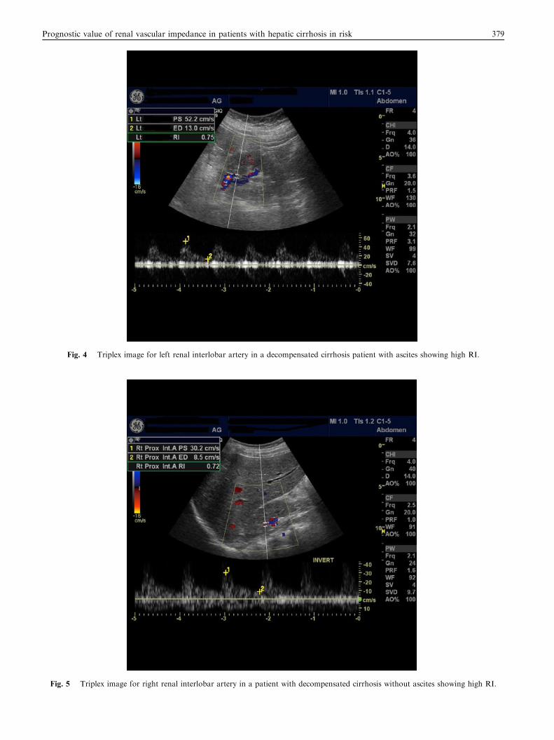
Fig. 4 Triplex image for left renal interlobar artery in a decompensated cirrhosis patient with ascites showing high RI.
Fig. 5 Triplex image for right renal interlobar artery in a patient with decompensated cirrhosis without ascites showing high RI.
Prognostic value of renal vascular impedance in patients with hepatic cirrhosis in risk 379

Fig. 6 Triplex image for right renal interlobar artery in a patient with decompensated cirrhosis without ascites showing high RI & PI.
Table 2 Sensitivity, specificity, PPV, and NPV.
Sensitivity Specificity PPV NPV
71% 96% 92% 86%
380 A.M. Ghandour, A.H.A. Arnaout
prediction of the development of HRS was not sufficiently eval-uated by researchers.
Calculation of the renovascular impedance––expressed in
terms of the resistance index and the pulsatility index––withuse of Doppler tracings at the level of the interlobar arteriesyields an estimation of renal arterial vasoconstriction (8,9,15).
Patients with cirrhosis have significantly higher renal RIand PI values compared with healthy subjects (8,9,13,19),and renovascular impedance increases as the liver disease pro-gresses (9,20). These increases are marked in patients with
ascitic cirrhosis (9,13). This is in accordance with our resultof 56% of decompensated hepatic cirrhosis with ascites pa-tients have high RI and 78% of the same group having high
PI. In Annalisa et al. (9) work correlating renovascular imped-ance with portal pressure in patients with liver cirrhosis theyfound that; the RI and PI in the right renal artery (RRA)
had a sensitivity of 52%, a specificity of 100%, a positive pre-dictive value of 100%, and a negative predictive value of 33%.The RI in the left renal artery (LRA) had a sensitivity of 46%,a specificity of 86%, a positive predictive value of 92%, and a
negative predictive value of 32%, while in the present workcombining and averaging the RI and PI of right and left renal
arteries gave comparative figures to the previous study – sensi-
tivity of 71%, specificity of 96%, PPV of 92%, and an NPV of86% – apart from the higher NPV in our study.
Colli et al. (21) found that in patients who have compen-
sated (i.e., Child-Pugh class A) cirrhosis without ascites, highrenovascular impedance correlates with the presence of esoph-ageal varices. This is in accordance with our findings of higher
impedance values in the group 2 than group 1 (RI 32%, PI41% and RI 8%, PI 3%, respectively).
Agostino et al. (7) found elevated RI in 36% of their pa-
tients with Child-Pugh class A cirrhosis without ascites, whichis related to 32% in the present study of same group ofpatients.
It has also been found that impedance values higher than
the threshold for normal have prognostic value in the identifi-cation of patients who are at high risk for refractory ascites,hepatorenal syndrome, and death (9,13,22,23). Existing evi-
dence therefore suggests that portal venous pressure is the ini-tiating event that causes renal vasoconstriction (9).
The presence of renal dysfunction is often missed in pa-
tients with cirrhosis. Because of a reduction in muscle massin these patients, serum creatinine may be within the normalrange, even with a very low GFR. The use of blood urea nitro-gen (BUN) concentration as a measure of renal function is
even less reliable, because BUN levels can be affected by thepresence of gastrointestinal bleeding or by the amount of pro-tein in the diet (6), and here comes the major role of renal du-
plex Doppler assessment of renovascular impedance to pick

Prognostic value of renal vascular impedance in patients with hepatic cirrhosis in risk 381
that sector of patients at high risk of developing renal failureand anticipating renal dysfunction.
6. Conclusion
From the present study we can conclude that renovascularimpedance values (RI and PI) are good specific and positive
predictive tools for hepatorenal syndrome development in pa-tients with hepatic cirrhosis with consequent better early man-agement and prognosis and also combining and averaging
both renal artery impedance values give better negative predic-tivity than using either artery.
References
(1) Wolf DC. Cirrhosis. Medscape 2008(Aug 11).
(2) Roberto JG. Hyperdynamic circulation of liver disease 40 years
later: pathophysiology and clinical consequences. Hepatology
1994;20:1359–63.
(3) Arroyo V. Pathophysiology, diagnosis and treatment of ascites in
cirrhosis. Ann Hepatol 2002;1:72–9.
(4) Sandeep M, Hemant K, Rowen K. Hepatorenal Syndrome.
eMedicine, Article Last Updated 2008; May 15.
(5) Yeung E, Yong E, Wong F. Renal dysfunction in cirrhosis:
diagnosis, treatment, and prevention. MedGenMed 2004;6(4):9.
(6) Hecker R, Sherlock S. Electrolyte and circulatory changes in
terminal liver failure. Lancet 1956;2:1221–5.
(7) Agostino C, Mirella F, Roberta P, Massimo C, Stefania V, Dario
C. Renovascular impedance and esophageal varices in patients
with Child-Pugh class A cirrhosis. Radiology 2001;219:712–5.
(8) Colli A, Cocciolo M, Riva C, Martinez E. Abnormal renovas-
cular impedance in patients with hepatic cirrhosis: detection with
duplex US. Radiology 1993;187:561–3.
(9) Berzigotti A, Casadei A, Magalotti D, Castaldini N, Losinno F,
Rossi C, et al. Renovascular impedance correlates with portal
pressure in patients with liver cirrhosis. Radiology
2006;240:581–6.
(10) Tomas B, Scott W, Biggins S. In: Zakim, Boyer, editors.
Hepatology a text book of liver disease. Philadelphia: Saunders;
2011. p. 837–52, Ch 47.
(11) Francoz C, Glotz D, Moreau R, Durand F. The evaluation of
renal function and disease in patients with cirrhosis. Hepatology
2010;52:605–13.
(12) Korula J. Hepatorenal syndrome. In: Kaplowitz N, editor. Liver
and biliary disease. Baltimore, Md: Williams & Wilkins; 1996. p.
603–13.
(13) Maroto A, Gine A, Salo J. Diagnosis of functional renal failure of
cirrhosis with Doppler sonography: prognostic value of resistive
index. Hepatology 1994;20:839–44.
(14) Platt J, Rubin J, Ellis J. Duplex Doppler US of the kidney:
differentiation of obstructive from non-obstructive dilatation.
Radiology 1989;171:515–7.
(15) Platt J, Ellis J, Rubin J, DiPietro M, Sedman A. Intrarenal
arterial Doppler sonography in patients with non-obstructive
renal disease: correlation of resistive index with biopsy findings.
AJR Am J Roentgenol 1990;154:1223–7.
(16) Platt J, Rubin J, Ellis J. Acute renal failure: possible role of
duplex Doppler US in distinction between acute prerenal failure
and acute tubular necrosis. Radiology 1991;179:419–23.
(17) Sacerdoti D, Bolognesi M, Merkel C, Angeli P, Gatta A. Renal
vasoconstriction in cirrhosis evaluated by duplex Doppler ultra-
sonography. Hepatology 1993;17:219–24.
(18) Rivolta R, Maggi A, Cazzaniga M, Daniela C, Anna P, Daniela
S, et al. Reduction of renal cortical blood flow assessed by
Doppler in cirrhotic patients with refractory ascites. Hepatology
1998;28:1235–40.
(19) Yang Y, Liu L, Bai W. Renal haemodynamics in patients with
liver cirrhosis assessed by colour ultrasonography. J Int Med Res
2011;39:249–55.
(20) Rendon U, Rojas M, Macias R. Doppler ultrasonography in the
assessment of renal hemodynamics in patients with chronic liver
disease. Rev Esp Enferm Dig 2000;92:799–805.
(21) Colli A, Fraquelli M, Pometta R. Renovascular impedance and
esophageal varices in patients with Child-Pugh class A cirrhosis.
Radiology 2001;219:712–5.
(22) Ljubicic N, Kujundzic M, Banic M, Vrkljan M. Predictive factors
influencing the therapeutic response to diuretic treatment of
ascites in nonazotemic cirrhotic patients. Scand J Gastroenterol
1998;33:441–7.
(23) Kastelan S, Ljubicic N, Kastelan Z. The role of duplex-doppler
ultrasonography in the diagnosis of renal dysfunction and
hepatorenal syndrome in patients with liver cirrhosis. Hepato-
gastroenterology 2004;51:1408–12.