Embed Size (px)
Citation preview
J O U R N A L O F T H E AM E R I C A N C O L L E G E O F C A R D I O L O G Y V O L . 6 4 , N O . 1 6 , 2 0 1 4
ª 2 0 1 4 B Y T H E A M E R I C A N CO L L E G E O F C A R D I O L O G Y F O U N DA T I O N I S S N 0 7 3 5 - 1 0 9 7 / $ 3 6 . 0 0
P U B L I S H E D B Y E L S E V I E R I N C . h t t p : / / d x . d o i . o r g / 1 0 . 1 0 1 6 / j . j a c c . 2 0 1 4 . 0 7 . 9 7 3Open access under CC BY-NC-ND license.
ORIGINAL INVESTIGATIONS
Prognostic Value ofFractional Flow Reserve
Linking Physiologic Severity to Clinical OutcomesNils P. Johnson, MD,1 Gábor G. Tóth, MD,2 Dejian Lai, PHD,3 Hongjian Zhu, PHD,3 Göksel Açar, MD,4
Pierfrancesco Agostoni, MD, PHD,5 Yolande Appelman, MD, PHD,6 Fatih Arslan, MD, PHD,5
Emanuele Barbato, MD, PHD,2 Shao-Liang Chen, MD,7 Luigi Di Serafino, MD, PHD,8
Antonio J. Domínguez-Franco, MD,9 Patrick Dupouy, MD,10 Ali M. Esen, MD,4 Özlem B. Esen, MD,11
Michalis Hamilos, MD, PHD,12 Kohichiro Iwasaki, MD,13 Lisette O. Jensen, MD, PHD,14
Manuel F. Jiménez-Navarro, MD, PHD,9 Demosthenes G. Katritsis, MD, PHD,15 Sinan A. Kocaman, MD,16
Bon-Kwon Koo, MD, PHD,17 Ramón López-Palop, MD, PHD,18 Jeffrey D. Lorin, MD,19 Louis H. Miller, MD,20
Olivier Muller, MD, PHD,21 Chang-Wook Nam, MD, PHD,22 Niels Oud, MD,6 Etienne Puymirat, MD, PHD,23
Johannes Rieber, MD,24 Gilles Rioufol, MD, PHD,25 Josep Rodés-Cabau, MD,26 Steven P. Sedlis, MD,19
Yasuchika Takeishi, MD, PHD,27 Pim A.L. Tonino, MD, PHD,28,29 Eric Van Belle, MD, PHD,30 Edoardo Verna, MD,31
Gerald S. Werner, MD, PHD,32 William F. Fearon, MD,33 Nico H.J. Pijls, MD, PHD,28,29 Bernard De Bruyne, MD, PHD,2
K. Lance Gould, MD1
JACC JOURNAL CME
This article has been selected as the month’s JACC Journal CME activity.
Accreditation and Designation Statement
The American College of Cardiology Foundation (ACCF) is accredited by
the Accreditation Council for Continuing Medical Education (ACCME) to
provide continuing medical education for physicians.
The ACCF designates this Journal-based CME activity for a maximum
of 1 AMA PRA Category 1 Credit(s). Physicians should only claim credit
commensurate with the extent of their participation in the activity.
Method of Participation and Receipt of CME Certificate
To obtain credit for JACC CME, you must:
1. Be an ACC member or JACC subscriber.
2. Carefully read the CME-designated article available online and in this
issue of the journal.
3. Answer the post-test questions. At least 2 out of the 3 questions
provided must be answered correctly to obtain CME credit.
4. Complete a brief evaluation.
5. Claim your CME credit and receive your certificate electronically by
following the instructions given at the conclusion of the activity.
CME Objective for This Article: After completing this article, the reader
should: 1) appreciate the continuous relationship between fractional
flow reserve (FFR) and subsequent major adverse clinical events
(MACE) in a broad range of clinical scenarios; 2) integrate the degree
of FFR abnormality with other clinical data and patient preference
to reach a personalized decision regarding coronary revascularization;
3) favor an FFR-assisted strategy to guide revascularization over an
anatomy-based strategy given that it lowers MACE while providing
superior angina relief; 4) recognize the prognostic value of FFR as an
indication of residual diffuse disease when measured immediately
following percutaneous coronary intervention (PCI); 5) consider
tracking the aggregate FFR distribution in his or her cardiac catheteri-
zation laboratory to optimize referral patterns and lesion selection; and
6) enrich future clinical trials of revascularization for preventing
hard endpoints of MI and mortality by selecting lesions with lower
FFR values.
CME Editor Disclosure: JACC CME Editor Ragavendra Baliga, MD, FACC,
has reported that he has no relationships to disclose.
Author Disclosures: Dr. Johnson has received internal funding from the
Weatherhead PET Center for Preventing and Reversing Atherosclerosis;
signed nonfinancial, nondisclosure agreements with St. Jude Medical and
Volcano Corporation to discuss coronary physiology projects; and
received significant institutional research support from both companies.
Drs. Lai and Zhu have received internal funding from the Weatherhead
PET Center for Preventing and Reversing Atherosclerosis. Dr. Barbato has
received institutional consultancy fees and research support from St.
Jude Medical. Dr. Hamilos has received consulting fees from St. Jude
Medical. Dr. Jensen has received grant support from St Jude Medical,
Terumo, and Biosensors. Dr. Koo has received institutional research
support from St. Jude Medical. Dr. Palop has received research grants
from Abbott Vascular, Terumo; and Medtronic; and honoraria for advi-
sory panels and presentations from Medtronic, Abbott Vascular, Boston
Scientific, Terumo, Volcano, St. Jude, Eli Lilly and AstraZeneca. Dr.
Muller has received research support from the Fondation Vaudoise de
Cardiologie, Lausanne, Switzerland; and has received honoraria for pre-
sentations from St. Jude Medical and Medtronic. Dr. Van Belle has served
as a consultant for St. Jude Medical and received speaker’s fees and
honoraria from Volcano and St. Jude Medical. Dr. Fearon has received
Johnson et al. J A C C V O L . 6 4 , N O . 1 6 , 2 0 1 4
FFR and Outcomes O C T O B E R 2 1 , 2 0 1 4 : 1 6 4 1 – 5 4
1642
research support from St. Jude Medical and Medtronic; speaker’s fees
from Medtronic; and owns minor stock options in HeartFlow Inc. Dr. Pijls
has served as a consultant to St. Jude Medical and HeartFlow Inc; and has
received institutional grant support from St. Jude Medical. Dr. De Bruyne
has received consulting fees and research support from St. Jude Medical;
his consulting fees are passed to Cardiovascular Research Center Aalst, a
not-for-profit organization. Dr. Gould has received internal funding from
the Weatherhead PET Center for Preventing and Reversing
Atherosclerosis; signed nonfinancial, nondisclosure agreements with St.
Jude Medical and Volcano Corporation to discuss coronary physiology
projects; 510(k) applicant for cfrQuant, a software package for quantifying
absolute flow using cardiac positron emission tomography, and all
royalties will go to a University of Texas scholarship fund, as the
From the 1Weatherhead PET Center For Preventing and Reversing Atherosc
icine, University of Texas Medical School and Memorial Hermann Hospital,
Belgium; 3Division of Biostatistics, University of Texas School of Public Health
Kosuyolu High Speciality Education and Research Hospital, Istanbul, Turk
Center Utrecht, Utrecht, the Netherlands; 6Department of Cardiology, VU Uni7Department of Cardiology, Nanjing First Hospital, Nanjing Medical Universi
Presidio Ospedaliero (P.O.) Di Venere, Bari, Italy; 9Unidad de Gestión C
Virgen de la Victoria, Instituto de Investigación Biomédica de Málaga (IBIM10Pôle Cardiovasculaire Interventionnel, Hôpital Privé d’Antony, Antony, F
pital, Istanbul, Turkey; 12Department of Cardiology, University Hospital of
ogy, Okayama Kyokuto Hospital, Okayama, Japan; 14Department of Cardiol15Department of Cardiology, Athens Euroclinic, Athens, Greece; 16Departm
cine, Ankara, Turkey; 17Department of Internal Medicine, Seoul National U
Cardiología, Hospital Universitario San Juan, Alicante, Spain; 19VA New Yor
School of Medicine, New York, New York; 20Division of Cardiology, Depar
Medicine, New York, New York; 21Department of Cardiology, University
Switzerland; 22Department of Internal Medicine, Keimyung University Don
Européen Georges Pompidou, Paris, France; 24Clinic for Cardiology and I
hausen, Munich, Germany; 25Interventional Cardiology Department, Hos
France; 26Québec Heart and Lung Institute, Laval University, Québec City
tology, Fukushima Medical University, Fukushima, Japan; 28Department
Netherlands; 29Department of Biomedical Engineering, Eindhoven Unive30Department of Cardiology, University Hospital, and EA2693, Lille-II-Un
Ospedale di Circolo e Fondazione Macchi, University Hospital, Varese, Ital
the 33Division of Cardiovascular Medicine, Stanford University Medical Ce
internal funding from the Weatherhead PET Center for Preventing an
nondisclosure agreements with St. Jude Medical and Volcano Corporation to
significant institutional research support from both companies. Drs. Lai
Weatherhead PET Center for Preventing and Reversing Atherosclerosis. Dr.
and research support from St. Jude Medical. Dr. Hamilos has received con
received grant support from St Jude Medical, Terumo, and Biosensors. Dr. K
St. Jude Medical. Dr. Palop has received research grants from Abbott Va
advisory panels and presentations from Medtronic, Abbott Vascular, Boston
AstraZeneca. Dr. Muller has received research support from the Fondation V
has received honoraria for presentations from St. Jude Medical and Medtro
Jude Medical and received speaker’s fees and honoraria from Volcano and
support from St. Jude Medical and Medtronic; speaker’s fees from Medtron
Dr. Pijls has served as a consultant to St. Jude Medical and HeartFlow Inc; a
Jude Medical. Dr. De Bruyne has received consulting fees and research sup
passed to Cardiovascular Research Center Aalst, a not-for-profit organizatio
Weatherhead PET Center for Preventing and Reversing Atherosclerosis; sign
Jude Medical and Volcano Corporation to discuss coronary physiology p
package for quantifying absolute flow using cardiac positron emission tom
Texas scholarship fund, as the University of Texas has a commercial, non
distribute and market cfrQuant in exchange for royalties, but Dr. Gould re
selected collaborators for research. All other authors have reported that the
this paper to disclose.
Listen to this manuscript’s audio summary by JACC Editor-in-Chief Dr. Va
You can also listen to this issue’s audio summary by JACC Editor-in-Chief
Manuscript received April 7, 2014; revised manuscript received June 11, 201
University of Texas has a commercial, nonexclusive agreement with
Positron Corporation to distribute and market cfrQuant in exchange for
royalties, but Dr. Gould retains the ability to distribute cost-free versions
to selected collaborators for research. All other authors have reported
that they have no relationships relevant to the contents of this paper to
disclose.
Medium of Participation: Print (article only); online (article and quiz).
CME Term of Approval
Issue Date: October 21, 2014
Expiration Date: October 20, 2015
lerosis, Division of Cardiology, Department of Med-
Houston, Texas; 2Cardiovascular Center Aalst, Aalst,
, Houston, Texas; 4Department of Cardiology, Kartal
ey; 5Department of Cardiology, University Medical
versity Medical Center, Amsterdam, the Netherlands;
ty, Nanjing City, China; 8Department of Cardiology,
línica del Corazón, Hospital Clínico Universitario
A), Universidad de Málaga (UMA), Málaga, Spain;
rance; 11Department of Cardiology, Memorial Hos-
Heraklion, Crete, Greece; 13Department of Cardiol-
ogy, Odense University Hospital, Odense, Denmark;
ent of Cardiology, Gazi University School of Medi-
niversity Hospital, Seoul, South Korea; 18Sección de
k Harbor Health Care System, New York University
tment of Medicine, New York University School of
of Lausanne Hospital Center (CHUV), Lausanne,
gsan Medical Center, Daegu, South Korea; 23Hôpital
nternal Intensive Care Medicine, Klinikum Bogen-
pices Civils de Lyon and CARMEN INSERM 1060,
, Canada; 27Department of Cardiology and Hema-
of Cardiology, Catharina Hospital, Eindhoven, the
rsity of Technology, Eindhoven, the Netherlands;
iversity, Lille, France; 31Department of Cardiology,
y; 32Klinikum Darmstadt, Darmstadt, Germany; and
nter, Stanford, California. Dr. Johnson has received
d Reversing Atherosclerosis; signed nonfinancial,
discuss coronary physiology projects; and received
and Zhu have received internal funding from the
Barbato has received institutional consultancy fees
sulting fees from St. Jude Medical. Dr. Jensen has
oo has received institutional research support from
scular, Terumo; and Medtronic; and honoraria for
Scientific, Terumo, Volcano, St. Jude, Eli Lilly and
audoise de Cardiologie, Lausanne, Switzerland; and
nic. Dr. Van Belle has served as a consultant for St.
St. Jude Medical. Dr. Fearon has received research
ic; and owns minor stock options in HeartFlow Inc.
nd has received institutional grant support from St.
port from St. Jude Medical; his consulting fees are
n. Dr. Gould has received internal funding from the
ed nonfinancial, nondisclosure agreements with St.
rojects; 510(k) applicant for cfrQuant, a software
ography, and all royalties will go to a University of
exclusive agreement with Positron Corporation to
tains the ability to distribute cost-free versions to
y have no relationships relevant to the contents of
lentin Fuster.
Dr. Valentin Fuster.
4, accepted July 8, 2014.
J A C C V O L . 6 4 , N O . 1 6 , 2 0 1 4 Johnson et al.O C T O B E R 2 1 , 2 0 1 4 : 1 6 4 1 – 5 4 FFR and Outcomes
1643
Prognostic Value of Fractional Flow Reserve
Linking Physiologic Severity to Clinical Outcomes
ABSTRACT
BACKGROUND Fractional flow reserve (FFR) has become an established tool for guiding treatment, but its graded
relationship to clinical outcomes as modulated by medical therapy versus revascularization remains unclear.
OBJECTIVES The study hypothesized that FFR displays a continuous relationship between its numeric value and
prognosis, such that lower FFR values confer a higher risk and therefore receive larger absolute benefits from
revascularization.
METHODS Meta-analysis of study- and patient-level data investigated prognosis after FFR measurement. An interaction
term between FFR and revascularization status allowed for an outcomes-based threshold.
RESULTS A total of 9,173 (study-level) and 6,961 (patient-level) lesions were included with a median follow-up of 16
and 14 months, respectively. Clinical events increased as FFR decreased, and revascularization showed larger net benefit
for lower baseline FFR values. Outcomes-derived FFR thresholds generally occurred around the range 0.75 to 0.80,
although limited due to confounding by indication. FFR measured immediately after stenting also showed an inverse
relationship with prognosis (hazard ratio: 0.86, 95% confidence interval: 0.80 to 0.93; p < 0.001). An FFR-assisted
strategy led to revascularization roughly half as often as an anatomy-based strategy, but with 20% fewer adverse events
and 10% better angina relief.
CONCLUSIONS FFR demonstrates a continuous and independent relationship with subsequent outcomes, modulated
by medical therapy versus revascularization. Lesions with lower FFR values receive larger absolute benefits from
revascularization. Measurement of FFR immediately after stenting also shows an inverse gradient of risk, likely from
residual diffuse disease. An FFR-guided revascularization strategy significantly reduces events and increases freedom
from angina with fewer procedures than an anatomy-based strategy. (J Am Coll Cardiol 2014;64:1641–54) © 2014 by the
American College of Cardiology Foundation. Open access under CC BY-NC-ND license.
M ost medical conditions form a continuumfrom nearly normal to extremely patho-logic, for example hypertension or hyper-
cholesterolemia. Often treatment of such gradeddiseases offers only a dichotomous choice, like revas-cularization in atherosclerotic coronary disease. Thespectrum of coronary artery disease (CAD) requiresa threshold of severity for making a binary decisionfor 1 therapy (optimal medical therapy alone) oranother (addition of percutaneous coronary interven-tion [PCI] or coronary artery bypass grafting [CABG]).
SEE PAGE 1655
Since its introduction more than 20 years ago (1),fractional flow reserve (FFR) has become an impor-tant tool for selecting revascularization or medicaltreatment alone for a coronary stenosis. On the basisof comparisons to noninvasive stress tests, most le-sions display some features of “ischemia” when theFFR falls below 0.75 to 0.80 (2). Consequently,
randomized outcomes trials of FFR utilized fixedcutoff values in this range (3–5). However, anindependent technique instead uses clinical out-comes to define a threshold. Accordingly, an analysisof prognosis with and without revascularization,along the spectrum of FFR values, offers an appro-priate and complementary alternative to the nonin-vasive tests originally examined at the clinicalintroduction of FFR.
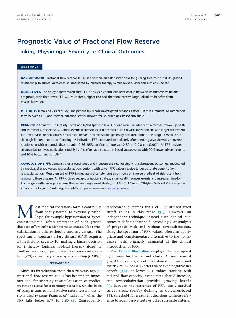
The Central Illustration displays the conceptualhypothesis for the current study. At near normal(high) FFR values, event rates should be lowest andthe risk of PCI or CABG offers no or even negative netbenefit (3,4). At lower FFR values tracking withreduced flow capacity, event rates should increase,and revascularization provides growing benefit(5). Between the extremes of FFR, the 2 survivalcurves cross, thereby defining an outcomes-basedFFR threshold for treatment decisions without refer-ence to noninvasive tests or other surrogate criteria.
CENTRAL ILLUSTRATION C
Similar to many continuous “bi
flow reserve (FFR) potentially
Near normal (high) FFR values
revascularization procedures eq
(low) FFR values increase the r
percutaneous coronary interven
accrues in parallel. In between
outcomes-based FFR optimal t
ABBR EV I A T I ON S
AND ACRONYMS
CABG = coronary artery
bypass grafting
CAD = coronary artery disease
CI = confidence interval
FFR = fractional flow reserve
MACE = major adverse
cardiac event(s)
MI = myocardial infarction
PCI = percutaneous
coronary intervention
Johnson et al. J A C C V O L . 6 4 , N O . 1 6 , 2 0 1 4
FFR and Outcomes O C T O B E R 2 1 , 2 0 1 4 : 1 6 4 1 – 5 4
1644
Using the existing FFR outcomes litera-ture, we sought to answer 3 questions linkingphysiologic severity to prognosis. First, doesFFR provide a continuous and independentmarker for clinical outcomes? Second, canFFR risk stratify prognosis for a coronarystenosis as modulated by revascularization?And, third, will the FFR value measuredimmediately after PCI predict subsequentevents?
METHODS
We used 2 parallel and complementary types ofanalysis. For the study-level meta-regression, eachpublished manuscript provided single data points ofsummary values for the group mean FFR and subse-quent major adverse cardiac event (MACE) rates. Forthe patient-level meta-analysis on the basis of publi-cations whose authors agreed to participate in thiscollaborative project, raw data for every patient
onceptual Relationship Between FFR and Outcomes
omarkers” such as blood pressure and lipids, fractional
relates to subsequent outcomes in a graded fashion.
indicate a favorable prognosis, where the risk from
uals or even exceeds any potential benefit. Worse
isk of events such that the absolute benefit from
tion (PCI) or coronary artery bypass grafting (CABG)
these extremes the curves cross, providing an
hreshold.
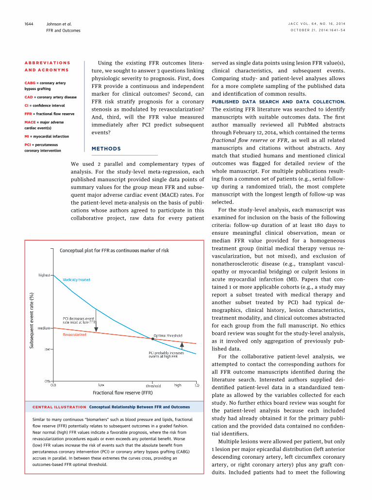
served as single data points using lesion FFR value(s),clinical characteristics, and subsequent events.Comparing study- and patient-level analyses allowsfor a more complete sampling of the published dataand identification of common results.PUBLISHED DATA SEARCH AND DATA COLLECTION.
The existing FFR literature was searched to identifymanuscripts with suitable outcomes data. The firstauthor manually reviewed all PubMed abstractsthrough February 12, 2014, which contained the termsfractional flow reserve or FFR, as well as all relatedmanuscripts and citations without abstracts. Anymatch that studied humans and mentioned clinicaloutcomes was flagged for detailed review of thewhole manuscript. For multiple publications result-ing from a common set of patients (e.g., serial follow-up during a randomized trial), the most completemanuscript with the longest length of follow-up wasselected.
For the study-level analysis, each manuscript wasexamined for inclusion on the basis of the followingcriteria: follow-up duration of at least 180 days toensure meaningful clinical observation, mean ormedian FFR value provided for a homogeneoustreatment group (initial medical therapy versus re-vascularization, but not mixed), and exclusion ofnonatherosclerotic disease (e.g., transplant vascul-opathy or myocardial bridging) or culprit lesions inacute myocardial infarction (MI). Papers that con-tained 1 or more applicable cohorts (e.g., a study mayreport a subset treated with medical therapy andanother subset treated by PCI) had typical de-mographics, clinical history, lesion characteristics,treatment modality, and clinical outcomes abstractedfor each group from the full manuscript. No ethicsboard review was sought for the study-level analysis,as it involved only aggregation of previously pub-lished data.
For the collaborative patient-level analysis, weattempted to contact the corresponding authors forall FFR outcome manuscripts identified during theliterature search. Interested authors supplied dei-dentified patient-level data in a standardized tem-plate as allowed by the variables collected for eachstudy. No further ethics board review was sought forthe patient-level analysis because each includedstudy had already obtained it for the primary publi-cation and the provided data contained no confiden-tial identifiers.
Multiple lesions were allowed per patient, but only1 lesion per major epicardial distribution (left anteriordescending coronary artery, left circumflex coronaryartery, or right coronary artery) plus any graft con-duits. Included patients had to meet the following
37 studies included6,061 total patients6,961 total lesions
Attempted tocontact all
correspondingauthors
22 studies overlap3,358 subjects overlap3,426 lesions overlap
Full-text articles assessedfor eligibility
(n = 120)
PubMed search through February 12, 2014for “fractional flow reserve” or “FFR”
(n = 925)
Patient-level analysis
51 studies included90 distinct cohorts8,418 total patients9,173 total lesions
Study-level analysis
805 records excluded374 lack outcome data141 review articles86 editorials68 case reports61 non-human studies64 letters to the editor11 non-cardiac
�������
68 articles excluded14 could not be retrieved12 mixed medical and PCI/CABG15 lacked mean FFR values
11 overlapped with prior work7 contained only post-FFR10 other reasons*
������
FIGURE 1 Details of PubMed Search
Details of PubMed search that led to study-level and patient-level data. *Other reasons for
exclusion were no (2 articles) or too short (1) follow-up, unclear or mixture of treatment
methods when describing outcomes (3), myocardial bridging (1), simulation-derived
fractional flow reserve (FFR) (1), trial design (1), and meta-analysis (1). CABG ¼ coronary
artery bypass grafting; PCI ¼ percutaneous coronary intervention.
J A C C V O L . 6 4 , N O . 1 6 , 2 0 1 4 Johnson et al.O C T O B E R 2 1 , 2 0 1 4 : 1 6 4 1 – 5 4 FFR and Outcomes
1645
criteria: either a clinical event of known date or aminimum of 180 days of MACE-free follow-up afterFFR measurement to ensure meaningful clinical ob-servation; pre-treatment FFR value; recorded treat-ment decision (medical or revascularization); andexclusion of nonatherosclerotic disease or culprit le-sions in acute MI. To explore the prognostic valueof post-revascularization FFR measurements, a small,additional group of patients was included that hadonly post-treatment FFR results. As variably collectedby each study, patient demographics, clinical history,lesion characteristics, treatmentmodality, and clinicaloutcomes were recorded.
Clinical events of interest to study- and patient-level analyses were death, MI, and target lesion orvessel revascularization. Clearly documented re-vascularizations in off-target vessels were excluded asbeing unrelated to the initial lesion studied byFFR. Too few studies and patient-level data specifiedcardiac versus noncardiac death to enable its separa-tion. Similarly, myocardial infarction was inconsis-tently noted if due to target vessel or elsewhere andtherefore could not be meaningfully distinguished.
Two composite MACE rates were studied: first, thetriad of death, MI, and target lesion or vessel revas-cularization; second, only death and MI. In thepatient-level meta-analysis, only the first event wasincluded if several occurred.GENERAL STATISTICAL METHODS. Statistical ana-lyses were performed in R version 3.0.2 (R Founda-tion for Statistical Computing, Vienna, Austria), usingthe metafor package (version 1.9-2) for meta-analysis.We used standard summary statistical tests. Quantile-quantile plots identified the following continuousvariables as significantly non-normal: FFR, weight,body mass index, minimal lumen diameter, andreference vessel diameter. Applicable tests were 2-tailed, and p < 0.05 was considered statistically sig-nificant. Further statistical methods can be found inthe Online Appendix.STUDY-LEVEL META-REGRESSION. For the study-level meta-regression, length of follow-up was het-erogeneous. To adjust for the differences, 2 methodswere compared. The simple method normalized eachMACE rate to 12 months. For example, 10 events in100 subjects over 8 months would yield a normalizedMACE rate of [(10/100)/8]$12 ¼ 15% at 1 year. Themore complex method adjusted the normalizedevent rates on the basis of a Poisson regression pre-dicting MACE as a continuous function of length offollow-up. Fixed and random (DerSimonian andLaird) effects meta-regressions of the incidence rateincluded the mean FFR value, revascularization as abinary variable, and an interaction term. The optimal
outcomes-based threshold occurred at the intersec-tion of the unrevascularized and revascularized fittedcurves. Note that some intersections did not occurwithin the plausible FFR range from 0 to 1, particu-larly with small or parallel event rates betweentreatment groups.PATIENT-LEVEL META-ANALYSIS. To explore thehypothesis that FFR might simply reflect de-mographic characteristics, classic cardiovascular riskfactors, or basic angiographic features, we studied thecapability of other variables to predict the measuredFFR value in the patient-level data. Each variablewas studied in isolation using logistic regression(quasibinomial link function in a generalized linearmodel). Continuous predictors also were examinedusing correlation methods, whereas binary predictorssummarized by median and interquartile range werecompared using the Mann-Whitney U test. Becauseonly quantitative percent diameter stenosis showed a
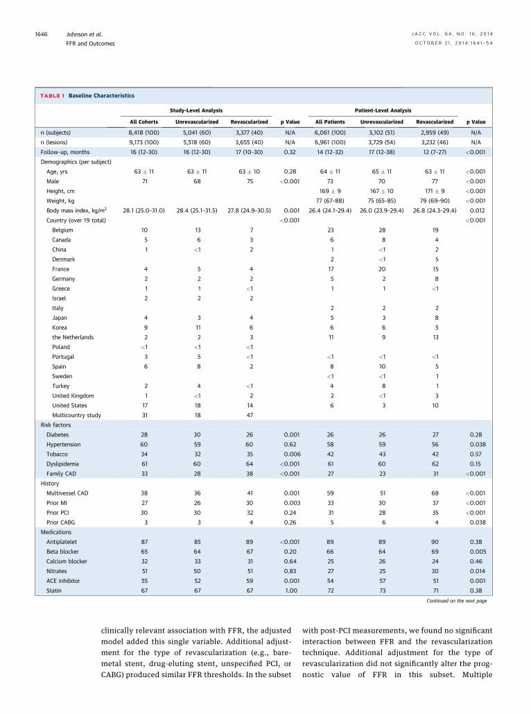
TABLE 1 Baseline Characteristics
Study-Level Analysis
p Value
Patient-Level Analysis
p ValueAll Cohorts Unrevascularized Revascularized All Patients Unrevascularized Revascularized
n (subjects) 8,418 (100) 5,041 (60) 3,377 (40) N/A 6,061 (100) 3,102 (51) 2,959 (49) N/A
n (lesions) 9,173 (100) 5,518 (60) 3,655 (40) N/A 6,961 (100) 3,729 (54) 3,232 (46) N/A
Follow-up, months 16 (12–30) 16 (12–30) 17 (10–30) 0.32 14 (12–32) 17 (12–38) 12 (7–27) <0.001
Demographics (per subject)
Age, yrs 63 � 11 63 � 11 63 � 10 0.28 64 � 11 65 � 11 63 � 11 <0.001
Male 71 68 75 <0.001 73 70 77 <0.001
Height, cm 169 � 9 167 � 10 171 � 9 <0.001
Weight, kg 77 (67–88) 75 (65–85) 79 (69–90) <0.001
Body mass index, kg/m2 28.1 (25.0–31.0) 28.4 (25.1–31.5) 27.8 (24.9–30.5) 0.001 26.4 (24.1–29.4) 26.0 (23.9–29.4) 26.8 (24.3–29.4) 0.012
Country (over 19 total) <0.001 <0.001
Belgium 10 13 7 23 28 19
Canada 5 6 3 6 8 4
China 1 <1 2 1 <1 2
Denmark 2 <1 5
France 4 5 4 17 20 15
Germany 2 2 2 5 2 8
Greece 1 1 <1 1 1 <1
Israel 2 2 2
Italy 2 2 2
Japan 4 3 4 5 3 8
Korea 9 11 6 6 6 5
the Netherlands 2 2 3 11 9 13
Poland <1 <1 <1
Portugal 3 5 <1 <1 <1 <1
Spain 6 8 2 8 10 5
Sweden <1 <1 1
Turkey 2 4 <1 4 8 1
United Kingdom 1 <1 2 2 <1 3
United States 17 18 14 6 3 10
Multicountry study 31 18 47
Risk factors
Diabetes 28 30 26 0.001 26 26 27 0.28
Hypertension 60 59 60 0.62 58 59 56 0.038
Tobacco 34 32 35 0.006 42 43 42 0.57
Dyslipidemia 61 60 64 <0.001 61 60 62 0.15
Family CAD 33 28 38 <0.001 27 23 31 <0.001
History
Multivessel CAD 38 36 41 0.001 59 51 68 <0.001
Prior MI 27 26 30 0.003 33 30 37 <0.001
Prior PCI 30 30 32 0.24 31 28 35 <0.001
Prior CABG 3 3 4 0.26 5 6 4 0.038
Medications
Antiplatelet 87 85 89 <0.001 89 89 90 0.38
Beta blocker 65 64 67 0.20 66 64 69 0.005
Calcium blocker 32 33 31 0.64 25 26 24 0.46
Nitrates 51 50 51 0.83 27 25 30 0.014
ACE inhibitor 55 52 59 0.001 54 57 51 0.001
Statin 67 67 67 1.00 72 73 71 0.38
Continued on the next page
Johnson et al. J A C C V O L . 6 4 , N O . 1 6 , 2 0 1 4
FFR and Outcomes O C T O B E R 2 1 , 2 0 1 4 : 1 6 4 1 – 5 4
1646
clinically relevant association with FFR, the adjustedmodel added this single variable. Additional adjust-ment for the type of revascularization (e.g., bare-metal stent, drug-eluting stent, unspecified PCI, orCABG) produced similar FFR thresholds. In the subset
with post-PCI measurements, we found no significantinteraction between FFR and the revascularizationtechnique. Additional adjustment for the type ofrevascularization did not significantly alter the prog-nostic value of FFR in this subset. Multiple
TABLE 1 Continued
Study-Level Analysis
p Value
Patient-Level Analysis
p ValueAll Cohorts Unrevascularized Revascularized All Patients Unrevascularized Revascularized
Presentation characteristics
Ejection fraction, % 60 � 12 60 � 11 60 � 13 0.08 61 � 13 62 � 13 61 � 13 0.07
Presentation <0.001 <0.001
Stable 71 64 89 68 64 74
Unstable angina 15 18 8 20 23 16
NSTEMI 13 16 3 9 9 9
STEMI 1 2 <1 3 4 1
CCS angina 2.4 � 1.1 2.1 � 1.0 2.6 � 1.1 <0.001 <0.001
No angina 9 18 3
Class I 16 15 16
Class II 45 45 45
Class III 23 16 28
Class IV 7 5 8
NYHA heart failure <0.001
No heart failure 29 30 29
Functional class I 42 55 29
Functional class II 23 13 33
Functional class III 6 2 9
Functional class IV <1 <1 <1
Procedure characteristics (per lesion)
Vessel <0.001 <0.001
LMCA 10 10 9 7 8 7
LAD 57 59 54 57 58 55
LCx 14 14 15 15 16 15
RCA 18 16 22 19 17 22
Graft (IMA, SVG, radial) <1 <1 <1 1 1 1
In-stent restenosis 20 22 18 0.17 4 3 5 0.008
%DS by QCA 51 � 15 46 � 12 56 � 16 <0.001 52 � 18 44 � 13 63 � 19 <0.001
MLD, mm 1.44 (1.03–1.91) 1.66 (1.28–2.07) 1.19 (0.85–1.69) <0.001 1.35 (1.00–1.70) 1.50 (1.26–1.81) 1.06 (0.75–1.44) <0.001
RVD, mm 3.00 (2.51–3.54) 3.06 (2.55–3.65) 2.94 (2.49–3.42) <0.001 2.90 (2.49–3.31) 2.85 (2.45–3.30) 2.99 (2.50–3.40) 0.014
Pd, mm Hg 71 � 17 83 � 14 63 � 15 <0.001 77 � 17 82 � 16 67 � 15 <0.001
Pa, mm Hg 94 � 16 94 � 15 94 � 17 0.77 94 � 17 94 � 17 93 � 17 0.28
FFR 0.80 (0.69–0.88) 0.87 (0.82–0.91) 0.69 (0.61–0.77) <0.001 0.81 (0.71–0.88) 0.87 (0.83–0.92) 0.70 (0.59–0.76) <0.001
FFR post-PCI 0.92 (0.87–0.96) N/A 0.92 (0.86–0.96) N/A
Hyperemia <0.001 <0.001
IC adenosine 42 51 28 59 71 48
IV adenosine 14 10 20 38 27 47
IC or IV adenosine 41 35 49
ATP or papaverine 4 4 3 3 1 5
Revascularization method N/A N/A
BMS 21 30
CABG 4 11
DES 9 27
PCI 44 30
CABG or PCI 14
Other (POBA, DEB) 9 2
Values are n (%), median (interquartile range), mean � SD, %.
ACE ¼ angiotensin-converting enzyme; ATP ¼ adenosine triphosphate; BMS ¼ bare-metal stent(s); CABG ¼ coronary artery bypass grafting; CAD ¼ coronary artery disease; CCS ¼ CanadianCardiovascular Society; DEB ¼ drug-eluting balloon; DES ¼ drug-eluting stent(s); %DS ¼ percent diameter stenosis; FFR ¼ fractional flow reserve; IC ¼ intracoronary; IMA ¼ internal mammaryartery; IV ¼ intravenous; LAD ¼ left anterior descending coronary artery; LCx ¼ left circumflex coronary artery; LMCA ¼ left main coronary artery; MLD ¼ minimal lumen diameter; N/A ¼ notapplicable; NSTEMI ¼ non-ST-segment elevation myocardial infarction; NYHA ¼ New York Heart Association; Pa ¼ aortic pressure; Pd ¼ distal coronary pressure; PCI ¼ percutaneous coronaryintervention; POBA ¼ plain old balloon angioplasty; QCA ¼ quantitative coronary angiography; RCA ¼ right coronary artery; RVD ¼ reference vessel diameter; STEMI ¼ ST-segment elevationmyocardial infarction; SVG ¼ saphenous vein graft.
J A C C V O L . 6 4 , N O . 1 6 , 2 0 1 4 Johnson et al.O C T O B E R 2 1 , 2 0 1 4 : 1 6 4 1 – 5 4 FFR and Outcomes
1647
Orde
red
Coho
rts
0.0 0.2 0.4 0.6 0.8 1.0 0.0 0.2 0.4 0.6 0.8 1.0
Mean Cohort FFR Fractional Flow Reserve (FFR)
Num
ber o
f Les
ions
FFR 0.87 (0.83-0.92)in 3,729 lesions
treated medically
FFR 0.70 (0.59-0.76)in 3,150 lesions
received PCI/CABG
FFR 0.75-0.80FFR 0.75-0.80
250
200
150
100
50
0
FFR 0.69 (0.61-0.77)in 3,377 patientsfrom 43 cohorts
received PCI/CABG
FFR 0.87 (0.82-0.91)in 5,041 patientsfrom 47 cohorts
treated medically
A B
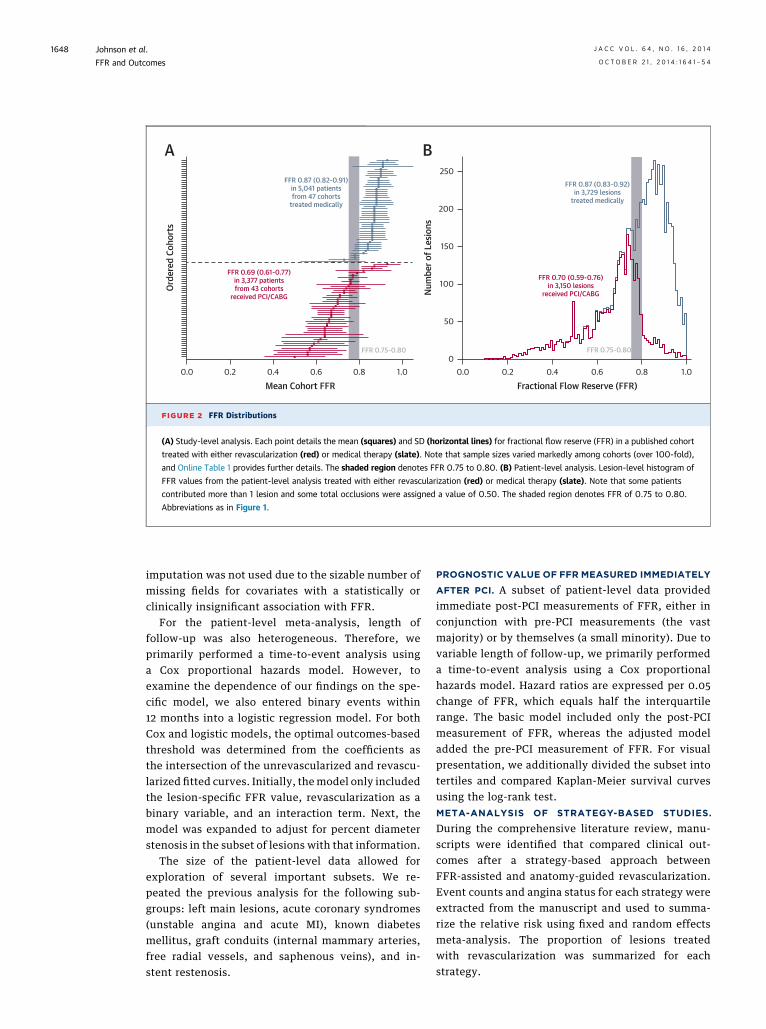
FIGURE 2 FFR Distributions
(A) Study-level analysis. Each point details the mean (squares) and SD (horizontal lines) for fractional flow reserve (FFR) in a published cohort
treated with either revascularization (red) or medical therapy (slate). Note that sample sizes varied markedly among cohorts (over 100-fold),
and Online Table 1 provides further details. The shaded region denotes FFR 0.75 to 0.80. (B) Patient-level analysis. Lesion-level histogram of
FFR values from the patient-level analysis treated with either revascularization (red) or medical therapy (slate). Note that some patients
contributed more than 1 lesion and some total occlusions were assigned a value of 0.50. The shaded region denotes FFR of 0.75 to 0.80.
Abbreviations as in Figure 1.
Johnson et al. J A C C V O L . 6 4 , N O . 1 6 , 2 0 1 4
FFR and Outcomes O C T O B E R 2 1 , 2 0 1 4 : 1 6 4 1 – 5 4
1648
imputation was not used due to the sizable number ofmissing fields for covariates with a statistically orclinically insignificant association with FFR.
For the patient-level meta-analysis, length offollow-up was also heterogeneous. Therefore, weprimarily performed a time-to-event analysis usinga Cox proportional hazards model. However, toexamine the dependence of our findings on the spe-cific model, we also entered binary events within12 months into a logistic regression model. For bothCox and logistic models, the optimal outcomes-basedthreshold was determined from the coefficients asthe intersection of the unrevascularized and revascu-larized fitted curves. Initially, themodel only includedthe lesion-specific FFR value, revascularization as abinary variable, and an interaction term. Next, themodel was expanded to adjust for percent diameterstenosis in the subset of lesions with that information.
The size of the patient-level data allowed forexploration of several important subsets. We re-peated the previous analysis for the following sub-groups: left main lesions, acute coronary syndromes(unstable angina and acute MI), known diabetesmellitus, graft conduits (internal mammary arteries,free radial vessels, and saphenous veins), and in-stent restenosis.
PROGNOSTIC VALUE OF FFR MEASURED IMMEDIATELY
AFTER PCI. A subset of patient-level data providedimmediate post-PCI measurements of FFR, either inconjunction with pre-PCI measurements (the vastmajority) or by themselves (a small minority). Due tovariable length of follow-up, we primarily performeda time-to-event analysis using a Cox proportionalhazards model. Hazard ratios are expressed per 0.05change of FFR, which equals half the interquartilerange. The basic model included only the post-PCImeasurement of FFR, whereas the adjusted modeladded the pre-PCI measurement of FFR. For visualpresentation, we additionally divided the subset intotertiles and compared Kaplan-Meier survival curvesusing the log-rank test.META-ANALYSIS OF STRATEGY-BASED STUDIES.
During the comprehensive literature review, manu-scripts were identified that compared clinical out-comes after a strategy-based approach betweenFFR-assisted and anatomy-guided revascularization.Event counts and angina status for each strategy wereextracted from the manuscript and used to summa-rize the relative risk using fixed and random effectsmeta-analysis. The proportion of lesions treatedwith revascularization was summarized for eachstrategy.
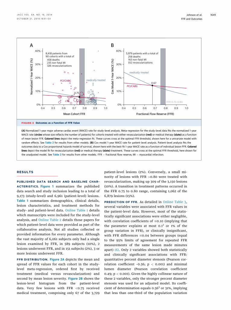
40%
30%
20%
10%
0%0.4 0.5 0.6 0.7 0.8 0.9 1.0
Fractional Flow Reserve (FFR)
5,979 patients with a total of258 deaths163 non-fatal MI552 revascularizations
FFR 0.75-0.80
0.67
0.75
medically treated
medically treated
PCI/CABG
PCI/CABG
Cox
Mod
el 1-
Year
MAC
E (%
)
40%
30%
20%
10%
0%0.4 0.5 0.6 0.7 0.8 0.9 1.0
Mean Cohort FFR
Norm
aliz
ed 1-
Year
MAC
E (%
)
8,418 patients from90 cohorts with a total of
458 deaths235 non-fatal MI326 revascularizations
A B
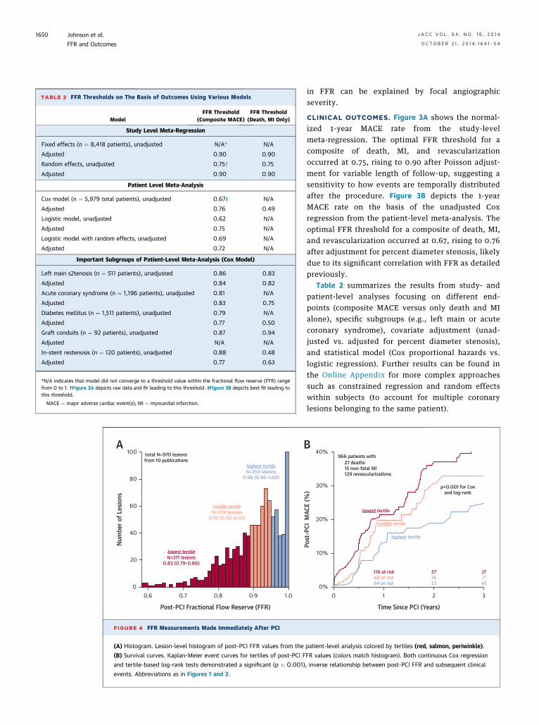
FIGURE 3 Outcomes as a Function of FFR Value
(A) Normalized 1-year major adverse cardiac event (MACE) rate for study-level analysis. Meta-regression for the study-level data fits the normalized 1-year
MACE rate (circles whose size reflects the number of patients) for cohorts treated with either revascularization (red) or medical therapy (slate) as a function
of mean lesion FFR. Colored lines depict the meta-regression fit. These curves cross at the optimal FFR threshold, shown here for a univariate model with
random effects. See Table 2 for results from other models. (B) Cox model 1-year MACE rate for patient-level analysis. Patient-level analysis fits the
outcomes data to a Cox proportional hazards model of survival, shown here with the best-fit 1-year MACE rate as a function of individual lesion FFR. Colored
lines depict the model fit for revascularization (red) or medical therapy (slate) treatment. These curves cross at the optimal FFR threshold, here shown for
the unadjusted model. See Table 2 for results from other models. FFR ¼ fractional flow reserve; MI ¼ myocardial infarction.
J A C C V O L . 6 4 , N O . 1 6 , 2 0 1 4 Johnson et al.O C T O B E R 2 1 , 2 0 1 4 : 1 6 4 1 – 5 4 FFR and Outcomes
1649
RESULTS
PUBLISHED DATA SEARCH AND BASELINE CHAR-
ACTERISTICS. Figure 1 summarizes the publisheddata search and study inclusion leading to a total of9,173 (study-level) and 6,961 (patient-level) lesions.Table 1 summarizes demographics, clinical details,lesion characteristics, and treatment methods forstudy- and patient-level data. Online Table 1 detailswhich manuscripts were included for the study-levelanalysis, and Online Table 2 details those papers forwhich patient-level data were provided as part of thecollaborative analysis. Not all studies collected orprovided information for every parameter. Althoughthe vast majority of 6,061 subjects only had a singlelesion examined by FFR, in 589 subjects (10%), 2lesions underwent FFR, and in 151 subjects (2%), 3 ormore lesions underwent FFR.
FFR DISTRIBUTION. Figure 2A depicts the mean andspread of FFR values for each cohort in the study-level meta-regression, ordered first by receivedtreatment (medical versus revascularization) andsecond by mean lesion severity. Figure 2B shows thelesion-level histogram from the patient-leveldata. Very few lesions with FFR <0.75 receivedmedical treatment, comprising only 67 of the 3,729
patient-level lesions (2%). Conversely, a small mi-nority of lesions with FFR >0.80 were treated withrevascularization, making up 309 of the 3,150 lesions(10%). A transition in treatment patterns occurred inthe FFR 0.75 to 0.80 range, containing 1,062 of the6,879 lesions (15%).
PREDICTION OF FFR. As detailed in Online Table 3,several variables were associated with FFR values inthe patient-level data. However, most of the statis-tically significant associations were either negligible,with correlation coefficients of #0.10 (implying thatthe parameter explains at most 0.12 or 1% of thegroup variation in FFR), or clinically insignificant,with FFR differences #0.04 between groups (equalto the 95% limits of agreement for repeated FFRmeasurements of the same lesion made minutesapart) (6). Only 2 variables showed both statisticallyand clinically significant associations with FFR:quantitative percent diameter stenosis (Pearson cor-relation coefficient –0.56; p < 0.001) and minimallumen diameter (Pearson correlation coefficient0.49; p < 0.001). Given the highly collinear nature ofthese 2 variables, only the stronger percent diameterstenosis was used for an adjusted model. Its coeffi-cient of determination equals 0.562 or 31%, implyingthat less than one-third of the population variation
highest tertileN=350 lesions
0.98 (0.96-1.00)
middle tertileN=309 lesions
0.92 (0.90-0.93)
lowest tertileN=311 lesions
0.83 (0.79-0.86)
total N=970 lesionsfrom 10 publications
Post-PCI Fractional Flow Reserve (FFR)
Num
ber o
f Les
ions
100
80
60
40
20
00.6 0.7 0.8 0.9 1.0
A B
FIGURE 4 FFR Measurements Made Immediately After PCI
(A) Histogram. Lesion-level histogram of post–PCI FFR values from the
(B) Survival curves. Kaplan-Meier event curves for tertiles of post-PCI F
and tertile-based log-rank tests demonstrated a significant (p < 0.001)
events. Abbreviations as in Figures 1 and 2.
TABLE 2 FFR Thresholds on The Basis of Outcomes Using Various Models
ModelFFR Threshold
(Composite MACE)FFR Threshold
(Death, MI Only)
Study Level Meta-Regression
Fixed effects (n ¼ 8,418 patients), unadjusted N/A* N/A
Adjusted 0.90 0.90
Random effects, unadjusted 0.75† 0.75
Adjusted 0.90 0.90
Patient Level Meta-Analysis
Cox model (n ¼ 5,979 total patients), unadjusted 0.67‡ N/A
Adjusted 0.76 0.49
Logistic model, unadjusted 0.62 N/A
Adjusted 0.75 N/A
Logistic model with random effects, unadjusted 0.69 N/A
Adjusted 0.72 N/A
Important Subgroups of Patient-Level Meta-Analysis (Cox Model)
Left main s2tenosis (n ¼ 511 patients), unadjusted 0.86 0.83
Adjusted 0.84 0.82
Acute coronary syndrome (n ¼ 1,196 patients), unadjusted 0.81 N/A
Adjusted 0.83 0.75
Diabetes mellitus (n ¼ 1,511 patients), unadjusted 0.79 N/A
Adjusted 0.77 0.50
Graft conduits (n ¼ 92 patients), unadjusted 0.87 0.94
Adjusted N/A N/A
In-stent restenosis (n ¼ 120 patients), unadjusted 0.88 0.48
Adjusted 0.77 0.63
*N/A indicates that model did not converge to a threshold value within the fractional flow reserve (FFR) rangefrom 0 to 1. †Figure 3A depicts raw data and fit leading to this threshold. ‡Figure 3B depicts best fit leading tothis threshold.
MACE ¼ major adverse cardiac event(s); MI ¼ myocardial infarction.
Johnson et al. J A C C V O L . 6 4 , N O . 1 6 , 2 0 1 4
FFR and Outcomes O C T O B E R 2 1 , 2 0 1 4 : 1 6 4 1 – 5 4
1650
in FFR can be explained by focal angiographicseverity.
CLINICAL OUTCOMES. Figure 3A shows the normal-ized 1-year MACE rate from the study-levelmeta-regression. The optimal FFR threshold for acomposite of death, MI, and revascularizationoccurred at 0.75, rising to 0.90 after Poisson adjust-ment for variable length of follow-up, suggesting asensitivity to how events are temporally distributedafter the procedure. Figure 3B depicts the 1-yearMACE rate on the basis of the unadjusted Coxregression from the patient-level meta-analysis. Theoptimal FFR threshold for a composite of death, MI,and revascularization occurred at 0.67, rising to 0.76after adjustment for percent diameter stenosis, likelydue to its significant correlation with FFR as detailedpreviously.
Table 2 summarizes the results from study- andpatient-level analyses focusing on different end-points (composite MACE versus only death and MIalone), specific subgroups (e.g., left main or acutecoronary syndrome), covariate adjustment (unad-justed vs. adjusted for percent diameter stenosis),and statistical model (Cox proportional hazards vs.logistic regression). Further results can be found inthe Online Appendix for more complex approachessuch as constrained regression and random effectswithin subjects (to account for multiple coronarylesions belonging to the same patient).
Time Since PCI (Years)
27 deaths15 non-fatal MI129 revascularizations
966 patients with
116 at risk68 at risk64 at risk
573653
372745
32100%
10%
20%
30%
40%
Post
-PCI
MAC
E (%
)
p<0.001 for Coxand log-rank
highest tertile
lowest tertile
middle tertile
patient-level analysis colored by tertiles (red, salmon, periwinkle).
FR values (colors match histogram). Both continuous Cox regression
, inverse relationship between post-PCI FFR and subsequent clinical
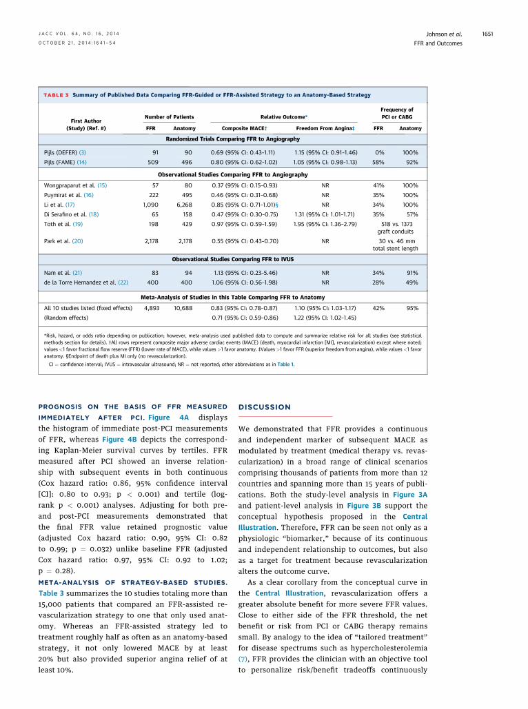
TABLE 3 Summary of Published Data Comparing FFR-Guided or FFR-Assisted Strategy to an Anatomy-Based Strategy
First Author(Study) (Ref. #)
Number of Patients Relative Outcome*Frequency ofPCI or CABG
FFR Anatomy Composite MACE† Freedom From Angina‡ FFR Anatomy
Randomized Trials Comparing FFR to Angiography
Pijls (DEFER) (3) 91 90 0.69 (95% CI: 0.43–1.11) 1.15 (95% CI: 0.91–1.46) 0% 100%
Pijls (FAME) (14) 509 496 0.80 (95% CI: 0.62–1.02) 1.05 (95% CI: 0.98–1.13) 58% 92%
Observational Studies Comparing FFR to Angiography
Wongpraparut et al. (15) 57 80 0.37 (95% CI: 0.15–0.93) NR 41% 100%
Puymirat et al. (16) 222 495 0.46 (95% CI: 0.31–0.68) NR 35% 100%
Li et al. (17) 1,090 6,268 0.85 (95% CI: 0.71–1.01)§ NR 34% 100%
Di Serafino et al. (18) 65 158 0.47 (95% CI: 0.30–0.75) 1.31 (95% CI: 1.01–1.71) 35% 57%
Toth et al. (19) 198 429 0.97 (95% CI: 0.59–1.59) 1.95 (95% CI: 1.36–2.79) 518 vs. 1373graft conduits
Park et al. (20) 2,178 2,178 0.55 (95% CI: 0.43–0.70) NR 30 vs. 46 mmtotal stent length
Observational Studies Comparing FFR to IVUS
Nam et al. (21) 83 94 1.13 (95% CI: 0.23–5.46) NR 34% 91%
de la Torre Hernandez et al. (22) 400 400 1.06 (95% CI: 0.56–1.98) NR 28% 49%
Meta-Analysis of Studies in this Table Comparing FFR to Anatomy
All 10 studies listed (fixed effects) 4,893 10,688 0.83 (95% CI: 0.78–0.87) 1.10 (95% CI: 1.03–1.17) 42% 95%
(Random effects) 0.71 (95% CI: 0.59–0.86) 1.22 (95% CI: 1.02–1.45)
*Risk, hazard, or odds ratio depending on publication; however, meta-analysis used published data to compute and summarize relative risk for all studies (see statisticalmethods section for details). †All rows represent composite major adverse cardiac events (MACE) (death, myocardial infarction [MI], revascularization) except where noted;values <1 favor fractional flow reserve (FFR) (lower rate of MACE), while values >1 favor anatomy. ‡Values >1 favor FFR (superior freedom from angina), while values <1 favoranatomy. §Endpoint of death plus MI only (no revascularization).
CI ¼ confidence interval; IVUS ¼ intravascular ultrasound; NR ¼ not reported; other abbreviations as in Table 1.
J A C C V O L . 6 4 , N O . 1 6 , 2 0 1 4 Johnson et al.O C T O B E R 2 1 , 2 0 1 4 : 1 6 4 1 – 5 4 FFR and Outcomes
1651
PROGNOSIS ON THE BASIS OF FFR MEASURED
IMMEDIATELY AFTER PCI. Figure 4A displaysthe histogram of immediate post-PCI measurementsof FFR, whereas Figure 4B depicts the correspond-ing Kaplan-Meier survival curves by tertiles. FFRmeasured after PCI showed an inverse relation-ship with subsequent events in both continuous(Cox hazard ratio: 0.86, 95% confidence interval[CI]: 0.80 to 0.93; p < 0.001) and tertile (log-rank p < 0.001) analyses. Adjusting for both pre-and post-PCI measurements demonstrated thatthe final FFR value retained prognostic value(adjusted Cox hazard ratio: 0.90, 95% CI: 0.82to 0.99; p ¼ 0.032) unlike baseline FFR (adjustedCox hazard ratio: 0.97, 95% CI: 0.92 to 1.02;p ¼ 0.28).
META-ANALYSIS OF STRATEGY-BASED STUDIES.
Table 3 summarizes the 10 studies totaling more than15,000 patients that compared an FFR-assisted re-vascularization strategy to one that only used anat-omy. Whereas an FFR-assisted strategy led totreatment roughly half as often as an anatomy-basedstrategy, it not only lowered MACE by at least20% but also provided superior angina relief of atleast 10%.
DISCUSSION
We demonstrated that FFR provides a continuousand independent marker of subsequent MACE asmodulated by treatment (medical therapy vs. revas-cularization) in a broad range of clinical scenarioscomprising thousands of patients from more than 12countries and spanning more than 15 years of publi-cations. Both the study-level analysis in Figure 3Aand patient-level analysis in Figure 3B support theconceptual hypothesis proposed in the CentralIllustration. Therefore, FFR can be seen not only as aphysiologic “biomarker,” because of its continuousand independent relationship to outcomes, but alsoas a target for treatment because revascularizationalters the outcome curve.
As a clear corollary from the conceptual curve inthe Central Illustration, revascularization offers agreater absolute benefit for more severe FFR values.Close to either side of the FFR threshold, the netbenefit or risk from PCI or CABG therapy remainssmall. By analogy to the idea of “tailored treatment”for disease spectrums such as hypercholesterolemia(7), FFR provides the clinician with an objective toolto personalize risk/benefit tradeoffs continuously
Johnson et al. J A C C V O L . 6 4 , N O . 1 6 , 2 0 1 4
FFR and Outcomes O C T O B E R 2 1 , 2 0 1 4 : 1 6 4 1 – 5 4
1652
instead of in binary fashion. Our data support theconcept that ischemia exists not as a dichotomousstate, but rather as a graded continuum.
The FFR distributions in Figure 2 highlight themajor limitation of our analysis, namely that the FFRvalue strongly influenced the treatment decision—what has been termed “confounding by indication” inthe epidemiology literature. As a result, we possess alimited understanding of the natural history of lowFFR lesions. For example, unrevascularized lesionswith an FFR <0.75 made up less than 2% of all medi-cally treated lesions in Figure 2B. Although we usedvarious statistical techniques to compensate, all FFRthreshold values in Table 2 should be consideredonly as hypothesis generating. The ongoing FAME-2(Fractional Flow Reserve versus Angiography forMultivessel Evaluation-2) trial (5) randomized 441patients to initial medical therapy with 625 lesionshaving FFR #0.80 (mean FFR 0.68 � 0.15). Comparedto the 443 medically treated lesions with FFR#0.80 inour meta-analysis (mean FFR 0.77), the 2-year primaryendpoint results from the FAME-2 trial will provide alarger, more severe, and randomized exploration ofoutcomes for medically treated lesions with low FFR.
Rather than changing the FFR thresholds of 0.75or 0.80 validated in randomized outcomes trials(3–5), our analysis should be interpreted generally assupporting a larger treatment benefit from revascu-larization at lower FFR values. As made explicit byFigure 3 and Table 2, a range of plausible FFR thresh-olds exists in our data depending on the clinicalendpoint, statistical model, patient versus populationanalysis, and subgroup. However, our results can beused to enrich revascularization benefit when usingFFR either for clinical care or in future research trials.
In the lesion-level FFR histogram in Figure 2B, amajority of FFR measurements (3,595 of 6,879; 52%)exceeded 0.80. This prevalence of high FFR reflects acombination of the patient population referred forinvasive angiography and also lesion selection byoperators for FFR measurement. As with other“normal rates” for medical tests or surgical pro-cedures (e.g., myocardial perfusion imaging, invasiveangiography, or appendectomy), no specific limitscan or should be imposed. In any particular case,clinical judgment and integration of all available datamust guide patient care. Rather, Figure 2B suggeststhat prevalence of high FFR could provide a system-level metric when studying patterns of CAD care, in-dependent of classic angiographic metrics thatcorrelate poorly with physiology.
Immediate post-PCI measurements of FFR carryprognostic value with an inverse relationship tosubsequent clinical events. Indeed, pre-PCI FFR
values no longer showed a statistically significantassociation with events after accounting for post-PCIFFR measurements in that subgroup. Althoughrevascularization can “reset” the numerical FFRvalue, clearly the event rates in Figure 4 after PCIremain higher than matched, unrevascularized levelsin Figure 3. Therefore, the prognostic value of thesame FFR number differs between no-PCI and im-mediate post-PCI scenarios. Additionally, these re-sults suggest a mechanism for the inverse prognosticgradient for post-PCI FFR, namely residual diffusedisease (assuming optimal stent implantation). Otherpotential mechanisms appear unlikely, as PCI largelyremoved focal disease. Microvascular dysfunction,which carries an adverse prognosis (8), would lowerhyperemic flow levels and therefore raise the FFRvalue, creating a direct relationship between post-PCIFFR and outcomes, opposite to the observed pattern.
The technique of meta-analysis historically devel-oped to summarize treatment effects. By contrast,FFR provides a diagnostic test. Although meta-analyses have been extended to diagnostic pro-cedures, they require a reference metric to judgeperformance. Our technique used outcomes as thepatient-relevant gold standard to link the diagnostictest of FFR to treatment choices of medical therapy orrevascularization for CAD. Alternatively, several priorstudies compared an FFR-assisted to an anatomy-based decision strategy that, however, does notdirectly address the “threshold continuum” of ouranalysis. As summarized in Table 3, their resultsindicate superior clinical outcomes and freedom fromangina with an FFR-guided strategy while reducingthe need for PCI or CABG. Together, these resultsprovide different yet additive clinical insights linkingFFR-based physiology to outcomes.COMPARISON TO EXISTING PUBLISHED DATA.
Because our analysis draws from the existing FFRliterature, its findings parallel but extend and inte-grate prior publications with new insights on thecontinuous spectrum of FFR and its outcomes. Theconceptual curve in the Central Illustration for FFRappears similar to work using nuclear perfusion im-aging (9) in that “ischemia” by either technique re-lates continuously to outcomes as modulated bytreatment (medical therapy vs. revascularization).
Preliminary results from the FAME-2 study (10)mirror the continuous relationship between FFR foruntreated lesions and subsequent outcomes as seenin our Figure 3. Additionally, the significant treatmentinteraction reported in the FAME-2 trial, showinglarger benefit for PCI when FFR <0.65 and smallerbenefit when FFR $0.65 (5), supports the similarthreshold values found in our Table 2. Both of these
PERSPECTIVES
COMPETENCY IN PATIENT CARE: A strategy based on FFR
leads to revascularization roughly half as often as one based only
on coronary anatomy and lowers major adverse cardiac events by
at least 20% while providing superior angina relief.
COMPETENCY IN INTERPERSONAL & COMMUNICATION
SKILLS: In discussions with patients about the balance between
risk and benefit from coronary revascularization, physicians can
explain FFR as an objective, continuous variable that informs
personalized treatment decisions.
TRANSLATIONAL OUTLOOK: The results of future clinical
trials will help refine the role of FFR to guide revascularization
decisions in various patient populations, including measurements
made immediately after PCI.
J A C C V O L . 6 4 , N O . 1 6 , 2 0 1 4 Johnson et al.O C T O B E R 2 1 , 2 0 1 4 : 1 6 4 1 – 5 4 FFR and Outcomes
1653
early findings from the FAME-2 trial will be clarifiedat completion of its primary 2-year endpoint analysis.Therefore, patient-level FAME-2 trial data wasexcluded from this analysis (5).STUDY LIMITATIONS. Similar to any meta-analysis,our study shares the limitations of its primary sour-ces. Almost all of the study- and patient-level datacomes from nonrandomized, observational designs.Baseline characteristics reported in Table 1 wereneither standardized nor collected uniformly amongpublications. Clinical endpoints (death, MI, andrevascularization) largely did not undergo blindedadjudication or have common definitions. PCI tech-niques spanned the entire spectrum from balloon-onlyangioplasty to the latest drug-eluting stents. Admin-istered treatment closely followed the FFR value andwas rarely randomized (“confounding by indication”).
As demonstrated by intravascular ultrasound, asignificant number of subsequent events do not arisefrom the stenosis of interest. Roughly half of all eventsin a prospective study following a baseline acute cor-onary syndrome arose from nonculprit sites (11.6%from nonculprits alone compared to 20.4% cumulativeevent rate) after a median of 3.4 years (11). Therefore,an important number of events in our meta-analysismay be unrelated to the lesion interrogated withFFR. Performance or deferral of focal revasculariza-tion on the lesion examined using FFR would not beexpected to alter the natural history of these remoteplaques. Similarly, noncardiac deaths and some car-diac deaths may not be caused by the lesion measuredwith FFR. Such nuanced classification of subsequentclinical events as related or unrelated to the FFR lesionwas largely absent from the primary studies.
Our analysis did not address in detail the amount ofmyocardium at risk distal to a lesion undergoing FFRmeasurement. We hypothesize that the same numericFFR value has greater prognostic importance whenthe distal mass is large. Therefore, although the higherFFR threshold seen for left main stenosis in Table 2makes intuitive sense, we could not explore theissue further due to lack of an angiographic risk score.
Finally, the conceptual curve in the CentralIllustration might be too simplistic. At low FFRvalues, a larger proportion of net myocardial flowoften comes from the collateral circulation (12). Abroad literature assessment has demonstrated a bet-ter prognosis in patients with more mature collaterals(13). Therefore, it may be possible that a low FFR in-flection point exists in the Central Illustration for un-treated lesions, below which event rates flatten oreven decrease. The natural history of medicallytreated lesions with very low FFR values will becomeavailable from the FAME-2 trial.
CONCLUSIONS
FFR demonstrates a continuous and independentrelationship between its numeric value and sub-sequent outcomes, modulated by medical therapyversus revascularization. Lesions with lower FFRvalues receive larger absolute benefits from PCI orCABG. Outcome-derived FFR thresholds on the basisof a composite MACE of death, MI, and revasculari-zation generally fall around the range of 0.75 to 0.80,although limited due to confounding by indication.Measurement of FFR immediately after PCI alsoshows an inverse gradient of risk, likely from residualdiffuse disease. An FFR-guided revascularizationstrategy significantly reduces MACE and increasesfreedom from angina with less PCI or CABG than ananatomy-based strategy.
ACKNOWLEDGMENTS The authors gratefully ac-knowledge the following individuals for assisting indata collection: Atiye Cengel, MD (Department ofCardiology, School of Medicine, Gazi University,Ankara, Turkey), Xingchen Mai (New York UniversitySchool of Medicine, New York, New York), TimurTimurkaynak, MD (now at the Department of Cardi-ology, Bayındır Hospital, Istanbul, Turkey), and BrianYuen, MD (New York University School of Medicine,New York, New York).
REPRINT REQUESTS AND CORRESPONDENCE: Dr.Nils P. Johnson, Weatherhead PET Center, Universityof Texas Medical School at Houston, 6431 FanninStreet, Room MSB 4.256, Houston, Texas 77030.E-mail: [email protected].
Johnson et al. J A C C V O L . 6 4 , N O . 1 6 , 2 0 1 4
FFR and Outcomes O C T O B E R 2 1 , 2 0 1 4 : 1 6 4 1 – 5 4
1654
RE F E RENCE S
1. Pijls NH, van Son JA, Kirkeeide RL, De Bruyne B,Gould KL. Experimental basis of determiningmaximum coronary, myocardial, and collateralblood flow by pressure measurements for assess-ing functional stenosis severity before and afterpercutaneous transluminal coronary angioplasty.Circulation 1993;87:1354–67.
2. Pijls NH, De Bruyne B, Peels K, et al. Mea-surement of fractional flow reserve to assessthe functional severity of coronary-artery ste-noses. N Engl J Med 1996;334:1703–8.
3. Pijls NH, van Schaardenburgh P, Manoharan G,et al. Percutaneous coronary interventionof functionally nonsignificant stenosis: 5-yearfollow-up of the DEFER Study. J Am Coll Cardiol2007;49:2105–11.
4. Tonino PA, De Bruyne B, Pijls NH, et al. Frac-tional flow reserve versus angiography for guidingpercutaneous coronary intervention. N Engl J Med2009;360:213–24.
5. De Bruyne B, Pijls NH, Kalesan B, et al. Frac-tional flow reserve-guided PCI versus medicaltherapy in stable coronary disease. N Engl J Med2012;367:991–1001.
6. Berry C, van ’t Veer M, Witt N, et al. VERIFY(VERification of Instantaneous Wave-Free Ratioand Fractional Flow Reserve for the Assessment ofCoronary Artery Stenosis Severity in EverydaYPractice): a multicenter study in consecutivepatients. J Am Coll Cardiol 2013;61:1421–7.
7. Hayward RA, Krumholz HM, Zulman DM,Timbie JW, Vijan S. Optimizing statin treatment forprimary prevention of coronary artery disease. AnnIntern Med 2010;152:69–77.
8. Fearon WF, Low AF, Yong AS, et al. Prognosticvalue of the Index of Microcirculatory Resistancemeasured after primary percutaneous coronaryintervention. Circulation 2013;127:2436–41.
9. Hachamovitch R, Hayes SW, Friedman JD,Cohen I, Berman DS. Comparison of the short-termsurvival benefit associated with revascularizationcompared with medical therapy in patients with noprior coronary artery disease undergoing stress
myocardial perfusion single photon emissioncomputed tomography. Circulation 2003;107:2900–7.
10. Barbato E, Toth G, Pijls NHJ, et al. AbstractP3978: Actual FFR value predicts natural historyof stenoses in patients with stable coronary dis-ease. A FAME 2 trial subanalysis. Eur Heart J 2013;34:716.
11. Stone GW, Maehara A, Lansky AJ, et al.A prospective natural-history study of coro-nary atherosclerosis. N Engl J Med 2011;364:226–35.
12. Pijls NH, Bech GJ, el Gamal MI, et al. Quanti-fication of recruitable coronary collateral bloodflow in conscious humans and its potential topredict future ischemic events. J Am Coll Cardiol1995;25:1522–8.
13. Seiler C. Collateral circulation of the heart.London: Springer, 2009.
14. Pijls NH, Fearon WF, Tonino PA, et al. Frac-tional flow reserve versus angiography for guid-ing percutaneous coronary intervention inpatients with multivessel coronary artery dis-ease: 2-year follow-up of the FAME (FractionalFlow Reserve Versus Angiography for MultivesselEvaluation) study. J Am Coll Cardiol 2010;56:177–84.
15. Wongpraparut N, Yalamanchili V, Pasnoori V,et al. Thirty-month outcome after fractional flowreserve-guided versus conventional multivesselpercutaneous coronary intervention. Am J Cardiol2005;96:877–84.
16. Puymirat E, Peace A, Mangiacapra F, et al.Long-term clinical outcome after fractional flowreserve-guided percutaneous coronary revascu-larization in patients with small-vessel disease.Circ Cardiovasc Interv 2012;5:62–8.
17. Li J, Elrashidi MY, Flammer AJ, et al. Long-term outcomes of fractional flow reserve-guidedvs. angiography-guided percutaneous coronaryintervention in contemporary practice. Eur Heart J2013;34:1375–83.
18. Di Serafino L, De Bruyne B, Mangiacapra F,et al. Long-term clinical outcome after fractionalflow reserve versus angio-guided percutaneouscoronary intervention in patients with intermedi-ate stenosis of coronary artery bypass grafts. AmHeart J 2013;166:110–8.
19. Toth G, De Bruyne B, Casselman F, et al.Fractional flow reserve-guided versus angiog-raphy-guided coronary artery bypass graft sur-gery. Circulation 2013;128:1405–11.
20. Park SJ, Ahn JM, Park GM, et al. Trends in theoutcomes of percutaneous coronary interventionwith the routine incorporation of fractional flowreserve in real practice. Eur Heart J 2013;34:3353–61.
21. Nam CW, Yoon HJ, Cho YK, et al. Outcomesof percutaneous coronary intervention in in-termediate coronary artery disease: fractionalflow reserve-guided versus intravascular ultra-sound-guided. J Am Coll Cardiol Intv 2010;3:812–7.
22. de la Torre Hernandez JM, Lopez-Palop R,Garcia Camarero T, et al. Clinical outcomesafter intravascular ultrasound and fractionalflow reserve assessment of intermediate cor-onary lesions. Propensity score matching oflarge cohorts from two institutions with adifferential approach. EuroIntervention 2013;9:824–30.
KEY WORDS fractional flow reserve,meta-analysis, prognosis, threshold
APPENDIX For expanded Methods andResults sections as well as supplemental tables,please see the online version of this article.
Go to http://cme.jaccjournals.orgto take the CME quiz for thisarticle.