Embed Size (px)
Citation preview

Kang et al Acquired Cardiovascular Disease
Prognostic predictors in pericardiectomy for chronicconstrictive pericarditis
Se Hun Kang, MD,a Jong-Min Song, MD, PhD,a Minsoo Kim, MD,a Suk Jung Choo, MD, PhD,b
Cheol Hyun Chung, MD, PhD,b Duk-Hyun Kang, MD, PhD,a and Jae-Kwan Song, MD, PhDa
From th
Medi
Disclos
Receive
for pu
Address
Cardi
Song
0022-52
Copyrig
http://dx
ACD
Objective: Prognosis after pericardiectomy remains to be clearly elucidated, especially in Asian countries,where the causes of constrictive pericarditis differ from those in Western countries. We aimed to investigatethe preoperative prognostic factors and clinical outcomes after pericardiectomy in patients with chronicconstrictive pericarditis.
Methods: Preoperative clinical and imaging characteristics were evaluated in 85 consecutive patients withchronic constrictive pericarditis without other valvular or ischemic heart diseases who underwent pericardiec-tomy. Causes were idiopathic in 49 patients (57.6%) and tuberculous in 36 patients (42.4%). All-cause deathwas observed for a median of 38.5 months.
Results: Of 85 patients, 15 (17.6%) died during follow-up. These 15 patients who died during follow-up hadhigher aspartate aminotransferase, smaller left ventricular end-systolic dimension index, and higher early dia-stolic mitral inflow velocity before pericardiectomy than the 70 patients who survived. Multivariate Cox propor-tional analysis showed that diabetes mellitus (hazard ratio, 4.610; P ¼ .024) and high early diastolic mitralinflow velocity (hazard ratio, 1.050/cm/s; P ¼ .002) before pericardiectomy were independent predictors ofmortality after pericardiectomy. The preoperative cutoff value for early diastolic mitral inflow velocity in pre-dicting mortality after pericardiectomy was 71 cm/s (sensitivity of 84.6% and specificity of 52.2%), and therewas a significant difference in survival between groups divided by this cutoff value of early diastolic mitralinflow velocity (P ¼ .029).
Conclusions: Preoperative high early diastolic mitral inflow velocity and diabetes mellitus were predictors ofpoor prognosis after pericardiectomy in patients with chronic constrictive pericarditis. These results suggestthat preoperative Doppler echocardiographic evaluation may be valuable not only for diagnosing constrictivepericarditis but also for predicting prognosis after pericardiectomy. (J Thorac Cardiovasc Surg 2013;-:1-8)
Constrictive pericarditis (CP) is a disease characterizedby impaired diastolic ventricular filling resulting froma constriction caused by scarred fibrotic pericardium.1
The development of noninvasive imaging techniques, in-cluding Doppler echocardiography, has facilitated earlyclinical recognition of CP. CP is usually a long-term con-sequence of acute or chronic forms of pericarditis, whichmay be caused by infectious diseases such as tuberculosis,viruses, and bacteria; by malignancies; and by complica-tions associated with cardiac surgery and radiationtherapy.
Pericardiectomy is the definitive therapy for CP, resultingin complete relief of symptoms inmanypatients.1-3However,
e Division of Cardiologya and Department of Cardiovascular Surgery,b Asan
cal Center, University of Ulsan College of Medicine, Seoul, Korea.
ures: Authors have nothing to disclose with regard to commercial support.
d for publication March 12, 2012; revisions received Nov 15, 2012; accepted
blication Jan 11, 2013.
for reprints: Jong-Min Song, MD, PhD, Asan Medical Center, Division of
ology, University of Ulsan College of Medicine, 388-1 Poongnap2-dong,
pa-gu, Seoul 138-736, South Korea (E-mail: [email protected]).
23/$36.00
ht � 2013 by The American Association for Thoracic Surgery
.doi.org/10.1016/j.jtcvs.2013.01.022
The Journal of Thoracic and C
pericardiectomy has been associated with a relatively highearly mortality/morbidity and low long-term survival,2,4-12
suggesting that risk stratification before pericardiectomymay be clinically essential. Most studies investigatingfactors clinically prognostic in patients undergoingpericardiectomy have been conducted in Westerncountries.2,4,7,8,11,13-16 However, preoperative prognosticfactors and clinical outcomes after pericardiectomy remainto be clearly elucidated, especially in patients from Asiancountries, where the causes of CP differ from those inWestern countries. Furthermore, despite the primary role ofDoppler echocardiographic parameters in the diagnosis ofCP, their prognostic implications have not beendetermined. We therefore assessed the clinical andechocardiographic prognostic factors and clinical outcomesafter pericardiectomy in patients with CP.
MATERIALS AND METHODSPatient Population
We enrolled 125 consecutive patients who underwent pericardiectomy
for treatment of CP at the Asan Medical Center from January 1, 1996, to
September 30, 2010. Diagnosis of CP was based on clinical features, Dopp-
ler echocardiography, and other imaging methods.1,17,18 Among those
patients, 12 with significant aortic or mitral valvular disease, 12 who
ardiovascular Surgery c Volume -, Number - 1
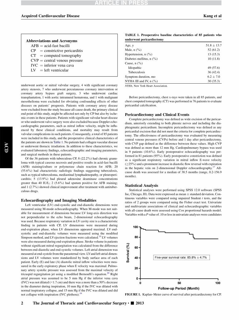
TABLE 1. Preoperative baseline characteristics of 85 patients who
underwent pericardiectomy
Age, y 51.8 � 13.7
Male, n (%) 52 (61.2)
Hypertension, n (%) 13 (15.3)
Diabetes mellitus, n (%) 10 (11.8)
Cause, n (%)
Idiopathic 49 (57.6)
Tuberculosis 36 (42.4)
Symptom duration, mo 6.2 � 7.0
NYHA III and IV, n (%) 30 (35.3)
NYHA, New York Heart Association.
FIGURE 1. Kaplan–Meier curve of survival after pericardiectomy for CP.
Abbreviations and AcronymsAFB ¼ acid-fast bacilliCP ¼ constrictive pericarditisCT ¼ computed tomographyCVP ¼ central venous pressureIVC ¼ inferior vena cavaLV ¼ left ventricular
Acquired Cardiovascular Disease Kang et al
ACD
underwent aortic or mitral valvular surgery, 4 with significant coronary
artery stenosis, 7 who underwent percutaneous coronary intervention or
coronary artery bypass graft surgery, 3 who underwent cardiac
transplantation, 1 with aortic intramural hematoma, and 1 with malignant
mesothelioma were excluded for obviating confounding effects of other
diseases on patients’ prognosis. Patients with coronary artery disease
were excluded from the study because all-cause death, the primary clinical
end point of this study, might be affected not only by CP but also by ische-
mic events in these patients. Patients with significant valvular heart disease
or who underwent valve surgery were also excluded because Doppler echo-
cardiographic parameters, such as mitral inflow velocity, might be influ-
enced by these clinical conditions, and mortality may result from
valvular complications in such patients. Consequently, a total of 85 patients
were included in this study, and the preoperative clinical characteristics of
the patients are shown in Table 1. No patients had collagen vascular disease
or underwent thoracic irradiation. In addition to these characteristics, we
evaluated laboratory findings, echocardiographic and imaging parameters,
and surgical methods in these patients.
Of the 36 patients with tuberculous CP, 8 (22.2%) had chronic granu-
loma with typical caseous necrosis and positive results in acid-fast bacilli
(AFB) staining/culture or polymerase chain reaction for AFB; 20
(55.6%) had characteristic radiologic findings suggesting tuberculosis,
such as typical tuberculoma, mediastinal lymphadenopathy, or pleuroperi-
carditis; 5 (13.9%) had pleural adenosine deaminase concentrations
greater than 40 IU/L; 2 (5.6%) had sputum positive for AFB staining;
and 1 (2.7%) showed clinical improvement after treatment with antituber-
culous drugs.
Echocardiography and Imaging ModalitiesLeft ventricular (LV) end-systolic and end-diastolic dimensions were
measured using M-mode echocardiography. When M-mode was not suit-
able for measurement of dimensions because LV long-axis direction was
not perpendicular to the echo beam, 2-dimensional echocardiography
was used. Because respiratory variation in LV cavity size is a characteristic
finding in patients with CP, LV dimensions were measured during
end-expiration phase, when LV dimensions appeared maximal. LV end-
systolic and end-diastolic volumes were measured using the modified
Simpson method, and LVejection fractions were calculated.19 LV volumes
were also measured during end-expiration phase. Stroke volume in patients
without significant mitral regurgitation was calculated from the difference
between end-diastolic and end-systolic volumes. Left atrial dimension was
measured at end-systole from the parasternal view. LVand left atrial dimen-
sions and LV volumes were standardized by body surface area of each
patient. Early (E) and late (A) diastolic mitral inflow velocities were mea-
sured in the early expiratory phase when E velocity was maximal. Pulmo-
nary artery systolic pressure was assessed from the maximal velocity of
tricuspid regurgitation jet using a modified Bernoulli’s equation.20 Right
atrial pressure was assumed to be 5 mm Hg if the inferior vena cava
(IVC) was not dilated (<1.7 cm) and there was a more than a 50% decrease
in the diameter during inspiration, 10 mm Hg if the IVC was dilated with
normal inspiratory collapse, and 15 mm Hg if the IVC was dilated and did
not collapse with inspiration (IVC plethora).19
2 The Journal of Thoracic and Cardiovascular Surger
Before pericardiectomy, chest x-rays were taken in all 85 patients, and
chest computed tomography (CT) was performed in 76 patients to evaluate
pericardial calcification.
Pericardiectomy and Clinical EventsComplete pericardiectomy was defined as wide excision of the pericar-
dium, anteriorly extending to both phrenic nerves and including the dia-
phragmatic pericardium. Incomplete pericardiectomy was defined as any
pericardial excision that did not meet the criteria for complete pericardiec-
tomy. The effectiveness of pericardiectomy was evaluated by measuring
central venous pressures (CVPs) before and 1 day after pericardiectomy,
with CVP gap defined as the difference between these values. High CVP
was defined as more than 12 mm Hg. Cardiopulmonary bypass was used
in 9 patients (10.6%). Early postoperative echocardiography was per-
formed in 81 patients (95%). Early postoperative constriction was defined
as a significant respiratory variation in mitral inflow E-wave velocity
(�25%) and a prominent increase in diastolic flow reversal with expiration
in the hepatic vein on 2-dimensional Doppler echocardiography.17 All-
cause death was assessed for a median of 38.5 months (range, 0.2-156.9
months).
Statistical AnalysisStatistical analyses were performed using SPSS 12.0 software (SPSS
Inc, Chicago, Ill). Data were expressed as mean� standard deviation. Con-
tinuous variables were compared using unpaired Student t tests, and the
ratios of 2 groups were compared using the Fisher exact test. Univariate
and multivariate associations of clinical and echocardiographic variables
with all-cause death were assessed using Cox proportional hazards model.
Variableswith aP value of .10 or less in univariate analyseswere candidates
y c - 2013
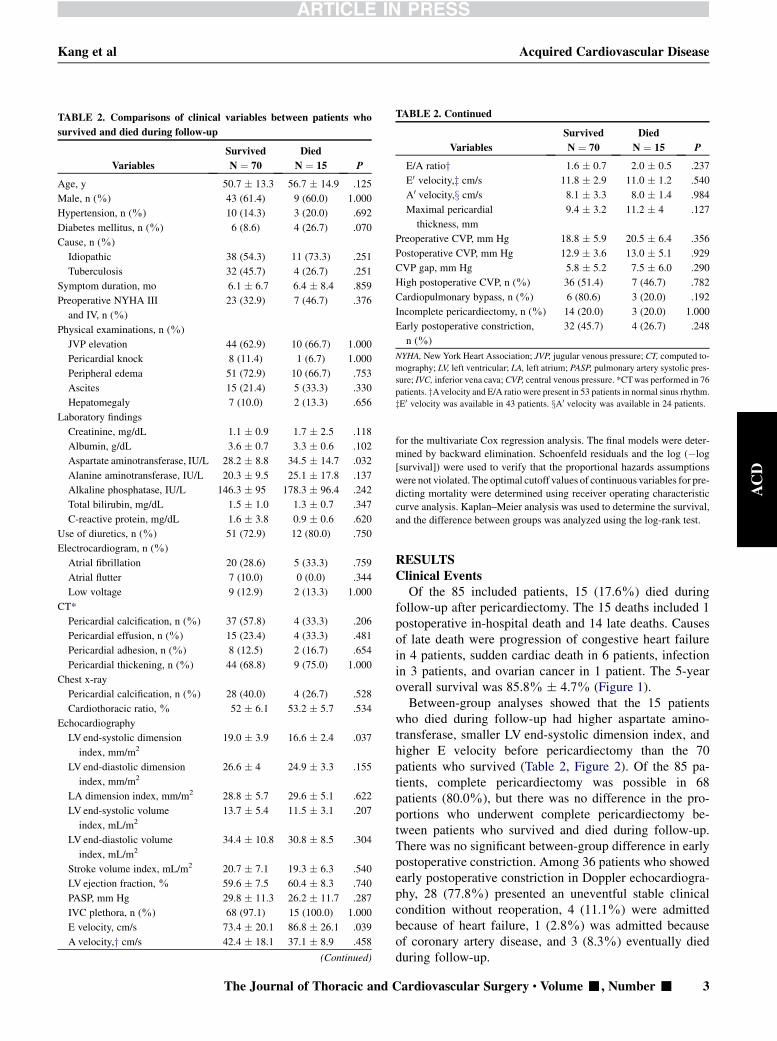
TABLE 2. Comparisons of clinical variables between patients who
survived and died during follow-up
Variables
Survived
N ¼ 70
Died
N ¼ 15 P
Age, y 50.7 � 13.3 56.7 � 14.9 .125
Male, n (%) 43 (61.4) 9 (60.0) 1.000
Hypertension, n (%) 10 (14.3) 3 (20.0) .692
Diabetes mellitus, n (%) 6 (8.6) 4 (26.7) .070
Cause, n (%)
Idiopathic 38 (54.3) 11 (73.3) .251
Tuberculosis 32 (45.7) 4 (26.7) .251
Symptom duration, mo 6.1 � 6.7 6.4 � 8.4 .859
Preoperative NYHA III
and IV, n (%)
23 (32.9) 7 (46.7) .376
Physical examinations, n (%)
JVP elevation 44 (62.9) 10 (66.7) 1.000
Pericardial knock 8 (11.4) 1 (6.7) 1.000
Peripheral edema 51 (72.9) 10 (66.7) .753
Ascites 15 (21.4) 5 (33.3) .330
Hepatomegaly 7 (10.0) 2 (13.3) .656
Laboratory findings
Creatinine, mg/dL 1.1 � 0.9 1.7 � 2.5 .118
Albumin, g/dL 3.6 � 0.7 3.3 � 0.6 .102
Aspartate aminotransferase, IU/L 28.2 � 8.8 34.5 � 14.7 .032
Alanine aminotransferase, IU/L 20.3 � 9.5 25.1 � 17.8 .137
Alkaline phosphatase, IU/L 146.3 � 95 178.3 � 96.4 .242
Total bilirubin, mg/dL 1.5 � 1.0 1.3 � 0.7 .347
C-reactive protein, mg/dL 1.6 � 3.8 0.9 � 0.6 .620
Use of diuretics, n (%) 51 (72.9) 12 (80.0) .750
Electrocardiogram, n (%)
Atrial fibrillation 20 (28.6) 5 (33.3) .759
Atrial flutter 7 (10.0) 0 (0.0) .344
Low voltage 9 (12.9) 2 (13.3) 1.000
CT*
Pericardial calcification, n (%) 37 (57.8) 4 (33.3) .206
Pericardial effusion, n (%) 15 (23.4) 4 (33.3) .481
Pericardial adhesion, n (%) 8 (12.5) 2 (16.7) .654
Pericardial thickening, n (%) 44 (68.8) 9 (75.0) 1.000
Chest x-ray
Pericardial calcification, n (%) 28 (40.0) 4 (26.7) .528
Cardiothoracic ratio, % 52 � 6.1 53.2 � 5.7 .534
Echocardiography
LV end-systolic dimension
index, mm/m2
19.0 � 3.9 16.6 � 2.4 .037
LV end-diastolic dimension
index, mm/m2
26.6 � 4 24.9 � 3.3 .155
LA dimension index, mm/m2 28.8 � 5.7 29.6 � 5.1 .622
LV end-systolic volume
index, mL/m2
13.7 � 5.4 11.5 � 3.1 .207
LV end-diastolic volume
index, mL/m2
34.4 � 10.8 30.8 � 8.5 .304
Stroke volume index, mL/m2 20.7 � 7.1 19.3 � 6.3 .540
LV ejection fraction, % 59.6 � 7.5 60.4 � 8.3 .740
PASP, mm Hg 29.8 � 11.3 26.2 � 11.7 .287
IVC plethora, n (%) 68 (97.1) 15 (100.0) 1.000
E velocity, cm/s 73.4 � 20.1 86.8 � 26.1 .039
A velocity,y cm/s 42.4 � 18.1 37.1 � 8.9 .458
(Continued)
TABLE 2. Continued
Variables
Survived
N ¼ 70
Died
N ¼ 15 P
E/A ratioy 1.6 � 0.7 2.0 � 0.5 .237
E0 velocity,z cm/s 11.8 � 2.9 11.0 � 1.2 .540
A0 velocity,x cm/s 8.1 � 3.3 8.0 � 1.4 .984
Maximal pericardial
thickness, mm
9.4 � 3.2 11.2 � 4 .127
Preoperative CVP, mm Hg 18.8 � 5.9 20.5 � 6.4 .356
Postoperative CVP, mm Hg 12.9 � 3.6 13.0 � 5.1 .929
CVP gap, mm Hg 5.8 � 5.2 7.5 � 6.0 .290
High postoperative CVP, n (%) 36 (51.4) 7 (46.7) .782
Cardiopulmonary bypass, n (%) 6 (80.6) 3 (20.0) .192
Incomplete pericardiectomy, n (%) 14 (20.0) 3 (20.0) 1.000
Early postoperative constriction,
n (%)
32 (45.7) 4 (26.7) .248
NYHA, New York Heart Association; JVP, jugular venous pressure; CT, computed to-
mography; LV, left ventricular; LA, left atrium; PASP, pulmonary artery systolic pres-
sure; IVC, inferior vena cava;CVP, central venous pressure. *CTwas performed in 76
patients. yAvelocity and E/A ratio were present in 53 patients in normal sinus rhythm.
zE0 velocity was available in 43 patients. xA0 velocity was available in 24 patients.
Kang et al Acquired Cardiovascular Disease
The Journal of Thoracic and C
ACD
for the multivariate Cox regression analysis. The final models were deter-
mined by backward elimination. Schoenfeld residuals and the log (�log
[survival]) were used to verify that the proportional hazards assumptions
were not violated. The optimal cutoff values of continuous variables for pre-
dicting mortality were determined using receiver operating characteristic
curve analysis. Kaplan–Meier analysis was used to determine the survival,
and the difference between groups was analyzed using the log-rank test.
RESULTSClinical EventsOf the 85 included patients, 15 (17.6%) died during
follow-up after pericardiectomy. The 15 deaths included 1postoperative in-hospital death and 14 late deaths. Causesof late death were progression of congestive heart failurein 4 patients, sudden cardiac death in 6 patients, infectionin 3 patients, and ovarian cancer in 1 patient. The 5-yearoverall survival was 85.8% � 4.7% (Figure 1).Between-group analyses showed that the 15 patients
who died during follow-up had higher aspartate amino-transferase, smaller LV end-systolic dimension index, andhigher E velocity before pericardiectomy than the 70patients who survived (Table 2, Figure 2). Of the 85 pa-tients, complete pericardiectomy was possible in 68patients (80.0%), but there was no difference in the pro-portions who underwent complete pericardiectomy be-tween patients who survived and died during follow-up.There was no significant between-group difference in earlypostoperative constriction. Among 36 patients who showedearly postoperative constriction in Doppler echocardiogra-phy, 28 (77.8%) presented an uneventful stable clinicalcondition without reoperation, 4 (11.1%) were admittedbecause of heart failure, 1 (2.8%) was admitted becauseof coronary artery disease, and 3 (8.3%) eventually diedduring follow-up.
ardiovascular Surgery c Volume -, Number - 3
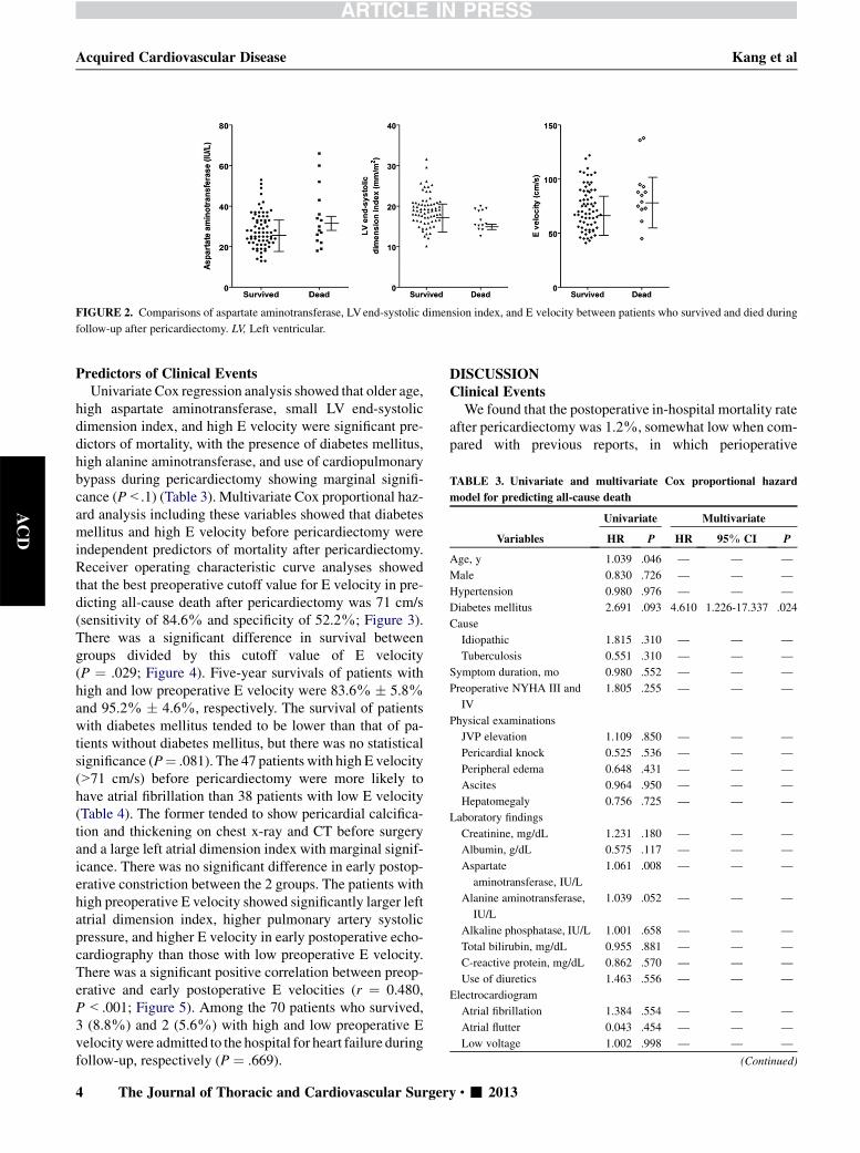
FIGURE 2. Comparisons of aspartate aminotransferase, LVend-systolic dimension index, and E velocity between patients who survived and died during
follow-up after pericardiectomy. LV, Left ventricular.
TABLE 3. Univariate and multivariate Cox proportional hazard
model for predicting all-cause death
Variables
Univariate Multivariate
HR P HR 95% CI P
Age, y 1.039 .046 — — —
Male 0.830 .726 — — —
Hypertension 0.980 .976 — — —
Diabetes mellitus 2.691 .093 4.610 1.226-17.337 .024
Cause
Idiopathic 1.815 .310 — — —
Tuberculosis 0.551 .310 — — —
Symptom duration, mo 0.980 .552 — — —
Preoperative NYHA III and
IV
1.805 .255 — — —
Physical examinations
JVP elevation 1.109 .850 — — —
Pericardial knock 0.525 .536 — — —
Peripheral edema 0.648 .431 — — —
Ascites 0.964 .950 — — —
Hepatomegaly 0.756 .725 — — —
Laboratory findings
Creatinine, mg/dL 1.231 .180 — — —
Albumin, g/dL 0.575 .117 — — —
Aspartate
aminotransferase, IU/L
1.061 .008 — — —
Alanine aminotransferase,
IU/L
1.039 .052 — — —
Alkaline phosphatase, IU/L 1.001 .658 — — —
Total bilirubin, mg/dL 0.955 .881 — — —
C-reactive protein, mg/dL 0.862 .570 — — —
Use of diuretics 1.463 .556 — — —
Electrocardiogram
Atrial fibrillation 1.384 .554 — — —
Atrial flutter 0.043 .454 — — —
Low voltage 1.002 .998 — — —
(Continued)
Acquired Cardiovascular Disease Kang et al
ACD
Predictors of Clinical EventsUnivariate Cox regression analysis showed that older age,
high aspartate aminotransferase, small LV end-systolicdimension index, and high E velocity were significant pre-dictors of mortality, with the presence of diabetes mellitus,high alanine aminotransferase, and use of cardiopulmonarybypass during pericardiectomy showing marginal signifi-cance (P<.1) (Table 3). Multivariate Cox proportional haz-ard analysis including these variables showed that diabetesmellitus and high E velocity before pericardiectomy wereindependent predictors of mortality after pericardiectomy.Receiver operating characteristic curve analyses showedthat the best preoperative cutoff value for E velocity in pre-dicting all-cause death after pericardiectomy was 71 cm/s(sensitivity of 84.6% and specificity of 52.2%; Figure 3).There was a significant difference in survival betweengroups divided by this cutoff value of E velocity(P ¼ .029; Figure 4). Five-year survivals of patients withhigh and low preoperative E velocity were 83.6% � 5.8%and 95.2% � 4.6%, respectively. The survival of patientswith diabetes mellitus tended to be lower than that of pa-tients without diabetes mellitus, but there was no statisticalsignificance (P¼ .081). The 47 patients with high E velocity(>71 cm/s) before pericardiectomy were more likely tohave atrial fibrillation than 38 patients with low E velocity(Table 4). The former tended to show pericardial calcifica-tion and thickening on chest x-ray and CT before surgeryand a large left atrial dimension index with marginal signif-icance. There was no significant difference in early postop-erative constriction between the 2 groups. The patients withhigh preoperative E velocity showed significantly larger leftatrial dimension index, higher pulmonary artery systolicpressure, and higher E velocity in early postoperative echo-cardiography than those with low preoperative E velocity.There was a significant positive correlation between preop-erative and early postoperative E velocities (r ¼ 0.480,P<.001; Figure 5). Among the 70 patients who survived,3 (8.8%) and 2 (5.6%) with high and low preoperative Evelocitywere admitted to the hospital for heart failure duringfollow-up, respectively (P ¼ .669).
4 The Journal of Thoracic and Cardiovascular Surger
DISCUSSIONClinical Events
We found that the postoperative in-hospital mortality rateafter pericardiectomy was 1.2%, somewhat low when com-pared with previous reports, in which perioperative
y c - 2013
TABLE 3. Continued
Variables
Univariate Multivariate
HR P HR 95% CI P
CT
Pericardial calcification 0.456 .204 — — —
Pericardial effusion 2.095 .241 — — —
Pericardial adhesion 2.930 .193 — — —
Pericardial thickening 1.454 .577 — — —
Chest x-ray
Pericardial calcification 0.559 .381 — — —
Cardiothoracic ratio, % 1.034 .505 — — —
Echocardiography
LV end-systolic dimension
index, mm/m2
0.821 .038 0.815 0.643-1.031 .089
LVend-diastolic dimension
index, mm/m2
0.907 .250 — — —
LA dimension index,
mm/m2
1.002 .974 — — —
LVend-systolic volume
index, mL/m2
0.948 .442 — — —
LVend-diastolic volume
index, mL/m2
0.97 .300 — — —
Stroke volume, mL/m2 0.953 .299 — — —
LVejection fraction, % 0.994 .856 — — —
PASP, mm Hg 0.99 .702 — — —
IVC plethora 22.355 .563 — — —
E velocity, cm/s 1.034 .011 1.050 1.018-1.083 .002
A velocity, cm/s 0.991 .717 — — —
E/A ratio 1.340 .546 — — —
E0 velocity, cm/s 1.000 .999 — — —
A0 velocity, cm/s 1.293 .486 — — —
Maximal pericardial
thickness, mm
1.156 .114 — — —
Preoperative CVP, mm Hg 1.022 .605 — — —
Postoperative CVP, mm Hg 1.031 .638 — — —
CVP gap, mm Hg 1.022 .658 — — —
High postoperative CVP 1.02 .970 — — —
Cardiopulmonary bypass 3.528 .060 — — —
Incomplete pericardiectomy 1.708 .426 — — —
Early postoperative
constriction
0.924 .899 — — —
HR, Hazard ratio; CI, confidence interval; NYHA, New York Heart Association;
JVP, jugular venous pressure; CT, computed tomography; LV, left ventricular;
LA, left atrium; PASP, pulmonary artery systolic pressure; IVC, inferior vena cava;
CVP, central venous pressure.
FIGURE 3. Receiver operating characteristic curve of transmitral E
velocity (E). Dot on the curve denotes best cutoff value. AUC, Area under
curve; CI, confidence interval.
Kang et al Acquired Cardiovascular Disease
ACD
mortality rates after pericardiectomy ranged from 2.3% to12%.2-9 The relatively good postoperative prognosis weobserved may be due to our patient population, in whichthe most common cause of CP was idiopathic. Patientswith idiopathic CP were reported to have the bestprognosis after pericardiectomy.6,8 Furthermore, weexcluded patients with significant valvular and coronaryartery disease, and other serious comorbidities to obviateconfounding effects of other diseases on patients’mortality and evaluate the pure prognosis of patientswith isolated CP after pericardiectomy. Although
The Journal of Thoracic and C
mortality rates after pericardiectomy in patients withpostradiotherapy CP have been reported to be high,2,6,8
none of our patients had this cause. Most patients withpostirradiation CP underwent radiotherapy to treatlymphoma and breast cancer. Patients with Hodgkin’slymphoma are frequently administered radiotherapy formediastinal lymph nodes. However, this disease is moreprevalent in the United States and Europe than in Easterncountries, including Korea. The most common subtype ofmalignant lymphoma in Korea is diffuse large B-celllymphoma,21 which is usually treated with chemotherapyand rarely with radiotherapy. In addition, 36% of our pa-tients had tuberculous CP, which was found to be a majorcause in only a few reports.3,9 We found that the 5-yearoverall survival after pericardiectomy (85.8%) tended tobe higher than in previous reports, in which survivalranged from 64% to 85%.4,5,7,9,11,16
Predictors of Clinical EventsWe found that diabetes mellitus and high E velocity be-
fore pericardiectomy were independent predictors of clini-cal events after pericardiectomy. A novel finding of ourstudy was that a Doppler echocardiographic parameter,early expiratory transmitral E velocity, which is importantin CP diagnosis, was also prognostic in these patients.The explanation of mechanism that high E velocity was as-sociated with mortality after pericardiectomy would bespeculative. However, this Doppler parameter may repre-sent the severity of CP, and a high transmitral E velocitymay reflect a high left atrial pressure resulting from severe
ardiovascular Surgery c Volume -, Number - 5
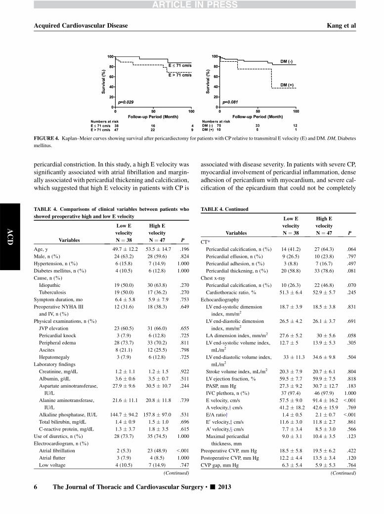
FIGURE 4. Kaplan–Meier curves showing survival after pericardiectomy for patients with CP relative to transmitral E velocity (E) and DM.DM,Diabetes
mellitus.
Acquired Cardiovascular Disease Kang et al
ACD
pericardial constriction. In this study, a high E velocity wassignificantly associated with atrial fibrillation and margin-ally associated with pericardial thickening and calcification,which suggested that high E velocity in patients with CP is
TABLE 4. Comparisons of clinical variables between patients who
showed preoperative high and low E velocity
Variables
Low E
velocity
N ¼ 38
High E
velocity
N ¼ 47 P
Age, y 49.7 � 12.2 53.5 � 14.7 .196
Male, n (%) 24 (63.2) 28 (59.6) .824
Hypertension, n (%) 6 (15.8) 7 (14.9) 1.000
Diabetes mellitus, n (%) 4 (10.5) 6 (12.8) 1.000
Cause, n (%)
Idiopathic 19 (50.0) 30 (63.8) .270
Tuberculosis 19 (50.0) 17 (36.2) .270
Symptom duration, mo 6.4 � 5.8 5.9 � 7.9 .753
Preoperative NYHA III
and IV, n (%)
12 (31.6) 18 (38.3) .649
Physical examinations, n (%)
JVP elevation 23 (60.5) 31 (66.0) .655
Pericardial knock 3 (7.9) 6 (12.8) .725
Peripheral edema 28 (73.7) 33 (70.2) .811
Ascites 8 (21.1) 12 (25.5) .798
Hepatomegaly 3 (7.9) 6 (12.8) .725
Laboratory findings
Creatinine, mg/dL 1.2 � 1.1 1.2 � 1.5 .922
Albumin, g/dL 3.6 � 0.6 3.5 � 0.7 .511
Aspartate aminotransferase,
IU/L
27.9 � 9.6 30.5 � 10.7 .244
Alanine aminotransferase,
IU/L
21.6 � 11.1 20.8 � 11.8 .739
Alkaline phosphatase, IU/L 144.7 � 94.2 157.8 � 97.0 .531
Total bilirubin, mg/dL 1.4 � 0.9 1.5 � 1.0 .696
C-reactive protein, mg/dL 1.3 � 3.7 1.8 � 3.5 .615
Use of diuretics, n (%) 28 (73.7) 35 (74.5) 1.000
Electrocardiogram, n (%)
Atrial fibrillation 2 (5.3) 23 (48.9) <.001
Atrial flutter 3 (7.9) 4 (8.5) 1.000
Low voltage 4 (10.5) 7 (14.9) .747
(Continued)
6 The Journal of Thoracic and Cardiovascular Surger
associated with disease severity. In patients with severe CP,myocardial involvement of pericardial inflammation, denseadhesion of pericardium with myocardium, and severe cal-cification of the epicardium that could not be completely
TABLE 4. Continued
Variables
Low E
velocity
N ¼ 38
High E
velocity
N ¼ 47 P
CT*
Pericardial calcification, n (%) 14 (41.2) 27 (64.3) .064
Pericardial effusion, n (%) 9 (26.5) 10 (23.8) .797
Pericardial adhesion, n (%) 3 (8.8) 7 (16.7) .497
Pericardial thickening, n (%) 20 (58.8) 33 (78.6) .081
Chest x-ray
Pericardial calcification, n (%) 10 (26.3) 22 (46.8) .070
Cardiothoracic ratio, % 51.3 � 6.4 52.9 � 5.7 .245
Echocardiography
LV end-systolic dimension
index, mm/m2
18.7 � 3.9 18.5 � 3.8 .831
LV end-diastolic dimension
index, mm/m2
26.5 � 4.2 26.1 � 3.7 .691
LA dimension index, mm/m2 27.6 � 5.2 30 � 5.6 .058
LV end-systolic volume index,
mL/m2
12.7 � 5 13.9 � 5.3 .305
LVend-diastolic volume index,
mL/m2
33 � 11.3 34.6 � 9.8 .504
Stroke volume index, mL/m2 20.3 � 7.9 20.7 � 6.1 .804
LV ejection fraction, % 59.5 � 7.7 59.9 � 7.5 .818
PASP, mm Hg 27.3 � 9.2 30.7 � 12.7 .183
IVC plethora, n (%) 37 (97.4) 46 (97.9) 1.000
E velocity, cm/s 57.5 � 9.0 91.4 � 16.2 <.001
A velocity,y cm/s 41.2 � 18.2 42.6 � 15.9 .769
E/A ratioy 1.4 � 0.5 2.1 � 0.7 <.001
E0 velocity,z cm/s 11.6 � 3.0 11.8 � 2.7 .861
A0 velocity,x cm/s 7.7 � 3.4 8.5 � 3.0 .566
Maximal pericardial
thickness, mm
9.0 � 3.1 10.4 � 3.5 .123
Preoperative CVP, mm Hg 18.5 � 5.8 19.5 � 6.2 .422
Postoperative CVP, mm Hg 12.2 � 4.4 13.5 � 3.4 .120
CVP gap, mm Hg 6.3 � 5.4 5.9 � 5.3 .764
(Continued)
y c - 2013
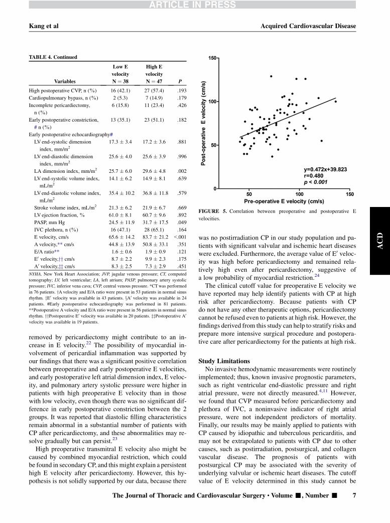
TABLE 4. Continued
Variables
Low E
velocity
N ¼ 38
High E
velocity
N ¼ 47 P
High postoperative CVP, n (%) 16 (42.1) 27 (57.4) .193
Cardiopulmonary bypass, n (%) 2 (5.3) 7 (14.9) .179
Incomplete pericardiectomy,
n (%)
6 (15.8) 11 (23.4) .426
Early postoperative constriction,
# n (%)
13 (35.1) 23 (51.1) .182
Early postoperative echocardiography#
LV end-systolic dimension
index, mm/m2
17.3 � 3.4 17.2 � 3.6 .881
LV end-diastolic dimension
index, mm/m2
25.6 � 4.0 25.6 � 3.9 .996
LA dimension index, mm/m2 25.7 � 6.0 29.6 � 4.8 .002
LV end-systolic volume index,
mL/m2
14.1 � 6.2 14.9 � 8.1 .639
LVend-diastolic volume index,
mL/m2
35.4 � 10.2 36.8 � 11.8 .579
Stroke volume index, mL/m2 21.3 � 6.2 21.9 � 6.7 .669
LV ejection fraction, % 61.0 � 8.1 60.7 � 9.6 .892
PASP, mm Hg 24.5 � 11.9 31.7 � 17.5 .049
IVC plethora, n (%) 16 (47.1) 28 (65.1) .164
E velocity, cm/s 65.6 � 14.2 83.7 � 21.2 <.001
A velocity,** cm/s 44.8 � 13.9 50.8 � 33.1 .351
E/A ratio** 1.6 � 0.6 1.9 � 0.9 .121
E0 velocity,yy cm/s 8.7 � 2.2 9.9 � 2.3 .175
A0 velocity,zz cm/s 8.3 � 2.5 7.3 � 2.9 .451
NYHA, New York Heart Association; JVP, jugular venous pressure; CT, computed
tomography; LV, left ventricular; LA, left atrium; PASP, pulmonary artery systolic
pressure; IVC, inferior vena cava; CVP, central venous pressure. *CTwas performed
in 76 patients. yA velocity and E/A ratio were present in 53 patients in normal sinus
rhythm. zE0 velocity was available in 43 patients. xA0 velocity was available in 24
patients. #Early postoperative echocardiography was performed in 81 patients.
**Postoperative A velocity and E/A ratio were present in 56 patients in normal sinus
rhythm. yyPostoperative E0 velocity was available in 20 patients. zzPostoperative A0
velocity was available in 19 patients.
FIGURE 5. Correlation between preoperative and postoperative E
velocities.
Kang et al Acquired Cardiovascular Disease
ACD
removed by pericardiectomy might contribute to an in-crease in E velocity.22 The possibility of myocardial in-volvement of pericardial inflammation was supported byour findings that there was a significant positive correlationbetween preoperative and early postoperative E velocities,and early postoperative left atrial dimension index, E veloc-ity, and pulmonary artery systolic pressure were higher inpatients with high preoperative E velocity than in thosewith low velocity, even though there was no significant dif-ference in early postoperative constriction between the 2groups. It was reported that diastolic filling characteristicsremain abnormal in a substantial number of patients withCP after pericardiectomy, and these abnormalities may re-solve gradually but can persist.23
High preoperative transmitral E velocity also might becaused by combined myocardial restriction, which couldbe found in secondary CP, and this might explain a persistenthigh E velocity after pericardiectomy. However, this hy-pothesis is not solidly supported by our data, because there
The Journal of Thoracic and C
was no postirradiation CP in our study population and pa-tients with significant valvular and ischemic heart diseaseswere excluded. Furthermore, the average value of E0 veloc-ity was high before pericardiectomy and remained rela-tively high even after pericardiectomy, suggestive ofa low probability of myocardial restriction.24
The clinical cutoff value for preoperative E velocity wehave reported may help identify patients with CP at highrisk after pericardiectomy. Because patients with CPdo not have any other therapeutic options, pericardiectomycannot be refused even to patients at high risk. However, thefindings derived from this study can help to stratify risks andprepare more intensive surgical procedure and postopera-tive care after pericardiectomy for the patients at high risk.
Study LimitationsNo invasive hemodynamic measurements were routinely
implemented; thus, known invasive prognostic parameters,such as right ventricular end-diastolic pressure and rightatrial pressure, were not directly measured.4,11 However,we found that CVP measured before pericardiectomy andplethora of IVC, a noninvasive indicator of right atrialpressure, were not independent predictors of mortality.Finally, our results may be mainly applied to patients withCP caused by idiopathic and tuberculous pericarditis, andmay not be extrapolated to patients with CP due to othercauses, such as postirradiation, postsurgical, and collagenvascular disease. The prognosis of patients withpostsurgical CP may be associated with the severity ofunderlying valvular or ischemic heart diseases. The cutoffvalue of E velocity determined in this study cannot be
ardiovascular Surgery c Volume -, Number - 7
Acquired Cardiovascular Disease Kang et al
ACD
applied to patients with significant mitral valvular diseaseand patients with a prosthetic mitral valve for predictingprognosis after pericardiectomy.
CONCLUSIONSPreoperative high E velocity and diabetes mellitus were
predictors of poor prognosis after pericardiectomy in pa-tients with chronic CP. These results suggest that preopera-tive Doppler echocardiographic evaluation may be valuablenot only for diagnosing CP but also for predicting prognosisafter pericardiectomy.
References1. Myers RB, Spodick DH. Constrictive pericarditis: clinical and pathophysiologic
characteristics. Am Heart J. 1999;138:219-32.
2. Ling LH, Oh JK, Schaff HV, Danielson GK, Mahoney DW, Seward JB, et al.
Constrictive pericarditis in the modern era: evolving clinical spectrum and im-
pact on outcome after pericardiectomy. Circulation. 1999;100:1380-6.
3. Nataf P, Cacoub P, Dorent R, Jault F, Bors V, Pavie A, et al. Results of subtotal
pericardiectomy for constrictive pericarditis. Eur J Cardiothorac Surg. 1993;7:
252-6.
4. Seifert FC, Miller DC, Oesterle SN, Oyer PE, Stinson EB, Shumway NE. Surgi-
cal treatment of constrictive pericarditis: analysis of outcome and diagnostic
error. Circulation. 1985;72:II264-73.
5. Aagaard MT, Haraldsted VY. Chronic constrictive pericarditis treated with total
pericardiectomy. Thorac Cardiovasc Surg. 1984;32:311-4.
6. Cameron J, Oesterle SN, Baldwin JC, Hancock EW. The etiologic spectrum of
constrictive pericarditis. Am Heart J. 1987;113:354-60.
7. DeValeria PA, Baumgartner WA, Casale AS, Greene PS, Cameron DE,
Gardner TJ, et al. Current indications, risks, and outcome after pericardiectomy.
Ann Thorac Surg. 1991;52:219-24.
8. Bertog SC, Thambidorai SK, Parakh K, Schoenhagen P, Ozduran V,
Houghtaling PL, et al. Constrictive pericarditis: etiology and cause-specific
survival after pericardiectomy. J Am Coll Cardiol. 2004;43:1445-52.
9. Arsan S, Mercan S, Sarigul A, Atasoy S, Demircin M, Dogan R, et al. Long-term
experience with pericardiectomy: analysis of 105 consecutive patients. Thorac
Cardiovasc Surg. 1994;42:340-4.
10. Hehrlein FW, Moosdorf R, Pitton M, Dapper F. The role of pericardiectomy in
pericardial disorders. Eur Heart J. 1991;12(Suppl D):7-9.
11. Tirilomis T, Unverdorben S, von der Emde J. Pericardectomy for chronic con-
strictive pericarditis: risks and outcome. Eur J Cardiothorac Surg. 1994;8:
487-92.
8 The Journal of Thoracic and Cardiovascular Surger
12. Gopaldas RR, Dao TK, Caron NR, Markley JG. Predictors of in-hospital compli-
cations after pericardiectomy: a nationwide outcomes study. J Thorac Cardio-
vasc Surg. 2012 May 9. [Epub ahead of print]
13. Ling LH, Oh JK, Breen JF, Schaff HV, Danielson GK, Mahoney DW, et al. Cal-
cific constrictive pericarditis: is it still with us? Ann Intern Med. 2000;132:
444-50.
14. Ha JW, Oh JK, Schaff HV, Ling LH, Higano ST, Mahoney DW, et al. Impact of
left ventricular function on immediate and long-term outcomes after pericardiec-
tomy in constrictive pericarditis. J Thorac Cardiovasc Surg. 2008;136:1136-41.
15. Chowdhury UK, SubramaniamGK, Kumar AS, Airan B, Singh R, Talwar S, et al.
Pericardiectomy for constrictive pericarditis: a clinical, echocardiographic, and
hemodynamic evaluation of two surgical techniques. Ann Thorac Surg. 2006;
81:522-9.
16. Bozbuga N, Erentug V, Eren E, Erdogan HB, Kirali K, Antal A, et al. Pericardiec-
tomy for chronic constrictive tuberculous pericarditis: risks and predictors of sur-
vival. Tex Heart Inst J. 2003;30:180-5.
17. Oh JK, Hatle LK, Seward JB, Danielson GK, Schaff HV, Reeder GS, et al. Diag-
nostic role of doppler echocardiography in constrictive pericarditis. J Am Coll
Cardiol. 1994;23:154-62.
18. Dal-Bianco JP, Sengupta PP, Mookadam F, Chandrasekaran K, Tajik AJ,
Khandheria BK. Role of echocardiography in the diagnosis of constrictive peri-
carditis. J Am Soc Echocardiogr. 2009;22:24-33; quiz 103-4.
19. Lang RM, Bierig M, Devereux RB, Flachskampf FA, Foster E, Pellikka PA, et al.
Recommendations for chamber quantification: a report from the American Soci-
ety of Echocardiography’s Guidelines and Standards Committee and the Cham-
ber Quantification Writing Group, developed in conjunction with the European
Association of Echocardiography, a branch of the European Society of Cardiol-
ogy. J Am Soc Echocardiogr. 2005;18:1440-63.
20. Yock PG, Popp RL. Noninvasive estimation of right ventricular systolic pressure
by doppler ultrasound in patients with tricuspid regurgitation. Circulation. 1984;
70:657-62.
21. Ko YH, Kim CW, Park CS, Jang HK, Lee SS, Kim SH, et al. REAL classification
of malignant lymphomas in the Republic of Korea: incidence of recently recog-
nized entities and changes in clinicopathologic features. Hematolymphoreticular
Study Group of the Korean Society of Pathologists. Revised European-American
lymphoma. Cancer. 1998;83:806-12.
22. Moosdorf R. Indications, results, and pitfalls in the surgery of constrictive peri-
carditis. Herz. 2000;25:794-8.
23. Senni M, Redfield MM, Ling LH, Danielson GK, Tajik AJ, Oh JK. Left ventric-
ular systolic and diastolic function after pericardiectomy in patients with con-
strictive pericarditis: Doppler echocardiographic findings and correlation with
clinical status. J Am Coll Cardiol. 1999;33:1182-8.
24. Garcia MJ, Rodriguez L, Ares M, Griffin BP, Thomas JD, Klein AL. Differenti-
ation of constrictive pericarditis from restrictive cardiomyopathy: assessment
of left ventricular diastolic velocities in longitudinal axis by Doppler tissue
imaging. J Am Coll Cardiol. 1996;27:108-14.
y c - 2013
Kang et al Acquired Cardiovascular Disease
000 Prognostic predictors in pericardiectomy for chronic constrictive pericarditisSe Hun Kang, MD, Jong-Min Song, MD, PhD, Minsoo Kim, MD, Suk Jung Choo, MD, PhD,
Cheol Hyun Chung, MD, PhD, Duk-Hyun Kang, MD, PhD, and Jae-Kwan Song, MD, PhD,
Seoul, Korea
To investigate the preoperative prognostic factors after pericardiectomy in patients with chronic
CP, we evaluated preoperative clinical and imaging characteristics of 85 consecutive patients with
chronic CP who underwent pericardiectomy. Preoperative high E velocity and diabetes mellitus
were predictors of poor prognosis after pericardiectomy in these patients.
The Journal of Thoracic and Cardiovascular Surgery c Volume -, Number -
ACD