Embed Size (px)
Citation preview
DEPARTMENT OF NEUROSURGERYDEPARTMENT OF INTENSIVE CAREHELSINKI UNIVERSITY CENTRAL HOSPITAL AND FACULTY OF MEDICINEDOCTORAL PROGRAMME IN CLINICAL RESEARCHUNIVERSITY OF HELSINKI
20/2014
cover.indd 1 5.11.2014 10:13:12
Department of Neurosurgery Department of Anesthesiology and Intensive Care
Helsinki University Central HospitalHelsinki, Finland
&
Faculty of Medicine and Doctoral School of Health ScienceDoctoral Programme in Clinical Research
University of HelsinkiHelsinki, Finland
Prognos c Models in Trauma c Brain Injury
Rahul Raj
ACADEMIC DISSERTATION
To be presented, with the permission of the Faculty of Medicine of the University of Helsinki,
for public examination in Lecture Hall 1, of Töölö Hospital on 19 December 2014, at 12 noon.
Helsinki 2014
Supervisors Associate Professor Jari Siironen, MD, PhD Department of Neurosurgery Helsinki University Central Hospital Helsinki, Finland
Associate Professor Markus B. Skrifvars, MD, PhD, EDIC, FCICM Department of Anaesthesiology and Intensive Care Helsinki University Central Hospital Helsinki, Finland
Reviewers Professor Juha Öhman, MD, PhD Department of Neurosurgery Tampere University Hospital Tampere, Finland Associate Professor Patrik Finne, MD, PhD Department of Medicine, Division of Nephrology Helsinki University Central Hospital Helsinki, Finland
Opponent Professor Andrew Maas, MD, PhD Department of Neurosurgery Antwerp University Hospital Antwerp, Belgium
© Rahul RajIllustrations © Rahul Raj, except where indicatedOriginal Cover Image © Grandeduc | Dreamstime.com
ISBN 978-951-51-0129-7 (paperback)ISSN 2342-3161 (print)
ISBN 978-951-51-0130-3 (PDF)ISSN 2342-317X (online)
HansaprintHelsinki, 2014Finland

Author’s contact information
Rahul RajDepartment of NeurosurgeryHelsinki University Central HospitalTopeliuksenkatu 5FI-00260, HelsinkiFinland
Mobile: +358443191190 E-mail: [email protected]


To my Mother & Father
Table of Contents
AbstractList of Original PublicationsAbbreviations1 Introduction ........................................................................................................................12 Review of the Literature ......................................................................................................2 2.1 Traumatic Brain injury ..............................................................................................2 2.1.1 Defi nition ......................................................................................................................2 2.1.2 Epidemiology ................................................................................................................2 2.1.3 Pathophysiology ...........................................................................................................2 2.1.4 Early Predictors of Patient Outcome .........................................................................3 2.1.4.1 Demographics ..................................................................................................3 2.1.4.2 Clinical Signs ....................................................................................................3 2.1.4.3 Radiological Findings .....................................................................................4 2.1.4.4 Secondary Insults .............................................................................................4 2.1.4.5 Laboratory Variables and Biomarkers ..........................................................4 2.1.5 Patient Outcome ..........................................................................................................5 2.2 Prognostic Models ......................................................................................................5 2.2.1 Defi nition ......................................................................................................................5 2.2.2 Development ................................................................................................................6 2.2.3 Internal Validation .......................................................................................................7 2.2.4 External Validation ......................................................................................................8 2.2.5 Performance assessment .............................................................................................8 2.2.5.1 Discrimination .................................................................................................8 2.2.5.2 Calibration ........................................................................................................9 2.2.5.3 Overall performance measures ....................................................................10 2.2.5.4 Net Reclassifi cation Index ............................................................................10 2.2.6 Customization ............................................................................................................10 2.2.7 Applications ................................................................................................................11 2.2.7.1 Quality Audits ................................................................................................11 2.2.7.2 Clinical Practice .............................................................................................11 2.2.7.3 Research ..........................................................................................................12 2.3 Traumatic Brain Injury Models ...............................................................................12 2.3.1 IMPACT ......................................................................................................................12 2.3.2 CRASH ........................................................................................................................14 2.3.3 CT Scoring Systems ...................................................................................................15 2.3.3.1 Marshall CT ....................................................................................................15 2.3.3.2 Rotterdam CT ................................................................................................15 2.4 Trauma Scoring Systems ..........................................................................................16 2.4.1 Anatomical Trauma Scoring Systems ......................................................................16 2.4.2 Physiological Trauma Scoring Systems ...................................................................16 2.4.3 Combined Anatomical and Trauma Scores ............................................................16 2.4.3.1 RISC ................................................................................................................16 2.5 Intensive Care Scoring Systems ................................................................................18 2.5.1 APACHE II .................................................................................................................18 2.5.2 SAPS II ........................................................................................................................18 2.5.3 SOFA ............................................................................................................................21
3 Purpose of the Study .........................................................................................................234 Subjects and Methods .......................................................................................................24 4.1 Study Setting and Population ...................................................................................24 4.1.1 Traumatic Brain Injury Models (I-III) ....................................................................24 4.1.2 Intensive Care Scoring Systems (IV) .......................................................................24 4.1.3 Trauma Scoring Systems (V) ....................................................................................24 4.2 Data collection ..........................................................................................................25 4.2.1 Traumatic Brain Injury Models (I-III) ....................................................................25 4.2.2 Intensive Care Scoring Systems (IV) .......................................................................26 4.2.3 Trauma Scoring Systems (V) ....................................................................................26 4.3 Statistical Analysis ....................................................................................................275 Results ........................................................................................................................28 5.1 Study Characteristics and Patient Outcome ............................................................28 5.2 Early Predictors of Outcome ....................................................................................30 5.2.1 Laboratory Variables and Extra-Cranial Injury .....................................................30 5.2.2 Computerized Tomography Abnormalities ...........................................................31 5.3 Comparison of Diff erent Types of Prognostic Models ............................................31 5.3.1 Traumatic Brain Injury Models ................................................................................32 5.3.2 Intensive Care Scoring Systems ................................................................................33 5.3.3 Trauma Scoring Systems ...........................................................................................34 5.4 Novel Prognostic Models ..........................................................................................36 5.4.1 IMPACT-APACHE II ................................................................................................36 5.4.2 Helsinki CT Score ......................................................................................................38 5.4.3 Modifi ed Intensive Care Scoring Systems ..............................................................416 Discussion ........................................................................................................................44 6.1 Key Findings .............................................................................................................44 6.1.1 Traumatic Brain Injury Models ................................................................................44 6.1.2 Intensive Care Scoring Systems ................................................................................45 6.1.3 Trauma Scoring Systems ...........................................................................................46 6.1.4 IMPACT-APACHE II ................................................................................................46 6.1.5 Helsinki CT Score ......................................................................................................47 6.2 Early Predictors of Outcome aft er TBI ....................................................................47 6.2.1 Markers of Coagulation ............................................................................................47 6.2.2 Major Extra-Cranial Injury ......................................................................................48 6.2.3 Early Computerized Tomography Characteristics ................................................48 6.3 Statistical Considerations ........................................................................................49 6.4 Patient Outcome aft er Traumatic Brain Injury .......................................................50 6.4.1 Outcome Assessment Aft er TBI ...............................................................................51 6.5 Limitations of the Study ...........................................................................................52 6.6 Future Implications ..................................................................................................53 6.6.1 Which Model To Use And For What? .....................................................................53 6.6.2 External Validation of the Proposed Models..........................................................53 6.7 Practical Examples of Prognostic Models in TBI Research ....................................547 Conclusions .......................................................................................................................55Acknowledgements .................................................................................................................56References ................................................................................................................................58
Abstract
Background: Prognostic models are important tools for heterogeneity adjustment in traumatic brain injury (TBI). Prognoses aft er TBI have been particularly challenging to predict, with limited availability of robust prognostic models. TBI patients are by defi nition trauma patients, and oft en treated in the intensive care unit (ICU). Several prognostic models for ICU and trauma patients have been developed, although their applicability in patients with TBI is uncertain. Recently, however, some new prognostic models specifi cally designed for patients with TBI were introduced. Still, the optimal type of prognostic model in TBI remains unknown.
Aim: To investigate the applicability of diff erent types of prognostic models in patients with TBI and to develop novel models with enhanced performance to previous models, focusing on long-term outcome prediction.
Methods: Four patient databases of patients with TBI treated in the ICU were used to validate three TBI specifi c models, two computerized tomography (CT) scoring systems, one trauma scoring system, and three intensive care scoring systems. Models were validated by assessing their discrimination using area under the curve (AUC), calibration, and explanatory variation. Logistic regression was used for model customization and development. Models were internally validated using a resample bootstrap technique or a split-sample technique. Primary outcome was six-month mortality and unfavorable neurological outcome by the Glasgow Outcome Scale. 30-day in-hospital mortality was used for the trauma scoring system.
Results: Study populations ranged from 342 to 9,915 patients. Th e TBI models showed the best performance with AUCs between 0.80 and 0.85, followed by the intensive care scoring systems and the CT scores with AUCs between 0.68 to 0.80 and 0.63 to 0.70, respectively. Most models showed poor calibration, although good calibration was achieved following customization. Th e trauma scoring system exhibited modest to good discrimination (AUC 0.76-0.89) for short-term mortality prediction, but poor calibration. Several new prognostic models, with statistically signifi cant superior performance to previous models were created, among them a combined TBI-ICU model (‘IMPACT-APACHE’) and a novel CT scoring system (‘Th e Helsinki CT score’). Using a TBI specifi c model, based on admission characteristics, up to 40 % of the patient’s fi nal long-term outcome could be predicted.
Conclusion: Th e TBI models showed superior predictive performance to the intensive care and trauma scoring systems, showing that TBI patients are a highly specifi c population in the trauma and ICU setting. Th us, the use of a TBI specifi c model is advocated in the setting of TBI. Th e newly proposed models were found to be signifi cant improvements over previous models, but require external validation to show generalizability.
List of Original Publica ons
Th is thesis is based on the following publications:
I Raj R, Siironen J, Kivisaari R, Hernesniemi J, Tanskanen P, Handolin L, Skrifvars MB. External Validation of the International Mission for Prognosis and Analysis of Clinical Trials Model and the Role of Markers of Coagulation, Neurosurgery, 2013;73(2):305-311
II Raj R, Kivisaari R, Siironen J, Skrifvars MB. Predicting Outcome Aft er Traumatic Brain Injury: Development of Prognostic Scores Based on the IMPACT and the APACHE II, Journal of Neurotrauma, 2014;31(20):1721-1732
III Raj R, Siironen J, Skrifvars MB, Lappalainen J, Hernesniemi J, Kivisaari R. Predicting Outcome in Traumatic Brain Injury: Development of a Novel Computerized Tomography Classifi cation Systems (Th e Helsinki CT Score), Neurosurgery;75(6):632-647
IV Raj R, Skrifvars MB, Bendel S, Selander T, Kivisaari R, Siironen J, Reinikainen M. Predicting Six-month Mortality of Patients with Traumatic Brain Injury: Usefulness of Common Intensive Care Severity Scores, Critical Care, 2014;18:R60
V Raj R, Brinck T, Skrifvars MB, Kivisaari R, Siironen J, Lefering R, Handolin L. Validation of the Revised Injury Severity Classifi cation Score in Patients With Moderate-to-Severe Traumatic Brain Injury, Injury, 2014 (In Press)
Th e publications are referred to in the text by their roman numerals. Th e original publications have been reprinted with the permission of the copyright holders.
Abbrevia ons
AIS, Abbreviated Injury SeverityAPACHE, Acute Physiology and Chronic Health EvaluationAUC, Area Under the CurveAUROC, Area Under the Receiver Operating Characteristic CurveBAC, Blood Alcohol ConcentrationsCER, Comparative Eff ectiveness ResearchCRASH, Corticosteroid Randomization Aft er Signifi cant Head InjuryCT, Computerized TomographyDTI, Diff usor Tension ImagingEDH, Epidural HematomaFICC, Finnish Intensive Care ConsortiumGCS, Glasgow Coma ScaleGiViTI, Gruppo Italiano per la Valutaione degli Interventi in Terapia IntensiveGoF, Goodness of FitGOS, Glasgow Outcome ScaleH-L, Hosmer-Lemeshow Ĉ statistic testICD-10, International Classifi cation of Diseases and Related Health Problems 10th EditionICH, Intracerebral HemorrhageICU, Intensive Care UnitIMPACT, International Mission for Prognosis and Analysis of Clinical TrialsISS, Injury Severity ScoreMRI, Magnetic Resonance ImagingIVH, Intraventricular HemorrhageNISS, New Injury Severity ScorePT, Th romboplastin TimePTT, Partial Th romboplastin TimeRCT, Randomized Controlled TrialRISC, Revised Injury Severity Classifi cationROC, Receiver Operator CharacteristicSAPS, Simplifi ed Acute Physiology ScoreSDH, Subdural HematomaSOFA, Sequential Organ Failure AssessmentTARN, Trauma Audit & Research Network TBI, Traumatic Brain InjuryTR-DGU, Trauma Registry of the German Society for Trauma Surgery®TRISS, Trauma Score - Injury Severity ScoreTR-THEL, Trauma Registry of Helsinki University HospitaltSAH, Traumatic Subarachnoid Hemorrhage
1
1 Introduc on
Traumatic brain injury (TBI) is a global health care and socioeconomic problem.1-7 Each year, about 1 in 200 Europeans and Americans will sustain some form of TBI. Of all TBIs approximately 10-20% are moderate or severe in nature, requiring intensive care unit (ICU) treatment.4,8 Of these patients one in two dies or is left with severe life-long disabilities, demonstrating the cruel prognosis of TBI.9,10 Establishing an early and reliable prognosis in patients with TBI has previously proved particularly challenging.11,12 However, advances in statistical modeling and large patient databases enable more accurate prognoses.13-16 Prognostic models, which generally characterize prognostic research, are statistical models that use two or more variables to calculate the probability of a pre-defi ned outcome.15 Prognostic models are broadly applicable to areas such as study design improvement, clinical audits, comparative eff ectiveness research (CER), disease characterization, support treatment decisions, resource allocation, and family counseling.17-19 In intensive care and trauma research, prognostic models have served for decades to evaluate and improve quality of care.20-24 Although trauma and intensive care populations include patients with TBI, similar exploitations of prognostic models in TBI research have been scarce, possibly because previous models for TBI have suff ered from poor quality.11,12 An accurate prognostic
model for TBI patients remains challenging, mainly because of the wide disease heterogeneity, including diff erences in cause, pathophysiology, treatment, and outcome.7,17
In 2008, prognostic research in TBI showed a marked advance aft er the introduction of two major new TBI prognostic models.9,10 Th e novel models off er great potential in TBI research in terms of adjusting for heterogeneity and increasing study power.18,25,26 However, the novel TBI models do not yet enjoy the same widespread use as some of the intensive care or trauma prognostic models routinely used around the world.22,27,28 In theory, these already implemented intensive care and trauma models could also be used in the TBI population, as TBI patients are trauma and intensive care patients as well. However, the applicability of the intensive care and trauma models in the setting of TBI is unknown. Furthermore, most intensive care and trauma models are designed to predict short-term outcomes, something that signifi cantly underestimates the long-term consequences of TBI.29 Accordingly, the aim was to investigate the applicability of some of the most widely used intensive care and trauma models in patients with TBI and compare them to TBI specifi c prognostic models, with focus on long-term outcome prediction. A secondary goal was to create novel prognostic models with enhanced performance compared to previous models.
Introduction
2
2.1 Trauma c Brain injury 2.1.1 Defi ni on
TBI is not just one disease, but includes a wide spectrum of diff erent pathologies and is characterized by a broad heterogeneity in terms of etiology, mechanism, pathology, and severity. Th e term ‘head injury’ is oft en used synonymously with TBI, but may refer to injury of the skull only with no pathological abnormalities in the brain. Accordingly, in this thesis, the term ‘traumatic brain injury’ or its abbreviation ‘TBI’ is used.
As of today, there is no diagnostic test for TBI. Th us, TBI is defi ned as “an alteration in brain function, or other evidence of brain pathology, caused by an external force.”30 Symptoms of TBI vary by patient but may include disorientation, confusion, headache, nausea and vomiting, drowsiness, loss of memory, decreased level of or loss of consciousness, and neurological defi cits (weakness, loss of balance, change in vision, sensory loss, paresis or paralysis).
2.1.2 Epidemiology
TBI is oft en referred to as ‘the silent epidemic’. In Europe, it is estimated approximately 2.5 million people suff er from some form of TBI annually, leading to 1 million hospitalizations, causing 75,000 deaths. Th is is further associated with economic costs exceeding 33 billion euros.6,31 Similarly, in the US, about two million emergency department visits and almost 300,000 hospitalizations occur annually due to TBI, with associated costs reaching 76.5 billion dollars.8 Th e majority of all TBIs are mild in nature, but up to 10% to 20% are considered moderate or severe, depending on the population and defi nition.4,7,32-34 In Finland, the incidence of hospitalized TBI is approximately 100/100,000 with a mortality rate of
18/100,000.35 By comparison, a systematic review of the epidemiology of TBI showed an overall incidence of 235/100,000 in Europe, 103/100,000 in USA, 226/100,000 in Australia, 344/100,000 in Asia, and 160/100,000 in India.4 One study found an incidence as high as 790/100,000 in New Zealand.33 However, rather than actual diff erences in incidence, these wildly diff erent fi gures probably instead reveal national variations in healthcare and registration systems.
Th e most common mechanisms leading to TBI are fall accidents, traffi c accidents, and assault related incidents.36 In low-and-middle income countries traffi c accidents dominate, while by contrast high-income countries show an increasing frequency of fall accidents.37 Th e World Health Organization (WHO) forecasts that by 2030, TBI will become a leading cause of disability and death globally.38 Th is growth is primarily due to the rising frequency of traffi c accidents in developing countries, but is also fueled by the developed world’s aging population and consequent increased susceptibility to fall accidents.37,38 Noteworthy also is that up to half of all TBI patients are under the infl uence of alcohol at the time of injury, something that seems to be a particular problem in Finland due to the traditional drinking pattern ‘low frequency and high quantity’.4,39-41
2.1.3 Pathophysiology
Th e pathological mechanism of TBI is traditionally divided into two phases: primary and secondary brain injury. Th e primary injury is the mechanical damage that occurs to the brain parenchyma (tissue, vessels) at the time of injury. Th e primary injury evolves over time, reaching its ictus in the succeeding hours and overlapping with the early phases of secondary brain injury. Th e secondary brain injury, originally initiated by the primary
2 Review of the Literature
Review of the Literature
3
injury, takes place in the ensuing hours and days. Secondary brain injury processes include: hypoxic-ischemic injury, cerebral edema, metabolic dysfunction, alterations in vascular permeability, diminished blood fl ow, diff use axonal injury, vasospasm, hydrocephalus, and the consequences of intracranial hypertension.7,42,43 Secondary injury is further exacerbated by systemic insults, such as: coagulopathy, hypoxemia, hypotension, hypertension, hyperthermia, hypoglycemia, hyperglycemia, hypocapnia, hypercapnia, anemia, hypernatremia, and acid-base disorders.42,44,45 Hence, TBI treatment focuses on inhibiting the progression of primary brain injury and preventing or even reversing secondary brain injury.42,46-48
2.1.4 Early Predictors of Pa ent Outcome
2.1.4.1 Demographics
Age is one of the strongest predictors of outcome aft er TBI,with a proposed linear relationship.49-53 Ethnic origin and gender may also be associated with outcome in TBI patients. A meta-analysis found slightly poorer quality of life in female compared to male TBI survivors,54 although this remains controversial, as the contrary has also been reported.55 Besides, men are more susceptible to TBI than women.4,49,53 Reports on gender diff erences in outcome aft er TBI have raised interest in possible hormonal infl uences of estrogen and progesterone. A recent Cochrane meta-analysis found evidence for the neuroprotective properties of progesterone on outcome aft er TBI, and there is currently a phase III trial investigating the eff ect of progesterone (ProTECT III, ClinicalTrials.gov Identifi er: NCT00822900) and a phase II trial investigating the eff ect of estrogen on outcome aft er TBI (RESCUE - TBI, ClinicalTrials.gov Identifi er: NCT00973674).56 Ethnic origin and outcome
aft er TBI is also a controversial topic. A recent meta-analysis found black patients to have poorer outcomes compared to Caucasian and Asian patients, probably due to genetic diff erences.53 Th us, there is certainly a multifaceted age-gender-ethnic relationship aff ecting outcome aft er TBI, though its specifi c dynamics remain largely unknown.
2.1.4.2 Clinical Signs
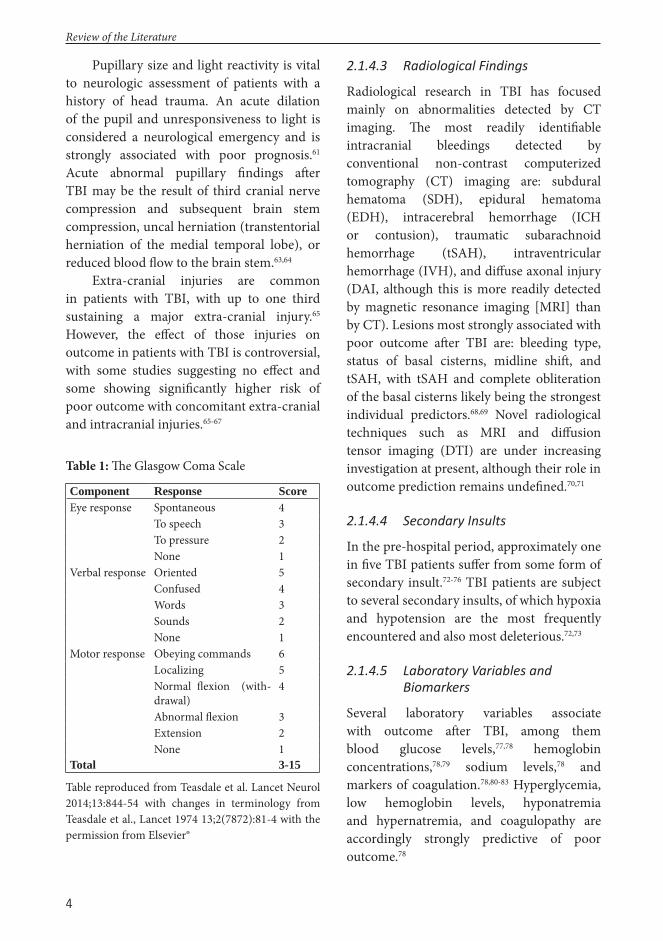
Level of consciousness aft er injury is a major determinant of TBI severity and oft en assessed by the Glasgow Coma Score (GCS).57 Th e GCS is traditionally used to classify TBI into mild (GCS 13-15), moderate (GCS 9-12), and severe (GCS 3-8). Although debate exists over whether GCS 13 should be classifi ed as moderate or mild, this stratifi cation system has been used for the last 40 years.58,59 Th e GCS was introduced in 1974 as a tool for “repeated bedside assessment” to detect “changing states” and measuring “duration of coma” in the fi rst 24 hours of observation in neurosurgical units.57 Th e GCS consists of three components: eye response, verbal response, and motor response, which are added together for a score from 3 to 15 (Table 1). Th e abbreviation ‘GCS’ is used inconsistently in the literature, as it may refer to both the individual components of the GCS (Glasgow Coma Scale) and the total score (Glasgow Coma Score).59 Th e scale is probably more useful for the individual patient and the score to summarize large groups of patients. Notably, GCS was never intended to be used in trauma or emergency medicine or even for its three components to be added together into a sum; despite the authors’ objections, it has been used in those manners ever since its introduction.59,60 However, the strong relationship between GCS and outcome aft er TBI and its simplicity still favors its use,59,61 although some contrary conclusions have been proposed.62
Review of the Literature
4
Pupillary size and light reactivity is vital to neurologic assessment of patients with a history of head trauma. An acute dilation of the pupil and unresponsiveness to light is considered a neurological emergency and is strongly associated with poor prognosis.61 Acute abnormal pupillary fi ndings aft er TBI may be the result of third cranial nerve compression and subsequent brain stem compression, uncal herniation (transtentorial herniation of the medial temporal lobe), or reduced blood fl ow to the brain stem.63,64
Extra-cranial injuries are common in patients with TBI, with up to one third sustaining a major extra-cranial injury.65 However, the eff ect of those injuries on outcome in patients with TBI is controversial, with some studies suggesting no eff ect and some showing signifi cantly higher risk of poor outcome with concomitant extra-cranial and intracranial injuries.65-67
Table 1: Th e Glasgow Coma Scale
Component Response ScoreEye response Spontaneous 4 To speech 3 To pressure 2 None 1Verbal response Oriented 5 Confused 4 Words 3 Sounds 2 None 1Motor response Obeying commands 6 Localizing 5 Normal fl exion (with-
drawal)4
Abnormal fl exion 3 Extension 2 None 1Total 3-15
Table reproduced from Teasdale et al. Lancet Neurol 2014;13:844-54 with changes in terminology from Teasdale et al., Lancet 1974 13;2(7872):81-4 with the permission from Elsevier®
2.1.4.3 Radiological Findings
Radiological research in TBI has focused mainly on abnormalities detected by CT imaging. Th e most readily identifi able intracranial bleedings detected by conventional non-contrast computerized tomography (CT) imaging are: subdural hematoma (SDH), epidural hematoma (EDH), intracerebral hemorrhage (ICH or contusion), traumatic subarachnoid hemorrhage (tSAH), intraventricular hemorrhage (IVH), and diff use axonal injury (DAI, although this is more readily detected by magnetic resonance imaging [MRI] than by CT). Lesions most strongly associated with poor outcome aft er TBI are: bleeding type, status of basal cisterns, midline shift , and tSAH, with tSAH and complete obliteration of the basal cisterns likely being the strongest individual predictors.68,69 Novel radiological techniques such as MRI and diff usion tensor imaging (DTI) are under increasing investigation at present, although their role in outcome prediction remains undefi ned.70,71
2.1.4.4 Secondary Insults
In the pre-hospital period, approximately one in fi ve TBI patients suff er from some form of secondary insult.72-76 TBI patients are subject to several secondary insults, of which hypoxia and hypotension are the most frequently encountered and also most deleterious.72,73
2.1.4.5 Laboratory Variables and Biomarkers
Several laboratory variables associate with outcome aft er TBI, among them blood glucose levels,77,78 hemoglobin concentrations,78,79 sodium levels,78 and markers of coagulation.78,80-83 Hyperglycemia, low hemoglobin levels, hyponatremia and hypernatremia, and coagulopathy are accordingly strongly predictive of poor outcome.78
Review of the Literature
5
Identifying laboratory abnormalities as predictors of outcome is important, as these can oft en be corrected. Confronting the question of causality however, is crucial before actively correcting abnormal laboratory values. For example, high levels of blood glucose concentrations independently predict poor outcome aft er TBI.77,78 However, recent evidence suggest that early hyperglycemia aft er TBI might be a benefi cial stress response, and thus, actively lowering blood glucose levels in early phases may reduce brain glucose availability and increase secondary brain injury.84-87
As of today, there is no accurate biomarker of TBI,88,89 though interest in biomarkers has been increasing in recent years. An accurate biomarker for mild TBI to establish diagnosis or for moderate to severe TBI to determine extent of injury would be of great clinical use, although no such biomarker has yet been identifi ed.88,89 Promising biomarkers for detecting brain injury include glial fi brillary acidic protein (GFAP), ubiquitin C-terminal hydrolase-L1 (ICH-L1), alfa-II spectric breakdown product (SBDP145), and S100B and neuron specifi c enolase, although few of these are routinely used in the clinical setting.90,91
2.1.5 Pa ent Outcome
Outcome aft er severe TBI is poor; about one in three patients dies and most survivors are left with severe lifelong disabilities.9,29,43,52 Furthermore, survivors of severe TBI face prolonged rehabilitation times, causing signifi cant patient and family suff ering as well as enormous economic costs.5,6,8,31 A recent large meta-analysis, including more than 140,000 patients from over 200 case series and a time period of almost 125 years (1885-2006), showed a general improvement in outcome aft er TBI.92 Notably, though, the improvement stagnated in 1990, suggesting no advances in patient outcome over the last quarter-century. Nonetheless, several studies
aft er 1990 have reported improvements in patient outcome as a result of, for instance, TBI care guideline development93,94 and aggressive neurointensive treatment regimes.95-98
A common and biased interpretation of improved patient outcome appears when comparing outcomes from recent randomized controlled trials (RCTs) with older observational studies. Such comparisons should be interpreted with great caution. Observational studies tend to have much broader inclusion criteria than RCTs, where patients with the most extreme prognosis (e.g. bilaterally dilated pupils, GCS 3, elderly patients) are oft en excluded. Th us, it is natural that patient outcome is better in RCTs than observational studies. However, the epidemiological shift of TBI patients towards older and sicker populations might, on the other hand, increase rates of poor outcome in observational studies.37 Hence, there are oft en substantial diff erences in patient case-mix between observational studies and RCTs, confounding inter-study comparisons.
2.2 Prognos c Models2.2.1 Defi ni on
A prognostic model is a statistical model, or a mathematical equation, that includes two or more prognostic factors, or variables, to calculate the probability of a pre-defi ned outcome. In medical research, the outcome is oft en dichotomized; examples include predicting the probability of being alive or dead at a certain time point, a tumor being benign or malign, or the risk of an adverse event occurring.
Diff erent terms for ‘prognostic models’ may be used, like ‘prediction model,’ ‘scoring system,’ or simply ‘score,’ oft en depending on the term used in the originating paper. In the present study, original terms are used when discussing individual models, but for general discussion the term ‘prognostic model’ or simply ‘model’ is used.
Review of the Literature
6
2.2.2 Development
Ideally, the factors used to create a prognostic model should all individually be statistically and clinically associated with the outcome, although this is not always the case. Th us, included variables should be chosen carefully; it is recommended to start with selected candidate variables, known from previous studies, aft er which variables from the own population are added.99 Generally, a higher number of variables improve the model’s explanatory eff ect, but using more variables also increases the risk of overfi tting and decreases clinical applicability. Accordingly, more than one researcher has suggested that a good model should include no more than fi ve to seven predictors.99,100
To create a prognostic model, complex statistical techniques are oft en necessary.
Th e most commonly used statistical method is logistic regression,99,101 but others include: discriminant analysis, artifi cial neural networks, and recursive partitioning. Logistic regression, however, has some key advantages over the other techniques, as it does not require variables to be normally distributed, linearly related, or to have equal within-group variances. Furthermore, logistic regression handles both categorical variables and continuous variables, and gives us easily interpretable outputs in the form of regression coeffi cients and odds ratios. Recursive partitioning, on the other hand, has the advantage of being easy to grasp visually, facilitating clinical applicability, but suff ers from problems of overfi tting and categorization of continuous data.102 Neural networking mimics the information processing of neurons in the brain and
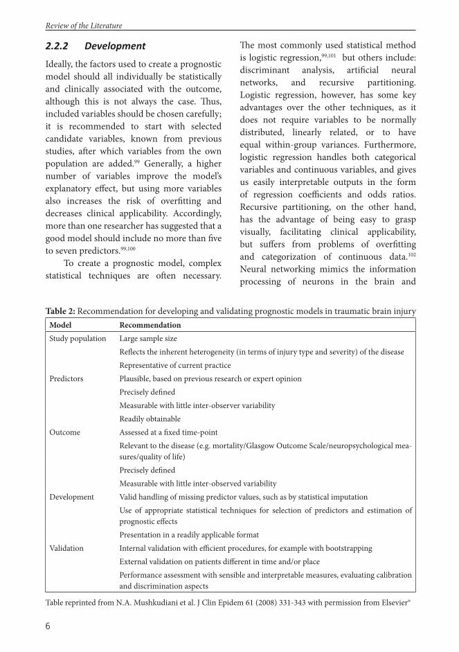
Table 2: Recommendation for developing and validating prognostic models in traumatic brain injuryModel RecommendationStudy population Large sample size
Refl ects the inherent heterogeneity (in terms of injury type and severity) of the diseaseRepresentative of current practice
Predictors Plausible, based on previous research or expert opinionPrecisely defi nedMeasurable with little inter-observer variabilityReadily obtainable
Outcome Assessed at a fi xed time-pointRelevant to the disease (e.g. mortality/Glasgow Outcome Scale/neuropsychological mea-sures/quality of life)Precisely defi nedMeasurable with little inter-observed variability
Development Valid handling of missing predictor values, such as by statistical imputationUse of appropriate statistical techniques for selection of predictors and estimation of prognostic eff ectsPresentation in a readily applicable format
Validation Internal validation with effi cient procedures, for example with bootstrappingExternal validation on patients diff erent in time and/or placePerformance assessment with sensible and interpretable measures, evaluating calibration and discrimination aspects
Table reprinted from N.A. Mushkudiani et al. J Clin Epidem 61 (2008) 331-343 with permission from Elsevier®
Review of the Literature
7
produces prognostic models. Th ese models are possibly superior to models created by logistic regression, in terms of statistical performance, but are also more complex.103 Th e complexity of neural networking is also its weakness, limiting its use. By combining neural networking with logistic regression, model complexity can be reduced while maintaining predictive accuracy, although this technique has yet to gain popularity.103-105
Still, more important than the statistical method is the selection of predictors.11,15,99,106,107 A systematic review of methodological improvements for prognostic models in TBI established recommendations for their development and validation (Table 2)99.
2.2.3 Internal Valida on
Internal validation refers to testing the model for reproducibility in a dataset similar to the one used to develop the model. All prognostic models should be at least internally validated before introduction in order to adjust for optimism,15 which is the term applied when the model performs worse than expected in a new dataset.106 Split-sample, cross-validation, jackknifi ng, and bootstrapping are the most common statistical techniques for internal validation.108
Th e split-sample technique is probably the most simple and straightforward method for internal validation.109 Th e dataset is randomly divided into two groups, making the groups similar but independent; one group is used for the development of the model (development set) and the other group is used for validation of the model (validation set). In this way the model is tested on similar but still independent data. Th e split-sample technique, however, heavily depends on sample size and requires adequately large patient groups. Furthermore, splitting data always results in lost data, and thus, reduces the statistical power of the model.108
Th e cross-validation technique is an extension of the split-sample technique, where patients are again randomly divided into two parts, one for model development and the other for validation.110 In cross-validation, this procedure is, however, repeated with the model now developed in the other dataset and validated in the original development dataset. Th e average of these two stages is taken as an estimate of performance. Th e cross-validation technique can further be extended to taking 90% of the data for model development and 10% for validation. Th e procedure is repeated for a total of ten iterations and the average represents the performance estimate. Th e most extreme variation of the cross-validation technique is the jackknife technique, where one patient is left out at a time, and the test is repeated hundreds or thousands of times.111
Th e bootstrap technique has been recognized as the most statistically robust method of internal validation.108 Bootstrapping is a computer-intensive resampling technique that draws random samples with replacements from the original dataset.111,112 Bootstrapping follows the logic of ‘the population is to the sample as the sample is to the bootstrap samples.’113 Th e bootstrap technique may be applied to a variety of performance measures, including the AUC, calibration slopes, and Nagelkerke R2 (see below). To assess the internal validity of a model using the bootstrap technique, an optimism-corrected performance is calculated as follows:114
Optimism corrected performance= apparent performance in sample-
optimism,where optimism= bootstrap performance-test performance
Review of the Literature
8
2.2.4 External Valida on
A prognostic model generally performs better on the dataset from which it was derived than on new data.115 External validation aims to assess the performance of a prognostic model in a diff erent, but plausibly related, population. External validation is essential to support the generalizability of prognostic models and to provide evidence that the model does in fact accurately predict outcomes.115,116 Th ere are several types of external validation variations, whether methodological (temporal, geographical, fully independent) or characteristic (prospective testing with more recent patients, multi-site testing, other investigators at another site).115-
118
2.2.5 Performance assessment 2.2.5.1 Discrimina on
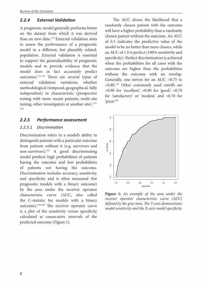
Discrimination refers to a model’s ability to distinguish patients with a particular outcome from patients without it (e.g. survivors and non-survivors).101 A good discriminating model predicts high probabilities of patients having the outcome and low probabilities of patients not having the outcome. Discrimination includes accuracy, sensitivity, and specifi city and is oft en measured (for prognostic models with a binary outcome) by the area under the receiver operator characteristic curve (AUC, also called the C-statistic for models with a binary outcome).119,120 Th e receiver operator curve is a plot of the sensitivity versus specifi city calculated at consecutive intervals of the predicted outcome (Figure 1).
Th e AUC shows the likelihood that a randomly chosen patient with the outcome will have a higher probability than a randomly chosen patient without the outcome. An AUC of 0.5 indicates the predictive value of the model to be no better than mere chance, while an AUC of 1.0 is perfect (100% sensitivity and specifi city). Perfect discrimination is achieved when the probabilities for all cases with the outcome are higher than the probabilities without the outcome with no overlap. Generally, one strives for an AUC >0.75 to >0.80.119 Other commonly used cutoff s are >0.90 for ‘excellent,’ >0.80 for ‘good,’ >0.70 for ‘satisfactory’ or ‘modest,’ and <0.70 for ‘poor.’101
Figure 1: An example of the area under the receiver operator characteristic curve (AUC) defi ned by the grey area. Th e Y-axis demonstrates model sensitivity and the X-axis model specifi city
Specificity
Sen
sitiv
ity
0.0
0.2
0.4
0.6
0.8
1.0
1.0 0.8 0.6 0.4 0.2 0.0
Review of the Literature
9
2.2.5.2 Calibra on
Model calibration refers to the concordance between predicted and observed outcomes over the whole risk spectra.101 Calibration testing is oft en overlooked in prognostic research, where many studies focus mainly on discrimination measures.121 Discrimination is considered more important when predicting outcome for the individual patient, but for risk stratifi cation and trial enrollment, calibration is more important than discrimination.121,122
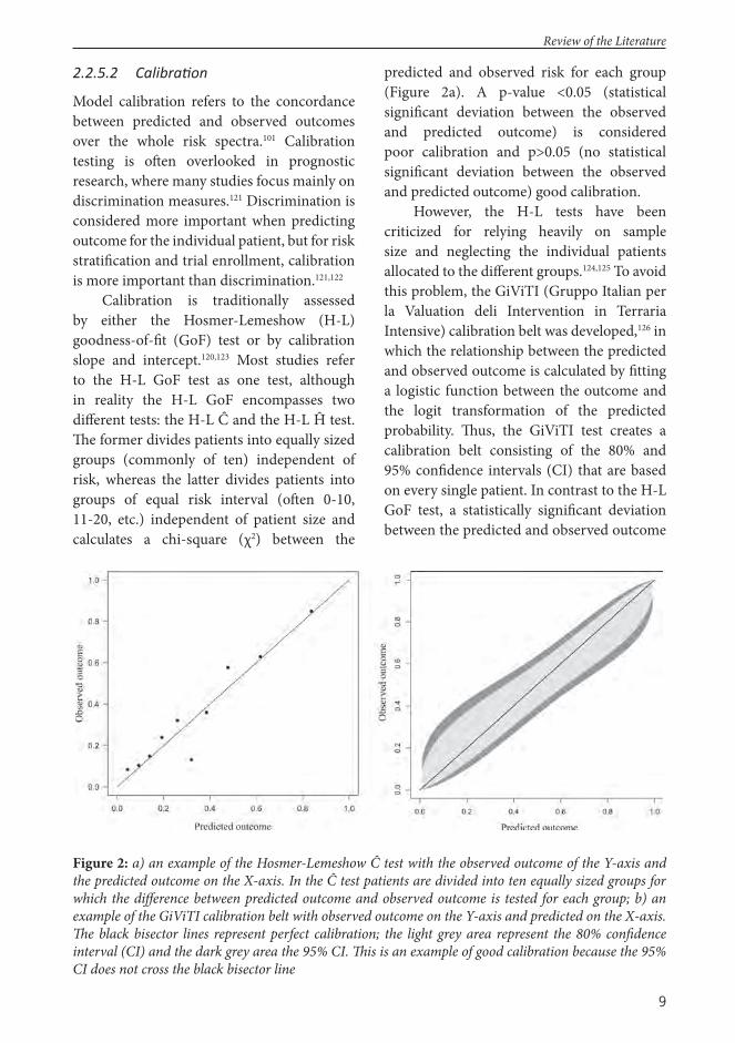
Calibration is traditionally assessed by either the Hosmer-Lemeshow (H-L) goodness-of-fi t (GoF) test or by calibration slope and intercept.120,123 Most studies refer to the H-L GoF test as one test, although in reality the H-L GoF encompasses two diff erent tests: the H-L Ĉ and the H-L Ĥ test. Th e former divides patients into equally sized groups (commonly of ten) independent of risk, whereas the latter divides patients into groups of equal risk interval (oft en 0-10, 11-20, etc.) independent of patient size and calculates a chi-square (χ2) between the
predicted and observed risk for each group (Figure 2a). A p-value <0.05 (statistical signifi cant deviation between the observed and predicted outcome) is considered poor calibration and p>0.05 (no statistical signifi cant deviation between the observed and predicted outcome) good calibration.
However, the H-L tests have been criticized for relying heavily on sample size and neglecting the individual patients allocated to the diff erent groups.124,125 To avoid this problem, the GiViTI (Gruppo Italian per la Valuation deli Intervention in Terraria Intensive) calibration belt was developed,126 in which the relationship between the predicted and observed outcome is calculated by fi tting a logistic function between the outcome and the logit transformation of the predicted probability. Th us, the GiViTI test creates a calibration belt consisting of the 80% and 95% confi dence intervals (CI) that are based on every single patient. In contrast to the H-L GoF test, a statistically signifi cant deviation between the predicted and observed outcome
Figure 2: a) an example of the Hosmer-Lemeshow Ĉ test with the observed outcome of the Y-axis and the predicted outcome on the X-axis. In the Ĉ test patients are divided into ten equally sized groups for which the diff erence between predicted outcome and observed outcome is tested for each group; b) an example of the GiViTI calibration belt with observed outcome on the Y-axis and predicted on the X-axis. Th e black bisector lines represent perfect calibration; the light grey area represent the 80% confi dence interval (CI) and the dark grey area the 95% CI. Th is is an example of good calibration because the 95% CI does not cross the black bisector line
Review of the Literature

10
occurs when the 95% CIs do not overlap the bisector line, indicating perfect calibration (Figure 2b). In this sense it is possible to identify visually areas of poor calibration and determine its direction (model overprediction or underprediction).
Th e calibration slope is the regression coeffi cient β in a logistic regression model with the linear predictor as the only covariate: observed = α + β linear predictor, where β is the intercept.127 Well calibrated models have a slope (α) of 1 and an intercept (β) of 0.128 Overpredicting models have a slope under 1, with the model tending to underestimate the incidence of outcome in low-risk patients and overpredict it in high-risk patients. Conversely, if the slope is greater than 1, the predicted risks are not suffi ciently diff erentiated across the risk strata.120 An intercept under 0 indicates that the predicted risks are systematically too high and an intercept over 0 indicates that the predicted risks are systematically too low.
2.2.5.3 Overall performance measures
Th e explanatory variation is considered an overall measure of model performance, including both discrimination and calibration. In linear regression, the R2 summarizes the grade of explanation of the dependent variables (or covariates) with the independent variable (the outcome). Larger values indicate a higher degree of explanation, with an R2 of 1.0 indicating that the model explains for 100% of the outcome, and an R2 of 0.0 meaning that the model explains for 0% of the outcome. In logistic regression, however, it is not possible to calculate a single R2 that has all the characteristics of the R2 in the linear regression. Th is has led to the development of several ‘pseudo R2’ measures, of which the Nagelkerke R2 test is probably the most frequently used.129,130 Th e Nagelkerke R2 is a variation of the earlier Cox and Snell R2 test for logistic regression, but in
contrast to it, the Nagelkerke R2 ranges from 0 to 1 (0-100%), to better mimic the ‘real’ linear regression R2.129
2.2.5.4 Net Reclassifi ca on Index
Th e added value of a variable to a prognostic model is oft en measured by comparing diff erences in AUC. Yet, for a model’s AUC to increase signifi cantly, the independent association between the new variable and the outcome has to be very strong. In other words, a predictor may be signifi cant without improving AUC, which might lead to neglecting important variables.121,131 In response, Pencina et al. introduced the Net Reclassifi cation Index (NRI).132 Th e NRI is a novel, more sensitive, statistical technique to measure the added value of a predictor when added to a prognostic model by reclassifi cation tables (which require a priori meaningful risk categories) or a continuous test. Th e NRI show how many patients are better classifi ed because of the added predictor, with an associated p-value.
2.2.6 Customiza on
All prognostic models become outdated over time.15,114 To improve the performance of a prognostic model and make it applicable to new settings, it may be customized. Customization aims to improve the performance of a particular prognostic model in a plausibly related but diff erent population from the original development population. Th ere are two methods for model customization, known as fi rst and second level customization.133-135
First level customization involves fi tting a new logistic regression equation with the observed outcome as the dependent variable and the logit-transformed original prediction as the independent variable. Th e infl uence of the individual variables does not change; rather, they recalibrate their joint eff ects on outcome.
Review of the Literature
11
Second level customization involves fi tting all the original predictors into a logistic regression model with the outcome as the dependent variable, and has been shown to be more eff ective than the fi rst level variety. Th us, given suffi cient sample size, second level customization is preferable.134 In general, customization does not aff ect discrimination but instead improves calibration.134,135
2.2.7 Applica ons 2.2.7.1 Quality Audits
It is impossible to improve results that are unknown; the aim of quality management is the delivery of improved care by monitoring clinical performance. Since the 1980s, risk-adjusted mortality rates have served as an important measure of hospital care quality.19,23 Comparing the observed outcome with the expected outcome has been shown to be a feasible method for improving quality of trauma and intensive care.22,136-139 For example, the Trauma Audit & Research Network (TARN) in the UK annually presents case-mix adjusted survival rates (oft en referred to as SMR for Standardized Mortality Rate) from their participating hospitals publicly at https://www.tarn.ac.uk/. Public comparisons of adjusted survival rates are, however, not without problems and it is important to know the limitation of such comparisons. Two things are absolutely vital for adjusted survival rates: 1) an accurate prognostic model and 2) a proper outcome measure.140,141 In TBI, trauma, and intensive care research, hospital mortality is the most commonly used outcome measure (and also in the TARN database).22,142-145 Hospital mortality, however, is known to underestimate mortality rates substantially in severely ill patients and is thus a source of biased results.142,143 Th is is a particular problem in patients with moderate to severe TBI, because as many as one third of hospital survivors die within six months following hospital discharge.29,50 Moreover,
diff erences in discharge policies vary not only between countries but also within them, and adjusting for those diff erences with a fi xed-time outcome measure is absolutely essential, lest hospitals feel pressured to discharge severely ill patients more rapidly to avoid them from dying to keep up their stats.140,143 Furthermore, using mortality rates as the primary outcome measure aft er TBI neglects important aspects of patient outcome, such as functional and neurological recovery and quality of life.
2.2.7.2 Clinical Prac ce
Prognostic models aim to aid, not replace, clinicians in estimating patient prognosis. Prognoses provided by a good prognostic model are probably more accurate than the predictions of an individual clinician.146 Prognostics estimation is a natural feature of every clinical environment when making treatment decisions (e.g. ‘will this patient benefi t from craniotomy?’), allocating resources (e.g. ‘will this patient benefi t from intensive care?’), and informing relatives. Decision making should, however, never be based solely upon a prognostic model.19
Th e value of prognosis estimation in the management of TBI was already demonstrated 30 years ago.147 In a survey, the vast majority of neurosurgeons thought that prognosis estimation was especially important when deciding which patients need decompressive craniectomy, intensive care, ICP monitoring and aggressive ICP treatment, and when treatment should be withdrawn.148 Moreover, Murray and Teasdale149 showed that computer-based prognosis estimation of patients with TBI increased the rate of intubation and ventilation, ICP monitoring, and osmotic administration in those with an estimated good prognosis but reduced them in patients with an estimated poor prognosis. It is important to note that the predictions did not alter decisions to provide or restrict
Review of the Literature
12
treatment and did not aff ect outcomes. Th us, using prognostic models as a part of the routine clinical evaluation of TBI is not only reasonable and practical but also becoming increasingly important in the era of personalized medicine, where decisions are deeply connected with individual patient characteristics.
2.2.7.3 Research
Th e gold standard of modern evidence-based medicine is RCTs. RCTs are, however, increasingly hard to conduct in TBI research. For example, it might be considered unethical to randomize TBI patients who require intensive care to ordinary ward care, just for research purposes. Moreover the nature of TBI (a life-threatening event with unconsciousness) makes patient consent as a rule impossible to obtain, leaving that decision to the families.
Th e failure of numerous clinical trials in TBI has been attributed to the broad heterogeneity of TBI.150 To limit heterogeneity, clinical trials oft en apply strict enrollment criteria that decrease result generalizability and thus weaken statistical power. Study generalizability and statistical power can, however, be improved by using prognostic models.26 Th is can be achieved by: baseline characteristic selection, prognostic targeting, and covariate adjustment. Of these methods covariate adjustment has proven most robust.151 Pre-specifi ed covariate adjustment has in simulation studies increased statistical effi ciency by 30% in observational studies and 16% in RCTs.151,152 Moreover, the use of prognostic models also allows for more sophisticated statistical analyses, such as the sliding dichotomy and proportional odds approaches, further increasing statistical power up to 50% when combined with prespecifi ed covariate adjustment.26,153,154 Increased study power also provides signifi cant benefi ts from the
fi nancial perspective in terms of shortened study duration and, thus, study costs.
RCTs are, however, not the only way to produce scientifi c evidence, especially in TBI.25 In fact, modern clinical practice in TBI is largely based on guideline development and results from observational studies rather than results from RCTs.25 Accordingly, there is currently a re-orientation of TBI research from RCTs towards more international observational collaboration studies and comparative eff ectiveness research (CER).18,25,155 CER is designed to inform healthcare decisions by providing evidence of eff ectiveness, benefi ts, and harms of diff erent treatment strategies.156 Th e Institute of Medicine (IOM) defi nes CER as “the generation and synthesis of evidence that compares the benefi ts and harms of alternative methods to prevent, diagnose, treat, and monitor for a clinical condition or to improve delivery of care”. In contrast to an RCT, where the main goal is to assess the effi cacy of a specifi c intervention on patient outcome, CER aims to assess the eff ectiveness of diff erent treatment strategies by measuring diff erences in patient outcome. CER generally allows much broader patient inclusion criteria than RCTs, making them more applicable to bedside medicine. Still, an accurate prognostic model is essential for proper CER in order to adjust for heterogeneity.18,25
2.3 Trauma c Brain Injury Models
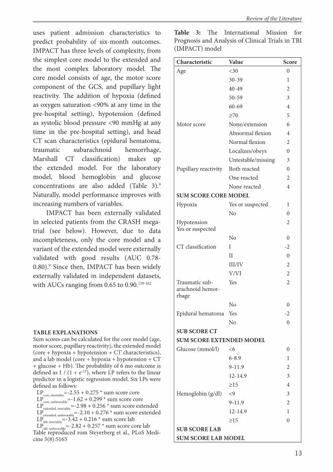
2.3.1 IMPACT
Th e International Mission for Prognosis and Analysis of Clinical Trials (IMPACT) study is the result of pooled data from eight RCTs and three observational studies conducted between 1984 and 1997 (Table 11).157,158 Th e IMPACT prognostic models (simply IMPACT below) were introduced in 2008 and are freely available online (http://www.tbi-impact.org/?p=impact/calc). IMPACT
Review of the Literature
13
uses patient admission characteristics to predict probability of six-month outcomes. IMPACT has three levels of complexity, from the simplest core model to the extended and the most complex laboratory model. Th e core model consists of age, the motor score component of the GCS, and pupillary light reactivity. Th e addition of hypoxia (defi ned as oxygen saturation <90% at any time in the pre-hospital setting), hypotension (defi ned as systolic blood pressure <90 mmHg at any time in the pre-hospital setting), and head CT scan characteristics (epidural hematoma, traumatic subarachnoid hemorrhage, Marshall CT classifi cation) makes up the extended model. For the laboratory model, blood hemoglobin and glucose concentrations are also added (Table 3).9 Naturally, model performance improves with increasing numbers of variables.
IMPACT has been externally validated in selected patients from the CRASH mega-trial (see below). However, due to data incompleteness, only the core model and a variant of the extended model were externally validated with good results (AUC 0.78-0.80).9 Since then, IMPACT has been widely externally validated in independent datasets, with AUCs ranging from 0.65 to 0.90.159-162
Table 3: Th e International Mission for Prognosis and Analysis of Clinical Trials in TBI (IMPACT) model
Characteristic Value ScoreAge <30 0
30-39 140-49 250-59 360-69 4≥70 5
Motor score None/extension 6Abnormal fl exion 4Normal fl exion 2Localizes/obeys 0Untestable/missing 3
Pupillary reactivity Both reacted 0One reacted 2None reacted 4
SUM SCORE CORE MODELHypoxia Yes or suspected 1
No 0HypotensionYes or suspected
2
No 0CT classifi cation I -2
II 0III/IV 2V/VI 2
Traumatic sub-arachnoid hemor-rhage
Yes 2
No 0Epidural hematoma Yes -2
No 0SUB SCORE CTSUM SCORE EXTENDED MODELGlucose (mmol/l) <6 0
6-8.9 19-11.9 212-14.9 3≥15 4
Hemoglobin (g/dl) <9 39-11.9 212-14.9 1≥15 0
SUB SCORE LABSUM SCORE LAB MODEL
TABLE EXPLANATIONSSum scores can be calculated for the core model (age, motor score, pupillary reactivity), the extended model (core + hypoxia + hypotension + CT characteristics), and a lab model (core + hypoxia + hypotension + CT + glucose + Hb). Th e probability of 6 mo outcome is defi ned as 1 / (1 + e-LP), where LP refers to the linear predictor in a logistic regression model. Six LPs were defi ned as follows:
LPcore, mortality=-2.55 + 0.275 * sum score coreLPcore, unfavorable=-1.62 + 0.299 * sum score coreLPextended, mortality=-2.98 + 0.256 * sum score extendedLPextended, unfavorable=-2.10 + 0.276 * sum score extendedLPlab, mortality=-3.42 + 0.216 * sum score labLPlab, unfavorable=-2.82 + 0.257 * sum score core lab
Table reproduced rom Steyerberg et al., PLoS Medi-cine 5(8):5165
Review of the Literature
14
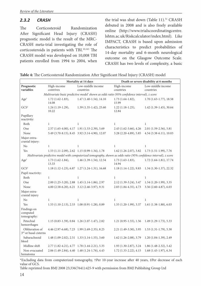
2.3.2 CRASH
Th e Corticosteroid Randomization Aft er Signifi cant Head Injury (CRASH) prognostic model is the result of the MRC-CRASH meta-trial investigating the role of corticosteroids in patients with TBI.10,163 Th e CRASH model was developed on 10,008 TBI patients enrolled from 1994 to 2004, when
the trial was shut down (Table 11).51 CRASH debuted in 2008 and is also freely available online (http://www.trialscoordinatingcentre.lshtm.ac.uk/Riskcalculator/index.html). Like IMPACT, CRASH is based upon admission characteristics to predict probabilities of 14-day mortality and 6-month neurological outcome on the Glasgow Outcome Scale. CRASH has two levels of complexity, a basic
Table 4: Th e Corticosteroid Randomization Aft er Signifi cant Head Injury (CRASH) modelMortality at 14 days Death or severe disability at 6 months
Prognostic variables
High-income contrives
Low-middle income countries
High-income countries
Low-middle income countries
Multivariate basic predictive model* shown as odds ratio (95% confi dence interval), z scoreAge† 1.72 (1.62-1.83),
14.081.47 (1.40-1.54), 14.10 1.73 (1.64-1.82),
15.991.70 (1.63-1.77), 18.58
GCS‡ 1.24 (1.19-1.29), 10.22
1.39 (1.35-1.42), 25.60 1.22 (1.18-1.25), 12.84
1.42 (1.39-1.45), 30.64
Pupillary reactivity: Both 1 1 1 1 One 2.57 (1.65-4.00), 4.17 1.91 (1.53-2.39), 5.69 2.43 (1.62-3.66), 4.26 2.01 (1.59-2.56), 5.81 None 5.49 (3.70-8.15), 8.45 3.92 (3.14-4.90), 12.07 3.28 (2.20-4.89), 5.85 4.54 (3.38-6.11), 10.03Major extra-cranial injury: No 1 1 1 1 Yes 1.53 (1.11-2.09), 2.62 1.15 (0.99-1.34), 1.78 1.62 (1.26-2.07), 3.82 1.73 (1.51-1.99), 7.76
Multivariate predictive model with computerized tomography, shown as odds ratio (95% confi dence interval), z scoreAge† 1.73 (1.62-1.84),
13.331.46 (1.39-1.54), 12.54 1.73 (1.63-1.83),
14.941.72 (1.64-1.81), 17.74
GCS‡ 1.18 (1.12-1.23), 6.87 1.27 (1.24-1.31), 16.68 1.18 (1.14-1.22), 9.83 1.34 (1.30-1.37), 22.32Pupil reactivity: Both 1 1 1 1 One 2.00 (1.25-3.20), 2.88 1.45 (1.14-1.86), 2.97 2.12 (1.39-3.24), 3.47 1.54 (1.20-1.99), 3.35 None 4.00 (2.58-6.20), 6.21 3.12 (2.46-3.97), 9.31 2.83 (1.84-4.35), 4.73 3.56 (2.60-4.87), 6.03Major extra-cranial injury No 1 1 1 1 Yes 1.53 (1.10-2.13), 2.53 1.08 (0.91-1.28), 0.89 1.55 (1.20-1.99), 3.37 1.61 (1.38-1.88), 6.03Findings on computed tomography: Petechial hemorrhages
1.15 (0.83-1.59), 0.84 1.26 (1.07-1.47), 2.82 1.21 (0.95-1.55), 1.56 1.49 (1.29-1.73), 5.33
Obliteration of 3rd or basal cisterns
4.46 (2.97-6.68), 7.23 1.99 (1.69-2.35), 8.25 2.21 (1.49-3.30), 3.95 1.53 (1.31-1.79), 5.30
Subarachnoid bleed
1.48 (1.09-2.02), 2.51 1.33 (1.14-1.55), 3.60 1.62 (1.26-2.08), 3.79 1.20 (1.04-1.39), 2.49
Midline shift 2.77 (1.82-4.21), 4.77 1.78 (1.44-2.21), 5.35 1.93 (1.30-2.87), 3.24 1.86 (1.48-2.32), 5.42 Non-evacuated hematoma
2.06 (1.49-2.84), 4.40 1.48 (1.24-1.76), 4.43 1.72 (1.33-2.22), 4.15 1.68 (1.43-1.97), 6.34
*Excluding data from computerized tomography, †Per 10-year increase aft er 40 years, ‡Per decrease of each value of GCS. Table reprinted from BMJ 2008 23;336(7641):425-9 with permission from BMJ Publishing Group Ltd
Review of the Literature
15
model, and an extended version with CT scan characteristics. Th e basic model includes age, GCS, pupillary light reaction, and presence of major extra-cranial injury. CT scan characteristics added for the extended model are presence of petechial hemorrhage, status of third ventricle and basal cisterns, presence of tSAH, midline shift , and mass lesion. Moreover, CRASH is calibrated diff erently for patients from low-and-middle income countries and high-income countries (Table 4). Similar to IMPACT, external validation studies of CRASH have yielded good results.10,164-166
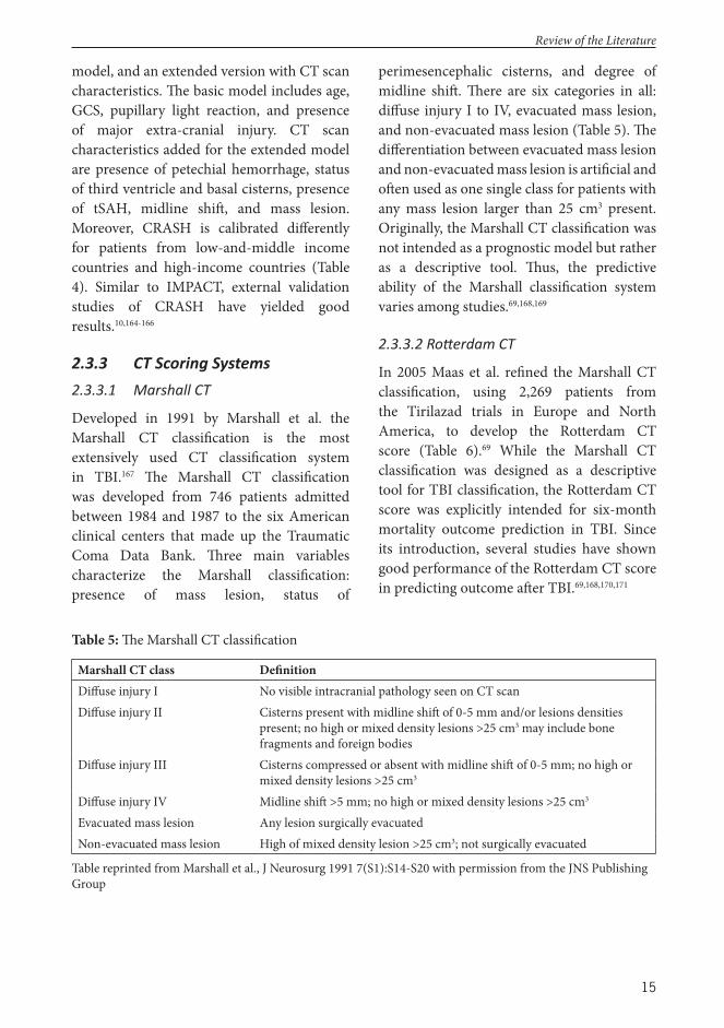
2.3.3 CT Scoring Systems2.3.3.1 Marshall CT
Developed in 1991 by Marshall et al. the Marshall CT classifi cation is the most extensively used CT classifi cation system in TBI.167 Th e Marshall CT classifi cation was developed from 746 patients admitted between 1984 and 1987 to the six American clinical centers that made up the Traumatic Coma Data Bank. Th ree main variables characterize the Marshall classifi cation: presence of mass lesion, status of
perimesencephalic cisterns, and degree of midline shift . Th ere are six categories in all: diff use injury I to IV, evacuated mass lesion, and non-evacuated mass lesion (Table 5). Th e diff erentiation between evacuated mass lesion and non-evacuated mass lesion is artifi cial and oft en used as one single class for patients with any mass lesion larger than 25 cm3 present. Originally, the Marshall CT classifi cation was not intended as a prognostic model but rather as a descriptive tool. Th us, the predictive ability of the Marshall classifi cation system varies among studies.69,168,169
2.3.3.2 Ro erdam CT
In 2005 Maas et al. refi ned the Marshall CT classifi cation, using 2,269 patients from the Tirilazad trials in Europe and North America, to develop the Rotterdam CT score (Table 6).69 While the Marshall CT classifi cation was designed as a descriptive tool for TBI classifi cation, the Rotterdam CT score was explicitly intended for six-month mortality outcome prediction in TBI. Since its introduction, several studies have shown good performance of the Rotterdam CT score in predicting outcome aft er TBI.69,168,170,171
Table 5: Th e Marshall CT classifi cation
Marshall CT class Defi nitionDiff use injury I No visible intracranial pathology seen on CT scanDiff use injury II Cisterns present with midline shift of 0-5 mm and/or lesions densities
present; no high or mixed density lesions >25 cm3 may include bone fragments and foreign bodies
Diff use injury III Cisterns compressed or absent with midline shift of 0-5 mm; no high or mixed density lesions >25 cm3
Diff use injury IV Midline shift >5 mm; no high or mixed density lesions >25 cm3
Evacuated mass lesion Any lesion surgically evacuatedNon-evacuated mass lesion High of mixed density lesion >25 cm3; not surgically evacuated
Table reprinted from Marshall et al., J Neurosurg 1991 7(S1):S14-S20 with permission from the JNS Publishing Group
Review of the Literature

16
Table 6: Th e Rotterdam CT score
Variable ScoreBasal cisterns Normal 0 Compressed 1 Absent 2Midline shift No shift or ≤5 mm 0 Shift >5mm 1Epidural mass lesion Present 0 Absent 1Intraventricular blood or traumatic subarachnoid hemorrhage Absent 0 Present 1Sumscore +1
Table reprinted from Maas et al., Neurosurgery 2005 57(6):1173-82
2.4 Trauma Scoring Systems2.4.1 Anatomical Trauma Scoring
Systems
Th e Injury Severity Score (ISS) and the New Injury Severity Score (NISS) are anatomical scoring systems providing an overall indicator of patient injury severity.172,173 In both systems the body is divided into six regions (head, face, chest, abdomen, extremities, and external) and each body region injury is assigned a score based on injury severity from 0 (no injury) to 6 (unsurvivable injury) on the Abbreviated Injury Scale (AIS).174 Th e ISS and NISS both use a range from 0 to 75. A patient with an AIS of 6 in any body region automatically gets a total ISS of 75; otherwise ISS is calculated by the sum of squares of the single highest AIS in each of the three most severely injured body regions. Th e NISS is a modifi cation of the ISS and calculated by the sum of squares of the patient’s three most severe AIS injuries, regardless of body region.
2.4.2 Physiological Trauma Scoring Systems
Th e Trauma Score (TS) was fi rst introduced in 1981 by Champion et al. and later revised in 1989 into the Revised Trauma Score (RTS).175,176 Th e TS and the RTS are physiological scores giving points for abnormal physiologic patient characteristics — the more abnormal the value, the lower the score and the higher the risk of death. Th e TS includes fi ve variables (respiratory rate, respiratory eff ort, systolic blood pressure, capillary refi ll, GCS), while the RTS uses only three (respiratory rate, systolic blood pressure, and GCS). Normal physiological measures (e.g. systolic blood pressure >90 mmHg) give a score of 4 — the more abnormal the value, the closer the assigned score is to 0 (e.g. GCS 3 gives 0 points). Accordingly, the TS ranges from 0-20, and the RTS from 0-12.
2.4.3 Combined Anatomical and Trauma Scores
Th e recognition of the close connection between anatomical injury severity and physiological response made way for new prognostic models that combine these two scoring systems. Since its introduction, the Trauma Score-Injury Severity Score (TRISS) has been considered the gold standard of injury severity classifi cation for general trauma patients.20,175,177 Th e TRISS uses values from the ISS, the RTS, patient age, and injury type (blunt vs. penetrating) to quantify the probability of survival.
2.4.3.1 RISC
In recent years, the TRISS approach for outcome prediction in trauma patients has been discussed critically.178,179 Th e TRISS has been cited for not considering adequately the importance of age and head injury in trauma patients.180
Review of the Literature
17
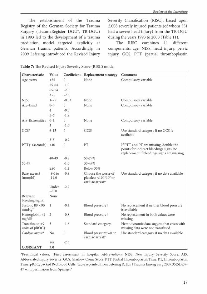
Th e establishment of the Trauma Registry of the German Society for Trauma Surgery (TraumaRegister DGU®, TR-DGU) in 1993 led to the development of a trauma prediction model targeted explicitly at German trauma patients. Accordingly, in 2009 Lefering introduced the Revised Injury
Severity Classifi cation (RISC), based upon 2,008 severely injured patients (of whom 551 had a severe head injury) from the TR-DGU during the years 1993 to 2000 (Table 11).
Th e RISC combines 11 diff erent components: age, NISS, head injury, pelvic injury, GCS, PTT (partial thromboplastin
Table 7: Th e Revised Injury Severity Score (RISC) model
Characteristic Value Coeffi cient Replacement strategy CommentAge, years <55 0 None Compulsory variable
55-64 -1.065-74 -2.0≥75 -2.3
NISS 1-75 -0.03 None Compulsory variableAIS-Head 0-3 0 None Compulsory variable
4 -0.55-6 -1.8
AIS-Extremities 0-4 0 None Compulsory variable5 -1.0
GCS* 6-15 0 GCS† Use standard category if no GCS is available
3-5 -0.9PTT† (seconds) <40 0 PT If PTT and PT are missing, double the
points for indirect bleedings signs; no replacement if bleedings signs are missing
40-49 -0.8 50-79%50-79 -1.0 30-49%
≥80 -1.2 Below 30%Base excess† (mmol/l)
-9.0 to -19.0
-0.8 Choose the worse of platelets <100*109 or cardiac arrest†
Use standard category if no data available
Under -20.0
-2.7
Relevant bleeding signs:
None
Systolic BP <90 mmHg*
1 -0.4 Blood pressure† No replacement if neither blood pressure is available
Hemoglobin <9 mg/dl†
2 -0.8 Blood pressure† No replacement in both values were missing
Transfusion >9 units of pROC†
3 -1.6 Standard category Hemodynamic data suggest that cases with missing data were not transfused
Cardiac arrest* No 0 Blood pressure*=0 or cardiac arrest†
Use standard category if no data available
Yes -2.5CONSTANT 5.0
*Preclinical values, †First assessment in hospital, Abbreviations: NISS, New Injury Severity Score; AIS, Abbreviated Injury Severity; GCS, Glashow Coma Score; PTT, Partial Th romboplastin Time; PT, Th romboplastin Time; pRBC, packed Red Blood Cells. Table reprinted from Lefering R, Eur J Trauma Emerg Surg 2009;35(5):437-47 with permission from Springer®
Review of the Literature
18
time), base excess, cardiac arrest, and relevant signs of bleeding (Table 7).180 With the exception of NISS, all the variables are categorical. According to the original methodology of the RISC, missing values are substituted, so that, for example, missing partial thromboplastin values are replaced by thromboplastin. Th e value of each predictor is associated with a given coeffi cient. For an individual patient, the point weights are subtracted from a constant of 5.0, resulting in the fi nal score X, which is transformed into a probability of hospital survival (Ps) with the logistic function: P(s)=1/(1+e-X)=eX/(1+eX). Th e RISC predicts in-hospital mortality and serves as the prediction model for one of the largest trauma databases in Europe (TR-DGU). It is noteworthy that the RISC has not been externally validated in independent populations outside Germany.
2.5 Intensive Care Scoring Systems
ICU scoring system-based models are among the most widely used prognostic models in healthcare.22,27 Th e fi rst intensive care scoring systems were introduced over 30 years ago, with the introduction of the Acute Physiology and Chronic Health Evaluation (APACHE181) in 1981 and the Simplifi ed Acute Physiology Score (SAPS182) in 1984. Since debuting, APACHE has been revised three times (APACHE II,183 APACHE III,184 APACHE IV185) and the SAPS twice (SAPS II,186 SAPS 3187,188). Moreover, although not originally developed as a prognostic tool, the Sequential Organ Failure Assessment (SOFA189-
191) score is frequently used for outcome prediction in ICU patients. Nevertheless, although routinely used in most ICUs in the world, the role of the intensive care scoring systems in the neurotrauma population is controversial.17,192,193
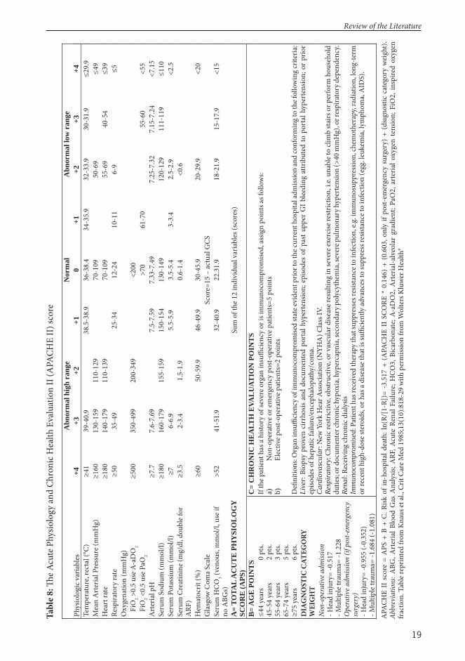
2.5.1 APACHE II
APACHE II is based on 5,815 patients, with various critical illnesses, admitted to 13 ICUs in North America during 1979-1982 (Table 11). Th e APACHE II score consists of three major blocks: 1) the acute physiology score, consisting of the most abnormal values of 12 diff erent physiological parameters measured during the fi rst 24 hours of ICU admission; 2) the age score; and 3) the chronic health score (Table 8).183 Each parameter yields a sub-score (Acute Physiology Score [APS], age points [B], chronic health evaluation [C], see Table 8) that add up to the total APACHE II score, ranging from 0 to 71 — the higher the score, the more severe the disease and the higher the risk of death. Th e relation between risk of death and APACHE II score is not linear but rather sigmoid-shaped and largely dependent on admission diagnosis.
Th e development cohort of APACHE II included 120 head injury patients (+517 possible patients belonging to the multiple trauma and neurologic sub-groups). Despite the relatively low number of TBI patients, previous studies have shown APACHE II to be a poor to good predictor of short-term mortality aft er TBI.194-196
2.5.2 SAPS II
SAPS II is based on 13,152 patients admitted to 137 ICUs in 12 countries in 1991 and 1992 (Table 11). Like the APACHE II score, SAPS II is calculated with 12 physiological parameters from the fi rst 24 hours of ICU admission and three disease-related variables (Table 9).186 SAPS also uses the most abnormal physiological value measured during the fi rst 24 hours of ICU admission. Th e points of the 15 variables are added together to yield the total score for SAPS II, which ranges from 0 to 163 — the higher the score, the more severe the disease and the higher the risk of death.
Review of the Literature
19
Tabl
e 8: Th
e A
cute
Phy
siolo
gy an
d Ch
roni
c Hea
lth E
valu
atio
n II
(APA
CHE
II) s
core
Abn
orm
al h
igh
rang
eN
orm
alA
bnor
mal
low
ran
gePh
ysio
logi
c var
iabl
es+4
+3+2
+10
+1+2
+3+4
Tem
pera
ture
, rec
tal (
°C)
≥41
39-4
0.9
38.5
-38.
936
-38.
434
-35.
932
-33.
930
-31.
9≤2
9.9
Mea
n A
rter
ial P
ress
ure (
mm
Hg)
≥160
130-
159
110-
129
70-1
0950
-69
≤49
Hea
rt ra
te≥1
8014
0-17
911
0-13
970
-109
55-6
940
-54
≤39
Resp
irato
ry ra
te≥5
035
-49
25-3
412
-24
10-1
16-
9≤5
Oxy
gena
tion
(mm
Hg)
Fi
O2 >
0.5
use A
-aD
O2
≥500
350-
499
200-
349
<200
Fi
O2 <
0.5
use P
aO2
>70
61-7
055
-60
<55
Art
eria
l pH
≥7.7
7.6-
7.69
7.5-
7.59
7.33
-7.4
97.
25-7
.32
7.15
-7.2
4<7
.15
Seru
m S
odiu
m (m
mol
/l)≥1
8016
0-17
915
5-15
915
0-15
413
0-14
912
0-12
911
1-11
9≤1
10Se
rum
Pot
assiu
m (m
mol
/l)≥7
6-6.
95.
5-5.
93.
5-5.
43-
3.4
2.5-
2.9
<2.5
Seru
m C
reat
inin
e (m
g/dl
, dou
ble f
or
ARF
)≥3
.52-
3.4
1.5-
1.9
0.6-
1.4
<0.6
Hem
atoc
rit (%
)≥6
050
-59.
946
-49.
930
-45.
920
-29.
9<2
0G
lasg
ow C
oma S
cale
Scor
e=15
– ac
tual
GCS
Seru
m H
CO3 (v
enou
s, m
mol
/l, u
se if
no
ABG
s)>5
241
-51.
932
-40.
922
.31.
918
-21.
915
-17.
9<1
5
A=
TOTA
L A
CU
TE
PH
YSI
OL
OG
Y
SCO
RE
(APS
)Su
m o
f the
12
indi
vidu
al v
aria
bles
(sco
res)
B=
AG
E P
OIN
TS
C=
CH
RO
NIC
HE
ALT
H E
VAL
UAT
ION
PO
INT
S≤4
4 ye
ars
45-5
4 ye
ars
55-6
4 ye
ars
65-7
4 ye
ars
≥75
year
s
0 pt
s.2
pts.
3 pt
s.5
pts.
6 pt
s.
If th
e pat
ient
has
a hi
story
of s
ever
e org
an in
suffi
cien
cy o
r is i
mm
unoc
ompr
omise
d, as
sign
poin
ts as
follo
ws:
a)
Non
-ope
rativ
e or e
mer
genc
y po
st-op
erat
ive p
atie
nts=
5 po
ints
b)
Elec
tive p
ost-o
pera
tive p
atie
nts=
2 po
ints
Defi
niti
ons:
Org
an in
suffi
cien
cy o
f im
mun
ocom
prom
ised
stat
e evi
dent
prio
r to
the c
urre
nt h
ospi
tal a
dmiss
ion
and
conf
orm
ing t
o th
e fol
low
ing c
riter
ia:
Live
r: Bi
opsy
pro
ven
cirr
hosis
and
doc
umen
ted
port
al h
yper
tens
ion;
epi
sode
s of
pas
t upp
er G
I ble
edin
g at
trib
uted
to p
orta
l hyp
erte
nsio
n; o
r pr
ior
episo
des o
f hep
atic
failu
re/e
ncep
halo
path
y/co
ma.
Car
diov
ascu
lar:
New
Yor
k H
ear A
ssoc
iatio
n (N
YHA
) Cla
ss IV
. Re
spir
ator
y: Ch
roni
c res
tric
tive,
obstr
uctiv
e, or
vas
cula
r dise
ase r
esul
ting
in se
vere
exer
cise
restr
ictio
n, i.
e. un
able
to cl
imb
stai
rs o
r per
form
hou
seho
ld
dutie
s; or
doc
umen
ter c
hron
ic h
ypox
ia, h
yper
capn
ia, s
econ
dary
pol
ycyt
hem
ia, s
ever
e pul
mon
ary h
yper
tens
ion
(>40
mm
Hg)
, or r
espi
rato
ry d
epen
denc
y. Re
nal:
Rece
ivin
g ch
roni
c dia
lysis
Im
mun
ocom
prom
ised
: Pat
ient
has
rece
ived
ther
apy t
hat s
uppr
esse
s res
istan
ce to
infe
ctio
n, e.
g. im
mun
osup
pres
sion,
chem
othe
rapy
, rad
iatio
n, lo
ng-te
rm
or re
cent
hig
h-do
se st
eroi
ds, o
r has
a di
seas
e tha
t is s
uffi c
ient
ly ad
vanc
es to
supp
ress
resis
tanc
e to
infe
ctio
n (e
gg. l
euke
mia
, lym
phom
a, A
IDS)
.
DIA
GN
OST
IC C
ATEG
ORY
W
EIG
HT
Non-
oper
ativ
e adm
issio
n- H
ead
inju
ry=
-0.5
17
- Mul
tiple
trau
ma=
-1.2
28
Ope
rativ
e adm
issio
n (if
pos
t-em
erge
ncy
surg
ery)
- Hea
d in
jury
= -0
.955
(-0.
352)
- Mul
tiple
trau
ma=
-1.6
84 (-
1.08
1)A
PACH
E II
sco
re =
APS
+ B
+ C
. Risk
of i
n-ho
spita
l dea
th: l
n(R/
[1-R
])=
-3.5
17 +
(A
PACH
E II
SCO
RE *
0.1
46)
+ (0
.603
, onl
y if
post-
emer
genc
y su
rger
y) +
(di
agno
stic
cate
gory
wei
ght).
Ab
brev
iatio
ns: A
BG, A
rter
ial B
lood
Gas
Ana
lysis
; ARF
, Acu
te R
enal
Fai
lure
; HCO
3, B
icar
bona
te; A
-aD
O2,
Art
eria
l-alv
eola
r gr
adie
nt; P
aO2,
art
eria
l oxy
gen
tens
ion;
FiO
2, in
spire
d ox
ygen
fr
actio
n. T
able
repr
inte
d fro
m K
naus
et al
., Cr
it Ca
re M
ed 1
985;
13(1
0):8
18-2
9 w
ith p
erm
issio
n fro
m W
olte
rs K
luw
er H
ealth
®
Review of the Literature
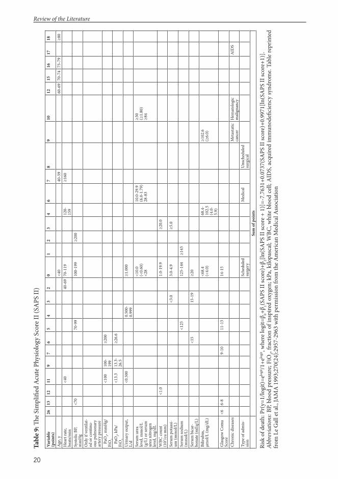
20
Review of the Literature
Tabl
e 9: Th
e S
impl
ifi ed
Acu
te P
hysio
logy
Sco
re II
(SA
PS II
)Va
riab
le
(poi
nts)
2613
1211
97
65
43
20
12
34
67
89
1012
1516
1718
Age
, y<4
040
-59
60-6
970
-74
75-7
9≥8
0H
eart
rate
, be
ats/
min
<40
40-6
970
-119
120-
159
≥160
Systo
lic B
P, m
mH
g<7
070
-99
100-
199
≥200
Onl
y if
vent
ilat-
ed o
r con
tinu-
ous p
ulm
onar
y ar
tery
pre
ssur
e
PaO
2, mm
Hg/
FiO
2
<100
100-
199
≥200
Pa
O2, k
Pa/
FiO
2
<13.
313
.3-
26.5
≥26.
6
Urin
ary
outp
ut,
L/d
<0.5
000.
500-
0.99
9≥1
.000
Seru
m u
rea
leve
l, m
mol
/L
(g/L
) or s
erum
ur
ea n
itrog
en
leve
l, m
g/dL
<10.
0(<
0.60
)<2
8
10.0
-29.
9(6
.0-1
.79)
28-8
3
≥30
(≥1.
80)
≥84
WBC
coun
t (1
03 /cu
mm
)<1
.01.
0-19
.9≥2
0.0
Seru
m p
otas
si-um
(mm
ol/L
)<3
.03.
0-4.
9≥5
.0
Seru
m so
dium
(m
mol
/L)
<125
125-
144
≥145
Seru
m b
icar
-bo
nate
(mEq
/L)
<15
15-1
9≥2
0
Bilir
ubin
, μm
ol/L
(mg/
dL)
<68.
4(<
4.0)
68.4
-10
2.5
(4.0
-5.
9)
≥102
.6(≥
6.0)
Gla
sgow
Com
a Sc
ore
<66-
89-
1011
-13
14-1
5
Chro
nic d
iseas
esM
etas
tatic
ca
ncer
Hem
atol
ogic
m
alig
nanc
yA
IDS
Type
of a
dmis-
sion
Sche
dule
d su
rger
yM
edic
alU
nsch
edul
ed
surg
ical
Sum
of p
oint
s
Risk
of d
eath
: Pr(
y=1/
logi
t)=elo
git /1
+elo
git , w
here
logi
t=β 0+β
1(SA
PS II
scor
e)+β
2[ln(S
APS
II sc
ore +
1)]
=-7.
7631
+0.0
737(
SAPS
II sc
ore)
+0.9
971[
ln(S
APS
II sc
ore+
1)].
Abbr
evia
tions
: BP,
bloo
d pr
essu
re; F
iO2, f
ract
ion
of in
spire
d ox
ygen
; kPa
, kilo
pasc
al; W
BC, w
hite
blo
od c
ell;
AID
S, a
cqui
red
imm
unod
efi c
ienc
y sy
ndro
me.
Tabl
e re
prin
ted
from
Le G
all e
t al.,
JAM
A 1
993;
270(
24):2
957-
2963
with
per
miss
ion
from
the A
mer
ican
Med
ical
Ass
ocia
tion
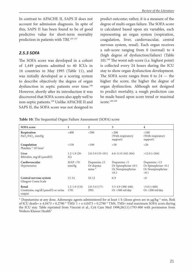
21
In contrast to APACHE II, SAPS II does not account for admission diagnosis. In spite of this, SAPS II has been found to be of good predictive value for short-term mortality prediction in patients with TBI.195-197
2.5.3 SOFA
Th e SOFA score was developed in a cohort of 1,449 patients admitted to 40 ICUs in 16 countries in May 1995 (Table 11), and was initially developed as a scoring system to describe objectively the degree of organ dysfunction in septic patients over time.198 However, shortly aft er its introduction it was discovered that SOFA scores also apply well to non-septic patients.199 Unlike APACHE II and SAPS II, the SOFA score was not designed to
predict outcome; rather, it is a measure of the degree of multi-organ failure. Th e SOFA score is calculated based upon six variables, each representing an organ system (respiration, coagulation, liver, cardiovascular, central nervous system, renal). Each organ receives a sub-score ranging from 0 (normal) to 4 (high degree of dysfunction/failure) (Table 10).198 Th e worst sub-score (i.e. highest point) is collected every 24 hours during the ICU stay to show organ dysfunction development. Th e SOFA score ranges from 0 to 24 — the higher the score, the higher the degree of organ dysfunction. Although not designed to predict mortality, a rough prediction can be made based upon score trend or maximal score.189,198
Table 10: Th e Sequential Organ Failure Assessment (SOFA) score
SOFA score 1 2 3 4
RespirationPaO2/FiO2, mmHg
<400 <300 <200(With respiratory support)
<100(With respiratory support)
CoagulationPlatelets * 103/mm3
<150 <100 <50 <20
LiverBilirubin, mg/dl (μmoll/l)
1.2-1.9 (20-32)
2.0-5.9 (33-101) 6.0-11.9 (102-204) >12.0 (>204)
CardiovascularHypotension
MAP <70 mmHg
Dopamine ≤5 Or doputa-mine *
Dopamine >5Or Epinephrine ≤0.1Or Norepinephrine ≤0.1
Dopamine >15Or Epinephrine >0.1Or Norepinephrine >0.1
Central nervous systemGlasgow Coma Scale
13-14 10-12 6-9 <6
RenalCreatinine, mg/dl (μmol/l) or urine output
1.2-1.9 (110-170)
2.0-3.4 (171-299)
3.5-4.9 (300-440) Or <500 ml/day
>5.0 (>440) Or <200 ml/day
* Doputamine at any dose, Adrenergic agents administered for at least 1 h (doses given are in μg/kg * min, Risk of ICU death= e-4.0473 + 0.2790 * TMS/ 1 + e-4.0473 + 0.2790 * TMS, TMS= total maximum SOFA score during the ICU stay. Table reprinted from Vincent et al., Crit Care Med 1998;26(11):1793-800 with permission from Wolters Kluwer Health®
Review of the Literature
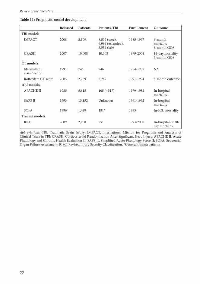
22
Table 11: Prognostic model development
Released Patients Patients, TBI Enrollement OutcomeTBI models IMPACT 2008 8,509 8,509 (core),
6,999 (extended), 3,554 (lab)
1985-1997 6-month mortality6-month GOS
CRASH 2007 10,008 10,008 1999-2004 14-day mortality6-month GOS
CT models Marshall CT classifi cation
1991 746 746 1984-1987 NA
Rotterdam CT score 2005 2,269 2,269 1991-1994 6-month outcomeICU models APACHE II 1985 5,815 105 (+517) 1979-1982 In-hospital
mortality SAPS II 1993 13,152 Unknown 1991-1992 In-hospital
mortality SOFA 1996 1,449 181* 1995 In-ICU mortalityTrauma models RISC 2009 2,008 551 1993-2000 In-hospital or 30-
day mortality
Abbreviations: TBI, Traumatic Brain Injury; IMPACT, International Mission for Prognosis and Analysis of Clinical Trials in TBI; CRASH, Corticosteroid Randomization Aft er Signifi cant Head Injury; APACHE II, Acute Physiology and Chronic Health Evaluation II; SAPS II, Simplifi ed Acute Physiology Score II; SOFA, Sequential Organ Failure Assessment; RISC, Revised Injury Severity Classifi cation, *General trauma patients
Review of the Literature
23
3 Purpose of the Study
Th e purpose of this study is to validate diff erent types of prognostic models for patients with TBI and to develop novel models with enhanced predictive performance, with focus on long-term outcome prediction. Th e following specifi c aims were addressed:
1. To investigate the predictive accuracy of TBI specifi c models (I-III), general intensive care scoring systems (IV), and general trauma scoring systems (V) for outcome prediction in patients with TBI.
2. To create new prognostic models with enhanced performance compared to previous models (II, III, IV).
Purpose of the Study
24
4 Subjects and Methods
4.1 Study Se ng and Popula on
4.1.1 Trauma c Brain Injury Models (I-III)
Studies I, II, and III investigate TBI models and are based on patients with TBI admitted to the ICUs of Töölö Hospital (Helsinki University Central Hospital [HUCH]) from January 2009 to December 2012 (Table 12). Located in Helsinki, Finland, Töölö Hospital is the HUCH trauma unit and the only Level I trauma center in Th e Hospital District of Helsinki and Uusimaa (HUS), a joint authority encompassing 24 municipalities and approximately two million inhabitants. Th e Department of Neurosurgery in Helsinki is the largest neurosurgical unit in Finland and one of the largest in Europe, performing more than 3,200 neurosurgical operations annually.
TBI was defi ned as a discharge ICD-10 (International Classifi cation of Diseases and Related Health Problems, 10th Edition) diagnosis of S06.1-S06.9, caused by an external force.30 Only blunt injury mechanism TBI and adult patients (≥14 years for I and III, ≥16 years for II) were included. Patients with a history of head trauma but no intracranial pathological fi ndings by CT imaging during the hospital stay and those with subacute injuries (>24 hours) were excluded.
Study I includes 342 patients with moderate to severe TBI (admission GCS 3-12) admitted from January 2009 to December 2010.
Studies II and III include patients with mild complicated TBI (defi ned as admission GCS 13-15 requiring ICU admission) and moderate to severe TBI (admission GCS 3-12) admitted from January 2009 to December 2012; there were totals of 890 (II) and 869 patients (III).
4.1.2 Intensive Care Scoring Systems (IV)
Study IV includes patients with moderate to severe TBI admitted to one of fi ve university hospitals participating in the Finnish Intensive Care Consortium (FICC) database from January 2003 to December 2012 (Table 12).
Th e FICC database is a multi-center database consisting of prospectively collected data from ICUs in 22 diff erent hospitals (of which 5 are university hospitals). Th e FICC database was established in 1994 as a co-operative benchmarking project to improve the quality of intensive care in Finland. Data from patient monitors, laboratory systems, and ventilators are automatically collected.200 Specially trained ICU personnel manually enter specifi c types of other data, such as co-morbidities and outcomes. All data are stored in a central database maintained by Tieto Healthcare & Welfare Ltd. (Kuopio, Finland). Patients with TBI were identifi ed by their APACHE III184 diagnosis. Moderate to severe TBI was defi ned as a worst GCS of 3 to 13 in the fi rst ICU day,57 leading to the inclusion of 1,625 patients in the study.
4.1.3 Trauma Scoring Systems (V)
Study V encompasses severely injured patients with moderate to severe TBI admitted to the TR-DGU and the Trauma Register of Helsinki University Hospital (TR-THEL) (Table 12). Moderate to severe traumatic brain injury was defi ned as head-region AIS of three or higher. Patients were further divided based on the severity of extra-cranial injuries into isolated TBI (no other body part AIS ≥ 2) and polytrauma TBI (at least one other body part AIS ≥ 2). For comparison reasons, mild TBI (AIS-head=1-2) and no TBI (AIS-head=0) were also defi ned.
Th e TR-DGU (TraumaRegister DGU® of the German Trauma Society) began in
Sujects and Methods
25
1993 with fi ve trauma units in Germany. Since then, the TR-DGU has grown to be one of Europe’s largest databases, entering over 30,000 patients annually from over 600 hospitals, more than 90% of which are in Germany. Inclusion criteria are admission via the emergency department with subsequent ICU care, or admission with vital signs but death before ICU admission. Data for the TR-DGU is gathered prospectively in a central web-based database hosted by the Akademie der Unfallchirurgie GmbH of the DGU. Scientifi c data analyses are approved by a peer-reviewed process by the Committee of Emergency Medicine, Intensive Care, and Trauma Management of the German Trauma Society (Sektion NIS). Study V was registered as TR-DGU Project ID: 2012-053 II.
Th e Trauma Registry of Helsinki University Hospital (TR-THEL) is a single-center trauma registry consisting of patients with severe trauma (defi ned as an ISS >15) admitted to Töölö Hospital. Th e TR-THEL was founded in 2006 as a benchmarking project for improving the quality of trauma care in the region. Data is collected according to the Utstein recommendations.201 From 2006 to 2011, 400 to 450 patients were entered into the database annually.
4.2 Data collec on4.2.1 Trauma c Brain Injury
Models (I-III)
Patient admission characteristics were recorded by emergency department physicians and were retrieved from electronic records. Two authors co-operatively classifi ed all admission head-CT images by Marshall CT classifi cation, Rotterdam CT score, and by a set of pre-defi ned characteristics:
• Mass lesion type (SDH, EDH, ICH [contusion])
• Mass lesion volume (≥25 cm3, <25 cm3)
• Presence, location, and thickness of tSAH (basal, cortical, both; thin, thick)
• Presence of IVH (yes or no)• Status of suprasellar cisterns
(normal, compressed, obliterated)• Status of ambiens cisterns (normal,
compressed, obliterated)• Status of fourth ventricle (normal,
abnormal)• Midline shift (in mm) • Cortical sulci eff acement (no
eff acement, unilateral eff acement, bilateral eff acement)
Patients scheduled for acute mass lesion evacuation were classifi ed as Marshall class V (evacuated mass lesion). Mass lesion was defi ned as any SDH, EDH, or ICH of any size, with ICH referring to both contusion and intracerebral hemorrhage, as no clear distinction between the two exists. Mass lesion volume was measured using the ABC/2 method, which is accurate for both intra-parenchymal and extra-parenchymal hemorrhage volume assessment, with little inter-observer variability.202-204
Laboratory values were measured on admission and retrieved from electronic hospital records. Th e international normalized ratio (INR) and platelet count were used as markers of coagulation, while ISS indicated extra-cranial injury.172 Th e ISS was calculated by an independent accredited nurse using the AIS 2005 revision.174 Hypotension and hypoxia were defi ned according to the Brain Trauma Foundation guidelines as a systolic blood pressure <90 mmHg and an oxygen saturation <90%, respectively, at any time prior to hospital admission.47 Th e APACHE II variables were extracted from the ICU soft ware (PICIS, Anesthesia Manager®) at fi ve-minute intervals to pinpoint the most abnormal physiological and laboratory values measured during the fi rst 24 hours in the ICU.
Sujects and Methods
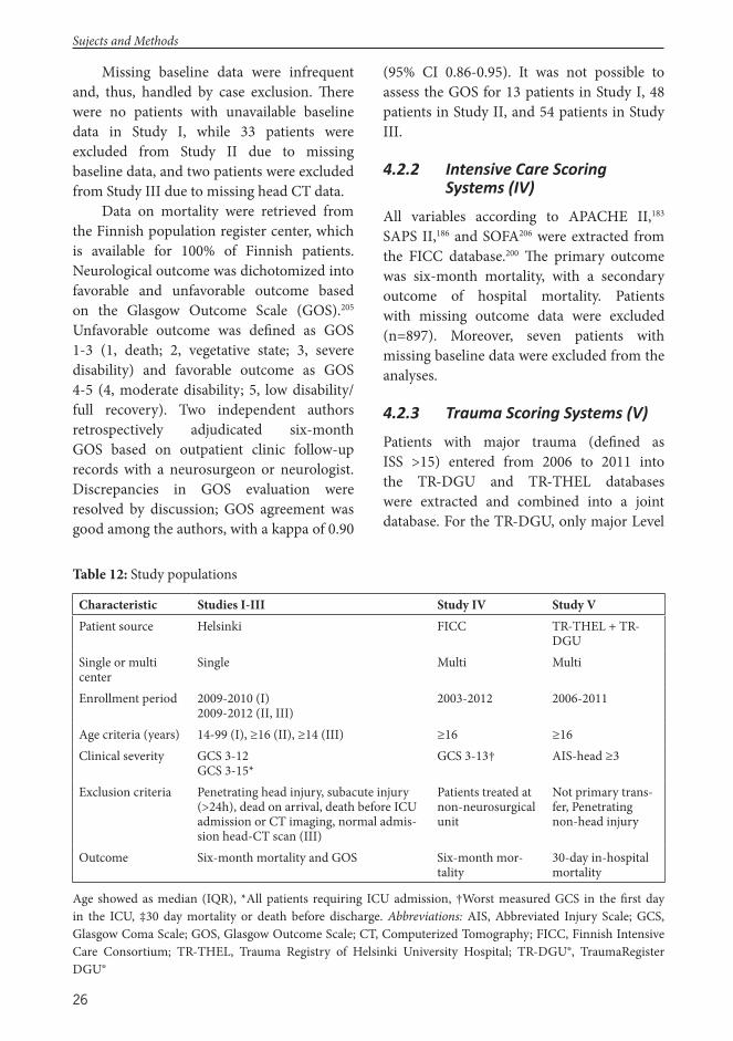
26
Missing baseline data were infrequent and, thus, handled by case exclusion. Th ere were no patients with unavailable baseline data in Study I, while 33 patients were excluded from Study II due to missing baseline data, and two patients were excluded from Study III due to missing head CT data.
Data on mortality were retrieved from the Finnish population register center, which is available for 100% of Finnish patients. Neurological outcome was dichotomized into favorable and unfavorable outcome based on the Glasgow Outcome Scale (GOS).205 Unfavorable outcome was defi ned as GOS 1-3 (1, death; 2, vegetative state; 3, severe disability) and favorable outcome as GOS 4-5 (4, moderate disability; 5, low disability/full recovery). Two independent authors retrospectively adjudicated six-month GOS based on outpatient clinic follow-up records with a neurosurgeon or neurologist. Discrepancies in GOS evaluation were resolved by discussion; GOS agreement was good among the authors, with a kappa of 0.90
(95% CI 0.86-0.95). It was not possible to assess the GOS for 13 patients in Study I, 48 patients in Study II, and 54 patients in Study III.
4.2.2 Intensive Care Scoring Systems (IV)
All variables according to APACHE II,183 SAPS II,186 and SOFA206 were extracted from the FICC database.200 Th e primary outcome was six-month mortality, with a secondary outcome of hospital mortality. Patients with missing outcome data were excluded (n=897). Moreover, seven patients with missing baseline data were excluded from the analyses.
4.2.3 Trauma Scoring Systems (V)
Patients with major trauma (defi ned as ISS >15) entered from 2006 to 2011 into the TR-DGU and TR-THEL databases were extracted and combined into a joint database. For the TR-DGU, only major Level
Table 12: Study populations
Characteristic Studies I-III Study IV Study VPatient source Helsinki FICC TR-THEL + TR-
DGUSingle or multi center
Single Multi Multi
Enrollment period 2009-2010 (I)2009-2012 (II, III)
2003-2012 2006-2011
Age criteria (years) 14-99 (I), ≥16 (II), ≥14 (III) ≥16 ≥16 Clinical severity GCS 3-12
GCS 3-15*GCS 3-13† AIS-head ≥3
Exclusion criteria Penetrating head injury, subacute injury (>24h), dead on arrival, death before ICU admission or CT imaging, normal admis-sion head-CT scan (III)
Patients treated at non-neurosurgical unit
Not primary trans-fer, Penetrating non-head injury
Outcome Six-month mortality and GOS Six-month mor-tality
30-day in-hospital mortality
Age showed as median (IQR), *All patients requiring ICU admission, †Worst measured GCS in the fi rst day in the ICU, ‡30 day mortality or death before discharge. Abbreviations: AIS, Abbreviated Injury Scale; GCS, Glasgow Coma Scale; GOS, Glasgow Outcome Scale; CT, Computerized Tomography; FICC, Finnish Intensive Care Consortium; TR-THEL, Trauma Registry of Helsinki University Hospital; TR-DGU®, TraumaRegister DGU®
Sujects and Methods
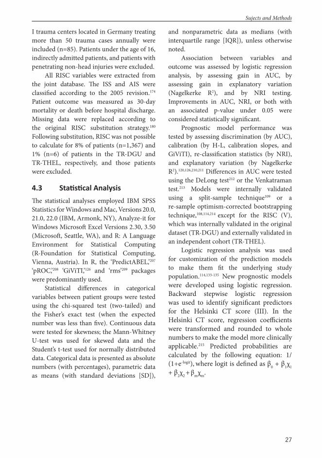
27
I trauma centers located in Germany treating more than 50 trauma cases annually were included (n=85). Patients under the age of 16, indirectly admitted patients, and patients with penetrating non-head injuries were excluded.
All RISC variables were extracted from the joint database. Th e ISS and AIS were classifi ed according to the 2005 revision.174 Patient outcome was measured as 30-day mortality or death before hospital discharge. Missing data were replaced according to the original RISC substitution strategy.180 Following substitution, RISC was not possible to calculate for 8% of patients (n=1,367) and 1% (n=6) of patients in the TR-DGU and TR-THEL, respectively, and those patients were excluded.
4.3 Sta s cal AnalysisTh e statistical analyses employed IBM SPSS Statistics for Windows and Mac, Versions 20.0, 21.0, 22.0 (IBM, Armonk, NY), Analyze-it for Windows Microsoft Excel Versions 2.30, 3.50 (Microsoft , Seattle, WA), and R: A Language Environment for Statistical Computing (R-Foundation for Statistical Computing, Vienna, Austria). In R, the ‘PredictABEL,’207 ‘pROC,’208 ‘GiViTI,’126 and ‘rms’209 packages were predominantly used.
Statistical diff erences in categorical variables between patient groups were tested using the chi-squared test (two-tailed) and the Fisher’s exact test (when the expected number was less than fi ve). Continuous data were tested for skewness; the Mann-Whitney U-test was used for skewed data and the Student’s t-test used for normally distributed data. Categorical data is presented as absolute numbers (with percentages), parametric data as means (with standard deviations [SD]),
and nonparametric data as medians (with interquartile range [IQR]), unless otherwise noted.
Association between variables and outcome was assessed by logistic regression analysis, by assessing gain in AUC, by assessing gain in explanatory variation (Nagelkerke R2), and by NRI testing. Improvements in AUC, NRI, or both with an associated p-value under 0.05 were considered statistically signifi cant.
Prognostic model performance was tested by assessing discrimination (by AUC), calibration (by H-L, calibration slopes, and GiViTI), re-classifi cation statistics (by NRI), and explanatory variation (by Nagelkerke R2).120,126,210,211 Diff erences in AUC were tested using the DeLong test212 or the Venkatraman test.213 Models were internally validated using a split-sample technique109 or a re-sample optimism-corrected bootstrapping technique,108,114,214 except for the RISC (V), which was internally validated in the original dataset (TR-DGU) and externally validated in an independent cohort (TR-THEL).
Logistic regression analysis was used for customization of the prediction models to make them fi t the underlying study population.114,133-135 New prognostic models were developed using logistic regression. Backward stepwise logistic regression was used to identify signifi cant predictors for the Helsinki CT score (III). In the Helsinki CT score, regression coeffi cients were transformed and rounded to whole numbers to make the model more clinically applicable.215 Predicted probabilities are calculated by the following equation: 1/(1+e-logit), where logit is defi ned as β0 + β1χ1 + β2χ2 + βmχm.
Sujects and Methods
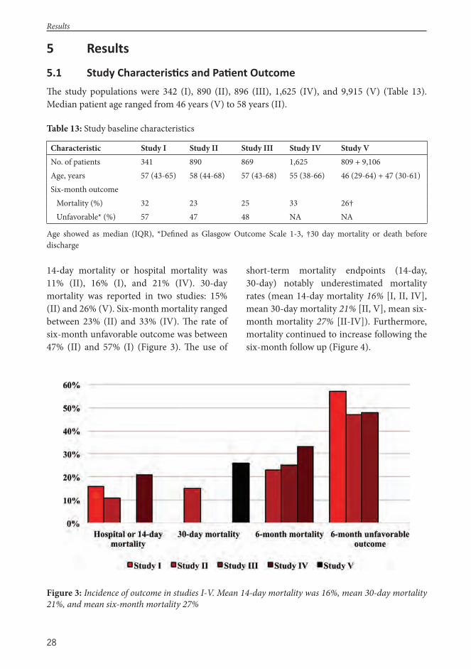
28
5 Results
5.1 Study Characteris cs and Pa ent OutcomeTh e study populations were 342 (I), 890 (II), 896 (III), 1,625 (IV), and 9,915 (V) (Table 13). Median patient age ranged from 46 years (V) to 58 years (II).
Table 13: Study baseline characteristics
Characteristic Study I Study II Study III Study IV Study VNo. of patients 341 890 869 1,625 809 + 9,106Age, years 57 (43-65) 58 (44-68) 57 (43-68) 55 (38-66) 46 (29-64) + 47 (30-61)Six-month outcome Mortality (%) 32 23 25 33 26† Unfavorable* (%) 57 47 48 NA NA
Age showed as median (IQR), *Defi ned as Glasgow Outcome Scale 1-3, †30 day mortality or death before discharge
Figure 3: Incidence of outcome in studies I-V. Mean 14-day mortality was 16%, mean 30-day mortality 21%, and mean six-month mortality 27%
Results
14-day mortality or hospital mortality was 11% (II), 16% (I), and 21% (IV). 30-day mortality was reported in two studies: 15% (II) and 26% (V). Six-month mortality ranged between 23% (II) and 33% (IV). Th e rate of six-month unfavorable outcome was between 47% (II) and 57% (I) (Figure 3). Th e use of
short-term mortality endpoints (14-day, 30-day) notably underestimated mortality rates (mean 14-day mortality 16% [I, II, IV], mean 30-day mortality 21% [II, V], mean six-month mortality 27% [II-IV]). Furthermore, mortality continued to increase following the six-month follow up (Figure 4).
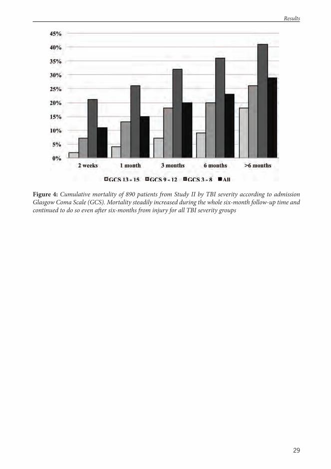
29
Figure 4: Cumulative mortality of 890 patients from Study II by TBI severity according to admission Glasgow Coma Scale (GCS). Mortality steadily increased during the whole six-month follow-up time and continued to do so even aft er six-months from injury for all TBI severity groups
Results

30
5.2 Early Predictors of Outcome 5.2.1 Laboratory Variables and
Extra-Cranial Injury
Th e role of coagulation markers and extra-cranial injury severity markers were investigated by multivariate logistic regression analysis, AUC comparison, and NRI testing.
In logistic regression analysis, INR independently predicted six-month mortality (OR 2.23, 95% CI 1.20-4.17, p=0.012), but not six-month unfavorable outcome (p=0.116; Table 14). Th e addition of INR to IMPACT signifi cantly increased AUC from 0.85 to 0.87 (AUC +0.02, p=0.034) for six-month mortality prediction, but not for six-month unfavorable outcome (AUC +0.00, p=0.721). Similarly, NRI testing found the
addition of INR to signifi cantly improve six-month mortality prediction (NRI 0.28, 95% CI 0.08-0.48, p=0006), but again, not unfavorable outcome prediction (NRI -0.05, 95% CI -0.25-[-0.16], p=0.658).
Platelet count was insignifi cant in logistic regression analysis for both six-month mortality (OR=1.00, p=0.578) and unfavorable outcome prediction (OR 1.00, p=0.169), with no increases in AUC (p>0.05).
ISS was used as a marker of extra-cranial injury severity and dichotomized to >15 vs. ≤15 and to >25 vs. ≤25. Logistic regression analysis revealed ISS to be statistically insignifi cant aft er adjusting for IMPACT covariates (p>0.05), with no gain in AUC (AUC +0.00, p>0.05).
Table 14: Gained prognostic value of markers of coagulation and markers of extra-cranial injury to the IMPACT model
Prognostic model AUC (95% CI) Gain in AUC P-Value* OR (95% CI) P-Value†
Six-month mortality
IMPACT-lab‡ 0.85 (0.81-0.89) Reference Reference
+ INR 0.87 (0.83-0.91) +0.02 0.034 2.23 (1.20-4.17) 0.012
+ Platelet count 0.85 (0.81-0.89) 0.00 0.944 1.00 (1.00-1.01) 0.578
+ ISS >15 0.85 (0.81-0.89) 0.00 0.777 1.26 (0.33-4.76) 0.735
+ ISS >25 0.85 (0.81-0.90) 0.00 0.334 1.13 (0.63-2.02) 0.683
Six-month unfavorable outcome
IMPACT-lab‡ 0.81 (0.76-0.86) Reference Reference
+ INR 0.81 (0.77-0.86) 0.00 0.721 0.52 (0.23-1.18) 0.116
+ Platelet count 0.81 (0.76-0.86) 0.00 0.764 1.00 (1.00-1.01) 0.169
+ ISS >15 0.81 (0.76-0.86) 0.00 0.819 0.96 (0.38-2.47) 0.937
+ ISS >25 0.81 (0.76-0.86) 0.00 0.841 1.03 (0.59-1.77) 0.929
Th e addition of INR signifi cantly improved the AUC of the IMPACT model with +0.02 units for mortality predic-tion. NRI testing confi rmed the relationship between INR and mortality. In contrast, INR was not signifi cant for neurological outcome prediction by either method, showing that one cannot expect the same variables to predict mortality and neurological outcome. Platelet count and ISS were not signifi cant by either test for either outcome measure. Abbreviations: IMPACT, International Mission for Prognosis and Analysis in Clinical Trials for TBI; AUC, Area Under the Receiver Characteristic Curve; INR, International Normalized Ratio; ISS, Injury Severity Score*Signifi cance tested for gain in AUC compared to the reference AUC†Signifi cance for the independent eff ect of the variable adjusted for the IMPACT lab‡IMPACT lab includes: age, motor score, pupillary reactivity, hypoxia, hypotension, Marshall CT class, presence of epidural hematoma, presence of traumatic subarachnoid hemorrhage, glucose concentration and hemoglobin concentration. Th e IMPACT lab model was fi rst level customized
Results
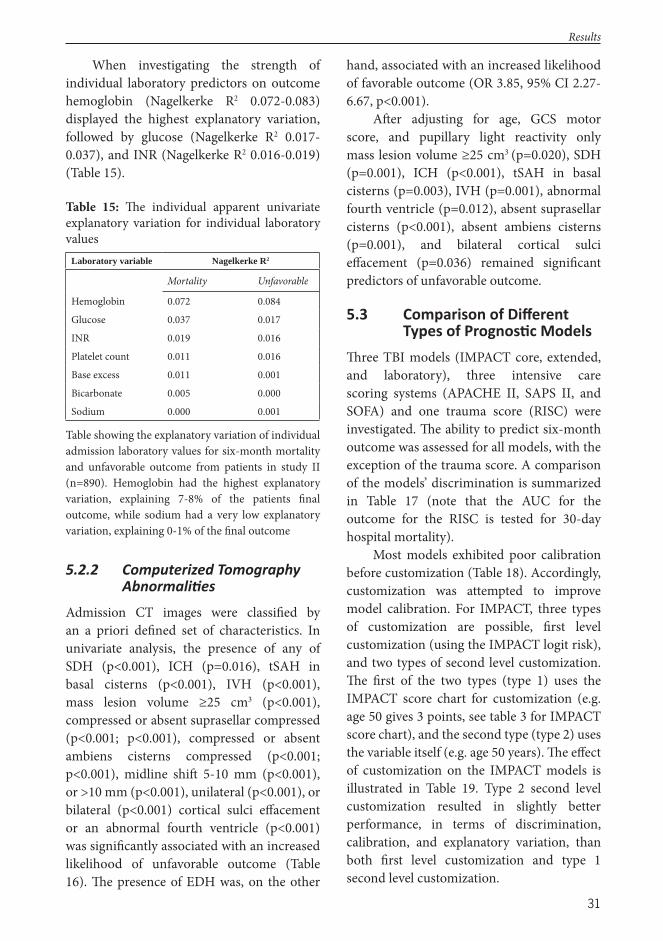
31
When investigating the strength of individual laboratory predictors on outcome hemoglobin (Nagelkerke R2 0.072-0.083) displayed the highest explanatory variation, followed by glucose (Nagelkerke R2 0.017-0.037), and INR (Nagelkerke R2 0.016-0.019) (Table 15).
Table 15: Th e individual apparent univariate explanatory variation for individual laboratory values
Laboratory variable Nagelkerke R2
Mortality Unfavorable
Hemoglobin 0.072 0.084
Glucose 0.037 0.017
INR 0.019 0.016
Platelet count 0.011 0.016
Base excess 0.011 0.001
Bicarbonate 0.005 0.000
Sodium 0.000 0.001
Table showing the explanatory variation of individual admission laboratory values for six-month mortality and unfavorable outcome from patients in study II (n=890). Hemoglobin had the highest explanatory variation, explaining 7-8% of the patients fi nal outcome, while sodium had a very low explanatory variation, explaining 0-1% of the fi nal outcome
5.2.2 Computerized Tomography Abnormali es
Admission CT images were classifi ed by an a priori defi ned set of characteristics. In univariate analysis, the presence of any of SDH (p<0.001), ICH (p=0.016), tSAH in basal cisterns (p<0.001), IVH (p<0.001), mass lesion volume ≥25 cm3 (p<0.001), compressed or absent suprasellar compressed (p<0.001; p<0.001), compressed or absent ambiens cisterns compressed (p<0.001; p<0.001), midline shift 5-10 mm (p<0.001), or >10 mm (p<0.001), unilateral (p<0.001), or bilateral (p<0.001) cortical sulci eff acement or an abnormal fourth ventricle (p<0.001) was signifi cantly associated with an increased likelihood of unfavorable outcome (Table 16). Th e presence of EDH was, on the other
hand, associated with an increased likelihood of favorable outcome (OR 3.85, 95% CI 2.27-6.67, p<0.001).
Aft er adjusting for age, GCS motor score, and pupillary light reactivity only mass lesion volume ≥25 cm3 (p=0.020), SDH (p=0.001), ICH (p<0.001), tSAH in basal cisterns (p=0.003), IVH (p=0.001), abnormal fourth ventricle (p=0.012), absent suprasellar cisterns (p<0.001), absent ambiens cisterns (p=0.001), and bilateral cortical sulci eff acement (p=0.036) remained signifi cant predictors of unfavorable outcome.
5.3 Comparison of Diff erent Types of Prognos c Models
Th ree TBI models (IMPACT core, extended, and laboratory), three intensive care scoring systems (APACHE II, SAPS II, and SOFA) and one trauma score (RISC) were investigated. Th e ability to predict six-month outcome was assessed for all models, with the exception of the trauma score. A comparison of the models’ discrimination is summarized in Table 17 (note that the AUC for the outcome for the RISC is tested for 30-day hospital mortality).
Most models exhibited poor calibration before customization (Table 18). Accordingly, customization was attempted to improve model calibration. For IMPACT, three types of customization are possible, fi rst level customization (using the IMPACT logit risk), and two types of second level customization. Th e fi rst of the two types (type 1) uses the IMPACT score chart for customization (e.g. age 50 gives 3 points, see table 3 for IMPACT score chart), and the second type (type 2) uses the variable itself (e.g. age 50 years). Th e eff ect of customization on the IMPACT models is illustrated in Table 19. Type 2 second level customization resulted in slightly better performance, in terms of discrimination, calibration, and explanatory variation, than both fi rst level customization and type 1 second level customization.
Results
32
5.3.1 Trauma c Brain Injury Models
IMPACT discrimination (by AUC) increased with rising model complexity from the core (AUC 0.81) to the laboratory model (AUC 0.85). Th e addition of INR signifi cantly increased the laboratory model’s AUC from 0.85 to 0.87 (AUC +0.02, p=0.034), which was the highest AUC achieved in the study. IMPACT discriminated in general better
for six-month mortality than neurological outcome (mean AUC for mortality prediction 0.82, and mean AUC for unfavorable neurological outcome prediction 0.81).
In close relationship with the TBI models, two CT scoring systems were also investigated: the Marshall CT classifi cation and the Rotterdam CT score. Both CT scores were of limited value for long-outcome prediction, with AUCs ranging from 0.64 to 0.70 (Table 17).
Table 16: Multivariate analysis showing relationship between individual admission CT characteristics and six-month unfavorable outcome
Univariate analysis Multivariate analysis*OR (95% CI) P-Value OR (95% CI) P-Value
Mass lesion volume >25 cm3 2.90 (2.19-3.85) <0.001 1.49 (1.07-2.10) 0.020Subdural hematoma 3.46 (2.51-4.77) <0.001 1.71 (1.18-2.49) 0.001Epidural hematoma 0.26 (0.15-0.44) <0.001 0.59 (0.32-1.09) 0.091Intracerebral hemorrhage 1.39 (1.06-1.82) 0.016 2.15 (1.53-3.01) <0.001Traumatic SAH No 1 1 Limited to cortical sulci 1.05 (0.78-1.43) 0.733 1.19 (0.83-1.71) 0.345 Also in basal cisterns 2.13 (1.48-3.06) <0.001 1.94 (1.24-3.01) 0.003Intraventricular hemorrhage 2.83 (1.84-4.34) <0.001 2.41 (1.45-3.98) 0.001Abnormal fourth ventricle 12.71 (2.97-54.40) <0.001 8.23 (1.60-42.87) 0.012Suprasellar cisterns Normal 1 1 Compressed 2.06 (1.49-2.84) <0.001 1.28 (0.87-1.87) 0.214 Absent 6.97 (4.44-10.95) <0.001 2.91 (1.62-5.25) <0.001Ambiens cisterns Normal 1 1 Compressed 2.13 (1.51-3.01) <0.001 1.31 (0.86-2.01) 0.206 Absent 5.62 (3.72-8.48) <0.001 2.47 (1.43-4.27) 0.001Midline shift <5 mm 1 1 5-10 mm 1.51 (1.08-2.11) <0.001 1.16 (0.79-1.71) 0.454 >10 mm 3.52 (2.49-4.96) <0.001 1.07 (0.69-1.67) 0.768Cortical sulci eff acement No eff acement 1 1 Unilateral eff acement 1.83 (1.33-2.53) <0.001 1.25 (0.86-1.82) 0.248 Bilateral eff acement 3.40 (2.38-4.83) <0.001 1.61 (1.03-2.50) 0.036
Table showing univariate and multivariate analysis of the association between admission CT characteristics and six-month unfavorable neurological outcome. In univariate analysis, all except traumatic SAH limited to cortical sulci were signifi cantly associates with the outcome. In an enter type multivariate logistic regression analysis, epidural hematoma cortical sulci traumatic SAH, compressed ambiens cisterns, midline shift and unilateral cortical sulci eff acement were found to be insignifi cant predictors of outcome. Data is from Study III, with a total patient number of 869. Abbreviations: SAH, subarachnoid hemorrhage.
*Multivariate analysis adjusted for age (continuous), GCS motor score (six categories) and pupillary light reactivity (normal, one reacts, no reaction)
Results
33
5.3.2 Intensive Care Scoring Systems
APACHE II and SAPS II showed the best discrimination of the three investigated intensive care scoring systems, with AUCs between 0.79 and 0.80. Th ere was, however, no statistical signifi cant diff erence in AUC between APACHE II and SAPS II (AUC 0.79 [95% CI 0.75-0.82] vs. 0.80 [95% CI
0.77-0.83], p>0.05) for six-month mortality prediction. In contrast, the SOFA score revealed signifi cantly poorer discrimination compared to APACHE II (AUC 0.79 vs. 0.68 [95% CI 0.64-0.72], p<0.001) and SAPS II (AUC 0.80 vs. 0.68 [95% CI 0.64-0.72], p<0.001). Moreover, APACHE II showed modest discrimination for six-month unfavorable neurological outcome prediction, with an AUC of 0.78 (95% CI 0.74-0.82).
Figure 5: Calibration of the APACHE II and the IMPACT laboratory shown by the GiViTI calibration belt. Left : calibration for six-month mortality; Right: calibration for six-month unfavourable outcome. Comparable calibration between the IMPACT and APACHE II models are noted for six-month mortality prediction while calibration of the IMPACT is notably better for unfavourable outcome prediction than APACHE II (top right). Both IMPACT and APACHE II are uncustomized
Results
34
Comparing discrimination between IMPACT and APACHE II, for six-month mortality prediction, revealed no statistically signifi cant diff erences in AUC between the models (AUC APACHE II 0.81 vs. IMPACT 0.81-0.82, p>0.05 between all models). In contrast, for six-month unfavorable outcome prediction IMAPCT signifi cantly outperformed APACHE II (AUC APACHE II 0.78 vs. IMPACT AUC 0.82-0.82, p<0.05, between all models).
5.3.3 Trauma Scoring Systems
RISC showed good discrimination for predicting 30-day mortality in a mixed trauma population (AUC TR-THEL 0.89, TR-DGU 0.92). Likewise, in a mixed cohort of patients with moderate to severe TBI RISC exhibited good discrimination (AUC TR-THEL 0.84, TR-DGU 0.89). Discrimination of RISC was higher for TBI patients with polytrauma than for patients with isolated TBI in both datasets (AUC TR-THEL 0.89 vs. 0.76; AUC TR-DGU 0.90 vs. 0.87).
Subgroup analysis revealed patients with isolated moderate to severe TBI to have the lowest AUC (0.76 in TR-THEL, 0.87 in TR-DGU). Moreover, RISC overpredicted risk of death for all TBI patients, particularly among high-risk TBI patients. Accordingly, RISC calibration was suboptimal for patients with isolated TBI in both datasets (H-L χ2 4366.7 for TR-DGU and χ2 111.6 for TR-THEL), but still, worse for polytrauma TBI patients (H-L χ2 449.0 for TR-DGU and χ2 49.1 for TR-THEL).
Table 17: Comparison of discriminative power between prognostic models
Prognostic model Area Under the Curve Mortality Unfavorable TBI Models IMPACT core* 0.81 0.81 IMPACT core‡ 0.83 0.81 IMPACT extended* 0.81 0.82 IMPACT lab* 0.82 0.82 IMPACT lab† 0.85 0.81 IMPACT lab + INR§ 0.87 0.81CT Models Marshall CT classifi cation‡ 0.64 0.63 Rotterdam CT score‡ 0.70 0.68 Helsinki CT score§ 0.75 0.75TBI + CT Models IMPACT core + Marshall CT§ 0.83 0.81 IMPACT core + Rotterdam CT§ 0.83 0.81 IMPACT core + Helsinki CT§ 0.84 0.83Intensive Care Scoring Systems APACHE II* 0.81 0.78 APACHE II† 0.79 NA SAPS II† 0.80 NA SOFA† 0.68 NA Adjusted SOFA§ 0.79 NA Reference§ 0.77 NATBI + Intensive Care Models IMPACTcore-APACHE II§ 0.84 0.83 IMPACText-APACHE II§ 0.84 0.83 IMPACTlab-APACHE II§ 0.85 0.83 Trauma Scoring Systems (Hospital Mortality) ¶ RISC* (TR-DGU®) Severe TBI 0.89 NA Isolated severe TBI 0.87 NA Polytrauma severe TBI 0.90 NA RISC* (TR-THEL) Severe TBI 0.84 NA Isolated severe TBI 0.76 NA Polytrauma severe TBI 0.89 NA
Th e TBI, TBI + CT and TBI + Intensive Care Scoring Systems had the highest discriminative power of the models, followed by the Intensive Care and Trauma Scoring Systems in isolation. Mortality is defi ned as death within six months from injury unless other stated. Unfavorable outcome defi ned as Glasgow Outcome Scale 1-3 six months from injury (dead, vegetative state, severe disability). *Not customized original models, †First level customized, ‡Second level customized, §New logistic regression based models, ¶Predicts risk of 30-day mortality or death before discharge - not comparable to the other models predicting six-month outcome
Results
35
Table 18: Original model calibration for mortality prediction prior to customization
Prognostic model N H-L p-value
TBI models (II)
IMPACT core 890 <0.001
IMPACT extended 890 <0.001
IMPACT lab 890 0.054
Intensive Care Scoring Systems (IV)
APACHE II 1,625 <0.001
SAPS II 1,625 0.002
SOFA 1,625 <0.001
Trauma models (V) (Hospital mortality)
RISC (TR-DGU) 9,106 <0.001
RISC (TR-THEL) 809 <0.001
Table showing the calibration of the original uncustomized models. Calibration was poor for every model prior to customization, except for the IMPACT lab, as indicated by the H-L p-value <0.05 (meaning that there is a signifi cant diff erence between predicted and observed outcome). As the H-L test is largely sample size dependent patient number is shown (N). Abbreviations: H-L; Hosmer-Lemeshow Ĉ-statistic
Results
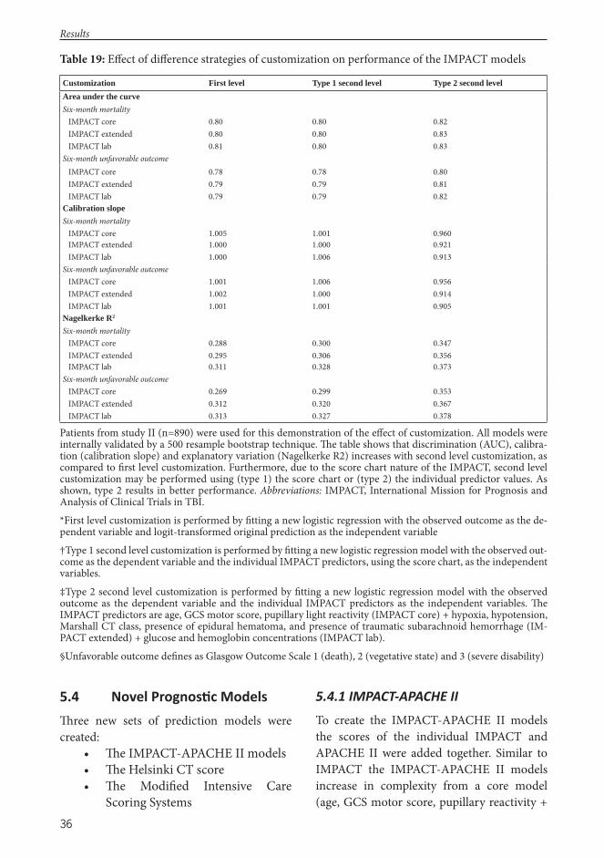
36
5.4 Novel Prognos c ModelsTh ree new sets of prediction models were created:
• Th e IMPACT-APACHE II models• Th e Helsinki CT score• Th e Modifi ed Intensive Care
Scoring Systems
5.4.1 IMPACT-APACHE II
To create the IMPACT-APACHE II models the scores of the individual IMPACT and APACHE II were added together. Similar to IMPACT the IMPACT-APACHE II models increase in complexity from a core model (age, GCS motor score, pupillary reactivity +
Table 19: Eff ect of diff erence strategies of customization on performance of the IMPACT models
Customization First level Type 1 second level Type 2 second levelArea under the curveSix-month mortality IMPACT core 0.80 0.80 0.82 IMPACT extended 0.80 0.80 0.83 IMPACT lab 0.81 0.80 0.83Six-month unfavorable outcome IMPACT core 0.78 0.78 0.80 IMPACT extended 0.79 0.79 0.81 IMPACT lab 0.79 0.79 0.82Calibration slopeSix-month mortality IMPACT core 1.005 1.001 0.960 IMPACT extended 1.000 1.000 0.921 IMPACT lab 1.000 1.006 0.913Six-month unfavorable outcome IMPACT core 1.001 1.006 0.956 IMPACT extended 1.002 1.000 0.914 IMPACT lab 1.001 1.001 0.905Nagelkerke R2
Six-month mortality IMPACT core 0.288 0.300 0.347 IMPACT extended 0.295 0.306 0.356 IMPACT lab 0.311 0.328 0.373Six-month unfavorable outcome IMPACT core 0.269 0.299 0.353 IMPACT extended 0.312 0.320 0.367 IMPACT lab 0.313 0.327 0.378
Patients from study II (n=890) were used for this demonstration of the eff ect of customization. All models were internally validated by a 500 resample bootstrap technique. Th e table shows that discrimination (AUC), calibra-tion (calibration slope) and explanatory variation (Nagelkerke R2) increases with second level customization, as compared to fi rst level customization. Furthermore, due to the score chart nature of the IMPACT, second level customization may be performed using (type 1) the score chart or (type 2) the individual predictor values. As shown, type 2 results in better performance. Abbreviations: IMPACT, International Mission for Prognosis and Analysis of Clinical Trials in TBI.*First level customization is performed by fi tting a new logistic regression with the observed outcome as the de-pendent variable and logit-transformed original prediction as the independent variable†Type 1 second level customization is performed by fi tting a new logistic regression model with the observed out-come as the dependent variable and the individual IMPACT predictors, using the score chart, as the independent variables. ‡Type 2 second level customization is performed by fi tting a new logistic regression model with the observed outcome as the dependent variable and the individual IMPACT predictors as the independent variables. Th e IMPACT predictors are age, GCS motor score, pupillary light reactivity (IMPACT core) + hypoxia, hypotension, Marshall CT class, presence of epidural hematoma, and presence of traumatic subarachnoid hemorrhage (IM-PACT extended) + glucose and hemoglobin concentrations (IMPACT lab).§Unfavorable outcome defi nes as Glasgow Outcome Scale 1 (death), 2 (vegetative state) and 3 (severe disability)
Results
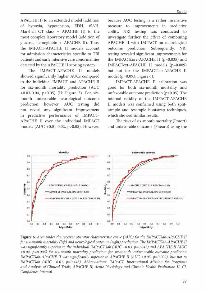
37
APACHE II) to an extended model (addition of hypoxia, hypotension, EDH, tSAH, Marshall CT class + APACHE II) to the most complex laboratory model (addition of glucose, hemoglobin + APACHE II). Th us, the IMPACT-APACHE II models account for admission characteristics specifi c to TBI patients and early intensive care abnormalities detected by the APACHE II scoring system.
Th e IMPACT-APACHE II models showed signifi cantly higher AUCs compared to the individual IMPACT and APACHE II for six-month mortality prediction (AUC +0.03-0.04, p<0.05) (II: Figure 3). For six-month unfavorable neurological outcome prediction, however, AUC testing did not reveal any signifi cant improvement in predictive performance of IMPACT-APACHE II over the individual IMPACT models (AUC +0.01-0.02, p>0.05). However,
because AUC testing is a rather insensitive measure to improvements in predictive ability, NRI testing was conducted to investigate further the eff ect of combining APACHE II with IMPACT on neurological outcome prediction. Subsequently, NRI testing revealed signifi cant improvements for the IMPACTcore-APACHE II (p=0.035) and IMPACText-APACHE II models (p=0.009) but not for the IMPACTlab-APACHE II model (p=0.093, Figure 6).
IMPACT-APACHE II calibration was good for both six-month mortality and unfavorable outcome prediction (p>0.05). Th e internal validity of the IMPACT-APACHE II models was confi rmed using both split-sample and resample bootstrap techniques, which showed similar results.
Th e risks of six-month mortality (Pmort) and unfavorable outcome (Pneuro) using the
Figure 6: Area under the receiver operator characteristic curve (AUC) for the IMPACTlab-APACHE II for six-month mortality (left ) and neurological outcome (right) prediction. Th e IMPACTlab-APACHE II was signifi cantly superior to the individual IMPACT lab (AUC +0.03, p=0.043) and APACHE II (AUC +0.04, p=0.006) for six-month mortality prediction; for six-month unfavourable outcome prediction IMPACTlab-APACHE II was signifi cantly superior to APACHE II (AUC +0.05, p=0.002), but not to IMPACTlab (AUC +0.01, p=0.448). Abbreviations: IMPACT, International Mission for Prognosis and Analysis of Clinical Trials; APACHE II, Acute Physiology and Chronic Health Evaluation II; CI, Confi dence Interval
Results
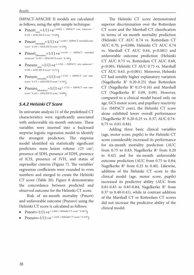
38
IMPACT-APACHE II models are calculated as follows, using the split-sample technique: Pmortcore=1/(1+e-(-6.004 + IMPACT core sumscore *
0.234 + APACHE II score * 0.160) Pmortextended=1/(1+e-(-6.265 + IMPACT extended sum-
score * 0.193 + APACHE II score * 0.158) Pmortlaboratory=1/(1+e-(-6.516 + IMPACT extended
sumscore * 0.187 + APACHE II score * 0.149) Pneurocore=1/(1+e-(-3.363 + IMPACT core sumscore *
0.198 + APACHE II score * 0.113) Pneuroextended=1/(1+e-(-3.551 + IMPACT core sum-
score * 0.175 + APACHE II score * 0.105) Pneurolaboratory=1/(1+e-(-3.732 + IMPACT core sum-
score * 0.162 + APACHE II score * 0.099)
5.4.2 Helsinki CT Score
In univariate analysis 11 of the predefi ned CT characteristics were signifi cantly associated with unfavorable six-month outcome. Th ese variables were inserted into a backward stepwise logistic regression model to identify the strongest predictors. Th e stepwise model identifi ed six statistically signifi cant predictors: mass lesion volume ≥25 cm3, presence of SDH, presence of EDH, presence of ICH, presence of IVH, and status of suprasellar cisterns (Figure 7). Th e variables’ regression coeffi cients were rounded to even numbers and merged to create the Helsinki CT score (Table 20). Figure 8 demonstrates the concordance between predicted and observed outcome for the Helsinki CT score.
Risk of six-month mortality (Pmort) and unfavorable outcome (Pneuro) using the Helsinki CT score is calculated as follows: Pmort=1/(1+e-(-2.666 + Helsinki CT score * 0.287)) Pneuro=1/(1+e-(-1.636 + Helsinki CT score * 0.319))
Th e Helsinki CT score demonstrated superior discrimination over the Rotterdam CT score and the Marshall CT classifi cation in terms of six-month mortality prediction (Helsinki CT AUC 0.74 vs. Rotterdam CT AUC 0.70, p=0.006; Helsinki CT AUC 0.74 vs. Marshall CT AUC 0.64, p<0.001) and unfavorable outcome prediction (Helsinki CT AUC 0.75 vs. Rotterdam CT AUC 0.68, p<0.001; Helsinki CT AUC 0.75 vs. Marshall CT AUC 0.63, p<0.001). Moreover, Helsinki CT had notably higher explanatory variation (Nagelkerke R2 0.20-0.25) than Rotterdam CT (Nagelkerke R2 0.15-0.16) and Marshall CT (Nagelkerke R2 0.09, 0.09). However, compared to a clinical model based only on age, GCS motor score, and pupillary reactivity (i.e. IMPACT core), the Helsinki CT score alone exhibited lower overall performance (Nagelkerke R2 0.20-0.25 vs. 0.37; AUC 0.74-0.75 vs. 0.81-0.84).
Adding three basic clinical variables (age, motor score, pupils) to the Helsinki CT score considerably increased its performance for six-month mortality prediction (AUC from 0.75 to 0.83; Nagelkerke R2 from 0.20 to 0.42) and for six-month unfavorable outcome prediction (AUC from 0.75 to 0.84; Nagelkerke R2 from 0.25 to 0.40). Likewise, addition of the Helsinki CT score to the clinical model (age, motor score, pupils) increased its predictive ability (AUC from 0.81-0.83 to 0.83-0.84; Nagelkerke R2 from 0.37 to 0.40-0.41), while in contrast addition of the Marshall CT or Rotterdam CT scores did not increase the predictive ability of the clinical model.
Results
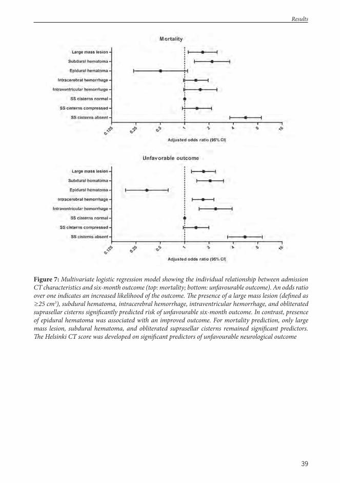
39
Figure 7: Multivariate logistic regression model showing the individual relationship between admission CT characteristics and six-month outcome (top: mortality; bottom: unfavourable outcome). An odds ratio over one indicates an increased likelihood of the outcome. Th e presence of a large mass lesion (defi ned as ≥25 cm3), subdural hematoma, intracerebral hemorrhage, intraventricular hemorrhage, and obliterated suprasellar cisterns signifi cantly predicted risk of unfavourable six-month outcome. In contrast, presence of epidural hematoma was associated with an improved outcome. For mortality prediction, only large mass lesion, subdural hematoma, and obliterated suprasellar cisterns remained signifi cant predictors. Th e Helsinki CT score was developed on signifi cant predictors of unfavourable neurological outcome
Results
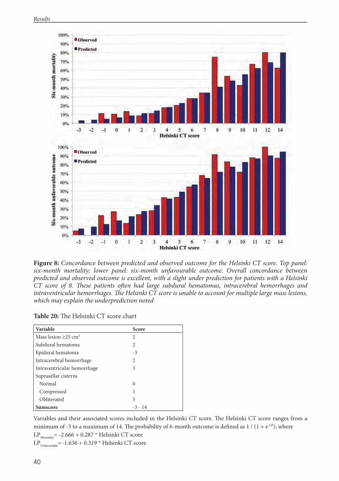
40
Figure 8: Concordance between predicted and observed outcome for the Helsinki CT score. Top panel: six-month mortality; lower panel: six-month unfavourable outcome. Overall concordance between predicted and observed outcome is excellent, with a slight under prediction for patients with a Helsinki CT score of 8. Th ese patients oft en had large subdural hematomas, intracerebral hemorrhages and intraventricular hemorrhages. Th e Helsinki CT score is unable to account for multiple large mass lesions, which may explain the underprediction noted
Table 20: Th e Helsinki CT score chart
Variable ScoreMass lesion ≥25 cm3 2Subdural hematoma 2Epidural hematoma -3Intracerebral hemorrhage 2Intraventricular hemorrhage 3Suprasellar cisterns Normal 0 Compressed 1 Obliterated 5Sumscore -3 - 14
Variables and their associated scores included in the Helsinki CT score. Th e Helsinki CT score ranges from a minimum of -3 to a maximum of 14. Th e probability of 6-month outcome is defi ned as 1 / (1 + e-LP), whereLPMortality= -2.666 + 0.287 * Helsinki CT scoreLPUnfavorable= -1.636 + 0.319 * Helsinki CT score
Results
41
5.4.3 Modifi ed Intensive Care Scoring Systems
Th e modifi ed intensive care scoring systems include customized versions of APACHE II and SAPS II, an adjusted SOFA score, and the reference model. APACHE II and SAPS II were originally developed for predicting risk of in-hospital mortality in the general ICU population. Th ese two models were specifi cally customized for six-month outcome prediction in patients with TBI treated in the ICU (Figure 9). Th e risks of six-month mortality (Pmort) using the APACHE II and SAPS II models are calculated as follows: PmortAPACHE II=1/(1+e-(-5.183 + APACHE II score *
0.193)) PmortSAPS II=1/(1+e-(-4.967 + SAPS II score * 0.092))
Th e risk of six-month unfavorable neurological outcome (Pneuro) using APACHE II is calculated as follows: PneuroAPACHE II=1/(1+e-(-3.335 + APACHE II score *
0.170)
Th e original SOFA showed poor performance for predicting outcome in patients with moderate to severe TBI (AUC 0.68). Th us, it was modifi ed to suit the TBI population. Accordingly, the adjusted SOFA is a modifi ed version of the original SOFA designed specifi cally for patients with moderate to severe TBI. In the adjusted SOFA, GCS is included as a separate variable to give it more infl uence. Like GCS, age is similarly included as a separate variable, which signifi cantly increased the predictive ability of (adjusted) SOFA to match APACHE II (AUC 0.79 vs. 0.79, p=0.920) and SAPS II (AUC 0.79 vs. 0.80, p=0.745).
Th e reference model is a simple prognostic model based solely on patient age and worst measured GCS during the fi rst 24 hours in the ICU. Age was categorized into ten-year intervals and GCS was dichotomized
based on the median, as this combination was found to give the highest performance. Th e reference model showed good discrimination (AUC 0.77, 95% CI 0.74-0.80) and calibration (H-L p-value 0.086). It is notable that there were no signifi cant diff erences in discrimination, calibration, or precision between the reference model and APACHE II (AUC -0.02, p=0.425), or between the reference model and SAPS II (AUC -0.03, p=0.218).
To test further the eff ect of age and GCS on outcome, patients from Study II and IV were pooled into a total study population of 2,430 patients with moderate and severe TBI (GCS 3-13, N=805 from II and N=1,625 from IV). Th e combined six-month mortality rate was 30% (II: 24%, IV: 33%), the median age 56 years (IQR 41-67, II: 58 years [IQR 45-68], IV: 55 years [IQR 38-66]), and the median GCS 7 (IQR 4-77, II: 9 [IQR 4-11], IV: 6 [IQR 4-10]). In this large set of pooled data, age and GCS were used as continuous variables to minimize loss of information. Th e risk of six-month mortality was calculated using the following equation: Pmortage+GCS=1/(1+e-(-0.801 + 0.037 * AGE + (-0.309)
* GCS))
Th e apparent AUC of the new combined model was 0.79 (95% CI 0.77-0.81) with good calibration (H-L p-value=0.410). Th e model was internally validated in Study II with an AUC of 0.81 (95% CI 0.77-0.84) and in Study IV with an AUC of 0.78 (95% CI 0.0.75-0.80). Calibration was good in both datasets (II: H-L p-value=0.088; IV: H-L p-value=0.142, Figure 10). Bootstrap validation of the model revealed close to no optimism with an AUC of 0.787 (and an optimism corrected Nagelkerke R2 of 0.29). A comparison of the calibration between the intensive care scoring systems from study IV is shown in Figure 11.
Results
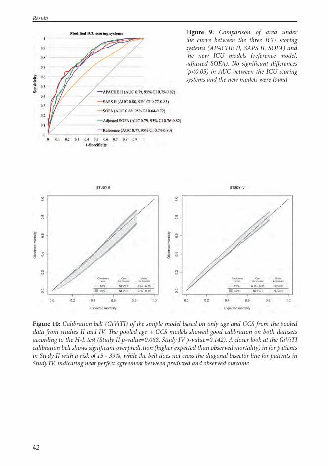
42
Figure 10: Calibration belt (GiViTI) of the simple model based on only age and GCS from the pooled data from studies II and IV. Th e pooled age + GCS models showed good calibration on both datasets according to the H-L test (Study II p-value=0.088, Study IV p-value=0.142). A closer look at the GiViTI calibration belt shows signifi cant overprediction (higher expected than observed mortality) in for patients in Study II with a risk of 15 - 39%, while the belt does not cross the diagonal bisector line for patients in Study IV, indicating near perfect agreement between predicted and observed outcome
Figure 9: Comparison of area under the curve between the three ICU scoring systems (APACHE II, SAPS II, SOFA) and the new ICU models (reference model, adjusted SOFA). No signifi cant diff erences (p<0.05) in AUC between the ICU scoring systems and the new models were found
Results
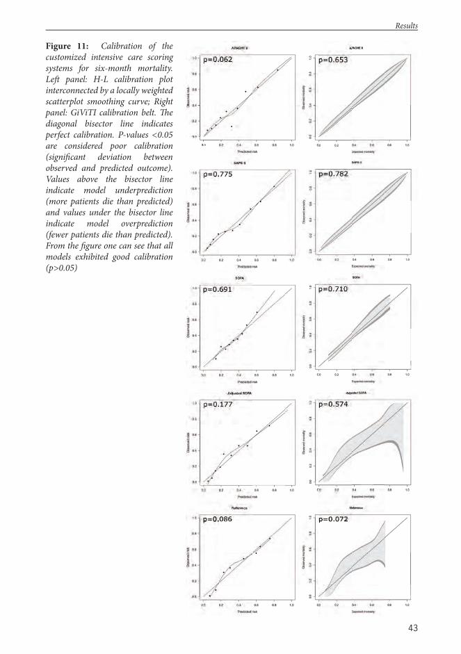
43
Figure 11: Calibration of the customized intensive care scoring systems for six-month mortality. Left panel: H-L calibration plot interconnected by a locally weighted scatterplot smoothing curve; Right panel: GiViTI calibration belt. Th e diagonal bisector line indicates perfect calibration. P-values <0.05 are considered poor calibration (signifi cant deviation between observed and predicted outcome). Values above the bisector line indicate model underprediction (more patients die than predicted) and values under the bisector line indicate model overprediction (fewer patients die than predicted). From the fi gure one can see that all models exhibited good calibration (p>0.05)
Results
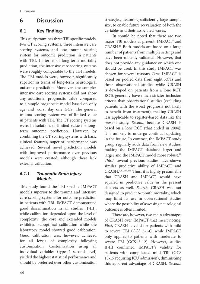
44
6 Discussion
6.1 Key FindingsTh is study examines three TBI specifi c models, two CT scoring systems, three intensive care scoring systems, and one trauma scoring system for outcome prediction in patients with TBI. In terms of long-term mortality prediction, the intensive care scoring systems were roughly comparable to the TBI models. Th e TBI models were, however, signifi cantly superior in terms of long-term neurological outcome prediction. Moreover, the complex intensive care scoring systems did not show any additional prognostic value compared to a simple prognostic model based on only age and worst day one GCS. Th e general trauma scoring system was of limited value in patients with TBI. Th e CT scoring systems were, in isolation, of limited value for long-term outcome prediction. However, by combining the CT scoring systems with basic clinical features, superior performance was achieved. Several novel prediction models with improved performance over previous models were created, although these lack external validation.
6.1.1 Trauma c Brain Injury Models
Th is study found the TBI specifi c IMPACT models superior to the trauma and intensive care scoring systems for outcome prediction in patients with TBI. IMPACT demonstrated good discrimination in all studies (I-III), while calibration depended upon the level of complexity: the core and extended models exhibited suboptimal calibration while the laboratory model showed good calibration. Good calibration was, however, achieved for all levels of complexity following customization. Customization using all individual variables (type 2 second level) yielded the highest statistical performance and should be preferred over other customization
strategies, assuming suffi ciently large sample size, to enable future reevaluation of both the variables and their associated scores.
In should be noted that there are two major TBI models at present: IMPACT9 and CRASH.10 Both models are based on a large number of patients from multiple settings and have been robustly validated. However, that does not provide any guidance on which one should be used. In this study IMPACT was chosen for several reasons. First, IMPACT is based on pooled data from eight RCTs and three observational studies while CRASH is developed on patients from a lone RCT. RCTs generally have much stricter inclusion criteria than observational studies (excluding patients with the worst prognosis not likely to benefi t from treatment), making CRASH less applicable to register-based data like the present study. Second, because CRASH is based on a lone RCT (that ended in 2004), it is unlikely to undergo continual updating in the future. In contrast, the IMPACT study group regularly adds data from new studies, making the IMPACT database larger and larger and the IMPACT model more robust.18 Th ird, several previous studies have shown similar predictive ability of IMPACT and CRASH.9,10,164,165 Th us, it is highly presumable that CRASH and IMPACT would have equaled in predictive value in the present datasets as well. Fourth, CRASH was not designed to predict 6-month mortality, which may limit its use in observational studies where the possibility of assessing neurological outcome is oft en limited.
Th ere are, however, two main advantages of CRASH over IMPACT that merit noting. First, CRASH is valid for patients with mild to severe TBI (GCS 3-14), while IMPACT only applies to patients with moderate to severe TBI (GCS 3-12). However, studies II-III confi rmed IMPACT’s validity for patients with complicated mild TBI (GCS 13-15 requiring ICU admission), diminishing this apparent advantage of CRASH. Second,
Discussion
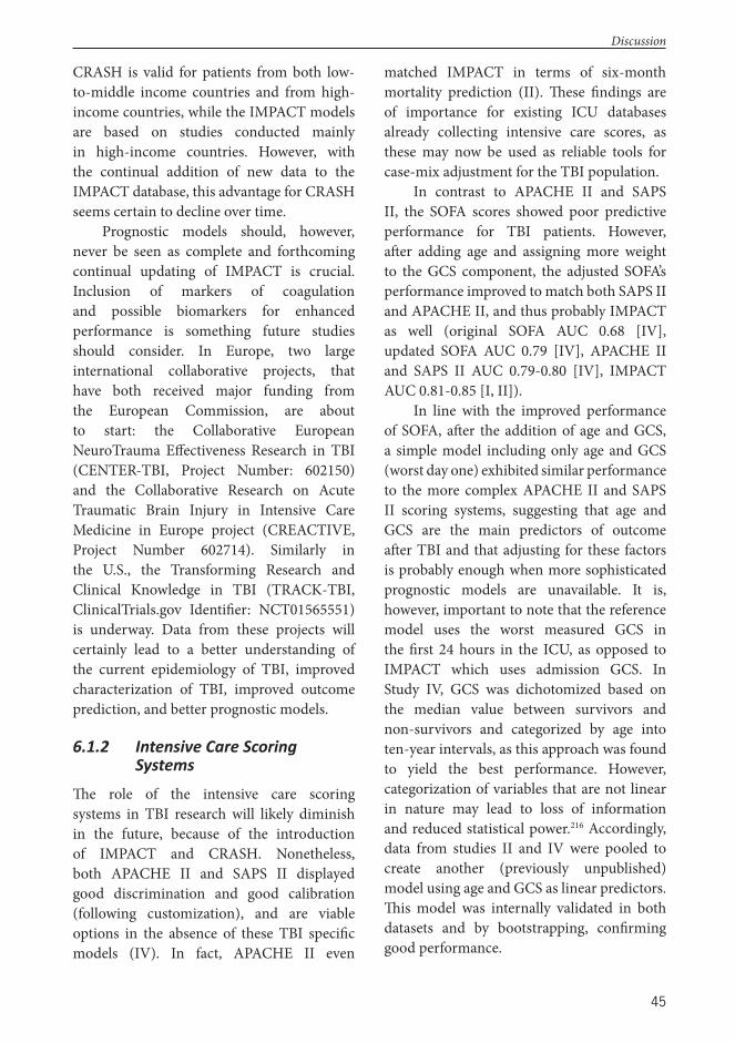
45
CRASH is valid for patients from both low-to-middle income countries and from high-income countries, while the IMPACT models are based on studies conducted mainly in high-income countries. However, with the continual addition of new data to the IMPACT database, this advantage for CRASH seems certain to decline over time.
Prognostic models should, however, never be seen as complete and forthcoming continual updating of IMPACT is crucial. Inclusion of markers of coagulation and possible biomarkers for enhanced performance is something future studies should consider. In Europe, two large international collaborative projects, that have both received major funding from the European Commission, are about to start: the Collaborative European NeuroTrauma Eff ectiveness Research in TBI (CENTER-TBI, Project Number: 602150) and the Collaborative Research on Acute Traumatic Brain Injury in Intensive Care Medicine in Europe project (CREACTIVE, Project Number 602714). Similarly in the U.S., the Transforming Research and Clinical Knowledge in TBI (TRACK-TBI, ClinicalTrials.gov Identifi er: NCT01565551) is underway. Data from these projects will certainly lead to a better understanding of the current epidemiology of TBI, improved characterization of TBI, improved outcome prediction, and better prognostic models.
6.1.2 Intensive Care Scoring Systems
Th e role of the intensive care scoring systems in TBI research will likely diminish in the future, because of the introduction of IMPACT and CRASH. Nonetheless, both APACHE II and SAPS II displayed good discrimination and good calibration (following customization), and are viable options in the absence of these TBI specifi c models (IV). In fact, APACHE II even
matched IMPACT in terms of six-month mortality prediction (II). Th ese fi ndings are of importance for existing ICU databases already collecting intensive care scores, as these may now be used as reliable tools for case-mix adjustment for the TBI population.
In contrast to APACHE II and SAPS II, the SOFA scores showed poor predictive performance for TBI patients. However, aft er adding age and assigning more weight to the GCS component, the adjusted SOFA’s performance improved to match both SAPS II and APACHE II, and thus probably IMPACT as well (original SOFA AUC 0.68 [IV], updated SOFA AUC 0.79 [IV], APACHE II and SAPS II AUC 0.79-0.80 [IV], IMPACT AUC 0.81-0.85 [I, II]).
In line with the improved performance of SOFA, aft er the addition of age and GCS, a simple model including only age and GCS (worst day one) exhibited similar performance to the more complex APACHE II and SAPS II scoring systems, suggesting that age and GCS are the main predictors of outcome aft er TBI and that adjusting for these factors is probably enough when more sophisticated prognostic models are unavailable. It is, however, important to note that the reference model uses the worst measured GCS in the fi rst 24 hours in the ICU, as opposed to IMPACT which uses admission GCS. In Study IV, GCS was dichotomized based on the median value between survivors and non-survivors and categorized by age into ten-year intervals, as this approach was found to yield the best performance. However, categorization of variables that are not linear in nature may lead to loss of information and reduced statistical power.216 Accordingly, data from studies II and IV were pooled to create another (previously unpublished) model using age and GCS as linear predictors. Th is model was internally validated in both datasets and by bootstrapping, confi rming good performance.
Discussion
46
Th ere are several limitations of the intensive care scores that may limit their use in TBI research. First, it takes a minimum of 24 hours to estimate patient risk using the ICU models, and thus, initial risk stratifi cation for clinical trial inclusion is usually not possible. Second, although modern computer science has made it possible to collect data automatically, it is still more resource- and time- consuming than models that include only admission characteristics. Th ird, any score that uses data collected from the fi rst 24 hours is aff ected by the quality of care provided, so that high scores may be the result of either poor care or high severity of illness or both.19,217 It should, however, be noted that there are ICU models based on ICU admission variables that do not require 24-hour data collection which were not investigated in the present study, such as the MPM0-II218
and the MPM0-III219 models.
6.1.3 Trauma Scoring Systems
Th e RISC was of limited value for patients with moderate to severe TBI. In a subgroup of patients with isolated TBI, AUC was ‘only’ 0.76. Generally, values over 0.75 are considered suffi cient.119 However, the predictive ability was only examined for 30-day hospital mortality, an outcome measure known to underestimate mortality rates aft er TBI signifi cantly.29 Furthermore, external validation studies of IMPACT have shown AUC values up to 0.90 for predicting six-month mortality.161 Accordingly, discrimination of the RISC was considered to be only modest for isolated TBI patients.
RISC calibration was notably poor in every subgroup of TBI patients. Calibration was measured by the H-L Ĉ-statistic, which may falsely generate p-values <0.05 for large sample sizes, indicating poor calibration even when calibration in fact is good.125 Th e RISC gave an H-L χ2 of 382 (p<0.001) with a study population of 9,106 (TR-DGU),
indicating poor calibration. In comparison, the development of APACHE IV included 110,558 patients and gave an H-L χ2 of 17 and an associated p-value of 0.08, showing that good calibration is achievable even is large sample sizes, if the model is good.185 Moreover, concordance between predicted and observed mortality for the RISC was defi cient, especially for the higher risk intervals (V: Figure 3). Th us, although the H-L test in Study V is aff ected by the large sample size, poor calibration was prominent.
Th e RISC has routinely been used as a benchmarking tool in one of Europe’s largest trauma databases, the TR-DGU. Because TBI is the leading cause of death in trauma it is crucial for correct benchmarking that the prognostic model used provide accurate outcome predictions in TBI.220 For accurate benchmarking in the future, the RISC ought to be updated to account better for patients with TBI. In fact, the importance of TBI has been acknowledged in the new, updated RISC II, which has now replaced the ‘old’ RISC.221
Most existing scoring systems like the RISC and TRISS are based largely on the anatomical injury severity scoring system AIS, oft en through the ISS or the NISS. Th e AIS is seldom calculated upon hospital admission, as it oft en requires primary, secondary, and tertiary patient surveys to assess all injuries completely. Th us, the trauma models cannot oft en be used as a tool for early baseline risk adjustment for study enrollment, one of the main purposes of prediction models in TBI research. Furthermore, the eff ect of extra-cranial injury on outcome was found to be negligible in the present study, which further questions the role of the general trauma scoring systems in TBI research.
6.1.4 IMPACT-APACHE II
Both IMPACT and APACHE II showed good predictive ability in TBI patients (II). Nevertheless, adjusting only for baseline
Discussion
47
prognostic risk by using IMPACT, later aspects of care are ignored, such as the quality of intensive care. Inter-center diff erences in process and quality of care are a confounding factor in most multi-center studies. For example, closer investigation of the IMPACT and the CRASH studies found large inter-center diff erences in outcome, even among European centers.222,223 On the other hand, adjusting only for abnormalities measured in the fi rst 24 hours of ICU admission ignores initial injury severity. To overcome these diffi culties, the IMPACT-APACHE II models were created as a combination of IMPACT and APACHE II with increasing levels of complexity (core, extended, and laboratory versions). Th e models account for both baseline injury severity (by IMPACT) and for early physiological abnormalities measured in the ICU (by APACHE II). Moreover, patient co-morbidity is taken into account as well (by APACHE II), which is nowadays a crucial aspect of the aging TBI population.37 Given the factors outlined, it came as no surprise that IMPACT-APACHE II showed superior predictive performance to the individual IMPACT models and APACHE II for outcome prediction. Th us, the novel IMPACT-APACHE II models off er a way to adjust for not only for baseline risk, but also for early aspects of intensive care and patient co-morbidities, and might come to serve as a powerful tool in increasing design and statistical power of forthcoming studies.
6.1.5 Helsinki CT Score
Th e Helsinki CT score ranges from -3 points (isolated small EDH) to a maximum of 14 points. In addition to the CT characteristics, patient age, GCS motor score, and pupillary reactivity can be added for increased performance. Th e Helsinki CT score was shown to be superior to both the Marshall CT classifi cation and the Rotterdam CT score for outcome prediction. However, some valuable
features of the Marshall and Rotterdam CT scores must be recognized.
Th e Marshall CT classifi cation was not developed as a prediction tool and its usefulness lies in its descriptive value, which the Helsinki CT score does not replace. Th e Rotterdam CT score, on the other hand, was designed to predict outcome. However, at the cost of predictive performance the Rotterdam CT score was designed to range from 1 to 6, mimicking the GCS motor score. Th us, the Rotterdam CT score could be argued to be easier to interpret than the Helsinki CT score. However, the Helsinki CT score is also presented in a user-friendly score chart and the probabilities can easily be calculated using a publically available Microsoft Offi ce Excel® worksheet (http://links.lww.com/NEU/A676). It should, however, be noted that the Rotterdam CT score was designed to predict six-month mortality, whereas the Helsinki CT score was designed to predict six-month unfavorable outcome. Th e diff erences between the Helsinki CT and Rotterdam CT scores reinforce the point that the same variables cannot be expected to predict both mortality and neurological outcome. In summary, the Helsinki CT score probably is probably advantageous over the Rotterdam CT score for predictive purposes, although external validation studies are needed to confi rm this.
6.2 Early Predictors of Outcome a er TBI
6.2.1 Markers of Coagula on
One in three patients with TBI has evidence of coagulopathy during the treatment course.83 Th e presence of coagulopathy signifi cantly increases risk of hemorrhagic and ischemic lesion progression and consequently risk for unfavorable outcome and even death.82,83,224,225 Results from the IMPACT study suggest that abnormalities in admission INR may be an important predictor of long-term outcome
Discussion
48
aft er TBI.78 Study I confi rmed the association between INR and outcome. Th e fact that INR signifi cantly increased the AUC of the IMPACT laboratory model (by +0.02) indicates a very strong association with mortality. Moreover, the explanatory variation of INR was similar to that of glucose, which is a known strong predictor of outcome.77,78 In contrast, no signifi cant relationship between INR and long-term neurological outcome could be established by any of logistic regression, AUC, or NRI testing, showing that one cannot expect the same predictors to predict neurological outcome and mortality. Study I further showed that platelet count was not an independent predictor of outcome in TBI patients, supporting the hypothesis that platelet function rather than platelet count is the determining factor of platelet associated coagulopathy.226
6.2.2 Major Extra-Cranial Injury
Major extra-cranial injury (MEI) is present in about 23% to 41% of patients with TBI, depending on the population and defi nition of TBI and MEI.10,227 Th e role of MEI on outcome in patients with TBI is, however, debated. Some studies suggest that outcome aft er TBI is mainly dependent upon severity of brain injury and that coexisting MEI plays little part, whereas other studies advocate that presence of concomitant MEI signifi cantly increases the likelihood of poor outcome.10,66,67,227,228
A meta-analysis including roughly 40,000 patients from the IMPACT, CRASH, and TARN databases found MEI (defi ned as AIS 3 or higher or an injury requiring hospital admission on its own) to be a strong predictor of outcome aft er TBI.65 However, the strength of the eff ect was inversely related to the degree of brain injury, so that the more severe the brain injury, the lesser the eff ect of MEI.
A likely reason why previous studies have yielded confl icting results is that no clear defi nition of MEI exists.229 A commonly used defi nition of MEI is ISS >15, although ISS cut-off points up to >25 have also been used.177,230 In Study I, MEI was defi ned as ISS >15 and >25, whereas in Study V, MEI was defi ned as a head-AIS of ≥3, plus at least one other body part at AIS ≥2. Both studies (I, V) systematically found MEI to remain an insignifi cant predictor of outcome, aft er adjusting for other markers of injury. In fact, in Study V patients with TBI and concomitant MEI (polytrauma TBI) had a slightly lower risk of mortality than TBI patients without MEI (isolated TBI). Th is fi nding, however, is probably explained by the fact that patients with isolated TBI had a more severe TBI (by head-AIS) than polytrauma TBI patients. Th us, the fi ndings support the hypothesis that severity of brain injury is the major determinant of outcome in patients with TBI.
A recent international consensus meeting proposed a new defi nition of MEI: at least two injuries with AIS ≥3, plus at least one of the following physiological derangements: hypotension, decreased level of consciousness, acidosis, or coagulopathy.231 Th is defi nition has, however, been neither investigated nor validated in the TBI population, and thus, the role of MEI on outcome aft er TBI remains controversial. Standardized data collection and uniform defi nitions are advocated for appropriate prognostic research, so that the best treatment for TBI patients with associated injuries can be determined.
6.2.3 Early Computerized Tomography Characteris cs
Th e admission CT characteristics associated with poor outcome irrespective of patient age, GCS motor score, or pupillary light reactivity were: mass lesion volume ≥25 cm3, type of mass lesion (SDH, ICH, EDH), tSAH in basal cisterns, presence of IVH, abnormal
Discussion
49
fourth ventricle, absent suprasellar cisterns, absent ambiens cisterns, and bilateral cortical sulci eff acement. Still, only six of these were independently associated with outcome: mass lesion volume ≥25 cm3, type of mass lesion (SDH, ICH, EDH), presence of IVH, and status of suprasellar cisterns (the variables together constituting the Helsinki CT score).
Several previous studies have suggested degree of midline shift and presence of tSAH to be the strongest predictors (detected by CT imaging) of poor outcome aft er TBI.10,68,69,167,232,233 In spite of this, both midline shift and tSAH were found to be insignifi cant and consequently omitted from the Helsinki CT score (III). Th e reasons for these diff erent results remain uncertain, but they should certainly be noted.
Midline shift , when measured on admission, is oft en the result of a space-occupying mass lesion, and thus, highly amendable to correction through mass lesion evacuation. In contrast, day one or post-operative midline shift is probably a much more informative predictor than admission or pre-operative midline shift , which might explain the weak association between midline shift and outcome in the present study.
Traumatic SAH was previously thought as one of the strongest predictors of poor outcome in TBI.68,69,233-236 In the IMPACT study, the presence of tSAH independently doubled the odds of poor outcome.9,68 Likewise, in the CRASH trial, presence of tSAH was strongly associated with poor outcome.10 In contrast, in the present study, tSAH was insignifi cant in multivariate analysis (I-III). Lack of statistical power might be one reason why tSAH was insignifi cant. In fact, most patients had tSAH (presence of tSAH in Study I: 67%, II: 57%, III: 58%) and the diff erence in incidence between those with good vs. poor outcome might have been too small to make any diff erence. Another reason might be that tSAH and
IVH were diff erentiated in this study. Th e presence of IVH and tSAH strongly correlates with the risk of developing post-traumatic hydrocephalus, which is strongly associated with poor outcome.237,238 Up to 40% of patients with moderate to severe TBI develop post-traumatic hydrocephalus and the presence of tSAH, and especially IVH, increases the risk.237,238 Th us, separating tSAH and IVH might explain why tSAH was found to be insignifi cant when adjusting for IVH.238
Th e Rotterdam CT and Marshall CT scoring systems were both of limited value for long-term outcome prediction (III). However, the addition of age, motor score, and pupils signifi cantly improved discrimination drastically, with AUCs rising from 0.63-0.75 to 0.81-0.84 and explanatory variation values rising from 9-16% to 38-39%. Th is shows that early CT fi ndings should not be used in isolation to establish patient prognosis, but should always be combined with relevant clinical features. It is notable that even aft er combining patient clinical characteristics with CT features, approximately 40% of the actual outcome was explained.
6.3 Sta s cal Considera onsGood discrimination does not necessarily mean good calibration and vice versa. In fact, perfectly calibrated models cannot achieve the theoretical AUC maximum of 1.121 Discrimination was measured in all studies using the AUC. Th e AUC is highly dependent on the underlying case-mix; a heterogeneous population increases AUC while a homogenous population decreases AUC. Studies I, II, and III included patients with mild, moderate, and severe TBI (GCS 3-15, all requiring ICU admission), whereas studies IV and V only included patients with moderate and severe TBI (GCS 3-13 in IV and AIS-head ≥3 in V). Th us, studies I, II and III may be considered more heterogeneous than studies IV and V, which could aff ect the
Discussion
50
AUC reported. To avoid such bias, a case-mix adjusted AUC has been proposed.239
We used three types of calibration tests: the H-L test (I, II, IV, V), the calibration slope (III), and the GiViTI calibration belt (II, IV). Th e H-L test and the calibration slope have been extensively used in the past. Studies II and IV are, however, among the fi rst to assess and compare the H-L and GiViTI calibration tests.240 Th e main advantage of the GiViTI calibration belt over the H-L test and the calibration slope is the possibility of estimating 95% confi dence intervals over the whole risk spectrum. Th us, the calibration belt provides valuable information about the degree and direction of miscalibration, such as whether it aff ects only a specifi c risk interval or if the overall calibration is poor. Th is is valuable not only for external validation of prognostic models but also for evaluating and comparing the quality of care at individual centers.
We found the H-L and the GiViTI tests to produce similar results for external validation studies (II, IV). Th us, for overall calibration testing, the H-L test is probably suffi cient. However, when the H-L test indicates poor calibration (p<0.05) one may utilize the GiViTI calibration belt to assess the signifi cance of miscalibration and pinpoint risk intervals of either under- or overprediction.126,211,241 Future studies are necessary to gain a better understanding of the benefi ts and possible pitfalls of using the GiViTI calibration belt.
6.4 Pa ent Outcome a er Trauma c Brain Injury
Overall six-month mortality rate ranged between 23% and 33% and six-month unfavorable outcome was between 47% and 57% (I-IV). For patients with severe TBI (GCS ≤8) the weighted six-month mortality rate was 40% and unfavorable outcome rate
63%. Th ese numbers are somewhat higher compared to some previous observational studies (Table 21, a non-systematic review of outcome in observational studies). It should, however, be noted that in the present study, median patient age was just below 60 years, whereas in previous studies it has generally been below 40 years (Figure 12). It is widely known that age is one of the strongest predictors of outcome aft er TBI.29,49-53 Th e relationship between older age and poor outcome has been suggested to be linear.49 Th us, the increasing age of TBI patients is most likely a primary reason why poor outcome was more frequently noted in the present study. Th is deduction also supports the theory of stagnated improvements in outcome aft er TBI because of the epidemiologic shift previously proposed.37,92
Th e importance of long-term follow-up in TBI patients cannot be overemphasized. Studies using hospital mortality as the primary outcome measure are severely biased for two main reasons. First, discharge policies vary among hospitals, biasing follow-up time.142,143 Second, a substantial number of TBI patients die following hospital discharge, biasing outcome rates.29,50-52 Supporting these concerns, Study II showed a 14-day mortality rate of 11%, compared to a 33% six-month mortality rate in Study IV, for a staggering 200% increase. Th us, hospital mortality severely underestimates mortality rates aft er TBI and should be discouraged as a primary outcome measure in TBI research. Furthermore, it was found that mortality rates steadily increased over the entire follow-up period, suggesting that even a six-month outcome may be too short to evaluate the full eff ects of TBI on patient outcome, especially in terms of functional outcome, neurological outcome, and quality of life.
Discussion
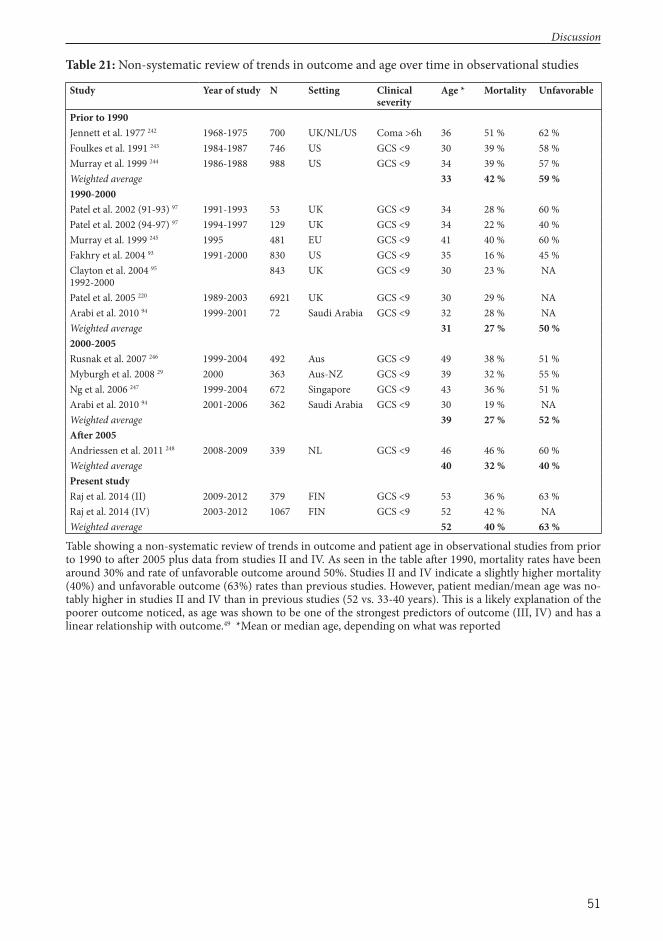
51
Table 21: Non-systematic review of trends in outcome and age over time in observational studies
Study Year of study N Setting Clinical severity
Age * Mortality Un favorable
Prior to 1990Jennett et al. 1977 242 1968-1975 700 UK/NL/US Coma >6h 36 51 % 62 %Foulkes et al. 1991 243 1984-1987 746 US GCS <9 30 39 % 58 %Murray et al. 1999 244 1986-1988 988 US GCS <9 34 39 % 57 %Weighted average 33 42 % 59 %1990-2000Patel et al. 2002 (91-93) 97 1991-1993 53 UK GCS <9 34 28 % 60 %Patel et al. 2002 (94-97) 97 1994-1997 129 UK GCS <9 34 22 % 40 %Murray et al. 1999 245 1995 481 EU GCS <9 41 40 % 60 %Fakhry et al. 2004 93 1991-2000 830 US GCS <9 35 16 % 45 %Clayton et al. 2004 95
1992-2000843 UK GCS <9 30 23 % NA
Patel et al. 2005 220 1989-2003 6921 UK GCS <9 30 29 % NAArabi et al. 2010 94 1999-2001 72 Saudi Arabia GCS <9 32 28 % NAWeighted average 31 27 % 50 %2000-2005Rusnak et al. 2007 246 1999-2004 492 Aus GCS <9 49 38 % 51 %Myburgh et al. 2008 29 2000 363 Aus-NZ GCS <9 39 32 % 55 %Ng et al. 2006 247 1999-2004 672 Singapore GCS <9 43 36 % 51 %Arabi et al. 2010 94 2001-2006 362 Saudi Arabia GCS <9 30 19 % NAWeighted average 39 27 % 52 %Aft er 2005Andriessen et al. 2011 248 2008-2009 339 NL GCS <9 46 46 % 60 %Weighted average 40 32 % 40 %Present studyRaj et al. 2014 (II) 2009-2012 379 FIN GCS <9 53 36 % 63 %Raj et al. 2014 (IV) 2003-2012 1067 FIN GCS <9 52 42 % NAWeighted average 52 40 % 63 %
Table showing a non-systematic review of trends in outcome and patient age in observational studies from prior to 1990 to aft er 2005 plus data from studies II and IV. As seen in the table aft er 1990, mortality rates have been around 30% and rate of unfavorable outcome around 50%. Studies II and IV indicate a slightly higher mortality (40%) and unfavorable outcome (63%) rates than previous studies. However, patient median/mean age was no-tably higher in studies II and IV than in previous studies (52 vs. 33-40 years). Th is is a likely explanation of the poorer outcome noticed, as age was shown to be one of the strongest predictors of outcome (III, IV) and has a linear relationship with outcome.49 *Mean or median age, depending on what was reported
Discussion
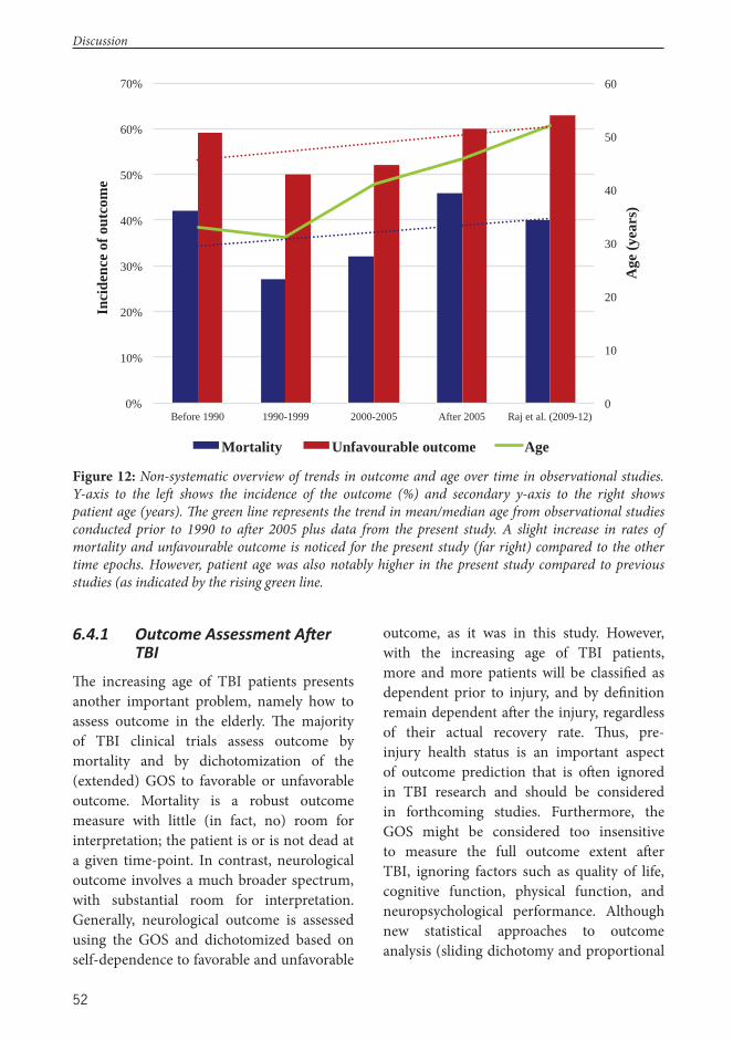
52
6.4.1 Outcome Assessment A er TBI
Th e increasing age of TBI patients presents another important problem, namely how to assess outcome in the elderly. Th e majority of TBI clinical trials assess outcome by mortality and by dichotomization of the (extended) GOS to favorable or unfavorable outcome. Mortality is a robust outcome measure with little (in fact, no) room for interpretation; the patient is or is not dead at a given time-point. In contrast, neurological outcome involves a much broader spectrum, with substantial room for interpretation. Generally, neurological outcome is assessed using the GOS and dichotomized based on self-dependence to favorable and unfavorable
outcome, as it was in this study. However, with the increasing age of TBI patients, more and more patients will be classifi ed as dependent prior to injury, and by defi nition remain dependent aft er the injury, regardless of their actual recovery rate. Th us, pre-injury health status is an important aspect of outcome prediction that is oft en ignored in TBI research and should be considered in forthcoming studies. Furthermore, the GOS might be considered too insensitive to measure the full outcome extent aft er TBI, ignoring factors such as quality of life, cognitive function, physical function, and neuropsychological performance. Although new statistical approaches to outcome analysis (sliding dichotomy and proportional
0
10
20
30
40
50
60
0%
10%
20%
30%
40%
50%
60%
70%
Before 1990 1990-1999 2000-2005 After 2005 Raj et al. (2009-12)
Age
(yea
rs)
Inci
denc
e of
out
com
e
Mortality Unfavourable outcome Age
Figure 12: Non-systematic overview of trends in outcome and age over time in observational studies. Y-axis to the left shows the incidence of the outcome (%) and secondary y-axis to the right shows patient age (years). Th e green line represents the trend in mean/median age from observational studies conducted prior to 1990 to aft er 2005 plus data from the present study. A slight increase in rates of mortality and unfavourable outcome is noticed for the present study (far right) compared to the other time epochs. However, patient age was also notably higher in the present study compared to previous studies (as indicated by the rising green line.
Discussion
53
odds analysis)26 might improve studies’ statistical power, multidimensional outcome analysis is crucial in forthcoming studies. Furthermore, the eff ect of genetic variations on outcome and complications aft er TBI (e.g. hydrocephalus, epilepsy, neurological defi cits) are other areas of key interest for future research.
6.5 Limita ons of the StudyTh ere are some limitations of the present study that must be acknowledged. First, the main limitation of the study is that few of the newly proposed models were externally validated. Although all models were internally validated, external validation in independent datasets is essential to demonstrate model generalizability. Th us, before application, proposed models such as IMPACT-APACHE II and the Helsinki CT score should be externally validated in independent datasets. Second, due to the retrospective nature of studies I-III, neurological outcome was assessed retrospectively according to the simple GOS205 and not to the more sensitive extended GOS.249 Th ird, all studies were register-based, and as in all register studies, the quality of data and data completeness should be considered. However, missing data were not a signifi cant problem in any of the studies and when there were missing data, these patients were excluded instead of using the more sophisticated statistical techniques, such as multiple imputation.250 Fourth, the performance of the RISC for long-term outcome prediction could not be assessed. Th us, the ‘true’ predictive ability of the RISC in patients with TBI could not be established. Fift h, variable interaction and transformation techniques were not exploited in the present study. Further studies should aim at investigating inter-variable interactions and look at the possibility of variable transformation, as this might increase model performance.
6.6 Future Implica ons6.6.1 Which Model To Use And For
What?
Based on this study’s results, the choice among TBI models, intensive care scoring systems, and trauma scoring systems is the TBI models wherever possible. IMPACT exhibited the best overall performance in the present study and is thus considered the most robust model in patients with TBI. Aside from superior statistical performance, accurate prognoses assessable directly upon hospital admission is an obvious advantage of IMPACT over other investigated models. However, if IMPACT is unavailable, customization of some of the intensive care scores is probably a valid substitute. Although the prognostic models provide seemly accurate estimations of patient prognosis, there are numerous factors that simply cannot be accounted for by standardized models. As indicated by the explanatory variation, about 40% at best of patient outcome was explained by IMPACT. Th at means that about 60% of the outcome remains to be explained. Th us, using prognostic models in the individual patient should be approached with due caution; current models are not accurate enough to provide individual prognoses and the role of current prognostic models is mainly for research purposes.
6.6.2 External Valida on of the Proposed Models
Future external validations studies of IMPACT-APACHE II and Helsinki CT score are essential to show model generalizability. Furthermore, the APACHE or intensive care component of IMPACT-APACHE II models should be modifi ed to include more neurointensive specifi c variables, such as ICP, CPP, PbtO2, and possibly biomarkers (e.g. S100B peaks251) and brain microdialysis markers. In addition, the individual scores included in the Helsinki CT score should
Discussion
54
be externally validated and evaluated in independent datasets, as it is possible that the regression coeffi cients and their associated scores might diff er in other settings.
6.7 Prac cal Examples of Prognos c Models in TBI Research
Prognostic model research in itself does not lead anywhere if the prognostic models remain underutilized. Below are descriptions of two examples of how diff erent types of prognostic models can be used in TBI research.
Th e role of hyperoxemia in the setting of TBI is a controversial topic.252,253 To investigate this a national ICU database (FICC) was used to assess the independent eff ect of hyperoxemia on long-term mortality in patients with moderate to severe TBI. Because the FICC lacks some of the essential data necessary to use IMPACT, a customized version of APACHE II was used to adjust for diff erences in severity of illness among TBI patients. Th e APACHE II was customized using the total score (level one customization), which resulted in good model discrimination and calibration (AUC 0.80, H-L p-value=0.10). Th is customized APACHE II thus provided an excellent tool for injury severity adjustment in multivariate analysis. Subsequently, hyperoxemia was
found not to be an independent predictor of outcome in patients with moderate to severe TBI treated in the ICU.50
Up to half of all TBI patients are alcohol intoxicated at the time of injury.4 Th e eff ect of acute alcohol intoxication on outcome aft er TBI is, however, a debated subject.254 Because high levels of blood alcohol concentration (BAC) are known to decrease level of consciousness, alcohol intoxicated patients may be wrongly classifi ed as having a more severe TBI than they really have.255 Th us, adjusting for IMPACT may be insuffi cient, as admission GCS is one of the most important predictors. Th e APACHE II, on the other hand, includes the worst day one GCS and thus enables identifi cation of patients with low initial GCS due to alcohol intoxication instead of brain injury. Th us, IMPACT-APACHE II provides an excellent tool to adjust for TBI severity with alcohol intoxication as a confounding factor. Subsequently, aft er adjusting for potential confounding factors, including IMPACT-APACHE II and the Rotterdam CT scores, low admission BAC (<2.3‰) were found to signifi cantly decrease the risk of six-month mortality compared to no BAC (0‰) or high BAC (≥2.3‰) (no BAC as reference; low BAC OR 0.41, 95% CI 0.19-0.88, p=0.021; high BAC OR 0.58, 95% CI 0.29-1.15, p=0.120).256
Discussion
55
7 Conclusions
1. Th e TBI specifi c IMPACT models displayed superior overall performance compared to the intensive care and trauma scoring systems, showing that patients with TBI are a highly specifi c population in the trauma and intensive care unit environment. Th us, the use of a TBI specifi c prognostic model, undergoing continual updating, is advocated (I-V).
2. Th ree novel models were developed: the TBI-ICU combination model (IMPACT-APACHE), the Helsinki CT score, and the reference model (age + GCS). Th e TBI-ICU model showed superior performance over the TBI and intensive care scoring system when used in isolation, and may be used to adjust for patient baseline prognostic risk and inter-center diff erences in quality of early intensive care (II). Th e Helsinki CT score may be used for early objective prediction of long-term outcome and for describing and comparing patient series (III). Th e reference model, based on only age and GCS, showed similar performance to the more complex intensive care scoring systems (which are also roughly comparable to IMPACT), showing that adjusting for these factors may provide adequate case-mix adjustment. Th is is of great importance for forthcoming epidemiological studies lacking the necessary data for more complex prognostications (IV). External validation studies of the newly proposed models are required to show generalizability.
Conclusions
56
Acknowledgements
Th is study was carried out at the Department of Neurosurgery and the Department of Anesthesiology and Intensive Care, Helsinki University Central Hospital from 2012 to 2014. Th e thesis is part of the Clinical Research Program of Faculty of Medicine and Doctoral School of Health Science (Dissertationes Scholae Doctoralis Ad Sanitatem Investigandam Universitatis Helsinkiensis).
I begin by presenting my deepest gratitude to my supervisors Jari Siironen and Markus Skrifvars. Jari, you were the fi rst to introduce me to the world of neurotrauma. Th ank you for taking me in as a young inexperienced medical student and guiding me in the right direction. You have been my role model, both inside and outside the hospital, since we fi rst started this project. Markus, you truly are the best supervisor one could wish for. You taught me everything from scientifi c writing to complex statistical analyses, and so much more. Without you none of this work would have been even remotely possible, and I am truly grateful to you.
I also wish to give my sincerest gratitude to Riku Kivisaari for the countless hours spent analyzing radiological images (even on your free time!). Although not serving as my offi cial supervisor, you always acted like one.
I express my sincere gratitude to my custos, Professor Juha Hernesniemi, the head, and mentor for the whole Department of Neurosurgery in Helsinki. His skills as a surgeon and compassion as a leader inspire not only me, but also a whole generation of future neurosurgeons.
My special thanks go to Juha Öhman and Patrik Finne, the offi cial reviewers of this thesis. Th ank you for your advice and constructive criticism that so improved the fi nal product.
I am deeply ful to thank Professor Andrew Maas for accepting the role of being my opponent and for sharing his visionary views and immense knowledge of neurotrauma on this special day.
I am grateful to Jaakko Lappalainen for all his critical revisions and excellent comments on my manuscripts thoughout the years. My deepest appreciation also goes to Matti Reinikainen for all the constructive comments, statistical advice, and clinical knowledge.
I thank all my co-authors, Tuomas Brinck, Lauri Handolin, Päivi Tanskanen, Stepani Bendel, Rolf Lefering, and Tuomas Selander, for making the studies in this dissertation possible.
My heartfelt thanks go to all the Anesthesiologists and Neurosurgeons at the Department of Neurosurgery at Töölö Hospital. Th ank you for your patience, instruction, and guidance the last two summers.
To my boys, Era Mikkonen, Gustav Strömberg, Walter Federolf, Erik Wahlström, and Rasmus Löfman: thank you for your support and patience over the years, but most of all, thank you for your friendship, which I hold so dear. I also wish to acknowledge the members of “Rahuls
Acknowledgements

57
klinikgrupp” (you know who you are) and the amazing journey through medical school that we have shared.
Also, to my boys back home, Daniel Fellman, David Nyman, Jonas Grönholm, Kristoff er Knuts, Martin Kjellman, Linus Korkea-Aho, and Robin Julin. Having known me for nearly 20 years, you are my oldest friends and know me better than anyone. Th ank you for all the adventures we have enjoyed and will continue to en joy together.
To my beautiful sisters Richika Raj and Rimmi Raj (or, as I like to say, Timon and Pumba): although it was not always the easiest task, thank you for your support and love.
Sara Johansson, my soul mate, and the love of my life. Not only being the sole woman, besides my mother, capable of putting up with me, you make my life worth living. Without you, none of this matters.
I dedicated this book to my mother (Renu Raj) and father (Bharat Raj). Mamma, my love for you is beyond words, and I cannot describe how proud I am of being your son. Pappa, coming from a tiny village in the mountains of Himalaya (Budhesu, India) to another tiny village in Finland (Jakobstad, Finland), your journey through life cannot be matched. I am truly grateful for everything you have given me. I stumble on my words, but you must know that you are my biggest inspiration in life.
Th e research and the writing of this book were fi nancially supported by grants from Finska Läkaresällskapet, Maire Taponen Foundation, Medicinska Understödsföreningen Liv och Hälsa, Svensk-Österbottniska Samfundet r.f., the Maud Kuistila Memorial Foundation, the Viktor Fagerström Foundation of the Finnish Medical Society Duodecim, and a Helsinki University Central Hospital EVO grant.
Helsinki, November 2014
Acknowledgements
58
References1. Jennett B. Epidemiology of head injury. J Neurol
Neurosurg Psychiatr 1996;60:362–9.
2. Lu JJ, Marmarou AA, Choi SS, Maas AA, Murray GG, Steyerberg EWE. Mortality from traumatic brain injury. Acta Neurochir Suppl 2004;95:281–5.
3. Langlois JA, Rutland-Brown W, Wald MM. Th e epidemiology and impact of traumatic brain injury: a brief overview. J Head Trauma Rehabil 2006;21:375–8.
4. Tagliaferri F, Compagnone C, Korsic M, Servadei F, Kraus J. A systematic review of brain injury epidemiology in Europe. Acta Neurochir (Wien) 2006;148:255–68.
5. Faul M, Wald MM, Rutland-Brown W, Sullivent EE, Sattin RW. Using a cost-benefi t analysis to es-timate outcomes of a clinical treatment guideline: testing the Brain Trauma Foundation guidelines for the treatment of severe traumatic brain injury. J Trauma 2007;63:1271–8.
6. Olesen J, Gustavsson A, Svensson M, Wittchen H-U, Jönsson B, CDBE2010 study group, et al. Th e economic cost of brain disorders in Europe. Eur J Neurol 2012;19:155–62.
7. Maas AIR, Stocchetti N, Bullock R. Moderate and severe traumatic brain injury in adults. Lancet Neurology 2008;7:728–41.
8. Coronado VG, McGuire LC, Sarmiento K, Bell J, Lionbarger MR, Jones CD, et al. Trends in Trau-matic Brain Injury in the U.S. and the public health response: 1995-2009. J Safety Res 2012;43:299–307.
9. Steyerberg EW, Mushkudiani N, Perel P, Butcher I, Lu J, McHugh GS, et al. Predicting outcome aft er traumatic brain injury: development and inter-national validation of prognostic scores based on admission characteristics. PLoS Med 2008;5:e165.
10. MRC CRASH Trial Collaborators, Perel P, Aran-go M, Clayton T, Edwards P, Komolafe E, et al. Predicting outcome aft er traumatic brain injury: practical prognostic models based on large cohort of international patients. BMJ 2008;336:425–9.
11. Hukkelhoven CWPM, Rampen AJJ, Maas AIR, Farace E, Habbema JDF, Marmarou A, et al. Some prognostic models for traumatic brain injury were not valid. J Clin Epidemiol 2006;59:132–43.
12. Perel P, Edwards P, Wentz R, Roberts I. Systematic review of prognostic models in traumatic brain injury. BMC Med Inform Decis Mak 2006;6:38.
13. Hemingway H, Croft P, Perel P, Hayden JA, Abrams K, Timmis A, et al. Prognosis research strategy (PROGRESS) 1: a framework for research-ing clinical outcomes. BMJ 2013;346:e5595.
14. Riley RD, Hayden JA, Steyerberg EW, Moons KGM, Abrams K, Kyzas PA, et al. Prognosis Re-search Strategy (PROGRESS) 2: prognostic factor research. PLoS Med 2013;10:e1001380.
15. Steyerberg EW, Moons KGM, van der Windt DA, Hayden JA, Perel P, Schroter S, et al. Prognosis Re-search Strategy (PROGRESS) 3: prognostic model research. PLoS Med 2013;10:e1001381.
16. Hingorani AD, Windt DAVD, Riley RD, Abrams K, Moons KGM, Steyerberg EW, et al. Prognosis research strategy (PROGRESS) 4: stratifi ed medi-cine research. BMJ 2013;346:e5793.
17. Lingsma HF, Roozenbeek B, Steyerberg EW, Mur-ray GD, Maas AIR. Early prognosis in traumatic brain injury: from prophecies to predictions. Lancet Neurology 2010;9:543–54.
18. Maas AIR, Murray GD, Roozenbeek B, Lingsma HF, Butcher I, McHugh GS, et al. Advancing care for traumatic brain injury: fi ndings from the IM-PACT studies and perspectives on future research. Lancet Neurology 2013;12:1200–10.
19. Selker HP. Systems for comparing actual and predicted mortality rates: characteristics to pro-mote cooperation in improving hospital care. Ann Intern Med 1993;118:820–2.
20. Champion HR, Copes WS, Sacco WJ, Lawnick MM, Keast SL, Bain LW, et al. Th e Major Trauma Outcome Study: establishing national norms for trauma care. J Trauma 1990;30:1356–65.
21. Knaus WA, Wagner DP, Zimmerman JE, Drap-er EA. Variations in mortality and length of stay in intensive care units. Ann Intern Med 1993;118:753–61.
22. Le Gall J-R. Th e use of severity scores in the intensive care unit. Intensive Care Medicine 2005;31:1618–23.
23. Breslow MJ, Badawi O. Severity scoring in the critically ill: Part 1: Interpretation and accuracy of outcome prediction scoring systems. Chest 2012;141:245–52.
24. Breslow MJ, Badawi O. Severity scoring in the crit-ically ill: Part 2: Maximizing value from outcome prediction scoring systems. Chest 2012;141:518–27.
25. Maas AIR, Menon DK, Lingsma HF, Pineda JA, Sandel ME, Manley GT. Re-orientation of clinical research in traumatic brain injury: report of an international workshop on comparative eff ective-ness research. J Neurotrauma 2012;29:32–46.
26. Maas AIR, Steyerberg EW, Marmarou A, McHugh GS, Lingsma HF, Butcher I, et al. IMPACT recom-mendations for improving the design and analysis of clinical trials in moderate to severe traumatic brain injury. Neurotherapeutics 2010;7:127–34.
27. Vincent J-L, Moreno R. Clinical review: scoring systems in the critically ill. Crit Care 2010;14:207.
28. Champion HR. Trauma scoring. Scand J Surg 2002;91:12–22.
29. Myburgh JA, Cooper DJ, Finfer SR, Venkatesh B, Jones D, Higgins A, et al. Epidemiology and 12-month outcomes from traumatic brain in-jury in Australia and New Zealand. J Trauma 2008;64:854–62.
References
59
30. Menon DK, Schwab K, Wright DW, Maas AI, Demographics and Clinical Assessment Work-ing Group of the International and Interagency Initiative toward Common Data Elements for Research on Traumatic Brain Injury and Psycho-logical Health. Position statement: defi nition of traumatic brain injury. Arch Phys Med Rehabil 2010;91:1637–40.
31. Olesen J, Leonardi M. Th e burden of brain diseases in Europe. Eur J Neurol 2003;10:471–7.
32. Kay A, Teasdale G. Head injury in the United Kingdom. World J Surg 2001;25:1210–20.
33. Feigin VL, Th eadom A, Barker-Collo S, Starkey NJ, McPherson K, Kahan M, et al. Incidence of traumatic brain injury in New Zealand: a popula-tion-based study. Lancet Neurology 2013;12:53–64.
34. Centers for Disease Control and Prevention (CDC). CDC grand rounds: reducing severe trau-matic brain injury in the United States. MMWR Morb Mortal Wkly Rep 2013;62:549–52.
35. Koskinen S, Alaranta H. Traumatic brain injury in Finland 1991-2005: a nationwide register study of hospitalized and fatal TBI. Brain Inj 2008;22:205–14.
36. Coronado VG, Xu L, Basavaraju SV, McGuire LC, Wald MM, Faul MD, et al. Surveillance for trau-matic brain injury-related deaths--United States, 1997-2007. MMWR Surveill Summ 2011;60:1–32.
37. Roozenbeek B, Maas AIR, Menon DK. Changing patterns in the epidemiology of traumatic brain injury. Nat Rev Neurol 2013;9:231–6.
38. Mathers CD, Loncar D. Projections of global mor-tality and burden of disease from 2002 to 2030. PLoS Med 2006;3:e442
39. Savola O, Niemelä O, Hillbom M. Alcohol intake and the pattern of trauma in young adults and working aged people admitted aft er trauma. Alco-hol and Alcoholism 2005;40:269–73.
40. Sundstrøm T, Sollid S, Wentzel-Larsen T, Wester K. Head injury mortality in the Nordic countries. J Neurotrauma 2007;24:147–53.
41. Paljärvi T, Mäkelä P, Poikolainen K. Pattern of drinking and fatal injury: a population-based follow-up study of Finnish men. Addiction 2005;100:1851–9.
42. Haddad SH, Arabi YM. Critical care management of severe traumatic brain injury in adults. Scand J Trauma Resusc Emerg Med 2012;20:12.
43. Rosenfeld JV, Maas AI, Bragge P, Morganti-Koss-mann MC, Manley GT, Gruen RL. Early man-agement of severe traumatic brain injury. Lancet 2012;380:1088–98.
44. Chesnut RM, Marshall LF, Klauber MR, Blunt BA, Baldwin N, Eisenberg HM, et al. Th e role of sec-ondary brain injury in determining outcome from severe head injury. J Trauma 1993;34:216–22.
45. Unterberg AW, Stover J, Kress B, Kiening KL. Edema and brain trauma. Neuroscience 2004;129:1021–9.
46. Smith M. Critical care management of severe head injury. Anaesthesia & Intensive Care Medicine 2014;15:164–7.
47. Brain Trauma Foundation, American Association of Neurological Surgeons, Congress of Neuro-logical Surgeons. Guidelines for the management of severe traumatic brain injury. J. Neurotrau-ma2007;24 Suppl 1:S1–106.
48. Helmy AA, Vizcaychipi MM, Gupta AKA. Trau-matic brain injury: intensive care management. Br J Anaesth 2007;99:32–42.
49. Hukkelhoven CWPM, Steyerberg EW, Rampen AJJ, Farace E, Habbema JDF, Marshall LF, et al. Patient age and outcome following severe trau-matic brain injury: an analysis of 5600 patients. J Neurosurg 2003;99:666–73.
50. Raj R, Bendel S, Reinikainen M, Kivisaari R, Siiro-nen J, Lång M, et al. Hyperoxemia and long-term outcome aft er traumatic brain injury. Crit Care 2013;17:R177.
51. Roberts I, Yates D, Sandercock P, Farrell B, Was-serberg J, Lomas G, et al. Eff ect of intravenous corticosteroids on death within 14 days in 10008 adults with clinically signifi cant head injury (MRC CRASH trial): randomised placebo-controlled trial. Lancet 2004;364:1321–8.
52. Edwards P, Arango M, Balica L, Cottingham R, El-Sayed H, Farrell B, et al. Final results of MRC CRASH, a randomised placebo-controlled trial of intravenous corticosteroid in adults with head in-jury-outcomes at 6 months. Lancet 2005;365:1957–9.
53. Mushkudiani NA, Engel DC, Steyerberg EW, Butcher I, Lu J, Marmarou A, et al. Prognostic value of demographic characteristics in traumatic brain injury: results from the IMPACT study. J Neurotrauma 2007;24:259–69.
54. Farace E, Alves WM. Do women fare worse: a metaanalysis of gender diff erences in traumatic brain injury outcome. J Neurosurg 2000;93:539–45.
55. Groswasser Z, Cohen M, Keren O. Female TBI patients recover better than males. Brain Injury 1998;12:805–8.
56. Junpeng M, Huang S, Qin S. Progesterone for acute traumatic brain injury. Cochrane Database Syst Rev 2011;CD008409.
57. Teasdale G, Jennett B. Assessment of coma and impaired consciousness. A practical scale. Lancet 1974;2:81–4.
58. Mena JHJ, Sanchez AIA, Rubiano AMA, Peitzman ABA, Sperry JLJ, Gutierrez MIM, et al. Eff ect of the modifi ed Glasgow Coma Scale score criteria for mild traumatic brain injury on mortality pre-diction: comparing classic and modifi ed Glasgow Coma Scale score model scores of 13. J Trauma 2011;71:1185–93.
References
60
59. Teasdale G, Maas A, Lecky F, Manley G, Stocchet-ti N, Murray G. Th e Glasgow Coma Scale at 40 years: standing the test of time. Lancet Neurology 2014;13:844–54.
60. Teasdale G, Jennett B. Assessment of coma and severity of brain damage. Anesthesiology 1978;49:225–6.
61. Marmarou A, Lu J, Butcher I, McHugh GS, Murray GD, Steyerberg EW, et al. Prognostic value of the Glasgow Coma Scale and pupil reactivity in trau-matic brain injury assessed pre-hospital and on enrollment: an IMPACT analysis. J Neurotrauma 2007;24:270–80.
62. Green SM. Cheerio, laddie! Bidding farewell to the Glasgow Coma Scale. Ann Emerg Med 2011;58:427–30.
63. Ritter AM, Muizelaar JP, Barnes T, Choi S, Fatou-ros P, Ward J, et al. Brain stem blood fl ow, pupil-lary response, and outcome in patients with severe head injuries. Neurosurgery 1999;44:941–8.
64. Ropper AH, Cole D, Louis DN. Clinicopathologic correlation in a case of pupillary dilation from ce-rebral hemorrhage. Arch Neurol 1991;48:1166–9.
65. van Leeuwen N, Lingsma HF, Perel P, Lecky F, Roozenbeek B, Lu J, et al. Prognostic value of ma-jor extracranial injury in traumatic brain injury: an individual patient data meta-analysis in 39,274 patients. Neurosurgery 2012;70:811–8.
66. Ho KM, Burrell M, Rao S. Extracranial injuries are important in determining mortality of neurotrau-ma. Crit Care Med 2010;38:1562–8.
67. Sarrafzadeh AS, Peltonen EE, Kaisers U, Küchler I, Lanksch WR, Unterberg AW. Secondary insults in severe head injury--do multiply injured patients do worse? Crit Care Med 2001;29:1116–23.
68. Maas AIR, Steyerberg EW, Butcher I, Dammers R, Lu J, Marmarou A, et al. Prognostic value of computerized tomography scan characteristics in traumatic brain injury: results from the IMPACT study. J Neurotrauma 2007;24:303–14.
69. Maas AIR, Hukkelhoven CWPM, Marshall LF, Steyerberg EW. Prediction of outcome in traumatic brain injury with computed tomographic char-acteristics: a comparison between the computed tomographic classifi cation and combinations of computed tomographic predictors. Neurosurgery 2005;57:1173–82.
70. Hulkower MB, Poliak DB, Rosenbaum SB, Zim-merman ME, Lipton ML. A decade of DTI in traumatic brain injury: 10 years and 100 articles later. AJNR Am J Neuroradiol 2013;34:2064–74.
71. Yuh EL, Cooper SR, Mukherjee P, Yue JK, Lingsma H, Gordon W, et al. Diff usion Tensor Imaging for Outcome Prediction in Mild Traumatic Brain Injury: A TRACK-TBI Study. J Neurotrauma 2014;31:1457–77.
72. Chesnut RM. Secondary brain insults aft er head injury: clinical perspectives. New Horiz 1995;3:366–75.
73. McHugh GS, Engel DC, Butcher I, Steyerberg EW, Lu J, Mushkudiani N, et al. Prognostic value of secondary insults in traumatic brain injury: results from the IMPACT study. J Neurotrauma 2007;24:287–93.
74. Manley G, Knudson MM, Morabito D, Damron S, Erickson V, Pitts L. Hypotension, hypoxia, and head injury: frequency, duration, and consequenc-es. Arch Surg 2001;136:1118–23.
75. Stocchetti N, Furlan A, Volta F. Hypoxemia and arterial hypotension at the accident scene in head injury. J Trauma 1996;40:764–7.
76. Stocchetti N, Colombo A, Ortolano F, Videtta W, Marchesi R, Longhi L, et al. Time course of intra-cranial hypertension aft er traumatic brain injury. J Neurotrauma 2007;24:1339–46.
77. Salim A, Hadjizacharia P, DuBose J, Brown C, Inaba K, Chan LS, et al. Persistent hyperglycemia in severe traumatic brain injury: an independent predictor of outcome. Am Surg 2009;75:25–9.
78. Van Beek JGM, Mushkudiani NA, Steyerberg EW, Butcher I, McHugh GS, Lu J, et al. Prognostic value of admission laboratory parameters in traumatic brain injury: results from the IMPACT study. J Neurotrauma 2007;24:315–28.
79. LeRoux P. Haemoglobin management in acute brain injury. Curr Opin Crit Care 2013;19:83–91.
80. Laroche M, Kutcher ME, Huang MC, Cohen MJ, Manley GT. Coagulopathy aft er traumatic brain injury. Neurosurgery 2012;70:1334–45.
81. Maegele M. Coagulopathy aft er traumatic brain injury: incidence, pathogenesis, and treatment options. Transfusion 2013;53 Suppl 1:28S–37S.
82. Lustenberger T, Talving P, Kobayashi L, Inaba K, Lam L, Plurad D, et al. Time course of coagulopa-thy in isolated severe traumatic brain injury. Injury 2010;41:924–8.
83. Harhangi BS, Kompanje EJO, Leebeek FWG, Maas AIR. Coagulation disorders aft er traumatic brain injury. Acta Neurochir (Wien) 2008;150:165–75.
84. NICE-SUGAR Study Investigators, Finfer S, Chit-tock DR, Su SY-S, Blair D, Foster D, et al. Intensive versus conventional glucose control in critically ill patients. N Engl J Med 2009;360:1283–97.
85. Vespa PM, McArthur D, O’Phelan K, Glenn T. Per-sistently low extracellular glucose correlates with poor outcome 6 months aft er human traumatic brain injury despite a lack of increased lactate: a microdialysis study. J Cereb Blood Flow Metab 2003;23:865–77.
86. Holbein M, Béchir M, Ludwig S, Sommerfeld J, Cottini SR, Keel M, et al. Diff erential infl uence of arterial blood glucose on cerebral metabolism following severe traumatic brain injury. Crit Care 2009;13:R13.
References
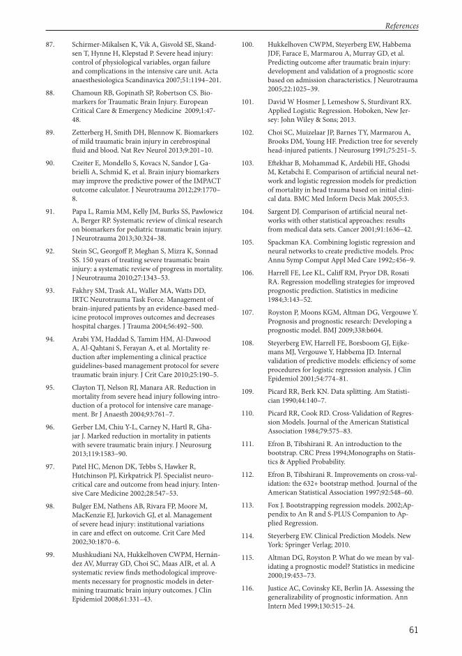
61
87. Schirmer-Mikalsen K, Vik A, Gisvold SE, Skand-sen T, Hynne H, Klepstad P. Severe head injury: control of physiological variables, organ failure and complications in the intensive care unit. Acta anaesthesiologica Scandinavica 2007;51:1194–201.
88. Chamoun RB, Gopinath SP, Robertson CS. Bio-markers for Traumatic Brain Injury. European Critical Care & Emergency Medicine 2009;1:47-48.
89. Zetterberg H, Smith DH, Blennow K. Biomarkers of mild traumatic brain injury in cerebrospinal fl uid and blood. Nat Rev Neurol 2013;9:201–10.
90. Czeiter E, Mondello S, Kovacs N, Sandor J, Ga-brielli A, Schmid K, et al. Brain injury biomarkers may improve the predictive power of the IMPACT outcome calculator. J Neurotrauma 2012;29:1770–8.
91. Papa L, Ramia MM, Kelly JM, Burks SS, Pawlowicz A, Berger RP. Systematic review of clinical research on biomarkers for pediatric traumatic brain injury. J Neurotrauma 2013;30:324–38.
92. Stein SC, Georgoff P, Meghan S, Mizra K, Sonnad SS. 150 years of treating severe traumatic brain injury: a systematic review of progress in mortality. J Neurotrauma 2010;27:1343–53.
93. Fakhry SM, Trask AL, Waller MA, Watts DD, IRTC Neurotrauma Task Force. Management of brain-injured patients by an evidence-based med-icine protocol improves outcomes and decreases hospital charges. J Trauma 2004;56:492–500.
94. Arabi YM, Haddad S, Tamim HM, Al-Dawood A, Al-Qahtani S, Ferayan A, et al. Mortality re-duction aft er implementing a clinical practice guidelines-based management protocol for severe traumatic brain injury. J Crit Care 2010;25:190–5.
95. Clayton TJ, Nelson RJ, Manara AR. Reduction in mortality from severe head injury following intro-duction of a protocol for intensive care manage-ment. Br J Anaesth 2004;93:761–7.
96. Gerber LM, Chiu Y-L, Carney N, Hartl R, Gha-jar J. Marked reduction in mortality in patients with severe traumatic brain injury. J Neurosurg 2013;119:1583–90.
97. Patel HC, Menon DK, Tebbs S, Hawker R, Hutchinson PJ, Kirkpatrick PJ. Specialist neuro-critical care and outcome from head injury. Inten-sive Care Medicine 2002;28:547–53.
98. Bulger EM, Nathens AB, Rivara FP, Moore M, MacKenzie EJ, Jurkovich GJ, et al. Management of severe head injury: institutional variations in care and eff ect on outcome. Crit Care Med 2002;30:1870–6.
99. Mushkudiani NA, Hukkelhoven CWPM, Hernán-dez AV, Murray GD, Choi SC, Maas AIR, et al. A systematic review fi nds methodological improve-ments necessary for prognostic models in deter-mining traumatic brain injury outcomes. J Clin Epidemiol 2008;61:331–43.
100. Hukkelhoven CWPM, Steyerberg EW, Habbema JDF, Farace E, Marmarou A, Murray GD, et al. Predicting outcome aft er traumatic brain injury: development and validation of a prognostic score based on admission characteristics. J Neurotrauma 2005;22:1025–39.
101. David W Hosmer J, Lemeshow S, Sturdivant RX. Applied Logistic Regression. Hoboken, New Jer-sey: John Wiley & Sons; 2013.
102. Choi SC, Muizelaar JP, Barnes TY, Marmarou A, Brooks DM, Young HF. Prediction tree for severely head-injured patients. J Neurosurg 1991;75:251–5.
103. Eft ekhar B, Mohammad K, Ardebili HE, Ghodsi M, Ketabchi E. Comparison of artifi cial neural net-work and logistic regression models for prediction of mortality in head trauma based on initial clini-cal data. BMC Med Inform Decis Mak 2005;5:3.
104. Sargent DJ. Comparison of artifi cial neural net-works with other statistical approaches: results from medical data sets. Cancer 2001;91:1636–42.
105. Spackman KA. Combining logistic regression and neural networks to create predictive models. Proc Annu Symp Comput Appl Med Care 1992;:456–9.
106. Harrell FE, Lee KL, Califf RM, Pryor DB, Rosati RA. Regression modelling strategies for improved prognostic prediction. Statistics in medicine 1984;3:143–52.
107. Royston P, Moons KGM, Altman DG, Vergouwe Y. Prognosis and prognostic research: Developing a prognostic model. BMJ 2009;338:b604.
108. Steyerberg EW, Harrell FE, Borsboom GJ, Eijke-mans MJ, Vergouwe Y, Habbema JD. Internal validation of predictive models: effi ciency of some procedures for logistic regression analysis. J Clin Epidemiol 2001;54:774–81.
109. Picard RR, Berk KN. Data splitting. Am Statisti-cian 1990;44:140–7.
110. Picard RR, Cook RD. Cross-Validation of Regres-sion Models. Journal of the American Statistical Association 1984;79:575–83.
111. Efron B, Tibshirani R. An introduction to the bootstrap. CRC Press 1994;Monographs on Statis-tics & Applied Probability.
112. Efron B, Tibshirani R. Improvements on cross-val-idation: the 632+ bootstrap method. Journal of the American Statistical Association 1997;92:548–60.
113. Fox J. Bootstrapping regression models. 2002;Ap-pendix to An R and S-PLUS Companion to Ap-plied Regression.
114. Steyerberg EW. Clinical Prediction Models. New York: Springer Verlag; 2010.
115. Altman DG, Royston P. What do we mean by val-idating a prognostic model? Statistics in medicine 2000;19:453–73.
116. Justice AC, Covinsky KE, Berlin JA. Assessing the generalizability of prognostic information. Ann Intern Med 1999;130:515–24.
References
62
117. Feldman Z, Contant CF, Robertson CS, Narayan RK, Grossman RG. Evaluation of the Leeds prognostic score for severe head injury. Lancet 1991;337:1451–3.
118. Bleeker SE, Moll HA, Steyerberg EW, Donders ART, Derksen-Lubsen G, Grobbee DE, et al. Exter-nal validation is necessary in prediction research: a clinical example. J Clin Epidemiol 2003;56:826–32.
119. Hanley JA, McNeil BJ. Th e meaning and use of the area under a receiver operating characteristic (ROC) curve. Radiology 1982;143:29–36.
120. Steyerberg EW, Vickers AJ, Cook NR, Gerds T, Gonen M, Obuchowski N, et al. Assessing the performance of prediction models: a framework for traditional and novel measures. Epidemiology 2010;21:128–38.
121. Cook NR. Use and misuse of the receiver operat-ing characteristic curve in risk prediction. Circula-tion 2007;115:928–35.
122. Vincent J-LJ, Sakr YY. SOFA so good for predicting long-term outcomes. Resuscitation 2012;83:537–8.
123. Lemeshow S, Hosmer DW. A review of good-ness of fi t statistics for use in the development of logistic regression models. Am J Epidemiol 1982;115:92–106.
124. Kramer AA, Zimmerman JE. Assessing the cali-bration of mortality benchmarks in critical care: Th e Hosmer-Lemeshow test revisited. Crit Care Med 2007;35:2052–6.
125. Bertolini G, D’Amico R, Nardi D, Tinazzi A, Apo-lone G. One model, several results: the paradox of the Hosmer-Lemeshow goodness-of-fi t test for the logistic regression model. J Epidemiol Biostat 2000;5:251–3.
126. Finazzi S, Poole D, Luciani D, Cogo PE, Bertolini G. Calibration belt for quality-of-care assessment based on dichotomous outcomes. PLoS ONE 2011;6:e16110.
127. Miller ME, Langefeld CD, Tierney WM, Hui SL, McDonald CJ. Validation of probabilistic predic-tions. Med Decis Making 1993;13:49–58.
128. Miller ME, Hui SL, Tierney WM. Validation tech-niques for logistic regression models. Statistics in medicine 1991;10:1213–26.
129. Nagelkerke N. A note on a general defi nition of the coeffi cient of determination. Biometrika 1991;78:691–2.
130. Murray GD, Butcher I, McHugh GS, Lu J, Mush-kudiani NA, Maas AIR, et al. Multivariable prognostic analysis in traumatic brain injury: results from the IMPACT study. J Neurotrauma 2007;24:329–37.
131. Pepe MS, Janes H, Longton G, Leisenring W, Newcomb P. Limitations of the odds ratio in gaug-ing the performance of a diagnostic, prognostic, or screening marker. Am J Epidemiol 2004;159:882–90.
132. Pencina MJ, D’Agostino RB, D’Agostino RB, Vasan RS. Evaluating the added predictive ability of a new marker: from area under the ROC curve to reclassifi cation and beyond. Statistics in medicine 2008;27:157–72.
133. Zhu BP, Lemeshow S, Hosmer DW, Klar J, Avrunin J, Teres D. Factors aff ecting the performance of the models in the Mortality Probability Model II sys-tem and strategies of customization: a simulation study. Crit Care Med 1996;24:57–63.
134. Moreno R, Apolone G. Impact of diff erent custom-ization strategies in the performance of a general severity score. Crit Care Med 1997;25:2001–8.
135. Bakhshi-Raiez F, Peek N, Bosman RJ, de Jonge E, de Keizer NF. Th e impact of diff erent prognostic models and their customization on institutional comparison of intensive care units. Crit Care Med 2007;35:2553–60.
136. Rothen HU, Takala J. Can outcome prediction data change patient outcomes and organizational out-comes? Curr Opin Crit Care 2008;14:513–9.
137. Woodhouse D, Berg M, van der Putten J, Houtep-en J. Will benchmarking ICUs improve outcome? Curr Opin Crit Care 2009;15:450–5.
138. Hemmila MR, Nathens AB, Shafi S, Calland JF, Clark DE, Cryer HG, et al. Th e Trauma Qual-ity Improvement Program: pilot study and initial demonstration of feasibility. J Trauma 2010;68:253–62.
139. Shafi S, Ahn C, Parks J, Nathens AB, Cryer HM, Gentilello LM, et al. Quality of Care Within a Trauma Center Is not Altered by Injury Type. J Trauma 2010;68:716–20.
140. Angus DC. Scoring system fatigue...and the search for a way forward. Crit Care Med 2000;28:2145–6.
141. Lemeshow S, Le Gall JR. Modeling the severity of illness of ICU patients. A systems update. JAMA 1994;272:1049–55.
142. Brinkman S, Abu-Hanna A, de Jonge E, de Keizer NF. Prediction of long-term mortality in ICU patients: model validation and assessing the eff ect of using in-hospital versus long-term mortali-ty on benchmarking. Intensive Care Medicine 2013;39:1925–31.
143. Pouw ME, Peelen LM, Moons KGM, Kalkman CJ, Lingsma HF. Including post-discharge mortality in calculation of hospital standardised mortality ratios: retrospective analysis of hospital episode statistics. BMJ 2013;347:f5913.
144. Shafi S, Nathens AB, Parks J, Cryer HM, Fildes JJ, Gentilello LM. Trauma quality improvement using risk-adjusted outcomes. J Trauma 2008;64:599–604.
145. Nathens AB, Xiong W, Shafi S. Ranking of trauma center performance: the bare essentials. J Trauma 2008;65:628–35.
References
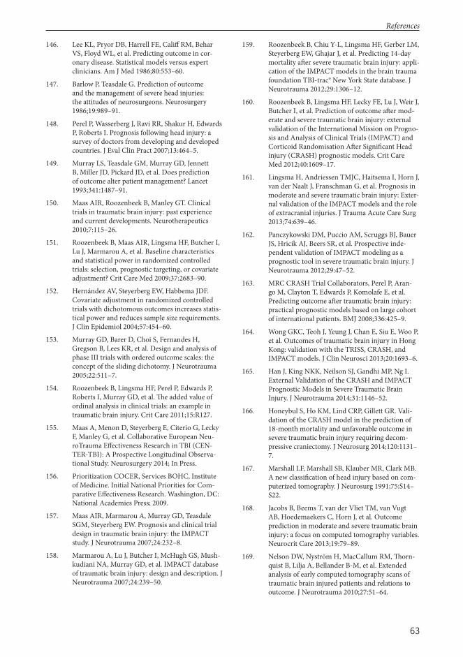
63
146. Lee KL, Pryor DB, Harrell FE, Califf RM, Behar VS, Floyd WL, et al. Predicting outcome in cor-onary disease. Statistical models versus expert clinicians. Am J Med 1986;80:553–60.
147. Barlow P, Teasdale G. Prediction of outcome and the management of severe head injuries: the attitudes of neurosurgeons. Neurosurgery 1986;19:989–91.
148. Perel P, Wasserberg J, Ravi RR, Shakur H, Edwards P, Roberts I. Prognosis following head injury: a survey of doctors from developing and developed countries. J Eval Clin Pract 2007;13:464–5.
149. Murray LS, Teasdale GM, Murray GD, Jennett B, Miller JD, Pickard JD, et al. Does prediction of outcome alter patient management? Lancet 1993;341:1487–91.
150. Maas AIR, Roozenbeek B, Manley GT. Clinical trials in traumatic brain injury: past experience and current developments. Neurotherapeutics 2010;7:115–26.
151. Roozenbeek B, Maas AIR, Lingsma HF, Butcher I, Lu J, Marmarou A, et al. Baseline characteristics and statistical power in randomized controlled trials: selection, prognostic targeting, or covariate adjustment? Crit Care Med 2009;37:2683–90.
152. Hernández AV, Steyerberg EW, Habbema JDF. Covariate adjustment in randomized controlled trials with dichotomous outcomes increases statis-tical power and reduces sample size requirements. J Clin Epidemiol 2004;57:454–60.
153. Murray GD, Barer D, Choi S, Fernandes H, Gregson B, Lees KR, et al. Design and analysis of phase III trials with ordered outcome scales: the concept of the sliding dichotomy. J Neurotrauma 2005;22:511–7.
154. Roozenbeek B, Lingsma HF, Perel P, Edwards P, Roberts I, Murray GD, et al. Th e added value of ordinal analysis in clinical trials: an example in traumatic brain injury. Crit Care 2011;15:R127.
155. Maas A, Menon D, Steyerberg E, Citerio G, Lecky F, Manley G, et al. Collaborative European Neu-roTrauma Eff ectiveness Research in TBI (CEN-TER-TBI): A Prospective Longitudinal Observa-tional Study. Neurosurgery 2014; In Press.
156. Prioritization COCER, Services BOHC, Institute of Medicine. Initial National Priorities for Com-parative Eff ectiveness Research. Washington, DC: National Academies Press; 2009.
157. Maas AIR, Marmarou A, Murray GD, Teasdale SGM, Steyerberg EW. Prognosis and clinical trial design in traumatic brain injury: the IMPACT study. J Neurotrauma 2007;24:232–8.
158. Marmarou A, Lu J, Butcher I, McHugh GS, Mush-kudiani NA, Murray GD, et al. IMPACT database of traumatic brain injury: design and description. J Neurotrauma 2007;24:239–50.
159. Roozenbeek B, Chiu Y-L, Lingsma HF, Gerber LM, Steyerberg EW, Ghajar J, et al. Predicting 14-day mortality aft er severe traumatic brain injury: appli-cation of the IMPACT models in the brain trauma foundation TBI-trac® New York State database. J Neurotrauma 2012;29:1306–12.
160. Roozenbeek B, Lingsma HF, Lecky FE, Lu J, Weir J, Butcher I, et al. Prediction of outcome aft er mod-erate and severe traumatic brain injury: external validation of the International Mission on Progno-sis and Analysis of Clinical Trials (IMPACT) and Corticoid Randomisation Aft er Signifi cant Head injury (CRASH) prognostic models. Crit Care Med 2012;40:1609–17.
161. Lingsma H, Andriessen TMJC, Haitsema I, Horn J, van der Naalt J, Franschman G, et al. Prognosis in moderate and severe traumatic brain injury: Exter-nal validation of the IMPACT models and the role of extracranial injuries. J Trauma Acute Care Surg 2013;74:639–46.
162. Panczykowski DM, Puccio AM, Scruggs BJ, Bauer JS, Hricik AJ, Beers SR, et al. Prospective inde-pendent validation of IMPACT modeling as a prognostic tool in severe traumatic brain injury. J Neurotrauma 2012;29:47–52.
163. MRC CRASH Trial Collaborators, Perel P, Aran-go M, Clayton T, Edwards P, Komolafe E, et al. Predicting outcome aft er traumatic brain injury: practical prognostic models based on large cohort of international patients. BMJ 2008;336:425–9.
164. Wong GKC, Teoh J, Yeung J, Chan E, Siu E, Woo P, et al. Outcomes of traumatic brain injury in Hong Kong: validation with the TRISS, CRASH, and IMPACT models. J Clin Neurosci 2013;20:1693–6.
165. Han J, King NKK, Neilson SJ, Gandhi MP, Ng I. External Validation of the CRASH and IMPACT Prognostic Models in Severe Traumatic Brain Injury. J Neurotrauma 2014;31:1146–52.
166. Honeybul S, Ho KM, Lind CRP, Gillett GR. Vali-dation of the CRASH model in the prediction of 18-month mortality and unfavorable outcome in severe traumatic brain injury requiring decom-pressive craniectomy. J Neurosurg 2014;120:1131–7.
167. Marshall LF, Marshall SB, Klauber MR, Clark MB. A new classifi cation of head injury based on com-puterized tomography. J Neurosurg 1991;75:S14–S22.
168. Jacobs B, Beems T, van der Vliet TM, van Vugt AB, Hoedemaekers C, Horn J, et al. Outcome prediction in moderate and severe traumatic brain injury: a focus on computed tomography variables. Neurocrit Care 2013;19:79–89.
169. Nelson DW, Nyström H, MacCallum RM, Th orn-quist B, Lilja A, Bellander B-M, et al. Extended analysis of early computed tomography scans of traumatic brain injured patients and relations to outcome. J Neurotrauma 2010;27:51–64.
References
64
170. Huang Y-H, Deng Y-H, Lee T-C, Chen W-F. Rotterdam computed tomography score as a prognosticator in head-injured patients under-going decompressive craniectomy. Neurosurgery 2012;71:80–5.
171. Mata-Mbemba D, Mugikura S, Nakagawa A, Murata T, Ishii K, Li L, et al. Early CT Findings to Predict Early Death in Patients with Traumatic Brain Injury: Marshall and Rotterdam CT Scoring Systems Compared in the Major Academic Ter-tiary Care Hospital in Northeastern Japan. Acad Radiol 2014;21:605–11.
172. Baker SP, O’Neill B, Haddon W, Long WB. Th e injury severity score: a method for describing patients with multiple injuries and evaluating emergency care. J Trauma 1974;14:187–96.
173. Osler T, Baker SP, Long W. A modifi cation of the injury severity score that both improves accuracy and simplifi es scoring. J Trauma 1997;43:922–5.
174. Th e abbreviated Injury Scale 2005 – Update 2008. Association for the Advancement of Automotive Medicine 2008;Des Plains, IL 60018.
175. Champion HR, Sacco WJ, Carnazzo AJ, Copes W, Fouty WJ. Trauma score. Crit Care Med 1981;9:672–6.
176. Champion HR, Sacco WJ, Copes WS, Gann DS, Gennarelli TA, Flanagan ME. A revision of the Trauma Score. J Trauma 1989;29:623–9.
177. Boyd CR, Tolson MA, Copes WS. Evaluating trauma care: the TRISS method. Trauma Score and the Injury Severity Score. J Trauma 1987;27:370–8.
178. Gabbe BJ, Cameron PA, Wolfe R. TRISS: does it get better than this? Academic Emergency Medi-cine 2004;11:181–6.
179. Demetriades D, Chan LS, Velmahos G, Berne TV, Cornwell EE, Belzberg H, et al. TRISS methodol-ogy in trauma: the need for alternatives. Br J Surg 1998;85:379–84.
180. Lefering R. Development and validation of the re-vised injury severity classifi cation score for severe-ly injured patients. European Journal of Trauma and Emergency Surgery 2009;35:437–47.
181. Knaus WA, Zimmerman JE, Wagner DP, Draper EA, Lawrence DE. APACHE-acute physiology and chronic health evaluation: a physiologically based classifi cation system. Crit Care Med 1981;9:591–7.
182. Le Gall JR, Loirat P, Alperovitch A, Glaser P, Granthil C, Mathieu D, et al. A simplifi ed acute physiology score for ICU patients. Crit Care Med 1984;12:975–7.
183. Knaus WA, Draper EA, Wagner DP, Zimmerman JE. APACHE II: a severity of disease classifi cation system. Crit Care Med 1985;13:818–29.
184. Knaus WA, Wagner DP, Draper EA, Zimmerman JE, Bergner M, Bastos PG, et al. Th e APACHE III prognostic system. Risk prediction of hospital mortality for critically ill hospitalized adults. Chest 1991;100:1619–36.
185. Zimmerman JE, Kramer AA, McNair DS, Malila FM. Acute Physiology and Chronic Health Evalu-ation (APACHE) IV: hospital mortality assessment for today’s critically ill patients. Crit Care Med 2006;34:1297–310.
186. Le Gall JRJ, Lemeshow SS, Saulnier FF. A new Simplifi ed Acute Physiology Score (SAPS II) based on a European/North American multicenter study. JAMA 1993;270:2957–63.
187. Metnitz PGH, Moreno RP, Almeida E, Jordan B, Bauer P, Campos RA, et al. SAPS 3--From evaluation of the patient to evaluation of the intensive care unit. Part 1: Objectives, methods and cohort description. Intensive Care Medicine 2005;31:1336–44.
188. Moreno RP, Metnitz PGH, Almeida E, Jordan B, Bauer P, Campos RA, et al. SAPS 3--From evalu-ation of the patient to evaluation of the intensive care unit. Part 2: Development of a prognostic model for hospital mortality at ICU admission. Intensive Care Medicine 2005;31:1345–55.
189. Ferreira FLF, Bota DPD, Bross AA, Mélot CC, Vincent JLJ. Serial evaluation of the SOFA score to predict outcome in critically ill patients. JAMA 2001;286:1754–8.
190. Minne L, Abu-Hanna A, de Jonge E. Evaluation of SOFA-based models for predicting mortality in the ICU: A systematic review. Crit Care 2008;12:R161.
191. Pettilä V, Pettilä M, Sarna S, Voutilainen P, Takku-nen O. Comparison of multiple organ dysfunction scores in the prediction of hospital mortality in the critically ill. Crit Care Med 2002;30:1705–11.
192. Menon DK, Zahed C. Prediction of outcome in severe traumatic brain injury. Curr Opin Crit Care 2009;15:437–41.
193. Hyam JA, Welch CA, Harrison DA, Menon DK. Case mix, outcomes and comparison of risk pre-diction models for admissions to adult, general and specialist critical care units for head injury: a secondary analysis of the ICNARC Case Mix Programme Database. Crit Care 2006;10 :S2.
194. Vassar MJ, Lewis FR, Chambers JA, Mullins RJ, O’Brien PE, Weigelt JA, et al. Prediction of outcome in intensive care unit trauma patients: a multicenter study of Acute Physiology and Chronic Health Evaluation (APACHE), Trauma and Injury Severity Score (TRISS), and a 24-hour intensive care unit (ICU) point system. J Trauma 1999;47:324–9.
195. Livingston BM, MacKirdy FN, Howie JC, Jones R, Norrie JD. Assessment of the performance of fi ve intensive care scoring models within a large Scot-tish database. Crit Care Med 2000;28:1820–7.
196. Rocca B, Martin C, Viviand X, Bidet PF, Saint-Gilles HL, Chevalier A. Comparison of four sever-ity scores in patients with head trauma. J Trauma 1989;29:299–305.
References
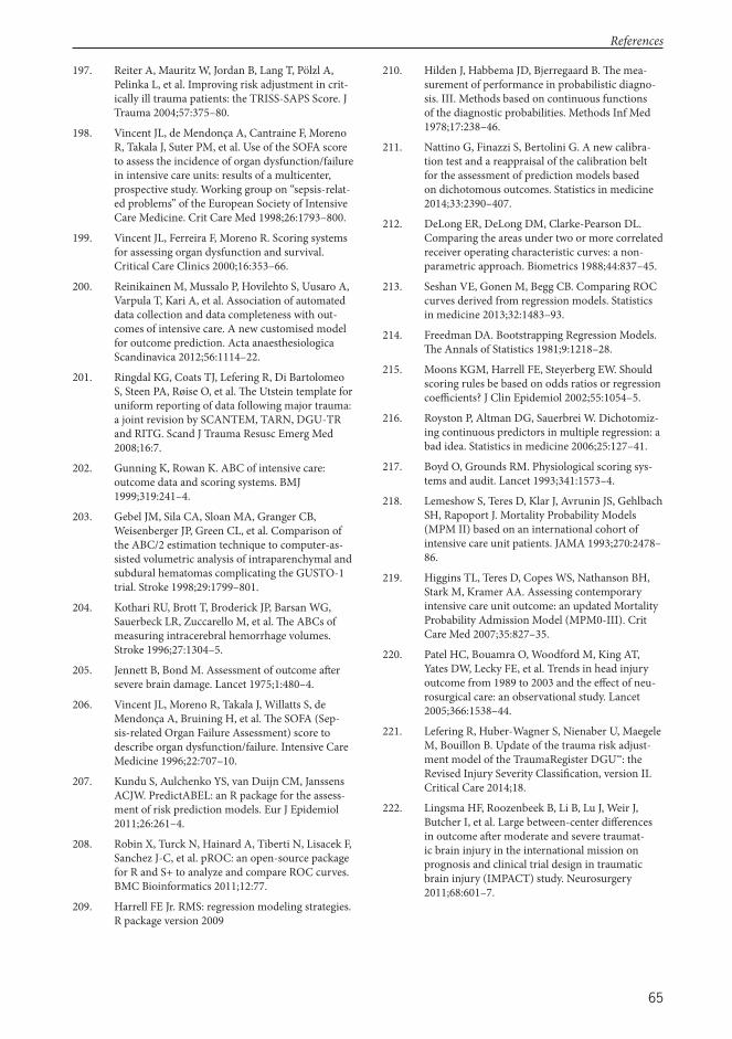
65
197. Reiter A, Mauritz W, Jordan B, Lang T, Pölzl A, Pelinka L, et al. Improving risk adjustment in crit-ically ill trauma patients: the TRISS-SAPS Score. J Trauma 2004;57:375–80.
198. Vincent JL, de Mendonça A, Cantraine F, Moreno R, Takala J, Suter PM, et al. Use of the SOFA score to assess the incidence of organ dysfunction/failure in intensive care units: results of a multicenter, prospective study. Working group on “sepsis-relat-ed problems” of the European Society of Intensive Care Medicine. Crit Care Med 1998;26:1793–800.
199. Vincent JL, Ferreira F, Moreno R. Scoring systems for assessing organ dysfunction and survival. Critical Care Clinics 2000;16:353–66.
200. Reinikainen M, Mussalo P, Hovilehto S, Uusaro A, Varpula T, Kari A, et al. Association of automated data collection and data completeness with out-comes of intensive care. A new customised model for outcome prediction. Acta anaesthesiologica Scandinavica 2012;56:1114–22.
201. Ringdal KG, Coats TJ, Lefering R, Di Bartolomeo S, Steen PA, Røise O, et al. Th e Utstein template for uniform reporting of data following major trauma: a joint revision by SCANTEM, TARN, DGU-TR and RITG. Scand J Trauma Resusc Emerg Med 2008;16:7.
202. Gunning K, Rowan K. ABC of intensive care: outcome data and scoring systems. BMJ 1999;319:241–4.
203. Gebel JM, Sila CA, Sloan MA, Granger CB, Weisenberger JP, Green CL, et al. Comparison of the ABC/2 estimation technique to computer-as-sisted volumetric analysis of intraparenchymal and subdural hematomas complicating the GUSTO-1 trial. Stroke 1998;29:1799–801.
204. Kothari RU, Brott T, Broderick JP, Barsan WG, Sauerbeck LR, Zuccarello M, et al. Th e ABCs of measuring intracerebral hemorrhage volumes. Stroke 1996;27:1304–5.
205. Jennett B, Bond M. Assessment of outcome aft er severe brain damage. Lancet 1975;1:480–4.
206. Vincent JL, Moreno R, Takala J, Willatts S, de Mendonça A, Bruining H, et al. Th e SOFA (Sep-sis-related Organ Failure Assessment) score to describe organ dysfunction/failure. Intensive Care Medicine 1996;22:707–10.
207. Kundu S, Aulchenko YS, van Duijn CM, Janssens ACJW. PredictABEL: an R package for the assess-ment of risk prediction models. Eur J Epidemiol 2011;26:261–4.
208. Robin X, Turck N, Hainard A, Tiberti N, Lisacek F, Sanchez J-C, et al. pROC: an open-source package for R and S+ to analyze and compare ROC curves. BMC Bioinformatics 2011;12:77.
209. Harrell FE Jr. RMS: regression modeling strategies. R package version 2009
210. Hilden J, Habbema JD, Bjerregaard B. Th e mea-surement of performance in probabilistic diagno-sis. III. Methods based on continuous functions of the diagnostic probabilities. Methods Inf Med 1978;17:238–46.
211. Nattino G, Finazzi S, Bertolini G. A new calibra-tion test and a reappraisal of the calibration belt for the assessment of prediction models based on dichotomous outcomes. Statistics in medicine 2014;33:2390–407.
212. DeLong ER, DeLong DM, Clarke-Pearson DL. Comparing the areas under two or more correlated receiver operating characteristic curves: a non-parametric approach. Biometrics 1988;44:837–45.
213. Seshan VE, Gonen M, Begg CB. Comparing ROC curves derived from regression models. Statistics in medicine 2013;32:1483–93.
214. Freedman DA. Bootstrapping Regression Models. Th e Annals of Statistics 1981;9:1218–28.
215. Moons KGM, Harrell FE, Steyerberg EW. Should scoring rules be based on odds ratios or regression coeffi cients? J Clin Epidemiol 2002;55:1054–5.
216. Royston P, Altman DG, Sauerbrei W. Dichotomiz-ing continuous predictors in multiple regression: a bad idea. Statistics in medicine 2006;25:127–41.
217. Boyd O, Grounds RM. Physiological scoring sys-tems and audit. Lancet 1993;341:1573–4.
218. Lemeshow S, Teres D, Klar J, Avrunin JS, Gehlbach SH, Rapoport J. Mortality Probability Models (MPM II) based on an international cohort of intensive care unit patients. JAMA 1993;270:2478–86.
219. Higgins TL, Teres D, Copes WS, Nathanson BH, Stark M, Kramer AA. Assessing contemporary intensive care unit outcome: an updated Mortality Probability Admission Model (MPM0-III). Crit Care Med 2007;35:827–35.
220. Patel HC, Bouamra O, Woodford M, King AT, Yates DW, Lecky FE, et al. Trends in head injury outcome from 1989 to 2003 and the eff ect of neu-rosurgical care: an observational study. Lancet 2005;366:1538–44.
221. Lefering R, Huber-Wagner S, Nienaber U, Maegele M, Bouillon B. Update of the trauma risk adjust-ment model of the TraumaRegister DGU™: the Revised Injury Severity Classifi cation, version II. Critical Care 2014;18.
222. Lingsma HF, Roozenbeek B, Li B, Lu J, Weir J, Butcher I, et al. Large between-center diff erences in outcome aft er moderate and severe traumat-ic brain injury in the international mission on prognosis and clinical trial design in traumatic brain injury (IMPACT) study. Neurosurgery 2011;68:601–7.
References
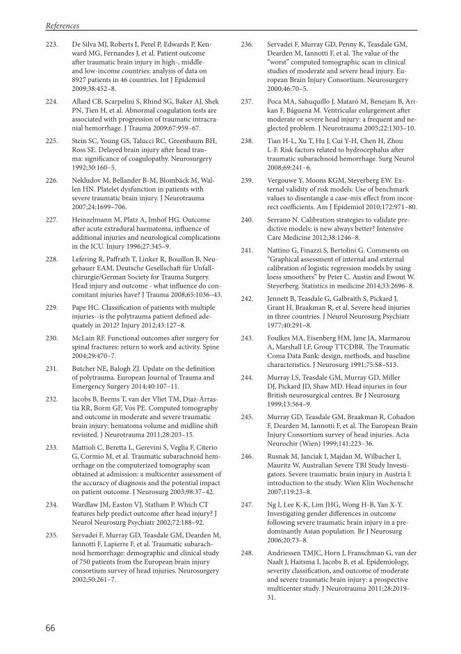
66
223. De Silva MJ, Roberts I, Perel P, Edwards P, Ken-ward MG, Fernandes J, et al. Patient outcome aft er traumatic brain injury in high-, middle- and low-income countries: analysis of data on 8927 patients in 46 countries. Int J Epidemiol 2009;38:452–8.
224. Allard CB, Scarpelini S, Rhind SG, Baker AJ, Shek PN, Tien H, et al. Abnormal coagulation tests are associated with progression of traumatic intracra-nial hemorrhage. J Trauma 2009;67:959–67.
225. Stein SC, Young GS, Talucci RC, Greenbaum BH, Ross SE. Delayed brain injury aft er head trau-ma: signifi cance of coagulopathy. Neurosurgery 1992;30:160–5.
226. Nekludov M, Bellander B-M, Blombäck M, Wal-len HN. Platelet dysfunction in patients with severe traumatic brain injury. J Neurotrauma 2007;24:1699–706.
227. Heinzelmann M, Platz A, Imhof HG. Outcome aft er acute extradural haematoma, infl uence of additional injuries and neurological complications in the ICU. Injury 1996;27:345–9.
228. Lefering R, Paff rath T, Linker R, Bouillon B, Neu-gebauer EAM, Deutsche Gesellschaft für Unfall-chirurgie/German Society for Trauma Surgery. Head injury and outcome - what infl uence do con-comitant injuries have? J Trauma 2008;65:1036–43.
229. Pape HC. Classifi cation of patients with multiple injuries--is the polytrauma patient defi ned ade-quately in 2012? Injury 2012;43:127–8.
230. McLain RF. Functional outcomes aft er surgery for spinal fractures: return to work and activity. Spine 2004;29:470–7.
231. Butcher NE, Balogh ZJ. Update on the defi nition of polytrauma. European Journal of Trauma and Emergency Surgery 2014;40:107–11.
232. Jacobs B, Beems T, van der Vliet TM, Diaz-Arras-tia RR, Borm GF, Vos PE. Computed tomography and outcome in moderate and severe traumatic brain injury: hematoma volume and midline shift revisited. J Neurotrauma 2011;28:203–15.
233. Mattioli C, Beretta L, Gerevini S, Veglia F, Citerio G, Cormio M, et al. Traumatic subarachnoid hem-orrhage on the computerized tomography scan obtained at admission: a multicenter assessment of the accuracy of diagnosis and the potential impact on patient outcome. J Neurosurg 2003;98:37–42.
234. Wardlaw JM, Easton VJ, Statham P. Which CT features help predict outcome aft er head injury? J Neurol Neurosurg Psychiatr 2002;72:188–92.
235. Servadei F, Murray GD, Teasdale GM, Dearden M, Iannotti F, Lapierre F, et al. Traumatic subarach-noid hemorrhage: demographic and clinical study of 750 patients from the European brain injury consortium survey of head injuries. Neurosurgery 2002;50:261–7.
236. Servadei F, Murray GD, Penny K, Teasdale GM, Dearden M, Iannotti F, et al. Th e value of the “worst” computed tomographic scan in clinical studies of moderate and severe head injury. Eu-ropean Brain Injury Consortium. Neurosurgery 2000;46:70–5.
237. Poca MA, Sahuquillo J, Mataró M, Benejam B, Ari-kan F, Báguena M. Ventricular enlargement aft er moderate or severe head injury: a frequent and ne-glected problem. J Neurotrauma 2005;22:1303–10.
238. Tian H-L, Xu T, Hu J, Cui Y-H, Chen H, Zhou L-F. Risk factors related to hydrocephalus aft er traumatic subarachnoid hemorrhage. Surg Neurol 2008;69:241–6.
239. Vergouwe Y, Moons KGM, Steyerberg EW. Ex-ternal validity of risk models: Use of benchmark values to disentangle a case-mix eff ect from incor-rect coeffi cients. Am J Epidemiol 2010;172:971–80.
240. Serrano N. Calibration strategies to validate pre-dictive models: is new always better? Intensive Care Medicine 2012;38:1246–8.
241. Nattino G, Finazzi S, Bertolini G. Comments on “Graphical assessment of internal and external calibration of logistic regression models by using loess smoothers” by Peter C. Austin and Ewout W. Steyerberg. Statistics in medicine 2014;33:2696–8.
242. Jennett B, Teasdale G, Galbraith S, Pickard J, Grant H, Braakman R, et al. Severe head injuries in three countries. J Neurol Neurosurg Psychiatr 1977;40:291–8.
243. Foulkes MA, Eisenberg HM, Jane JA, Marmarou A, Marshall LF, Group TTCDBR. Th e Traumatic Coma Data Bank: design, methods, and baseline characteristics. J Neurosurg 1991;75:S8–S13.
244. Murray LS, Teasdale GM, Murray GD, Miller DJ, Pickard JD, Shaw MD. Head injuries in four British neurosurgical centres. Br J Neurosurg 1999;13:564–9.
245. Murray GD, Teasdale GM, Braakman R, Cohadon F, Dearden M, Iannotti F, et al. Th e European Brain Injury Consortium survey of head injuries. Acta Neurochir (Wien) 1999;141:223–36.
246. Rusnak M, Janciak I, Majdan M, Wilbacher I, Mauritz W, Australian Severe TBI Study Investi-gators. Severe traumatic brain injury in Austria I: introduction to the study. Wien Klin Wochenschr 2007;119:23–8.
247. Ng I, Lee K-K, Lim JHG, Wong H-B, Yan X-Y. Investigating gender diff erences in outcome following severe traumatic brain injury in a pre-dominantly Asian population. Br J Neurosurg 2006;20:73–8.
248. Andriessen TMJC, Horn J, Franschman G, van der Naalt J, Haitsma I, Jacobs B, et al. Epidemiology, severity classifi cation, and outcome of moderate and severe traumatic brain injury: a prospective multicenter study. J Neurotrauma 2011;28:2019–31.
References
67
249. Weir J, Steyerberg EW, Butcher I, Lu J, Lings-ma HF, McHugh GS, et al. Does the extended Glasgow Outcome Scale add value to the conven-tional Glasgow Outcome Scale? J Neurotrauma 2012;29:53–8.
250. McHugh GS, Butcher I, Steyerberg EW, Lu J, Mushkudiani N, Marmarou A, et al. Statistical approaches to the univariate prognostic analysis of the IMPACT database on traumatic brain injury. J Neurotrauma 2007;24:251–8.
251. Th elin EP, Nelson DW, Bellander B-M. Secondary peaks of S100B in serum relate to subsequent radiological pathology in traumatic brain injury. Neurocrit Care 2014;20:217–29.
252. Fehlings MG, Baker A. Is there a role for hyper-oxia in the management of severe traumatic brain injury? J Neurosurg 2007;106:525–5.
253. Kumaria A, Tolias CM. Normobaric hyperoxia therapy for traumatic brain injury and stroke: a review. Br J Neurosurg 2009;23:576–84.
254. Opreanu RC, Kuhn D, Basson MD. Infl uence of alcohol on mortality in traumatic brain injury. J Am Coll Surg 2010;210:997–1007.
255. Shahin H, Gopinath SP, Robertson CS. Infl uence of alcohol on early Glasgow Coma Scale in head-in-jured patients. J Trauma 2010;69:1176–81.
256. Raj R, Skrifvars MB, Kivisaari R, Hernesniemi J, Lappalainen J, Siironen J. Acute alcohol intoxi-cation and long-term outcome in patients with traumatic brain injury. J Neurotrauma 2014; In Press.
References