Embed Size (px)
Citation preview
British Journal of Ophthalmology, 1979, 63, 735-743
Prognosis for rubeosis iridis following centralretinal vein occlusionSTEPHEN H. SINCLAIR AND EVANGELOS S. GRAGOUDASFrom the Eye Research Institute of Retina Foundation, and the Retina Service,Massachusetts Eye and Ear Infirmary, Boston, Massachusetts
SUMMARY The records of 57 patients with recent central retinal vein occlusion were reviewed inorder to predict the development of rubeosis iridis (RI) and neovascular glaucoma (NVG) fromthe initial clinical examination, colour fundus photographs, and fluorescein angiograms. Twelvepatients (21 %) developed both RI and NVG, and this complication appeared to be correlated mostsignificantly with clinical and fluoroangiographic evidence of severe retinal ischaemia. The corre-lation between RI and other findings was as follows: widespread capillary occlusion (86 % developedRI); absent perifoveal network (80 %); 10 or more cotton-wool spots (75 %); A-V transit time ofgreater than 20 seconds (75 %); severe large vessel leakage (31 %); and severe retinal oedema (60 %).Less significant correlation of RI was obtained with poor visual acuity and with associated systemicdiseases. Factors of no statistical prognostic value included the patient's age, history, or evidenceof pre-existing open-angle glaucoma, degree of fundus haemorrhage, capillary dilatation, discoedema, disc capillary dilatation and leakage, leakage of small vessels in the posterior pole, anddevelopment of disc collateral vessels. Multivariate linear discriminant analysis of the data supportedthe above factors as being the variables that best predicted the development of RI, and a derivedlinear equation predicted 91 % of all patients studied who had central retinal vein occlusion andwho developed rubeosis iridis.
Rubeosis iridis (RI) and neovascular glaucoma(NVG) are severe complications of central retinalvein occlusion (CRVO) which occur in 12% to 30%of all cases (Smith, 1961; Vannas, 1961; Laatikainenand Kohner, 1976). The stimulus for anterior seg-ment neovascularisation is poorly understood butis thought to be related to severe retinal ischaemia(Smith, 1961; Ashton, 1961; Shilling and Kohner,1976). Fluorescein angiography adds considerableinformation to the understanding of the patho-physiology of CRVO by enabling qualitativeevaluation of the retinal vascular responses to anischaemic lesion. These responses include retinalcapillary nonperfusion, dilatation of small vessels,microaneurysm formation, and abnormal vascularpermeability secondary to endothelial cell damage.Studies in diabetics (Laatikainen and Kohner,1976; Kohner et al., 1976) have suggested a relationbetween the development of fundus or iris neovas-cularisation and widespread areas of retinal capillarynonperfusion. A similar relation has been reported
for branch vein occlusion (Kohner et al., 1976;Sedney, 1976; Laatikainen and Blach, 1977) andCRVO (Laatikainen and Kohner, 1976; Hayreh,1976, Kottow et al., 1976; Laatikainen, 1977a).
Prognosticating this complication has becomeimportant, since recent studies (Sedney, 1976;May et al., 1976; Laatikainen, 1977b) have reportedthat panretinal photocoagulation may be used toreverse or prevent iris neovascularisation afterCRVO. It has been suggested, however, that treat-ment should be completed either early in thecourse of rubeosis, before extensive peripheralanterior synechiae are established, or prophylacti-cally before clinical rubeosis is recognised. Thisobviously necessitates the early identification ofthose patients with CRVO who are at high risk ofdeveloping neovascular complications. This studywas undertaken to extend previous studies (Laati-kainen and Kohner, 1976; Laatikainen and Blach,1977) in order to determine the features of eitherthe initial clinical examination, the fluorescein
735
on 18 May 2018 by guest. P
rotected by copyright.http://bjo.bm
j.com/
Br J O
phthalmol: first published as 10.1136/bjo.63.11.735 on 1 N
ovember 1979. D
ownloaded from
Stephen H. Sinclair and Evangelos S. Gragoudas
angiogram, or both, that might be statisticallycorrelated with the subsequent development ofanterior segment neovascularisation.
Materials and methods
The records of patients with CRVO who had beenreferred to the Massachusetts Eye and Ear InfirmaryFluorescein Service were reviewed for developmentof clinically unequivocal rubeosis iridis, neovascularglaucoma, or both. Only patients with relatively'fresh' vein occlusions were selected; patients with'old' occlusions, either by history or by funduscriteria, were excluded. The duration of symptomsprior to the initial examination of the 57 patientsincluded is shown in Table 1. Fifty-four cases wereunilateral and 3 bilateral, in which a CRVO hadoccurred recently after an older occlusion in thefellow eye. The average age of the patients was 60years, with a range of 23 to 93 years. All patientswere followed up until resolution of the CRVO oruntil they developed clinically unequivocal rubeosisiridis or neovascular glaucoma. The averagefollow-up period of eyes that did not develop rubeo-sis was 10 months (range 4 to 36 months). Mostpatients received no treatment after initial evalua-tion; only a few were treated with antifibrinolyticagents, anticoagulants, or steroids.
Patients' records, which included stereo fundusphotographs, stereo fundus fluorescein angiograms,and data from the initial and follow-up examina-tions, were reviewed for the following details: (1)history: duration and type of symptoms, age,associated systemic, ocular, or orbital disease; (2)initial ocular examination: visual acuity, intraocularpressure, afferent pupillary defect; fellow eyevisual acuity, intraocular pressure, disc or fieldchanges, and retinal pathology; (3) course: finalvisual acuity, time required for resolution of haem-orrhages and oedema, development of macularcysts, disc collaterals, unequivocal iris neovascu-larisation (aberrant surface vessels visible onbiomicroscopy), or neovascular glaucoma.
Table 1 Duration of symptoms in patients with CRVO
Duration of symptoms No. ofpatients (Y.)
1 mo 29 (51)
2-3 mo 13 (23)
>3 mo 6 (11)
Undetermined length of time 5 (9)
Routine examination without symptoms 4 (7)
CRVO=central retinal vein occlusion.
For the purpose of this study the photographedretina of the involved eye was divided into 3 terri-tories for evaluation of various areas: (1) macula:area within a 2-disc radius from the fovea; (2)posterior pole: disc and area within the majortemporal vascular arcades, except for the maculararea described above; (3) periphery: area peripheralto the disc and major temporal arcades. Colourstereo fundus photographs were used to assess theseverity or extent of retinal haemorrhages, oedema,and cotton-wool spots within these 3 territories,and collateral disc vessels.
In each of the above territories the intraretinal,deep, superficial, and subhyaloid retinal haemor-rhages were graded: none (0), mild (1), moderate (2),or severe (3) (the number in parentheses is the gradeof severity for each variable that we included in themultivariate discriminant analysis; see 'Statisticalanalysis'). Retinal oedema was graded: none (0),mild (1), or severe (2), with notation of the forma-tion of macular cysts (3). The number of cotton-wool spots was recorded, as well as their papillary,peripapillary, or peripheral location. Opticociliaryveins were either present (1) or absent (0).
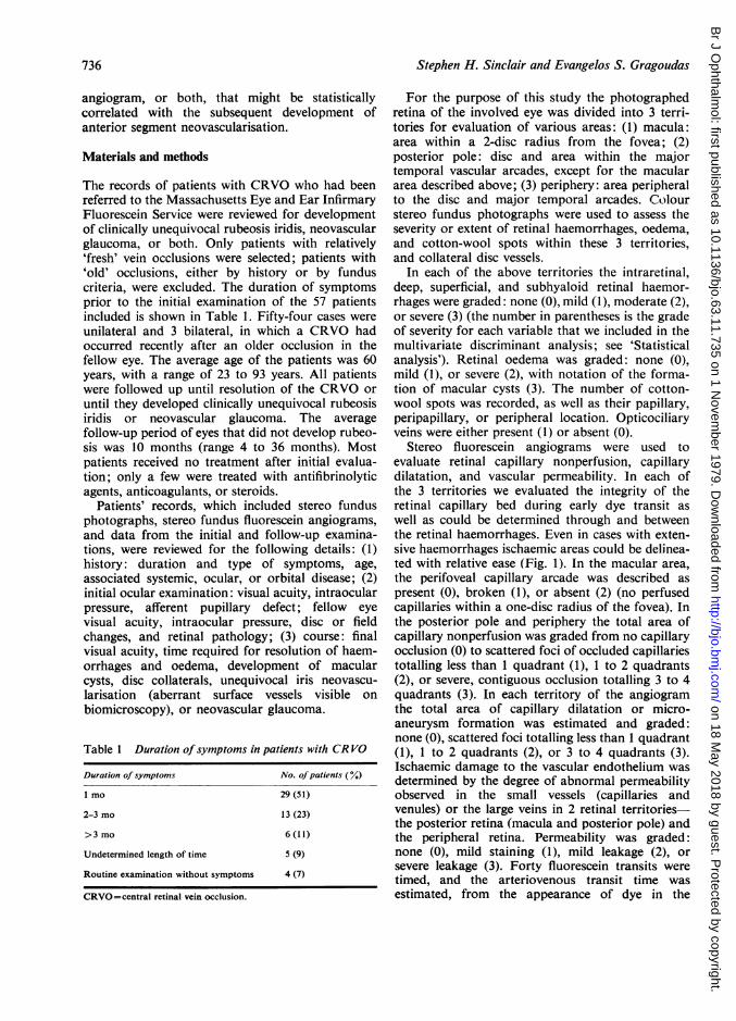
Stereo fluorescein angiograms were used toevaluate retinal capillary nonperfusion, capillarydilatation, and vascular permeability. In each ofthe 3 territories we evaluated the integrity of theretinal capillary bed during early dye transit aswell as could be determined through and betweenthe retinal haemorrhages. Even in cases with exten-sive haemorrhages ischaemic areas could be delinea-ted with relative ease (Fig. 1). In the macular area,the perifoveal capillary arcade was described aspresent (0), broken (1), or absent (2) (no perfusedcapillaries within a one-disc radius of the fovea). Inthe posterior pole and periphery the total area ofcapillary nonperfusion was graded from no capillaryocclusion (0) to scattered foci of occluded capillariestotalling less than 1 quadrant (1), 1 to 2 quadrants(2), or severe, contiguous occlusion totalling 3 to 4quadrants (3). In each territory of the angiogramthe total area of capillary dilatation or micro-aneurysm formation was estimated and graded:none (0), scattered foci totalling less than 1 quadrant(1), 1 to 2 quadrants (2), or 3 to 4 quadrants (3).Ischaemic damage to the vascular endothelium wasdetermined by the degree of abnormal permeabilityobserved in the small vessels (capillaries andvenules) or the large veins in 2 retinal territories-the posterior retina (macula and posterior pole) andthe peripheral retina. Permeability was graded:none (0), mild staining (1), mild leakage (2), orsevere leakage (3). Forty fluorescein transits weretimed, and the arteriovenous transit time wasestimated, from the appearance of dye in the
736
on 18 May 2018 by guest. P
rotected by copyright.http://bjo.bm
j.com/
Br J O
phthalmol: first published as 10.1136/bjo.63.11.735 on 1 N
ovember 1979. D
ownloaded from
Prognosis for rubeosis iridis following central retinal vein occlusion
Fig. I Fundus photograph and mid-capillary-transit fluorescein angiogram ofpatient with CRVO. A, B: Mild,scattered foci of capillary occlusion; C, D: Extensive capillary occlusion
retinal arteries to the complete filling of the majorcentral retinal veins.
STATISTICAL ANALYSISAll of the collected data and their association withrubeosis iridis were subjected to statistical analysisby x2 tests for the resultant 3 x 2 or 4x 2 tables. Thedata were also subjected to a computer analysiswhich derived a multivariate, linear, discriminantcoefficient equation. This analysis evaluates thepotential of each variable, alone and in combinationwith others, to predict which patients will developrubeosis iridis as a complication of CRVO. Theprogramme derives a linear equation of thosevariables that are the best predictors. A weightedcoefficient, assigned to each variable, is multipliedby the graded severity of the variable: X = K +C1 (V1) + C2 (V2) + C3 (V3) + . . . + Cn (Vn)where K is a constant, C1 . . . Cn are the weighted
coefficients for the best-predicting variables, andVi . . . Vn are the graded values of the patient foreach variable. The resultant discriminant score (X)indicates the tendency of that patient to developrubeosis iridis. We can thus derive a weightedcombination of the best-predicting variables whichmay serve better than any 1 variable alone topredict which patients will develop rubeosis iridis.A discriminant score was calculated for eachpatient by means of coefficients of the best-predict-ing variables derived by the programme along withtheir graded values (the parenthetical values des-cribed above).
Results
Rubeosis iridis developed in 12 of 57 patients (21%)with early CRVO within a period of 3 weeks to7 months after the onset of symptoms (Table 2),
737
on 18 May 2018 by guest. P
rotected by copyright.http://bjo.bm
j.com/
Br J O
phthalmol: first published as 10.1136/bjo.63.11.735 on 1 N
ovember 1979. D
ownloaded from
Stephen H. Sinclair and Evangelos S. Gragoudas
Patients with R I EJ
Macula
4035
uv 30c.1ui25 5
0- 20-*0 150 10. 95% 85%
5 86%
Intact Broken Absentfoveal foveal fovealnet net net
Patients wthout RI EPosterior pole
Mild Severe Mild(0-1) (3-4) (0-1quadrant quadrant quadocclusion occlusion occlt
Periphery
81%
I Severe(3-4)
lrant quadrantusion occlusion
Table 2 Interval between onset ofsymptoms anddevelopment of rubeosis in CRVO
Interval (months) No. ofpatients
<I 1
1- 2
2- 3
3- 3
4+ 3
Total 12
CRVO =central retinal vein occlusion.
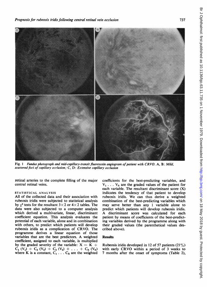
and in all these patients angle neovascularisationand glaucoma ensued. The development of rubeosisiridis was most significantly correlated with thosefeatures of the initial clinical examination andfluorescein angiogram that indicated severe retinalischaemia. As shown in Fig. 2, rubeosis iridiscorrelated most directly with the extent of capillarynonperfusion that was observed on the fluoresceinangiogram. Rubeosis developed in 80-86% of eyeswith an absent parafoveal net of 3 to 4 quadrantsof posterior pole or peripheral capillary occlusion,but appeared in only 3-9% of those with an intactparafoveal net and less than 1 quadrant of capillaryocclusion. These results were significant, P <0 001level. The view of the perifoveal arcade was suffi-ciently blocked by haemorrhage to prevent evalua-tion in 6 patients.
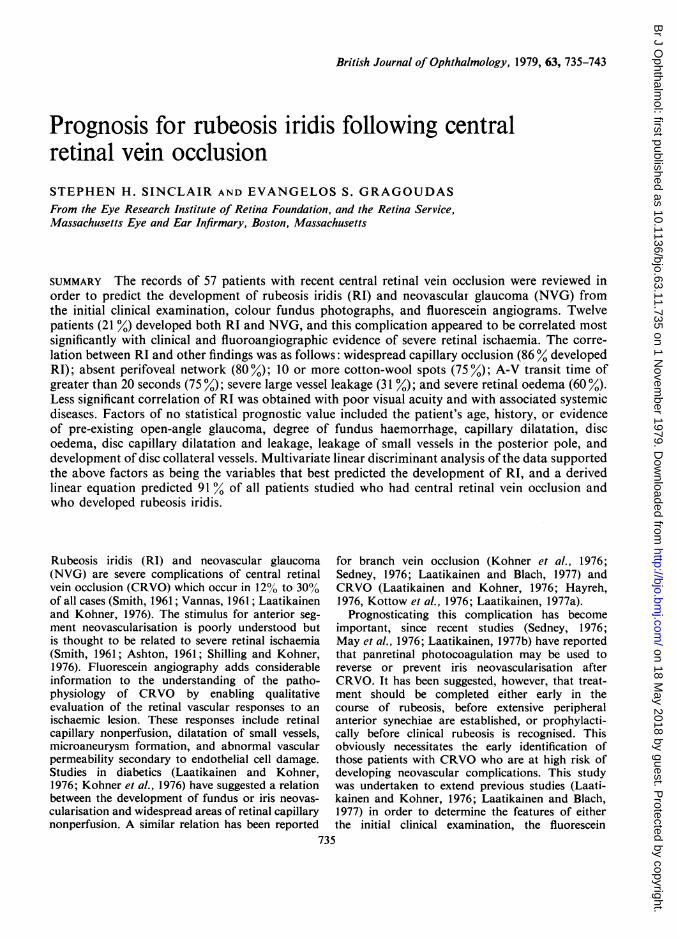
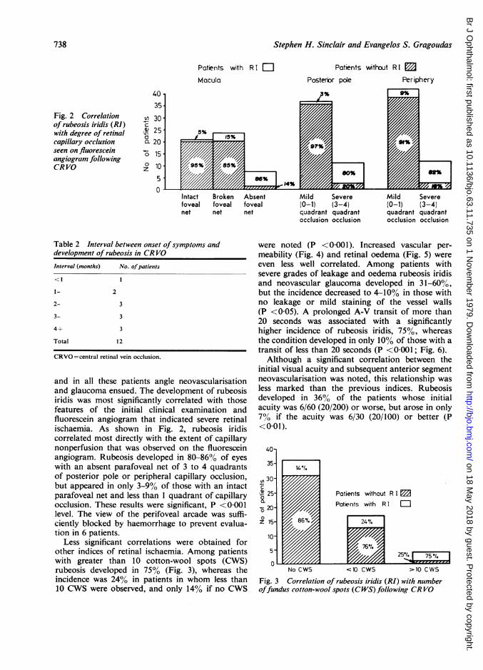
Less significant correlations were obtained forother indices of retinal ischaemia. Among patientswith greater than 10 cotton-wool spots (CWS)rubeosis developed in 75% (Fig. 3), whereas theincidence was 24% in patients in whom less than10 CWS were observed, and only 14% if no CWS
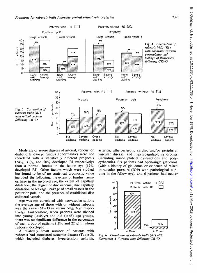
were noted (P <0 001). Increased vascular per-meability (Fig. 4) and retinal oedema (Fig. 5) wereeven less well correlated. Among patients withsevere grades of leakage and oedema rubeosis iridisand neovascular glaucoma developed in 31-60%,but the incidence decreased to 4-10% in those withno leakage or mild staining of the vessel walls(P <0 05). A prolonged A-V transit of more than20 seconds was associated with a significantlyhigher incidence of rubeosis iridis, 75%, whereasthe condition developed in only 10% of those with atransit of less than 20 seconds (P <0 001; Fig. 6).Although a significant correlation between the
initial visual acuity and subsequent anterior segmentneovascularisation was noted, this relationship wasless marked than the previous indices. Rubeosisdeveloped in 36% of the patients whose initialacuity was 6/60 (20/200) or worse, but arose in only7% if the acuity was 6/30 (20/100) or better (P<0-01).
0
z
No CWS
Patients without R I 1Patients with RII
<10 CWS >10 CWS
Fig. 3 Correlation of rubeosis iridis (RI) with numberoffundus cotton-wool spots (CWS) following CRVO
Fig. 2 Correlationof rubeosis iridis (RI)with degree of retinalcapillary occlusionseen on fluoresceinangiogram followingCR VO
738
on 18 May 2018 by guest. P
rotected by copyright.http://bjo.bm
j.com/
Br J O
phthalmol: first published as 10.1136/bjo.63.11.735 on 1 N
ovember 1979. D
ownloaded from
Prognosis for rubeosis iridis following central retinal vein occlusion
Patients with R I Eli
Posterior pole
Large vessels Small vessels
Patients without RI EDPeriphery
Large vessels Small vessels
Fig. 4 Correlation ofrubeosis iridis (RI)with abnormal vascularpermeability andleakage offluoresceinfollowing CR VO
Fig. 5 Correlation ofrubeosis iridis (RI)with retinal oedemafollowing CR VO 0
z
Patients with R I
No Severe Cystic No Severe No Severeoedema oedema oedema oedema oedema oedema oedema
Moderate or severe degrees of arterial, venous, or
diabetic fellow-eye fundus abnormalities were notcorrelated with a statistically different prognosis(24%, 33%, and 20% developed RI respectively)than a normal fundus in the fellow eye (17%developed RI). Other factors which were studiedbut found to be of no statistical prognostic valueincluded the following: the extent of fundus haem-orrhage in the involved eye, the extent of capillarydilatation, the degree of disc oedema, disc capillarydilatation or leakage, leakage of small vessels in theposterior pole, and the presence of established disccollateral vessels.Age was not correlated with neovascularisation;
the average age of those with or without rubeosiswas the same (63±19yr versus 59±16yr respec-tively). Furthermore, when patients were dividedinto young (<45 yr) and old (>45) age groups,there was no significant difference in the percentageof each group of patients (18% and 22%) in whomrubeosis developed.A relatively small number of patients with
rubeosis had associated systemic disease (Table 3),which included diabetes, hypertension, arthritis,
arteritis, atherosclerotic cardiac and/or peripheralvascular disease, and hypercoagulable syndromes(including minor platelet dysfunctions and poly-cythaemia). Six patients had open-angle glaucoma(with a history of glaucoma or evidence of raisedintraocular pressure (IOP) with pathological cup-
ping in the fellow eye), and 6 patients had ocular
40
35 -
Ln
a._
z
Patients without R I mPatients with R I Fli
0 Z,_,1_,1,A_,A<20sec >20sec
Fig. 6 Correlation of rubeosis iridis (RI) withfluorescein A- V transit time following CRVO
4c35.
@ 30J 25a. 20o 150z
5-
739
on 18 May 2018 by guest. P
rotected by copyright.http://bjo.bm
j.com/
Br J O
phthalmol: first published as 10.1136/bjo.63.11.735 on 1 N
ovember 1979. D
ownloaded from
Stephen H. Sinclair and Evangelos S. Gragoudas
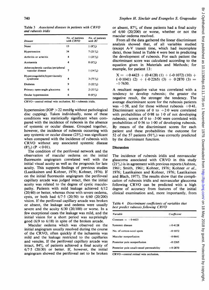
Table 3 Associated diseases in patients with CRVOand rubeosis iridis
No. ofpatients No. of patientsDisease with CRVO with RI
None 13 1 (8%)
Hypertension 34 7 (21°,)
Arthritis or arteritis 4 1 (25%)
Azotaemia 4 0 (0%)
Atherosclerotic cardiac/peripheralvascular disease 7 1 (14%)
Hypercoagulable/hyperviscositysyndrome 4 3 (75%)
Diabetes 9 2 (22%)
Primary open-angle glaucoma 6 2 (33 %)
Ocular hypertension 6 0 (0 %)
CRVO=central retinal vein occlusion. RI=rubeosis iridis.
hypertension (IOP > 22 mmHg without pathologicaldisc cupping). Taken individually, none of theseconditions was statistically significant when com-pared with the incidence of rubeosis in the absenceof systemic or ocular disease. Grouped together,however, the incidence of rubeosis occurring withany systemic or ocular disease (25%) was significantwhen compared with the incidence of rubeosis withCRVO without any associated systemic disease(8%) (P < 0 01).The condition of the perifoveal network and the
observation of macular oedema on the initialfluorescein angiogram correlated well with theinitial visual acuity as well as the prognosis for lateacuity. This supports findings of previous studies(Laatikainen and Kohner, 1976; Kohner, 1976). Ifon the initial fluorescein angiogram the perifovealcapillary arcade was judged intact, then the initialacuity was related to the degree of cystic maculo-pathy. Patients with mild leakage achieved 6/12(20/40) or better, whereas those with severe oedema,cysts, or both had 6/7 5 (20/50) to 6/60 (20/200)vision. If the perifoveal capillary arcade was brokenor absent, the leakage and oedema were usuallysevere and the acuity 6/30 (20/100) or worse. In a
few exceptional cases the leakage was mild, and theinitial vision for a short period was sui prisinglygood (6/9 to 6/18) in spite of the broken arcade.
Macular oedema which was observed on theinitial angiogram usually resolved during the courseof the CRVO, often quickly if the ischaemia was
mild and the leakage restricted to the capillariesand venules. If the perifoveal capillary arcade was
intact, 84% of patients achieved a final acuity of6/7 5 (20/30) or better. If, however, the initialangiogram showed the perifoveal net to be broken
or absent, 87% of these patients had a final acuityof 6/60 (20/200) or worse, whether or not themacular oedema resolved.From all the data gathered the linear discriminant
analysis showed that, of all variables studied(except A-V transit time, which had incompletedata), those listed in Table 4 were best in predictingthe development of rubeosis. For each patient thediscriminant score was calculated according to theequation given in Materials and Methods; forexample, for patient 13:
X = +0-4423 + (0 4128) (1) + (-0 1072) (10) +(-0 1041) (2) + (-0 2265) (3) + 0 2870 (3) --1 7630.
A resultant negative value was correlated with atendency to develop rubeosis; the greater thenegative result, the stronger the tendency. Theaverage discriminant score for the rubeosis patientswas -1 58, and for those without rubeosis +0 41.Discriminant scores of 0 to +2-0 were correlatedwith probabilities of 0 98 to 1-0 of not developingrubeosis; scores of 0 to -3 00 were correlated withprobabilities of 0-56 to 1-00 of developing rubeosis.By means of the discriminant scores for eachpatient and these probabilities the outcome for52 of the 57 patients (91%) was correctly predictedby the discriminant function.
Discussion
The incidence of rubeosis iridis and neovascularglaucoma associated with CRVO in this study(21%) is in agreement with previous reports (Ashton,1961; Smith, 1961; Kohner, 1976; Kohner et al.,1976; Laatikainen and Kohner, 1976; Laatikainenand Blach, 1977). The results show that the compli-cation of rubeosis iridis and neovascular glaucomafollowing CRVO can be predicted with a highdegree of accuracy from features of the initialclinical examination and, more importantly, from
Table 4 Discriminant coefficients of variables thatbest predict rubeosis following CR VO
Variable Coefficient
Constant = + 0-4423
Systemic disease +0-4128
No. of cotton-wool spots -01072
Macular nonperfusion -08641
Posterior pole nonperfusion -0 2265
Posterior pole small-vessel permeability +0-2870
CRVO=central retinal vein occlusion.
740
on 18 May 2018 by guest. P
rotected by copyright.http://bjo.bm
j.com/
Br J O
phthalmol: first published as 10.1136/bjo.63.11.735 on 1 N
ovember 1979. D
ownloaded from
Prognosis for rubeosis iridis following central retinal vein occlusion
findings of the initial fluorescein angiogram. Thesevariables, summarised in Tables 5 and 6, are director indirect manifestations of an ischaemic infarctand support the concept that anterior segmentneovascularisation is correlated with the degree orthe extent of fundus ischaemia, or both. Thestrongest one-to-one correlation was with wide-spread capillary occlusion as seen on the fluoresceinangiogram. Rubeosis iridis was correlated alsowith the funduscopic observation of cotton-woolspots, but less well than with the extent of capillaryocclusion. This was probably because cotton-wool
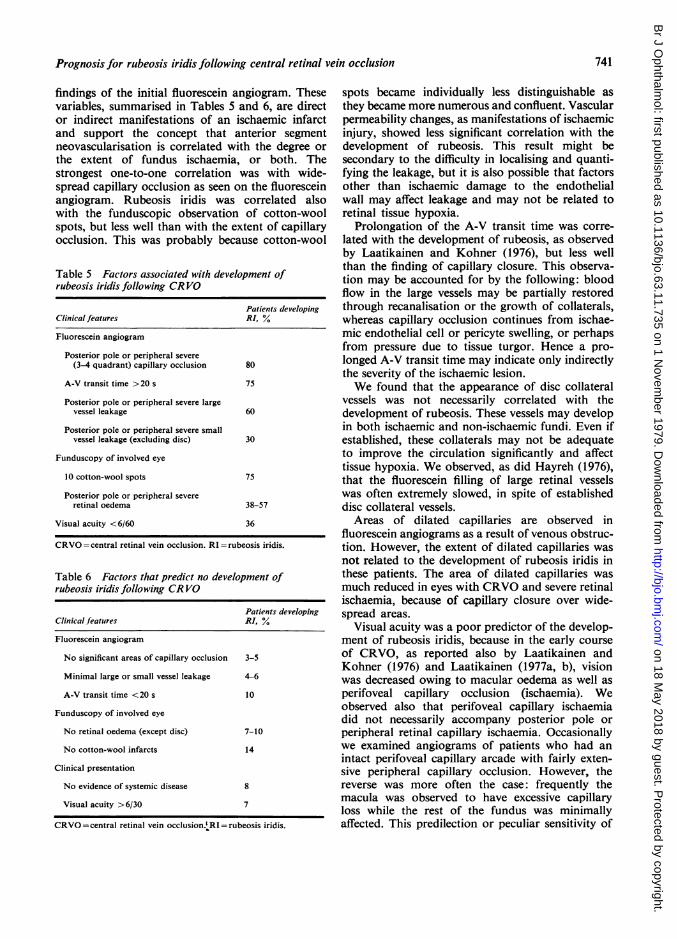
Table 5 Factors associated with development ofrubeosis iridis following CRVO
Patients developingClinical features RI, Y
Fluorescein angiogram
Posterior pole or peripheral severe(3-4 quadrant) capillary occlusion 80
A-V transit time >20 s 75
Posterior pole or peripheral severe largevessel leakage 60
Posterior pole or peripheral severe smallvessel leakage (excluding disc) 30
Funduscopy of involved eye
10 cotton-wool spots 75
Posterior pole or peripheral severeretinal oedema 38-57
Visual acuity <6/60 36
CRVO =central retinal vein occlusion. RI=rubeosis iridis.
Table 6 Factors that predict no development ofrubeosis iridis following CR VO
Patients developingClinical features RI, %
Fluorescein angiogram
No significant areas of capillary occlusion 3-5
Minimal large or small vessel leakage 4-6
A-V transit time <20 s 10
Funduscopy of involved eye
No retinal oedema (except disc) 7-10
No cotton-wool infarcts 14
Clinical presentation
No evidence of systemic disease 8
Visual acuity >6/30 7
CRVO =central retinal vein occlusion.'RI =rubeosis iridis.
spots became individually less distinguishable asthey became more numerous and confluent. Vascularpermeability changes, as manifestations of ischaemicinjury, showed less significant correlation with thedevelopment of rubeosis. This result might besecondary to the difficulty in localising and quanti-fying the leakage, but it is also possible that factorsother than ischaemic damage to the endothelialwall may affect leakage and may not be related toretinal tissue hypoxia.
Prolongation of the A-V transit time was corre-lated with the development of rubeosis, as observedby Laatikainen and Kohner (1976), but less wellthan the finding of capillary closure. This observa-tion may be accounted for by the following: bloodflow in the large vessels may be partially restoredthrough recanalisation or the growth of collaterals,whereas capillary occlusion continues from ischae-mic endothelial cell or pericyte swelling, or perhapsfrom pressure due to tissue turgor. Hence a pro-longed A-V transit time may indicate only indirectlythe severity of the ischaemic lesion.We found that the appearance of disc collateral
vessels was not necessarily correlated with thedevelopment of rubeosis. These vessels may developin both ischaemic and non-ischaemic fundi. Even ifestablished, these collaterals may not be adequateto improve the circulation significantly and affecttissue hypoxia. We observed, as did Hayreh (1976),that the fluorescein filling of large retinal vesselswas often extremely slowed, in spite of establisheddisc collateral vessels.
Areas of dilated capillaries are observed influorescein angiograms as a result of venous obstruc-tion. However, the extent of dilated capillaries wasnot related to the development of rubeosis iridis inthese patients. The area of dilated capillaries wasmuch reduced in eyes with CRVO and severe retinalischaemia, because of capillary closure over wide-spread areas.
Visual acuity was a poor predictor of the develop-ment of rubeosis iridis, because in the early courseof CRVO, as reported also by Laatikainen andKohner (1976) and Laatikainen (1977a, b), visionwas decreased owing to macular oedema as well asperifoveal capillary occlusion (ischaemia). Weobserved also that perifoveal capillary ischaemiadid not necessarily accompany posterior pole orperipheral retinal capillary ischaemia. Occasionallywe examined angiograms of patients who had anintact perifoveal capillary arcade with fairly exten-sive peripheral capillary occlusion. However, thereverse was more often the case: frequently themacula was observed to have excessive capillaryloss while the rest of the fundus was minimallyaffected. This predilection or peculiar sensitivity of
741
on 18 May 2018 by guest. P
rotected by copyright.http://bjo.bm
j.com/
Br J O
phthalmol: first published as 10.1136/bjo.63.11.735 on 1 N
ovember 1979. D
ownloaded from
Stephen H. Sinclair and Evangelos S. Gragoudas
the posterior pole to ischaemic injury has beennoted by Hayreh (1976) and may perhaps be ex-plained by the higher density of photoreceptors andneural elements in the posterior fundus, whichcause this area to have higher oxygen requirements,or perhaps by different haemodynamics withindifferent segments of the fundus.
In this study a CRVO associated with systemic,orbital, or ocular disease was correlated with asignificantly increased risk of developing anteriorsegment neovascularisation. However, none of theassociated diseases alone showed a statisticallysignificant difference from the cases of rubeosis withno associated disease. This seems to be contrary tothe impression of others that rubeosis occurs morefrequently in CRVO patients with primary glaucomaor systemic disease (Vannas, 1961; Kottow et al.,1976). Our observation that age was not significantlycorrelated with rubeosis iridis is also contrary tofindings by Vannas (1961) and Kottow et al.,(1976), but the increased number of elderly patientshaving rubeosis with CRVO in this study merelyreflected the increased incidence of CRVO in theelderly. This result, however, could be caused by anunrepresentative sample composed of the more severeCRVO patients referred to the Fluorescein Service.
In a previous study of CRVO by Laatikainen andKohner (1976) it was suggested that a fluoresceinangiogram taken 3 months after the occlusion wasmore accurate in evaluating perifoveal capillary netintegrity than the initial angiogram or one takenwithin 1 month of the onset of symptoms. This wasbecause of progressive deterioration (increased areaof capillary occlusion) noted in follow-up angio-grams of a few patients. Progression of the ischae-mia, if common in CRVO cases, would prohibitearly prognostication for rubeosis iridis. To evaluatethis we reviewed follow-up fluorescein angiogramstaken within 2 to 5 months in 15 patients in whomthe initial angiogram was taken within 1 month ofthe onset of symptoms. The follow-up angiogramin 12 patients did not change significantly; theremaining 3 (20%) showed significant progressionor worsening of capillary occlusion, dilatation, andleakage. In 1 of these patients, who had no ischaemiainitially, we found scattered foci of capillary occlu-sion over less than 1 total quadrant. In the secondpatient, with no initial ischaemia, moderately severe3-quadrant ischaemia was seen. The third patient,with patchy capillary occlusion, later showedsevere confluent occlusion. However, rubeosis iridisdeveloped in none of these 3. There was no particularfeature of the initial history, ocular examination,or fluorescein angiogram that distinguished thesepatients from those who showed no progressivedeterioration.
These results suggest that progressive ischaemicdeterioration may occur in CRVO, but that theinitial fluorescein angiogram and examination areaccurate for most cases in prognosticating thecomplication of anterior segment neovascularisa-tion. If, however, clinical findings suggest a pro-gression of ischaemia, then perhaps angiographyshould be repeated. From reviewing these 15angiograms we agree, however, with Laatikainenand Kohner (1976) that, for the purpose of prog-nosticating the final visual acuity in CRVO, the3-month angiogram may be more accurate than theinitial angiogram taken within 1 month of the onsetof symptoms.
In conclusion, this study has shown that certainfeatures of the initial clinical examination and,more particularly, of the initial fluorescein angio-gram which indicate severe retinal ischaemia canprognosticate the development of anterior segmentneovascularisation after CRVO. If capillary occlu-sion over 3 to 4 quadrants is recognised on thefluorescein angiogram, rubeosis iridis will developin 80-86% of these patients, but rubeosis willdevelop in only 3-9% if minimal occlusion isobserved. These extremes, however, represent onlya small segment of all CRVO cases. If a discriminantscore is calculated utilising capillary occlusion incombination with the other variables that alsoindicate retinal ischaemia, then up to 9100 of allCRVOs might be accurately prognosticated.
Herbert Kayne performed statistical analysis and S. FlaviaBlackwell provided editorial assistance.
Presentation of this paper at ARVO 77 was supported byFight for Sight, Inc., New York, through a grant fromHouse of Vision, Inc.
References
Ashton, N. (1961). Neovascularisation in ocular disease.Transactions of the Ophthalmological Societies of theUnited Kingdom, 81, 145-161.
Hayreh, S. S. (1976). So-called central retinal vein occlusion.Ophthalmologica, 172, 1-37.
Kohner, E. M. (1976). Vision and Circulation, pp. 246-250.C. V. Mosby: St. Louis.
Kohner. E. M., Shilling, J. S., and Hamilton, A. M. (1976).The role of avascular retina in new vessel formation.Metabolic Ophthalmology, 1, 15-23.
Kottow, M., Metzler, U., and Hendrickson, P. (1976).Vision and Circulation, pp. 251-264. C. V. Mosby: St.Louis.
Laatikainen, L. (1977a). Photocoagulation in retinal venousocclusion. Acta Ophthalmologica, 55, 478-488.
Laatikainen, L. (1977b). Preliminary report on effect ofretinal panphotocoagulation on rubeosis iridis andneovascular glaucoma. British Journal of Ophthalmology,61, 278-284.
Laatikainen, L., and Blach, R. K. (1977). Behaviour of theiris vasculature in central retinal vein occlusion: a fluores-cein angiographic study of the vascular response of the
742
on 18 May 2018 by guest. P
rotected by copyright.http://bjo.bm
j.com/
Br J O
phthalmol: first published as 10.1136/bjo.63.11.735 on 1 N
ovember 1979. D
ownloaded from
Prognosis for rubeosis iridis following central retinal vein occlusion
retina and the iris. British Journal of Ophthalmology, 61,272-277.
Laatikainen, L., and Kohner, E. M. (1976). Fluorescein
angiography and its prognostic significance in centralretinal vein occlusion. British Journal of Ophthalmology,60, 411-418.
May, D. R., Klein, M. L., and Peyman, G. A. (1976). A
prospective study of xenon arc photocoagulation forcentral retinal vein occlusion. British Journal of Ophthal-mology, 60, 816-818.
Sedney, S. C. (1976). Photocoagulation in retinal vein occlu-sion. Documenta Ophthalmologica, 40, 1-241.
Shilling, J. S., and Kohner, E. M. (1976). New vessel forma-tion in retinal branch vein occlusion. British Journal ofOphthalmology, 60, 810-815.
Smith, R. (1961). Neovascularisation in ocular disease.Transactions of the Ophthalmological Societies of theUnited Kingdom, 81, 125-144.
Vannas, S. (1961). Glaucoma due to thrombosis of thecentral vein of the retina. Ophthalmologica, 142, 266-282.
743
on 18 May 2018 by guest. P
rotected by copyright.http://bjo.bm
j.com/
Br J O
phthalmol: first published as 10.1136/bjo.63.11.735 on 1 N
ovember 1979. D
ownloaded from