Embed Size (px)
Citation preview
Archives of Disease in Childhood, 1970, 45, 553.
ProgeriaCase Report with Long-term Studies of Serum Lipids
B. G. P. MACNAMARA, K. T. FARN, A. K. MITRA, JUNE K. LLOYD,and AUDREY S. FOSBROOKE
From the George Eliot Hospital, Nuneaton, and the Department of Child Health, University of London
Macnamara, B. G. P., Farn, K. T., Mitra, A. K., Lloyd, J. K., and Fosbrooke,A. S. (1970). Archives of Disease in Childhood, 45, 553. Progeria: case reportwith long-term studies of serum lipids. A girl with the typical features ofprogeria died at the age of 10 years from cardiac failure following repeated myocardialinfarctions. Serial studies of the serum lipids were made from the age of 2 years3 months. Variable hyperlipidaemia, increased f,- and pre-fl-ipoproteins, andimpaired clearing of absorbed dietary fat were shown. A polyunsaturated fat diet,which was started at the age of 2 years 5 months and continued throughout the rest ofher life, controlled the hyperlipidaemia and the clearing of fat became normal.Pre-,B-lipoprotein could still, however, be seen occasionally. The dietary regimendid not delay the development of atherosclerosis, the pathogenesis of which, inprogeria, may be due to factors other than hyperlipidaemia.
Progeria was first described by Hutchinson in1886 and named by Hastings Gilford in 1897. Theterm means prematurely old. Individuals appearnormal at birth and weight gain is usually satis-factory during the first year of life, but thereaftervirtually ceases. Growth in height is retarded andrarely exceeds that of a normal 4- or 5-year-old.Intelligence is normal. The appearance has beenlikened to a 'plucked bird' or very old man becauseof baldness and lack of subcutaneous fat; the skin isatrophic and often shows brown pigmentation.The nails are atrophic or absent. The eyes areprominent and the nose beaked; the chin recessiveand the maxilla hypoplastic. The chest is narrowand the abdomen protrudes. Clinical manifesta-tions of osteoarthritis and arteriosclerosis mayappear before the sixth year. The commonestcause of death is myocardial infarction and survivalbeyond the second decade is rare. The radiologicalfeatures, which consist of hypoplastic facial bones,delay of cranial suture and fontanelle closure, thinshort clavicles, coxa valgus, and progressiveacro-osteolysis of the terminal phalanges have beenrecently reviewed by Margolin and Steinbach(1968), Ozonoff and Clemett (1967), and Macleod(1966).
Received 30 January 1970.
About 45 cases have been reported to date and theliterature was reviewed in 1950 by Thomson andForfar, and in 1964 by Ghosh and Varma. Theaetiology is unknown. Disordered pituitary functionwas suggested originally by Hutchinson and byGilford but no conclusive evidence ofpituitary dys-function has been demonstrated. Talbot et al. in1945 postulated that the defect was due to an inbornerror ofmetabolism. In a more recent report Villee,Nichols, and Talbot (1969) described metabolicstudies on two boys with progeria. These childrenwere unresponsive to growth hormone, relativelyinsulin resistant, and had highly cross-linkedcollagen. The authors concluded that the disorderwas due to mesenchymal dysplasia in which con-nective tissue cells are unresponsive to growthinfluences. They speculated that the dysplasia ledto structural changes in the arterial walls. Fibro-blast cultures obtained from a 9-year-old boy withprogeria have been shown to live for only 2 sub-cultures, in comparison with 20-30 subculturesin age-matched controls (Goldstein, 1969). Agenetic component in the aetiology is suggested bythe report of two affected sibs whose parents werefirst cousins (Mostafa and Gabr, 1954), and thereport of a further case in the offspring of a con-sanguineous marriage (Bhakoo, Garg, and Sehgal,1965).
553
copyright. on January 24, 2020 by guest. P
rotected byhttp://adc.bm
j.com/
Arch D
is Child: first published as 10.1136/adc.45.242.553 on 1 A
ugust 1970. Dow
nloaded from
Macnamara, Famn, Mitra, Lloyd, and FosbrookeA relation between the atherosclerosis and
disordered lipid metabolism has been suggested byseveral workers. Of the 36 cases reviewed byGhosh and Varma (1964), raised serum cholesterolwas reported in 14. Raised ,-lipoprotein levelshave been reported in some cases (Keay, Oliver, andBoyd, 1955; Rosenthal et al., 1956), and Keay et al.also found the ratio of cholesterol to phospholipidto be increased. In some patients, however,cholesterol and lipoproteins have been found to benormal (Villee et al., 1969; Kaiman, Lambie, andMetzl, 1969).
This report describes a patient whose day-to-daycare from birth to death has been supervised by oneof us (K.T.F.), and in whom the finding of abnor-malities of the serum lipids resulted in dietarytreatment in an attempt to prevent, or at least delay,the onset of atherosclerosis.
Case ReportThe patient, a girl, was the second child of healthy un-
related parents; her brother was also healthy. She wasborn in September 1958 by breech delivery at 35 weeks'gestation and weighed 2 * 6 kg.; there had been meconiumstaining of the liquor, but her condition at birth wassatisfactory and she gave no cause for concern until theage of 3 months when puckering of the skin of the legs
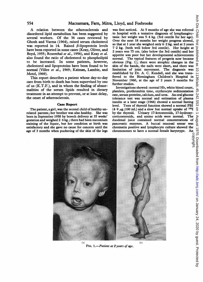
was first noticed. At 5 months of age she was referredto hospital with a tentative diagnosis of lymphangiec-tasia: her weight was 5 4 kg. (3rd centile for her age).Over the next 18 months her weight progress slowed,so that at 1 year she weighed only 6 * 2 kg. and at 2 years7-2 kg. (both well below 3rd centile). Her height at2 years was 75 cm. (also below the 3rd centile) and herappetite was poor but her developmental achievementsnormal. The typical features of progeria now becameobvious (Fig. 1); there were atrophic changes in theskin of the hands, the nails were short, and there waslimitation of joint movement. The diagnosis wasestablished by Dr. A. C. Kendall, and she was trans-ferred to the Birmingham Children's Hospital inNovember 1960, at the age of 2 years 3 months forfurther studies.
Investigations showed: normal Hb, white blood count,platelets, prothrombin time, erythrocyte sedimentationrate, serum proteins, calcium, and urea. An oral glucosetolerance test was normal and estimation of plasmainsulin at a later stage (1966) showed a normal fastinglevel. Tests of thyroid function showed a normal PBI(4 9 zg./l100 ml.) and a slow but normal uptake of 132Iby the thyroid. Urinary 17-ketosteroids, 17-hydroxy-corticosteroids, and amino acids were normal. Theduodenal juice contained normal concentrations ofpancreatic enzymes. A buccal mucosal smear waschromatin positive and lymphocyte culture showed thechromosomes to have a normal female karyotype. An
....::-
....r-..-,,HM:,.v.w 2.;.
(a)FIG. 1.-Patient at 2years of age.
(b)
554
copyright. on January 24, 2020 by guest. P
rotected byhttp://adc.bm
j.com/
Arch D
is Child: first published as 10.1136/adc.45.242.553 on 1 A
ugust 1970. Dow
nloaded from
During 1962 and 1963, though growth remained poor,she became increasingly active and entered into thenormal activities of children in her home area. Herintelligence was normal and she was accepted into thevillage school. The provision of a wig greatly helpedher social adjustment. By the age of 3i years the ECGshowed evidence of left ventricular preponderance andthis abnormality persisted in subsequent tracings. Bythe age of 7 she was showing marked osteoarthriticchanges with limitation of movement of virtually alljoints, and by the age of 8 had begun to have angina oneffort. In April 1967, at the age of 8i years, she wasadmitted to the George Eliot Hospital, Nuneaton, withan anterior myocardial infarction. After her dischargeshe attended Bramcote Hospital School travelling to andfrom home by taxi. Her intelligence remained normaland she spent her spare time at home reading, writing,and painting.
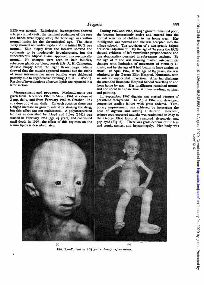
In September 1967 digoxin was started because ofpersistent tachycardia. In April 1969 she developedcongestive cardiac failure with gross oedema. Tem-porary improvement was achieved by increasing thedose of digoxin and adding a diuretic. However,relapse soon occurred and she was readmitted in May tothe George Eliot Hospital, cyanosed, dyspnoeic, andpop-eyed (Fig. 2). There was gross oedema of the legsand trunk, ascites, and hepatomegaly. Her body was
EEG was normal. Radiological investigations showeda large cranial vault; the terminal phalanges of the toesand hands were hypoplastic; the bone age was withinnormal limits for the chronological age. The chestx-ray showed no cardiomegaly and the initial ECG wasnormal. Skin biopsy from the forearm showed theepidermis to be moderately hyperkeratotic, but thesubcutaneous adipose tissue appeared microscopicallynormal. No changes were seen in hair follicles,sebaceous glands, or blood vessels (Dr. A. H. Cameron).Muscle biopsy from the right flexor carpi radialisshowed that the muscle appeared normal but the axonsof some intramuscular nerve bundles were thickenedpossibly due to degenerative swelling (Dr. A. L. Woolf).Results of investigations of serum lipids are reported in alater section.
Management and progress. Methandienone wasgiven from December 1960 to March 1961 at a dose of2 mg. daily, and from February 1962 to October 1963at a dose of 0 4 mg. daily. On each occasion there wasa slight increase in growth rate after starting the drug,but this effect was not maintained. A polyunsaturatedfat diet as described by Lloyd and Jukes (1961) wasstarted in February 1961 (age 21 years) and continueduntil death in 1969; the effect of this regimen on theserum lipids is described later.
FIG. 2.-Patient at 10 years shortly before death.9
Progeria 555
copyright. on January 24, 2020 by guest. P
rotected byhttp://adc.bm
j.com/
Arch D
is Child: first published as 10.1136/adc.45.242.553 on 1 A
ugust 1970. Dow
nloaded from
Macnamara, Famn, Mitra, Lloyd, and Fosbrookeemaciated and she had conspicuous arthritis especiallyin her knees and hands. Despite intensification of hertherapy she died four days after admission at the age of10 years 9 months.
Necropsy. (A.K.M.) This showed the typicalappearances of progeria, with complete absence of hairincluding eyebrows, receding jaw, beaked nose, thinextremities with lack of skin turgor. The heart wasmuch enlarged mainly due to left ventricular dilatationweight 175 g.). The myocardium showed numerousinfarcts, mainly anterior and apical. The left ventriclecontained adherent thrombus and the endocardium wasthickened. The left coronary artery was of smallcalibre. Both main branches showed calcification of thewall and insufficiency. There was no obvious recentthrombus in the vessels. The aorta showed yellowplaques in the descending portion, and in the ascendingportion a few atheromatous ulcers. The lungs showedcongestion and consolidation of the lower lobes due tomild bronchopneumonia. The liver showed a slightnutmeg pattern. The brain showed no abnormality,nor did the adrenals, urogenital system, or the alimentarytract.
Studies of Serum LipidsAll analyses were made on venous blood. Total lipid
was estimated by the method of De la Huerga, Yesinick,and Popper (1953), total cholesterol by the method ofSackett (1925), total light dispersing material by themethod of Fosbrooke and Rudd (1966), and non-esteri-fled fatty acids by the method of Dole and Meinertz(1960). Serum lipoproteins were separated by paperelectrophoresis according to the method of Salt andWolff (1957) and ultracentrifugation was carried out bythe method ofCornwell et al. (1961). Before ultracentri-fugation the serum was subjected to a spin at 10,000 x gfor 30 minutes.
The clearing of absorbed dietary fat was investigatedby making serial estimations of serum lipids after a
breakfast meal containing 2-2 g. fat per kg. (eitherordinary fat largely of animal origin or corn oil) togetherwith carbohydrate and protein; during the remainder ofthe test period a fat-free diet was given. Blood forbaseline values at the start of these tests was obtainedafter an overnight fast (at least 12 hours). Theseinvestigations were carried out using each type of fatbefore the introduction of the corn oil diet, and wererepeated 1 and 3 years after the start of the diet asindicated in Fig. 3. During the follow-up period themajority of blood samples were obtained about 3 hoursafter a standardized breakfast.
Results
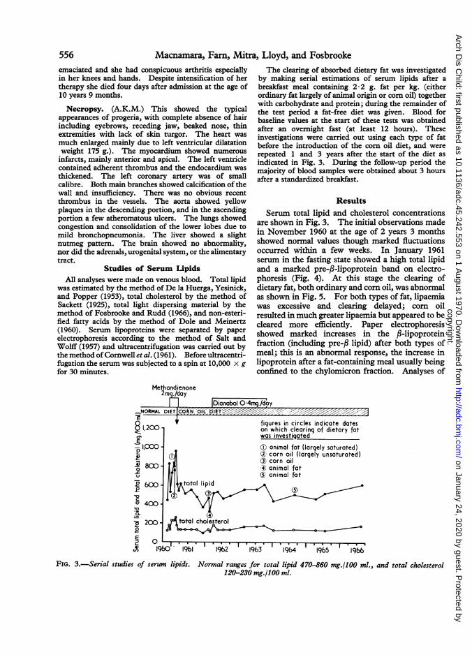
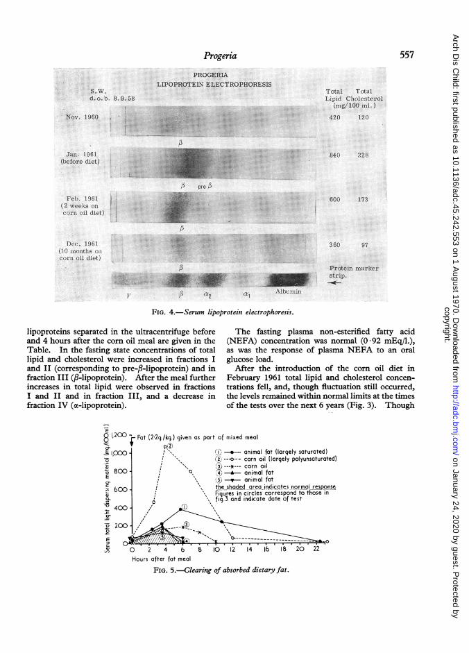
Serum total lipid and cholesterol concentrationsare shown in Fig. 3. The initial observations madein November 1960 at the age of 2 years 3 monthsshowed normal values though marked fluctuationsoccurred within a few weeks. In January 1961serum in the fasting state showed a high total lipidand a marked pre-,B-lipoprotein band on electro-phoresis (Fig. 4). At this stage the clearing ofdietary fat, both ordinary and corn oil, was abnormalas shown in Fig. 5. For both types of fat, lipaemiawas excessive and clearing delayed; corn oilresulted in much greater lipaemia but appeared to becleared more efficiently. Paper electrophoresisshowed marked increases in the f-lipoproteinfraction (including pre-,B lipid) after both types ofmeal; this is an abnormal response, the increase inlipoprotein after a fat-containing meal usually beingconfined to the chylomicron fraction. Analyses of
Methandienone2mj/day
IDianabol 0-4mgq.doy-NORMAL DIETk[OkN O D-IfK<i'.
figures in circles indicote doteson which clearing of dietory fatwos investigated
(D onimol fot (lorgely soturoted)(2t corn oil (largely unsoturoted)Q) corn oil
animal fat@ animal fat
1961 1962 19b3 1964 19b5 1966
FIG. I-Serial studies of serwn lj.pids. Norinal ranges for total lipid 470-860 mg.1100 .ml., and total cholesterol120-230 mg.1100 ml.
.1%1 . 1 '.
556
--I
E 481,200C71E
1,(,Uo10
...VIU 800 -
6 1_r_v
j 6W -
-vcC' 400 -
ma-.L
-a 200-I.-
.0-Em 0L-IVV)
copyright. on January 24, 2020 by guest. P
rotected byhttp://adc.bm
j.com/
Arch D
is Child: first published as 10.1136/adc.45.242.553 on 1 A
ugust 1970. Dow
nloaded from
557Progeria
FIG. 4.-Serum lipoprotein electrophoresis.
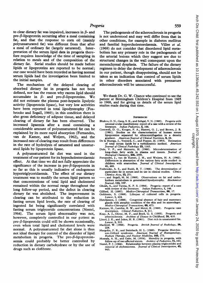
lipoproteins separated in the ultracentrifuge before The fiand 4 hours after the corn oil meal are given in the (NEFA)Table. In the fasting state concentrations of total as was tilipid and cholesterol were increased in fractions I glucose lIcand II (corresponding to pre-/-lipoprotein) and in After tfraction III (,B-lipoprotein). After the meal further Februaryincreases in total lipid were observed in fractions trations fI and II and in fraction III, and a decrease in the levelsfraction IV (oc-lipoprotein). of the tes
E
1200 - Fat (2-2g./kg.) given as port of mixed meal
E 1,Coo - a-* anima0 (2 ---a--~~~~~~~cornc
800 C t--*-- corno 800- animaE - anima
600 \ the shoded areao '!Figures in circle. 0 " fig.3 and indical-v 400- , "
o 200
° 0 2 4 b 8 10 12 14
asting plasma non-esterified fatty acidconcentration was normal (0 92 mEq/1.),he response of plasma NEFA to an oraload.the introduction of the corn oil diet in1961 total lipid and cholesterol concen-
Fell, and, though fluctuation still occurred,remained within normal limits at the times;ts over the next 6 years (Fig. 3). Though
i1 fat (largely saturated)oil (largely polyunsoturated)oil3I fat3I fata_indicates normal response!S correspond to those inite date of test
16 18 20 22Hours after fat meal
FIG. 5.-Clearing of absorbed dietary fat.
. . .
copyright. on January 24, 2020 by guest. P
rotected byhttp://adc.bm
j.com/
Arch D
is Child: first published as 10.1136/adc.45.242.553 on 1 A
ugust 1970. Dow
nloaded from
558 Macnamara, Famn, Mitra, Lloyd, and FosbrookeTABLE
Ultracentrifugal Analysis of Serum Lipoproteins Obtained Before and 4 Hours After a MealContaining Corn Oil (2 2 g./kg.)
Total Lipid Total Cholesterol i Normal Valuest (mg./100 ml.)Ultracentrifugal (mg./100 ml.) (mg./100 ml.) (means and ranges)
FractionBefore AtrBefore Atr TalLpd Total(fasting) After (fasting) After Total Lipid Cholesterol
I + II (Sf 10-400) 202 290 54 50 115 27(71-193) (18-39)
III (Sf 3-9) 317 375 120 131 213 84(164-269) (64-97)
IV (high density) 197 104 54 26 260 70(221-302) (53-78)
Cleared serum* 790 790 236 212 596 175(500-640) (147-206)
Whole serum 790 1060 236 227
*Serum subjected to centrifugation at 10,000 x g for 30 minutes before ultracentrifugation. This process removes chylomicron materialbut may also remove a small amount of very low density lipoprotein which otherwise would appear in fractions I + II.tData obtained from 10 healthy children.
pre-f-lipoprotein also decreased with the fall intotal lipid (Fig. 4) a pre-,B band was observedoccasionally during the subsequent follow-upperiod. Retesting of the responses to dietary fatin 1962 and 1964 showed normal clearing ofordinary fat but slight delay in the clearing of cornoil (Fig. 5).
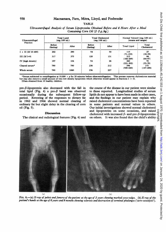
DiscussionThe clinical and radiological features (Fig. 6) and
the course of the disease in our patient were similarto those reported. Longitudinal studies of serumlipids do not appear to have been made in other cases,and the findings in our patient may explain whyraised cholesterol concentrations have been reportedin some patients and normal values in others.Our initial investigations showed normal cholesteroland lipoproteins on some occasions, and raisedcholesterol with increased /- and pre-/3-lipoproteinson others. It was also found that the child's ability
FIG. 6.-a) X-ray of pelvis andfemora of thepatient at the age of 2 years showing marked coxa valga. (b) X-ray of thepatient's hands at the age of6years and 9 months showing sclerosis and destruction of terminal phalanges ('acro-osteolysis').
copyright. on January 24, 2020 by guest. P
rotected byhttp://adc.bm
j.com/
Arch D
is Child: first published as 10.1136/adc.45.242.553 on 1 A
ugust 1970. Dow
nloaded from
Progeria 559to clear dietary fat was impaired, increases in f,- andpre-,-lipoprotein occurring after a meal containingfat, and that the response to corn oil (mainlypolyunsaturated fat) was different from that aftera meal of ordinary fat (largely saturated). Inter-pretation of the serum lipid data in progeria there-fore requires knowledge of the time of sampling inrelation to meals and of the composition of thedietary fat. Serial studies should be made beforelipids or lipoproteins are reported to be normal;our case would have been recorded as having normalserum lipids had the investigation been limited tothe initial samples.The mechanism of the delayed clearing of
absorbed dietary fat in progeria has not beendefined, nor has the reason why excess lipid shouldaccumulate in ,B- and pre-f-lipoproteins. Wedid not estimate the plasma post-heparin lipolyticactivity (lipoprotein lipase), but very low activitieshave been reported in total lipodystrophy (Fos-brooke and Segall, 1969); in this condition there isalso gross deficiency of adipose tissue, and delayedclearing of dietary fat has been observed. Theincreased lipaemia after a meal containing aconsiderable amount of polyunsaturated fat can beexplained by its more rapid absorption (Fernandes,van de Kamer, and Weijers, 1962), and theincreased rate of clearing may result from differencesin the rate of hydrolysis of saturated and unsatur-ated lipids by lipoprotein lipase.A polyunsaturated fat diet was used in the
treatment of our patient for its hypocholesterolaemiceffect. At that time we did not fully appreciate thesignificance of the increase in pre-f-lipoprotein inso far as this is usually indicative of endogenoushypertriglyceridaemia. The effect of our dietarytreatment was to modify the serum lipid pattern sothat concentrations of total lipid and cholesterolremained within the normal range throughout thelong follow-up period, and the defect in clearingdietary fat was abolished. The improvement inclearing can be attributed to the reduction infasting serum lipid levels, the rate of clearing ofingested fat being significantly correlated withfasting serum triglyceride concentrations (Nestel,1964). The serum lipid abnormality was not,however, completely controlled in our patient aspre-f-lipoprotein could still be shown occasionally,even when total lipid and cholesterol levels werenormal. A polyunsaturated fat diet alone is thusnot ideal therapy for control of the disorder of lipidmetabolism in progeria. The pre-/3-lipoprotein-aemia could probably be better controlled byreduction in dietary carbohydrate or by the use ofdrugs such as clofibrate.
The pathogenesis ofthe atherosclerosis in progeriais not understood and may well differ from that inother conditions, for example in diabetes mellitusand familial hypercholesterolaemia. Villee et al.(1969) do not consider that disordered lipid meta-bolism has any primary role in the pathogenesis ofthe arterial lesions which they suggest are due tostructural changes in the wall consequent upon themesenchymal dysplasia. The failure of the dietaryregimen to delay the development of atherosclerosisin our patient, though disappointing, should not betaken as an indication that control of serum lipidsin other disorders associated with prematureatherosclerosis will be unsuccessful.
We thank Dr. G. W. Chance who continued to see thepatient at Birmingham Children's Hospital from 1965to 1968, and for giving us details of the serum lipidstudies made during that time.
REFERENCESBhakoo, 0. N., Garg, S. K., and Sehgal, V. N. (1965). Progeria with
unusual ocular manifestions: report of a case with a review of theliterature. Indian Pediatrics, 2, 164.
Cornwell, D. G., Kruger, F. A., Hamwi, G. J., and Brown, J. B.(1961). Studies on the characterisation of human serumlipoproteins separated by ultracentrifugation in a densitygradient. American Journal of Clinical Nutrition, 9, 24.
De la Huerga, J., Yesinick, C., and Popper, H. (1953). Estimationof total serum lipids by a turbidimetric method. Americanjournal of Clinical Pathology 23, 1163.
Dole, V. P., and Meinertz, H. (1960). Microdetermination oflong-chain fatty acids in plasma and tissues. Journal ofBiological Chemistry, 235, 2595.
Fernandes, J., van de Kamer, J. H., and Weijers, H. A. (1962).Differences in absorption of the various fatty acids studied inchildren with steatorrhea. Journal of Clinical Investigation,41, 488.
Fosbrooke, A. S., and Rudd, B. T. (1966). The determination ofparticulate fat in serum and its use in clinical studies. ClinicaChimica Acta, 13, 251.
-, and Segall, M. M. (1969). Observations on fat and carbo-hydrate metabolism in generalized lipodystrophy. BiochemicalJournal, 112, 33P.
Ghosh, S., and Varma, K. P. S. (1964). Progeria: report of a casewith review of the literature. Indian Pediatrics, 1, 146.
Gilford. H. (1897). Medico-Chirurgical Transactions, 80, 17.Goldstein, S. (1969). Lifespan of cultured cells in progeria.
Lancet, 1, 424.Hutchinson, J. (1886). Congenital absence of hair and mammary
glands with atrophic condition of the skin and its appendages.Medico-Chirurgical Transactions, 69, 473.
Kaiman, H., Lambie, R. W., and Metzl, K. (1969). Progeria: casedescription. Clinical Pediatrics, 8, 411.
Keay, A. J., Oliver, M. F., and Boyd, G. S. (1955). Progeria andatherosclerosis. Archives of Disease in Childhood, 30, 410.
Lloyd, J. K., and Jukes, H. R. (1961). Diet rich in unsaturated fat.Lancet, 1, 312.
Macleod, W. (1966). Progeria. British Journal of Radiology, 39,224.
Margolin, F. R., and Steinbach, H. L. (1968). Progeria: Hutchin-son-Gilford syndrome. American Journal of Roentgenology,Radium Therapy, and Nuclear Medicine, 103, 173.
Mostafa, A. H., and Gabr, M. (1954). Heredity in progeria, withfollow-up of two affected sisters. Archivc: of Pediatrics, 71, 163.
Nestel, P. J. (1964). Relationship between plasma triglycerides andremoval of chylomicrons. Journal of Clinical Investigation, 43,943.
copyright. on January 24, 2020 by guest. P
rotected byhttp://adc.bm
j.com/
Arch D
is Child: first published as 10.1136/adc.45.242.553 on 1 A
ugust 1970. Dow
nloaded from

560 Macnamara, Famn, Mitra, Lloyd, and FosbrookeOzonoff, M. B., and Clemett, A. R. (1967). Progressive osteolysis
in progeria. American Journal of Roentgenology, RadiumTherapy and Nuclear Medicine, 100, 75.
Rosenthal, I. M., Bronstein, I. P., Dallenbach, F. D., Pruzansky, S.,and Rosenwald, A. K. (1956). Progeria: report of a case withcephalometric roentgenograms and abnormally high concen-trations of lipoproteins in the serum. Pediatrics, 18, 565.
Sackett, G. E. (1925). Modification of Bloor's method for thedetermination of cholesterol in whole blood or serum. Journalof Biological Chemistrv, 64, 203.
Salt, H. B., and Wolff, 0. H. (1957). The applications of serumlipoprotein electrophoresis in paediatric practice. Archives ofDisease in Childhood, 32, 404.
Talbot, N. B., Butler, A. M., Pratt, E. L., MacLachlan, E. A., andTannheimer, J. (1945). Progeria: clinical, metabolic andpathologic studies on a patient. American3'ournal of Diseases ofChildren, 69, 267.
Thomson, J., and Forfar, J. 0. (1950). Progeria (Hutchinson-Gilford syndrome): report of a case and review of the literature.Archives of Disease in Childhood, 25, 224.
Villee, D. B., Nichols, G., Jr., and Talbot, N. B. (1969). Metabolicstudies in two boys with classical progeria. Pediatrics, 43, 207.
Correspondence to Dr. B. G. P. Macnamara, GeorgeEliot Hospital, College Street, Nuneaton.
copyright. on January 24, 2020 by guest. P
rotected byhttp://adc.bm
j.com/
Arch D
is Child: first published as 10.1136/adc.45.242.553 on 1 A
ugust 1970. Dow
nloaded from