Embed Size (px)
Citation preview
1
Normo- and hyperandrogenic women with polycystic ovary syndrome exhibit an adverse metabolic profile through life.
Pekka Pinola1,2, Katri Puukka2,3, Terhi T. Piltonen1,2, Johanna Puurunen1,2, Eszter Vanky4,5, Inger Sundström-Poromaa6, Elisabet Stener-Victorin7, Angelica Lindén Hirschberg8, Pernille Ravn9, Marianne Skovsager Andersen10, Dorte Glintborg10, Jan Roar Mellembakken11, Aimo Ruokonen 2,3, Juha S. Tapanainen2,12 and Laure C. Morin-Papunen1,2
1Department of Obstetrics and Gynecology, Oulu University Hospital, Finland
2University of Oulu and Medical Research Center Oulu, Finland 3NordLab Oulu, Oulu University Hospital and Department of Clinical Chemistry, University of Oulu, Oulu, Finland 4Institute of Laboratory Medicine, Children's and Women's Health, Norwegian University of Science and Technology, Trondheim, Norway 5Department of Obstetrics and Gynecology, St. Olav's University Hospital Trondheim, Norway 6Department of Women's and Children's Health, Uppsala University, Sweden 7Department of Physiology and Pharmacology, Karolinska Institutet, Stockholm, Sweden 8Department of Obstetrics and Gynecology, Karolinska University Hospital, Sweden 9Department of Gynecology and Obstetrics, Odense University Hospital, Odense C, Denmark 10Department of Endocrinology and Metabolism, Odense University Hospital, Odense C, Denmark 11Section of Reproductive Medicine, Department of Gynecology, Women’s division, Oslo University Hospital, Oslo, Norway 12Department of Obstetrics and Gynecology, University of Helsinki and Helsinki University Hospital, Helsinki, Finland Abstract Context: Women with polycystic ovary syndrome (PCOS) exhibit various metabolic risks such as obesity, impaired glucose tolerance, insulin resistance, dyslipidemia and chronic inflammation, but studies regarding age-related metabolic risk profiles of the syndrome are scarce. Objective: To compare the metabolic profiles of normo- and hyperandrogenic women with PCOS with those of control women throughout their reproductive life spans. Design: Case-control study. Setting: Eight study sites in Nordic countries. Patients: In all, 1526 women with PCOS (normoandrogenic, N=686 and hyperandrogenic, N=842) and 447 controls were divided into three age groups (< 30, 30–39 and > 39 years). Interventions: None. Main Outcome measures: Body mass index (BMI), waist circumference, blood pressure, glucose, insulin, cholesterol, lipoproteins, triglycerides and high-sensitivity C-reactive protein. Results: Both normo- and hyperandrogenic women with PCOS were more obese, especially abdominally. They had increased serum levels of insulin (fasting and in oral glucose tolerance tests), triglycerides, LDL, total cholesterol, blood pressure, and lower high-density lipoprotein levels (P < 0.001) independently of BMI when compared with the control population as early as from young adulthood until menopause. The prevalence of metabolic syndrome was two- to fivefold higher in women with PCOS compared with controls, depending on age and phenotype, and the highest prevalence was observed in hyperandrogenic women with PCOS at late reproductive age. Conclusions: When evaluating metabolic risks in women with PCOS, androgenic status, and especially abdominal obesity and age should be taken into account, which would allow tailored management of the syndrome from early adulthood on. Introduction Polycystic ovary syndrome (PCOS) is the most common endocrine disorder affecting reproductive-aged women (incidence 6–18% depending on the population and the diagnostic criteria used) (1, 2). It is known to have multifaceted unfavorable effects on women’s health. In addition to anovulatory infertility, which is typically associated with the syndrome, women with PCOS exhibit numerous metabolic risk factors, such as abdominal obesity, insulin resistance, impaired glucose tolerance, dyslipidemia and chronic inflammation (3-6). Hyperandrogenism (HA) plays a central role in the syndrome and has been associated with a more severe metabolic profile in some, but not in all studies (7-11). Obesity is common in PCOS and it exacerbates the symptoms and promotes negative health consequences by increasing the risks of type 2-diabetes (T2DM) and
2 cardiovascular disease (CVD). However, as regards the interaction between weight, weight gain and hyperandrogenism the results of previous studies have been inconsistent. Some data suggest that the metabolic risks linked to PCOS are mainly related to obesity and insulin resistance, whereas others support the hypothesis that the syndrome per se and especially HA, independently of BMI, are the most important cardiovascular risks (12-14). Nevertheless, according to the results of previous studies, the unfavorable metabolic profile linked to PCOS seems to worsen after menopausal transition, highlighting the lifelong health burden of the syndrome (3, 15).
In women without PCOS an impaired metabolic profile is associated with obesity and weight gain. Especially during the menopausal transition, weight gain has been shown to have an adverse effect on glucose metabolism (16). Further, circulating levels of ovarian and adrenal androgens do not seem to have a significant impact on chronic inflammation markers or lipid profile in women in the general population, whereas a low serum level of sex hormone-binding globulin (SHBG) is an independent predictor of an increased risk of CVD (17, 18).
Given the lack of data regarding age-related metabolic changes in women with PCOS, the aim of the present study was to explore and compare metabolic profiles in women with PCOS and in control women throughout their reproductive life spans in a cross-sectional dataset. We also aimed to identify specific metabolic risk factors and the impact of hyperandrogenism in PCOS during early adulthood. Materials and methods Study population Women taking part in eight Nordic PCOS studies contributed to the present investigation. Two centers in Finland, three in Sweden, two in Norway and one in Denmark were involved, and there were totals of 1528 women with PCOS (age range 14–59 years) and 447 control women without the syndrome (age range 18–62 years) (6, 19-26). Circulating levels of androgens in a subpopulation of this Nordic PCOS cohort population have been recently published (27).
Diagnoses of PCOS were made according to Rotterdam criteria (28). Diagnosis of PCOS in peri- and postmenopausal women was based on the presence of oligo-amenorrhea combined with hyperandrogenism (either biochemical or clinical) reported at reproductive age. Thus, retrospectively, all the women met the Rotterdam criteria. Ovarian morphology was examined using transvaginal ultrasonography in all participants. Biochemical hyperandrogenism was defined in relation to the upper limits used in the respective laboratories, depending on the method used, and clinical hyperandrogenism was diagnosed when a subject had a Ferriman–Gallwey score of > 7. The PCOS population was further divided into two groups: hyperandrogenic women with PCOS (HA-PCOS, including biochemical and/or clinical hyperandrogenism) and normoandrogenic women with PCOS (NA-PCOS, including women with both oligo-amenorrhea and polycystic ovaries observed in ultrasonography). The control population consisted of women with normal ovaries (in ultrasonography) and absence of PCOS-related symptoms, e.g. oligo- or anovulation and/or hirsutism and/or elevated serum testosterone (T) levels. The PCOS and control groups were grouped according to age as follows: women under 30 years, 30–39 years and over 39 years. Women using hormonal preparations or medication affecting androgen levels were excluded from the study. Alternatively, a 2-month washout period was required prior to entering the study.
Body mass index (BMI) was calculated (kg/m2). Waist circumference was assessed according to a generally accepted method. Both systolic and diastolic blood pressures (BPs) were measured as millimeters of mercury (mmHg) in a sitting position after 15 minutes of resting.
A diagnosis of metabolic syndrome (MetS) was made according to the Rotterdam consensus (28), requiring three out of the following five criteria: waist circumference > 88 cm and/or BMI ≥ 30 kg/m2, triglycerides ≥ 150 mg/dL (1.70 mmol/L), HDL ≤ 50 mg/dL (1.30 mmol/L), systolic BP ≥ 130 mmHg and/or diastolic BP ≥ 85 mmHg, and fasting glucose ≥ 110–126 mg/dL (6.11–6.99 nmol/L and/or 2-hour OGTT glucose ≥ 140–199 mg/dL (7.78–11.04 nmol/L). Laboratory methods The metabolic variables (glucose, insulin, cholesterol, lipoproteins, triglycerides and high-sensitivity C-reactive protein) were assayed using the routine methods employed in the laboratories of the different study centers (6, 19-26). According to the Nordic Reference Interval Project 2000 (29) the reference ranges for the serum lipid levels were similar in all subpopulations of the study.
In five study sites assay of T was performed by using liquid chromatography-mass spectrometry (LC/MS) and SHBG was analyzed by chemiluminometric immunoassay, as reported earlier (27). In the remaining three study sites (not included in our previous publication) T was analyzed by radioimmunoassay (25, 26).
3
The mean oral glucose tolerance test (OGTT) plasma glucose level and mean OGTT serum insulin levels were calculated as the means of concentrations at different time points ([basal + 2-hour] / 2).
Conversion factors ([concentration, metric unit] × [conversion factor] = SI unit) to SI units for the laboratory parameters were as follows: glucose 0.056 (nmol/L), triglycerides 0.011 (mmol/L) and for total cholesterol, HDL and LDL, 0.026 (mmol/L). Statistical methods Comparisons of continuous variables were carried out by using Student’s t-test or the (nonparametric) Mann–Whitney U-test, depending on the distribution of the variable. Adjustments for BMI and age were applied using linear regression analysis. For categorical variables the χ2-test was used. Statistical analyses were performed by using SPSS 21.0 and 22.0 software (IBM-SPSS Inc., Chicago, Illinois, USA). Of note, the number of participants varied between analyses, as a result of lack of measurements in some cases.
Informed consent was obtained from all women at the original study sites and the study was approved by the Ethics Committees of the respective study sites. Results The women with PCOS (N=1550) had higher BMIs and waist circumferences than the control population (N=447). They also presented with significantly lower levels of high-density lipoprotein (HDL) and higher levels of insulin (fasting and during OGTTs), total cholesterol, low-density lipoprotein (LDL), triglycerides and BP (both systolic and diastolic) compared with the control women, after adjusting for BMI (Table 1).
The anthropometric and metabolic parameters in the HA-PCOS group (N=842), the NA-PCOS group (N=684) and in control women (N=447) are shown in Table 2 and Figures 1 and 2. Only the results that remained significant after adjustment for BMI and age are presented in the text. Table 1. Anthropometric and metabolic parameters in the PCOS and control women shown as means
with standard deviations in parentheses.
aStatistical significance (P-value < 0.05) remains after adjustment for age and BMI.
Metabolic parameters
N
Controls
N
PCOS
P-value
Age [year] 447 33.5 (9.9) 1550 30.0 (7.2) <0.001
BMI [kg/m2] 447 25.9 (5.4) 1497 29.2 (6.9) <0.001
Waist [cm] 312 87.6 (14.6) 1204 92.9 (17.5) <0.001
Fasting glucose [mg/dL] 376 91.9 (16.2) 1104 91.9 (10.8) NS
Fasting insulin [mU/l] 372 7.4 (6.0) 1093 12.3 (11.1) <0.001a
Total cholesterol [mg/dL] 364 177.9 (34.8) 982 185.6 (38.7) 0.003a
HDL [mg/dL] 346 58.0 (11.6) 960 54.1 (15.5) <0.001a
LDL [mg/dL] 347 100.5 (30.9) 863 112.1 (34.8) <0.001a
Triglycerides [mg/dL] 366 79.7 (44.3) 974 115.1 (70.9) <0.001a
OGTT glucose 2h [mg/dL]
140 90.1 (23.4) 681 104.5 (32.4) <0.001
OGTT mean glucose [mg/dL]
140 90.1 (14.4) 681 99.1 (19.8) <0.001
OGTT insulin 2h [mU/l] 152 27.4 (20.5) 860 71.7 (73.9) <0.001a
OGTT mean insulin [mU/l]
152 17.2 (12.0) 840 42.6 (41.6) <0.001a
Systolic blood pressure [mmHg]
318 118 (16) 1276 123 (16) <0.001a
Diastolic blood pressure [mmHg]
318 74 (12) 1276 79 (12) <0.001a
hsCRP [mg/l] 159 1.5 (3.0) 761 2.8 (3.8) <0.001
4 Table 2. Anthropometric and metabolic parameters in the study populations shown as means with
standard deviations in parentheses.
aP-value between reference group and NA-PCOS, bP-value between reference group and HA-PCOS, cP-value between NA- and HA-PCOS groups. dStatistical significance (P-value < 0.05) after adjustment for age and BMI
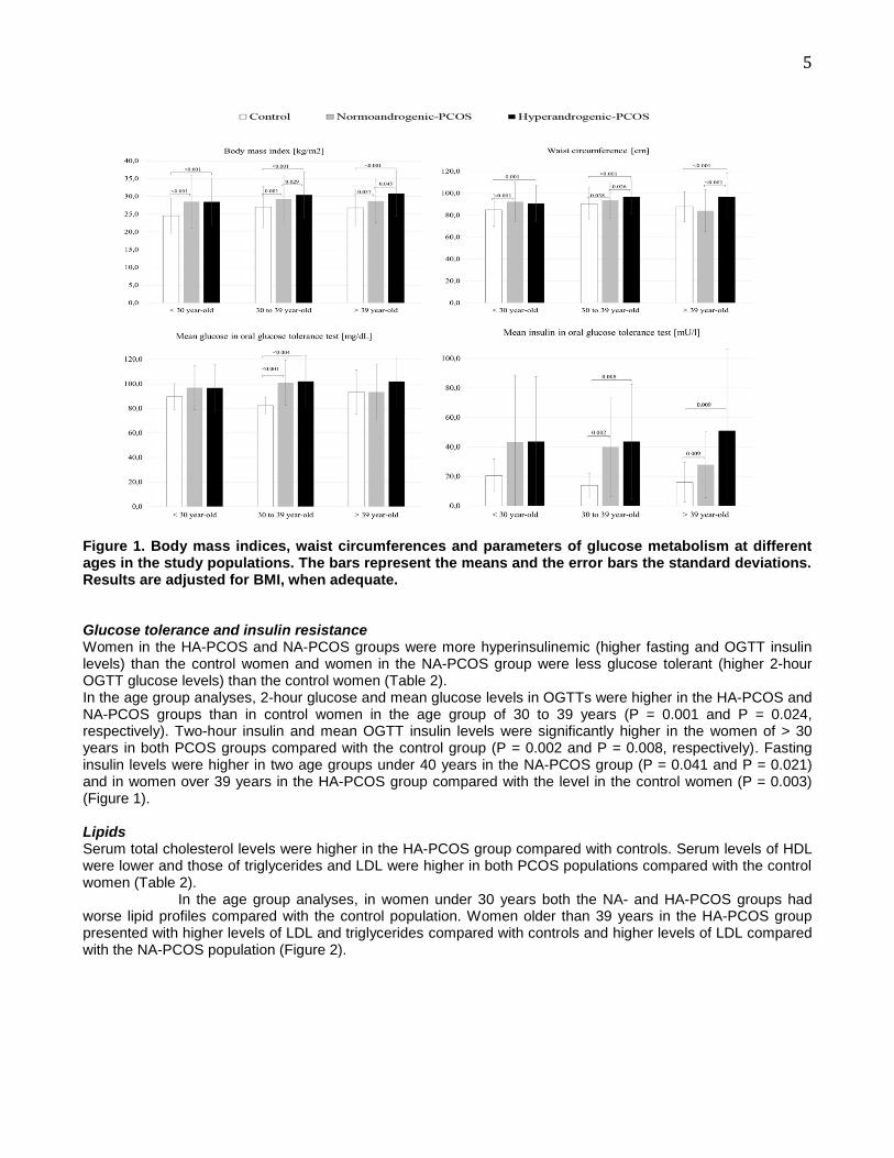
Comparisons between the HA-PCOS, NA-PCOS and control groups BMI and Waist circumference BMIs and waist circumferences were significantly greater in both the HA- and NA-PCOS subpopulations compared with the control group. In addition, BMI was greater in the HA-PCOS population compared with the NA-PCOS women (Table 2).
In age group analyses, women in the HA-PCOS and NA-PCOS groups had greater BMIs and waist circumferences in all age groups compared with the control group. In the age groups of 30 to 39 years and > 39 years, the women in the HA-PCOS group had higher BMIs and waist circumferences than the women in the NA-PCOS group (Figure 1).
Metabolic parameters
N Controls N NA-PCOS N HA-PCOS P-valuea P-valueb
P-valuec
Age [year] 447 33.5 (9.9) 684 29.9 (7.0) 842 30.0 (7.4) <0.001 <0.001 NS
BMI [kg/m2] 447 25.9 (5.4) 666 28.8 (7.0) 811 29.4 (6.7) <0.001 <0.001 0.027
Waist [cm] 312 87.6 (14.6) 590 92.1 (17.8) 604 93.6 (17.0) <0.001 <0.001 NSd
Fasting glucose [mg/dL]
376 91.9 (16.2) 542 91.9 (10.8) 552 91.9 (10.8) NS NS NS
Fasting insulin [mU/l]
372 7.4 (6.0) 544 12.0 (11.4) 537 12.4 (10.7) <0.001d <0.001d NS
Total cholesterol [mg/dL]
364 177.9 (34.8) 368 181.7 (34.8)
603 185.6 (38.7) 0.041 0.004d NS
HDL [mg/dL] 346 58.0 (11.6) 364 50.3 (19.3) 586 54.1 (15.5) <0.001d <0.001d 0.013
LDL [mg/dL] 347 100.5 (30.9) 349
108.3 (30.9)
504 112.1 (34.8) <0.001d <0.001d NS
Triglycerides [mg/dL]
366 79.7 (44.3) 367 106.3 (62.0)
596 106.3 (70.9) <0.001d <0.001d NS
OGTT Glucose 2h [mg/dL]
140 90.1 (23.4) 442 104.5 (30.6)
238 106.3 (34.2) <0.001d <0.001 NS
OGTT mean glucose [mg/dL]
140 90.1 (14.4) 442 97.3 (18.0) 238 99.1 (19.8) <0.001 <0.001 NS
OGTT Insulin 2h [mU/l]
152 27.4 (20.5) 476 67.8 (69.4) 376 76.0 (79.0) <0.001d <0.001d NS
OGTT mean insulin [mU/l]
152 17.2 (12.0) 465 40.9 (40.1) 367 44.5 (43.3) <0.001d <0.001d NS
Systolic blood pressure [mmHg]
318 118 (16) 605 123 (16) 657 124 (17) <0.001d <0.001d NS
Diastolic blood pressure [mmHg]
318 74 (12) 605 78 (12) 657 79 (12) <0.001d <0.001d NS
hsCRP [mg/L]
159 1.5 (3.0) 468 2.8 (3.6) 291 2.9 (4.0) <0.001 <0.001 NS
5
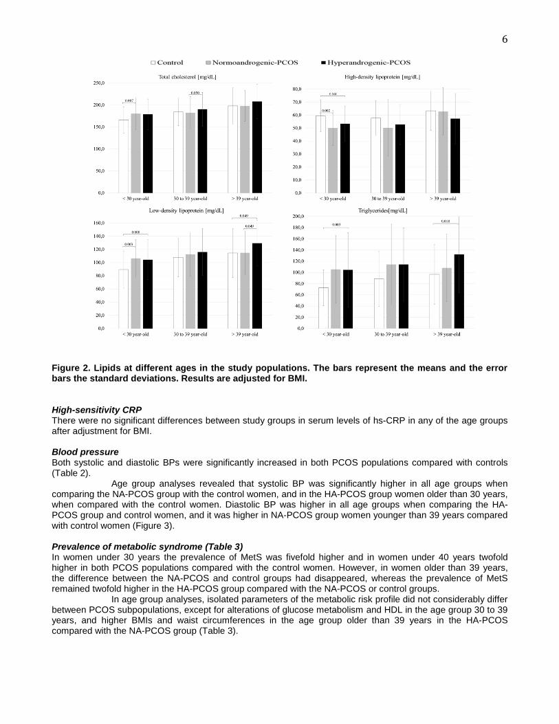
Figure 1. Body mass indices, waist circumferences and parameters of glucose metabolism at different ages in the study populations. The bars represent the means and the error bars the standard deviations. Results are adjusted for BMI, when adequate. Glucose tolerance and insulin resistance Women in the HA-PCOS and NA-PCOS groups were more hyperinsulinemic (higher fasting and OGTT insulin levels) than the control women and women in the NA-PCOS group were less glucose tolerant (higher 2-hour OGTT glucose levels) than the control women (Table 2). In the age group analyses, 2-hour glucose and mean glucose levels in OGTTs were higher in the HA-PCOS and NA-PCOS groups than in control women in the age group of 30 to 39 years (P = 0.001 and P = 0.024, respectively). Two-hour insulin and mean OGTT insulin levels were significantly higher in the women of > 30 years in both PCOS groups compared with the control group (P = 0.002 and P = 0.008, respectively). Fasting insulin levels were higher in two age groups under 40 years in the NA-PCOS group (P = 0.041 and P = 0.021) and in women over 39 years in the HA-PCOS group compared with the level in the control women (P = 0.003) (Figure 1). Lipids Serum total cholesterol levels were higher in the HA-PCOS group compared with controls. Serum levels of HDL were lower and those of triglycerides and LDL were higher in both PCOS populations compared with the control women (Table 2). In the age group analyses, in women under 30 years both the NA- and HA-PCOS groups had worse lipid profiles compared with the control population. Women older than 39 years in the HA-PCOS group presented with higher levels of LDL and triglycerides compared with controls and higher levels of LDL compared with the NA-PCOS population (Figure 2).
6
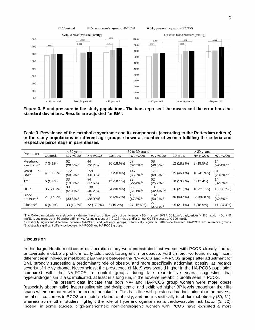
Figure 2. Lipids at different ages in the study populations. The bars represent the means and the error bars the standard deviations. Results are adjusted for BMI. High-sensitivity CRP There were no significant differences between study groups in serum levels of hs-CRP in any of the age groups after adjustment for BMI. Blood pressure Both systolic and diastolic BPs were significantly increased in both PCOS populations compared with controls (Table 2).
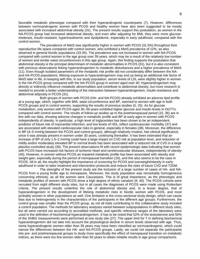
Age group analyses revealed that systolic BP was significantly higher in all age groups when comparing the NA-PCOS group with the control women, and in the HA-PCOS group women older than 30 years, when compared with the control women. Diastolic BP was higher in all age groups when comparing the HA-PCOS group and control women, and it was higher in NA-PCOS group women younger than 39 years compared with control women (Figure 3). Prevalence of metabolic syndrome (Table 3) In women under 30 years the prevalence of MetS was fivefold higher and in women under 40 years twofold higher in both PCOS populations compared with the control women. However, in women older than 39 years, the difference between the NA-PCOS and control groups had disappeared, whereas the prevalence of MetS remained twofold higher in the HA-PCOS group compared with the NA-PCOS or control groups.
In age group analyses, isolated parameters of the metabolic risk profile did not considerably differ between PCOS subpopulations, except for alterations of glucose metabolism and HDL in the age group 30 to 39 years, and higher BMIs and waist circumferences in the age group older than 39 years in the HA-PCOS compared with the NA-PCOS group (Table 3).
7
Figure 3. Blood pressure in the study populations. The bars represent the means and the error bars the standard deviations. Results are adjusted for BMI. Table 3. Prevalence of the metabolic syndrome and its components (according to the Rotterdam criteria) in the study populations in different age groups shown as number of women fulfilling the criteria and respective percentage in parentheses.
Parameter < 30 years 30 to 39 years > 39 years
Controls NA-PCOS HA-PCOS Controls NA-PCOS HA-PCOS Controls NA-PCOS HA-PCOS
Metabolic syndromea
7 (5.1%) 62 (26.3%)b
64 (26.7%)c 16 (18.0%) 57
(37.5%)b
68 (40.0%)c 12 (18.2%) 8 (19.5%) 14
(42.4%)c,d
Waist or BMIa
41 (33.6%) 172 (53.6%)b
159 (50.3%)c 57 (50.0%) 147
(65.6%)b
171 (69.8%)c 35 (46.1%) 18 (41.9%) 31
(73.8%)c,d
TGa 5 (2.9%) 33 (19.0%)b
54 (17.6%)c 12 (10.1%) 33
(22.4%)b
62 (25.2%)c 10 (13.2%) 8 (17.4%)
14 (32.6%)c
HDLa 35 (21.9%) 89 (51.1%)b
138 (45.2%)c 34 (30.9%) 88
(61.1%)b
101 (42.4%)c,d 16 (21.3%) 10 (21.7%) 13 (30.2%)
Blood pressurea
21 (15.9%) 111 (33.5%)b
131 (38.0%)c 28 (25.2%) 108
(47.8%)b
132 (50.2%)c 30 (40.5%) 23 (50.0%)
30 (62.5%)c
Glucosea 4 (8.0%) 33 (13.3%) 22 (17.2%) 5 (15.2%) 27 (16.6%) 27 (27.6%)d 15 (21.1%) 7 (18.9%) 11 (34.4%)
aThe Rotterdam criteria for metabolic syndrome, three out of five: waist circumference > 88cm and/or BMI ≥ 30 kg/m2, triglycerides ≥ 150 mg/dL, HDL ≤ 50 mg/dL, blood pressure ≥130 and/or ≥85 mmHg, fasting glucose ≥ 110-126 mg/dL and/or 2-hour OGTT glucose 140-199 mg/dL. bStatistically significant difference between NA-PCOS and reference groups, cStatistically significant difference between HA-PCOS and reference groups, dStatistically significant difference between NA-PCOS and HA-PCOS groups.
Discussion In this large, Nordic multicenter collaboration study we demonstrated that women with PCOS already had an unfavorable metabolic profile in early adulthood, lasting until menopause. Furthermore, we found no significant differences in individual metabolic parameters between the NA-PCOS and HA-PCOS groups after adjustment for BMI, strongly suggesting a predominant role of obesity, and more specifically abdominal obesity, as regards severity of the syndrome. Nevertheless, the prevalence of MetS was twofold higher in the HA-PCOS population compared with the NA-PCOS or control groups during late reproductive years, suggesting that hyperandrogenism is also implicated, at least in a long run, in the adverse metabolic profile seen in PCOS.
The present data indicate that both NA- and HA-PCOS group women were more obese (especially abdominally), hyperinsulinemic and dyslipidemic, and exhibited higher BP levels throughout their life spans when compared with the control population. This is in line with previous data indicating that the adverse metabolic outcomes in PCOS are mainly related to obesity, and more specifically to abdominal obesity (30, 31), whereas some other studies highlight the role of hyperandrogenism as a cardiovascular risk factor (5, 32). Indeed, in some studies, oligo-amenorrheic normoandrogenic women with PCOS have exhibited a more
8 favorable metabolic phenotype compared with their hyperandrogenic counterparts (7). However, differences between normoandrogenic women with PCOS and healthy women have also been suggested to be mostly associated with increased abdominal obesity (31). The present results support this postulation, as women in the NA-PCOS group had increased abdominal obesity, and even after adjusting for BMI, they were more glucose-intolerant, insulin-resistant, hyperinsulinemic and dyslipidemic, especially in early adulthood, compared with the control women. The prevalence of MetS was significantly higher in women with PCOS (31.5%) throughout their reproductive life spans compared with control women, who exhibited a MetS prevalence of 12%, as also observed in general Nordic populations (33-35). The prevalence was not increased in women with NA-PCOS compared with control women in the age group over 39 years, which may be a result of the relatively low number of women and similar waist circumferences in this age group. Again, this finding supports the postulation that abdominal obesity is the principal determinant of metabolic abnormalities in PCOS (31), but it is also consistent with previous observations linking hyperandrogenism to metabolic disturbances and a higher prevalence of MetS (11). Even though isolated parameters of the metabolic risk profile did not considerably differ between the NA- and HA-PCOS populations, lifelong exposure to hyperandrogenism may end up being an additional risk factor of MetS later in life. In keeping with this, in our study population, serum levels of LDL were slightly higher in women in the HA-PCOS group compared with the NA-PCOS group in women aged over 40. Hyperandrogenism may directly or indirectly influence metabolic abnormalities and contribute to abdominal obesity, but more research is needed to provide a better understanding of the interaction between hyperandrogenism, insulin resistance and abdominal adiposity in PCOS (31).
Both groups of women with PCOS (HA- and NA-PCOS) already exhibited an adverse lipid profile at a young age, which, together with BMI, waist circumference and BP, seemed to worsen with age in both PCOS groups and in control women, supporting the results of previous studies (5, 25). As for glucose metabolism, only women with PCOS aged 30–39 years exhibited higher glucose and insulin levels in OGTTs compared with control women. The results of follow-up studies up to the postmenopausal period (15, 36) are in line with our data, showing adverse changes in metabolic profile and BP at early ages in women with PCOS independently of obesity. In particular, a high level of triglycerides has been shown to be an independent predictor of future risk of myocardial infarction, and low levels of HDL reflect cardiovascular morbidity (37) and are associated with elevated cardiovascular risks in general, especially in females (38). Moreover, the difference in BP (4–5 mmHg between the PCOS and control groups), although relatively modest, has clinical significance, since it was already present in women under 30 years, continuing thereafter. It has been estimated that an increase of BP of only 1.5–2 mmHg could have a large impact on CVD risk at population level, and reduction of mildly and/or moderately elevated BP to normal levels has been associated with a reduced risk of CVD in a large placebo-controlled study (39). The present observations fit with recent epidemiologic data indicating that women with PCOS have increased risk factors of ischemic heart and cerebrovascular diseases, independently of obesity (40). However, in women without PCOS an impaired metabolic profile has been associated with obesity and weight gain, especially during the period of menopausal transition (16), and this also seems to be the case in PCOS. All in all, the results highlight the importance of screening for PCOS and overweight/obesity in early adulthood in order to tailor treatment and intervention protocols and reduce the risks of future CVD and T2DM.
The strengths of the present study are the inclusion of a large number of cases of HA- and NA-PCOS from a young fertile age to menopause. Moreover, the study population was remarkably homogeneous concerning ethnicity, as all the women were Caucasians. This is of great importance, as the phenotypic and metabolic profiles of women with PCOS show a high degree of ethnic variation (8, 40). The PCOS cohorts were recruited from eight different study sites, but in all cases the diagnoses of PCOS were made using Rotterdam criteria. The present results underline the role of abdominal obesity and, to a lesser degree, that of hyperandrogenism in the development of lifelong metabolic risks in Nordic women with PCOS, and most probably women in other populations as well. One limitation is the cross-sectional study design, which can cause bias due to heterogeneity in the characteristics of the participants in the different age groups. Furthermore, the control group was smaller than the PCOS group, as not all trials contributing to this collaborative study included a control population. The methods for laboratory analyses varied between subpopulations in different study sites, but all were carried out according to accredited methods, and specific reference ranges of the laboratory were used in the definition of biochemical hyperandrogenism. It has to be noted that 52% of the testosterone and 50% of the SHBG measurements were performed at one study site (27). The upper limit for T in defining biochemical hyperandrogenism did not take into account the physiological decline in serum levels observed with age. Thus, some hyperandrogenic women older than 39 years may have been classified as normoandrogenic, which could narrow the differences between the HA- and NA-PCOS groups. Lastly, we could not separate the participants into pre- and postmenopausal groups to study more specifically the effect of menopausal transition on metabolic indices, as there were too few women older than 50 years to obtain reliable results in age group comparisons.
9 Conclusions This Nordic multicenter study showed that women with PCOS presented a worsened metabolic profile compared with a control population, from early adulthood to menopause. Even though lifelong exposure to hyperandrogenemia seemed to contribute, at least partly, to a higher prevalence of MetS, metabolic disturbances were present in both the HA- and NA-PCOS groups, and abdominal obesity appeared to be the principal determinant of metabolic abnormalities in PCOS. We conclude that when evaluating metabolic risks in women with PCOS, androgenic status, obesity (especially abdominal obesity) and age should all be taken into account to allow tailored management of the syndrome, focusing on prevention of abdominal obesity, starting from early adulthood. However, only a long-term, longitudinal follow-up study will reveal whether the metabolic risk factors linked to PCOS, and more specifically to HA, translate into later cardiovascular morbidity and mortality.
10
References
1. Franks S 1995 Polycystic ovary syndrome. N Engl J Med 333(13):853-861
2. March WA, Moore VM, Willson KJ, Phillips DI, Norman RJ, Davies MJ 2010 The prevalence of polycystic ovary syndrome in a community sample assessed under contrasting diagnostic criteria. Hum Reprod 25(2):544-551
3. Puurunen J, Piltonen T, Morin-Papunen L, Perheentupa A, Jarvela I, Ruokonen A, Tapanainen JS 2011 Unfavorable hormonal, metabolic, and inflammatory alterations persist after menopause in women with PCOS. J Clin Endocrinol Metab 96(6):1827-1834
4. Daan NM, Louwers YV, Koster MP, Eijkemans MJ, de Rijke YB, Lentjes EW, Fauser BC, Laven JS 2014 Cardiovascular and metabolic profiles amongst different polycystic ovary syndrome phenotypes: Who is really at risk? Fertil Steril 102(5):1444-1451.e3
5. Johnstone EB, Davis G, Zane LT, Cedars MI, Huddleston HG 2012 Age-related differences in the reproductive and metabolic implications of polycystic ovarian syndrome: Findings in an obese, united states population. Gynecol Endocrinol 28(10):819-822
6. Morin-Papunen LC, Vauhkonen I, Koivunen RM, Ruokonen A, Tapanainen JS 2000 Insulin sensitivity, insulin secretion, and metabolic and hormonal parameters in healthy women and women with polycystic ovarian syndrome. Hum Reprod 15(6):1266-1274
7. Barber TM, Wass JA, McCarthy MI, Franks S 2007 Metabolic characteristics of women with polycystic ovaries and oligo-amenorrhoea but normal androgen levels: Implications for the management of polycystic ovary syndrome. Clin Endocrinol (Oxf) 66(4):513-517
8. Welt CK, Gudmundsson JA, Arason G, Adams J, Palsdottir H, Gudlaugsdottir G, Ingadottir G, Crowley WF 2006 Characterizing discrete subsets of polycystic ovary syndrome as defined by the rotterdam criteria: The impact of weight on phenotype and metabolic features. J Clin Endocrinol Metab 91(12):4842-4848
9. Barber TM, Vojtechova P, Franks S 2013 The impact of hyperandrogenism in female obesity and cardiometabolic diseases associated with polycystic ovary syndrome. Horm Mol Biol Clin Investig 15(3):91-103
10. O'Reilly MW, Taylor AE, Crabtree NJ, Hughes BA, Capper F, Crowley RK, Stewart PM, Tomlinson JW, Arlt W 2014 Hyperandrogenemia predicts metabolic phenotype in polycystic ovary syndrome: The utility of serum androstenedione. J Clin Endocrinol Metab 99(3):1027-1036
11. Sung YA, Oh JY, Chung H, Lee H 2014 Hyperandrogenemia is implicated in both the metabolic and reproductive morbidities of polycystic ovary syndrome. Fertil Steril 101(3):840-845
12. Park HT, Cho GJ, Ahn KH, Shin JH, Kim YT, Hur JY, Kim SH, Lee KW, Kim T 2010 Association of insulin resistance with anti-mullerian hormone levels in women without polycystic ovary syndrome (PCOS). Clin Endocrinol (Oxf) 72(1):26-31
13. Piouka A, Farmakiotis D, Katsikis I, Macut D, Gerou S, Panidis D 2009 Anti-mullerian hormone levels reflect severity of PCOS but are negatively influenced by obesity: Relationship with increased luteinizing hormone levels. Am J Physiol Endocrinol Metab 296(2):E238-43
14. Nisenblat V, Norman RJ 2009 Androgens and polycystic ovary syndrome. Curr Opin Endocrinol Diabetes Obes 16(3):224-231
15. Schmidt J, Brannstrom M, Landin-Wilhelmsen K, Dahlgren E 2011 Reproductive hormone levels and anthropometry in postmenopausal women with polycystic ovary syndrome (PCOS): A 21-year follow-up study of
11 women diagnosed with PCOS around 50 years ago and their age-matched controls. J Clin Endocrinol Metab 96(7):2178-2185
16. Polotsky HN, Polotsky AJ 2010 Metabolic implications of menopause. Semin Reprod Med 28(5):426-434
17. Bell RJ, Davison SL, Papalia MA, McKenzie DP, Davis SR 2007 Endogenous androgen levels and cardiovascular risk profile in women across the adult life span. Menopause 14(4):630-638
18. Wang Q, Kangas AJ, Soininen P, Tiainen M, Tynkkynen T, Puukka K, Ruokonen A, Viikari J, Kahonen M, Lehtimaki T, Salomaa V, Perola M, Davey Smith G, Raitakari OT, Jarvelin MR, Wurtz P, Kettunen J, Ala-Korpela M 2015 Sex hormone-binding globulin associations with circulating lipids and metabolites and the risk for type 2 diabetes: Observational and causal effect estimates. Int J Epidemiol 44(2):623-637
19. Vanky E, Kjotrod S, Salvesen KA, Romundstad P, Moen MH, Carlsen SM 2004 Clinical, biochemical and ultrasonographic characteristics of scandinavian women with PCOS. Acta Obstet Gynecol Scand 83(5):482-486
20. Puurunen J, Piltonen T, Jaakkola P, Ruokonen A, Morin-Papunen L, Tapanainen JS 2009 Adrenal androgen production capacity remains high up to menopause in women with polycystic ovary syndrome. J Clin Endocrinol Metab 94(6):1973-1978
21. Stener-Victorin E, Holm G, Labrie F, Nilsson L, Janson PO, Ohlsson C 2010 Are there any sensitive and specific sex steroid markers for polycystic ovary syndrome? J Clin Endocrinol Metab 95(2):810-819
22. Hudecova M, Holte J, Olovsson M, Sundstrom Poromaa I 2009 Long-term follow-up of patients with polycystic ovary syndrome: Reproductive outcome and ovarian reserve. Hum Reprod 24(5):1176-1183
23. Piltonen T, Koivunen R, Perheentupa A, Morin-Papunen L, Ruokonen A, Tapanainen JS 2004 Ovarian age-related responsiveness to human chorionic gonadotropin in women with polycystic ovary syndrome. J Clin Endocrinol Metab 89(8):3769-3775
24. Piltonen T, Puurunen J, Hedberg P, Ruokonen A, Mutt SJ, Herzig KH, Nissinen A, Morin-Papunen L, Tapanainen JS 2012 Oral, transdermal and vaginal combined contraceptives induce an increase in markers of chronic inflammation and impair insulin sensitivity in young healthy normal-weight women: A randomized study. Hum Reprod 27(10):3046-3056
25. Glintborg D, Mumm H, Ravn P, Andersen M 2012 Age associated differences in prevalence of individual rotterdam criteria and metabolic risk factors during reproductive age in 446 caucasian women with polycystic ovary syndrome. Horm Metab Res 44(9):694-698
26. Nybacka A, Carlstrom K, Stahle A, Nyren S, Hellstrom PM, Hirschberg AL 2011 Randomized comparison of the influence of dietary management and/or physical exercise on ovarian function and metabolic parameters in overweight women with polycystic ovary syndrome. Fertil Steril 96(6):1508-1513
27. Pinola P, Piltonen TT, Puurunen J, Vanky E, Sundstrom-Poromaa I, Stener-Victorin E, Ruokonen A, Puukka K, Tapanainen JS, Morin-Papunen LC 2015 Androgen profile through life in women with polycystic ovary syndrome: A nordic multicenter collaboration study. J Clin Endocrinol Metab 100(9):3400-3407
28. Rotterdam ESHRE/ASRM-Sponsored PCOS consensus workshop group 2004 Revised 2003 consensus on diagnostic criteria and long-term health risks related to polycystic ovary syndrome (PCOS). Hum Reprod 19(1):41-47
29. Rustad P, Simonsson P, Felding P, Pedersen M 2004 Nordic reference interval project bio-bank and database (NOBIDA): A source for future estimation and retrospective evaluation of reference intervals. Scand J Clin Lab Invest 64(4):431-438
12 30. Tzeng CR, Chang YC, Chang YC, Wang CW, Chen CH, Hsu MI 2014 Cluster analysis of cardiovascular and metabolic risk factors in women of reproductive age. Fertil Steril 101(5):1404-1410
31. Moran L, Teede H 2009 Metabolic features of the reproductive phenotypes of polycystic ovary syndrome. Hum Reprod Update 15(4):477-488
32. Macut D, Micic D, Parapid B, Cvijovic G, Sumarac M, Kendereski A, Milic N, Tulic L, Muharemagic A, Zoric S, Pejkovic D 2002 Age and body mass related changes of cardiovascular risk factors in women with polycystic ovary syndrome. Vojnosanit Pregl 59(6):593-599
33. Ilanne-Parikka P, Eriksson JG, Lindstrom J, Hamalainen H, Keinanen-Kiukaanniemi S, Laakso M, Louheranta A, Mannelin M, Rastas M, Salminen V, Aunola S, Sundvall J, Valle T, Lahtela J, Uusitupa M, Tuomilehto J, Finnish Diabetes Prevention Study Group 2004 Prevalence of the metabolic syndrome and its components: Findings from a finnish general population sample and the diabetes prevention study cohort. Diabetes Care 27(9):2135-2140
34. Nystrom PK, Carlsson AC, Leander K, de Faire U, Hellenius ML, Gigante B 2015 Obesity, metabolic syndrome and risk of atrial fibrillation: A swedish, prospective cohort study. PLoS One 10(5):e0127111
35. Hildrum B, Mykletun A, Hole T, Midthjell K, Dahl AA 2007 Age-specific prevalence of the metabolic syndrome defined by the international diabetes federation and the national cholesterol education program: The norwegian HUNT 2 study. BMC Public Health 7:220
36. Pasquali R, Gambineri A, Anconetani B, Vicennati V, Colitta D, Caramelli E, Casimirri F, Morselli-Labate AM 1999 The natural history of the metabolic syndrome in young women with the polycystic ovary syndrome and the effect of long-term oestrogen-progestagen treatment. Clin Endocrinol (Oxf) 50(4):517-527
37. Stampfer MJ, Krauss RM, Ma J, Blanche PJ, Holl LG, Sacks FM, Hennekens CH 1996 A prospective study of triglyceride level, low-density lipoprotein particle diameter, and risk of myocardial infarction. JAMA 276(11):882-888
38. Ulmer H, Kollerits B, Kelleher C, Diem G, Concin H 2005 Predictive accuracy of the SCORE risk function for cardiovascular disease in clinical practice: A prospective evaluation of 44 649 austrian men and women. Eur J Cardiovasc Prev Rehabil 12(5):433-441
39. Hart R, Doherty DA 2015 The potential implications of a PCOS diagnosis on a woman's long-term health using data linkage. J Clin Endocrinol Metab 100(3):911-919
40. Glintborg D, Mumm H, Hougaard D, Ravn P, Andersen M 2010 Ethnic differences in rotterdam criteria and metabolic risk factors in a multiethnic group of women with PCOS studied in denmark. Clin Endocrinol (Oxf) 73(6):732-738